MECHANISMS OF WNT SIGNALING IN DEVELOPMENT - Stanford University

Molecular Mechanisms of Fibroblast GrowthFactor Signaling in Physiology and Pathology
Artur A. Belov and Moosa Mohammadi
Department of Biochemistry and Molecular Pharmacology, New York University School of Medicine,New York, New York 10016
Correspondence: [email protected]
Fibroblast growth factors (FGFs) signal in a paracrine or endocrine fashion to mediate amyriad of biological activities, ranging from issuing developmental cues, maintainingtissue homeostasis, and regulating metabolic processes. FGFs carry out their diverse func-tions by binding and dimerizing FGF receptors (FGFRs) in a heparan sulfate (HS) cofactor- orKlotho coreceptor-assisted manner. The accumulated wealth of structural and biophysicaldata in the past decade has transformed our understanding of the mechanism of FGF signal-ing in human health and development, and has provided novel concepts in receptor tyrosinekinase (RTK) signaling. Among these contributions are the elucidation of HS-assisted recep-tor dimerization, delineation of the molecular determinants of ligand–receptor specificity,tyrosine kinase regulation, receptor cis-autoinhibition, and tyrosine trans-autophosphoryla-tion. These structural studies have also revealed how disease-associated mutations highjackthe physiological mechanisms of FGFR regulation to contribute to human diseases. In thispaper, we will discuss the structurally and biophysically derived mechanisms of FGF signal-ing, and how the insights gained may guide the development of therapies for treatment of adiverse array of human diseases.
Fibroblast growth factor (FGF) signaling ful-fills essential roles in metazoan development
and metabolism. A wealth of literature has doc-umented the requirement for FGF signaling inmultiple processes during embryogenesis, in-cluding implantation (Feldman et al. 1995), gas-trulation (Sun et al. 1999), somitogenesis (Du-brulle and Pourquie 2004; Wahl et al. 2007; Leeet al. 2009; Naiche et al. 2011; Niwa et al. 2011),body plan formation (Martin 1998; RodriguezEsteban et al. 1999; Tanaka et al. 2005; Marianiet al. 2008), morphogenesis (Metzger et al. 2008;Makarenkova et al. 2009), and organogenesis
(Goldfarb 1996; Kato and Sekine 1999; Sekineet al. 1999; Sun et al. 1999; Colvin et al. 2001;Serls et al. 2005; Vega-Hernandez et al. 2011).Recent clinical and biochemical data have un-covered unexpected roles for FGF signaling inmetabolic processes, including phosphate/vita-min D homeostasis (Consortium 2000; Razza-que and Lanske 2007; Nakatani et al. 2009; Gat-tineni et al. 2011; Kir et al. 2011), cholesterol/bile acid homeostasis (Yu et al. 2000a; Holt et al.2003), and glucose/lipid metabolism (Fu et al.2004; Moyers et al. 2007). Highlighting its di-verse biology, deranged FGF signaling contrib-
Editors: Joseph Schlessinger and Mark A. Lemmon
Additional Perspectives on Signaling by Receptor Tyrosine Kinases available at www.cshperspectives.org
Copyright # 2013 Cold Spring Harbor Laboratory Press; all rights reserved; doi: 10.1101/cshperspect.a015958
Cite this article as Cold Spring Harb Perspect Biol 2013;5:a015958
1
on March 12, 2022 - Published by Cold Spring Harbor Laboratory Press http://cshperspectives.cshlp.org/Downloaded from

utes to many human diseases, such as congenitalcraniosynostosis and dwarfism syndromes (Nas-ki et al. 1996; Wilkie et al. 2002, 2005), Kallmannsyndrome (Dode et al. 2003; Pitteloud et al.2006a), hearing loss (Tekin et al. 2007, 2008),and renal phosphate wasting disorders (Shima-da et al. 2001; White et al. 2001), as well as manyacquired forms of cancers (Rand et al. 2005; Pol-lock et al. 2007; Gartside et al. 2009; di Martinoet al. 2012). Endocrine FGFs have also been im-plicated in the progression of acquired metabol-ic disorders, including chronic kidney disease(Fliser et al. 2007), obesity (Inagaki et al. 2007;Moyers et al. 2007; Reinehr et al. 2012), and in-sulin resistance (Fu et al. 2004; Chen et al. 2008b;Chateau et al. 2010; Huang et al. 2011), givingrise to many opportunities for drug discovery inthe field of FGF biology (Beenken and Moham-madi 2012).
Based on sequence homology and phyloge-ny, the 18 mammalian FGFs are grouped into sixsubfamilies (Ornitz and Itoh 2001; Popoviciet al. 2005; Itoh and Ornitz 2011). Five of thesesubfamilies act in a paracrine fashion, namely,the FGF1 subfamily (FGF1 and FGF2), the FGF4subfamily (FGF4, FGF5, and FGF6), the FGF7subfamily (FGF3, FGF7, FGF10, and FGF22), theFGF8 subfamily (FGF8, FGF17, and FGF18), andthe FGF9 subfamily (FGF9, FGF16, and FGF20).In contrast, the FGF19 subfamily (FGF19,FGF21, and FGF23) signals in an endocrinemanner (Beenken andMohammadi 2012). FGFsexert their pleiotropic effects by binding and ac-tivating the FGF receptor (FGFR) subfamily ofreceptor tyrosine kinases that are coded by fourgenes (FGFR1, FGFR2, FGFR3, and FGFR4) inmammals (Johnson and Williams 1993; Mo-hammadi et al. 2005b). The extracellular do-main of FGFRs consists of three immunoglobu-lin (Ig)-like domains (D1, D2, and D3), and theintracellular domain harbors the conserved ty-rosine kinase domain flanked by the flexibleamino-terminal juxtamembrane linker and car-boxy-terminal tail (Lee et al. 1989; Dionne et al.1991; Givol and Yayon 1992). Aunique feature ofFGFRs is the presence of acontiguous segment ofglutamic and aspartic acids in the D1–D2 linker,termed the acid box (AB). The two-membraneproximal D2 and D3 and the intervening D2–
D3 linker are necessary and sufficient for ligandbinding/specificity (Dionne et al. 1990; Johnsonet al. 1990), whereas D1 and the D1–D2 linkerare implicated in receptor autoinhibition (Wanget al. 1995; Roghani and Moscatelli 2007; Kali-nina et al. 2012). Alternative splicing and trans-lational initiation further diversify both ligandsand receptors. The amino-terminal regions ofFGF8 and FGF17 can be differentially splicedto yield FGF8a, FGF8b, FGF8e, FGF8f (Gemelet al. 1996; Blunt et al. 1997), and FGF17a andFGF17b isoforms (Xu et al. 1999), whereas cyto-sine-thymine-guanine (CTG)-mediated trans-lational initiation gives rise to multiple highmolecular weight isoforms of FGF2 and FGF3(Florkiewicz and Sommer 1989; Prats et al. 1989;Acland et al. 1990). The tissue-specific alterna-tive splicing in D3 of FGFR1, FGFR2, and FGFR3yields “b” and “c” receptor isoforms which,along with their temporal and spatial expressionpatterns, is the major regulator of FGF–FGFRspecificity/promiscuity (Orr-Urtreger et al.1993; Ornitz et al. 1996; Zhang et al. 2006). Alarge body of structural data on FGF–FGFRcomplexes has begun to reveal the intricatemechanisms by which different FGFs and FGFRscombine selectively to generate quantitativelyand qualitatively different intracellular signals,culminating in distinct biological responses. Inaddition, these structural data have unveiledhow pathogenic mutations hijack the normalphysiological mechanisms of FGFR regulationto lead to pathogenesis. We will discuss the cur-rent state of the structural biology of the FGF–FGFR system, lessons learned from studying themechanism of action of pathogenic mutations,and how the structural data are beginning toshape and advance the translational research.
STRUCTURE–FUNCTION RELATIONSHIPOF FGFs
FGFs range in size from �150–300 amino acids(Basilico and Moscatelli 1992; Mohammadiet al. 2005b). Crystal structures with at leastone representative from each subfamily, namelyFGF1 and FGF2 (Eriksson et al. 1991; Zhanget al. 1991; Zhu et al. 1991), FGF4 (Bellostaet al. 2001), FGF7 (Ye et al. 2001), FGF8b (Olsen
A.A. Belov and M. Mohammadi
2 Cite this article as Cold Spring Harb Perspect Biol 2013;5:a015958
on March 12, 2022 - Published by Cold Spring Harbor Laboratory Press http://cshperspectives.cshlp.org/Downloaded from
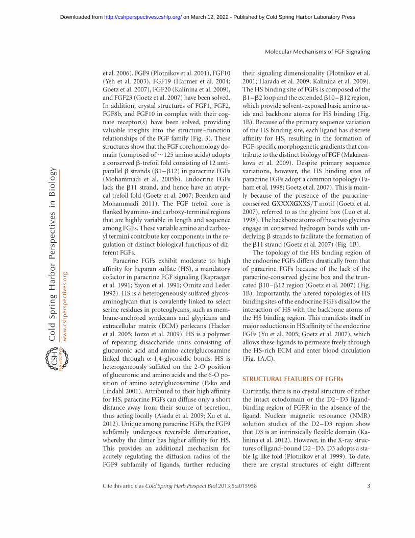
et al. 2006), FGF9 (Plotnikov et al. 2001), FGF10(Yeh et al. 2003), FGF19 (Harmer et al. 2004;Goetz et al. 2007), FGF20 (Kalinina et al. 2009),and FGF23 (Goetz et al. 2007) have been solved.In addition, crystal structures of FGF1, FGF2,FGF8b, and FGF10 in complex with their cog-nate receptor(s) have been solved, providingvaluable insights into the structure–functionrelationships of the FGF family (Fig. 3). Thesestructures show that the FGF core homology do-main (composed of �125 amino acids) adoptsa conserved b-trefoil fold consisting of 12 anti-parallel b strands (b1–b12) in paracrine FGFs(Mohammadi et al. 2005b). Endocrine FGFslack the b11 strand, and hence have an atypi-cal trefoil fold (Goetz et al. 2007; Beenken andMohammadi 2011). The FGF trefoil core isflanked byamino- and carboxy-terminal regionsthat are highly variable in length and sequenceamong FGFs. These variable amino and carbox-yl termini contribute key components in the re-gulation of distinct biological functions of dif-ferent FGFs.
Paracrine FGFs exhibit moderate to highaffinity for heparan sulfate (HS), a mandatorycofactor in paracrine FGF signaling (Rapraegeret al. 1991; Yayon et al. 1991; Ornitz and Leder1992). HS is a heterogeneously sulfated glycos-aminoglycan that is covalently linked to selectserine residues in proteoglycans, such as mem-brane-anchored syndecans and glypicans andextracellular matrix (ECM) perlecans (Hackeret al. 2005; Iozzo et al. 2009). HS is a polymerof repeating disaccharide units consisting ofglucuronic acid and amino acteylglucosaminelinked through a-1,4-glycosidic bonds. HS isheterogeneously sulfated on the 2-O positionof glucuronic and amino acids and the 6-O po-sition of amino acteylglucosamine (Esko andLindahl 2001). Attributed to their high affinityfor HS, paracrine FGFs can diffuse only a shortdistance away from their source of secretion,thus acting locally (Asada et al. 2009; Xu et al.2012). Unique among paracrine FGFs, the FGF9subfamily undergoes reversible dimerization,whereby the dimer has higher affinity for HS.This provides an additional mechanism foracutely regulating the diffusion radius of theFGF9 subfamily of ligands, further reducing
their signaling dimensionality (Plotnikov et al.2001; Harada et al. 2009; Kalinina et al. 2009).The HS binding site of FGFs is composed of theb1–b2 loop and the extendedb10–b12 region,which provide solvent-exposed basic amino ac-ids and backbone atoms for HS binding (Fig.1B). Because of the primary sequence variationof the HS binding site, each ligand has discreteaffinity for HS, resulting in the formation ofFGF-specific morphogenetic gradients that con-tribute to the distinct biology of FGF (Makaren-kova et al. 2009). Despite primary sequencevariations, however, the HS binding sites ofparacrine FGFs adopt a common topology (Fa-ham et al. 1998; Goetz et al. 2007). This is main-ly because of the presence of the paracrine-conserved GXXXXGXXS/T motif (Goetz et al.2007), referred to as the glycine box (Luo et al.1998). The backbone atoms of these two glycinesengage in conserved hydrogen bonds with un-derlying b strands to facilitate the formation ofthe b11 strand (Goetz et al. 2007) (Fig. 1B).
The topology of the HS binding region ofthe endocrine FGFs differs drastically from thatof paracrine FGFs because of the lack of theparacrine-conserved glycine box and the trun-cated b10–b12 region (Goetz et al. 2007) (Fig.1B). Importantly, the altered topologies of HSbinding sites of the endocrine FGFs disallow theinteraction of HS with the backbone atoms ofthe HS binding region. This manifests itself inmajor reductions in HS affinity of the endocrineFGFs (Yu et al. 2005; Goetz et al. 2007), whichallows these ligands to permeate freely throughthe HS-rich ECM and enter blood circulation(Fig. 1A,C).
STRUCTURAL FEATURES OF FGFRs
Currently, there is no crystal structure of eitherthe intact ectodomain or the D2–D3 ligand-binding region of FGFR in the absence of theligand. Nuclear magnetic resonance (NMR)solution studies of the D2–D3 region showthat D3 is an intrinsically flexible domain (Ka-linina et al. 2012). However, in the X-ray struc-tures of ligand-bound D2–D3, D3 adopts a sta-ble Ig-like fold (Plotnikov et al. 1999). To date,there are crystal structures of eight different
Molecular Mechanisms of FGF Signaling
Cite this article as Cold Spring Harb Perspect Biol 2013;5:a015958 3
on March 12, 2022 - Published by Cold Spring Harbor Laboratory Press http://cshperspectives.cshlp.org/Downloaded from
FGF–FGFR complexes that feature unique li-gand–receptor combinations, including FGF1with FGFR1c (Plotnikov et al. 2000; Beenkenet al. 2012), FGFR2c (Stauber et al. 2000),FGFR3c (Olsen et al. 2004), FGFR2b (Beenken
et al. 2012), FGF2 with FGFR1c (Plotnikov et al.1999; Schlessinger et al. 2000), FGFR2c (Plotni-kov et al. 2000), FGF8b with FGFR2c (Olsenet al. 2006), and FGF10 with FGFR2b (Yehet al. 2003). In all these structures, the receptor
Adipose tissue
A
B
C
α/β Klothocoreceptors
FGFRc
EndocrineFGF(21)
Endocrine FGF signaling
Bloodstream
EndocrineFGF(21)
FGFRc
ParacrineFGF(10)
Mesenchymal
Paracrine FGF signaling
Epithelial
FGF2-HS (PDB: 1FQ9) FGF19-HS (PDB: 2P23)
Heparin chip surface
400 nM FGF
FGF2
FGF23
50 150 250Time (sec)
350
FGF21FGF29
1800
1600
1400
1200
1000R
espo
nse
units
800
600
400
200
0
FGFRb
ParacrineFGF(8)
Liver
Figure 1. FGF signaling in the liver and adipose tissue. (A) The paracrine FGF signaling loop in the liver. FGFs areexpressed in both the epithelial or mesenchymal tissue, and signal in a paracrine fashion through their cognatereceptors, which are expressed in the opposite tissues. Shown are two examples of paracrine ligands, FGF8 andFGF10, which signal exclusively in an epithelial-to-mesenchyme and mesenchyme-to-epithelial manner, re-spectively. (B) Comparison of the crystal structures of FGF2 (PDB: 1FQ9) and FGF19 (PDB: 2P23) provides thestructural basis for the low affinity of endocrine ligands for HS. (C) Comparison of the binding interactions ofFGF2, FGF19, FGF21, and FGF23 with HS using surface-plasmon resonance spectroscopy. The low affinity ofFGF19 family members (such as FGF21) allows them to permeate freely through the HS-dense intercellularspace and enter into the blood. This enables them to act as hormones in target tissues in which a/b Klothocoreceptors are expressed (top panel in part A).
A.A. Belov and M. Mohammadi
4 Cite this article as Cold Spring Harb Perspect Biol 2013;5:a015958
on March 12, 2022 - Published by Cold Spring Harbor Laboratory Press http://cshperspectives.cshlp.org/Downloaded from
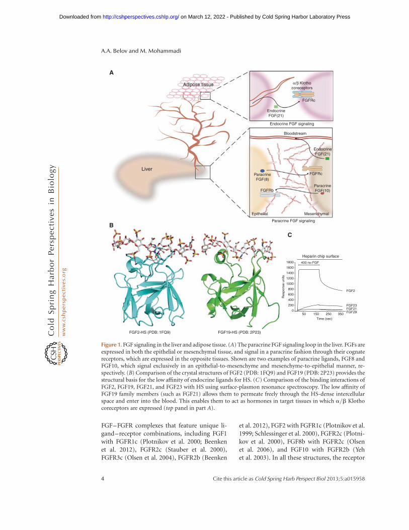
adopts an extended conformation displayingsignificant differences in the relative orientationof D2 and D3 to one another, suggesting thatthe overall receptor orientation is dictated by li-gand binding (Fig. 3). This is harmonious withthe fact that there are no intramolecular contactsbetween D2 and D3 that would constrain therelative disposition of the two domains in theabsence of the ligand.
As anticipated based on sequence homology(Bateman and Chothia 1995), D1 and D2 adoptIg folds that belong to the I set of the Ig super-family (Plotnikov et al. 1999; Hung et al. 2005;Kiselyov et al. 2006) (Fig. 2A). In contrast, D3has an unusual Ig-fold in that the region between
the bC0 and bE strands (referred to as the bC0 –bE loop) adopts a different conformation in dif-ferent complexes, suggesting that it is highly mo-bile. In all but the FGF8b–FGFR2c structure, D3lacks the analogous bD strand of the Ig-like do-mains D1 and D2. Notably, alternative splicingof D3 occurs at the junction between the bC0
strand and the bC0–bE loop, diversifying theprimary sequence of the loop (Johnson et al.1991; Yeh et al. 2003) (Fig. 2A). As wewill discusslater, this, together with the inherent flexibilityof the loop, plays a major role in determiningligand-binding specificity/promiscuity. The HSbinding site of the receptor resides in D2 and iscomprised of several surface-exposed residues
PDB: 1FQ9 and 2CR3
D3
D2
D1–D2 Linker
A B
C
D2–D3 Linker
D1
Nβ9
βG
βG
β4
β5
β12
β1
β8
β9βC
βC′–βE
gA
βB′–βC
βF–βG
C
βD
βG
βA
βF
βA′
β1A167
Y103
P169
G160
N162
Y164 R251
D283
S282
R78
Q285
E154
V248P141
Prototypical contacts at the FGF–D2 Interface
Prototypical contacts at the FGF–D2–D3 linker and D3 interface
L140
Y24
L165
180°
Figure 2. Structural features of a prototypical FGF receptor and FGF-conserved FGF–FGFR contacts. (A) The X-ray structure of FGF2–FGFR1c (PDB: 1FQ9) and an NMR structure of D1 (PDB: 2CR3) were linked arbitrarilywith a modeled D1–D2 linker to construct a model of a full-length FGF receptor. The acid box is red and the HSbinding region in the FGF2–FGFR1c complex is blue. FGFR1c and FGF2 are cyan and salmon, respectively. Thealternatively spliced portion of D3 is magenta. (B) Prototypical contacts (PDB: 1FQ9) between the ligand andreceptor D2 are illustrated. Dashed lines denote hydrogen bonds. Hydrophobic contacts are indicated usingtransparent surfaces. Oxygen and nitrogen atoms are red and blue, respectively, hereafter. (C) The conservedhydrogen bonds at the interface between the D2–D3 linker and D3 of the FGFR and FGF ligand as observed inthe FGF10–FGFR2b structure (PDB: 1NUN). The dashed box within panel C highlights the hydrogen bondsbetween the D2–D3 linker of FGFR and FGF.
Molecular Mechanisms of FGF Signaling
Cite this article as Cold Spring Harb Perspect Biol 2013;5:a015958 5
on March 12, 2022 - Published by Cold Spring Harbor Laboratory Press http://cshperspectives.cshlp.org/Downloaded from

emanating from the bB, bE, bD strands, the gAhelix, and the loop between the bA and bA0
strands of D2 (Schlessinger et al. 2000). Unlikethe FGF–HS interaction, only the side chains ofD2 participate in HS coordination, which ex-plains the significantly lower affinity of FGFRsfor HS compared to that of FGFs (Powell et al.2002; Ibrahimi et al. 2004c; Asada et al. 2009;Trueb 2011).
The bottom edge of D2, the D2–D3 linker,and the top portion of D3 compose the ligand-binding pocket (Plotnikov et al. 1999) (Fig 2A).The FGF straddles D3 via the bottom end of thetrefoil (the top end being the HS binding site).The bB0 –bC and bF–bG loops in D3 are en-gulfed in a depression formed between the b1,b2, b4, b5, b8, and b9 strands and the inter-vening loops, whereas the D2–D3 linker coop-erates with D3 to further fix the ligand in itsobserved position (Fig. 2C). The bA0 and bFstrands at the bottom edge of D2 sharply engagetheb1 throughb2 strands, theb3–b4 loop, andb9 and b12 of the ligand (Plotnikov et al. 2000)(Fig. 2A).
THE FGF–FGFR INTERFACE
Conserved Ligand–Receptor Contacts
Each subdomain makes several highly conservedcontacts with the ligand, unambiguously dem-onstrating that the D2–D3 fragment is the min-imal binding region of FGFs (Stauber et al. 2000;Olsen et al. 2004; Mohammadi et al. 2005b). Atthe FGF–D2 interface, two conserved tyrosineresidues, one from the b1 strand and anotherfrom the b8–b9 loop, along with highly con-served proline in the b12 strand of the ligandengage in hydrophobic and hydrogen-bondinginteractions with conserved residues on the bA0
and bG strands in D2 (Fig. 2B). The interfacebetween FGF and the D2–D3 linker is by far themost conserved residue. Here, an invariant argi-nine from the D2–D3 linker region makes threehydrogen bonds with a side chain of a residue inb9 and a backbone of the b8–b9 loop in theligand (Fig. 2C). Theb9 residue is an asparaginein all FGFs, with the exception of the FGF8 sub-family, which has a threonine instead. Notably
the D2–D3 linker arginine is also engaged inintramolecular hydrogen bonding with a con-served aspartic acid in the bB0 –bC loop of D3.This primes the arginine for FGF recognition,thus minimizing the entropic penalty associatedwith ligand binding. The focal role of these hy-drogen bonds in providing general FGF–FGFRaffinity is evidenced by the fact that fibroblasthomologous factors (FHFs) contain a valine inthe equivalent position of b9 asparagine, whichhinders them from binding/activating FGFRs(Goldfarb 1996; Olsen et al. 2003; Goetz et al.2009; Wang et al. 2012). Mutation of the D2–D3linker arginine to glutamine leads to a loss offunction in the Kallmann syndrome (Dode et al.2003; Pitteloud et al. 2006a,b), further high-lighting the importance of these conserved hy-drogen bonds in FGF–FGFR binding. The FGF–D3 interface harbors two highly conserved con-tacts. Here the backbone atoms of the bB0 –bCloop are engaged in three strong hydrogen bondswith an arginine fromb1 and glutamic acid fromb8 of the ligand (Fig. 2C). Together, the afore-mentioned contacts provide general FGF–FGFRbinding affinity, whereas specificity is primarilydecided by divergent contacts at the FGF–D3.
ALTERNATIVE SPLICING IN D3 IS A MAINMECHANISM IN REGULATION OF FGF–FGFR SPECIFICITY
In FGFR1–3, two alternative exons (“b” and“c”) code for the second half of D3 (Johnsonet al. 1991; Miki et al. 1992; Yayon et al. 1992)that are spliced in tissue-specific fashion (Orr-Urtreger et al. 1993; Wuechner et al. 1996; Beeret al. 2000). Generally, the b-splice variants areexpressed in the epithelial tissue, whereas the c-splice isoforms are expressed in the mesenchy-mal tissue (Orr-Urtreger et al. 1993; McEwenand Ornitz 1997). Paracrine FGFs also showtissue-specific expression patterns with ligandsfor FGFRb isoforms being expressed in mesen-chyme, and ligands for FGFRc isoforms ex-pressed in epithelium (Finch et al. 1989). Thisresults in an epithelial–mesenchymal FGF sig-naling loop that is crucial for tissue homeostasisand organogenesis (McIntosh et al. 2000; Itohand Ornitz 2011), as evidenced by the fact that
A.A. Belov and M. Mohammadi
6 Cite this article as Cold Spring Harb Perspect Biol 2013;5:a015958
on March 12, 2022 - Published by Cold Spring Harbor Laboratory Press http://cshperspectives.cshlp.org/Downloaded from

derangements of this signaling loop contributeto human skeletal disorders and cancer (Ibra-himi et al. 2005; Beenken and Mohammadi2009, 2011). The tissue-specific alternative splic-ing in D3 is the chief mechanism in the regula-tion of FGF–FGFR binding specificity (Yayonet al. 1992; Ornitz et al. 1996; Zhang et al.2006). Crystallographic studies of eight differentFGF–FGFR complexes have revealed that thisalternative splicing controls FGF–FGFR bind-ing specificity/promiscuity by altering the com-position of FGF binding sites in D3, includingthe bC0 –bE and bF–bG loops. Moreover, thebC0 –bE loop is inherently flexible, and is capa-ble of adapting uniquely to each ligand. The re-gions of FGFs that engage the alternativelyspliced half of D3, in particular, the amino-ter-minal region of ligands, is divergent in the pri-mary sequence. These structural data flag theFGF–D3 interface as the key mediator of FGF–FGFR binding specificity/promiscuity. The cur-rent structural data on FGF–FGFR complexeshave disclosed two distinct modes by which con-tacts at the FGF–D3 interface mediate FGF–FGFR binding specificity.
GENERAL MODE OF FGF–FGFR SPECIFICITY
The hallmark of the general mode, observedin the FGF1-, FGF2-, and FGF10-receptor com-plexes, is a cleft in D3 that forms between bB0 –bC and the alternatively spliced bC0 –bE on li-gand binding. This cleft is induced by hydro-phobic contacts between residues from the b7and b8 loops and the b5 strand in the FGF core,and a hydrophobic residue at the apex of thealternatively spliced bC0 –bE of the receptor(Fig. 3A–C). Residues from the b4, b5 strandsand the intervening loop as well as the amino-terminal tail of the ligand engage the bC0 –bEloop, further stabilizing the cleft. The bF–bGloop, which is also alternatively spliced, engagesresidues from the b4–b5 loop and the b8strand.
FGF7 Subfamily
FGF7 subfamily members are secreted by themesenchyme and act exclusively on the FGFR2b
resident in the epithelial tissue to constitute themesenchymal-to-epithelial arm of the signalingloop (Mason et al. 1994). The FGF10–FGFR2bstructure (Yeh et al. 2003) shows that F146,Y131, and A122 in FGF10 form a hydrophobicsurface that tethers Ile-317 from the alternative-ly spliced loop promoting formation of the D3cleft (Fig 3B). According to the structure, theFGF7 subfamily’s preference for FGFR2b can betraced to the highly specific hydrogen bondsbetween Asp-76, a unique amino-terminal res-idue in the FGF7 subfamily, and Ser-315 fromthe alternatively spliced bC0 –bE loop in the D3cleft. A p-cation interaction between Y345 inthe bF–bG (a residue unique to b-splice iso-forms of receptors) and R155 in the b8 strandof FGF10 further reinforces the specificity (Fig.3A,B). Interestingly, the substitution of the con-served tyrosine in the b1 strand for phenylala-nine in the FGF7 subfamily acts in concert withthe above-mentioned specific FGF–D3 contactsto further narrow the specificity of the FGF7subfamily for FGFR2b. This Y!F substitutiondisables this subfamily form hydrogen bondingwith D2 (Fig. 2B), thus minimizing the contri-bution of D2 in providing ligand-bindingaffinity. By primarily relying on the alternativelyspliced loops of D3 of the receptor to attainspecificity and affinity, the FGF7 subfamily issolely able to bind and activate FGFR2b. Inthe absence of these hydrogen bonds that im-pose constrains on the orientation of D3, D2 isobserved to rotate about the D2–D3 linker, re-sulting in a distinct orientation of the HS bind-ing site in D2, relative to the ligand. This struc-tural change has been postulated to play a role indetermining the HS selectivity of the FGF7 sub-family FGFR2b complexes (Mohammadi et al.2005a).
FGF1 Subfamily
Unlike the FGF7 subfamily, whose membersshare a common receptor specificity profile, themembers of the FGF1 subfamily, namely FGF1and FGF2, have a distinct receptor binding spe-cificity/promiscuity profile (Ibrahimi et al.2004a,b). Both FGF1 and FGF2 are promiscu-ous and can bind more than one FGFR isoform.
Molecular Mechanisms of FGF Signaling
Cite this article as Cold Spring Harb Perspect Biol 2013;5:a015958 7
on March 12, 2022 - Published by Cold Spring Harbor Laboratory Press http://cshperspectives.cshlp.org/Downloaded from

FGF2 binds equally well to the “c” isoforms ofFGFR1 and FGFR2 but has negligible binding tothe “b” isoforms (Ornitz et al. 1996). In con-trast, FGF1 overrides the specificity barrier setby alternative splicing and interacts indiscrimi-nately with all seven FGFRs (Zhang et al. 2006).
Crystal structures of FGF2 with both of itscognate receptors have been solved (Plotnikovet al. 1999, 2000), revealing the molecular basisfor the FGFR binding specificity/promiscuityof FGF2. Reminiscent of the FGF10–FGFR2bstructure, a valine/isoleucine at the apex ofthe bC0 –bE loop makes hydrophobic contactswith Y73 (from the b6 strand) and V88 and F93(from the b7–b8 loop), resulting in the forma-tion of the D3 cleft (Fig. 3C). The specificity/promiscuity of FGF2 can be traced mostly tospecific hydrogen bonds between Q56 fromthe b4 strand of FGF2 and D321 in the D3 cleft.
F17 from the amino terminus is immersed ina hydrophobic pocket created by I288, P286,and V280 in the D3 cleft, while also engagingin hydrogen-aromatic interactions with D321(Fig. 3C). The bC0 –bE loop of “b-”splice iso-forms would not be able to endorse these spe-cific contacts.
The crystal structure of FGF1 in complexwith four of its cognate FGFRs, namely, FGFR1c(Plotnikov et al. 2000; Beenken et al. 2012),FGFR2c (Stauber et al. 2000), FGFR3c (Olsenet al. 2004), and FGFR2b (Beenken et al. 2012),have been solved. Analysis of these four FGF1–FGFR structures show that the promiscuity ofFGF1 can be traced to the unusual ability ofFGF1 to adapt to the alternatively splicedbC0 –bE loop on the receptor. The versatilityin the interactions of FGF1 with the bC0 –bEloop manifests itself in the observed divergent
General mode FGF8 subfamily-specific mode
FGF8b–FGFR2c
180°
FGF1–FGFR2b
A B C D
PDB: 30JM
Y79
P90
Y70
1317
I288
1317
A122
F146
Y131R103
β9
β8 βF-βG
βC′–βEβC′–βE
βC′–βE
“βC′–βE”
βG
β9β5
β4β8
β1
βFβCβG βF
βC
I350
L343F352
βC′
βF-βG
β5 β9
β8
β1
R155
βCA322
β4
S315
Y345
Y73
F93
V88
V317
Q56P286
D321
I288 V280
L309
F32
V36
F93
S347
S95
Q285
I291
F17
D76
βC βC βF βG
E323
V280
Tyr23P286Y345
V342
β5
β4
PDB: 1NUN PDB: 1EVT PDB: 2FDB
FGF10–FGFR2b FGF2–FGFR2c
Figure 3. General and FGF8-specific modes of FGF–FGFR binding. FGF1 and FGF10 in complex with FGFR2bare illustrated with the alternatively spliced regions of D3 shown in slate. FGF2 and FGF8b in complex withFGFR2c are also illustrated, with the alternatively spliced regions of D3 in pink. FGF1, FGF10, FGF2, and FGF8bare light pink, gray, wheat, and salmon, respectively. The constant regions of FGFRs are lime green. Eachsubpanel illustrates the specific contacts the ligands make with D3. In the subpanels A and B, the b4 strandsfrom FGF1 and FGF10 are made transparent to allow for the visualization of thep-cation interactions at thebF–bG loop outside of the D3 cleft.
A.A. Belov and M. Mohammadi
8 Cite this article as Cold Spring Harb Perspect Biol 2013;5:a015958
on March 12, 2022 - Published by Cold Spring Harbor Laboratory Press http://cshperspectives.cshlp.org/Downloaded from
conformation of the bC0 –bE loop in the fourFGF1–FGFR structures. The FGF1–FGFR1c,FGF1–FGFR2c, and FGF1–FGFR2b complexesall feature the characteristic D3 cleft, whereasthe FGF1–FGFR3c structure lacks it. Three ami-no-terminal residues of FGF1, namely, F16, N22,and Y23 FGF1 make variable interactions withD3, depending on which receptor they interactwith (Beenken et al. 2012). Replacing the cor-responding three amino-terminal residues inFGF2 with that of FGF1 bestows on FGF2 theability to bind to the FGFR2b isoform, thus ver-ifying the structural data. As acorollary to FGF10specificity, subtle changes at the FGF1–D2 con-tacts augment the promiscuityof FGF1. Notably,FGF1 has an L135 instead of an M142 in FGF2 atthe FGF–D2 interface, which enables FGF1 toengage in stronger hydrophobic contacts withD2, thereby gaining more affinity through D2contacts. This enhances the promiscuityof FGF1as it reduces the dependency of FGF1 on D3 forreceptor binding.
Structural and biochemical studies of path-ogenic mutations affecting the extracellular do-main of FGFRs strongly support the regulatorymechanisms of FGF–FGFR binding specificity/promiscuity deduced from FGF–FGFR crys-tal structures (Wilkie 2005). For example, theS252W and P253R mutations in the D2–D3linker region of FGFR2, responsible for the Apertsyndrome (Wilkie et al. 1995), introduce addi-tional conserved contacts with FGFs that resultin a generalized increase in affinity of the recep-tor for all FGFs, thus minimizing the reliance ofFGF on specific contacts with D3 for receptorbinding (Anderson et al. 1998; Ibrahimi et al.2001; Yu and Ornitz 2001; Glaser et al. 2003;Yoon et al. 2009). This enables the mesenchy-mallyexpressed diseased FGFR2c to illegitimate-ly bind and become activated in an autocrinefashion by mesenchymmal FGF10, therebyshort-circuiting the epithelial-to-mesenchymesignaling polarity (Yu et al. 2000b). Likewise,structural studies of the D321A mutation, whichmaps onto the bC0–bE loop of FGFR2c, showthat this mutation removes the electrostatic re-pulsion and steric conflict that prohibits bind-ing of FGFR2c to FGF10, thereby enabling theillegitimate activation of the “diseased” receptor
by FGF10 in the mesenchyme (Ibrahimi et al.2004a).
FGF8 SUBFAMILY-SPECIFIC MODEOF FGF–FGFR SPECIFICITY
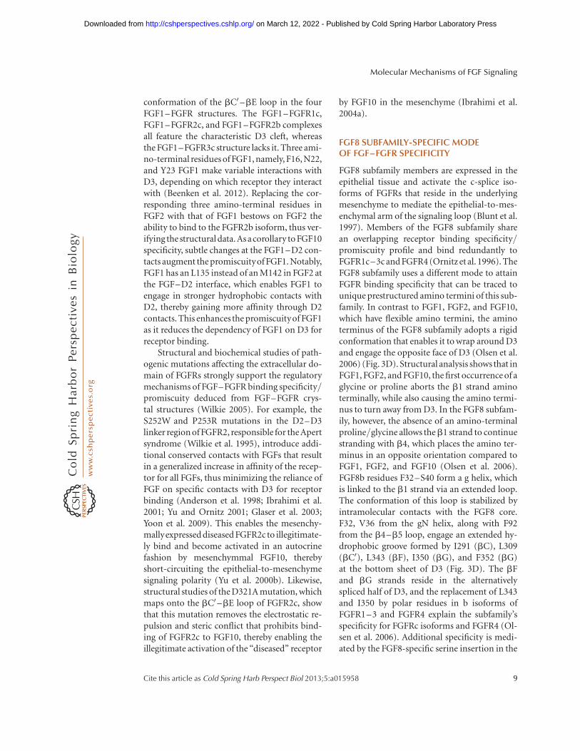
FGF8 subfamily members are expressed in theepithelial tissue and activate the c-splice iso-forms of FGFRs that reside in the underlyingmesenchyme to mediate the epithelial-to-mes-enchymal arm of the signaling loop (Blunt et al.1997). Members of the FGF8 subfamily sharean overlapping receptor binding specificity/promiscuity profile and bind redundantly toFGFR1c–3c and FGFR4 (Ornitz et al. 1996). TheFGF8 subfamily uses a different mode to attainFGFR binding specificity that can be traced tounique prestructured amino termini of this sub-family. In contrast to FGF1, FGF2, and FGF10,which have flexible amino termini, the aminoterminus of the FGF8 subfamily adopts a rigidconformation that enables it to wrap around D3and engage the opposite face of D3 (Olsen et al.2006) (Fig. 3D). Structural analysis showsthat inFGF1, FGF2, and FGF10, the first occurrence of aglycine or proline aborts the b1 strand aminoterminally, while also causing the amino termi-nus to turn away from D3. In the FGF8 subfam-ily, however, the absence of an amino-terminalproline/glycine allows theb1 strand to continuestranding with b4, which places the amino ter-minus in an opposite orientation compared toFGF1, FGF2, and FGF10 (Olsen et al. 2006).FGF8b residues F32–S40 form a g helix, whichis linked to the b1 strand via an extended loop.The conformation of this loop is stabilized byintramolecular contacts with the FGF8 core.F32, V36 from the gN helix, along with F92from the b4–b5 loop, engage an extended hy-drophobic groove formed by I291 (bC), L309(bC0), L343 (bF), I350 (bG), and F352 (bG)at the bottom sheet of D3 (Fig. 3D). The bFand bG strands reside in the alternativelyspliced half of D3, and the replacement of L343and I350 by polar residues in b isoforms ofFGFR1–3 and FGFR4 explain the subfamily’sspecificity for FGFRc isoforms and FGFR4 (Ol-sen et al. 2006). Additional specificity is medi-ated by the FGF8-specific serine insertion in the
Molecular Mechanisms of FGF Signaling
Cite this article as Cold Spring Harb Perspect Biol 2013;5:a015958 9
on March 12, 2022 - Published by Cold Spring Harbor Laboratory Press http://cshperspectives.cshlp.org/Downloaded from
b4–b5 loop, which forms a unique network ofhydrogen bonds with the backbone atoms ofthe alternatively spliced bB0 –bC and bF–bGloops.
The bC0 –bE loop conformation is totallyrearranged to make room for the unique FGF8amino terminus. In fact, a section of this loopforms the canonical Ig-folded bD strand that isconnected to bE through a short loop. All theloops connecting the strands of the top sheetlocalize on one side, and, as a result, the FG-F8b–FGFR2c structure lacks the D3 cleft. Theunique mode of FGF8b–FGFR2c binding in-duces a unique D3 rotation that pivots about theD2–D3 linker. Modeling studies show that themembrane insertion points of receptor mono-mers in the FGF8b–FGFR2c dimer would becloser by �15 A vis-a-vis the FGF2–FGFR2cdimer. These topological differences have beenpostulated to contribute to the distinct signalingcapacity by different FGFs (Olsen et al. 2006).The L341S loss-of-function mutation in FGF-R1, which is responsible for the Kallmann syn-drome, maps to the D3 groove that the FGF8subfamily engages (Dode et al. 2003; Pitteloudet al. 2006a; Falardeau et al. 2008). The substi-tution of leucine for the polar serine in this hy-drophobic groove severely impairs the FGF8bbinding, thus implicating the FGF8 subfamilyin the etiology of the Kallmann syndrome. In-deed, subsequent genetic screening of a cohortof patients led to the identification of loss-of-function mutations in FGF8 and FGF17 (Falar-deau et al. 2008; Trarbach et al. 2010; McCabeet al. 2011).
In summary, contacts between FGF and al-ternatively spliced regions in D3 dictate FGF–FGFR specificity and promiscuity, whereas con-tacts between FGF and D2 and the D2–D3 linkerserve primarily to provide basal ligand-bindingaffinity. Importantly, differences in the contactsbetween FGF and D2 and/or D2–D3 contactscan enhance specificity/promiscuity of FGFsby modifying the basal FGF–FGFR affinity.The fidelity of FGF–FGFR binding specificity/promiscuity combined with ligand-dependentdifferences in receptor orientation would allowfor the precise regulation of FGF-induced sig-naling.
HS-ASSISTED PARACRINE FGF–FGFRDIMERIZATION
HS is a mandatory cofactor in paracrine FGFsignaling (Imamura and Mitsui 1987; Rap-raeger et al. 1991; Yayon et al. 1991; Olwin andRapraeger 1992; Ornitz et al. 1992), as docu-mented by the fact that mice and flies with de-fects in components of FGF signaling or HSbiosynthetic enzymes share overlapping pheno-types (Lin et al. 1999; Inatani et al. 2003). Struc-tural data have shown that HS promotes theformation of a symmetric 2:2:2 dimer betweenFGF, FGFR, and HS, which is required for sig-nal transmission across the plasma membrane(Schlessinger et al. 2000). In the dimeric com-plex, FGFs engage D2, D3, and the D2–D3 link-er of their primary receptor (as discussed indepth above). In addition, residues from theb8–b9 and b11–b12 loops of FGFs interactwith the bC0 –bD and bE–bF loops in D2 ofthe neighboring (secondary) receptor (Fig. 4A).Notably, the primary sequence of the b11–b12loop shows considerable variation among FGFs,indicating that additional FGF–FGFR signalingspecificity may be achieved on receptor dimeri-zation. The dimer interface is further fortifiedby the direct interactions between FGFRs medi-ated via the bottom end of their D2 domains(Fig. 2A, B). A 2:2:1 FGF–FGFR–HS asymmet-ric model has also been proposed (Pellegriniet al. 2000); however, analysis of the mechanismof action of pathogenic mutations has lent un-biased support for the symmetric mode of di-merization (Mohammadi et al. 2005a). For ex-ample, the A172F gain-of-function mutationthat is implicated in the Pfeifer syndromemaps to the D2–D2 receptor interface thathas been shown to cause gain of function byfacilitating ligand-dependent FGFR dimeriza-tion (Ibrahimi et al. 2005).
The HS binding sites of the ligands and re-ceptors are adjacent to one other, forming a con-tinuous basic canyon on the membrane distalend of the dimer. The HS binding residues ofFGFs and FGFRs act in concert to recruit twoHS molecules in a symmetric fashion (Fig. 4A).Each HS oligosaccharide makes a total of 30hydrogen bonds with a single FGF and both
A.A. Belov and M. Mohammadi
10 Cite this article as Cold Spring Harb Perspect Biol 2013;5:a015958
on March 12, 2022 - Published by Cold Spring Harbor Laboratory Press http://cshperspectives.cshlp.org/Downloaded from
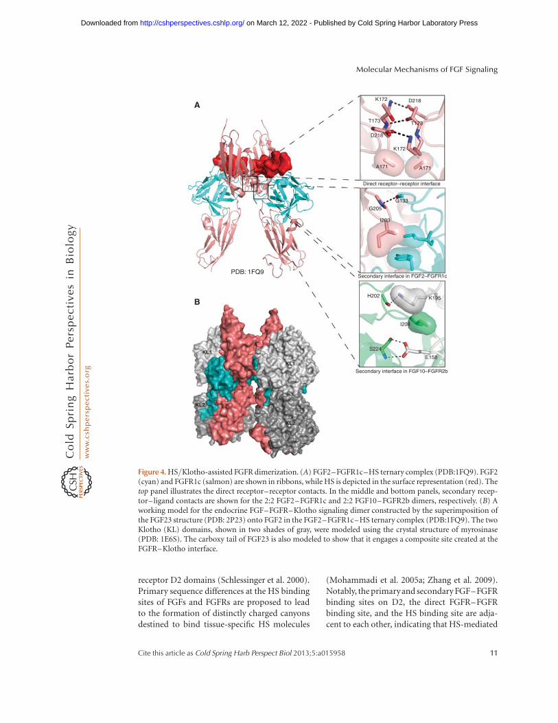
receptor D2 domains (Schlessinger et al. 2000).Primary sequence differences at the HS bindingsites of FGFs and FGFRs are proposed to leadto the formation of distinctly charged canyonsdestined to bind tissue-specific HS molecules
(Mohammadi et al. 2005a; Zhang et al. 2009).Notably, the primaryand secondary FGF–FGFRbinding sites on D2, the direct FGFR–FGFRbinding site, and the HS binding site are adja-cent to each other, indicating that HS-mediated
K172
T173
D218
K172
A171
Direct receptor–receptor interface
Secondary interface in FGF2–FGFR1c
Secondary interface in FGF10–FGFR2b
H202 K195
I204
E158
S224
G205
PDB: 1FQ9
A
B
KL1
KL2
KL2
KL1
I203
G133
A171
T173
D218
Figure 4. HS/Klotho-assisted FGFR dimerization. (A) FGF2–FGFR1c–HS ternary complex (PDB:1FQ9). FGF2(cyan) and FGFR1c (salmon) are shown in ribbons, while HS is depicted in the surface representation (red). Thetop panel illustrates the direct receptor–receptor contacts. In the middle and bottom panels, secondary recep-tor–ligand contacts are shown for the 2:2 FGF2–FGFR1c and 2:2 FGF10–FGFR2b dimers, respectively. (B) Aworking model for the endocrine FGF–FGFR–Klotho signaling dimer constructed by the superimposition ofthe FGF23 structure (PDB: 2P23) onto FGF2 in the FGF2–FGFR1c–HS ternary complex (PDB:1FQ9). The twoKlotho (KL) domains, shown in two shades of gray, were modeled using the crystal structure of myrosinase(PDB: 1E6S). The carboxy tail of FGF23 is also modeled to show that it engages a composite site created at theFGFR–Klotho interface.
Molecular Mechanisms of FGF Signaling
Cite this article as Cold Spring Harb Perspect Biol 2013;5:a015958 11
on March 12, 2022 - Published by Cold Spring Harbor Laboratory Press http://cshperspectives.cshlp.org/Downloaded from
dimerization is a cooperative process (Fig. 4A,middle and bottom panels). By engaging ligandand receptors in the dimer, HS promotes thekinetics and thermodynamics of FGF–FGFRbinding and dimerization, allowing for thetransmission of a sustained and robust intracel-lular signal as opposed to the transient down-stream signaling that is observed in HS-deficientcells (Yayon et al. 1991; Nugent and Edelman1992; Ornitz et al. 1992; Mathieu et al. 1995;Delehedde et al. 2000).
KLOTHO CORECEPTOR-DEPENDENTENDOCRINE FGF SIGNALING
In addition to exhibiting a negligible HS bind-ing affinity (Fig. 1C), the endocrine FGFs alsohave poor affinity for their cognate FGFRs(Goetz et al. 2012b). Modeling studies show keyresidues at their predicted receptor binding site ofendocrine FGFs are substituted for residues thatare suboptimal for receptor binding (Goetz et al.2007) (Fig. 1B). For example, substitution of theconserved arginine in theb1 strand and glutam-ic acid in the b8 strand with glycine and histi-dine, respectively, in FGF23 should cause a ma-jor reduction in receptor binding affinity of thisligand. As discussed earlier, each of these tworesidues make conserved hydrogen bonds withD3 to provide general receptor binding affinity(Fig. 2B,C). The poor HS binding affinity, alongwith negligible FGFR binding affinity rendersHS ineffective in promoting endocrine FGF–FGFR binding and dimerization (Goetz et al.2007; Beenken and Mohammadi 2012). Instead,these ligands must rely on a/b Klotho corecep-tors to signal (Urakawa et al. 2006; Kurosu et al.2007; Ogawa et al. 2007; Kharitonenkov et al.2008; Suzuki et al. 2008; Kuro-o 2012). Klothocoreceptors are single-pass transmembrane pro-teins whose ectodomain consists of tandem KLdomains, which are homologous to b-glucosi-dases (Kuro-o et al. 1997; Ito et al. 2000). TheKlotho coreceptors have been shown to associ-ate constitutively with the c-splice isoforms ofFGFR1–3 and FGFR4 to promote binding anddimerization of endocrine FGF–FGFR com-plexes (Kurosu et al. 2006, 2007; Goetz et al.2012a). The Klotho dependency confines the
target tissue specificity of endocrine FGFs tothose that express a Klotho and b Klotho (Wuet al. 2007; Kurosu and Kuro-o 2008). Signalingspecificity is further reinforced by the inherentspecificity of FGF19 subfamily members forFGFRs (Goetz et al. 2012a). For example,FGF21 primarily activates the FGFR1c–b Klo-tho complex (Yie et al. 2012), whereas FGF19 isable to activate both FGFR1c–b Klotho as wellas FGFR4–bKlotho. FGF23, on the other hand,binds promiscuously to FGFR1c–a Klotho,FGFR3c–a Klotho, and FGFR4–a Klotho (Yuet al. 2005; Goetz et al. 2012a).
Although the structural basis for ternarycomplex formation remains to be elucidated,biochemical studies have already provided sig-nificant insights into the molecular interactionsbetween the components in the ternary complex(Fig. 4B). These studies have shown that the a
Klotho and b Klotho coreceptors employ twodifferent mechanisms to promote ternary com-plex formation. a Klotho combines with FGF-R1c to create a de novo site for the FGF23 car-boxy tail, whereas b Klotho uses two distinctsites to bind independently to the FGFR and toeither the FGF19 or FGF21 carboxy tail (Wuet al. 2008; Goetz et al. 2012a). Consistent withthe key role of the carboxy tail of FGF23 in sig-naling, the biological activity of FGF23 is down-regulated by a naturally occurring proteolyticcleavage at an RXXR motif following the b-tre-foil core (Shimada et al. 2001). Interestingly, theproteolytically cleaved carboxy tail can compet-itively inhibit binding of native FGF23 to theFGFR1c–a Klotho complex (Goetz et al. 2010,2012a), indicating that this cleavage acts at twolevels to inhibit FGF23 signaling: by inactivatingFGF23 as well as by generating an endogenousinhibitor of FGF23 signaling. Pathogenic muta-tions of the RXXR motif abrogate proteolyticcleavage of the ligand (Shimada et al. 2002)and elevate the serum concentration of full-length bioactive FGF23, which accelerates phos-phate excretion in the kidney and results in au-tosomal dominant hypophosphatemic rickets(ADHR) (White et al. 2001). Recent biochemi-cal data show that the mutation of residuesthat comprises the D3 hydrophobic groove inFGFRc isoforms and FGFR4, which mediates
A.A. Belov and M. Mohammadi
12 Cite this article as Cold Spring Harb Perspect Biol 2013;5:a015958
on March 12, 2022 - Published by Cold Spring Harbor Laboratory Press http://cshperspectives.cshlp.org/Downloaded from
binding of the FGF8 subfamily, also abolishesKlotho binding (Goetz et al. 2012a). Consistentwith the overlap between FGF8 and Klotho bind-ing sites on FGFR, the association of the Klothocoreceptor with FGFRs retards the ability ofthese receptors to respond to FGF8, indicatingthat endocrine and paracrine FGF signaling im-pact each other.
The insights gained into the mechanism ofendocrine FGF signaling are already being ex-ploited to develop agonists and antagonists ofthe endocrine FGF system for treating metabolicdiseases, including diabetes, obesity, and disor-ders associated with perturbed phosphate andbile acid homeostasis (Goetz et al. 2010). Forexample, a novel FGF21 agonist has been engi-neered by knocking out the HS binding affinityof FGF2 and swapping its carboxy-terminal tailwiththatofFGF21orFGF19(Goetzetal.2012b).This engineered FGF21 agonist is superior tonative FGF21 in its insulin-sensitizing potentialandiscurrently being evaluated inamouse mod-el for obesity. Using the same approach, FGF2was converted into an FGF23 agonist, which maybe used to treat patients with familial tumoralcalcinosis, an inherited disorder, associated withloss-of-function mutations in FGF23 (Goetz etal. 2012b). Conversely, the carboxy-terminal tailof FGF23 is being developed for treatment ofrenal phosphate wasting disorders, and possiblyfor combating the cardiovascular morbidity fac-tor in chronic kidneydisease thathasbeen shownto be caused by elevated FGF23 serum levels(Goetz et al. 2010).
ALTERNATIVE SPLICING OF D1 AND D1–D2LINKER CONTROLS RECEPTORAUTOINHIBITION
In FGFR1–3, a second major alternative splic-ing event (involving exons encoding D1 and theAB-containing D1–D2 linker regions) gener-ates receptor isoforms lacking D1, the D1–D2linker, or both (Johnson et al. 1991; Givol andYayon 1992; Hou et al. 1992; Xu et al. 1992;Shimizu et al. 2001). The loss of D1 and theD1–D2 linker enhances the affinity of FGFR forboth HS and FGF, indicating that they play anautoinhibitory role in FGFR regulation (Wang
et al. 1995; Roghani and Moscatelli 2007). Themolecular basis by which these regions exertreceptor autoinhibition has been interrogatedusing solution NMR and surface plasmon res-onance (SPR) spectroscopies (Kalinina et al.2012). The data show that the negatively chargedAB subregion of the linker electrostatically en-gages the positively charged HS binding site onD2 in cis, thereby directly suppressing the HSaffinity of the receptor (illustrated in Fig. 2A).Because of the close proximity of the HS bindingsite to the primary and secondary ligand-bind-ing sites, as well as the direct receptor–receptorbinding sites on D2, the cis electrostatic AB:HSinteractions also sterically autoinhibits FGF–FGFR binding and dimerization. Consistentwith the AB subregion playing a key role inFGFR autoinhibition, the amino acid sequenceof the AB subregion is strongly conserved amongFGFR orthologs (Kalinina et al. 2012).
MECHANISM OF FGFR KINASEREGULATION
HS or Klotho-dependent FGF–FGFR dimeriza-tion juxtaposes the cytoplasmic kinase domains,providing them with sufficient opportunity totrans-phosphorylate each other on specific tyro-sine residues. A-loop tyrosine phosphorylationleads to a local rearrangement of the A loop intoits active state, which in turn stabilizes the activeconformation of the kinase globally and culmi-nates in the upregulation of the intrinsic kinaseactivity (Hubbard 1999). Secondary phosphor-ylation events on tyrosines in the JM ( juxta-membrane), kinase insert, and carboxy-tail re-gions are then ensued, providing docking sitesfor SH2-containing downstream signaling sub-strates, such as PLCg and CrkL (Eswarakumaret al. 2005; Seo et al. 2009). Crystallographicstudies of FGFR kinases have afforded major in-sights into the molecular mechanisms of FGFRkinase regulation and deregulation in patholog-ical states. The crystal structures of the FGFR1kinase (FGFR1K) (Mohammadi et al. 1996; Baeet al. 2010) and FGFR2K (Chen et al. 2007) havebeen solved in both their unphosphorylated(low activity) and A-loop tyrosine phosphor-ylated (activated) states. Reminiscent of all cur-
Molecular Mechanisms of FGF Signaling
Cite this article as Cold Spring Harb Perspect Biol 2013;5:a015958 13
on March 12, 2022 - Published by Cold Spring Harbor Laboratory Press http://cshperspectives.cshlp.org/Downloaded from
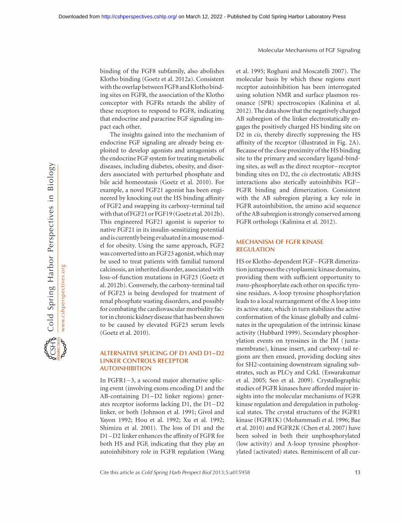
rently solved A-loop phosphorylated RTK ki-nase domains, including IRK (Hubbard 1997),IGF1R (Favelyukis et al. 2001; Pautsch et al.2001), and MUSK (Bergamin et al. 2010), thephosphate moiety of A-loop tyrosine (pY654 inFGFRK1 and pY657 in FGFRK2) engages in twohydrogen bonds with an RTK-invariant argininein the first half of the A-loop (R646 in FGFR1and R649 in FGFR2) to stabilize the active con-formation of the A-loop (Chen et al. 2007; Baeet al. 2010) (Fig. 5C,D). This in turn triggers amore intimate interaction between the aminolobe and the carboxy lobe of kinase, optimallyaligning residues from the A-loop, the catalytic
loop, and the aC-helix to catalyze phospho-transfer reactions (Fig. 5D).
In sharp contrast to other unphosphory-lated RTKs, in which the ATP and substratebinding pockets are occluded in cis by the kinaseA-loop or JM region, these sites remain acces-sible in FGFRKs, although the conformationof the carboxyl-terminal end of the A-loop isnot optimal for substrate binding (Mohammadiet al. 1996). A crystallographic analysis of FGF-R2Ks harboring pathogenic gain-of-functionmutations, responsible for craniosynostosis syn-dromes and cancers, has proved to be instru-mental in unraveling the molecular mechanism
Kinase hinge of unphosphorylated FGFR2
Kinase hinge of phosphorylated FGFR2A-loop of phosphorylated FGFR2
R649
Y656
K659
N662
I654
Y657
A-loop of unphosphorylated FGFR2
E565
A C
DB
R649
I654
Y657
Y656
N662
N549
I547
H544K545
E565
N549
K641
I547
H544 K545
PDB: 2PVF and 2PSQ
K641
Figure 5. Comparison of FGFR2 kinases in the unphosphorylated and A-loop phosphorylated states. (A) Theinhibitory network of hydrogen bonds, termed the “molecular brake,” at the kinase hinge/interlobe region of theunphosphorylated low-activity FGFR2 kinase (PDB: 2PSQ, cyan). (B) Disengagement of the molecular brake inthe A-loop phosphorylated activated FGFR2 kinase (PDB: 2PVF, gray). (C) The unphosphorylated A-loopconformation and select amino acids are shown. (D) The phosphorylated A-loop is held in an active confor-mation by hydrogen bonds between the phosphate moiety of phosphorylated A-loop tyrosine and basic residues(Arg-?? and Lys-??) within the A-loop. Note that on phosphorylation of A-loop Y656 and Y657, the A-loopundergoes a major conformational change as evidenced by the pistonlike conformational switch in the orien-tation of I654 and N662.
A.A. Belov and M. Mohammadi
14 Cite this article as Cold Spring Harb Perspect Biol 2013;5:a015958
on March 12, 2022 - Published by Cold Spring Harbor Laboratory Press http://cshperspectives.cshlp.org/Downloaded from

of FGFRK autoinhibition (Chen et al. 2007). Acomparison of these unphosphorylated “dis-eased” FGFR2Ks with the wild-type unphos-phorylated and A-loop phosphorylated FGFRKsshows that kinase autoinhibition is controlled atthe level of protein dynamics. Specifically, a net-work of hydrogen bonds mediated by a triad ofresidues from the kinase hinge (E565), the aC–b4 loop (N549), and the b8 strand (K641), areengaged in an autoinhibitory network of hydro-gen bonds (termed “molecular brake”) at thekinase-hinge region that restricts the transitionof the kinase into the active state, thus stabiliz-ing the low activity state (Chen et al. 2007)(Fig. 5A,B). A-loop tyrosine phosphorylationor pathogenic gain-of-function mutations dis-engage this molecular break either directly orindirectly through allosteric communicationbetween the A-loop and kinase hinge, therebystabilizing the active state (Chen et al. 2007).The constituents of this molecular brake areconserved in other RTKs, including PDGFR,CSFLR, and KIT, and indicate that kinase regu-lation by a molecular brake may also apply toother RTKs.
Loss-of-function mutations in the tyrosinekinase domains of FGFRs are also implicated inhuman diseases, such as Kallmann syndrome,lacrimo-auriculo-dento-digital (LADD) syn-drome, and cleft lip and palate. The crystal struc-ture of FGFR2K, containing the A628T muta-tion responsible for LADD syndrome shows thatthe introduction of the polar and bulkier threo-nine sterically hinders formation of hydrogenbonds between Arg630 and Asp626 in the cata-lytic loop, thus hampering substrate tyrosinebinding and phosphate-transfer reaction (Lewet al. 2007).
STRUCTURAL BASIS FOR FGFR KINASETRANS-AUTOPHOSPHORYLATION
As introduced earlier, tyrosine trans-autophos-phorylation fulfills two key roles in RTK signal-ing: (1) the upregulation of kinase activity, and(2) the generation of docking sites for down-stream signaling proteins (Lemmon and Schles-singer 2010). Hence, elucidation of the struc-tural basis for trans-phosphorylation is essential
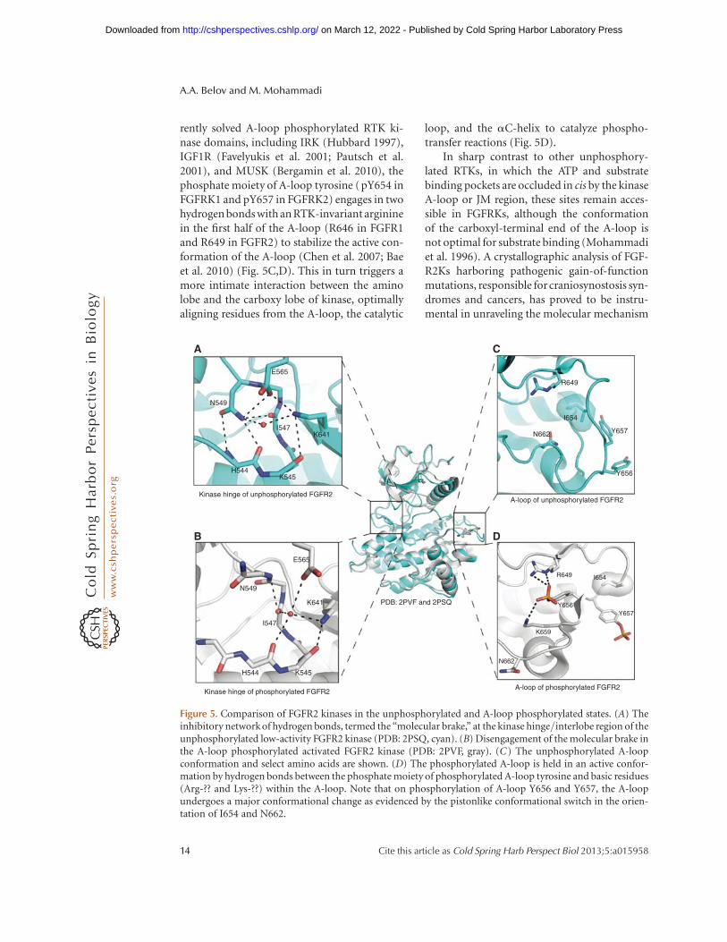
for grasping the mechanism of RTK signal trans-duction in physiological and pathological statesand for identifying new strategies for targeteddrug discovery. Two recent studies have capturedsnapshotsofFGFR1andFGFR2kinasescaught inthe act of trans-phosphorylation (Chen et al.2008a; Bae et al. 2010). One kinase acts as theenzyme whereas the other kinase offers a phos-phorylatable tyrosine from the kinase insert(Y583F in FGFR1) (Fig. 6B) or the carboxy-tailregion (Y769 in FGFR2) (Chen et al. 2008a) (Fig.6A). The structural data show that tyrosine trans-phosphorylation entails both a substantial degreeof sequence specificity and structural comple-mentarities. Both trans-phosphorylation com-plexes are asymmetric, whereby the carboxy lobeof the “substrate” engages both the amino lobeand carboxy lobe of the enzyme, burying a 1650A2 (kinase-insert tyrosine trans-phosphorylationcomplex) and a 1850 A2 (carboxy-tail tyrosinephosphorylation complex) solvent-exposed sur-face area. The enzyme–substrate interface can beroughly divided into a proximal (vicinity of thecatalytic cleft) and a distal (remote from the cat-alytic cleft) site (Fig. 6A,B). At the proximal inter-face, the phosphorylatable tyrosine and its imme-diate surrounding sequences from the substrateengage the catalytic cleft of the enzyme, whereasin the distal interface, the carboxy-lobe regionsof the substrate engage the amino lobe of theenzyme remote from the enzyme catalytic cleft.
The structural mode by which the enzymeand the substrate embrace each other is very dif-ferent between these two trans-phosphorylationcomplexes. Importantly, the kinase-insert tyro-sine and the carboxy-tail tyrosine of the sub-strate dock into the active site of the enzymefrom the opposite direction (Fig. 6A,B). Theoverall topology of the carboxy-tail tyrosinetrans-phosphorylation complex is dictated bythe latching of the carboxyl-terminal end (neg-ative pole) of the aI helix of the substrate ontothe amino-terminal end (positive pole) of theaG helix in the substrate at the proximal inter-face. This interaction assists Y769 (P-0) and itssurrounding P-4 (T765) to P þ 3 (L772) resi-duesto optimallyengage the active site and the Pþ 1 pocket of the enzyme to mediate the prox-imal specificity. Prominent contacts at the prox-
Molecular Mechanisms of FGF Signaling
Cite this article as Cold Spring Harb Perspect Biol 2013;5:a015958 15
on March 12, 2022 - Published by Cold Spring Harbor Laboratory Press http://cshperspectives.cshlp.org/Downloaded from

imal site include the salt bridge between E767(P-2) of the substrate with R573 of the enzyme,hydrogen bonds of T765 (P-4) and N766 (P-3)with theaG helix of the enzyme, and hydropho-bic contacts of L770 (P þ 1) and L772 (P þ 3)with a P þ 1 pocket (Fig. 6A, lower panel). Spe-
cificity is augmented by specific contacts at thedistal interface between the carboxy lobe of thesubstrate and the nucleotide-binding loop inthe amino lobe of the enzyme. Prominent con-tacts at the distal site include perpendicular ar-omatic interactions between F600 of substrate
FGFR2 carboxy-tail transphosphorylationPDB: 3CLY
αG
αD
C αE
G488A
B
G490N730
F492
F600
Distal site
Mg2+
Mg2+
Mg2+
AMP–PCPR573
R630
E767Y769
K668
S702
Proximal site
D519
R577
Distal site
AMP–PCP
D623
F583R627
R570
FGFR1 kinase-insert transphophorylationPDB: 3GQl
Kinase insert
E582
Proximal site
P666
D626αl
CN
Figure 6. Comparison of the carboxy-tail and kinase-insert trans-phosphorylation complexes. (A) The crystalstructure of FGFR2 kinases caught in the act of trans-phosphorylation on carboxy-tail tyrosine Tyr-769 (PDB:3CLY). The enzyme-acting kinase is cyan, and the substrate kinase is salmon. The aI helix and kinase insert aregreen, with the amino to carboxy polarity indicated with arrows. (B) The kinase-insert trans-phosphorylationcomplex (PDB: 3GQI) is colored as in A. Note that the kinase insert Tyr-583 has been mutated to phenylalanine.Compared to the carboxy-tail tyrosine trans-phosphorylation complex, there are fewer interactions between theenzyme and the substrate in the kinase-insert tyrosine trans-phosphorylation complex.
A.A. Belov and M. Mohammadi
16 Cite this article as Cold Spring Harb Perspect Biol 2013;5:a015958
on March 12, 2022 - Published by Cold Spring Harbor Laboratory Press http://cshperspectives.cshlp.org/Downloaded from
and F492 of enzyme, and hydrogen bonds be-tween N730 of substrate and backbone carbonyloxygens of the nucleotide binding loop in theenzyme (Chen et al. 2008a).
In contrast, the enzyme–substrate relation-ship in the kinase-insert trans-phosphorylationcomplex is determined by contacts between thecarboxy-lobe of substrate and the amino-lobe ofenzyme at the distal site (Bae et al. 2010). Asidefrom the hydrogen bonds between R577 (fromthe amino-terminal end of the kinase insert) andbackbone carbonyl oxygen atoms in the b3–aC loop, all the remaining interactions at thisdistal contact site are of van der Waals nature.Interestingly, as in the carboxy-tail tyrosinetrans-phosphorylation complex, the nucleotidebinding loop of the enzyme partakes in the distalsite, indicating that this loop, in addition tocoordinating ATP, may also participate in sub-strate recognition. At the proximal site, the pseu-dosubstrate Phe583 (Tyr583) occupies a verysimilar position in the catalytic pocket of theenzyme-acting kinase, even though phenylala-nine is not in position to form hydrogen bondswith the catalytic base D623 and R627 (Fig. 6B).Akin to the carboxy-tail tyrosine trans-phos-phorylation complex, residues P-0 to P þ 3form an antiparallel strand with b10 in the A-loop. Compared to the carboxy-tail tyrosinetrans-phosphorylation complex, there are sig-nificantly fewer interactions at the proximalsite in the FGFR1 kinase-insert trans-phosphor-ylation complex. In fact the hydrophobic P þ 1pocket remains unengaged in this complex.Overall, the enzyme interface in the kinase-in-sert trans-phosphorylation complex containsfewer contacts and possesses lower shape com-plementarity than in the carboxy-tail tyrosinetrans-phosphorylation complex (0.62 vs. 0.72).These differences are consistent with the kinaseautophosphorylation data showing that phos-phorylation of carboxy-tail tyrosine precedesthat of kinase-insert tyrosine (Furdui et al.2006; Chen et al. 2008a).
FUTURE PERSPECTIVES
The structural studies have provided the molec-ular bases for several major signaling events in
FGF signaling, including FGF–FGFR specific-ity, HS-assisted FGF–FGFR dimerization, andFGFR kinase regulation. Many imminent ques-tions in the FGF field remain for structural bi-ologists to address, however. Elucidation of thecrystal structure of the endocrine FGF–FGFR–Klotho ternary complex is a top priority, as itwill provide a novel mechanism by which FGF–FGFR dimerization is achieved. Crystal struc-tures with a representative from the FGF4 andFGF9 subfamilies in complex with their cognateFGFR should complete our understanding ofthe molecular mechanisms governing the spe-cificity/promiscuity of this complex system. Atthe intracellular level, future work should bedirected toward obtaining additional structuresof trans-phosphorylation events, especially ofthe A-loop tyrosine phosphorylation, the gate-keeping phosphorylation event. Crystal struc-tures of downstream signaling substrates incomplex with the intracellular kinase domainwill undoubtedly yield valuable informationon how FGFR substrates are docked onto thekinase and are phosphorylated. The crystallo-graphic data indicate that intrinsic protein dy-namics may also modulate the activity of boththe extracellular domain and intracellular do-mains of FGFRs (Chen et al. 2007; Kalininaet al. 2012). Ideally, the crystallographic snap-shots should be complemented by NMR dy-namic studies and computational methods toprovide a more accurate four-dimensional per-spective of FGF signaling. These structural datawill be valuable in directing future biochemicalinterrogations of this complex signaling net-work, and provide a framework for a compre-hensive understanding of the basis of diseaseand structure-based drug discovery for manyhuman diseases where perturbed FGF signalingis implicated.
ACKNOWLEDGMENTS
We thank Yang Liu for critically reading themanuscript, and Jinghong Ma for assistance infigure preparation. This work is supported bythe National Institute of Dental and Craniofa-cial Research Grant DE13686 (to M.M.). A.A.B.is partially supported by the Macromolecular
Molecular Mechanisms of FGF Signaling
Cite this article as Cold Spring Harb Perspect Biol 2013;5:a015958 17
on March 12, 2022 - Published by Cold Spring Harbor Laboratory Press http://cshperspectives.cshlp.org/Downloaded from

Structure and Mechanism Training Grant5T32GM088118-03.
REFERENCES
Acland P, Dixon M, Peters G, Dickson C. 1990. Subcellularfate of the int-2 oncoprotein is determined by choice ofinitiation codon. Nature 343: 662–665.
Anderson J, Burns HD, Enriquez-Harris P, Wilkie AO,Heath JK. 1998. Apert syndrome mutations in fibroblastgrowth factor receptor 2 exhibit increased affinity for FGFligand. Hum Mol Genet 7: 1475–1483.
Asada M, Shinomiya M, Suzuki M, Honda E, Sugimoto R,Ikekita M, Imamura T. 2009. Glycosaminoglycan affinityof the complete fibroblast growth factor family. BiochimBiophys Acta 1790: 40–48.
Bae JH, Boggon TJ, Tome F, Mandiyan V, Lax I, Schles-singer J. 2010. Asymmetric receptor contact is requiredfor tyrosine autophosphorylation of fibroblast growthfactor receptor in living cells. Proc Natl Acad Sci 107:2866–2871.
Basilico C, Moscatelli D. 1992. The FGF family of growthfactors and oncogenes. Adv Cancer Res 59: 115–165.
Bateman A, Chothia C. 1995. Outline structures for theextracellular domains of the fibroblast growth factor re-ceptors. Nat Struct Biol 2: 1068–1074.
Beenken A, Mohammadi M. 2009. The FGF family: Biology,pathophysiology and therapy. Nat Rev Drug Discov 8:235–253.
Beenken A, Mohammadi M. 2011. Craniosynostoses—Mo-lecular genetics, principles of diagnosis, and treatment.Karger, Basel, Switzerland.
Beenken A, Mohammadi M. 2012. The structural biology ofthe FGF19 subfamily. Adv Exp Med Biol 728: 1–24.
Beenken A, Eliseenkova AV, Ibrahimi OA, Olsen SK, Mo-hammadi M. 2012. Plasticity in interactions of fibroblastgrowth factor 1 (FGF1) N terminus with FGF receptorsunderlies promiscuity of FGF1. J Biol Chem 287:3067–3078.
Beer HD, Vindevoghel L, Gait MJ, Revest JM, Duan DR,Mason I, Dickson C, Werner S. 2000. Fibroblast growthfactor (FGF) receptor 1-IIIb is a naturally occurring func-tional receptor for FGFs that is preferentially expressed inthe skin and the brain. J Biol Chem 275: 16091–16097.
Bellosta P, Iwahori A, Plotnikov AN, Eliseenkova AV, Ba-silico C, Mohammadi M. 2001. Identification of receptorand heparin binding sites in fibroblast growth factor 4 bystructure-based mutagenesis. Mol Cell Biol 21: 5946–5957.
Bergamin E, Hallock PT, Burden SJ, Hubbard SR. 2010. Thecytoplasmic adaptor protein Dok7 activates the receptortyrosine kinase MuSK via dimerization. Mol Cell 39:100–109.
Blunt AG, Lawshe A, Cunningham ML, Seto ML, Or-nitz DM, MacArthur CA. 1997. Overlapping expressionand redundant activation of mesenchymal fibroblastgrowth factor (FGF) receptors by alternatively splicedFGF-8 ligands. J Biol Chem 272: 3733–3738.
Chateau MT, Araiz C, Descamps S, Galas S. 2010. Klothointerferes with a novel FGF-signalling pathway and insu-
lin/Igf-like signalling to improve longevity and stress re-sistance in Caenorhabditis elegans. Aging (Albany NY) 2:567–581.
Chen H, Ma J, Li W, Eliseenkova AV, Xu C, Neubert TA,Miller WT, Mohammadi M. 2007. A molecular brake inthe kinase hinge region regulates the activity of receptortyrosine kinases. Mol Cell 27: 717–730.
Chen H, Xu CF, Ma J, Eliseenkova AV, Li W, Pollock PM,Pitteloud N, Miller WT, Neubert TA, Mohammadi M.2008a. A crystallographic snapshot of tyrosine trans-phosphorylation in action. Proc Natl Acad Sci 105:19660–19665.
Chen WW, Li L, Yang GY, Li K, Qi XY, Zhu W, Tang Y, Liu H,Boden G. 2008b. Circulating FGF-21 levels in normalsubjects and in newly diagnose patients with Type 2 dia-betes mellitus. Exp Clin Endocrinol Diabetes 116: 65–68.
Colvin JS, White AC, Pratt SJ, Ornitz DM. 2001. Lung hy-poplasia and neonatal death in Fgf9-null mice identifythis gene as an essential regulator of lung mesenchyme.Development 128: 2095–2106.
Delehedde M, Seve M, Sergeant N, Wartelle I, Lyon M,Rudland PS, Fernig DG. 2000. Fibroblast growth factor-2 stimulation of p42/44MAPK phosphorylation and IkBdegradation is regulated by heparan sulfate/heparin inrat mammary fibroblasts. J Biol Chem 275: 33905–33910.
di Martino E, Tomlinson DC, Knowles MA. 2012. A decadeof FGF receptor research in bladder cancer: Past, present,and future challenges. Adv Urol 2012: 429213.
Dionne CA, Crumley G, Bellot F, Kaplow JM, Searfoss G,Ruta M, Burgess WH, Jaye M, Schlessinger J. 1990. Clon-ing and expression of two distinct high-affinity receptorscross-reacting with acidic and basic fibroblast growthfactors. EMBO J 9: 2685–2692.
Dionne CA, Jaye M, Schlessinger J. 1991. Structural diversityand binding of FGF receptors. Ann NY Acad Sci 638:161–166.
Dode C, Levilliers J, Dupont JM, De Paepe A, Le Du N,Soussi-Yanicostas N, Coimbra RS, Delmaghani S, Com-pain-Nouaille S, Baverel F, et al. 2003. Loss-of-functionmutations in FGFR1 cause autosomal dominant Kall-mann syndrome. Nat Genet 33: 463–465.
Dubrulle J, Pourquie O. 2004. fgf8 mRNA decay establishes agradient that couples axial elongation to patterning in thevertebrate embryo. Nature 427: 419–422.
Eriksson AE, Cousens LS, Weaver LH, Matthews BW. 1991.Three-dimensional structure of human basic fibroblastgrowth factor. Proc Natl Acad Sci 88: 3441–3445.
Esko JD, Lindahl U. 2001. Molecular diversity of heparansulfate. J Clin Invest 108: 169–173.
Eswarakumar VP, Lax I, Schlessinger J. 2005. Cellular sig-naling by fibroblast growth factor receptors. CytokineGrowth Factor Rev 16: 139–149.
Faham S, Linhardt RJ, Rees DC. 1998. Diversity does makea difference: Fibroblast growth factor–heparin interac-tions. Curr Opin Struct Biol 8: 578–586.
Falardeau J, Chung WC, Beenken A, Raivio T, Plummer L,Sidis Y, Jacobson-Dickman EE, Eliseenkova AV, Ma J,Dwyer A, et al. 2008. Decreased FGF8 signaling causesdeficiency of gonadotropin-releasing hormone in hu-mans and mice. J Clin Invest 118: 2822–2831.
A.A. Belov and M. Mohammadi
18 Cite this article as Cold Spring Harb Perspect Biol 2013;5:a015958
on March 12, 2022 - Published by Cold Spring Harbor Laboratory Press http://cshperspectives.cshlp.org/Downloaded from
Favelyukis S, Till JH, Hubbard SR, Miller WT. 2001. Struc-ture and autoregulation of the insulin-like growth factor1 receptor kinase. Nat Struct Biol 8: 1058–1063.
Feldman B, Poueymirou W, Papaioannou VE, DeChiara TM,Goldfarb M. 1995. Requirement of FGF-4 for postimplan-tation mouse development. Science 267: 246–249.
Finch PW, Rubin JS, Miki T, Ron D, Aaronson SA. 1989.Human KGF is FGF-related with properties of a paracrineeffector of epithelial cell growth. Science 245: 752–755.
Fliser D, Kollerits B, Neyer U, Ankerst DP, Lhotta K,Lingenhel A, Ritz E, Kronenberg F, Kuen E, Konig P,et al. 2007. Fibroblast growth factor 23 (FGF23) predictsprogression of chronic kidney disease: The mild to mod-erate kidney disease (MMKD) Study. J Am Soc Nephrol 18:2600–2608.
Florkiewicz RZ, Sommer A. 1989. Human basic fibroblastgrowth factor gene encodes four polypeptides: Three ini-tiate translation from non-AUG codons. Proc Natl AcadSci 86: 3978–3981.
Fu L, John LM, Adams SH, Yu XX, Tomlinson E, Renz M,Williams PM, Soriano R, Corpuz R, Moffat B, et al. 2004.Fibroblast growth factor 19 increases metabolic rate andreverses dietary and leptin-deficient diabetes. Endocrinol-ogy 145: 2594–2603.
Furdui CM, Lew ED, Schlessinger J, Anderson KS. 2006.Autophosphorylation of FGFR1 kinase is mediated by asequential and precisely ordered reaction. Mol Cell 21:711–717.
Gartside MG, Chen H, Ibrahimi OA, Byron SA, Curtis AV,Wellens CL, Bengston A, Yudt LM, Eliseenkova AV, Ma J,et al. 2009. Loss-of-function fibroblast growth factor re-ceptor-2 mutations in melanoma. Mol Cancer Res 7:41–54.
Gattineni J, Twombley K, Goetz R, Mohammadi M, BaumM. 2011. Regulation of serum 1,25(OH)2 vitamin D3levels by fibroblast growth factor 23 is mediated by FGFreceptors 3 and 4. Am J Physiol Renal Physiol 301: F371–377.
Gemel J, Gorry M, Ehrlich GD, MacArthur CA. 1996. Struc-ture and sequence of human FGF8. Genomics 35: 253–257.
Givol D, Yayon A. 1992. Complexity of FGF receptors: Ge-netic basis for structural diversity and functional specif-icity. FASEB J 6: 3362–3369.
Glaser RL, Broman KW, Schulman RL, Eskenazi B, Wyro-bek AJ, Jabs EW. 2003. The paternal-age effect in Apertsyndrome is due, in part, to the increased frequency ofmutations in sperm. Am J Hum Genet 73: 939–947.
Goetz R, Beenken A, Ibrahimi OA, Kalinina J, Olsen SK,Eliseenkova AV, Xu C, Neubert TA, Zhang F, Linhardt RJ,et al. 2007. Molecular insights into the klotho-dependent,endocrine mode of action of fibroblast growth factor 19subfamily members. Mol Cell Biol 27: 3417–3428.
Goetz R, Dover K, Laezza F, Shtraizent N, Huang X,Tchetchik D, Eliseenkova AV, Xu CF, Neubert TA, OrnitzDM, et al. 2009. Crystal structure of a fibroblast growthfactor homologous factor (FHF) defines a conserved sur-face on FHFs for binding and modulation of voltage-gated sodium channels. J Biol Chem 284: 17883–17896.
Goetz R, Nakada Y, Hu MC, Kurosu H, Wang L, Nakatani T,Shi M, Eliseenkova AV, Razzaque MS, Moe OW, et al.2010. Isolated C-terminal tail of FGF23 alleviates hypo-
phosphatemia by inhibiting FGF23–FGFR–Klotho com-plex formation. Proc Natl Acad Sci 107: 407–412.
Goetz R, Ohnishi M, Ding X, Kurosu H, Wang L, Akiyoshi J,Ma J, Gai W, Sidis Y, Pitteloud N, et al. 2012a. Klothocoreceptors inhibit signaling by paracrine fibroblastgrowth factor 8 subfamily ligands. Mol Cell Biol 32:1944–1954.
Goetz R, Ohnishi M, Kir S, Kurosu H, Wang L, Pastor J, Ma J,Gai W, Kuro OM, Razzaque MS, et al. 2012b. Conversionof a paracrine fibroblast growth factor into an endocrinefibroblast growth factor. J Biol Chem 287: 29134–29146.
Goldfarb M. 1996. Functions of fibroblast growth factors invertebrate development. Cytokine Growth Factor Rev 7:311–325.
Hacker U, Nybakken K, Perrimon N. 2005. Heparan sul-phate proteoglycans: The sweet side of development.Nat Rev Mol Cell Biol 6: 530–541.
Harada M, Murakami H, Okawa A, Okimoto N, Hiraoka S,Nakahara T, Akasaka R, Shiraishi Y, Futatsugi N, Mizu-tani-Koseki Y, et al. 2009. FGF9 monomer–dimer equi-librium regulates extracellular matrix affinity and tissuediffusion. Nat Genet 41: 289–298.
Harmer NJ, Pellegrini L, Chirgadze D, Fernandez-Recio J,Blundell TL. 2004. The crystal structure of fibroblastgrowth factor (FGF) 19 reveals novel features of theFGF family and offers a structural basis for its unusualreceptor affinity. Biochem 43: 629–640.
Holt JA, Luo G, Billin AN, Bisi J, McNeill YY, Kozarsky KF,Donahee M, Wang DY, Mansfield TA, Kliewer SA, et al.2003. Definition of a novel growth factor-dependent sig-nal cascade for the suppression of bile acid biosynthesis.Genes Dev 17: 1581–1591.
Hou J, Kan M, Wang F, Xu JM, Nakahara M, McBride G,McKeehan K, McKeehan WL. 1992. Substitution of pu-tative half-cystine residues in heparin-binding fibroblastgrowth factor receptors. Loss of binding activity in bothtwo and three loop isoforms. J Biol Chem 267: 17804–17808.
Huang Z, Wang H, Lu M, Sun C, Wu X, Tan Y, Ye C, Zhu G,Wang X, Cai L, et al. 2011. A better anti-diabetic recom-binant human fibroblast growth factor 21 (rhFGF21)modified with polyethylene glycol. PLoS ONE 6: e20669.
Hubbard SR. 1997. Crystal structure of the activated insulinreceptor tyrosine kinase in complex with peptide sub-strate and ATP analog. EMBO J 16: 5572–5581.
Hubbard SR. 1999. Structural analysis of receptor tyrosinekinases. Prog Biophys Mol Biol 71: 343–358.
Hung KW, Kumar TK, Kathir KM, Xu P, Ni F, Ji HH,Chen MC, Yang CC, Lin FP, Chiu IM, et al. 2005. Solutionstructure of the ligand binding domain of the fibroblastgrowth factor receptor: Role of heparin in the activationof the receptor. Biochem 44: 15787–15798.
Ibrahimi OA, Eliseenkova AV, Plotnikov AN, Yu K, OrnitzDM, Mohammadi M. 2001. Structural basis for fibro-blast growth factor receptor 2 activation in Apert syn-drome. Proc Natl Acad Sci 98: 7182–7187.
Ibrahimi OA, Zhang F, Eliseenkova AV, Itoh N, LinhardtRJ, Mohammadi M. 2004a. Biochemical analysis ofpathogenic ligand-dependent FGFR2 mutations sug-gests distinct pathophysiological mechanisms for cranio-facial and limb abnormalities. Hum Mol Genet 13:2313–2324.
Molecular Mechanisms of FGF Signaling
Cite this article as Cold Spring Harb Perspect Biol 2013;5:a015958 19
on March 12, 2022 - Published by Cold Spring Harbor Laboratory Press http://cshperspectives.cshlp.org/Downloaded from
Ibrahimi OA, Zhang F, Eliseenkova AV, Linhardt RJ, Mo-hammadi M. 2004b. Proline to arginine mutations inFGF receptors 1 and 3 result in Pfeiffer and Muenke cra-niosynostosis syndromes through enhancement of FGFbinding affinity. Hum Mol Genet 13: 69–78.
Ibrahimi OA, Zhang F, Hrstka SC, Mohammadi M, Lin-hardt RJ. 2004c. Kinetic model for FGF, FGFR, and pro-teoglycan signal transduction complex assembly. Bio-chem 43: 4724–4730.
Ibrahimi OA, Chiu ES, McCarthy JG, Mohammadi M. 2005.Understanding the molecular basis of Apert syndrome.Plas Reconstr Surg 115: 264–270.
Imamura T, Mitsui Y. 1987. Heparan sulfate and heparin asa potentiator or a suppressor of growth of normal andtransformed vascular endothelial cells. Exp Cell Res 172:92–100.
Inagaki T, Dutchak P, Zhao G, Ding X, Gautron L, Para-meswara V, Li Y, Goetz R, Mohammadi M, Esser V,et al. 2007. Endocrine regulation of the fasting responseby PPARa-mediated induction of fibroblast growth fac-tor 21. Cell Metab 5: 415–425.
Inatani M, Irie F, Plump AS, Tessier-Lavigne M, Yama-guchi Y. 2003. Mammalian brain morphogenesis andmidline axon guidance require heparan sulfate. Science302: 1044–1046.
Iozzo RV, Zoeller JJ, Nystrom A. 2009. Basement membraneproteoglycans: Modulators par excellence of cancergrowth and angiogenesis. Mol Cell 27: 503–513.
Ito S, Kinoshita S, Shiraishi N, Nakagawa S, Sekine S,Fujimori T, Nabeshima YI. 2000. Molecular cloning andexpression analyses of mouse b klotho, which encodes anovel Klotho family protein. Mech Dev 98: 115–119.
Itoh N, Ornitz DM. 2011. Fibroblast growth factors: Frommolecular evolution to roles in development, metabolismand disease. J Biochem 149: 121–130.
Johnson DE, Williams LT. 1993. Structural and functionaldiversity in the FGF receptor multigene family. Adv Can-cer Res 60: 1–41.
Johnson DE, Lee PL, Lu J, Williams LT. 1990. Diverse formsof a receptor for acidic and basic fibroblast growth fac-tors. Mol Cell Biol 10: 4728–4736.
Johnson DE, Lu J, Chen H, Werner S, Williams LT. 1991. Thehuman fibroblast growth factor receptor genes: A com-mon structural arrangement underlies the mechanismsfor generating receptor forms that differ in their thirdimmunoglobulin domain. Mol Cell Biol 11: 4627–4634.
Kalinina J, Byron SA, Makarenkova HP, Olsen SK, Eliseen-kova AV, Larochelle WJ, Dhanabal M, Blais S, Ornitz DM,Day LA, et al. 2009. Homodimerization controls the fi-broblast growth factor 9 subfamily’s receptor binding andheparan sulfate-dependent diffusion in the extracellularmatrix. Mol Cell Biol 29: 4663–4678.
Kalinina J, Dutta K, Ilghari D, Beenken A, Goetz R, Eli-seenkova AV, Cowburn D, Mohammadi M. 2012. Thealternatively spliced acid box region plays a key role inFGF receptor autoinhibition. Structure 20: 77–88.
Kato S, Sekine K. 1999. FGF–FGFR signaling in vertebrateorganogenesis. Cell Mol Biol (Noisy-le-grand) 45: 631–638.
Kharitonenkov A, Dunbar JD, Bina HA, Bright S, MoyersJS, Zhang C, Ding L, Micanovic R, Mehrbod SF, Knier-
man MD, et al. 2008. FGF-21/FGF-21 receptor interac-tion and activation is determined by b Klotho. J CellPhysiol 215: 1–7.
Kir S, Beddow SA, Samuel VT, Miller P, Previs SF, Suino-Powell K, Xu HE, Shulman GI, Kliewer SA, Mange-lsdorf DJ. 2011. FGF19 as a postprandial, insulin-inde-pendent activator of hepatic protein and glycogen syn-thesis. Science 331: 1621–1624.
Kiselyov VV, Bock E, Berezin V, Poulsen FM. 2006. NMRstructure of the first Ig module of mouse FGFR1. ProteinSci 15: 1512–1515.
Kuro-o M. 2012. Klotho and b Klotho. Adv Exp Med Biol728: 25–40.
Kuro-o M, Matsumura Y, Aizawa H, Kawaguchi H, Suga T,Utsugi T, Ohyama Y, Kurabayashi M, Kaname T, Kume E,et al. 1997. Mutation of the mouse klotho gene leads to asyndrome resembling ageing. Nature 390: 45–51.
Kurosu H, Kuro-o M. 2008. The Klotho gene family and theendocrine fibroblast growth factors. Curr Opin NephrolHypertens 17: 368–372.
Kurosu H, Ogawa Y, Miyoshi M, Yamamoto M, Nandi A,Rosenblatt KP, Baum MG, Schiavi S, Hu MC, Moe OW,et al. 2006. Regulation of fibroblast growth factor-23signaling by klotho. J Biol Chem 281: 6120–6123.
Kurosu H, Choi M, Ogawa Y, Dickson AS, Goetz R, Eliseen-kova AV, Mohammadi M, Rosenblatt KP, Kliewer SA,Kuro-o M. 2007. Tissue-specific expression of b Klothoand fibroblast growth factor (FGF) receptor isoforms de-termines metabolic activity of FGF19 and FGF21. J BiolChem 282: 26687–26695.
Lee PL, Johnson DE, Cousens LS, Fried VA, Williams LT.1989. Purification and complementary DNA cloning ofa receptor for basic fibroblast growth factor. Science 245:57–60.
Lee HC, Tseng WA, Lo FY, Liu TM, Tsai HJ. 2009. FoxD5mediates anterior–posterior polarity through upstreammodulator Fgf signaling during zebrafish somitogenesis.Dev Biol 336: 232–245.
Lemmon MA, Schlessinger J. 2010. Cell signaling by recep-tor tyrosine kinases. Cell 141: 1117–1134.
Lew ED, Bae JH, Rohmann E, Wollnik B, Schlessinger J.2007. Structural basis for reduced FGFR2 activity inLADD syndrome: Implications for FGFR autoinhibitionand activation. Proc Natl Acad Sci 104: 19802–19807.
Lin X, Buff EM, Perrimon N, Michelson AM. 1999. Heparansulfate proteoglycans are essential for FGF receptor sig-naling during Drosophila embryonic development. De-velopment 126: 3715–3723.
Luo Y, Lu W, Mohamedali KA, Jang JH, Jones RB, Gabriel JL,Kan M, McKeehan WL. 1998. The glycine box: A deter-minant of specificity for fibroblast growth factor. Bio-chem 37: 16506–16515.
Makarenkova HP, Hoffman MP, Beenken A, Eliseenkova AV,Meech R, Tsau C, Patel VN, Lang RA, Mohammadi M.2009. Differential interactions of FGFs with heparan sul-fate control gradient formation and branching morpho-genesis. Sci Signal 2: ra55.
Mariani FV, Ahn CP, Martin GR. 2008. Genetic evidence thatFGFs have an instructive role in limb proximal–distalpatterning. Nature 453: 401–405.
A.A. Belov and M. Mohammadi
20 Cite this article as Cold Spring Harb Perspect Biol 2013;5:a015958
on March 12, 2022 - Published by Cold Spring Harbor Laboratory Press http://cshperspectives.cshlp.org/Downloaded from

Martin GR. 1998. The roles of FGFs in the early develop-ment of vertebrate limbs. Genes Dev 12: 1571–1586.
Mason IJ, Fuller-Pace F, Smith R, Dickson C. 1994. FGF-7(keratinocyte growth factor) expression during mousedevelopment suggests roles in myogenesis, forebrain re-gionalisation and epithelial–mesenchymal interactions.Mech Dev 45: 15–30.
Mathieu M, Chatelain E, Ornitz D, Bresnick J, Mason I,Kiefer P, Dickson C. 1995. Receptor binding and mito-genic properties of mouse fibroblast growth factor 3.Modulation of response by heparin. J Biol Chem 270:24197–24203.
McCabe MJ, Gaston-Massuet C, Tziaferi V, Gregory LC,Alatzoglou KS, Signore M, Puelles E, Gerrelli D, Fa-rooqi IS, Raza J, et al. 2011. Novel FGF8 mutations asso-ciated with recessive holoprosencephaly, craniofacial de-fects, and hypothalamo-pituitary dysfunction. J ClinEndocrinol Metab 96: E1709–E1718.
McEwen DG, Ornitz DM. 1997. Determination of fibroblastgrowth factor expression in mouse, rat and human sam-ples using a single primer pair. Biotechniques 22: 1068–1070.
McIntosh I, Bellus GA, Jab EW. 2000. The pleiotropic effectsof fibroblast growth factor receptors in mammalian de-velopment. Cell Struct Funct 25: 85–96.
Metzger RJ, Klein OD, Martin GR, Krasnow MA. 2008. Thebranching programme of mouse lung development. Na-ture 453: 745–750.
Miki T, Bottaro DP, Fleming TP, Smith CL, Burgess WH,Chan AM, Aaronson SA. 1992. Determination of ligand-binding specificity by alternative splicing: Two distinctgrowth factor receptors encoded by a single gene. ProcNatl Acad Sci 89: 246–250.
Mohammadi M, Schlessinger J, Hubbard SR. 1996. Struc-ture of the FGF receptor tyrosine kinase domain reveals anovel autoinhibitory mechanism. Cell 86: 577–587.
Mohammadi M, Olsen SK, Goetz R. 2005a. A protein can-yon in the FGF–FGF receptor dimer selects from an a lacarte menu of heparan sulfate motifs. Curr Opin StructBiol 15: 506–516.
Mohammadi M, Olsen SK, Ibrahimi OA. 2005b. Structuralbasis for fibroblast growth factor receptor activation. Cy-tokine Growth Factor Rev 16: 107–137.
Moyers JS, Shiyanova TL, Mehrbod F, Dunbar JD, Nob-litt TW, Otto KA, Reifel-Miller A, Kharitonenkov A.2007. Molecular determinants of FGF-21 activity-synergyand cross-talk with PPARg signaling. J Cell Physiol 210:1–6.
Naiche LA, Holder N, Lewandoski M. 2011. FGF4 and FGF8comprise the wavefront activity that controls somitogen-esis. Proc Natl Acad Sci 108: 4018–4023.
Nakatani T, Sarraj B, Ohnishi M, Densmore MJ, Taguchi T,Goetz R, Mohammadi M, Lanske B, Razzaque MS. 2009.In vivo genetic evidence for klotho-dependent, fibroblastgrowth factor 23 (Fgf23) -mediated regulation of system-ic phosphate homeostasis. FASEB J 23: 433–441.
Naski MC, Wang Q, Xu J, Ornitz DM. 1996. Graded activa-tion of fibroblast growth factor receptor 3 by mutationscausing achondroplasia and thanatophoric dysplasia. NatGenet 13: 233–237.
Niwa Y, Shimojo H, Isomura A, Gonzalez A, Miyachi H,Kageyama R. 2011. Different types of oscillations inNotch and Fgf signaling regulate the spatiotemporal pe-riodicity of somitogenesis. Genes Dev 25: 1115–1120.
Nugent MA, Edelman ER. 1992. Kinetics of basic fibroblastgrowth factor binding to its receptor and heparan sulfateproteoglycan: A mechanism for cooperactivity. Biochem31: 8876–8883.
Ogawa Y, Kurosu H, Yamamoto M, Nandi A, RosenblattKP, Goetz R, Eliseenkova AV, Mohammadi M, Kuro-oM. 2007. b Klotho is required for metabolic activity offibroblast growth factor 21. Proc Natl Acad Sci 104:7432–7437.
Olsen SK, Garbi M, Zampieri N, Eliseenkova AV, OrnitzDM, Goldfarb M, Mohammadi M. 2003. Fibroblastgrowth factor (FGF) homologous factors share structuralbut not functional homology with FGFs. J Biol Chem 278:34226–34236.
Olsen SK, Ibrahimi OA, Raucci A, Zhang F, Eliseenkova AV,Yayon A, Basilico C, Linhardt RJ, Schlessinger J, Moham-madi M. 2004. Insights into the molecular basis for fi-broblast growth factor receptor autoinhibition andligand-binding promiscuity. Proc Natl Acad Sci 101:935–940.
Olsen SK, Li JY, Bromleigh C, Eliseenkova AV, Ibrahimi OA,Lao Z, Zhang F, Linhardt RJ, Joyner AL, Mohammadi M.2006. Structural basis by which alternative splicing mod-ulates the organizer activity of FGF8 in the brain. GenesDev 20: 185–198.
Olwin BB, Rapraeger A. 1992. Repression of myogenic dif-ferentiation by aFGF, bFGF, and K-FGF is dependent oncellular heparan sulfate. J Cell Biol 118: 631–639.
Ornitz DM, Itoh N. 2001. Fibroblast growth factors. GenomeBiol 2: REVIEWS3005.
Ornitz DM, Leder P. 1992. Ligand specificity and heparindependence of fibroblast growth factor receptors 1 and 3.J Biol Chem 267: 16305–16311.
Ornitz DM, Yayon A, Flanagan JG, Svahn CM, Levi E,Leder P. 1992. Heparin is required for cell-free bindingof basic fibroblast growth factor to a soluble receptor andfor mitogenesis in whole cells. Mol Cell Biol 12: 240–247.
Ornitz DM, Xu J, Colvin JS, McEwen DG, MacArthur CA,Coulier F, Gao G, Goldfarb M. 1996. Receptor specificityof the fibroblast growth factor family. J Biol Chem 271:15292–15297.
Orr-Urtreger A, Bedford MT, Burakova T, Arman E, Zim-mer Y, Yayon A, Givol D, Lonai P. 1993. Developmentallocalization of the splicing alternatives of fibroblastgrowth factor receptor-2 (FGFR2). Dev Biol 158: 475–486.
Pautsch A, Zoephel A, Ahorn H, Spevak W, HauptmannR, Nar H. 2001. Crystal structure of bisphosphorylatedIGF-1 receptor kinase: Insight into domain movementsupon kinase activation. Structure 9: 955–965.
Pellegrini L, Burke DF, von Delft F, Mulloy B, Blundell TL.2000. Crystal structure of fibroblast growth factor recep-tor ectodomain bound to ligand and heparin. Nature407: 1029–1034.
Pitteloud N, Acierno JS Jr, Meysing A, Eliseenkova AV,Ma J, Ibrahimi OA, Metzger DL, Hayes FJ, Dwyer AA,Hughes VA, et al. 2006a. Mutations in fibroblast growthfactor receptor 1 cause both Kallmann syndrome and
Molecular Mechanisms of FGF Signaling
Cite this article as Cold Spring Harb Perspect Biol 2013;5:a015958 21
on March 12, 2022 - Published by Cold Spring Harbor Laboratory Press http://cshperspectives.cshlp.org/Downloaded from

normosmic idiopathic hypogonadotropic hypogonad-ism. Proc Natl Acad Sci 103: 6281–6286.
Pitteloud N, Meysing A, Quinton R, Acierno JS Jr, Dwy-er AA, Plummer L, Fliers E, Boepple P, Hayes F, Semi-nara S, et al. 2006b. Mutations in fibroblast growth factorreceptor 1 cause Kallmann syndrome with a wide spec-trum of reproductive phenotypes. Mol Cell Endocrinol254– 255: 60–69.
Plotnikov AN, Schlessinger J, Hubbard SR, Mohammadi M.1999. Structural basis for FGF receptor dimerization andactivation. Cell 98: 641–650.
Plotnikov AN, Hubbard SR, Schlessinger J, Mohammadi M.2000. Crystal structures of two FGF–FGFR complexesreveal the determinants of ligand–receptor specificity.Cell 101: 413–424.
Plotnikov AN, Eliseenkova AV, Ibrahimi OA, Shriver Z,Sasisekharan R, Lemmon MA, Mohammadi M. 2001.Crystal structure of fibroblast growth factor 9 reveals re-gions implicated in dimerization and autoinhibition. JBiol Chem 276: 4322–4329.
Pollock PM, Gartside MG, Dejeza LC, Powell MA, MallonMA, Davies H, Mohammadi M, Futreal PA, Stratton MR,Trent JM, et al. 2007. Frequent activating FGFR2 muta-tions in endometrial carcinomas parallel germline muta-tions associated with craniosynostosis and skeletal dys-plasia syndromes. Oncogene 26: 7158– 7162.
Popovici C, Roubin R, Coulier F, Birnbaum D. 2005. Anevolutionary history of the FGF superfamily. BioEssays27: 849–857.
Powell AK, Fernig DG, Turnbull JE. 2002. Fibroblast growthfactor receptors 1 and 2 interact differently with heparin/heparan sulfate. Implications for dynamic assembly of aternary signaling complex. J Biol Chem 277: 28554–28563.
Prats H, Kaghad M, Prats AC, Klagsbrun M, Lelias JM,Liauzun P, Chalon P, Tauber JP, Amalric F, Smith JA,et al. 1989. High molecular mass forms of basic fibroblastgrowth factor are initiated by alternative CUG codons.Proc Natl Acad Sci 86: 1836–1840.
Rand V, Huang J, Stockwell T, Ferriera S, Buzko O, Levy S,Busam D, Li K, Edwards JB, Eberhart C, et al. 2005.Sequence survey of receptor tyrosine kinases reveals mu-tations in glioblastomas. Proc Natl Acad Sci 102: 14344–14349.
Rapraeger AC, Krufka A, Olwin BB. 1991. Requirement ofheparan sulfate for bFGF-mediated fibroblast growth andmyoblast differentiation. Science 252: 1705–1708.
Razzaque MS, Lanske B. 2007. The emerging role of thefibroblast growth factor-23-klotho axis in renal regula-tion of phosphate homeostasis. J Endocrinol 194: 1–10.
Reinehr T, Woelfle J, Wunsch R, Roth CL. 2012. Fibroblastgrowth factor 21 (FGF-21) and its relation to obesity,metabolic syndrome, and nonalcoholic fatty liver in chil-dren: A longitudinal analysis. J Clin Endocrinol Metab 97:2143–2150.
Rodriguez Esteban C, Capdevila J, Economides AN, Pas-cual J, Ortiz A, Izpisua Belmonte JC. 1999. The novelCer-like protein Caronte mediates the establishment ofembryonic left–right asymmetry. Nature 401: 243–251.
Roghani M, Moscatelli D. 2007. Prostate cells express twoisoforms of fibroblast growth factor receptor 1 with dif-
ferent affinities for fibroblast growth factor-2. Prostate 67:115–124.
Schlessinger J, Plotnikov AN, Ibrahimi OA, Eliseenkova AV,Yeh BK, Yayon A, Linhardt RJ, Mohammadi M. 2000.Crystal structure of a ternary FGF–FGFR–heparin com-plex reveals a dual role for heparin in FGFR binding anddimerization. Mol Cell 6: 743–750.
Sekine K, Ohuchi H, Fujiwara M, Yamasaki M, Yoshizawa T,Sato T, Yagishita N, Matsui D, Koga Y, Itoh N, et al. 1999.Fgf10 is essential for limb and lung formation. Nat Genet21: 138–141.
Seo JH, Suenaga A, Hatakeyama M, Taiji M, Imamoto A.2009. Structural and functional basis of a role for CRKLin a fibroblast growth factor 8-induced feed-forwardloop. Mol Cell Biol 29: 3076–3087.
Serls AE, Doherty S, Parvatiyar P, Wells JM, Deutsch GH.2005. Different thresholds of fibroblast growth factorspattern the ventral foregut into liver and lung. Develop-ment 132: 35–47.
Shimada T, Mizutani S, Muto T, Yoneya T, Hino R, Takeda S,Takeuchi Y, Fujita T, Fukumoto S, Yamashita T. 2001.Cloning and characterization of FGF23 as a causativefactor of tumor-induced osteomalacia. Proc Natl AcadSci 98: 6500–6505.
Shimada T, Muto T, Urakawa I, Yoneya T, Yamazaki Y,Okawa K, Takeuchi Y, Fujita T, Fukumoto S, Yamashita T.2002. Mutant FGF-23 responsible for autosomal domi-nant hypophosphatemic rickets is resistant to proteolyticcleavage and causes hypophosphatemia in vivo. Endocri-nology 143: 3179–3182.
Shimizu A, Tada K, Shukunami C, Hiraki Y, Kurokawa T,Magane N, Kurokawa-Seo M. 2001. A novel alternativelyspliced fibroblast growth factor receptor 3 isoform lack-ing the acid box domain is expressed during chondro-genic differentiation of ATDC5 cells. J Biol Chem 276:11031–11040.
Stauber DJ, DiGabriele AD, Hendrickson WA. 2000. Struc-tural interactions of fibroblast growth factor receptorwith its ligands. Proc Natl Acad Sci 97: 49–54.
Sun X, Meyers EN, Lewandoski M, Martin GR. 1999. Tar-geted disruption of Fgf8 causes failure of cell migration inthe gastrulating mouse embryo. Genes Dev 13: 1834–1846.
Suzuki M, Uehara Y, Motomura-Matsuzaka K, Oki J, Koya-ma Y, Kimura M, Asada M, Komi-Kuramochi A, Oka S,Imamura T. 2008. b Klotho is required for fibroblastgrowth factor (FGF) 21 signaling through FGF receptor(FGFR) 1c and FGFR3c. Mol Endocrinol 22: 1006–1014.
Tanaka Y, Okada Y, Hirokawa N. 2005. FGF-induced vesic-ular release of Sonic hedgehog and retinoic acid in left-ward nodal flow is critical for left–right determination.Nature 435: 172–177.
Tekin M, Hismi BO, Fitoz S, Ozdag H, Cengiz FB, Sirmaci A,Aslan I, Inceoglu B, Yuksel-Konuk EB, Yilmaz ST, et al.2007. Homozygous mutations in fibroblast growth factor3 are associated with a new form of syndromic deafnesscharacterized by inner ear agenesis, microtia, and micro-dontia. Am J Hum Genet 80: 338–344.
Tekin M, Ozturkmen Akay H, Fitoz S, Birnbaum S, Cen-giz FB, Sennaroglu L, Incesulu A, Yuksel Konuk EB, Ha-sanefendioglu Bayrak A, Senturk S, et al. 2008. Homozy-gous FGF3 mutations result in congenital deafness with
A.A. Belov and M. Mohammadi
22 Cite this article as Cold Spring Harb Perspect Biol 2013;5:a015958
on March 12, 2022 - Published by Cold Spring Harbor Laboratory Press http://cshperspectives.cshlp.org/Downloaded from

inner ear agenesis, microtia, and microdontia. Clin Genet73: 554–565.
Trarbach EB, Abreu AP, Silveira LF, Garmes HM, Bap-tista MT, Teles MG, Costa EM, Mohammadi M,Pitteloud N, Mendonca BB, et al. 2010. Nonsense muta-tions in FGF8 gene causing different degrees of humangonadotropin-releasing deficiency. J Clin EndocrinolMetab 95: 3491–3496.
Trueb B. 2011. Biology of FGFRL1, the fifth fibroblast growthfactor receptor. Cell Mol Life Sci 68: 951–964.
Urakawa I, Yamazaki Y, Shimada T, Iijima K, Hasegawa H,Okawa K, Fujita T, Fukumoto S, Yamashita T. 2006. Klo-tho converts canonical FGF receptor into a specific re-ceptor for FGF23. Nature 444: 770–774.
Vega-Hernandez M, Kovacs A, De Langhe S, Ornitz DM.2011. FGF10/FGFR2b signaling is essential for cardiacfibroblast development and growth of the myocardium.Development 138: 3331–3340.
Wahl MB, Deng C, Lewandoski M, Pourquie O. 2007. FGFsignaling acts upstream of the NOTCH and WNTsignal-ing pathways to control segmentation clock oscillationsin mouse somitogenesis. Development 134: 4033–4041.
Wang F, Kan M, Yan G, Xu J, McKeehan WL. 1995. Alter-nately spliced NH2-terminal immunoglobulin-like LoopI in the ectodomain of the fibroblast growth factor (FGF)receptor 1 lowers affinity for both heparin and FGF-1. JBiol Chem 270: 10231–10235.
Wang C, Chung BC, Yan H, Lee SY, Pitt GS. 2012. Crystalstructure of the ternary complex of a NaV C-terminaldomain, a fibroblast growth factor homologous factor,and calmodulin. Structure 20: 1167–1176.
White KE, Evans WE, O’Riordan JLH, Speer MC, EconsMJ, Lorenz-Depiereux B, Grabowski M, Meitinger T,Strom TM. 2000. Autosomal dominant hypophospha-taemic rickets is associated with mutations in FGF23.Nat Genet 26: 345–348.
White KE, Carn G, Lorenz-Depiereux B, Benet-Pages A,Strom TM, Econs MJ. 2001. Autosomal-dominant hypo-phosphatemic rickets (ADHR) mutations stabilize FGF-23. Kidney Int 60: 2079–2086.
Wilkie AO. 2005. Bad bones, absent smell, selfish testes: Thepleiotropic consequences of human FGF receptor muta-tions. Cytokine Growth Factor Rev 16: 187–203.
Wilkie AO, Slaney SF, Oldridge M, Poole MD, AshworthGJ, Hockley AD, Hayward RD, David DJ, Pulleyn LJ,Rutland P, et al. 1995. Apert syndrome results from lo-calized mutations of FGFR2 and is allelic with Crouzonsyndrome. Nat Genet 9: 165–172.
Wilkie AO, Patey SJ, Kan SH, van den Ouweland AM, Ha-mel BC. 2002. FGFs, their receptors, and human limbmalformations: clinical and molecular correlations. AmJ Med Genet 112: 266–278.
Wu X, Ge H, Gupte J, Weiszmann J, Shimamoto G, Stevens J,Hawkins N, Lemon B, Shen W, Xu J, et al. 2007. Co-receptor requirements for fibroblast growth factor-19 sig-naling. J Biol Chem 282: 29069–29072.
Wu X, Lemon B, Li X, Gupte J, Weiszmann J, Stevens J,Hawkins N, Shen W, Lindberg R, Chen JL, et al. 2008.C-terminal tail of FGF19 determines its specificity to-ward Klotho co-receptors. J Biol Chem 283: 33304–33309.
Wuechner C, Nordqvist AC, Winterpacht A, Zabel B, Schall-ing M. 1996. Developmental expression of splicing vari-ants of fibroblast growth factor receptor 3 (FGFR3) inmouse. Int J Dev Biol 40: 1185–1188.
Xu J, Nakahara M, Crabb JW, Shi E, Matuo Y, Fraser M,Kan M, Hou J, McKeehan WL. 1992. Expression andimmunochemical analysis of rat and human fibroblastgrowth factor receptor (flg) isoforms. J Biol Chem 267:17792–17803.
Xu J, Lawshe A, MacArthur CA, Ornitz DM. 1999. Genomicstructure, mapping, activity and expression of fibroblastgrowth factor 17. Mech Dev 83: 165–178.
Xu R, Ori A, Rudd TR, Uniewicz KA, Ahmed YA, Gui-mond SE, Skidmore MA, Siligardi G, Yates EA, Fer-nig DG. 2012. Diversification of the structural determi-nants of fibroblast growth factor–heparin interactions:Implications for binding specificity. J Biol Chem.
Yayon A, Klagsbrun M, Esko JD, Leder P, Ornitz DM. 1991.Cell surface, heparin-like molecules are required forbinding of basic fibroblast growth factor to its high affin-ity receptor. Cell 64: 841–848.
Yayon A, Zimmer Y, Shen GH, Avivi A, Yarden Y, Givol D.1992. A confined variable region confers ligand specificityon fibroblast growth factor receptors: Implications for theorigin of the immunoglobulin fold. EMBO J 11: 1885–1890.
Ye S, Luo Y, Lu W, Jones RB, Linhardt RJ, Capila I, Toida T,Kan M, Pelletier H, McKeehan WL. 2001. Structural basisfor interaction of FGF-1, FGF-2, and FGF-7 with differentheparan sulfate motifs. Biochem 40: 14429–14439.
Yeh BK, Igarashi M, Eliseenkova AV, Plotnikov AN, Sher I,Ron D, Aaronson SA, Mohammadi M. 2003. Structuralbasis by which alternative splicing confers specificity infibroblast growth factor receptors. Proc Natl Acad Sci 100:2266–2271.
Yie J, Wang W, Deng L, Tam LT, Stevens J, Chen MM, Li Y,Xu J, Lindberg R, Hecht R, et al. 2012. Understandingthe physical interactions in the FGF21/FGFR/b-Klothocomplex: Structural requirements and implications inFGF21 signaling. Chem Biol Drug Des 79: 398–410.
Yoon SR, Qin J, Glaser RL, Jabs EW, Wexler NS, Sokol R,Arnheim N, Calabrese P. 2009. The ups and downs of mu-tation frequencies during aging can account for the Apertsyndrome paternal age effect. PLoS Genet 5: e1000558.
Yu K, Ornitz DM. 2001. Uncoupling fibroblast growth factorreceptor 2 ligand binding specificity leads to Apert syn-drome-like phenotypes. Proc Natl Acad Sci 98: 3641–3643.
Yu C, Wang F, Kan M, Jin C, Jones RB, Weinstein M,Deng CX, McKeehan WL. 2000a. Elevated cholesterolmetabolism and bile acid synthesis in mice lacking mem-brane tyrosine kinase receptor FGFR4. J Biol Chem 275:15482–15489.
Yu K, Herr AB, Waksman G, Ornitz DM. 2000b. Loss offibroblast growth factor receptor 2 ligand-binding specif-icity in Apert syndrome. Proc Natl Acad Sci 97: 14536–14541.
Yu X, Ibrahimi OA, Goetz R, Zhang F, Davis SI, Garringer HJ,Linhardt RJ, Ornitz DM, Mohammadi M, White KE.2005. Analysis of the biochemical mechanisms for theendocrine actions of fibroblast growth factor-23. Endocri-nology 146: 4647–4656.
Molecular Mechanisms of FGF Signaling
Cite this article as Cold Spring Harb Perspect Biol 2013;5:a015958 23
on March 12, 2022 - Published by Cold Spring Harbor Laboratory Press http://cshperspectives.cshlp.org/Downloaded from
Zhang JD, Cousens LS, Barr PJ, Sprang SR. 1991. Three-dimensional structure of human basic fibroblast growthfactor, a structural homolog of interleukin 1b. Proc NatlAcad Sci 88: 3446–3450.
Zhang X, Ibrahimi OA, Olsen SK, Umemori H, Moha-mmadi M, Ornitz DM. 2006. Receptor specificity of thefibroblast growth factor family. The complete mammalianFGF family. J Biol Chem 281: 15694–15700.
Zhang F, Zhang Z, Lin X, Beenken A, Eliseenkova AV,Mohammadi M, Linhardt RJ. 2009. Compositionalanalysis on heparin/heparan sulfate interacting withFGF†FGFR complexes. Biochemistry 48: 8379–8386.
Zhu X, Komiya H, Chirino A, Faham S, Fox GM, Arakawa T,Hsu BT, Rees DC. 1991. Three-dimensional structures ofacidic and basic fibroblast growth factors. Science 251:90–93.
A.A. Belov and M. Mohammadi
24 Cite this article as Cold Spring Harb Perspect Biol 2013;5:a015958
on March 12, 2022 - Published by Cold Spring Harbor Laboratory Press http://cshperspectives.cshlp.org/Downloaded from

2013; doi: 10.1101/cshperspect.a015958Cold Spring Harb Perspect Biol Artur A. Belov and Moosa Mohammadi Physiology and PathologyMolecular Mechanisms of Fibroblast Growth Factor Signaling in
Subject Collection Signaling by Receptor Tyrosine Kinases
CSF-1 Receptor Signaling in Myeloid CellsE. Richard Stanley and Violeta Chitu
The Genesis of Tyrosine PhosphorylationTony Hunter
Tyrosine KinasesThe EGFR Family: Not So Prototypical Receptor
Kathryn M. FergusonMark A. Lemmon, Joseph Schlessinger and
the Plasma Membrane of Living CellsStructure-Function Relationships of ErbB RTKs in
Thomas M. JovinDonna J. Arndt-Jovin, Michelle G. Botelho and
and SignalingTie2 and Eph Receptor Tyrosine Kinase Activation
Seegar, et al.William A. Barton, Annamarie C. Dalton, Tom C.M.
Two DecadesReceptor Tyrosine Kinases: Legacy of the First
Joseph Schlessinger
Receptors: Insights from MicroscopyThe Spatiotemporal Organization of ErbB
LidkeChristopher C. Valley, Keith A. Lidke and Diane S.
Kinases in Wnt Signal TransductionThe Role of Ryk and Ror Receptor Tyrosine
AmerongenJennifer Green, Roel Nusse and Renée van
Insulin-Resistant StatesInsulin Receptor Signaling in Normal and
Ronald KahnJérémie Boucher, André Kleinridders and C.
ProcessingRegulation of Receptor Tyrosine Kinase Ligand
Colin Adrain and Matthew Freeman
Central Role of RET in Thyroid CancerMassimo Santoro and Francesca Carlomagno
Tyrosine Kinase SignalingPTB-Domain-Containing Proteins in Receptor Molecular Mechanisms of SH2- and
Liu, et al.Melany J. Wagner, Melissa M. Stacey, Bernard A.
Receptor Tyrosine Kinase-Mediated Angiogenesis
Saharinen, et al.Michael Jeltsch, Veli-Matti Leppänen, Pipsa
Eph Receptor Signaling and Ephrins
PasqualeErika M. Lisabeth, Giulia Falivelli and Elena B.
http://cshperspectives.cshlp.org/cgi/collection/ For additional articles in this collection, see
Copyright © 2013 Cold Spring Harbor Laboratory Press; all rights reserved
on March 12, 2022 - Published by Cold Spring Harbor Laboratory Press http://cshperspectives.cshlp.org/Downloaded from

Biology of the TAM ReceptorsGreg Lemke Receptor Tyrosine Kinases
Effects of Membrane Trafficking on Signaling by
Marta Miaczynska
http://cshperspectives.cshlp.org/cgi/collection/ For additional articles in this collection, see
Copyright © 2013 Cold Spring Harbor Laboratory Press; all rights reserved
on March 12, 2022 - Published by Cold Spring Harbor Laboratory Press http://cshperspectives.cshlp.org/Downloaded from