Molecular mechanisms associated with survival of ...
91
University of Tennessee, Knoxville University of Tennessee, Knoxville TRACE: Tennessee Research and Creative TRACE: Tennessee Research and Creative Exchange Exchange Masters Theses Graduate School 8-2014 Molecular mechanisms associated with survival of Molecular mechanisms associated with survival of Salmonella Salmonella enterica enterica in broiler feed are serovar and strain dependent in broiler feed are serovar and strain dependent Ana Gissel Andino Dubón University of Tennessee - Knoxville, [email protected] Follow this and additional works at: https://trace.tennessee.edu/utk_gradthes Recommended Citation Recommended Citation Andino Dubón, Ana Gissel, "Molecular mechanisms associated with survival of Salmonella enterica in broiler feed are serovar and strain dependent. " Master's Thesis, University of Tennessee, 2014. https://trace.tennessee.edu/utk_gradthes/2788 This Thesis is brought to you for free and open access by the Graduate School at TRACE: Tennessee Research and Creative Exchange. It has been accepted for inclusion in Masters Theses by an authorized administrator of TRACE: Tennessee Research and Creative Exchange. For more information, please contact [email protected].
Transcript of Molecular mechanisms associated with survival of ...

University of Tennessee, Knoxville University of Tennessee, Knoxville
TRACE: Tennessee Research and Creative TRACE: Tennessee Research and Creative
Exchange Exchange
Masters Theses Graduate School
8-2014
Molecular mechanisms associated with survival of Molecular mechanisms associated with survival of Salmonella Salmonella
entericaenterica in broiler feed are serovar and strain dependent in broiler feed are serovar and strain dependent
Ana Gissel Andino Dubón University of Tennessee - Knoxville, [email protected]
Follow this and additional works at: https://trace.tennessee.edu/utk_gradthes
Recommended Citation Recommended Citation Andino Dubón, Ana Gissel, "Molecular mechanisms associated with survival of Salmonella enterica in broiler feed are serovar and strain dependent. " Master's Thesis, University of Tennessee, 2014. https://trace.tennessee.edu/utk_gradthes/2788
This Thesis is brought to you for free and open access by the Graduate School at TRACE: Tennessee Research and Creative Exchange. It has been accepted for inclusion in Masters Theses by an authorized administrator of TRACE: Tennessee Research and Creative Exchange. For more information, please contact [email protected].

To the Graduate Council:
I am submitting herewith a thesis written by Ana Gissel Andino Dubón entitled "Molecular
mechanisms associated with survival of Salmonella enterica in broiler feed are serovar and
strain dependent." I have examined the final electronic copy of this thesis for form and content
and recommend that it be accepted in partial fulfillment of the requirements for the degree of
Master of Science, with a major in Food Science and Technology.
Irene B. Hanning-Jarquin, Major Professor
We have read this thesis and recommend its acceptance:
Faith Critzer, Michael O. Smith
Accepted for the Council:
Carolyn R. Hodges
Vice Provost and Dean of the Graduate School
(Original signatures are on file with official student records.)

Molecular mechanisms associated with survival of
Salmonella enterica in broiler feed are serovar
and strain dependent
A Thesis Presented for the
Master of Science
Degree
The University of Tennessee, Knoxville
Ana Gissel Andino Dubón
August 2014

ii
Acknowledgments
I would like to express my deepest gratitude to my advisor Dr. Irene Hanning for
giving me the opportunity to work with her, guiding me and sharing all her experience
with me, and for always encouraging me at a professional and personal level.
I also want to thank my committee members: Dr. Faith Critzer for all her valuable
input in this work and Dr. Mike Smith for his appreciated feedback with this manuscript
and also in my professional development.
My heartfelt gratitude also goes to my mom Anarda Dubón and my aunt Tita
Dubón, for being hardworking and strong women, you are a truly inspiration for me. Also
want to thank my brother Jorge Andino for his constant encouragement, and my little
nieces and nephew: Fernanda, Ariana and JJ for being a constant source of happiness.
I would like to express my gratitude to my colleagues from the Food Science
Department for their friendship and valuable advises, specially to: Virginia Artegoitia,
Malcond Valladares, Sean Pendleton, Nan Zhang, Stella Chen, Carrie Yard, and Bridgshe’
Hardy.
I must acknowledge my friends in Knoxville: Justin West, Luciana Prestes,
Dzmitry Yuran, Marisol Sánchez, Olga Khalioukova, Laura Rubio, Virginia, Malcond,
Mauricio González, Nelly Cantillo, Carolina Tovar, Andrés García, Toño Coello, Diana
Orozco, Alexandra López, and Pamela Dossey. All of you became my little family away
from home, you were there when I most needed encouragement and laughter; I will always
have you in my heart.
My gratefulness is extended to Dr. Abel Gernat, professor, mentor and friend since
I started my studies at Zamorano University, Honduras and to Dr. Phil Perkins for his
guidance and valued advices.
iii
Abstract
Food animals including poultry are known as a major reservoir for Salmonella.
Poultry and poultry products are the leading sources of non-Typhi serotypes of Salmonella
enterica. Feed has been recognized as a source of Salmonella in chickens. However,
considering the fact that feed components have very low water activity of 0.4
approximately. The mechanisms of Salmonella survival in the feed and subsequent
colonization of poultry are unknown. Given the conditions of the source of the main
ingredients, processing, transportation and storage, poultry feed has a higher potential than
other sources to become contaminated with Salmonella. Data indicate that prevalence of
Salmonella enterica in human foodborne illness is not related to their prevalence of
isolation from feed. Thus, it appears that survival in poultry feed may be an independent
factor unrelated to virulence of specific serovars of Salmonella.
In this research, we examine the survival rates and gene expression of Salmonella
in poultry feed. Fifteen different serovars isolated from human infections or poultry
inoculated in poultry feed were assayed to determine survival rates at 0, 4, 8, 24 hours, 4
and 7 days. In addition, genes associated with colonization (hilA, invA) and survival via
fatty acids synthesis (cfa, fabA, fabB, fabD) were evaluated using real-time PCR at four
different time points, 0, 2, 4, and 24 hours after inoculation. This study demonstrated that
the ability of Salmonella enterica to survive over storage time in poultry feed was serovar
and strain dependent. Furthermore, the data indicate that the upregulation of short chain
fatty acid synthesis and down regulation of virulence genes may be associated with
survival in poultry feed.

iv
Table of Contents
Chapter I. Literature Review ............................................................................................. 1 Introduction .................................................................................................................................. 1
Salmonella general characteristics ............................................................................................ 1 Foodborne Illness ...................................................................................................................... 2 Specific to poultry ...................................................................................................................... 3
Differences in Salmonella serovars ............................................................................................. 5 Diseases in chickens ................................................................................................................... 5 Diseases in humans .................................................................................................................... 7
Prevalence ................................................................................................................................... 11 Cattle ........................................................................................................................................ 11 Pigs .......................................................................................................................................... 12 Poultry ...................................................................................................................................... 13 From food products .................................................................................................................. 14
Survival (Different Stresses) ..................................................................................................... 15 Temperature ............................................................................................................................. 15 Chemicals ................................................................................................................................. 17 Desiccation............................................................................................................................... 19 Cross-protection effects ........................................................................................................... 22
Conclusions ................................................................................................................................. 22
Chapter II. Survival of Salmonella enterica in poultry feed is serovar and strain
dependent ........................................................................................................................... 24 Abstract ....................................................................................................................................... 25 Introduction ................................................................................................................................ 27 Materials and Methods .............................................................................................................. 28
Bacteria and culturing conditions ............................................................................................ 28 Spiking and analysis of feed sample ......................................................................................... 28 RNA Preparation ...................................................................................................................... 29 Quantitative Reverse Transcriptase Real Time PCR (qRT-PCR) ............................................ 30 Analysis of gene expression ..................................................................................................... 30 Statistical Analysis ................................................................................................................... 31
Results ......................................................................................................................................... 31 Discussion .................................................................................................................................... 33 Conclusions ................................................................................................................................. 35
References .......................................................................................................................... 36
Appendix ............................................................................................................................ 61
Vita ...................................................................................................................................... 82

v
List of Tables
Table 1. Examples of some genomic characteristics of Salmonella serovars .................................. 62
Table 2. Number of national Salmonella foodborne outbreaks linked to farm animals from 2006 to 2011 (CDC, 2013) .................................................................................................................... 63
Table 3. Number of national Salmonella foodborne outbreaks linked to crops from 2006 to 2011 (CDC, 2013) ............................................................................................................................. 64
Table 4. Examples of Salmonella serovars isolated from foodborne outbreaks in humans and most common food items related to each serovar from 2007 to 2011. (CDC, 2013). ...................... 65
Table 5. Examples of Salmonella serovars (total % serotypes) profile of Pathogen Reduction/ Hazard Analysis and Critical Control Point (PR/HACCP) systems verification samples from broilers (USDA/FSIS, 2010) .................................................................................................... 66
Table 6. Examples of Salmonella serovars profile of Pathogen Reduction/ Hazard Analysis and Critical Control Point (PR/HACCP) systems verification samples from ground chicken (USDA/FSIS, 2010) ................................................................................................................. 67
Table 7. Examples of characteristic features of enteric fever and non-typhoidal salmonellosis ..... 68
Table 8. Examples of severity of disease and outcome from Salmonella serovars related to infection in humans from 1996 to 2006 (Adapted from Jones et al., 2008) ............................................ 69
Table 9. Examples of Non-Typhoidal Salmonella isolates from humans and resistance profile of specific antimicrobial agents (NARMS, 2010) ........................................................................ 70
Table 10. Examples of Non-Typhoidal Salmonella isolates from humans and their multidrug resistance profile (NARMS, 2010) .......................................................................................... 71
Table 11. Examples of Salmonella serovars profile of analyzed Pathogen Reduction/ Hazard Analysis and Critical Control Point (PR/HACCP) systems verification samples from cows and bulls (USDA/FSIS, 2010) ........................................................................................................ 72
Table 12. Examples of Salmonella serovars profile of analized Pathogen Reduction/ Hazard Analysis and Critical Control Point (PR/HACCP) systems verification samples from steers and heifers (USDA/FSIS, 2010) .............................................................................................. 73
Table 13. Examples of Salmonella serovars profile of analized Pathogen Reduction/ Hazard Analysis and Critical Control Point (PR/HACCP) systems verification samples from ground beef (USDA/FSIS, 2010) ......................................................................................................... 74
Table 14. Examples of Salmonella serovars profile of analized Pathogen Reduction/ Hazard Analysis and Critical Control Point (PR/HACCP) systems verification samples from market hogs (USDA/FSIS, 2010) ........................................................................................................ 75

vi
Table 15. Salmonella enterica serovars, source of the strains and references describing characteristics of the strains utilized in this work. ................................................................... 76
Table 16. The formulation and ingredient list of the starter/grower feed (CO-OP Chick) feed used in this study: ............................................................................................................................. 77
Table 17. A list of the genes, primer sequences and references for the primers that were used to evaluate gene expression changes of Salmonella enterica strains used in this study. ............. 78
Table 18. Measurement of water activity (aw) in the poultry feed, before being spiked with S.
enterica cultures, and after spiking at specific time points. ..................................................... 79
Table 19. Changes in the counts of culturable S. enterica serovars (CFU/g feed) expressed in log recovered from artificially inoculated feed at specific time points. ......................................... 80

vii
List of Figures
Figure 1. A heat map of relative fold change in gene expression of genes involved in virulence and colonization (hilA, InvA) and fatty acid synthesis (cfa, fabB, fadD, fabA) in 15 S. enterica serovars artifically inoculated into poultry feed and sampled after incubation at room temperature at 4h (panel A), 8h (panel B) and 24h (panel C). Maps are sorted based on the cfa
gene in ascending order of regulation for each time point. ...................................................... 81

1
Chapter I. Literature Review
Introduction
Salmonella general characteristics
Salmonellae are facultative anaerobic Gram-negative rod-shaped bacteria generally
2-5 microns long by 0.5-1.5 microns wide and motile by peritrichous flagella. Genome
sizes of Salmonella vary among serovars (Table 1) with ranges from 4460 to 4857 kb.
Salmonellae belong to the family Enterobacteriaceae and are a medically important
pathogen for both humans and animals. Salmonellae form a complex group of bacteria
consisting of two species, six subspecies and include more than 2,579 serovars (Grimont
and Weill, 2007; Malorny et al., 2011). Two species are currently recognized in the genus
Salmonella, S. enterica and S. bongori (Tindall et al., 2005). S. enterica can be subdivided
into the subspecies enterica, salamae, arizonae, diarizonae, houtenae and indica based on
biochemical and genomic modifications (Brenner et al., 2000). The majority of Salmonella
are lactose fermenters, hydrogen sulfite producers, oxidase negative and catalase positive.
Other biochemical properties that allow identification of Salmonella include the ability to
grow on citrate as a sole carbon source, decarboxylate lysine, and ability to hydrolyze urea
(Jensen and Hoorfar 2000; Abulreesh 2012).
The main niche of Salmonella serovars is the intestinal tract of humans and farm
animals. It can also be present in the intestinal tract of wild birds, reptiles, and occasionally
insects. Feedstuff, soil, bedding, litter and fecal matter are commonly identified as sources
of Salmonella contamination in farms (Le Minor 1991; Sanchez, 2002; Rodriguez et al.,
2006; Hoelzer et al., 2011). As Salmonella colonizes the gastrointestinal tract, the
2
organisms are excreted in feces from which they may be transmitted by insects and other
animals to a large number of places and are generally found in polluted water. Salmonellae
do not originate in water therefore their presence denotes fecal contamination (Albureesh
2012). Humans and animals that consume polluted water may shed the bacteria through
fecal matter continuing of the cycle of contamination.
Foodborne Illness
Like many other infectious diseases, the course and outcome of the infection
depends on variable factors including the dose of inoculation, the immune status of the
host and the genetic background of both the host and the virulence of the pathogen
(Sanderson and Nair, 2013). In the U.S., Salmonella is the leading foodborne pathogen,
causing the largest number of deaths and has the highest cost burden (Batz et al., 2011).
The annual costs associated with salmonellosis for 2010 were estimated at $2.71 billion for
1.4 million cases (USDA, 2013). The highest numbers of Salmonella outbreaks from the
past decade are related to land animals, with poultry as a major reservoir (Table 2). From
1998 to 2008, poultry and eggs were involved in the majority of Salmonella outbreaks. A
considerable number of outbreaks are related to crops (Table 3). From 1998 to 2008 fruits
and nuts were the largest source of Salmonella outbreaks in plant products, followed by
vine stalk vegetables and sprouts. More than 70% of human salmonellosis in the US has
been attributed to the consumption of contaminated chicken, turkey or eggs (CDC, 2013).
In Batz et al. (2012) study, Salmonella appears eight times between the top 20 ranked
pathogen-food combinations and is most notably associated with poultry, produce and
eggs. It is not always easy to identify specific serovars in an outbreak, in many cases
Salmonella cannot be linked to a specific food component due to complex food

3
preparations using a variety of ingredients. In data from foodborne outbreaks related to
human illness collected from 2007 to 2011, 89% of serotypes were identified (CDC, 2013).
Serovar Enteritidis was the most frequently isolated followed by Typhimurium, Newport,
Heidelberg and Montevideo (Table 4). The food vehicles associated with these serovars
include a wide variety of products such as eggs, chicken, pork, leafy greens, peanut butter,
turkey, dairy products and vegetables (Table 4).
Specific to poultry
Close to 145 Salmonella outbreaks have been associated with poultry meat, while
117 outbreaks were sourced to eggs from 1998 to 2008, causing illness in 2580 and 2,938
people respectively (CDC, 2013). Salmonellae can enter and survive in the farm
enviroment for long periods of time. Prevalence of Salmonella in farm enviroments ranges
from 10 to 26% (Rodriguez et al., 2006). Feed contamination with fecal matter has a great
potential of incidence in conventional farms, being able to horizontally spread Salmonella
contamination. Presence of Salmonella in feed and feed ingredients is well documented
(Alali et al., 2010; Bailey et al., 2001; Maciorowski et al., 2004; Rodriguez et al., 2006).
However, very low levels of Salmonella have been obtained from drinking water samples
from broiler farms. Conversly, recovery of Salmonella was easily accomplished in samples
from stagnant water where the bacteria can form biofilm layers in water pipes or hoses
(Alali et al., 2001; Bailey et al., 2001; Lilebjelke et al., 2005). Variety and prevalence of
Salmonella serovars differs among studies in different regions and types of farms. Yet,
there is some consistency in recovery rates of specific serovars: Heidelberg, Kentucky,
Enteritidis, Typhimurium, Montevideo, Seftenberg and Thompson as these are the highest
recovered serotypes (Bailey et al., 2001; Roy et al., 2002; Lilebjelke et al., 2005). In a one

4
year experiment in a integrated operation, Bailey et al. (2001) found that hatchery transport
pads, flies, drag swabs and boot swabs exhibited the highest prevalence of Salmonella. The
most frequently identified serotypes from those farm samples were Seftenberg, Thompson
and Montevideo. While in farms samples, serotypes Kentucky, Enteritidis, Heidelberg,
Typhimurium and antigenic formula I 4, 5,12:i:- were commonly isolated from broilers
(Table 5) and ground chicken (Table 6) according to reports from the monitoring system
by the USDA through the Food and Safety Inspection Service (FSIS) from 2000 to 2009.
Shell eggs are a major vehicle for S. Enteritidis in humans. By 1994 S. Enteritidis
became the most frequently serovar reported in US causing human salmonellosis. From
1985 to 2003 in 75% of S. Enteritidis outbreak cases, eggs were confirmed as the primary
ingredient or food vehicle of contamination (CDC, 2013). A major outbreak occurred in
1994 where tanker trailers that previously carried S. Enteritidis contaminated liquid eggs
caused the cross-contamination of ice-cream prepared at the same facility (Hennessy et al.,
1996). Serovar Enteriditis is known to be very well adapted to the hen house environment,
the bird, and the egg. Most commonly, eggs are infected with S. Enteritidis by vertical
transmission through transovarian infection from laying hens (Braden, 2006). S.
Typhimurium and other serovars usually contaminate eggs externally by penetrating the
egg shell (Martelli and Davies, 2012). Surveys conducted in US report Salmonella
contamination in table eggs by other serovars including Heidelberg and Montevideo (Jones
and Musgrove, 2007; Martelli and Davies, 2012). Enhanced biosecurity practices, post
harvest intervention methods (sanitizing and decontamination) and egg pasteurization can
reduce the risk factors for Salmonella infection in laying hen operations (Howard et al.,
2002).

5
Differences in Salmonella serovars
Diseases in chickens
Poultry are a specific host for S. Pullorum and S. Gallinarum and these rarely cause
illness in humans. These Salmonella serovars are non-motile, host-specific that causes
Pullorum disease (PD) and Fowl Typhoid (FT), respectively (Rettger 1909).
Pullorum disease was first described as “fatal septicemia” or “white diarrhea”
(Rettger 1909). Clinical signs are predominantly observed in young chickens, showing lack
of appetite, depression, respiratory distress, caseous core diarrhea and early death a few
days after hatching. In laying hens symptoms include reduced egg production, fertility and
hatchability (Bullis, 1977; Lister and Barrow, 2009; Hafez 2010). S. Pullorum may cause
severe systemic lesions including peritonitis, liver and spleen enlargement, and organs may
be streaked with hemorrhages. Furthermore, animals can also develop white focal necrosis
in the case of young birds and abnormal color and shape in ovaries in older birds. Pullorum
disease mortality rate is variable, but maybe as high as 100% in critical cases.
Fowl typhoid disease is caused by S. Gallinarum and affects chickens, turkeys,
guinea fowl and birds of all ages and breeds (Shivaprasad et al., 2013). The first described
outbreak was characterized by high mortality and signs of the disease that began with
yellow-to-green diarrhea with the birds dying a few days after infection (Rettger1909).
Conversley to S. Pullorum, S. Gallinarum is more frequently seen in growers or older birds
than young birds. One of the first signs of this disease is an increase in mortality rate,
followed by a decline in feed consumption and therefore a drop in egg production and
weight gain (Lister and Barrow, 2009). Histological examination reveals fatty degeneration

6
of the liver, occasionally accompanied by areas of necrosis, disintegration of muscle fibers
and congestion and perivascular infiltration of mononuclear cells in the kidneys
(Shivaprasad 2000).
Salmonella Pullorum and S. Gallinarum have been eradicated in developing regions
including the U.S., Canada and Western Europe but are still problems in other parts of the
world. Control programs that incorporated good hygiene management, biosecurity
enforcement, serological tests and slaughter policies helped with the eradication of these
pathogens. In 1935, the U.S. Federal Government executed the National Poultry
Improvement Plan (NPIP) in order to reduce the mortality of chickens from Pullorum and
Gallinarum disease. In the 1950’s poultry breeders and hatchers in the U.S. implemented
tests (blood analysis, tube agglutination and rapid serum test) for S. Pullorum and S.
Gallinarum on a regular basis while uniform national management standards were adopted.
Furthermore, in the 1950’s vaccination was implemented to control pullorum disease and
fowl typhoid. Two decades later both diseases were eradicated and by 1975 there was no
evidence of infection in commercial poultry (Bullis 1977; Boyd 2001; Kabir 2010).
It has been suggested that clearing poultry flocks of S. Gallinarum and S. Pullorum
opened a favorable niche for S. Enteritidis (Baulmer et al., 2000; Cogan and Humphrey,
2003; Kumar et al., 2009). The use of mathematical models with data from Europe and
U.S. indicates that S. Gallinarum excluded S. Enteritidis from poultry (Rabsch et al.,
2000). Coincidently, S. Enteritidis detection was on the rise after eradication of S.
Gallinarum and S. Pullorum, and by the 1990’s it was the most frequently reported
serovars in the U.S. Unlike avian Salmonella pathogens, serovar Enteritidis has rodents as
reservoirs, making it more difficult to control on the farms. S. Enteritidis and S. Gallinarum
7
are antigenically similar, both belonging to serogroup D1 possessing a similar
lipopolysaccharide structure and O9 antigens. When commercial flocks were cleared from
S. Gallinarum, serovar Enteritidis was able to colonize chickens without noticeable signs
of disease or without producing anti- O9 titers. It is believed that seropositive S. Pullorum
chickens had an enhanced immunity dominant O9 antigen that protected against S.
Enteritidis infection (Baulmer et al., 2000).
Diseases in humans
Clinically, salmonellosis may be manifested as gastroenteritis, septicemia, or
enteric fever. Enteric fevers are caused by the human-specific pathogens S. enterica
serovars Typhi and Paratyphi. Infection severity may vary by the resistance of each
individual and the immune system as well as the virulence of the Salmonella isolate
(Gianella and Jay, 2008).
Typhoid and paratyphoid fevers
Salmonella Typhi is a motile, non-lactose fermenting bacillus that causes most
endemic and epidemic cases of typhoid fever globally (Connor and Schwartz 2005; Crump
et al., 2008). Enteric fevers cause 200,000 deaths and 22 million illnesses per year, with
the highest incidence happening in Southeast and Central Asia where it is endemic (Crump
et al., 2004). Doses from 103 – 109 CFU of Salmonella Typhi are known to cause enteric
fever. (Fangtham and Wilde, 2008).

8
Non-typhoidal salmonellosis
Like enteric fevers, non-typhoidal salmonellosis (NTS) are spread via the fecal-oral
route, but estimated cases of NTS worldwide greatly surpass those for enteric fevers.
Unlike Typhi and Paratyphi, non-typhoidal Salmonellae are not human-restricted. Many
serovars closely related to foodborne outbreaks include S. Typhimurium, S. Enteritidis, S.
Newport and S. Heidelberg and have reservoirs in farm animals (Rabsch et al., 2001;
Rodriguez et al., 2006). Among other foodborne pathogens, NTS is the leading cause of
death and hospitalizations (Scallan et al., 2011). In NTS, cases are characterized by
gastroenteritis or bacteraemia, symptoms may involve nausea, vomiting, diarrhea, and are
typically self-limiting lasting approximately 7 days. Salmonella can also induce chronic
conditions including aseptic reactive arthritis and Reiter’s syndrome.
Differences among serovars with respect to disease severity
Different Salmonella serovars may demonstrate unique reservoirs and
pathogeneses. It is still poorly understood why a few Salmonella serovars are responsible
for a majority of human diseases, but nearly all of them belong to subspecies enterica. In a
1995 global survey, serotypes Enteritidis and Typhimurium were the most prevalent
serovars of all isolates (Herikstad et al., 2002). The biggest difference among severity and
treatment methods are between enteric fever salmonellae and non-typhoid salmonellae
(Table 7). It is suggested that a combination of factors specific to each serovar including
the presence of plasmid virulence genes (spv), surface cell structure, flagellin and
pathogenity islands (SPIs) are involved in severity of salmonellosis. It has been
demonstrated that S. Seftenberg and S. Litchfield have large deletions in invasion related
genes, which might have been the result of a selective advantage in the intestinal

9
environment (Ginocchio et al., 1997). Jones et al. (2008) analyzed data of more than 50
salmonellosis cases from 1996 to 2006 assesing differences among serovars in terms of
severity (Table 8). From these data, the most common salmonellosis outcomes were
related to serovars Typhimurium, Enteritidis and Newport, while fatality rates reported
were in most cases related to serovars Dublin, Muenster and Choleraesuis.
Differences among serovars with respect to antibiotic resistance
Resistant Salmonella strains are commonly found in food animal sources (Swartz
2002; Su et al., 2004). Mismanagement of antimicrobial agents for treatment in humans
and animals and the use of growth promoters in livestock has promoted antimicrobial
resistance in Salmonellae (Su et al., 2004; Hur et al., 2012). The occurrence of Salmonella
serovars resistant to quinolones, fluoroquinones, and third generation cephalosporins
which are medically significant treatments has increased (Rajashekara et al., 2000; Martin
et al., 2004; Mather et al., 2013). According to a NARMS report in 2010, the serovars with
greater resistance to antimicrobials are Typhimurium specifically to ampicillin,
chloramphenicol, streptomicin, sulfamethoxazole/sulfisoxazole, and tretracycline
(ACSSuT), as well as Enteritidis with resistance to naldixic acid. Serovars Newport,
Heidelberg, Dublin and I4, [5], 12:i:- were also shown to be resistant to various
antimicrobial groups (Table 9). In terms of multidrug resistance (more than 5
antimicrobials) the most prevalent serovars of epidemiological importance are
Typhimurium, Heidelberg, Dublin, Paratyphi B and I4, [5], 12:i:- (Table 10). Although S.
Enteritidis is highly prevalent in human infections, it has lower antimicrobial resistance
compared to other serovars. Antimicrobial resistance in Salmonella can be associated with
horizontal transference of antibiotic resistant genes characteristically found on mobile

10
genetic elements among Salmonella strains and other Enterobacteria or by clonal spread of
antimicrobial drug resistant serovars that are particularly effective in worldwide
dissemination (Davies et al., 2002; Butaye et al., 2006; Michael et al., 2006; Alcaine et al.,
2007). The mechanisms from which Salmonella develops resistance include production of
enzymes that can degrade cell permeability to antibiotics, activation of antimicrobial efflux
pumps, and production of β-lactamase to degrade the chemical structure of antimicrobial
agents (Sefton 2002; Foley and Lynne 2008).
Farm animals have been a common source of isolation for antimicrobial resistant
Salmonella serovars (Dunne et al., 2000; Gupta et al., 2003; Zhao et al., 2003). A
predominantly infectious S. Typhimurium DT104 emerged in the 1980’s and has managed
to spread worldwide. This serovar commonly carries chromosomally based resistance to
five antimicrobials (ACSSuT) and it is believed that it was disseminated worldwide by
human travel and then spread locally by the absence of effective antimicrobials (Glynn et
al., 1998; Acheson and Hohmann 2001; Davies et al., 2002). Salmonella Newport has been
identified to harbor plasmids encoding ACSSuT and produces β-lactamase, which
inactivates cephalosporins, providing resistance to ampicillin and chloramphenicol
(AmpC). In human isolates from S. Heidelberg showing high invasive infections, large
plasmids (IncA/C and IncI1) were found to carry multiple resistance genes (Han et al.,
2011; Hur et al., 2012). It is believed that horizontal transmission of virulence genes in
multi-drug resistant Salmonella strains can increase virulence, invasiveness and cause
higher mortality rates compared to susceptible Salmonella (Glynn et al., 1998; Angulo and
Molbak 2005; Varma et al., 2005).

11
Prevalence
On the farm
Cattle
Salmonellosis in cattle is caused by numerous serovars, with S. Typhimurium and
S. Dublin being the most common (La Ragione et al., 2013). Salmonella Dublin serovar is
commonly detected in calves and adult cattle. Most infections are introduced into
Salmonella free herds by the purchase of infected animals that might have acquired
infection on farm premises, in transit or on dealer’s premises (Wray et al., 1990). Another
route of contamination can be water-borne infection. During the early stages of the acute
enteric disease affected animals develop fever, dullness, loss of appetite, depressed milk
yield and adult pregnant animals may abort (Kahrs et al., 1972; La Ragione et al., 2013).
Infection with S. Dublin in humans is commonly developed after contact with carrier
animals but can also be transmitted through contaminated food and may cause
gastroenteritis (Fone and Barker, 1994; Uzzau et al., 2000).
In samples taken by FSIS/USDA from 2000 to 2009 from cows and bulls, the
increasing prevalence of serovars Montevideo, Newport, Agona, Kentucky and
Mbandanka is notable over the last decade (Table 11). Furthermore, when steers and
heifers were submitted to the same testing S. Dublin, S. Montevideo, S. Typhimirium, S.
Anatum and S. Newport were more prevalent than other serovars (Table 12). Beef products
are among the top five products related to Salmonella foodborne outbreaks (Table 2).
When ground beef was tested, a constant increase in S. Montevideo and S. Dublin isolates
was detected from 2004 to 2009, followed by serovars Newport, Typhimurium and

12
Anatum (Table 13). In the previous decade, a multistate sample collection from dairy cows
revealed 7.3% of the samples were positive for Salmonella and the five most dominant
serotypes were Meleagridis, Montevideo, Typhimurium, Kentucky and Agona (Blau et al.,
2005). However, 83% of the isolates were susceptible to all the antimicrobial drugs tested.
Pigs
Pigs are an important reservoir of human non-typhoidal salmonellosis and the
isolation of the organism from pork and pork products is very common. Porcine
salmonellae consist of two groups separated by host range and clinical presentation. The
first group consists of the host-adapted serovar S. Choleraesuies, which tends to elicit
systemic disease in the form of septicaemia with a high mortality rate in young pigs. The
second group consists of all the other serovars, which have a broader host range and tend
to produce momentary enteritis, for example S. Typhimurium. Like other animal farms, the
prevalence of Salmonella from swine varies depending on the region and type of farm
surveyed. Prevalence of Salmonella in samples taken from swine farm environments
ranges from 3- 33% (Davies et al., 1999; Rodriguez et al., 2006; Foley et al., 2007). When
fecal samples were taken from grower and finisher pigs, the prevalence among serovars
was higher for S. Derby and S. Typhimurium followed by Agona and Anatum, which are
among the serovars with highest incidence in human foodborne outbreaks (APHIS/ USDA,
2009). Moreover, 79.6% isolates were resistant to at least one antibiotic (APHIS/ USDA,
2009). Antimicrobial resistance has been more likely associated with S. Typhimurium and
S. Derby and pigs can become asymptomatic carriers (Boyen et al., 2007).
In the US, from 2000 to 2009 the most prevalent serovars isolated from market
hogs were Derby, Typhimurium, Johannesburg, Infantis and Anatum, two of which were
13
also in the top five serotypes isolated from humans in the same period (Haley et al., 2012).
Other serovars commonly isolated from pigs in recent years include Heidelberg, Saintpaul
and Agona (Table 14). Since the early 1990’s there has been a shift in the predominant
serovar isolated from swine, where Cholerasuis had a higher incidence this serovar has
been replaced by S. Typhimurium.
Poultry
Chicks may acquire Salmonella via vertical transmission from the parent, but
horizontal transmission from environmental facilities, transportation, feed, vectors
including humans, rodents and insects can be a significant problem (Foley et al., 2007;
Wales and Davies, 2013). Among commercial layers, contaminated eggs will typically
result from flock infections acquired via persistent environmental Salmonella, and are
associated with the serovar Enteritidis (van de Giessen et al., 1994; Kinde et al., 1996;
Wales et al., 2006). In studies conducted in poultry farms, Salmonella prevalence ranges
between 5 - 100% among various environmental and fecal samples (Jones et al., 1991;
Carramiñana et al., 1997; Bailey et al., 2002; Rodriguez et al., 2006). It appears,
Salmonella Enteritidis filled an ecological niche that was available after eradication of
serovars Pullorum and Gallinarum. S. Enteritidis was the most prevalent serovar isolated
from chickens during the 1990’s but that has changed in the following decade. In recent
years the serotypes commonly associated with chickens are Enteritidis, Kentucky,
Heidelberg, Typhimurium and I 4, [5], 12:i:- (Table 5 and Table 6).

14
From food products
Salmonella outbreaks linked to consumption of non-meat foods has rapidly
increased during the last decade. Recent data indicates that 13% of the Salmonella
outbreaks in the US have been related to contaminated non-meat foods (Doyle and
Erickson, 2008; Hanning et al., 2009). Salmonella Saintpaul, S. Rubislaw and S. Javiana
spread by paprika and paprika-powdered potato chips caused outbreaks with more than
1000 infected people (Lehmacher et al., 1995). An increase of S. Oranienburg infections
was registered in the early 2000’s where multi-state control studies revealed the
consumption of chocolate as the apparent cause of infection (Werber et al., 2005).
Epidemiological and environmental investigations indicate that cross-contamination in the
manufacturing plants may be the cause of outbreaks associated with low moisture foods
(Doyle and Buchanan 2013). Salmonella Typhimurium, S. Ofda, S. Tennessee and S.
Poona were isolated from sesame paste and sesame seed which were sold for raw
consumption in cereals (Brookmann et al., 2004). It is known that bacteria on plant
surfaces may form large biofilm with other bacteria (Cooke et al., 2007). The persistence
of these biofilms makes it difficult to clean and sanitize the crops. These factors are
thought to contribute to outbreaks related to plant products including fruits, nuts and vine
stalk vegetables (Table 3). Outbreaks of salmonellosis associated with seafood that
occurred in the U.S. could be from cross-contamination during farming, processing,
preparation and transportation. From 1999 to 2011, serovars Newport, Typhimurium,
Dublin, Montevideo and Java were reported to have caused outbreaks associated with
consumption of milk and cheese products in the US (Doyle and Buchanan 2013). The
reason some Salmonella serovars are more prevalent in specific food products is not

15
completely understood. It is suggested that Salmonellae react in a serovar dependent
manner to environmental stresses including differences in temperature, chemical and low-
nutrient available conditions which can vary by food.
Survival (Different Stresses)
Temperature
Salmonella is considered to be mesophilic with some strains being able to survive
at extreme low or high temperatures (2oC to 54oC). Sigma factors are proteins that
compose fundamental subunits of prokaryotic RNA polymerase and provide a mechanism
for cellular responses by redirecting transcription initiation (Kazmierczak et al., 2005).
Alternate sigma factors control the gene expression of bacteria by sensing the changes in
the environment. The sigma factors can sense perturbation in the outer membrane and
activate genes in response to heat stress in order to adapt to high temperatures. The
mechanism used is by specific activation and transcription of rpoH genes under high
temperature. RpoH is a virulence factor of Salmonella and other enteric bacteria and
provides protection against heat stress in the cytoplasm (Spector and Kenyon, 2012).
Transcription of rpoH genes in S. Enteritidis showed the highest level when cultured at
42oC. Additionally all virulence genes were upregulated in response to high temperature
(Brumell et al., 2001; Yang et al., 2014).
Water activity (aw) in foods is defined as the ratio of the vapor pressure of water in
a food matrix compared to that of pure water at the same temperature. High time and
temperature are required to kill 90% of Salmonella populations (D-value) in low aw foods
and may reflect the low efficiency of thermal inactivation in dry foods involved in

16
Salmonella related outbreaks including flour, nuts, butter, dry milk and chocolate (Scott et
al., 2009; Doyle and Buchanan 2013). The surrounding moisture and the conformation of
the food matrix can influence the thermo tolerance of Salmonella by increasing the
temperature required to inactivate the organism. Under low aw conditions in high
carbohydrate or high fat products, the heat resistance of S. Seftenberg strain 775W was
greater than S. Typhimurium (Goepfert and Biggie 1968; Moats et al., 1971; Gibson 1973;
Mattick et al., 2001). It is widely known that S. Seftenberg strain 775W has high resistance
to heat, with a thermotolerance approximately 30 times more than S. Typhimurium. The
thermotolerance of Salmonella in poultry products including liquid egg yolks and chicken
meat highlights the distinctiveness of S. Seftenberg to survive high cooking temperatures.
Other strains of S. Seftenberg and S. Bedford have shown similar inactivation temperatures
to strain 775W. Salmonella Senftenberg and S. Typhimurium exhibited higher resistance to
heat in chicken litter among other Salmonella serovars (Murphy et al., 1999; Doyle and
Mazzota, 2000; Chen et al., 2013). Furthermore, heat stress encountered during feed
processing increased the thermotolerance of S. Enteritidis strains and may induce
expression of virulence gene hilA in S. Enteritidis, S. Typhimurium and S. Seftenberg
(Churi et al., 2010; Park et al., 2011). It is believed that heat resistance development
increases with pre-adaptation to temperature and it is influenced by the strain tested and
culture conditions (Mañas et al., 1991; Shah et al., 1991).
Salmonella uses cold shock proteins (CSP) as a response for quick adaptation to a
temperature downshift in the environment. The CSPs are created during the acclimation
phase from 30oC to 10oC. During the downshift CSPs are synthesized for the cell to later
resume growth (Jeffreys et al., 1997; Craig et al., 1998; Kim et al., 2001). Many studies

17
have been conducted on the ability of salmonellae to increase its survival rate by
expressing a CSP when treated at low temperature (5oC to 10oC) prior to freezing. S.
Enteritidis was able to survive in chicken parts at 2oC, and in shell eggs at 4oC, while S.
Typhimurium survived in minced chicken at 2oC. Salmonella Panama has also shown a
elevated propensity to survive in agar at 4oC and S. Typhimurium and S. Tennessee had the
ability to survive in estuarine environments below 10oC (Rhodes and Kator, 1988).
Chemicals
There are a wide variety of potential chemical stresses, including pH, oxidation,
membrane disruption, and denaturation of critical macromolecules or metabolic poisons
that can affect pathogenic bacteria (Lambert, 2008; Wales et al., 2010). Chlorine,
commonly used to disinfect water, can be antimicrobial to Salmonella. Salmonellae are
capable of producing biofilms providing the organism with an exopolysaccharide matrix
that inhibits chemical attack (McDonnell and Russell, 1999; Solano et al., 2002; Lapidot et
al., 2006; White et al, 2006). Chlorine in recommended doses (2-5ppm of available
chlorine) is able to control bacterial biofilm formation in poultry drinking systems and
reduce incidence of Salmonella in the crop and ceca of broilers (Byrd et al., 2003; Amaral,
2004). However, chlorination by itself is not enough to reduce Salmonella incidence and
its degree of infection in birds. Other factors influencing the quality of drinking water for
birds are the type of drinker system, pH (optimal pH 6-8) and overall contamination in the
environment (Poppe et al., 1986; Amaral, 2004). In chickens, Salmonella first reaches the
crop (pH 4-5), as a result of bacterial lactic acid fermentation. If adaptation to that pH
occurs, Salmonella can survive and adapt to a lower pH and therefore oppose antibacterial
effects of the stomach (Rychlik and Barrow, 2005). Decontamination of broiler carcasses

18
occurs during immersion in the chilling tank and the bacterial load in each carcass is
expected to be lower than the initial count. The use of chlorine at range of 20- 50 ppm in
the chilling tank is enough to remove Salmonella biofilm on stainless steel. Chlorine is also
used as a sanitizing method in poultry processing plants along with organic acids,
inorganic phosphates and other organic preservatives. Treatments for decontamination of
carcasses were performed on different strains of Salmonella in the presence of acidified
sodium chlorite varied widely with serotype, the highest resistance levels were shown by
serotypes Typhimurium, Newport, and Derby (Capita, 2007). Among organic acids the use
of acetic and propionic acid have shown inhibitory effects against Salmonella (Chung and
Goepfert 1970; Tamblyn and Conner 1996). Equipment sanitization is also important, and
previous studies have shown the importance of combining sanitizing agents, including
detergents and acids. Treatments with sanitizers and detergent successfully inactivated S.
Enteritidis cells compared with a 50% inactivation by using sanitizers only (Zolotta and
Sasahara, 1994). In general, chlorate preparations act as selective toxic agents to enteric
pathogens by disrupting cell membrane causing the leakage of intracellular components in
bacterium. In the case of organic acids their bactericidal activity is related to pH, affecting
creation of un-dissociated acids that will acidify the cytoplasm and disrupt key
biochemical processes.
Many virulence factors in bacteria, including Salmonella, are regulated via the
PhoP/PhoQ system. PhoP genes act on the bacterial cell envelope by increasing the
resistance to low pH and enhancing survival within the macrophage (Ernst et al., 1999).
Salmonella responds to acidic environmental challenges of pH 5.5 to 6.0 (pre-shock)
followed by exposure of the adapted cells to pH 4.5 (acid shock), then activates a complex

19
acid tolerance response (ATR) that increases the potential of Salmonella survival under
extremely acid environments (pH 3.0 to 4.0) (Alvarez-Ordoñez et al., 2012). The ATR
mechanism requires acid shock proteins including RpoS sigma factor and PhoPQ. It has
been shown that RpoS and PhoPQ provide protection against inorganic acids, while
regulators RpoS, iron regulatory protein Fur and adaptive response protein Ada had a
major tolerance to stress in organic acids (Foster and Hall, 1992; Bearson et al., 1998;
Rychlik and Barrow, 2005). The PhoP locus is a crucial virulence determinant and
Salmonella phoP strains are very sensitive to microbial peptides. Several genes, including
rpoS, and some acid shock proteins and heat shock proteins are implicated in Salmonella
virulence. Commonly isolated from chicken carcasses S. Kentucky shows more acid
sensitivity (pH 5.5) than other Salmonella serovars (Enteritidis, Mbandaka and
Typhimurium) (Joerger et al., 2009). When virulence gene presence was surveyed, acid
adaptive stress genes including rpoS, fur and phoPQ were detected in S. Kentucky (Joerger
et al., 2009). Virulent S. Typhimurium strains with mutations in the rpoS gene were unable
to develop a full ATR and had significantly reduced virulence potential (Leyer and
Johnson, 1993; Foster and Spector, 1995; Lee et al., 1995).
It is known that virulence can be activated by acetic acid stress through the hilA
gene. Virulence gene expression using hilA in response to pH showed up-regulation in
strains Typhimurium 23595, Typhimurium 14028, Seftenberg, Heidelberg, Mbandanka,
Montevideo and Infantis (Durant et al., 2000; Gonzalez-Gil 2012).
Desiccation
Salmonella is heat tolerant, persistent in nature, survives long periods of time in dry
products, but requires aw > 0.93 for growth. Increasing numbers of multistate Salmonella

20
outbreaks associated with dry foods have occurred (Li et al., 2012; Podolack et al., 2010).
Some of these outbreaks have been characterized by a low infectious dose. It is believed
that enhanced virulence is induced by up-regulation of other stresses including acid and
heat. Salmonellae can be exposed to desiccation stress in the poultry farm environment by
numerous factors. Persistence of Salmonella cells in poultry house surroundings, dust, dry
fecal matter, floor materials, and equipment remaining contaminated after cleaning and
sanitization procedures can expose Salmonella to desiccation. The incapacity to detect
dormant Salmonella cells may undermine routine hygiene checks (Sarlin et al., 1998).
The genetic mechanism of Salmonella survival is related to the proP (Proline
permease II) gene. When a proP deletion was assayed, mutants could not survive
desiccation for long periods and became undetectable after four weeks. Sigma factor RpoS
also plays a role in protecting cells from drying by stabilizing membranes and enzymes by
threhalose synthesis, resulting in a more stable structure in the cell.
The formation of multicellular filamentous cells by rdar (red, dry and rough
colony) morphology is a major change induced in Salmonella by low aw exposure. Rdar
morphology promotes formation of aggregative fimbriae and cellulose increases
desiccation resistance in Salmonella cells, and these cells can remain viable for months
(White and Surette 2006; Finn et al., 2013). The aw of food matrices, product formulation
and storage temperature critically affect the survival of Salmonella in dry food matrices
(Troller, 1986). When bacteria are exposed to desiccation stress, the aw in the cell is
lowered. Strains Enteritidis, Typhimurium, Mbandaka have been found to have greater
persistence (over one year) than Seftenberg, but most authors agree than S. Seftenberg is
the most tolerant to desiccation, surviving exposure to detergents and disinfectants up to 30
21
months (Derrick and Mackey 1982; Davies and Wray 1996; Kumar and Kumar 2003;
Pedersen et al., 2008).
More recently a cell shrinkage strategy for Salmonella has been studied as a
mechanism of protection during desiccation. A scatter plot analysis showed that the
conversion from rod shape to cocci occurred at a greater extent in S. Tennessee (strong
desiccation resistance) than S. Typhimurium LT2 (weak desiccation resistance) responding
to a 5 day desiccation treatment. Gene expression profile for the two strains significantly
differed with S. Tennessee having no change in genes involved in cell elongation (rodA,
rodZ, mrdB, mreB, mrdA, mrcA, and mrcB) after 24-hours of desiccation while S. LT2
cell morphology genes up-regulated from 38 to 91-fold (Megalis 2013).
Fatty acid associated genes
Adaptive mechanisms of Salmonella related to survival and virulence in low aw
foods include a modification of the fatty acid profile. Salmonella will induce and express
genes encoding enzymes involved in the modification of the fatty acids, which will
increase osmotolerance.
Increase in cyclopropane fatty acids is considered to be an indicator of starvation or
desiccation stress (Kieft et al., 1994). Fatty acid profiles affect the lipid membrane and
increases osmotolerance. Salmonella enterica raises membrane fluidity via fabA and fabB
pathway (Baysee and O’Gara 2007). The cfa gene encodes enzymes that increase
membrane fluidity (Kim et al., 2005). Up-regulation of short chain fatty acid related genes
including, fabA, fabB and cfa was determined when Salmonella was inoculated in poultry
feed (Andino et al., 2014). Up-regulation of fatty acid catabolic genes has been identified

22
when Salmonella is exposed to dehydration stress under aerobic conditions (Li et al., 2012;
Finn et al., 2013)
Cross-protection effects
It is believed that cross-protection between different factors including heat and acid
stress can affect the virulence of Salmonella, although it is generally acknowledged that
several genes, including rpoS, and some acid and heat shock proteins have related effects
(Leyer and Johnson, 1992; Foster and Spector, 1995). For example, desiccation tolerance
of Salmonella enterica can have a cross-tolerance effect for other stresses. S. Enteritidis, S.
Newport, and S. Infantis and S. Typhimurium can show resistance to commonly used
desinfectants, dry heat and UV irradiation when exposed to a previous dehydration stress.
The interaction between temperature and pH is also important. Because cross protection
effects can impact the survival and virulence of Salmonella, it is important evaluate these
factors during formulation, processing and preservation of food products.
Conclusions
Salmonella is the leading foodborne pathogen, causing the largest number of deaths
and the highest cost burden in the US. Poultry and poultry products have been related to a
majority of Salmonella outbreaks in the past decade. Salmonella contamination in the farm
environment and feed is a major concern.
Salmonella serovars are resilient microorganisms with a complex genomic system
that makes the organism able to react to different harsh environmental conditions at the
farm, during processing and in the gastrointestinal tract. Different stress factors include
temperature, pH, osmotic shifts, and aw beyond their normal growth range. These factors

23
pose a great risk to food safety during processing and storage of foods. Furthermore, more
research is needed to understand why a few Salmonella serovars are responsible for a
majority of human diseases and demonstrate such unique reservoirs and pathogenesis.
With the description of stress mechanisms, mitigation methods can be implemented to
contrast the probability of Salmonella contamination.

24
Chapter II. Survival of Salmonella enterica in poultry feed is serovar and strain
dependent
A. Andino, S. Pendleton, N. Zhang, W. Chen, F. Critzer and I. Hanning*
Department of Food Science and Technology, University of Tennessee, 2605 River Dr.,
Knoxville TN, 37996.
Running title: Survival of Salmonella enterica in poultry feed is strain dependent
Keywords: Salmonella, poultry feed, virulence, survival, gene regulation, desiccation.
*Corresponding author: E-mail: [email protected]; phone 865-946-1110; fax 865-974-
7332.

25
Abstract
Feed components have low water activity making bacterial survival difficult. The
mechanisms of Salmonella survival in feed and subsequent colonization of poultry are
unknown. The purpose of this research was to compare the ability of Salmonella serovars
and strains to survive in broiler feed and to evaluate molecular mechanisms associated with
survival and colonization by measuring the expression of genes associated with
colonization (hilA, invA) and survival via fatty acids synthesis (cfa, fabA, fabB, fabD).
Feed was inoculated with one of 15 strains of Salmonella enterica consisting of 11
serovars (S. Typhimurium, S. Enteriditis, S. Kentucky, S. Seftenburg, S. Heidelberg, S.
Mbandanka, S. Newport, S. Bairely, S. Javiana, S. Montevideo and S. Infantis). To
inoculate feed, cultures were suspended in phosphate buffered saline (PBS) and survival
was evaluated by plating samples onto XLT4 agar plates at specific time points (0h, 4h, 8h,
24h, 4d and 7d). To evaluate gene expression, RNA was extracted from the samples at the
specific time points (0, 4, 8 and 24h) and gene expression measured with real time PCR
(qRT-PCR). The largest reduction in Salmonella occurred at the first and third sampling
time points (4 hours and 4 days) with the average reductions being 1.9 and 1.6 log cfu per
g, respectively. For the remaining time points (8h, 24h, and 7d) the average reduction was
less than 1 log cfu per g (0.6, 0.4, and 0.6, respectively). Most strains up-regulated cfa
(cyclopropane fatty acid synthesis) within 8 hours which would modify the fluidity of the
cell wall to aid in survival. There was a weak negative correlation between survival and
virulence gene expression indicating down-regulation in order to focus energy on other
gene expression efforts such as survival related genes. These data indicate the ability of
strains to survive over time in poultry feed was strain dependent and that up-regulation of

26
cyclopropane fatty acid synthesis and down regulation of virulence genes were associated
with a response to desiccation stress.

27
Introduction
Each year 31 identified pathogens caused an estimated 9.4 million episodes of
foodborne illness in the U.S. (Scallan et al., 2011). Among these foodborne pathogens,
nontyphoidal Salmonella enterica is the leading cause of death and hospitalizations
(Scallan et al., 2011). Foodborne pathogens can be acquired by food producing animals,
which may transmit zoonotic pathogens through the food chain and subsequently cause
human foodborne illness (Crump et al., 2002). Poultry and poultry products are the leading
source of non-Typhi serotypes of S. enterica in the U.S. (Braden, 2006). Poultry may be
colonized with S. enterica but not cause any signs or symptoms of disease in the birds.
Thus, if intestinal contents are released during processing, contamination of the carcasses
may occur (Rigby, 1980).
The initial source of S. enterica to the birds can be transmitted from a number of
vectors (Jarquin et al., 2009). Protein and by-product ingredients originating from animals,
which are used in feed, have been suggested as a source of S. enterica (Williams, 1981;
Davies et al., 2004). Given the conditions of the source of the main ingredients,
processing, transportation and storage, poultry feed has a higher potential than other
sources to become contaminated with S. enterica (Jones 2011).
Currently, S. enterica serovar Kentucky is the dominant serovar isolated from
poultry and poultry products in the United States (Foley et al. 2008), but this serovar rarely
causes foodborne illness. Conversely, even though isolation of serovar Enteritidis from
poultry products has declined, infections with this serovar have increased (CDC 2010).
Thus it appears that survival on the farm and in other poultry related environments
including feed may not be related to the ability of S. enterica to cause disease (Foley et al.

28
2008). Therefore, the main objective of this study was to compare the survival capabilities
of S. enterica serovars and strains in broiler feed over time in storage. A second objective
was to investigate molecular mechanisms associated with survival and virulence by
evaluating expression of specific genes associated with these characteristics.
Materials and Methods
Bacteria and culturing conditions
In these studies a total of 11 serovars consisting of 15 strains of S. enterica were
utilized (Table 1). All S. enterica strains were initially cultured on tryptic soy agar (TSA,
Becton, Dickinson and Company, Sparks, MD) and incubated at 37°C for 24h. After
incubation, a 10µl loop of culture was inoculated into 30 mL of tryptic soy broth (TSB,
Becton, Dickinson and Company; pH 7.2) and incubated in a shaking water bath at 37°C
for 15h. From this culture, 1mL was inoculated into TSB and incubated in a shaking water
bath at 37°C for 3h. The culture then was centrifuged at 8,000 x g for 5 min and the
supernatant discarded. The culture was washed 3 times by resuspending the pellet in
phosphate buffered saline (PBS, Becton, Dickinson and Company), centrifuging at 8000 x
g for 5 min at 25oC and finally resuspending in PBS. Salmonella suspensions were
standardized to 0.15 at 630 nm by spectrophotometry so that all serovars were used at
approximately the same concentrations (7 log CFU mL-1). A dilution series was also
conducted on the suspension to precisely determine the initial S. enterica concentration.
Spiking and analysis of feed sample
A Chick Starter/Grower-AMP BMD feed was purchased from a local Co-op
(Knoxville, TN) and was sieved through a screen (No. 8; 2.38 mm openings) to remove

29
dust and small particles. The composition of the formulated starter feed is presented in
Table 2. Water activity of the feed was measured using a water activity meter (Aqua Lab;
Decagon Services, Inc. Pullman, WA). For the survival studies, 10µl aliquots of the S.
enterica suspension prepared as described in the previous section were placed into 2 g of
the feed in 5 mL tubes and mixed by agitation. The inoculated feed was stored at 25°C. At
specific time points (0, 4, 8, 24 h, 4 and 7 d), S. enterica survival was evaluated using
standard microbiological methods and a standard dilution series. We chose to use seven
days because this is the average time of storage of poultry feed on poultry farms. Briefly,
the sample was suspended in 2 mL of PBS, vortexed and a 100µl portion of the solution
was used in a dilution series that was inoculated on XLT4 (xylose lysine tergitol-4, Becton,
Dickinson and Company) agar which was incubated at 37°C for 24h. A unioculated sample
of the poultry feed acted as the negative control. Triplicate samples were evaluated with
two repetitions performed for each serovar.
RNA Preparation
Total RNA was isolated from the samples as described by Gonzalez-Gil et al.
(2012) with some modification. At specific time points (0h, 4h, 8h and 24h) and equal
volume of RNA protect bacterial reagent (Qiagen, Valenica, CA) was added to a 2ml
microfuge tube containing the Salmonella feed suspensions and allowed to stand at room
temperature for 5 min. Subsequently, RNA was extracted from the samples using the
RNeasy mini kit (Qiagen) as directed by the manufacturer. After extraction, the RNA
samples were subjected to a DNase treatment utilizing the Qiagen DNase kit (Qiagen) as
directed by the manufacturer. All samples then were quantified using spectrophotometry
(Nanodrop ND-1000; ThermoScientific; Pittsburgh, PA).

30
Quantitative Reverse Transcriptase Real Time PCR (qRT-PCR)
After purification, cDNA was synthesized from the RNA using the iScript™ cDNA
Synthesis Kit (Bio Rad, Hercules, CA). All qRT-PCR reactions were performed as
described by Gonzalez-Gil et al. (2012) using the ABI 7100 RT-PCR system (Applied
Biosystems; Carlsbad, CA). Briefly, a 20µl total volume consisted of 10µl of Power
SYBR® Green PCR Master Mix (Life Technologies Corporation, Carlsbad, CA), 300 nM
of each primer, 100 ng of cDNA template and water to volume. With the exception of hilA
and 16S rRNA, primers were designed using the NCBI Primer-BLAST tool and evaluated
for specificity (Table 3). All primers were synthesized by Integrated DNA Technologies
(Coralville, IA). The qRT-PCR reactions were optimized to the conditions of 95°C for 15
min for the initial activation of Taq polymerase. This was followed by 35 cycles of
denaturation at 94°C for 15 sec., annealing at 55°C for 30 sec. and amplification at 60°C
for 30 sec. with fluorescence being measured during the extension phase. Melting curves
were conducted subsequently and consisted of 95°C for 15 sec., 60°C for 5 min. to a final
temperature of 95°C for 15 sec. All reactions were performed independently and in
triplicate.
Analysis of gene expression
Samples were normalized using the 16S rRNA gene as an internal standard (Table
3). The relative changes (n-fold) in gene expression between samples were calculated
using the 2(-Delta Delta C(T)) method as described by Livak and Schimittgen (2001). Fold
change in expression for specific target gene was determined and these data were utilized
to generate heat maps within a Microsoft® Excel® 14.3.5 (Microsoft Corporation,
Redmond, WA) spread sheet using the conditional formatting and color scale functions.

31
Statistical Analysis
For survival and water activity experiments, each strain was sampled in duplicate
with triplicate repetitions, and culturable CFU counts were analyzed via mixed ANOVA
analysis (p <0.05) to determine statistical differences between strains. Results are
expressed as least-square means with standard error of the means (SEM). For water
activity measurements, each strain was sampled in triplicate for each time point and
analyzed as above for the survival experiments. The software utilized was SAS® 9.3.
Results
The water activity of the sample of spiked feed was measured at specific times of 0,
4, 8, 24h, 4 and 7d (Table 4). This was done in order to correlate water activity in the feed
with any impact on the survival of S. enterica. Not surprisingly, there was some correlation
between the water activity in the spiked feed and the survival rates of the bacteria. Water
activity consistently decreased over the course of the experiments, as did the counts of
culturable S. enterica. However, the correlation coefficients indicated that there was no
significant correlation between water activity and reduction in culturable Salmonella. This
is most likely due to the large variation in reduction of Salmonella counts between each
time point.
The culturable S. enterica populations (log CFU g-1) were determined at 0, 4, 8,
24h, 4 and 7d, and differences in the survival of the bacteria were found to be dependent
on serovar and strain (Table 5). After 7d, nearly 3 logs (CFU per g of feed) of S. Enteriditis
(WT) and S. Typhimurium ATCC 23595 (LT2) were recovered from the feed samples.
After 4d of incubation at room temperature, S. Typhimurium 14028 and S. Montevideo

32
could not be recovered. Both strains of S. Kentucky and S. Typhimurium 14028 had the
most rapid decrease after 4h with approximately 3 logs (CFU per g of feed) less than the
initial inoculum recovered from the feed. Both strains of S. Enteritidis, S. Seftenburg, S.
Mbandanka and S. Infantis, had the lowest decrease (approximately 1 log CFU g-1) in
recoverable bacteria after 4 h. The remaining strains decreased by approximately 2 log
CFU g-1 from the initial inoculum levels after 4 h of incubation at room temperature.
Interestingly, data regarding strains of the same serovar was quite variable. The three
Typhimurium strains had different patterns in reduction of Salmonella, while the strains of
Kentucky and Enteritidis had similar patterns when comparing data of the same serovar.
Relative fold change in gene expression for each gene was calculated and heat
maps generated for the 3 time points sampled over the course of the experiment (Figure 1).
These maps then were sorted from ascending to descending for each gene. In this way, it
was visually apparent that the cfa gene was up-regulated in most serovars after 4h.
Furthermore, it appeared that there was a correlation between regulation of the cfa gene
and the fabB gene at the 8 and 24 h time points (0.93 and 0.90, respectively). There were
no other apparent gene regulation and gene correlations consistent among all strains.
Correlation analysis was performed to determine if survival of the S. enterica
serovars was correlated to expression of specific genes. A low positive coefficient of
correlation was obtained between bacterial survival and the genes cfa, fabA and fabB (0.23,
0.04, and 0.13, respectively). For the genes invA, fabD and hilA, a low negative correlation
(-0.24, -0.04, and -0.28) was correlated with the survival capability of the S. enterica
strains tested. Although the values of correlation were numerically different, they were not
statistically significant (P>0.05).

33
Discussion
According to Ha et al. (1998), S. enterica survival in feed can vary and is
dependent on formulation. In their study, Ha et al. (1998) also found that aerobic bacterial
counts recovered from feeds containing meat and bone meal were greater than those
containing soybean meals. However, Pektar et al. (2011) reported that there were no
differences in the abilities of S. enterica to survive in conventional versus organic feed
where the conventional feed contained bone and poultry meal which was replaced in the
organic feed with alfala meal. S. enterica contamination on individual ingredients of the
feed is also an important fact to consider, since S. enterica has been isolated from feed
ingredients including, grains, oilseed meal, feather and fish meal and meat by-products
(Maciorowski et al., 2004).
Survival of S. enterica in low water activity foods is well documented (Tamminga
et al., 1976; Juven et al., 1984; Rowe et al., 1987; Lehmacher et al., 1995; Beuchat 2009).
Interestingly, previous studies suggest that S. enterica survival is higher in foods with aw
between 0.43 and 0.55 than foods at aw 0.75 (Juven et al., 1984 and Pektar et al. 2011).
Since water activity did not drop below 0.61 in this study, water activity may have been
suboptimal for the S. enterica strains we evaluated for survival in feed.
The invA gene allows Salmonella to enter epithelial cells, playing an important role
in the invasion and disease process (Galán et al., 1992). The second virulence gene
evaluated in this study, hilA, regulates the expression of invasion genes in response to
environmental stimuli including osmolarity, oxygen levels, and pH (Durant et al., 2000;
Fluit, 2005; Chuanchuen et al., 2010; Park et al. 2011; Gonzalez-Gil et al. 2012). In the
present study, there was an overall negative correlation between survival and up-regulation

34
of these two genes indicating that perhaps efforts for virulence were shifted away from
these genes and instead focused on up-regulation of stress responses (Gonzalez-Gil et al.,
2012).
To survive the stress of desiccation, some bacteria increase membrane fluidity
(Baysse and O’Gara 2007). For S. enterica, membrane fluidity can be modified with an
increase in de novo synthesis of unsaturated fatty acids (UFA’s), which occurs via the
fabA-fabB pathway. Likewise, the cfa gene encodes CFA (cyclopropane fatty acid)
synthase, an enzyme which cyclizes UFA to improve membrane fluidity (Kim et al. 2005).
Conversely, fabD is activated to produce saturated fatty acids, which decrease membrane
fluidity. Thus the up-regulation of cfa in this study at the 4 h time point was not surprising
as an increase in CFAs is considered to be an indicator of starvation or desiccation stress
(Kieft et al. 1994).
Low water activity food products can become cross contaminated after processing
by factors including poor sanitization practices, poor equipment design and poor ingredient
control, which presents a significant food safety risk (Podolack et al., 2010). Some
research indicates the infectious dose of S. enterica is lower when infection occurs via a
contaminated low aw food (Greenwood and Hooper 1983; Rowe et al., 1987). The reason
for this is not exactly known. However, data from this study indicates that this may not be
due to up-regulation of virulence associated genes hilA and invA as our data showed a
tendency for these genes to be down-regulated in lower water activity. Instead, the lower
infectious dose may be an adaptive tolerance response where cells that survived the low
water activity are more stress resistant making it easier for these cells to survive the
subsequent stress of passage through the acidic gastrointestinal environment (Ma et al.

35
2009). It has also been suggested that pathogens in low water activity foods are typically
metabolically inactive, and this metabolic state makes the cells less susceptible to stresses
such as those encountered in the gastrointestinal environment (Barat et al. 2012).
Conclusions
The data indicate that differences in survival and gene expression vary by serovars
of S. enterica, caution should be taken if applying the results of this study to other serovars
of S. enterica that have not been evaluated. In addition, because only one type of feed and
incubation temperature were used, additional experiments are necessary to understand how
these variables may impact the results. In conclusion, this study demonstrated that the
ability of S. enterica to survive over storage time in poultry feed was serovar and strain
dependent. Furthermore, the data indicate that the upregulation of short chain fatty acid
synthesis and down regulation of virulence genes may be associated with survival in the
poultry feed component.
Acknowledgements
This study was funded by a HATCH grant awarded to Dr. Irene Hanning by the
University of Tennessee Experiment Station.

36
References

37
Abulreesh, H.H. 2012. Salmonellae in the environment. In: Annous, B., and J.B. Gurtler
(Eds.). Salmonella - Distribution, adaptation, control measures and molecular technologies:
InTech. 19-50.
Acheson, D., and E.L. Hohmann. 2001. Nontyphoidal salmonellosis. Clin. Infect. Dis. 32:
263-269.
Alali, W.Q., S. Thakur, R.D. Berghaus, M.P. Martin, and W.A. Gebreyes. 2010.
Prevalence and distribution of Salmonella in organic and conventional broiler poultry
farms. Foodborne Pathog. Dis. 7: 1363- 1371.
Alcaine, S.D., L.D Warnick, and M. Wiedmann. 2007. Antimicrobial resistance in
nontyphoidal Salmonella. J. Food Protect. 70: 780-790.
Álvarez-Ordóñez, A., M. Prieto, A. Bernardo, C. Hill, and M. López. 2012. The Acid
Tolerance Response of Salmonella spp.: An adaptive strategy to survive in stressful
environments prevailing in foods and the host. Food Res. Int. 45: 482-492.
do Amaral, L.A. 2004. Drinking water as a risk factor to poultry health. Revista Brasileira
de Ciência Avícola, 6: 191-199.
Andino, A., S. Pendleton, N. Zhang, W. Chen, F. Critzer, and I. Hanning. 2014. Survival
of Salmonella enterica in poultry feed is strain dependent. Poult. Sci. 93: 441-447.
Angulo, F.J., and K. Mølbak. 2005. Human Health Consequences of Antimicrobial Drug-
Resistant Salmonella and Other Foodborne Pathogens. Clin. Infect. Dis. 41: 1613-1620.
Barat, S., B. Steeb, A. Maze, and D. Bumann. 2012. Extensive In Vivo Resilience of
Persistent Salmonella. PLoS ONE. 7: e42007

38
Bailey, J.S., N.J. Stern, P. Fedorka-Cray, S.E. Craven, N.A. Cox, D.E. Cosby, S. Ladely,
and M.T. Musgrove. 2001. Sources and movement of Salmonella through integrated
poultry operations: a multistate epidemiological investigation. J. Food Protect. 64: 1690-
1697.
Bailey, J.S., N.A. Cox, S.E. Craven, and D.E. Cosby. 2002. Serotype tracking of
Salmonella through integrated broiler chicken operations. J. Food Protect. 65: 742-745.
Batz, M.B., S. Hoffmann, and J.G. Morris Jr. 2012. Ranking the disease burden of 14
pathogens in food sources in the United States using attribution data from outbreak
investigations and expert elicitation. J. Food Protect. 75: 1278-1291.
Baysse, C. and F. O’Gara. 2007. Role of membrane structure during stress signaling and
adaptation in Pseudomonas. In Pseudomonas: A model system in Biology. J.L Ramos and
A. Filloux (Eds.). Springer Netherlands. Pg. 193-224.
Bäumler, A.J., B.M. Hargis, and R.M. Tsolis. 2000. Tracing the origins of Salmonella
outbreaks. Science, 287: 50-52.
Bearson, B.L., L. Wilson, and J.W. Foster. 1998. A low pH-inducible, PhoPQ-dependent
acid tolerance response protects Salmonella Typhimurium against inorganic acid stress. J.
Bacteriol. 180: 2409-2417.
Beuchat, L.R. Behavior of Salmonella in foods with low water activity. Presentation at
IAFP Rapid response symposium “Salmonella in peanut butter products: understanding the
risk and controlling the process,” Arlington, VA, 26 March 2009.

39
Blau, D.M., B.J. McCluskey, S.R. Ladely, D.A. Dargatz, P.J. Fedorka-Cray, K.E. Ferris,
and M.L. Headrick. 2005. Salmonella in dairy operations in the United States: prevalence
and antimicrobial drug susceptibility. J. Food Protect. 68: 696-702.
Boyd, W. 2001. Making meat: Science, technology, and American poultry production.
Technol. Cult. 42: 631-664.
Boyen, F., F. Haesebrouck, D. Maes, F. Van Immerseel, R. Ducatelle, and F. Pasmans.
2008. Non-typhoidal Salmonella infections in pigs: A closer look at epidemiology,
pathogenesis and control. Vet. Microbiol. 130: 1-19.
Braden, C.R. 2006. Salmonella enterica serotype Enteritidis and eggs: A national epidemic
in the United States. Clin. Infect. Dis. 43:512–517.
Brenner, F.W., R.G. Villar, F.J. Angulo, R. Tauxe, and B. Swaminathan. 2000. Salmonella
nomenclature. J. Clin. Microbiol. 38: 2465-2467.
Brockmann, S.O., I. Piechotowski, and P. Kimmig. 2004. Salmonella in sesame seed
products. J. Food Protect. 67: 178-180.
Brumell, J.H., C.M. Rosenberger, G.T. Gotto, S.L. Marcus, and B.B. Finlay. 2001. SifA
permits survival and replication of Salmonella Typhimurium in murine macrophages. Cell.
Microbiol. 3: 75-84.
Bullis, K. L. 1977. The history of avian medicine in the US II. Pullorum disease and fowl
typhoid. Avian Dis. 422-429.

40
Butaye, P., G.B. Michael, S. Schwarz, T.J. Barrett, A. Brisabois, and D.G. White. 2006.
The clonal spread of multidrug-resistant non-typhi Salmonella serotypes. Microbes Infect.
8: 1891-1897.
Byrd, J.A., J.R. DeLoach, D.E. Corrier, D.J. Nisbet, and L.H. Stanker. 1998. Evaluation of
Salmonella serotype distributions from commercial broiler hatcheries and grower houses.
Avian Dis. 43: 39-47.
Capita, R. 2007. Variation in Salmonella resistance to poultry chemical decontaminants,
based on serotype, phage type, and antibiotic resistance patterns. J. Food Protect. 70:
1835-1843.
Carraminana, J.J., J. Yanguela, D. Blanco, C. Rota, A.I. Agustin, A. Arino, and A. Herrera.
1997. Salmonella incidence and distribution of serotypes throughout processing in a
Spanish poultry slaughterhouse. J. Food Protect. 60: 1312-1317.
Centers for Disease Control and Prevention (CDC). 2010. Preliminary FoodNet data on the
incidence of infection with pathogens transmitted commonly through food—10 States,
2009. U.S. Department of Health and Human Services, CDC, Atlanta, GA.
Center for Disease Control and Prevention (CDC). 2013. Foodborne Outbreak Online
Database (FOOD). Available at: http://wwwn.cdc.gov/foodborneoutbreaks/Default.aspx
(Accessed 10 March 2014)
Chen, Z., J. Diao, M. Dharmasena, C. Ionita, X. Jiang, and J. Rieck. 2013. Thermal
Inactivation of Desiccation-Adapted Salmonella spp. in Aged Chicken Litter. Appl.
Environ. Microbiol. 79: 7013-7020.

41
Chiu, C.H., P. Tang, C. Chu, S. Hu, Q. Bao, J. Yu, and Y.S. Lee. 2005. The genome
sequence of Salmonella enterica serovar Choleraesuis, a highly invasive and resistant
zoonotic pathogen. Nucleic acids Res. 33: 1690-1698.
Chuanchuen, R., K. Ajariyakhajorn, and C. Koowatananukul. 2010. Antimicrobial
resistance and virulence genes in Salmonella enterica isolates from dairy cows. Foodborne
Pathog. Dis. 7:63–69.
Chung, K.C. and J.M. Goepfert. 1970. Growth of Salmonella at low pH. J. Food Sci. 35:
326-328.
Churi, A., V.I. Chalova, I.B. Zabala-Diaz, C.L. Woodward, and S.C. Ricke. 2010.
Increased temperature influences hilA gene fusion expression in a Salmonella
Typhimurium poultry isolate. Food Biotechnol. 24: 51-61.
Clement, A., I. Hanning, S.H. Park, S.M. Melendez, S. Pendleton, A. Woo-ming, E.E.
Scott, and S.C. Ricke. 2010. Processing treatments and rearing conditions effects on
Salmonella, Campylobacter, and aerobic bacteria present on whole carcass chickens.
American Society for Microbiology 110th General Meeting. San Diego, CA. 2010.
Connor B.A., and E. Schwartz. 2005. Typhoid and paratyphoid fever in travellers. Lancet
Infect. Dis. 5: 623-628.
Cogan, T.A., and T.J. Humphrey. 2003. The rise and fall of Salmonella Enteritidis in the
UK. J. Appl. Microbiol. 94: 114- 119.

42
Cooke, F., E. Threlfall, and J. Wain. 2007. Current trends in the spread and occurrences of
human salmonellosis: Molecular typing and emerging antibiotic resistance Salmonella.
Mol. Biol. Pathogen. 1-34.
Craig, J.E., D. Boyle, K.P. Francis, and M.P. Gallagher. 1998. Expression of the cold-
shock gene cspB in Salmonella Typhimurium occurs below a threshold temperature.
Microbiology. 144: 697- 704.
Crump, J.A., P.M. Griffin, and F.J. Angulo. 2002. Bacterial contamination of animal feed
and its relationship to human foodborne illness. Clin. Infect. Dis. 235:859–865.
Crump, J.A., S.P. Luby, and E.D. Mintz. 2004. The global burden of typhoid fever. Bull.
World Health Organ. 82: 346-353.
Crump, J.A., P.K. Ram, S.K. Gupta, M.A. Miller, and E.D. Mintz. 2008. Part I. Analysis of
data gaps pertaining to Salmonella enterica serotype Typhi infections in low and medium
human development index countries, 1984–2005. Epidemiol. Infect. 136: 436- 448.
Davies, R.H., and C. Wray. 1996. Persistence of Salmonella Enteritidis in poultry units and
poultry food. Brit. Poultry Sci. 37: 589-596.
Davies, R.H., I.M. McLaren, and S. Bedford. 1999. Observations on the distribution of
Salmonella in a pig abattoir. Vet. Rec. 145: 655- 661.
Davies, P.R., H.S. Hurd, J.A. Funk, P.J. Fedorka-Cray, and F.T. Jones FT. 2004. The role
of contaminated feed in the epidemiology and control of Salmonella enterica in pork
production. Foodborne Pathog. Dis. 1:202–215.

43
Davis, M.A., D.D. Hancock, and T.E. Besser. 2002. Multiresistant clones of Salmonella
enterica: The importance of dissemination. J. Lab. Clin. Med. 140: 135- 141.
Deng, W., S.R. Liou, G. Plunkett III, G.F. Mayhew, D.J. Rose, V. Burland, D.C. Schwartz,
and F.R. Blattner. 2003. Comparative genomics of Salmonella enterica serovar Typhi
strains Ty2 and CT18. J. Bacteriol. 185: 2330- 2337.
Doyle, M.P., and M.C. Erickson. 2006. Reducing the carriage of foodborne pathogens in
livestock and poultry. Poultry Sci. 85: 960- 973.
Doyle, M.E., and A.S. Mazzotta. 2000. Review of studies on the thermal resistance of
Salmonellae. J. Food Protect. 63: 779- 795.
Doyle, M.P., and R.L. Buchanan. 2013. “Salmonella Species.” in Food microbiology:
fundamentals and frontiers (No. Ed. 4) ASM Press. 225: 261
Dunne, E.F., P.D. Fey, P. Kludt, R. Reporter, F. Mostashari, P. Shillam, and F.J. Angulo.
2000. Emergence of domestically acquired ceftriaxone-resistant Salmonella infections
associated with AmpC β-lactamase. Jama. 284: 3151-3156.
Durant, J.A., D.E. Corrier, and S.C. Ricke. 2000. Short-chain volatile fatty acids modulate
the expression of the hilA and invF genes of Salmonella Typhimurium. J. Food Prot.
63:573–578.
Durant, J.A., D.E. Corrier, L.H. Stanker, and S.C. Ricke. 2000. Expression of the hilA
Salmonella Typhimurium gene in a poultry Salm. enteritidis isolate in response to lactate
and nutrients. J. Appl. Microbiol. 89: 63- 69.

44
Ernst, R.K., T. Guina, and S.I. Miller. 1999. How intracellular bacteria survive: surface
modifications that promote resistance to host innate immune responses. J. Infect. Dis. 179:
326- 330.
Fangtham, M., and H. Wilde. 2008. Emergence of Salmonella paratyphi A as a major
cause of enteric fever: need for early detection, preventive measures, and effective
vaccines. J. Travel Med. 15: 344-350.
Finn, S., K. Händler, O. Condell, A. Colgan, S. Cooney, P. McClure, and S. Fanning.
2013. ProP is required for the survival of desiccated Salmonella enterica serovar
Typhimurium cells on a stainless steel surface. Appl. Environ. Microbiol. 79: 4376-4384.
Finn, S., O. Condell, P. McClure, A. Amézquita, and S. Fanning. 2013. Mechanisms of
survival, responses and sources of Salmonella in low-moisture environments. Frontiers in
microbiology. 4: 331.
Fluit, A.C. 2005. Towards more virulent and antibiotic-resistant Salmonella? Immunol.
Med. Microbiol. 43:1–11.
Foley, S. L., A. M. Lynne, and R. Nayak. 2008. Salmonella challenges: prevalence in
swine and poultry and potential pathogenicity of such isolates. J. Anim. Sci. 86:E149–
E162.
Foley, S.L., and A.M. Lynne. 2008. Food animal-associated Salmonella challenges:
pathogenicity and antimicrobial resistance. J. Animal Sci. 86: 173- 187.
Foley, S.L., A.M. Lynne, and R. Nayak. 2008. Salmonella challenges: prevalence in swine
and poultry and potential pathogenicity of such isolates. J. Animal Sci. 86: 149- 162.

45
Fone, D.L., and R.M. Barker. 1994. Associations between human and farm animal
infections with Salmonella Typhimurium DT104 in Herefordshire. Com. Dis. Report. CDR
review. 4: 136-40.
Foster, J.W., and H.K. Hall. 1990. Adaptive acidification tolerance response of Salmonella
Typhimurium. J. Bacteriol. 172: 771- 778.
Foster, J.W., and M.P. Spector. 1995. How Salmonella survive against the odds. Annu Rev.
Microbiol. 49: 145-174.
Galán, J.E., C. Ginocchio, and P. Costeas. 1992. Molecular and functional characterization
of the Salmonella invasion gene invA: homology of invA to members of a new protein
family. J. Bacteriol. 174: 4338–4349.
Gibson, B. 1973. The Effect of High Sugar Concentrations on the Heat Resistance of
Vegetative Micro�organisms. J. Appl. Bacteriol. 36: 365-376.
Ginocchio, C.C., K. Rahn, R.C. Clarke, and J.E. Galán. 1997. Naturally occurring
deletions in the centisome 63 pathogenicity island of environmental isolates of Salmonella
spp. Infect. Immun. 65: 1267- 1272.
Glynn, M.K., C. Bopp, W. Dewitt, P. Dabney, M. Mokhtar, and F.J. Angulo, 1998.
Emergence of Multidrug-Resistant Salmonella enterica Serotype Typhimurium DT104
Infections in the United States. New Engl J Med. 338: 1333-1339.
Goepfert, J.M., and R.A. Biggie. 1968. Heat resistance of Salmonella Typhimurium and
Salmonella Senftenberg 775W in milk chocolate. Appl. Microbiol. 16: 1939.

46
Gonzalez-Gil, F., A.L. Bolloch, S. Pendleton, N. Zhang, A. Wallis, and I. Hanning. 2012.
Expression of hilA in response to mild stress in Salmonella enterica is serovar and strain
dependent. J. Food Sci. 77:292–297.
Greenwood, M.H. and W.L. Hooper. 1993. Chocolate bars contaminated with Salmonella
napoli: an infectivity study. Br. Med. J. 286: 1394.
Grimont, P.A., and F.X. Weill. 2007. Antigenic formulae of the Salmonella serovars. WHO
Collaborating Centre for Reference and Research on Salmonella, Institut Pasteur, Paris,
France.
Gupta, A., J. Fontana, C. Crowe, B. Bolstorff, A. Stout, S. Van Duyne, M.P. Hoekstra,
J.M. Whichard, T.J. Barrett, and F.J. Angulo. 2003. Emergence of multidrug-resistant
Salmonella enterica serotype Newport infections resistant to expanded-spectrum
cephalosporins in the United States. J. Infect. Dis. 188: 1707- 1716.
Ha, S.D., K.G. Maciorowsk, Y.M. Kwon, F.T. Jones, and S.C. Ricke. 1998. Indigenous
feed microflora and Salmonella typhimurium marker strain survival in poultry mash diets
containing varying levels of protein. Anim. Feed Sci. Technol. 76:23–33.
Hafez, H.M., and S. Jodas. 2000. Salmonella infections in turkeys. Salmonella in domestic
animals 1: 133-156.
Haley, C.A., D.A. Dargatz, E.J. Bush, M.M. Erdman, and P.J. Fedorka-Cray. 2012.
Salmonella prevalence and antimicrobial susceptibility from the National Animal Health
Monitoring System Swine 2000 and 2006 studies. J. Food Protect. 75: 428-436.

47
Han, J., D.E. David, J. Deck, A.M. Lynne, P. Kaldhone, R. Nayak, R. Stefanova, and S.L.
Foley. 2011. Comparison of Salmonella enterica serovar Heidelberg isolates from human
patients with those from animal and food sources. J. Clin. Microbiol. 49: 1130- 1133.
Hanning, I.B., J.D. Nutt, and S.C. Ricke. 2009. Salmonellosis outbreaks in the United
States due to fresh produce: sources and potential intervention measures. Foodborne
Pathog. Dis. 6: 635- 648.
Hennessy, T.W., C.W. Hedberg, L. Slutsker, K.E. White, J.M. Besser-Wiek, M.E. Moen,
and M.T. Osterholm. 1996. A national outbreak of Salmonella Enteritidis infections from
ice cream. New Engl. J. Med. 334: 1281-1286.
Herikstad, H., Y. Motarjemi, and R.V. Tauxe. 2002. Salmonella surveillance: a global
survey of public health serotyping. Epidemiol. Infect. 129: 1- 8.
Hoelzer, K., A.M. Switt, and M. Wiedmann. 2011. Animal contact as a source of human
non-typhoidal salmonellosis. Vet. Res. 42: 34.
Howard, Z.R., C.A. O'Bryan, P.G. Crandall, and S.C. Ricke. 2012. Salmonella Enteritidis
in shell eggs: Current issues and prospects for control. Food Res. Int. 45: 755- 764.
Hur, J., C. Jawale, and J.H. Lee. 2012. Antimicrobial resistance of Salmonella isolated
from food animals: A review. Food Res. Int. 45: 819- 830.
Jeffreys, A.G., K.M. Hak, R.J. Steffan, J.W. Foster, and A.K. Bej. 1998. Growth, survival
and characterization of cspA in Salmonella Enteritidis following cold shock. Curr.
Microbiol. 36: 29- 35.

48
Jensen, A.N., and J. Hoorfar. 2000. Immediate differentiation of Salmonella-resembling
colonies on brilliant agar green. J. Rapid Meth. Aut. Microbiol. 8: 219- 225.
Joerger, R.D., C.A. Sartori, and K.E. Kniel. 2009. Comparison of genetic and
physiological properties of Salmonella enterica isolates from chickens reveals one major
difference between serovar Kentucky and other serovars: response to acid. Foodborne
Path. Dis. 6: 503-512.
Jones, D.R., and M.T. Musgrove. 2007. Pathogen prevalence and microbial levels
associated with restricted shell eggs. J. Food Protect. 70: 2004- 2007.
Jones, F.T., R.C. Axtell, D.V. Rives, S.E. Scheideler, F.R. Tarver Jr, R.L. Walker, R. L.,
and M.J. Wineland. 1991. A survey of Salmonella contamination in modern broiler
production. J. Food Protect. 54: 502-507
Jones, T.F., L.A. Ingram, P.R. Cieslak, V.J. Vugia, M. Tobin-D'Angelo, S. Hurd, and F.J.
Angulo. 2008. Salmonellosis outcomes differ substantially by serotype. J. Infect. Dis. 198:
109- 114.
Kabir, S.M. 2010. Avian colibacillosis and salmonellosis: a closer look at epidemiology,
pathogenesis, diagnosis, control and public health concerns. Int. J. Environ. Res. Publ.
Health. 7: 89-114.
Kahrs, R.F., J. Bentinck-Smith, G.R. Bjorck, D.W. Bruner, J.M. King, and N.F. Lewis.
1972. Epidemiologic investigation of an outbreak of fatal enteritis and abortion associated
with dietary change and Salmonella Typhimurium infection in a dairy herd: A case report.
Cornell Vet. 62: 175.
49
Kazmierczak, M.J., M. Wiedmann, and K.J. Boor. 2005. Alternative sigma factors and
their roles in bacterial virulence. Microbiol. Mol. Biol. Rev. 69: 527- 543.
Kieft, T.L, D. B. Ringelberg, and D.C. White. 1994. Changes in ester-linked phospholipid
fatty acid profiles of subsurface bacteria during starvation and desiccation in a porous
medium. Appl. Environ. Micro. 60:3292-3299.
Kim, B.H., I.S. Bang, S.Y. Lee, S.K. Hong, S.H. Bang, I.S. Lee, and Y.K. Park. 2001.
Expression of cspH, encoding the cold shock protein in Salmonella enterica serovar
Typhimurium UK-1. J. Bacteriol. 183: 5580-5588.
Kim, B.H., S. Kim, H.G. Kim, J. Lee, I.S. Lee, and Y.K. Park. 2005. The formation of
cyclopropane fatty acids in Salmonella enterica serovar Typhimurium. Microbiol.
151:209-218.
Kinde, H., D.H. Read, R.P. Chin, A.A. Bickford, R.L. Walker, A. Ardans, and I.A
Gardner. 1995. Salmonella Enteritidis, phase type 4 infection in a commercial layer flock
in southern California: bacteriologic and epidemiologic findings. Avian Dis. 40: 665-671.
Kumar, A., and S. Kumar. 2003. Survival kinetics of Salmonella enterica serotype
senftenberg (S. senftenberg) after heat and acid stress. World J. Microbiol. Biot. 19: 985-
987.
Kumar, Y., A. Sharma, R. Sehgal, and S. Kumar. 2009. Distribution trends of Salmonella
serovars in India (2001–2005). T. Roy. Soc. Trop Med. H. 103: 390-394.
Jarquin, R., I. Hanning, S. Ahn, and S.C. Ricke. 2009. Development of rapid detection and
genetic characterization of Salmonella in poultry breeder feeds. Sensors 9:5308–5323.

50
Jones, F.T. 2011. A review of practical Salmonella control measures in animal feed. J Appl
Poult Res 20:102–113.
Juven, B.J., J.S. Cox, J.S. Bailey, J.E. Thompson, O.W. Charles, and J.V. Shutze. 1984.
Survival of Salmonella in dry food and feed. J. Food Prot. 47:445–448.
Kim, B.H., S. Kim, H.G. Kim, J. Lee, I.S. Lee, and Y.K. Park. 2005. The formation of
cyclopropane fatty acids in Salmonella enterica serovar Typhimurium. Microbiol.
151:209-218.
Lambert, P.A. 2008. Mechanisms of action of biocides. Principles and Practice of
Disinfection, Preservation and Sterilization, Fraise AP, PA Lambert and JY Maillard
(Eds.) Blackwell Publishing Ltd., ISBN, 830222892, 139-153.
Le Minor, L. 1992. The genus Salmonella. The prokaryotes: A handbook on the biology of
bacteria: ecophysiology, isolation, identification, applications. 2nd ed. New York:
Springer-Verlag. 1: 2760- 2774.
Lee, I., J. Lin, H.K. Hall, B. Bearson, and J.W. Foster. 1995. The stationary�phase sigma
factor σS (RpoS) is required for a sustained acid tolerance response in virulent Salmonella
Typhimurium. Mol. Microbiol. 17: 155- 167.
Lehmacher, A., J. Bockemühl, and S. Aleksic. 1995. Nationwide outbreak of human
salmonellosis in Germany due to contaminated paprika and paprika-powdered potato
chips. Epidemiol. Infect. 115:501–511.
Leyer, G.J., and E.A. Johnson. 1992. Acid adaptation promotes survival of Salmonella spp.
in cheese. Appl. Environmental Microbiol. 58: 2075- 2080.

51
Leyer, G.J., and E.A. Johnson. 1993. Acid adaptation induces cross-protection against
environmental stresses in Salmonella Typhimurium. Appl. Environ. Microbiol. 59: 1842-
1847.
Li, H., A. Bhaskara, C. Megalis, and M.L. Tortorello. 2012. Transcriptomic analysis of
Salmonella desiccation resistance. Foodborne Pathog. Dis. 9: 1143- 1151.
Liljebjelke, K.A., C.L. Hofacre, T. Liu, D.G. White, S. Ayers, S. Young, and J.J. Maurer.
2005. Vertical and horizontal transmission of Salmonella within integrated broiler
production system. Foodborne Pathog. Dis. 2: 90- 102.
Lister, S.A., and P. Barrow. 2008. Bacterial Diseases. Pages 110-145 in Poultry Diseases.
M. Pattison, ed. Edinburgh, New York, USA.
Livak, K., and T. Schmittgen. 2001. Analysis of relative gene expression data using real-
time quantitative PCR and the 2(-Delta Delta C(T)) Method. J. Microbiol. Methods
25:402–408.
Ma, L., G. Zhang, P. Gerner-Smidt, Y. Mantripragada, I. Ezeoke, and M. P. Doyle. 2009.
Thermal inactivation of Salmonella in peanut butter. J. Food Prot. 8:1596- 1601.
Maciorowski, K.G., F.T. Jones, S.D. Pillai, and S.C. Ricke. 2004. Incidence, sources, and
control of foodborne Salmonella spp. in poultry feeds. World Poultry Sci. J. 60: 446–457.
Malorny, B., E. Hauser, and R. Dieckmann. 2011. New Approaches in Subspecies-level
Salmonella Classification. Pages 1-23 in Salmonella From Genome to Function. S.
Porwollik, ed. Norfolk, UK.
52
Mackey, B.M., and C. Derrick. 1990. Heat shock protein synthesis and thermotolerance in
Salmonella Typhimurium. J. Appl. Microbiol. 69: 373- 383.
Mañas, P., R. Pagán, I. Alvarez, and S. Condón Usón. 2003. Survival of Salmonella
senftenberg 775W to current liquid whole egg pasteurization treatments. Food Microbiol.
20: 593-600.
Martelli, F. and R.H. Davies. 2012. Salmonella serovars isolated from table eggs: An
overview. Food Res. Int. 45: 745- 754.
Martin, L.J., M. Fyfe, K. Doré, J.A. Buxton, F. Pollari, B. Henry, and S.A. McEwen. 2004.
Increased burden of illness associated with antimicrobial-resistant Salmonella enterica
serotype Typhimurium infections. J. Infect. Dis. 189: 377- 384.
Mather, A. E., S.W. Reid, D.J. Maskell, J. Parkhill, M.C. Fookes, S.R. Harris, and N.R.
Thomson. 2013. Distinguishable epidemics of multidrug-resistant Salmonella
Typhimurium DT104 in different hosts. Science. 341: 1514-1517.
Mattick, K. L., F. Jørgensen, P. Wang, J. Pound, M.H. Vandeven, L.R. Ward, and T.J.
Humphrey. 2001. Effect of challenge temperature and solute type on heat tolerance of
Salmonella serovars at low water activity. Appl. Environ. Microbiol. 67: 4128- 4136.
McClelland, M., K.E. Sanderson, J. Spieth, S.W. Clifton, P. Latreille, L. Courtney, and
R.K. Wilson. 2001. Complete genome sequence of Salmonella enterica serovar
Typhimurium LT2. Nature. 413: 852- 856.

53
McClelland, M., K.E. Sanderson, S.W. Clifton, P. Latreille, S. Porwollik, A. Sabo, and
R.K. Wilson. 2004. Comparison of genome degradation in Paratyphi A and Typhi, human-
restricted serovars of Salmonella enterica that cause typhoid. Nat. Genet. 36: 1268- 1274.
McDonnell, G., and A.D. Russell. 1999. Antiseptics and disinfectants: activity, action, and
resistance. Clin. Microbiol. Rev. 12: 147-179.
Megalis, C. 2013. Cell Shrinkage Strategy for Salmonella during desiccation. Unpublished
data.
Melendez, S., I. Hanning, J. Han, R. Nayak, A.R. Clement, A. Wooming, P. Herrera, F.T.
Jones, S.L. Foley, and S.C. Ricke. 2010. Salmonella isolated from pasture poultry exhibit
antimicrobial resistance and the presence of class I integrons. J. Applied Microbiol.
109:1957–1966.
Michael, G. B., P. Butaye, A. Cloeckaert, and S. Schwarz. 2006. Genes and mutations
conferring antimicrobial resistance in Salmonella: an update. Microbes Infect. 8: 1898-
1914.
Moats, W.A., R. Dabbah, and V.M. Edwards. 1971. Survival of Salmonella Anatum heated
in various media. Appl. Microbiol. 21: 476- 481.
Murphy, R.Y., B.P. Marks, E.R. Johnson, M.G. Johnson. 1999. Inactivation of Salmonella
and Listeria in ground chicken breast meat during thermal processing. J. Food Protect. 62:
980-985.
National Antimicrobial Resistance Monitoring System (NARMS) 2010 Executive report.
Available at:

54
http://www.fda.gov/AnimalVeterinary/SafetyHealth/AntimicrobialResistance/NationalAnti
microbialResistanceMonitoringSystem/ucm312356.htm (Accessed 13 February 2014).
Park, S.H., R. Jarquin, I. Hanning, G. Almeida, S.C. Ricke. 2011. Detection of Salmonella
spp. survival and virulence in poultry feed by targeting the hilA gene. J. Appl. Microbiol.
111:426–32.
Parkhill, J., G. Dougan, K.D. James, N.R. Thomson, D. Pickard, J. Wain, and B.G. Barrell.
2001. Complete genome sequence of a multiple drug resistant Salmonella enterica serovar
Typhi CT18. Nature. 413: 848- 852.
Pedersen, T., J. Elmerdahl Olsen, and M. Bisgaard. 2008. Persistence of Salmonella
Senftenberg in poultry production environments and investigation of its resistance to
desiccation. Avian Pathol. 37: 421- 427.
Pektar, A., W.Q. Alali, M.A. Harrison, and L.R. Beuchat. 2011. Survival of Salmonella in
organic and conventional broiler feed as affected by temperature and water activity. Agric.
Food and Anal. Bacteriol. 1:175–185.
Podolack, R., E. Enache, W. Stone, D.G. Black, P.H. Elliot. 2010. Sources and risk factors
for contamination, survival, persistence, and heat resistance of Salmonella in low-moisture
foods. J. Food Prot. 73:1919–1936.
Poppe, C., D.A.Barnum, and W.R. Mitchell. 1986. Effect of chlorination of drinking water
on experimental Salmonella infection in poultry. Avian Dis. 362-369.

55
Rabsch, W., B.M. Hargis, R.M. Tsolis, R.A. Kingsley, K.H. Hinz, H. Tschape, and A.J.
Baumler. 2000. Competitive exclusion of Salmonella Enteritidis by Salmonella Gallinarum
in poultry. Emerg. Infect. Dis. 6: 5.
Rabsch, W., H. Tschäpe, and A.J. Bäumler. 2001. Non-typhoidal salmonellosis: emerging
problems. Microbes Infect. 3: 237- 247.
la Ragione, R., H.J. Metcalfe, B. Villarreal-Ramos, D. Werling, P.A. Barrow, and U.
Methner. 2013. Salmonella infections in cattle. Salmonella in domestic animals, (Ed. 2),
233- 262.
Rajashekara, G., E. Haverly, D.A. Halvorson, K.E. Ferris, D.C. Lauer, and K.V. Nagaraja.
2000. Multidrug-resistant Salmonella Typhimurium DT104 in poultry. J. Food Protect. 63:
155- 161.
Rettger, L.F. 1909. Further studies on fatal septicemia in young chickens, or “white
diarrhea”. J Med. Res. 21.1: 115.
Rhodes, M. W., and H. Kator. 1988. Survival of Escherichia coli and Salmonella spp. in
estuarine environments. Appl. Environ. Microbiol. 54: 2902- 2907.
Rigby, C.E., J.R. Pettit, M.F. Baker, A.H. Bentley, M. Salomons, and H. Lior. 2010. Flock
infection and transport as sources of Salmonellae in broiler chickens and carcasses. Can. J.
Comp. Med. 44:328–337.
Rodriguez, A., P. Pangloli, H.A. Richards, J.R. Mount, and F.A. Draughon. 2006.
Prevalence of Salmonella in diverse environmental farm samples. J. Food Prot. 69: 2576–
2580.

56
Roy, P., A.S. Dhillon, L.H. Lauerman, D.M. Schaberg, D. Bandli, and S. Johnson. 2002.
Results of Salmonella isolation from poultry products, poultry, poultry environment, and
other characteristics. Avian Dis. 46: 17-24.
Rowe, B., D.N. Hutchinson, R.J. Gilbert, B.H. Hales, N.T. Begg, H.C. Dawkins, M. Jacob,
F.A. Rae, and M. Jepson. 1987. Salmonella ealing infections associated with consumption
of infant dried milk. The Lancet 330:900–903.
Rychlik, I., and P.A. Barrow. 2005. Salmonella stress management and its relevance to
behaviour during intestinal colonization and infection. FEMS Microbiol. Rev. 29: 1021-
1040.
Sanchez, S., C.L. Hofacre, M.D. Lee, J.J. Maurer and M.P. Doyle. 2002. Animal sources
of salmonellosis in humans. J. Am. Vet. Med. Assoc. 221: 492-497.
Sanderson, K.E., and M. Demerec. 1965. The linkage map of Salmonella Typhimurium.
Genetics. 51.6: 897.
Sarlin, L.L., E.T. Barnhart, R.W. Moore, D.E. Corrier, L.H. Stanker, L. H, and B.M.
Hargis. 1998. Comparison of enrichment methods for recovery and chick infectivity of
chlorine-injured Salmonella Enteritidis. J. Food Protect. 61: 1504- 1506.
Scallan, E., R.M. Hoekstra, F.J. Angulo, R.V. Tauxe, M.A. Widdowson, S.L. Roy and
P.M. Griffin. 2011. Foodborne illness acquired in the United States--major pathogens.
Emerg. Infect. Dis. 17: 7-15.
Scott, V.N., Y. Chun, J. Kuehm, M. Moorman, J. Meyer, T. Morille-Hinds, L. Post,
L.Smoot, S. Hood, J. Shebuski, and J. Banks. 2009. Control of Salmonella in low-moisture

57
foods I: Minimizing entry of Salmonella into a processing facility. Food Prot. Trends. 29:
342- 353.
Sefton, A.M. 2002. Mechanisms of antimicrobial resistance. Drugs. 62: 557- 566.
Shah, D.B., J.G. Bradshaw, and J.T. Peeler. 1991. Thermal Resistance of Egg�Associated
Epidemic Strains of Salmonella Enteritidis. J. Food Sci. 56: 391-393.
Shivaprasad, H.L. 2000. Fowl typhoid and pullorum disease. Revue scientifique et
technique (International Office of Epizootics) 19.2: 405-424.
Shivaprasad, H.L., U. Methner, and P.A. Barrow. 2013. Salmonella infections in the
domestic fowl. Pages 162-192 in Salmonella in domestic animals. U. Methner, and P.A.
Barrow, ed. Wallingford, Oxfordshire, UK.
Solano, C., B. García, J. Valle, C. Berasain, J.M. Ghigo, C. Gamazo, and I. Lasa. 2002.
Genetic analysis of Salmonella Enteritidis biofilm formation: critical role of cellulose. Mol.
Microbiol. 43: 793- 808.
Spector, M.P., and W.J. Kenyon. 2012. Resistance and survival strategies of Salmonella
enterica to environmental stresses. Food Res. Int. 45: 455-481.
Su, L.H., C.H. Chiu, C. Chu, and J.T. Ou. 2004. Antimicrobial resistance in nontyphoid
Salmonella serotypes: a global challenge. Clin. Infect. Dis. 39: 546-551.
Swartz, M.N. 2002. Human diseases caused by foodborne pathogens of animal origin.
Clin. Infect. Dis. 34: 111-122.

58
Swords, W.E., B. Cannon, and W. Benjamin. 1997. A virulence of LT2 strains of
Salmonella Typhimurium results from a defective rpoS Gene. Infect. Inmun. 65:2451–
2453.
Tamblyn, K.C., and D.E. Conner. 1997. Bactericidal activity of organic acids against
Salmonella Typhimurium attached to broiler chicken skin. J. Food Protect. 60: 629- 633.
Tamminga, S.K., R.R. Beumer, E.H. Kampelmacher, and F.M. Van Leusden FM. 1976.
Survival of Salmonella eastbourne and Salmonella typhimurium in chocolate. J. Hyg.
76:41–47.
Threlfall, J. 2000. Epidemic Salmonella Typhimurium DT 104 – A truly international
multi-resistant clone. J. Antimicro. Chemo. 46:7–10.
Thomson, N.R., D.J. Clayton, D. Windhorst, G. Vernikos, S. Davidson, C. Churcher, C.,
and J. Parkhill. 2008. Comparative genome analysis of Salmonella Enteritidis PT4 and
Salmonella Gallinarum 287/91 provides insights into evolutionary and host adaptation
pathways. Genome Res. 18: 1624-1637.
Tindall, B.J., P.A. Grimont, G.M. Garrity, and J.P. Euzeby. 2005. Nomenclature and
taxonomy of the genus Salmonella. Int. J. Syst. Evol. Microbiol. 55: 521- 524.
Troller, J.A. 1986. Water relations of foodborne bacterial pathogens--an updated review. J.
Food Protect. 49: 656- 670.
USDA. 2013. Foodborne illness cost calculator: Salmonella. Available at:
http://www.ers.usda.gov/topics/food-safety/foodborne-illness/salm_intro.asp (Accessed 13
February 2014)

59
USDA/FSIS. 2010. Serotypes Profile of Salmonella Isolates from Meat and Poultry
Products January 1998 through December 2011. Available at:
http://www.fsis.usda.gov/wps/wcm/connect/26c0911bb61e4877a63023f314300ef8/salmon
ella-serotype-annual-2011.pdf (Accessed 3 February 2014).
Uzzau, S., D.J. Brown, and T. Wallis, S. Rubino, S., Leori, S. Bernard, and J.E. Olsen.
2000. Host adapted serotypes of Salmonella enterica. Epidemiol. Infect. 125: 229- 255.
Van de Giessen, A.W., A.J. Ament, and S.H. Notermans. 1994. Intervention strategies for
Salmonella Enteritidis in poultry flocks: a basic approach. Int. J. Food Microbiol. 21: 145-
154.
Varma, J.K., K.D. Greene, J. Ovitt, T.J. Barrett, F. Medalla, and F.J. Angulo. 2005.
Hospitalization and antimicrobial resistance in Salmonella outbreaks, 1984-2002. Emerg.
Infect. Dis. 11: 6.
Wales, A.D., V.M. Allen, and R.H. Davies. 2010. Chemical treatment of animal feed and
water for the control of Salmonella. Foodborne Pathog. Dis. 7: 3- 15.
Wales, A., M. Breslin, and R. Davies. 2006. Semiquantitative assessment of the
distribution of Salmonella in the environment of caged layer flocks. J. Applied Microbiol.
101: 309-318.
Wales, A., R.H. Davies, P.A. Barrow and U. Methner. 2013. Environmental aspects of
Salmonella. Pages 399-425 in Salmonella in domestic animals. U. Methner, and P.A.
Barrow, ed. Wallingford, Oxfordshire, UK.

60
Werber, D., J. Dreesman, F. Feil, U. Van Treeck, G. Fell, S. Ethelberg, S., and A. Ammon
2005. International outbreak of Salmonella Oranienburg due to German chocolate. BMC
Infect. Dis. 5: 7.
White, A.P., and M.G. Surette. 2006. Comparative genetics of the rdar morphotype in
Salmonella. J. Bacteriol. 188: 8395- 8406.
White, A.P., D.L. Gibson, W. Kim, W.W. Kay, and M.G. Surette. 2006. Thin aggregative
fimbriae and cellulose enhance long-term survival and persistence of Salmonella. J.
Bacteriol.188: 3219- 3227.
Williams, J.E. 1981. Salmonellas in poultry feeds—a worldwide review. Part I:
Introduction. World’s Poult. Sci. J. 37:6–19.
Wray, C., N. Todd, I. McLaren, Y. Beedell, and B. Rowe. 1990. The epidemiology of
Salmonella infection of calves: the role of dealers. Epidemiol. Infect. 105: 295- 305.
Yang, Y., W.J. Khoo, Q. Zheng, H.J. Chung, and H.G. Yuk. 2014. Growth temperature
alters Salmonella Enteritidis heat/acid resistance, membrane lipid composition and
stress/virulence related gene expression. Int. J. Food Microbiol. 172: 102- 109.
Zhao, S., S. Qaiyumi, S. Friedman, R. Singh, S.L. Foley, D.G. White, and R.D. Walker.
2003. Characterization of Salmonella enterica serotype Newport isolated from humans and
food animals. J. Clin. Microbiol. 41: 5366- 5371.
Zottola, E.A., and K.C. Sasahara. 1994. Microbial biofilms in the food processing
industry—should they be a concern? Int. J. Food Microbiol. 23: 125- 148.

61
Appendix

62
Table 1. Examples of some genomic characteristics of Salmonella serovars
a No plasmid
Serovar Genome size
(kb) G+C (%) Plasmid size (kb) Reference
Typhi CT18 4809 52.09 pHCM1: 218 pHCM2:106 Parkhill et al. 2001
Typhimurium LT2 4857 53 94 McClelland et al. 2001
a Typhi Ty2 4792 52.02 Deng et al. 2003
a Paratyphi A (ATCC 9150) 4585 53 McClelland et al. 2004
Choleraesuis SC-1367 4755 52.11 pSC: 138 pSCV: 50 Chiu et al. 2005
a Enteritidis PT4 4685 52.17 Thomson et al. 2008
a Gallinarum 4658 52.22 Thomson et al. 2008

63
Table 2. Number of national Salmonella foodborne outbreaks linked to farm animals from 2006 to 2011 (CDC, 2013)
Food Animals Number of outbreaks Number of Illness
Poultry 145 2580
Eggs 117 2938
Pork 43 1043
Beef 37 1138
Dairy 21 682
Game 4 48

64
Table 3. Number of national Salmonella foodborne outbreaks linked to crops from 2006 to 2011 (CDC, 2013)
Food Number of outbreaks Number of Illness
Fruits/nuts 36 2359
Sprouts 21 711
Vine stalk vegetables 21 3216
Leafy vegetables 11 306
Roots 6 172
Grains/beans 5 259
Oil/sugar 1 14
Fungus 1 10

65
Table 4. Examples of Salmonella serovars isolated from foodborne outbreaks in humans and most common food items related to each serovar from 2007 to 2011. (CDC, 2013).
Serovar # Outbreaks % Ill Hospitalized Deaths Most common food vehicles
Enteritidis 167 27% 4972 394 2 Egg, chicken, pork, beef
Typhimurium 84 14% 2043 342 9 Chicken, leafy greens, peanut butter
Heidelberg 44 7% 1875 212 5 Chicken, turkey, dairy products
Newport 63 10% 1581 209 2 Sprouts, vegetables, tomatoes, pork, poultry
Montevideo 21 3% 1154 141 0 Beef, pepper, pork, cheese
Braenderup 19 3% 203 29 1 Pork, chicken, vegetables
Muenchen 17 3% 229 34 1 Sprouts, deli meat, fruit
Infantis 16 3% 363 34 0 Pork, turkey, beans
Javiana 14 2% 876 73 1 Chicken, pork, fruits, vegetables
Saintpaul 10 2% 1866 340 2 Peppers, tomatoes, poultry, beef

66
Table 5. Examples of Salmonella serovars (total % serotypes) profile of Pathogen Reduction/ Hazard Analysis and Critical Control Point (PR/HACCP) systems verification samples from broilers (USDA/FSIS, 2010)
Salmonella serovar 2000 2001 2002 2003 2004 2005 2006 2007 2008 2009
Kentucky 25.49 33.59 36.28 35.96 42.74 45.18 48.97 47.14 36.83 39.61
Enteritidis 2.68 1.62 3.13 3.51 6.06 7.71 13.66 10.82 18.31 20.78
Heidelberg 23.05 24.81 24.88 19.85 15.15 14.52 11.34 13.43 12.96 14.07
b Typhimurium 6.4 6.39 4.37 6.05 5.22 9.45 8.08 8.96 11.52 6.49
aI 4,5,12:i:- 3.03 4.18 4.3 2.49 3.29 2.16
Montevideo 4.31 3.05 1.9 2.06 2.09 3.47 1.63 2.24 2.06 1.73
Schwarzengrund 2.91 3.05 1.71 2.82 2.83 1.29 1.44 1.3
Typhimurium (var. Copenhagen)
6.64 3.34 6.36 9.56 8.78
Hadar 4.89 2.96 4.37 1.82 1.03
Thompson 3.14 2.48 2.18 2.06 1.16
Infantis 1.33 1.25 1.03 1.49 2.06
a Prior to 2004, isolates fitting the designation were included in the unidentified isolates category.
b After 2005 Typhimurium includes Typhimurium 5- (formerly Copenhagen).

67
Table 6. Examples of Salmonella serovars profile of Pathogen Reduction/ Hazard Analysis and Critical Control Point (PR/HACCP) systems
verification samples from ground chicken (USDA/FSIS, 2010)
% Total Serotyped
Salmonella serovar 2000 2001 2002 2003 2004 2005 2006 2007 2008 2009
Kentucky 26.53 18 16 20 12.89 31.91 42 24.81 28.57 30.88
Enteritidis 4.08
4.8
1.8 31.91 16 25.56 20 29.41
Heidelberg 18.37 26 29.6 25.71 1.55 12.77 16 20.3 24.76 10.29
Typhimurium 12.24 10 9.6 0.95 1.8 6.38 4 6.02 5.71 7.35
a I 4,5,12:i:- 0.26 2.13 4 5.26 0.95 4.41
Braenderup 0.26 2.94
Infantis 4.08 3.2 3.81 0.52 3 2.26 1.9 1.47
Montevideo
4.8 1.9 1.29
1.9
Schwarzengrund 2.04 20 3.2 1.29
3 1.5
Hadar 6.12 4 3.2 27.62 0.26 2.13 1
Thompson 4.08 4 3.2 5.71 1.03 2.13 2.26
a Prior to 2004, isolates fitting the designation were included in the unidentified isolates category.

68
Table 7. Examples of characteristic features of enteric fever and non-typhoidal salmonellosis
Enteric fever NTS
Natural host Humans Food Animals, reptiles, insects
Common related serovars
Typhi and Paratyphi Enteritidis, Typhimurium, Heidelberg
Incubation period 7 - 14 days 6 - 12 hours
Common symptoms Fever, coated tongue, bradycardia, rose spots on chest, myalgia
Nausea, vomiting, fever, chills, abdominal pain, myalgia
Treatment Fluoroquinone (5-7 days), chloramphenicol, amoxicillin a
Antibiotic treatment not recommended for systemic disease. Fluoroquinones b
Vaccination Available in endemic areas c Not available
a Depending on local patterns of antibiotic resistance, severity of the disease, availability and cost
b Fluroquinones are usually prefered if antibiotic treatment is appropiate
c Licensed available vaccines. Efficacy of the vaccine is 60 – 80% and protection for up to 7 year

69
Table 8. Examples of severity of disease and outcome from Salmonella serovars related to infection
in humans from 1996 to 2006 (Adapted from Jones et al., 2008)
Serovar Total % Hospitalization Invasive disease Death
All 46,639 100 22.8 6.7 0.5
Typhimurium 10,894 23.4 24.2 5.7 0.6
Enteritidis 7572 16.2 20.6 6.7 0.5
Newport 4779 10.2 21.9 1.4 0.3
Heidelberg 2830 6.1 26.2 13.4 0.4
Sandiego 164 0.4 22.6 18.9 0
Tennessee 155 0.3 29.7 4.5 1.3
Dublin 100 0.2 67 64 3
Muenster 98 0.2 26.5 11.2 2
Cerro 55 0.1 16.4 7.3 1.8
Choleraesuis 55 0.1 60 56.4 1.8

70
Table 9. Examples of Non-Typhoidal Salmonella isolates from humans and resistance profile of specific antimicrobial agents (NARMS, 2010)
Antimicrobial Agent group
Cephems Quinolones Phenicols
Folate
Pathway
Inhibitors
Penicillins Aminoglycosides Tetracycline
Serovar Ceftriaxone Naldixic
Acid Chloramphenicol Sulfisoxale Ampicillin Streptomicin Tetracycline
Newport 22 31% 1 2% 22 18% 23 10% 23 10% 25 12% 25 9%
Typhimurium 18 26% 5 10% 74 61% 105 47% 96 43% 94 44% 106 39%
Enteritidis
27 55% 3 2% 10 4% 12 5% 3 1% 11 4%
Heidelberg 15 21%
1 1% 7 3% 24 11% 17 8% 15 5%
Dublin 3 4%
22 8%
I 4,[5],12:i:- 2 3% 4.1 8% 1 1% 15 7% 17 8% 15 7%
Montevideo
2 1% 3 1%
Cubana 1 1% 1 2%
Kentucky 1 1% 1 2%
Choleraesuis
1 2%
Paratyphi B
8 7% 9 4% 9 4% 10 5% 10 4%
Other 11 9% 41 31 14% 42 20% 68 25%

71
Table 10. Examples of Non-Typhoidal Salmonella isolates from humans and their multidrug resistance profile (NARMS, 2010)
Multidrug
Serovar Resistant to >5
Antimicrobials ACSSuT 1 ACSSuTAuCx 2 ACT/S 3
Newport 22 17.2% 22 20.6% 22 66.7% 4 36.4%
Typhimurium 76 59.4% 68 63.6% 7 21.2% 4 36.4%
Heidelberg 6 4.7% 1 0.9%
Dublin 3 2.3% 3 2.8% 3 9.1% 1 9.1%
I 4, [5],12:i:- 3 2.3% 1 0.9%
Infantis 1 0.8% 1 0.9% 1 3.0%
Cubana 2 1.6% 1 0.9%
1 9.1%
Concord 2 1.6%
Denver 1 0.8%
Kentucky 2 1.6%
Choleraesuis 2 1.6% 1 0.9%
1 9.1%
Paratyphi B 7 5.5% 7 6.5%
Unknown 1 0.8% 1 0.9%
1 ACSSuT: ampicillin, chloramphenicol, streptomicin, sulfamethoxazole/sulfisoxazole, and tretracycline
2 ACSSuTAuCx: ACSSuT, amoxicillin-clavilinic acid, and ceftriaxone
3 ACT/S: ampicillin, chloramphenicol, and trimethoprim-sulfamethoxazole

72
Table 11. Examples of Salmonella serovars profile of analyzed Pathogen Reduction/ Hazard Analysis and Critical Control Point (PR/HACCP) systems verification samples from cows and bulls (USDA/FSIS, 2010)
% Total Serotyped
Serovar 2000 2001 2002 2003 2004 2005 2006 2007 2008 2009
Montevideo 10 13.46 5.48 2.63 4.17 11.5 15.79 9.52 16.67 25
Newport 15 5.77 24.66 13.16 8.33 3.85
16.67 8.33 16.67
Agona
6.85 5.26 4.17 7.69 10.53
16.67 8.33
Kentucky 7.5 9.62 6.85
7.69 21.05 2.38 8.33 8.33
Mbandaka 2.5 3.85 4.11
5.26 2.38
8.33
Cerro
7.89 8.33 7.69 5.26 11.9 16.67
Anatum
9.62
2.63 4.17 7.69
16.67 8.33
Muenster 12
10.96 18.42 8.33 7.69 10.53 9.52 8.33
Typhimurium 10 7.69 6.85 7.89 8.33 11.54
Dublin 2.5 5.77
8.33 3.85 5.26
Meleagridis
3.85
5.26 4.17 3.85 5.26 2.38
Infantis 2.5
5.48 2.63 4.17 7.69
4.76
Derby 2.5
4.11 5.26 8.33 3.85
Enteritidis 5.26 2.38

73
Table 12. Examples of Salmonella serovars profile of analized Pathogen Reduction/ Hazard Analysis and Critical Control Point (PR/HACCP) systems verification samples from steers and heifers (USDA/FSIS, 2010)
% Total Serotyped
Serovars 2000 2001 2002 2003 2004 2005 2006 2007 2008 2009
Dublin
18.18
8.33 16.67
22.22 22.22
Montevideo 50 9.09 7.14 10.53
10 11.11 11.11 10
Typhimurium 25
8.33
10
11.11 10
Anatum
10.53 8.33
10 11.11 11.11
Newport
5.26 8.33 8.32 20 11.11 11.11
Mbandanka
5.26
11.11
Muenster
7.14
8.32 10
10
Muenchen
16.67
10
Poona
16.67
10
Derby
36.36 7.14 15.79 33.33
Heidelberg
9.09 7.14 5.26
Kentucky 9.09 14.29 10.53 11.11

74
Table 13. Examples of Salmonella serovars profile of analized Pathogen Reduction/ Hazard Analysis and Critical Control Point (PR/HACCP) systems verification samples from ground beef (USDA/FSIS, 2010)
% Total Serotyped
Serovars 2000 2001 2002 2003 2004 2005 2006 2007 2008 2009
Montevideo 12.72 14.05 11.32 10 14.06 13.89 16.86 23.43 24.51 31.1
Dublin
5.31 4.95 4.17 5.14 9.81 12.25 12.8
Newport 8.25 10.91 10.69 11.02 7.52 6.48 6.86 5.99 7.35 9.15
a Typhimurium 6.31 5.53 4.07 5.51 4.16 9.26 6 5.18 6.62 8.54
Anatum 6.8 9.27 9.8 9.18 10.89 9.26 7.71 3.81 7.6 4.88
Cerro 5.05 3.89 3.82
3.7 6.29 4.9 5.15 4.88
Kentucky 4.27 6.88 4.83 4.69 4.16
2.72 4.41 4.88
Typhimurium var. Copenhagen)
7.77 3.74 6.49 5.51 3.56
Muenster 4.47 7.77 8.27 4.9 9.31 7.87 9.71 7.63 3.92
Mbandaka 4.37 5.38 4.58 4.49 3.37 5.56 4 6.27 4.17
Agona 6.62 5.92 7.13 3.24 4.09
a After 2005 Typhimurium includes Typhimurium 5- (formerly Copenhagen).

75
Table 14. Examples of Salmonella serovars profile of analized Pathogen Reduction/ Hazard Analysis and Critical Control Point (PR/HACCP) systems verification samples from market hogs (USDA/FSIS, 2010)
% Total Serotyped
Serovars 2000 2001 2002 2003 2004 2005 2006 2007 2008 2009
Derby 22.6 33.01 30.38 17.22 28.34 29.8 18.49 13.3 21.1 19.44
a Typhimurium 3.08 2.94 2.95 3.97
13.47 8.22 20.69 10.09 16.67
Johannesburg 8.22 3.59 2.95 4.64 3.64 3.67 9.59 9.85 4.59 9.26
Infantis 6.85 8.5 5.91 7.28 7.69 8.98 5.48 8.37 12.84 7.41
Anatum 3.42 7.19 5.49 5.3 10.93 5.31 21.58 6.4 5.5 5.56
Adelaide
4.05 3.27
4.93
4.63
Agona
3.42 3.94 5.5 4.63
Heidelberg 5.82 4.25 2.95 6.62
2.45 4.45
3.7
Saintpaul 2.4 4.58 5.91 5.3
4.49 5.48 6.4 6.42 3.7
Typhimurium (var. Copenhagen)
16.1 6.86 13.08 10.6 17
Reading 2.4 4.25 3.38 3.31 3.24 4.08
a After 2005 Typhimurium includes Typhimurium 5- (formerly Copenhagen).

76
Table 15. Salmonella enterica serovars, source of the strains and references describing
characteristics of the strains utilized in this work.
Salmonella enterica serovar Source Reference
S. Typhimurium DT104 Human infection Threlfall 2000
S. Typhimurium ATCC 23595 (LT2) Laboratory strain Swords et al. 1997
S. Typhimurium ATCC 14028 Laboratory strain None
S. Enteritidis (WT) Human infection None
S. Enteritidis ATCC 13076 Human infection None
S. Kentucky Poultry carcass Clement et al. 2010
S. Kentucky Poultry carcass Clement et al. 2010
S. Seftenburg Poultry farm Rodriguez et al. 2006
S. Heidelberg Poultry farm Rodriguez et al. 2006
S. Mbandanka Poultry carcass Melendez et al. 2010
S. Newport Poultry carcass Melendez et al. 2010
S. Bairely Poultry carcass Melendez et al. 2010
S. Javana Poultry Farm Rodriguez et al. 2006
S. Montevideo Swine farm Rodriguez et al. 2006
S. Infantis Poultry Farm Rodriguez et al. 2006

77
Table 16. The formulation and ingredient list of the starter/grower feed (CO-OP Chick) feed used in this study:
Guaranteed Analysis
Component %
Crude Protein 19 Lysine 0.82
Methionine 0.27 Crude Fat 3.5
Crude Fiber 4.5 Calcium 0.80-1.30
Phosphorus 0.7 Salt 0.25-0.75
Active drug ingredients g/t
Amprolium 125.11 Bactracin Methylene Disalicylate 220.46
Ingredients: Grain Products, Plant Protein Products, Processed Grain By-Products,
Molasses Products, Propionic Acid, Calcium Carbonate, Calcium Phosphate, Salt, Choline
Chloride, Yucca Schidegera Extract, Bacillus subtilis, Niacin Supplement, Vitamin E
Supplement, Calcium Pantothenate, Riboflavin Supplement, Vitamin A Acetate,
Menadione Dimethylpyrimidinol Bisulfite, Vitamin D-3 Supplement, Biotin, Vitamin B-12
Supplement, Pyridoxine Hydrochloride, Folic Acid, Thiamine, Ferrous Sulfate,
Manganous Oxide, Zinc Oxide, Copper Oxide, Calcium Iodate, Sodium Selenite, Cobalt
Carbonate.

78
Table 17. A list of the genes, primer sequences and references for the primers that were used to evaluate gene expression changes of Salmonella enterica strains used in this study.
Target gene
Sequence (5’ to 3’) References
16S
Forward: GCGGCCCCCTGGACAAAGAC Reverse: TAGCTCCGGAAGCCACGCCT
Gonzalez-Gil et al. 2012
hilA
Forward: ATGCCATAGCATTTTTATCC Reverse: GATTTAATCTGTATCAGG
Park et al. 2011
invA
Forward: CTGTCTGGCGGTGACGCTGG Reverse: ACGCGCCATTGCTCCACGAA
Own design. NCBI Reference Sequence: NC_003198.1
cfa
Forward: GCTGGTGGGAATGCGAGCGT Reverse: CAGCACACGCATCCCCGGTT
Own design. NCBI Reference Sequence: NC_011294.1
fabA
Forward: ACTCCCTGCGCCGAACATGC Reverse: CACTTCGCCCACGCCCAGAG
Own design.NCBI Reference Sequence: NC_011294.1
fabB
Forward: CCGCGTGGTCTGAAAGCCGT Reverse: GGACAGTGCGCCCATCGCAT
Own design.NCBI Reference Sequence: NC_011294.1
fabD
Forward: ACCCAGCAAGGTCCAGCGG Reverse: TTCGCGCCAGCGGCTTTACA
Own design.NCBI Reference Sequence: NC_011294.1

79
Table 18. Measurement of water activity (aw) in the poultry feed, before being spiked with S.
enterica cultures, and after spiking at specific time points.
Sample aw
Un-spiked 0.35±0.001a 0h 0.74±0.001b
4h 0.70±0.003c 8h 0.69±0.003d 24h 0.67±0.001e 4d 0.65±0.002f 7d 0.61±0.001g
1Values of Standard Error of the Mean ± from triplicates from each S. enterica strain.
Mean values within a column that do not have the same superscript letter are significantly
different (P < 0.05).

80
Table 19. Changes in the counts of culturable S. enterica serovars (CFU/g feed) expressed in log recovered from artificially inoculated feed at specific time points.
Strain Changes between time points1
0h to 4h 4h to 8h 8h to 24h 24h to 4d 4d to 7d
S. Typhimurium DT104 2.17±0.10a 0.38±0.10bc 0.51±0.12b 2.71±0.49a -0.58±0.78abcd
S. Typhimurium ATCC 23595 (LT2) 1.79±0.11ab 0.03±0.14bc 0.83±0.16bc 0.73±0.27ab -0.22±0.28d
S. Typhimurium ATCC 14028 3.47±0.80abc -0.15±0.80abc 1.59±0.29a 1.42±0.45b NC±0.002d
S. Enteritidis (WT) 1.40±0.10bc 0.13±0.05c 0.55±0.09b 1.19±0.14a 0.42±0.10cd
S. Enteritidis ATCC 13076 1.03±0.05c 0.74±0.28abc 0.29±0.17bcd 1.50±0.06a 2.10±0.00a
S. Kentucky A 3.01±0.81abc 0.36±1.07abc 0.69±0.74abc 0.75±0.44ab 0.7±0.44bcd
S. Kentucky F 2.95±0.47ab 0.20±0.64bc 0.92±0.75abc 0.00±0.94ab 0.35±0.65abcd
S. Seftenburg 0.97±0.21abc -0.22±0.27abc 0.38±0.24bcd 3.09±0.47a 1.05±0.47abcd
S. Heidelburg 1.57±0.35abc 1.28±0.11a -0.38±0.09d 1.75±0.54ab 1.42±0.64b
S. Mbandanka 1.35±0.14bc 0.59±0.11b -0.02±0.08cd 2.21±0.62ab 0.33±0.72abcd
S. Newport 2.30±0.27abc 0.85±0.24abc 0.87±0.11b 1.15±0.43ab 1.41±0.64abcd
S. Bairely 1.97±0.20abc 0.43±0.20abc 0.18±0.21bcd 0.94±0.22ab 2.02±0.29ab
S. Javiana 2.09±0.32abc 0.77±0.35abc 0.44±0.05b 1.42±0.60ab 1.94±0.63abcd
S. Montevideo 1.94±0.27abc 0.93±0.49abc 1.21±0.71abcd 2.16±0.68ab NC±0.001d
S. Infantis 0.82±0.16c 0.71±0.20abc 0.40±0.11bc 2.14±0.43a 1.75±0.5abcd
1Values± standard error of the mean from triplicates with duplicate repetition samples.
Mean values within a column that do not have the same superscript letter are significantly
different (P < 0.05).
2NC: No change between timepoints.
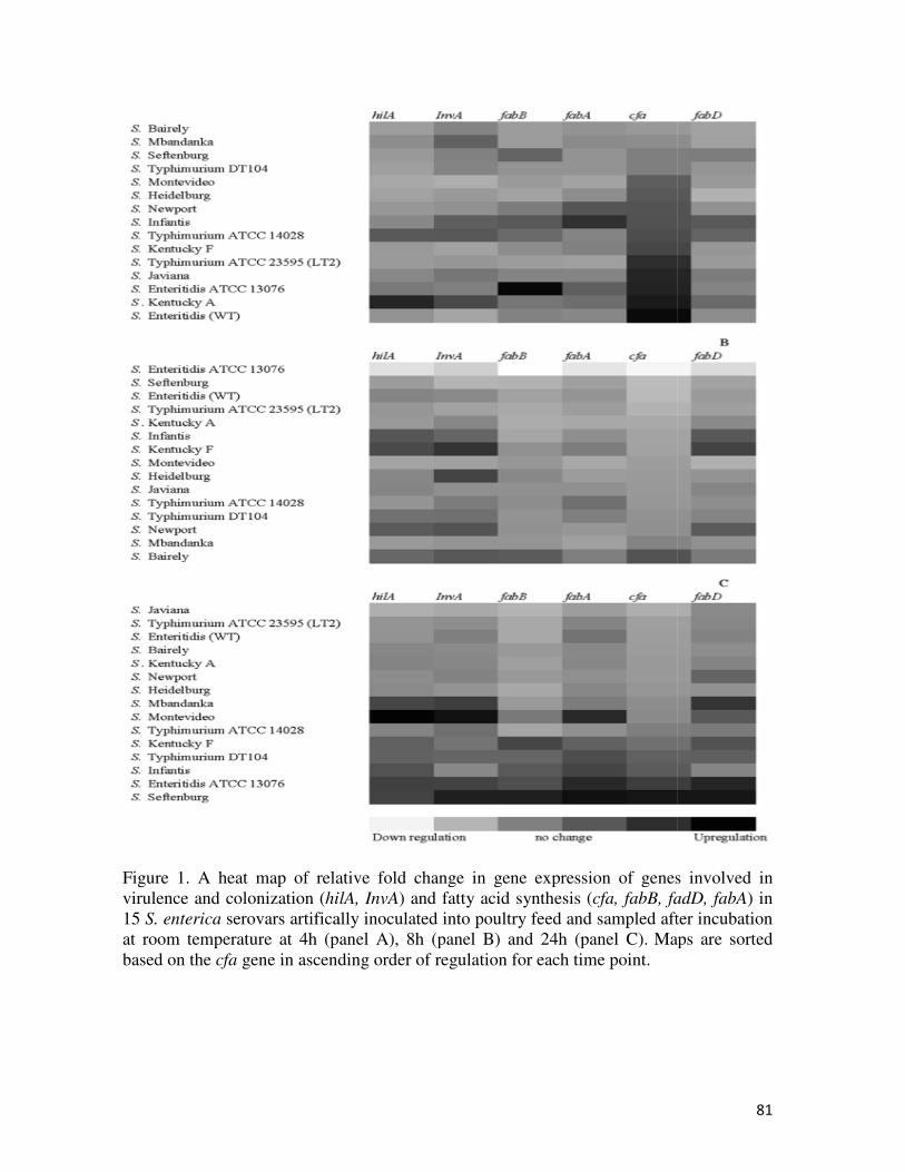
Figure 1. A heat map of relative virulence and colonization (hilA, InvA
15 S. enterica serovars artifically inoculated into poultry feed and sampled after incubation at room temperature at 4h (panel A), 8h (panel B) and 24h (panel C).based on the cfa gene in ascending order of regulation for each time point.
A heat map of relative fold change in gene expression of genes involved in hilA, InvA) and fatty acid synthesis (cfa, fabB, fadD, fabA
serovars artifically inoculated into poultry feed and sampled after incubation e at 4h (panel A), 8h (panel B) and 24h (panel C). Maps are sorted
gene in ascending order of regulation for each time point.
81
fold change in gene expression of genes involved in cfa, fabB, fadD, fabA) in
serovars artifically inoculated into poultry feed and sampled after incubation Maps are sorted

82
Vita
Ana Andino Dubón was born in Tegucigalpa, Honduras. She attended Zamorano
University, where she received her B.S. degree in Agriculture Science and Production while
working under Dr. Gernat. After graduation, she worked at Central Amenrican Poultry Company in
Honduras for five years. In 2009 she received a Master’s degree in Business Management from
UNITEC, Honduras and IEDE Business School, Madrid. In 2011 she joined Dr. Hanning’s
research group to later enroll in the Master’s Program. Ana received a Master’s Degree in Food
Science and Technology from the University of Tennessee in August, 2014. After graduation she
moved to Miami, FL to start a job as Technical Sales Representative for Wincorp International,
Inc.