Molecular mechanical simulations d(CGCGCGC)d(GCGCGCG ... · 5736 Biophysics: Raoand Kollman The...
5
Proc . NatI. Acad. Sci. USA Vol. 84, pp. 5735-5739, August 1987 Biophysics Molecular mechanical simulations on double intercalation of 9-amino acridine into d(CGCGCGC)d(GCGCGCG): Analysis of the physical basis for the neighbor-exclusion principle (molecular dynamics/counterion/conformation/normal mode/entropy) SHASHIDHAR N. RAO AND PETER A. KOLLMAN* Department of Pharmaceutical Chemistry, School of Pharmacy, University of California, San Francisco, CA 94143 Communicated by I. Tinoco, Jr., February 9, 1987 (received for review Dec-ember 22, 1986) ABSTRACT The neighbor-exclusion principle is one of the most general and interesting rules describing intercalative DNA binding by small molecules. It suggests that such binding can only occur at every other base-pair site, reflecting a very large negative cooperativity in the binding process. We have carried out molecular mechanics and molecular dynamics simulations to study intercalation complexes between 9-amino acridine and the base-paired heptanucleotide d(CGCGCGC) d(GCGCGCG), in which the neighbor-exclusion principle was both obeyed and violated. Our studies find no stereochemical preference that favors the neighbor-exclusion-obeying struc- tures over the neighbor-exclusion-violating structures. Alter- native explanations for the existence of the neighbor-exclusion principle are vibrational entropy effects that we calculate to favor the more flexible neighbor-exclusion models over the more rigid neighbor-exclusion-violating models and polyelec- trolyte (counterion release) effects. The neighbor-exclusion principle proposed by Crothers (1) is one of the most general rules for intercalative binding of planar drugs to DNA. According to this principle, every second (next-neighbor) intercalation site along the length of the DNA double helix remains unoccupied. Experimental evidence for this principle has been found in fiber diffraction studies on nucleic acid fibers bound to metallointercalative agents (2, 3), in single-crystal studies (4-8) on complexes between dinucleoside monophosphates complexed with in- tercalating drugs, and solution studies on binding of ethidium ion to oligonucleotides of ribose and deoxyribose sugars (9). Based on earlier crystallographic studies (4-6) that suggested mixed sugar puckering at the intercalation site, a stereochem- ical basis for the neighbor-exclusion principle was proposed. However, this mixed sugar puckering scheme has not been shown to be essential for creating intercalating sites in double-helical DNA through several crystal structure and model building studies (7-11). The first evidence suggesting violation of the neighbor- exclusion model was obtained from viscometric studies (12-14) on interactions between bi- and triderivatives of acridine and DNA, where it was suggested that a single base pair was sandwiched between two acridines. 1H NMR studies on the binding of a series of bis(acridine) compounds to d(AT)5d(AT)5, in which the two acridine rings were linked through linker chains of various lengths, found no evidence of violation of the neighbor-exclusion principle (15, 16). However, it was also noted that these studies have not ruled out violation of the neighbor-exclusion principle under dif- ferent salt conditions and temperatures or a different nucle- otide sequence (15). It has been postulated (17) that the anticooperativity of ethidium binding to DNA could be explained without reference to the neighbor-exclusion rule and is solely due to polyelectrolyte effects. Why do some simple monofunctional intercalators such as 9-amino acridine not intercalate at neighboring sites and violate the neighbor-exclusion principle? In this paper, we present theoretical investigations into the energetic basis for the neighbor-exclusion principle. Crystallographic data on complexes between oligonucleotides containing three or more base pairs and only intercalating drugs daunomycin (18) and triostin (19) are available. This limited data and the lack of sufficient experimental data on violation of neighbor- exclusion models make the theoretical investigations a chal- lenging task. Model-building and energy-minimization stud- ies on deoxytetranucleotides have been used to investigate the possibilities of creating intercalation sites obeying and violating the nearest-neighbor rule, in the framework of helical structures (20). Sawaryn et al. (21) have reported energy calculations on neighbor-exclusion models using an idealized dinucleoside monophosphate system to "create" structures violating this principle, but those calculations are very limited in scope. In the present investigations, we have considered the double intercalation of 9-amino acridine into the base-paired heptadeoxyribonucleotide d(CGCGCGC) d(GCGCGCG) and have model built both neighbor-exclu- sion-obeying and -violating structures. These structures will be called Oby and Vio, respectively. We find, using molec- ular mechanics and molecular dynamics simulations, that it is possible to build energetically reasonable structures with and without the neighbor-exclusion principle being violated by the intercalating molecules. Entropy effects could be a critical factor in the neighbor-exclusion principle. METHODS Intercalation geometries were created using the computer graphics program CHEM (22) on the E and S PS2 at the computer graphics laboratory of the University of California at San Francisco in the following way. The oligonucleotide was "cut" on the two chains at the phosphates in the intercalation site. Then, the two double-stranded segments were separated along the helix axis till the distance between the base pairs on either side of the cut was '-7 A. The two segments were then rotated relative to one another about the helix axis to achieve maximum overlap between these two base pairs. The planar acridine was then intercalated between the two base pairs, and the backbone dihedrals (except the sugar geometry) in intercalation sites were altered to mini- mize the distance between the bonded atoms. These model- built structures were then subjected to energy minimizations using the program AMBER(UCSF) (23-25). Abbreviations: Oby and Vio, structures that obey or violate, respec- tively, the neighbor-exclusion principle. 5735 The publication costs of this article were defrayed in part by page charge payment. This article must therefore be hereby marked 'advertiseiment" in accordance with 18 U.S.C. §1734 solely to indicate this fact.
Transcript of Molecular mechanical simulations d(CGCGCGC)d(GCGCGCG ... · 5736 Biophysics: Raoand Kollman The...
Proc . NatI. Acad. Sci. USAVol. 84, pp. 5735-5739, August 1987Biophysics
Molecular mechanical simulations on double intercalation of9-amino acridine into d(CGCGCGC)d(GCGCGCG): Analysisof the physical basis for the neighbor-exclusion principle
(molecular dynamics/counterion/conformation/normal mode/entropy)
SHASHIDHAR N. RAO AND PETER A. KOLLMAN*Department of Pharmaceutical Chemistry, School of Pharmacy, University of California, San Francisco, CA 94143
Communicated by I. Tinoco, Jr., February 9, 1987 (received for review Dec-ember 22, 1986)
ABSTRACT The neighbor-exclusion principle is one of themost general and interesting rules describing intercalativeDNA binding by small molecules. It suggests that such bindingcan only occur at every other base-pair site, reflecting a verylarge negative cooperativity in the binding process. We havecarried out molecular mechanics and molecular dynamicssimulations to study intercalation complexes between 9-aminoacridine and the base-paired heptanucleotide d(CGCGCGC)d(GCGCGCG), in which the neighbor-exclusion principle wasboth obeyed and violated. Our studies find no stereochemicalpreference that favors the neighbor-exclusion-obeying struc-tures over the neighbor-exclusion-violating structures. Alter-native explanations for the existence of the neighbor-exclusionprinciple are vibrational entropy effects that we calculate tofavor the more flexible neighbor-exclusion models over themore rigid neighbor-exclusion-violating models and polyelec-trolyte (counterion release) effects.
The neighbor-exclusion principle proposed by Crothers (1) isone of the most general rules for intercalative binding ofplanar drugs to DNA. According to this principle, everysecond (next-neighbor) intercalation site along the length ofthe DNA double helix remains unoccupied. Experimentalevidence for this principle has been found in fiber diffractionstudies on nucleic acid fibers bound to metallointercalativeagents (2, 3), in single-crystal studies (4-8) on complexesbetween dinucleoside monophosphates complexed with in-tercalating drugs, and solution studies on binding of ethidiumion to oligonucleotides of ribose and deoxyribose sugars (9).Based on earlier crystallographic studies (4-6) that suggestedmixed sugar puckering at the intercalation site, a stereochem-ical basis for the neighbor-exclusion principle was proposed.However, this mixed sugar puckering scheme has not beenshown to be essential for creating intercalating sites indouble-helical DNA through several crystal structure andmodel building studies (7-11).The first evidence suggesting violation of the neighbor-
exclusion model was obtained from viscometric studies(12-14) on interactions between bi- and triderivatives ofacridine and DNA, where it was suggested that a single basepair was sandwiched between two acridines. 1H NMR studieson the binding of a series of bis(acridine) compounds tod(AT)5d(AT)5, in which the two acridine rings were linkedthrough linker chains of various lengths, found no evidenceof violation of the neighbor-exclusion principle (15, 16).However, it was also noted that these studies have not ruledout violation of the neighbor-exclusion principle under dif-ferent salt conditions and temperatures or a different nucle-otide sequence (15). It has been postulated (17) that theanticooperativity of ethidium binding to DNA could be
explained without reference to the neighbor-exclusion ruleand is solely due to polyelectrolyte effects.Why do some simple monofunctional intercalators such as
9-amino acridine not intercalate at neighboring sites andviolate the neighbor-exclusion principle? In this paper, wepresent theoretical investigations into the energetic basis forthe neighbor-exclusion principle. Crystallographic data oncomplexes between oligonucleotides containing three ormore base pairs and only intercalating drugs daunomycin (18)and triostin (19) are available. This limited data and the lackof sufficient experimental data on violation of neighbor-exclusion models make the theoretical investigations a chal-lenging task. Model-building and energy-minimization stud-ies on deoxytetranucleotides have been used to investigatethe possibilities of creating intercalation sites obeying andviolating the nearest-neighbor rule, in the framework ofhelical structures (20). Sawaryn et al. (21) have reportedenergy calculations on neighbor-exclusion models using anidealized dinucleoside monophosphate system to "create"structures violating this principle, but those calculations arevery limited in scope. In the present investigations, we haveconsidered the double intercalation of 9-amino acridine intothe base-paired heptadeoxyribonucleotide d(CGCGCGC)d(GCGCGCG) and have model built both neighbor-exclu-sion-obeying and -violating structures. These structures willbe called Oby and Vio, respectively. We find, using molec-ular mechanics and molecular dynamics simulations, that it ispossible to build energetically reasonable structures with andwithout the neighbor-exclusion principle being violated bythe intercalating molecules. Entropy effects could be acritical factor in the neighbor-exclusion principle.
METHODSIntercalation geometries were created using the computergraphics program CHEM (22) on the E and S PS2 at thecomputer graphics laboratory of the University of Californiaat San Francisco in the following way. The oligonucleotidewas "cut" on the two chains at the phosphates in theintercalation site. Then, the two double-stranded segmentswere separated along the helix axis till the distance betweenthe base pairs on either side of the cut was '-7 A. The twosegments were then rotated relative to one another about thehelix axis to achieve maximum overlap between these twobase pairs. The planar acridine was then intercalated betweenthe two base pairs, and the backbone dihedrals (except thesugar geometry) in intercalation sites were altered to mini-mize the distance between the bonded atoms. These model-built structures were then subjected to energy minimizationsusing the program AMBER(UCSF) (23-25).
Abbreviations: Oby and Vio, structures that obey or violate, respec-tively, the neighbor-exclusion principle.
5735
The publication costs of this article were defrayed in part by page chargepayment. This article must therefore be hereby marked 'advertiseiment"in accordance with 18 U.S.C. §1734 solely to indicate this fact.

5736 Biophysics: Rao and Kollman
The molecular mechanical energies were evaluated byusing equation 1 in ref. 23 with the force field parameterspresented by Weiner et al. (26), and the structures wereenergy refined until an rms gradient of 0.1 kcal/mol.A (1 cal= 4.184 J) was achieved. In all the calculations a distance-dependent dielectric constant was used (26). The charges on9-amino-acridine (Fig. 1) were determined using the quantumchemically derived electrostatic potentials (27) with a STO-3G basis set and can be obtained from the authors. Theequilibrium values for C-C, N-C, and N-H bond lengthsin 9-amino acridine were taken as 1.378, 1.40, and 1.01 A,respectively, with the corresponding force constants being480, 460, and 434 kcal/mol, respectively. All the bond angleshad equilibrium values of 1200 with a force constant of 80kcal/mol. The dihedral force constants were chosen to be thesame for analogous dihedral angles in proteins and nucleicacids (26).The energy-minimized structures have been labeled as
Obyl and Viol, corresponding to the two classes of model-built complexes mentioned above. Constrained energy min-imizations were also carried out (as detailed in a later section)in which a few heptamer backbone torsions were constrainedto take up specific values, using a dihedral force constant of100 kcal/mol. The structures thus obtained were furtherrefined, after removing the constraints, to compare theirrelative stabilities to the structures obtained after energyminimizations with no constraints. In addition, we have alsoenergy minimized the heptanucleotide in the B form withoutthe intercalators.
In addition to molecular mechanical simulations, molecu-lar dynamical simulations were also carried out on theenergy-refined structures Oby and Vio, with a view toexplore the conformational space around the minima. Thiswas done using the molecular dynamics module in theprogram AMBER(UCSF) on the VAX11/780 with an FPS-264 attachment (25). These simulations were carried out at300 K for a 50-ps time period in "hydrated" sodium coun-terions (28) that were placed along the bisector of the pendantoxygens in the phosphate groups at distance of 5 A.
Initially, unconstrained dynamics were carried out inwhich the entire system, consisting of the heptamer, the twoacridines, and counterions, was allowed to move. This led tostructures in which the acridines were not fully intercalatedbetween the base pairs but were hydrogen bonded to phos-phates and bases in the vicinity of intercalation site throughthe N1(-H1O and 9-amino groups. To avoid such largedistortions of the structures, which are due to our primitivetreatment of electrostatic effects, we constrained the centralbase pairs (four in Vio and five in Oby) in and around theintercalation sites to their coordinates obtained after energyrefinement of initial model-built structures (Obyl and Viol).The molecular dynamics simulations were then carried outwith freedom of movement in cartesian space being allowedto the rest of the system, namely, the end base pairs, botholigonucleotide backbones, the acridines, and the counteri-ons. Structures obtained at the end of every 5 ps in such asimulation were then subjected to energy minimization in thepresence of counterions until the above stated gradient
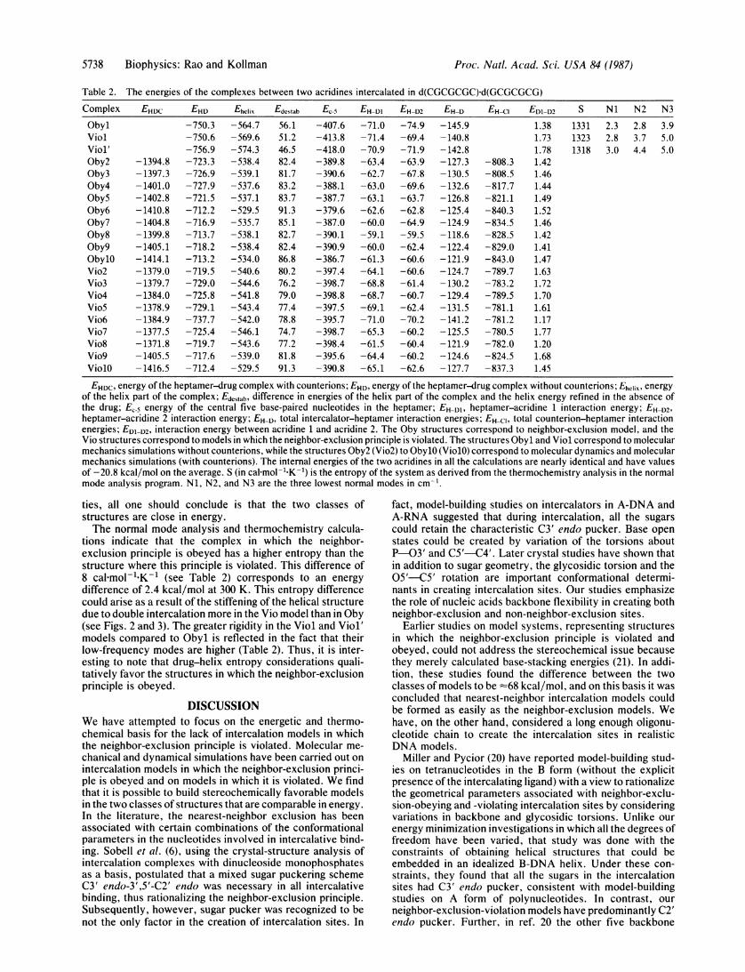
HN9A HN9B
N9
FIG. 1. Schematic rep-8 9
1 1 resentation of 9-amino acri-C ~~~~~~~~C2dine (in the united atom rep-
resentation) protonated at
C6- -C3 N10 used in the double-in-
N 6/ Ptercalation complexes with
IN C4 d(CGCGCGC)*d(GCGCG-H1O CG).
criterion was achieved. All of these structures are numberedas, for example, Oby2 and Vio2.We have also carried out normal mode analysis and
thermochemistry calculations on the energy-minimizedstructures obtained in the molecular-mechanics simulationsand have computed the entropy of the two structures usingnormal mode analysis (25). The energies of the molecularmechanically simulated structures were further refined tovery low gradients (10-5 kcal/mol'A), and the resultantstructures were entered into the normal mode program todetermine the normal modes and thermochemical parame-ters.
In the two structures that were model built and energyrefined, the starting structure of the oligonucleotide wasB-DNA (29). The nucleotides in the DNA fragment arereferred to by the serial number of the base and the base namewith the numbering being continuous into the second strand.Thus, Cyt-3 and Gua-12 are paired in the Watson-Crickconfiguration. In Vio, one of the acridines was intercalatedbetween the base pairs Cyt-3-Gua-12 and Gua-4 Cyt-11, whilethe other acridine was intercalated between the base pairsCyt-5 Gua-10 and Gua-4 Cyt-11. In Oby, the first acridine wasintercalated between Gua-2-Cyt-13 and Cyt-3Gua-12 basepairs, while the second one was intercalated between Cyt-5'Gua-10 and Gua-4-Cyt-11. The two acridines will be re-ferred to as acridine 1 and acridine 2, where the former liesat the 5' side of strand 1 (containing Cyt-1) in either structure.
RESULTSConformations of the Heptanucleotides in Oby and Vio. As
expected, the alterations in the conformations of the oligo-nucleotides in the two double-intercalation complexes fromvalues typical of B-DNA are predominantly confined to theregions of intercalation. The conformations of the oligonu-cleotide backbone and bases in the regions of intercalation(covering Cyt-3, Gua-4, Cyt-5, and Gua-6 and their comple-mentary base-paired nucleotides in Obyl and Cyt-3, Gua-4,and Cyt-5 and their complementary base-paired nucleotidesin Viol) are listed in Table 1. Also listed in this table are thebackbone and glycosidic conformations in the crystal struc-tures of 9-amino acridine complexed with 5-iodo-CpG (31)and triostin-d(CGTACG) (19). The conformation that differsmost significantly between the crystals and theoretical mod-els is the sugar geometry at the 5' end of the intercalation site.
Table 1. Conformational parameters
9AA-Obyl Viol CpG Trio
Torsion Si S2 S1 S2 Si S2 11 12
((5') 160 124 147 164 148 158 144 158 72 85 138 88a 180 182 186 179 180 190 186 191 236 216 177 197Is 275 264 262 224 311 270 185 274 311 296 180 307y 286 288 278 273 187 177 188 170 280 300 329 2068 173 175 178 166 190 173 196 171 220 229 159 143E 58 61 58 55 182 180 176 179 67 38 6 55((3') 132 147 150 88 158 147 158 143 110 130 133 84X(S') 75 61 68 107 67 68 87 72 17 38 100 94X(3') 72, 63 76 32 68 70 72 66 119 109 113 82
Conformational parameters (in degrees) in the intercalation sites ofObyl, Viol, and crystal structures of intercalation complexes 5-iodo-CpG-9-amino acridine (9AA-CpG) (29) and triostin-d(CGT-ACG) (Trio) (18). Si and S2 refer to the two strands of theoligonucleotide duplex, while 11 and 12 refer to the two intercalationsites in Trio (only the backbone and glycosidic conformations of oneof the two strands were reported in ref. 18). The nomenclature for therepresentation of torsion angles in the backbone of nucleic acids isthat adopted by Seeman et al. (30).
Proc. Natl. Acad. Sci. USA 84 (1987)
Proc. Natl. Acad. Sci. USA 84 (1987) 5737
In the latter, they are predominantly in the C2' endo region,while in the crystals, they are predominantly C3' endo.Only two kinds of conformations not usually seen in the
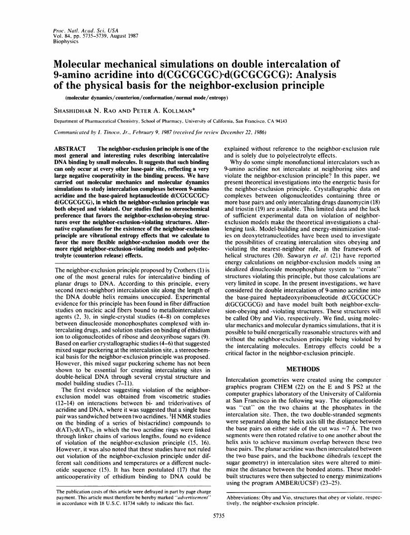
crystal structures either of the complexes between simpleintercalators and deoxyoligonucleotides or of deoxyoligonu-cleotides alone have been observed in the energy-refinedmodels obeying and violating the neighbor-exclusion princi-ple. They are the (trans,trans) phosphodiester conformations(/,y) as at the 3' end of Cyt-3 in Viol and Viol' and a gauche'conformation about C5'-05' (8) as at the 5' end of Cyt-3 inOby complexes. The unusual phosphodiester conformationleads to a slightly more stretched helix in the case of modelsviolating the neighbor-exclusion principle. Though this con-formation has never been observed in DNA structures, it isnot uncommon in the structures of tRNA (32). The unusualconformation about C5'-05' has been shown to be ofreasonable energy in conformational energy calculations ondinucleoside monophosphates (33). Figs. 2 and 3 show stereoviews of the energy-refined complexes Obyl and Viol.The conformational properties of Oby and Vio structures
are reflected in the overall helical properties of the heptamerduplex in them. For example, the helix in Viol is longer thanin Obyl by 1l.5 A, due to the stretched (trans,trans)phosphodiester conformations in Viol. The twist and tilts ofmost of the base pairs in these two structures are similar tothose in the B-DNA (Figs. 2 and 3). The only exception is inViol, where the twist of the base pairs involving Gua-4 is lessby 100 than in the energy-minimized heptamer alone. Theoverall helix is kinked slightly more in Viol (by -7°) than inObyl. In Obyl, the helix is hardly unwound at the firstintercalation site involving Cyt-3 and Gua-4, whereas theextent of unwinding is =20° at the other site. In Viol,however, the unwinding is smaller (413°) at the first inter-calation site. The overall unwinding of the helixes in Obyland Viol are, respectively, 20° and 10°. These values lie in therange (-3° to 260) obtained from x-ray crystal studies oncomplexes between dinucleoside monophosphates and inter-calators (see chapter 16 in ref. 34). However, the totalunwinding angle for Obyl is higher than that in daunomycin-d(CGTACG) (160) (18) and circular DNA (11°) (35) but islower than in the triostin-d(CGTACG) (19) complex (27°).These differences could be due to several structural factorsinduced by the intercalation of the more complex drugs in theexperimental studies such as Hoogsteen base-pair formation(19).
Energetics. Table 2 lists the molecular mechanical energiesof the Vio and Oby structures. The energy components inthese two classes of structures obtained by molecular me-chanical simulations alone are Obyl, Viol, and Viol'. The
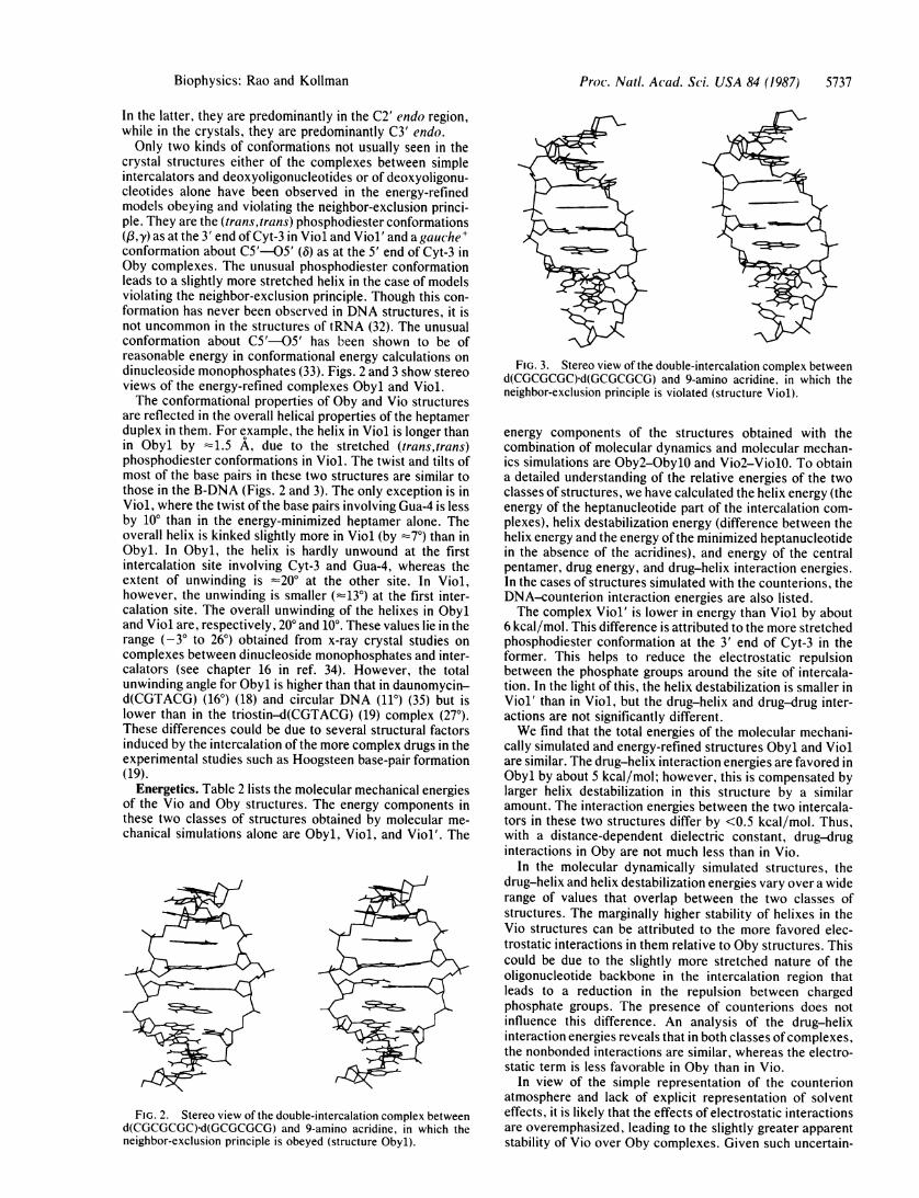
FIG. 2. Stereo view of the double-intercalation complex betweend(CGCGCGC)-d(GCGCGCG) and 9-amino acridine, in which theneighbor-exclusion principle is obeyed (structure Obyl).
FIG. 3. Stereo view of the double-intercalation complex betweend(CGCGCGC)-d(GCGCGCG) and 9-amino acridine, in which theneighbor-exclusion principle is violated (structure Viol).
energy components of the structures obtained with thecombination of molecular dynamics and molecular mechan-ics simulations are Oby2-ObylO and Vio2-ViolO. To obtaina detailed understanding of the relative energies of the twoclasses of structures, we have calculated the helix energy (theenergy of the heptanucleotide part of the intercalation com-plexes), helix destabilization energy (difference between thehelix energy and the energy of the minimized heptanucleotidein the absence of the acridines), and energy of the centralpentamer, drug energy, and drug-helix interaction energies.In the cases of structures simulated with the counterions, theDNA-counterion interaction energies are also listed.The complex Viol' is lower in energy than Viol by about
6 kcal/mol. This difference is attributed to the more stretchedphosphodiester conformation at the 3' end of Cyt-3 in theformer. This helps to reduce the electrostatic repulsionbetween the phosphate groups around the site of intercala-tion. In the light of this, the helix destabilization is smaller inViol' than in Viol, but the drug-helix and drug-drug inter-actions are not significantly different.We find that the total energies of the molecular mechani-
cally simulated and energy-refined structures Obyl and Violare similar. The drug-helix interaction energies are favored inObyl by about 5 kcal/mol; however, this is compensated bylarger helix destabilization in this structure by a similaramount. The interaction energies between the two intercala-tors in these two structures differ by <0.5 kcal/mol. Thus,with a distance-dependent dielectric constant, drug-druginteractions in Oby are not much less than in Vio.
In the molecular dynamically simulated structures, thedrug-helix and helix destabilization energies vary over a widerange of values that overlap between the two classes ofstructures. The marginally higher stability of helixes in theVio structures can be attributed to the more favored elec-trostatic interactions in them relative to Oby structures. Thiscould be due to the slightly more stretched nature of theoligonucleotide backbone in the intercalation region thatleads to a reduction in the repulsion between chargedphosphate groups. The presence of counterions does notinfluence this difference. An analysis of the drug-helixinteraction energies reveals that in both classes ofcomplexes,the nonbonded interactions are similar, whereas the electro-static term is less favorable in Oby than in Vio.
In view of the simple representation of the counterionatmosphere and lack of explicit representation of solventeffects, it is likely that the effects of electrostatic interactionsare overemphasized, leading to the slightly greater apparentstability of Vio over Oby complexes. Given such uncertain-
Biophysics: Rao and Kollman
5738 Biophysics: Rao and Kollman
Table 2. The energies of the complexes between two acridines intercalated in d(CGCGCGC)-d(GCGCGCG)
EHCI EDI-D21.381.731.78
-808.3 1.42-808.5 1.46-817.7 1.44-821.1 1.49-840.3 1.52-834.5 1.46-828.5 1.42-829.0 1.41-843.0 1.47-789.7 1.63-783.2 1.72-789.5 1.70-781.1 1.61-781.2 1.17-780.5 1.77-782.0 1.20-824.5 1.68-837.3 1.45
S
133113231318
Ni N2 N3
2.3 2.8 3.92.8 3.7 5.03.0 4.4 5.0
EHDC, energy of the heptamer-drug complex with counterions; EHD, energy of the heptamer-drug complex without counterions; EhcliX, energyof the helix part of the complex; Edcstab, difference in energies of the helix part of the complex and the helix energy refined in the absence ofthe drug; EC-5 energy of the central five base-paired nucleotides in the heptamer; EH-D1, heptamer-acridine 1 interaction energy; EH-D2,heptamer-acridine 2 interaction energy; EH-D, total intercalator-heptamer interaction energies; EH-C, total counterion-heptamer interactionenergies; EDI-D2, interaction energy between acridine 1 and acridine 2. The Oby structures correspond to neighbor-exclusion model, and theVio structures correspond to models in which the neighbor-exclusion principle is violated. The structures Obyl and Viol correspond to molecularmechanics simulations without counterions, while the structures Oby2 (Vio2) to ObylO (ViolO) correspond to molecular dynamics and molecularmechanics simulations (with counterions). The internal energies of the two acridines in all the calculations are nearly identical and have valuesof -20.8 kcal/mol on the average. S (in cal mol-'"K-1) is the entropy of the system as derived from the thermochemistry analysis in the normalmode analysis program. Ni, N2, and N3 are the three lowest normal modes in cm-'.
ties, all one should conclude is that the two classes ofstructures are close in energy.The normal mode analysis and thermochemistry calcula-
tions indicate that the complex in which the neighbor-exclusion principle is obeyed has a higher entropy than thestructure where this principle is violated. This difference of8 calmol-"K-' (see Table 2) corresponds to an energydifference of 2.4 kcal/mol at 300 K. This entropy differencecould arise as a result of the stiffening of the helical structuredue to double intercalation more in the Vio model than in Oby(see Figs. 2 and 3). The greater rigidity in the Viol and Viol'models compared to Obyl is reflected in the fact that theirlow-frequency modes are higher (Table 2). Thus, it is inter-esting to note that drug-helix entropy considerations quali-tatively favor the structures in which the neighbor-exclusionprinciple is obeyed.
DISCUSSIONWe have attempted to focus on the energetic and thermo-chemical basis for the lack of intercalation models in whichthe neighbor-exclusion principle is violated. Molecular me-
chanical and dynamical simulations have been carried out onintercalation models in which the neighbor-exclusion princi-ple is obeyed and on models in which it is violated. We findthat it is possible to build stereochemically favorable modelsin the two classes of structures that are comparable in energy.In the literature, the nearest-neighbor exclusion has beenassociated with certain combinations of the conformationalparameters in the nucleotides involved in intercalative bind-ing. Sobell et al. (6), using the crystal-structure analysis ofintercalation complexes with dinucleoside monophosphatesas a basis, postulated that a mixed sugar puckering schemeC3' endo-3',5'-C2' endo was necessary in all intercalativebinding, thus rationalizing the neighbor-exclusion principle.Subsequently, however, sugar pucker was recognized to benot the only factor in the creation of intercalation sites. In
fact, model-building studies on intercalators in A-DNA andA-RNA suggested that during intercalation, all the sugarscould retain the characteristic C3' endo pucker. Base openstates could be created by variation of the torsions aboutP-03' and C5'-C4'. Later crystal studies have shown thatin addition to sugar geometry, the glycosidic torsion and the05'-C5' rotation are important conformational determi-nants in creating intercalation sites. Our studies emphasizethe role of nucleic acids backbone flexibility in creating bothneighbor-exclusion and non-neighbor-exclusion sites.
Earlier studies on model systems, representing structuresin which the neighbor-exclusion principle is violated andobeyed, could not address the stereochemical issue becausethey merely calculated base-stacking energies (21). In addi-tion, these studies found the difference between the twoclasses of models to be ='7'68 kcal/mol, and on this basis it wasconcluded that nearest-neighbor intercalation models couldbe formed as easily as the neighbor-exclusion models. Wehave, on the other hand, considered a long enough oligonu-cleotide chain to create the intercalation sites in realisticDNA models.
Miller and Pycior (20) have reported model-building stud-ies on tetranucleotides in the B form (without the explicitpresence of the intercalating ligand) with a view to rationalizethe geometrical parameters associated with neighbor-exclu-sion-obeying and -violating intercalation sites by consideringvariations in backbone and glycosidic torsions. Unlike ourenergy minimization investigations in which all the degrees offreedom have been varied, that study was done with theconstraints of obtaining helical structures that could beembedded in an idealized B-DNA helix. Under these con-straints, they found that all the sugars in the intercalationsites had C3' endo pucker, consistent with model-buildingstudies on A form of polynucleotides. In contrast, ourneighbor-exclusion-violation models have predominantly C2'endo pucker. Further, in ref. 20 the other five backbone
Complex
ObylViolViol'Oby2Oby3Oby4ObySOby6Oby7Oby8Oby9ObylOVio2Vio3Vio4Vio5Vio6Vio7Vio8Vio9ViolO
EHDC
-1394.8-1397.3-1401.0-1402.8-1410.8-1404.8-1399.8-1405.1-1414.1-1379.0-1379.7-1384.0-1378.9-1384.9-1377.5-1371.8-1405.5-1416.5
EHD-750.3-750.6-756.9-723.3-726.9-727.9-721.5-712.2-716.9-713.7-718.2-713.2-719.5-729.0-725.8-729.1-737.7-725.4-719.7-717.6-712.4
EhClix
-564.7-569.6-574.3-538.4-539.1-537.6-537.1-529.5-535.7-538.1-538.4-534.0-540.6-544.6-541.8-543.4-542.0-546.1-543.6-539.0-529.5
Edcstab56.151.246.582.481.783.283.791.385.182.782.486.880.276.279.077.478.874.777.281.891.3
-407.6-413.8-418.0-389.8-390.6-388.1-387.7-379.6-387.0-390.1-390.9-386.7-397.4-398.7-398.8-397.5-395.7-398.7-398.4-395.6-390.8
EH-DI-71.0-71.4-70.9-63.4-62.7-63.0-63.1-62.6-60.0-59.1-60.0-61.3-64.1-68.8-68.7-69.1-71.0-65.3-61.5-64.4-65.1
EH-D2-74.9-69.4-71.9-63.9-67.8-69.6-63.7-62.8-64.9-59.5-62.4-60.6-60.6-61.4-60.7-62.4-70.2-60.2-60.4-60.2-62.6
EH-D-145.9-140.8-142.8-127.3-130.5-132.6-126.8-125.4-124.9-118.6-122.4-121.9-124.7-130.2-129.4-131.5-141.2-125.5-121.9-124.6-127.7
Proc. Natl. Acad. Sci. USA 84 (1987)

Proc. Natl. Acad. Sci. USA 84 (1987) 5739
angles in a mononucleotide were either trans or intermediatebetween trans and gauche-, and the glycosidic torsions werehigher by 200 to 300 than in our models. Based on theconformational features they suggested (20) a low possibilityof occurrence of neighbor-exclusion-violating models inshort oligonucleotide chains but did not exclude the possi-bility in a polymer.
Polyelectrolyte effects have been shown to be important inthe anticooperativity effects in the cases of ethidium andactinomycin binding to DNA (17). Upon the inclusion of sucheffects, it was shown unnecessary to invoke multiple-siteexclusion to explain the binding. Thus, counterion effectscould be important in competition between the two classes ofmodels under study. The entropic and enthalpic contribu-tions obtainable from the release of counterions into thesolution, as a result of intercalation of positively chargedacridines, could favor the neighbor-exclusion model. In theVio model, the counterion release would be less because thepresence of another intercalator at -7 A has already "re-leased" some of the cations in the vicinity. In the moleculardynamically simulated models, we do see a more favoredinteraction-energy profile between the heptamer and coun-terions in Oby than in Vio models. To further assess thecounterion effects in complexes involving charged intercala-tors, similar studies with neutral species such as actinomycinD are in order. In view of the charged nature of our systemand the simple treatment of electrostatic effects in our forcefield, it is clear that electrostatic interactions have not beenquantitatively simulated here. Unfortunately, "simple" neu-tral intercalators do not interact very well with DNA.
CONCLUSIONSIn summary, we could consider four contributions to therelative free energies of neighbor-exclusion-violating andneighbor-exclusion-obeying double-intercalation complexesbetween DNA and simple intercalators like acridine. The fourare stereochemical energies, vibrational entropy, counterionrelease, and specific solvent-solute interactions. Our resultssuggest that stereochemical energies cannot be used to ruleout the neighbor-exclusion-violating structures. The twoclasses of models are energetically similar within the limits ofaccuracy in our force field and simple model. On the otherhand, our results suggest that vibrational entropy consider-ations could play an important role in favoring the moreflexible neighbor-exclusion structure over the more rigidneighbor-exclusion-violating structure. Even though the en-ergy difference associated with this entropy difference issmaller than the internal energy difference (Table 2), it isexpected to be far less sensitive to an accurate representationof solvent environment.One of the referees has pointed out that the effects that
prevent nearest-neighbor intercalation could come into playonly when the acridines intercalate into long stretches ofnucleic acids. However, it may be noted that spectroscopicstudies (15, 16) that find no evidence for the violation of theneighbor-exclusion principle involve the intercalation of twoacridines connected at N9 positions through a series of linkerchains into a pentanucleotide double helix. Thus, it is clearthat the neighbor-exclusion rule holds for smaller DNAfragments. Therefore, it is likely that our model of a hepta-nucleotide should be adequate to understand the physicalbasis of the neighbor-exclusion principle.
Counterion release could be an important factor in desta-bilizing the latter class of structures. In the framework of theforce field employed, we cannot explicitly consider suchcounterion release effects; however, insight into the signifi-cance of this effect could be obtained by looking at ionic-strength-dependent effects or intercalation by neutral analogs
of the simple intercalators. Of course, there has been noexplicit inclusion of solvent interactions in these simulations.Thus, we cannot rule out that solute-solvent interactions ordifferences in solvent-solvent interactions could influencethe relative energies of neighbor-exclusion-violating and-obeying structures. More extensive simulations includingboth counterions and solvent will be required to assess thepossible role of counterions and solvent in providing a moreprecise energetic basis for the neighbor-exclusion rule.We thank Dr. U. Chandra Singh for his help with the normal mode
analysis program in AMBER(UCSF). We gratefully acknowledge thesupport from Grant CA-25644 from the National Cancer Institute inthis research. The use of the facilities of the University of California,San Francisco, Computer Graphics Laboratory (R. Langridge,director, and T. Ferrin, facility manager), supported by GrantRR-1081 from the National Institutes of Health, is also gratefullyacknowledged. The purchase of the FPS-264 array processor wasmade possible through Grants RR-02441 from the National Institutesof Health and DMB-84-13762 from the National Science Foundation,and their support for this is much appreciated.
1. Crothers, D. M. (1968) Biopolymers 6, 575-584.2. Bond, P. J., Langridge, R., Jennette, K. W. & Lippard, S. J. (1975)
Proc. Nail. Acad. Sci. USA 72, 4825-4829.3. Arnott, S., Bond, P. J. & Chandrasekaran, R. (1980) Nature (London)
287, 561-563.4. Tsai, C., Jain, S. C. & Sobell, H. M. (1977) J. Mol. Biol. 114, 301-315.5. Jain, S. C., Tsai, C. & Sobell, H. M. (1977) J. Mol. Biol. 114, 317-331.6. Sobell, H. M., Tsai, C., Jain, S. C. & Gilbert, S. G. (1977) J. Mol. Biol.
114, 333-365.7. Neidle, S., Achari, A., Taylor, G. L., Berman, H. M., Carrell, H. L.,
Glukser, J. P. & Stallings, W. C. (1977) Nature (London) 269, 304-307.8. Berman, H. M., Stallings, W. C., Carrell, H. L., Glukser, J. P., Neidle,
S., Taylor, G. L. & Achari, A. (1979) Biopolvmers 18, 2405-2429.9. Nelson, J. W. & Tinoco, I., Jr. (1984) Biopolymers 23, 213-233.
10. Alden, C. J. & Arnott, S. (1975) Nucleic Acids Res. 2, 1701-1717.11. Alden, C. J. & Arnott, S. (1977) Nucleic Acids Res. 4, 3855-3861.12. Wakelin, L. P. G., Romanos, M., Chen, T. K., Glaubiger, D., Canel-
lakis, E. S. & Waring, M. J. (1978) Biochemistry 17, 5057-5063.13. Atwell, G. J., Leupin, W., Twigden, S. J. & Denny, W. A. (1983) J. Am.
Chem. Soc. 105, 2913-2914.14. Denny, W. A., Atwell, G. J., Baguley, B. C. & Wakelin, L. P. G. (1985)
J. Med. Chem. 28, 1568-1574.15. Assa-Munt, U., Denny, W. A., Leupin, W. & Kearns, D. R. (1985)
Biochemistry 24, 1441-1449.16. Assa-Munt, U., Leupin, W., Denny, W. A. & Kearns, D. R. (1985)
Biochemistry 24, 1449-1460.17. Friedman, R. A. G. & Manning, G. S. (1984) Biopolymers 23, 2671-2714.18. Quigley, G. J., Wang, A. H.-J., Ughetto, G., van der Marel, G., van Boom,
J. H. & Rich, A. (1980) Proc. Nati. Acad. Sci. USA 77, 7204-7208.19. Wang, A. H.-J., Ughetto, G., Quigley, G. J., Hakoshima, T., van der
Marel, G. A., van Boom, J. H. & Rich, A. (1984) Science 225, 1115-1121.20. Miller, K. J. & Pycior, J. F. (1979) Biopolymers 18, 2683-2719.21. Sawaryn, A., Leps, B. & Bradaczek, H. (1983) J. Comp. Chem. 4,
333-336.22. Dearing, A. (1981) CHEM, A Molecular Display Program (Univ. of
California, San Francisco).23. Weiner, P. K. & Kollman, P. A. (1981) J. Comp. Chem. 2, 287-310.24. Weiner, P. K., Singh, U. C., Kollman, P. A., Caldwell, J. W. & Case,
D. (1985) AMBER, A Molecular Mechanics and Dynamics Program(Univ. of California, San Francisco), Version 2.0.
25. Singh, U. C., Weiner, P. K., Caldwell, J. W. & Kollman, P. A. (1986)AMBER, A Molecular Mechanics and Dynamics Program (Univ. ofCalifornia, San Francisco), Version 3.0 (developed for FPS).
26. Weiner, S. J., Kollman, P. A., Case, D., Singh, U. C., Ghio, C.,Alagona, J., Profeta, S., Jr., & Weiner, P. K. (1984) J. Am. Chem. Soc.106, 765-785.
27. Singh, U. C. & Kollman, P. A. (1984) J. Comp. Chem. 5, 129-144.28. Singh, U. C., Weiner, S. J. & Kollman, P. A. (1985) Proc. Nati. Acad.
Sci. USA 82, 755-759.29. Arnott, S., Campbell-Smith, P. & Chandrasekaran, R. (1976) in CRC
Handbook of Biochemistry, ed. Fasman, G. D. (CRC, Cleveland, OH),Vol. 2, pp. 411-422.
30. Seeman, N. C., Rosenberg, J. M., Suddath, F. L., Kim, J. J. P. & Rich,A. (1976) J. Mol. Biol. 104, 109-144.
31. Sakore, T. D., Reddy, B. S. & Sobell, H. M. (1979) J. Mol. Biol. 135,763-785.
32. Kim, S. H. (1981) in Topics in Nuicleic Acid Struc-ture, ed. Neidle, S.(Wiley, New York), pp. 83-112.
33. Yathindra, N. & Sundaralingam (1975) Biopolymers 14, 2387-2404.34. Saenger, W. (1984) Principles of Nucleic Acid Structure (Springer, New
York), pp. 51-101.35. Wang, J. C. (1974) J. Mol. Biol. 89, 783-801.
Biophysics: Rao and Kollman