
Molecular Machinery
11
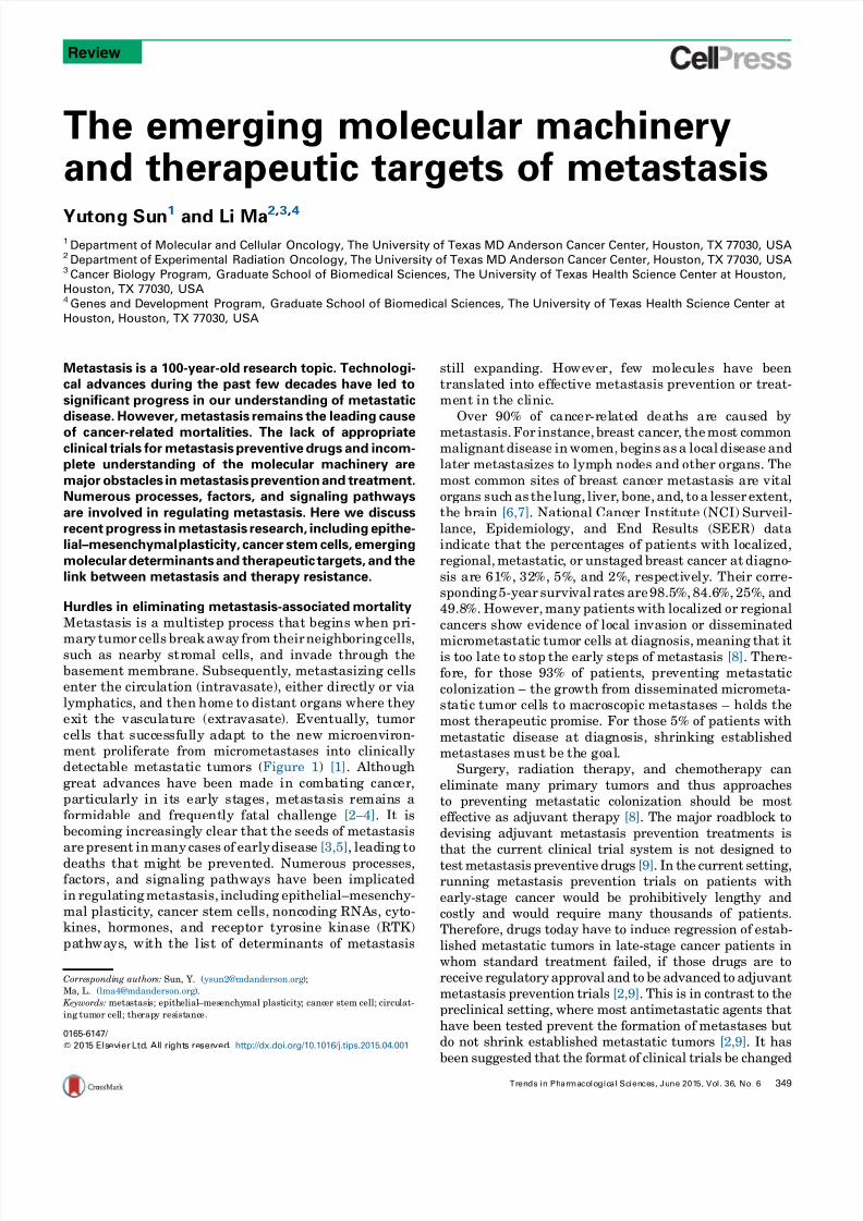
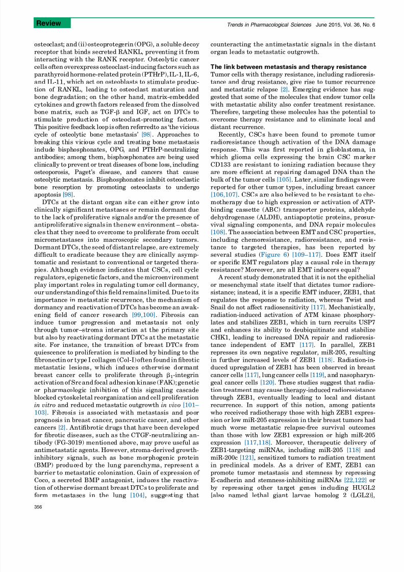
The emergingmolecularmachinery andtherapeutictargetsof metastasis Yutong Sun 1 and Li Ma 2, 3, 4 1 Department of MolecularandCellular Oncology, TheUniversityof TexasMDAndersonCancerCenter,Houston,TX77030, USA 2 Department of Experimental Radiation Oncology, TheUniversityof TexasMDAndersonCancerCenter,Houston, TX77030, USA 3 CancerBiologyProgram, GraduateSchoolof Biomedical Sciences, TheUniversityof TexasHealthScienceCenteratHouston, Houston, TX77030, USA 4 GenesandDevelopment Program, GraduateSchoolof Biomedical Sciences, TheUniversityof TexasHealthScienceCenter at Houston, Houston,TX77030, USA Metastasis isa100-year-oldresearchtopic. Technologi- caladvancesduringthepastfewdecadeshaveledto significant progressinourunderstanding ofmetastatic disease. However, metastasis remainstheleadingcause of can cer -re lat ed mortalities. Thelackofappropriate clinical trialsformetastasis preventivedrugsandincom- pleteunderstanding ofthemolecular machineryare majorobstacles inmetastasis preventionandtreatment. Numerous processes, factors,andsignalingpathways areinvolvedinregulatingmetastasis. Herewediscuss recentprogress inmetastasis research, includingepithe- lial–mesenchymal plasticity, cancerstemcells,emerging molecular determinants andtherapeutic targets, andthe linkbetweenmetastasis andtherapyresistance. Hurdlesineliminatingmetastasis-associated mortality Met astasis isamultis tep processthat be gins when pri- mar y tumor cel lsbreak away fromtheir neighboring cells , suchas nearby st romal cells, andinvadethro ugh the basement membr ane.Subseq uently, metastasiz ing cells enterthe cir cul ation(intravasate),eitherdir ectly or via lymphatics,andthen home to distant organswhere they exit the vascul at ure(extravasate). Eventua lly,tumor ce ll s that succ essf ul ly adapttothe new mi croenviron- mentproliferatefrommicrometastasesintoclinically detectable metastatic tumors(Figure 1) [1]. Altho ugh greatadvanceshavebeenmadein combating canc er, particularly inits ea rl y s tages, metastasis remainsa formidableandfrequently fatalchallenge [2–4].Itis becoming incre asingl y clearthat the seedsof metastasis are prese ntin many casesof early diseas e[3,5], leadingto deathsthat mi ghtbe prevent ed.Numerousprocesses, factors, andsignal ing pathwayshavebeenimplicated in regulating metast asis, including epithelial–mesenchy- mal plast icity,cancerstemcel ls, noncodi ng RNAs, cyt o- kines,hormones,and rece ptortyrosine kinase(RTK) pa thways , wi th the li st of determinantsof metastasis stillexpanding.Ho we ve r, f ew molec ul eshave been transl ated into eff ectivemetas tasis preventionortreat- ment in the cl inic. Over90% of cance r- r el ate d dea th s a re caused by metastasis. For insta nce, breastcancer , the mostcommon malignant diseasein women, begi ns as aloc al dis ease and later meta stasizestolymph nodesand other organs.The mostcommonsitesof breastcanc er metastasisare vi tal organssuch asthe lung,li ver ,bon e,and, toalesserextent, the br ai n [6,7].Nati onal Canc erInsti tute (NCI) Surveil- lance, Epidemiology, and End Results (SEER) data indi cate that the percentages of patients wi th localized, regional, metastatic,or unst agedbreastcancerat diagno - sisa re 6 1% , 32 %,5%, and 2%, re specti vely. The ir cor re- sponding 5-yearsurvi val ratesare98.5%,84.6%, 25%, and 49.8 %. Howe ver, many patientswithlocalized or regi onal cancers sho wevidence of local invasi onordisseminated micr ometa stati c tumo r cell s atdiagnosis, meaning thatit istoolate tostop the earl y stepsof metastasis [8]. Ther e- for e, for those93% of patients , pre venting met ast atic colonization –the gro wthfromdisseminated micrometa- st at ic tumo r ce lls tomacroscopic met astases –holds the mosttherapeutic promise. Forthose5% of patientswith metastatic diseaseat diagnosi s, shrinking establis hed metastasesmust be the goal. Surgery,radiationtherapy,andchemotherapy can eliminatemany primary tumorsandthusapproaches topreventing metastaticcolonizationshouldbemost effectiveasadjuvanttherapy [8].Themajorroadblock to devising adjuvantmetastasispreventiontreatmentsis thatthecurrentclinicaltrialsystemisnotdesignedto testmetastasispreventivedrugs[9].Inthecurrentsetting, running metastasispreventiontrialsonpatientswith early-stagecancerwouldbeprohibitively lengthy and costly andwouldrequiremany thousandsof patients. Therefore,drugstoday havetoinduceregressionof est ab- lishedmetastatictumorsinlate-stagecancerpatientsin whomstandardtreatmentfailed,if thosedrugsareto receive regulatory approvalandtobeadvancedtoadjuvant metastasispreventiontrials[2,9].Thisisincontrasttothe preclinicalsetting,wheremostantimetastaticagentsthat havebeentestedpreventtheformationof met astasesbut donotshrink establishedmetastatictumors[2,9].Ithas beensuggestedthattheformatof clinicaltrialsbechanged Review 0165-6147/ 2015 Els evierLtd. Al l ri ght s reser ved . http://dx.doi.org/10.1016/j.tips.2015.04.001 Corre spondi ng author s: Sun, Y. ([email protected] ); Ma, L. ([email protected] ). Keywords: meta stasi s; epith elial –mes enchy mal plast icity ; canc er stem cell; circu lat- ing tumor cell; the rap y res ist ance. Trends in Pharmacologic al Scien ces, June 201 5, Vol. 36, No. 6 349
-
Upload
anonymous-ceyk4p4 -
Category
Documents
-
view
213 -
download
0
Transcript of Molecular Machinery
7/23/2019 Molecular Machinery
http://slidepdf.com/reader/full/molecular-machinery 1/11
The emerging molecular machineryand therapeutic targets of metastasis
Yutong Sun1 and Li Ma2,3,41Department of Molecular and Cellular Oncology, The University of Texas MD Anderson Cancer Center, Houston, TX 77030, USA2Department of Experimental Radiation Oncology, The University of Texas MD Anderson Cancer Center, Houston, TX 77030, USA3Cancer
Biology
Program,
Graduate
School
of
Biomedical
Sciences,
The
University
of
Texas
Health
Science
Center
at
Houston,
Houston, TX 77030, USA4Genes
and
Development
Program,
Graduate
School
of
Biomedical
Sciences,
The
University
of
Texas
Health
Science
Center
at
Houston, Houston, TX 77030, USA
Metastasis is a 100-year-old research topic. Technologi-
cal
advances
during
the
past
few
decades
have
led
to
significant progress in our understanding of metastatic
disease. However, metastasis remains the leading cause
of cancer-related mortalities. The lack of appropriateclinical trials for metastasis preventive drugs and incom-
plete understanding of the molecular machinery are
major obstacles in metastasis prevention and treatment.
Numerous processes, factors, and signaling pathways
are involved in regulating metastasis. Here we discuss
recent progress in metastasis research, including epithe-
lial–mesenchymal
plasticity,
cancer
stem
cells,
emerging
molecular determinants and therapeutic targets, and the
link between metastasis and therapy resistance.
Hurdles in eliminating metastasis-associated mortality
Metastasis is a multistep process that begins when pri-
mary tumor cells
breakaway from
theirneighboringcells,such as nearby stromal cells, and invade through the
basement membrane.
Subsequently, metastasizing cells
enter
the circulation
(intravasate),
either
directly or
via
lymphatics, and then home to distant organs where they
exit the vasculature
(extravasate). Eventually,
tumor
cells that successfully adapt to the new microenviron-
ment
proliferate
from
micrometastases
into
clinically
detectable metastatic tumors
(Figure 1) [1].
Although
great advances have been made in combating cancer,
particularly in
its early stages,
metastasis remains a
formidable and frequently fatal challenge [2–4]. It is
becoming increasingly clear that the seeds of metastasis
arepresent
inmany
cases
of
earlydisease
[3,5], leading to
deaths that might be prevented. Numerous processes,
factors, and
signaling pathways
have
been
implicated
in regulatingmetastasis, including epithelial–mesenchy-
mal plasticity,
cancer
stem
cells, noncoding
RNAs, cyto-
kines,
hormones,
and receptor
tyrosine kinase
(RTK)
pathways, with the list of determinants of metastasis
still expanding. However, few molecules have been
translated into effective
metastasis prevention
or
treat-
ment in the clinic.
Over 90% of cancer-related deaths are caused by
metastasis. For instance, breast cancer, themost commonmalignant
disease
inwomen, begins as a
local disease and
later metastasizes to lymph nodes and other organs. The
most common sites of breast cancer metastasis are vital
organs such as the lung, liver, bone, and, to a lesser extent,
the brain [6,7].
National Cancer
Institute (NCI)
Surveil-
lance, Epidemiology, and End Results (SEER) data
indicate that the percentages of patients with localized,
regional,metastatic,
or unstaged
breast
cancer
at diagno-
sis are 61%, 32%, 5%, and 2%, respectively. Their corre-
sponding5-year
survival rates
are98.5%, 84.6%, 25%,
and
49.8%. However,many
patients
with
localized or regional
cancers show evidence of local invasion or disseminated
micrometastatic tumor cells at
diagnosis,
meaning
that
itis too late to stop the early steps of metastasis [8]. There-
fore, for
those
93% of
patients, preventing metastatic
colonization – the growth from disseminated micrometa-
static tumor cells to
macroscopic metastases –
holds the
most
therapeutic
promise.
For
those
5% of
patients
with
metastatic disease at diagnosis, shrinking established
metastases
must be the goal.
Surgery,
radiation
therapy,
and
chemotherapy
can
eliminate many primary tumors and thus approaches
to
preventing
metastatic
colonization
should
be
most
effective
as
adjuvant
therapy
[8]. The
major
roadblock
to
devising
adjuvant
metastasis
prevention
treatments
is
that
the
current
clinical
trial
system
is
not
designed
to
test metastasis preventive drugs [9]. In the current setting,
running
metastasis
prevention
trials
on
patients
with
early-stage cancer would be prohibitively lengthy and
costly
and
would
require
many
thousands
of
patients.
Therefore,
drugs
today
have
to
induce
regression
of estab-
lished metastatic tumors in late-stage cancer patients in
whom
standard
treatment
failed,
if
those
drugs
are
to
receive
regulatory
approval
and
to
be
advanced
to
adjuvant
metastasis prevention trials [2,9]. This is in contrast to the
preclinical
setting,
where
most
antimetastatic
agents
that
have been tested prevent the formation of metastases but
do
not
shrink
established
metastatic
tumors
[2,9].
It
has
been suggested that the format of clinical trials be changed
Review
0165-6147/
2015 Elsevier Ltd. All rights reserved. http://dx.doi.org/10.1016/j.tips.2015.04.001
Corresponding authors: Sun, Y. ([email protected] );
Ma, L. ([email protected] ).
Keywords: metastasis; epithelial–mesenchymal plasticity; cancer stem cell; circulat-
ing tumor cell; therapy resistance.
Trends in Pharmacological Sciences, June 2015, Vol. 36, No. 6 349

7/23/2019 Molecular Machinery
http://slidepdf.com/reader/full/molecular-machinery 2/11
to
accommodate
metastasis
preventive
drugs
[9], but
to
do
so would require a new approach to defining patient eligi-
bility
and
to
predicting
drug
response.
Ideally,
trials
of
metastasis
preventive
drugs
wouldenroll
patients
with
early-stage
disease
who
are
at
high
risk of developing metastases as well as those who already
have
metastases
and
are
at
risk
of
developing
more
[9].
One
major
obstacle
to
this
ideal,
however,
is
that
we
have
no
goodmeans of identifying these high-risk patients. We also
do not
know
how
to
select
patients
who
might
benefit
from
specific
metastasis
preventive
agents.
To
surmount
these
barriers and facilitate metastasis prevention trials, we
need
to
find
better
prognostic
markers
for
metastasis,
effective antimetastatic drugs, and predictive markers
for drug response.
In addition to the lack of appropriate clinical trials for
metastasis
prevention,
the
heterogeneity
of
metastatic
tumor
cells
may
account
for
the
failure
in
therapeutic
targeting of a specific pathway, since different subpopula-
tions
of
metastatic
tumor
cells
could
employ
distinct
mo-
lecular
machinery.
For
instance,
treatment
of
patients
with triple-negative breast cancer (TNBC), which metas-
tasizes
more
frequently
than
other
breast
cancer
subtypes
and
is
associated
with
poor
clinical
outcomes,
has
been
challenging due to the heterogeneity of this disease and the
lack
of
well-defined
therapeutic
targets
[10]. Thus,
there
is
a
pressing
need
to
understand
tumor
heterogeneity
and
elucidate
the
mechanisms
by
which
different
metastases
originate from different subpopulations of cancer cells
coexisting
within
a
tumor.
In
this
review
we
dissect
the
processes
of
metastatic
progression. These processes depend on genetic and epige-
netic
aberrations
in
tumor
cells
and
alterations
in
the
associated
microenvironment.
In
addition
to
reviewing the
emerging
molecular
determinants
and
therapeutic
targets at each step of the invasion–metastasis cascade,
we
discuss
the
molecular
basis
of
the
cellular
plasticity
of
tumor
cells.
Such
plasticity
is
likely
to
underlie
therapy
resistance and metastatic relapse, which suggests the
importance
of
understanding
tumor
heterogeneity
and
the
need
to
develop
new
combination
therapies
to
target
all types of cancer cell subpopulations, including cancer
stem
cells
(CSCs),
circulating
tumor
cells
(CTCs),
dissemi-
nated tumor cells (DTCs), and differentiated cancer cells.
Role of epithelial–mesenchymal plasticity and cancer
stem
cells
in
metastasis
The ability of cancer cells to
metastasizedepends
on
their
genetic and epigenetic alterations as well as themicroen-
vironmental cues
they
receive.
Recent studies
suggest
that many of
the properties
associated
with invasion
and metastasis do not arise as purely cell-autonomous
processes;
instead,
the surrounding tumor stroma
becomes
‘activated’ during primary tumor progression
andbeginsto release signals suchas transforminggrowth
factor
beta
(TGF-b),
hepatocyte
growth
factor
(HGF),
tumor necrosis
factor
(TNF) alpha, Wnt,
and platelet-
derived growth
factor
(PDGF).
Subsequent adaptation
of carcinoma cells to these heterotypic signals can lead
to
the acquisition of highlymalignantcell-biological traits
Primary tumor
MicrometastasisMacrometastasis
DormancyReacvaon
EMT
Invasion
Intravasaon
Extravasaon
CTCsEarly detecon
Prognosis
Therapeuc intervenon
Oncogenic
mutaons
Epithelial cells
Early disseminaon
MET
Colonizaon
Proliferaon
MET
TRENDS in Pharmacological Sciences
Figure 1.
Schematic of the invasion–metastasis cascade.Metastasis involvesa succession of discrete steps, beginning with local invasion, then intravasation of cancer cells
into blood and lymphatic vessels andtransit of circulating tumor cells (CTCs) through thevasculature, followed by extravasation to the parenchyma of distant organs, and
finally proliferation frommicrometastases into macrometastases.
Review Trends in Pharmacological Sciences June
2015,
Vol.
36,
No.
6
350

7/23/2019 Molecular Machinery
http://slidepdf.com/reader/full/molecular-machinery 3/11
through processes
such as the epithelial–mesenchymal
transition (EMT) (Figure 2) [11].
EMT
is
characterized by repression
of
epithelial
mark-
er expression, acquisition of mesenchymal markers, lossof
cell adhesion, and
increased cell motility and
invasive-
ness
(Figure 2)
[12,13].
During development, the EMT
program,
together
with its reverse
process,
the mesen-
chymal–epithelial transition
(MET),
enables
cells to
move
from one part of the embryo to another and then differen-
tiate,
which
contributesto
the formationof
various
organs
[13,14].
In
adult
cells, the EMT program
is usually silent
butcan be reactivated inprocesses such as woundhealing
[15].
Recent
studies
suggest that cancer
cells
can resur-
rect
this developmental
program:
while inducing
EMT in
epithelial tumor cells facilitates migration, invasion, and
dissemination,
the MET process
enables
metastatic
colo-
nization [16,17]. Microenvironmental
stimuli emanating from the tumor stroma,
such
as TGF-b, can
activate
the
expression of several master regulators of embryogenesis,
including
the transcription factors
Twist [18], Snail
[19,20], Slug
[21], ZEB1 [22], and
ZEB2 [23], which have
been identified as inducers of EMTand tumormetastasis
(Figure 2). Despite
initial skepticism, in vivo
models and
studies investigating EMT features
in
clinical tumor
samples have provided strong evidence for the involve-
ment
of
EMT and MET in metastasis [24,25].
In an
elegant study, Yang and colleagues generated mice with
a skin-specific, doxycycline-inducible Twist transgene
and induced skin tumors using chemical carcinogens;
either
oral (to induce Twist in both primary and
dissemi-
nated skin tumor cells)
or
topical
(to induce Twist in
primary skin tumor cells only) administration of doxycy-
cline
promoted
EMT, tumor invasion,
and dissemination.
Strikingly,
mice receiving
topical
doxycycline
had many
more lung metastases than mice receiving oral doxycy-
cline,
and the metastatic tumors from
mice treated
with
oral
or
topical
doxycycline lost
Twist expression and
had
epithelial features, indicating reversion of EMT
[16]. These findings suggest that both EMT and MET
are essential
for tumor cells to
accomplish the inva-
sion–metastasis cascade
in certain
cancers.
However,
it
should be noted that EMT and MET may not be the
prerequisite formetastasis
in all
tumortypes;
alternative
mechanisms
such
as ‘collective
invasion’
[26]
and ‘amoe-
boid movement’ [27] have been proposed.
Another
model
proposes
that
CSCs,
which
are
defined
operationally as tumor-initiating cells, are responsible forgenerating
secondary
tumors
[28]. Interestingly,
induction
of the
EMT
program
in
carcinoma
cells
can
generate
cells
with
properties
of
CSCs
(Figure 2) [29,30]. Hence,
the
invasion
and
intravasation
steps
of
metastasis
may
involve
EMT, which confers both motility and ‘stemness’ on carci-
noma
cells,
while
the
metastatic
colonization
step
may
require
the
MET
program,
which
facilitates
the
differenti-
ation of CSCs into non-CSCs. Moreover, the epithelial–
mesenchymal
plasticity
may
underlie
the
non-CSC-to-CSC
plasticity.
For
instance,
a
recent
study
demonstrated
that
TGF-b-induced expression of ZEB1 can drive basal breast
cancer
cells
to
undergo
EMT
and
convert
from
a
non-CSC
state
to
a
CSC
state
[31], while
ZEB1-targeting
miRNAssuch
as
miR-205
and
the
miR-200
family
have
been
found
to promote MET and suppress CSC properties [32–34]. In-
terestingly,
ZEB1
binds
to
the
promoter
region
of
miR-200
genes
and
represses
their
transcription,
forming
a
double-
negative feedback loop [35]. Consistent with its MET-
inducing
effect,
the
miR-200
family
has
been
found
to
suppress
cancer
cell
migration
and
invasion
[33,35]
but
enhance metastatic colonization after tumor cells have
already
disseminated
[36,37].
The implication of EMT and CSCs in metastasis has
offered potential opportunities for therapeutic interven-
tion [24,25]. Small-molecule inhibitors of ALK5, MEK, and
Src
were
found
to
block
EMT
induction
byHGF,
epidermal
growth
factor
(EGF),
or
insulin-like
growth
factor
1
(IGF-1)
[38], while rapamycin (an mTOR inhibitor) and 17-allyla-
mino-17-demethoxygeldanamycin
(17-AAG)
(an
HSP90
inhibitor)
were
identified
as
inhibitors
of
TGF-b-induced
EMT, migration, and invasion [39]. These approaches
designed
to
inhibit
EMT
induction
are
likely
to
block
tumor
cell
invasion
in
early-stage
carcinomas;
however,
in
patients with disseminated micrometastatic tumor cells,
killing
mesenchymal
cancer
cells
or
preventing
MET
should
be
the
goal.
For
instance,
salinomycin
was
identi-
fied
as
a
compound
that
induced
selective
killing
of
mesenchymal-type breast cancer cells and reduced the
proportion
of
breast
CSCs
[40]. To
date,
the
signals
that
EMT-inducing factors
TGF-β, HGF, TNF-α, Wnt, PDGF, etc.
Twist, Snail, Slug, ZEB1, ZEB2, etc.
Hypoxia
Inflammaon
Epithelial tumor cells
Mesenchymal tumor cellsLoss of E-cadherin and cell–cell adhesion
Gain of vimenn, N-cadherin, fibronecn, molity, and invasiveness
CSC properes
Self-renewal
Chemoresistance
Radioresistance
TRENDS in Pharmacological Sciences
Figure 2. The epithelial–mesenchymal transition (EMT). Hypoxia, inflammation, and extracellular factors present in the tumor stroma, such as transforminggrowth factor
beta (TGF-b), hepatocyte growth factor (HGF), tumor necrosis factor alpha (TNF-a), Wnt, and platelet-derived growth factor (PDGF), can activate the expression of
transcription factors including Twist, Snail, Slug, ZEB1, andZEB2, which are regardedas the coreEMT regulators. InducingEMT in carcinoma cells leads to lossof epithelial
markers and cell–cell adhesion and the acquisition of mesenchymal markers, motility, invasiveness, and cancer-stem-cell (CSC) properties including self-renewal,
chemoresistance, and radioresistance.
Review Trends in Pharmacological Sciences June
2015,
Vol.
36,
No.
6
351

7/23/2019 Molecular Machinery
http://slidepdf.com/reader/full/molecular-machinery 4/11
trigger MET at the metastatic site remain unclear. Identi-
fying
such
signals
may
reveal
new
therapeutic
targets
to
prevent
metastatic
colonization.
Molecular determinants of the metastatic process
Oncoproteins
and
oncomirs:
therapeutic
targets
for
both
primary tumors and metastases
A primary tumor can be initiated by various alternative
oncogenic
mutations
or
amplifications.
Certain
cancer-
causing
proteins
and
miRNAs
(oncomirs)
also
confer
advan-
tages for migration, invasion, or metastatic colonizationand
thus
targeting
these tumor-initiating
molecules
could
be
beneficial even in advanced cancer, including metastatic
disease. One of the most important advances in cancer
treatment is the development of drugs that inhibit oncogen-
ic kinases.
The
monoclonal
human
EGF
receptor
(EGFR) 2
(HER2) antibody
Herceptin1 and
small-molecule
HER2
inhibitors are effective in treating breast cancers driven
by
the
receptor
tyrosine
kinase
HER2.
HER2
serves
not
only
as
a
drug
target
but
also
as
a
predictive
marker
to
select
responsive patients [41]. Herceptin1 in combination with
first-line chemotherapy
significantly
increased
the
survival
of
womenwithmetastatic
breast
cancer
that
overexpressed
HER2 [42]. Similarly, agents targeting mutant ALK kinase
in
advanced
non-small-cell lung
cancer
[43]
or
mutant
BRAF
kinase
in
metastatic
melanoma
[44]
also
showed
clinical
benefits.
To determine whether targeting specific oncoproteins
can
also
benefit
patients
with
metastatic
disease,
it
will
be
of great interest to define the role of known oncogenic
signaling
pathways
in
metastasis,
including
RTK
signal-
ing
cascades,
cell
cycle
regulators,
and
DNA
repair
path-ways.
In
addition,
recent
evidence
indicates
that
deregulation of signaling pathways that control organ size,
such
as
the
Hippo
pathway
(Figure
3), can
lead
to
tumori-
genesis
and
metastasis.
As
the
core
component
of mam-
malian Hippo signaling, mammalian Ste20-like kinase
(MST),
which
is
the
mammalian
Hippo
homolog,
phosphor-
ylates
and
activates
large
tumor
suppressor
(LATS)
kinase
and LATS kinase in turn phosphorylates two mammalian
Yorkie
homologs,
YES-associated
protein
(YAP)
and
tran-
scriptional coactivator with PDZ-binding motif (TAZ), lead-
ing to cytoplasmic retention and functional inactivation of
these two transcriptional coactivators [45]. Genetic abla-
tion
of
Mst1/2
[46]
or
transgenic
overexpression
of Yap
[47]
in mice
increased
liver
size
and
ultimately
induced
hepa-
tocellular carcinoma, demonstrating a critical role ofHippo
signaling
in
organ
growth
and
tumorigenesis.
Moreover,
deletion
of Yap
in
the
mouse
mammary
gland
strongly
suppressed oncogene-induced mammary tumorigenesis
andmetastasis
[48], while
overexpression
of
YAP
in
breast
cancer
and
melanoma
cells
promoted
tumor
growth
and
metastasis [49]. Several upstream regulators provide
inputs
feeding
into
the
core
Hippo–YAP
pathway
[45];
among
them,
G
protein-coupled
receptors
(GPCRs)
have
been
found
to
regulate
LATS
and
YAP
phosphorylation,
although they act in a Hippo-independent manner and do
not
regulate
MST
[50]. Recently,
leukemia
inhibitory
g p 1 3 0
L I F R
S c r i b b l e
MST1/2LATS1/2
P
P
TEAD
YAP/TAZ
CTGF, AREG, BIRC5, etc.
Verteporfin
VGLL4-mimicking pepde
Organ growth, tumorigenesis, and metastasis
CTGFFG-3019
YAP/TAZ
LATS1/2
Gs
G12/13
YAP/TAZP
14-3-3
PP2A
YAP/TAZP
P
P
TRENDS in Pharmacological Sciences
Figure 3.
The Hippo–YES-associated protein (YAP) pathway regulates organ growth, tumorigenesis, and metastasis. The cell membrane receptors leukemia inhibitory
factor receptor(LIFR) andG protein-coupled receptors (GPCRs) regulate themammalian Ste20-like kinase (MST)–large tumor suppressor (LATS) kinase–YAP/transcriptional
coactivator with PDZ-binding motif (TAZ) phosphorylation cascade. Phosphorylation of YAP leads to its cytoplasmic retention and functional inactivation, whereas
dephosphorylated YAP translocates to the nucleus and acts as a transcriptional coactivator. Therapeutic agents targeting the Hippo–YAP pathway include the small-
molecule YAP inhibitor verteporfin, a peptidemimicking the YAP antagonist vestigial-like family member 4 (VGLL4), and FG-3019, a monoclonal antibody that neutralizes
the functional YAP target connective tissue growth factor (CTGF).
Review Trends in Pharmacological Sciences June
2015,
Vol.
36,
No.
6
352

7/23/2019 Molecular Machinery
http://slidepdf.com/reader/full/molecular-machinery 5/11
factor
receptor
(LIFR)
was
identified
as
a
cell
membrane
receptor
that
inhibits
breast
cancer
metastasis
by
activat-
ing
the
MST–LATS–YAP
phosphorylation
cascade
[51]. Mechanistically, LIFR promotes cell membrane re-
cruitment
of
the
adaptor
protein
Scribble,
which
in
turn
bridges
MST,
LATS,
and
YAP
together
and
facilitates
this
phosphorylation cascade [51]. Therapeutic agents target-
ing
the
Hippo–YAP
pathway,
including
the
small-molecule YAP inhibitor verteporfin [52], a peptide mimicking the
YAP
antagonist
vestigial-like
family
member
4
(VGLL4)
[53], and
FG-3019,
a
monoclonal
antibody
that
neutralizes
the functional YAP target connective tissue growth factor
(CTGF)
[54,55], have
shown
antitumor
or
antimetastatic
effect in preclinical models (Figure 3). Notably, a Phase II
study
of
FG-3019
treatment
in
combination
with
gemcita-
bine
demonstrated
a
dose-dependent
increase
in
the
sur-
vival of patients with pancreatic cancer [66 of 75 with stage
4
metastatic
disease;
2014
American
Society
of
Clinical
Oncology Annual
Meeting,
Abstract
#4138 (http://
meetinglibrary.asco.org/content/134242-144)]. It would
be
of
interest
to
determine
whether
the
blood
level
of
CTGF
can serve as a predictive marker for anti-CTGF therapy response;
if so,
this
would
resemble
the
HER2
paradigm
and facilitate biomarker-driven personalization of metas-
tasis
prevention
or
treatment.
Several
oncomirs
are
also
prometastatic
[56,57]. In
Tet-
Off miR-21 transgenic mice, miR-21-driven tumors
regressed
completely
in
a
few
days
after
doxycycline
treat-
ment
[58], providing
a
proof
of
principle
for
oncomir
addic-
tion that might be exploited therapeutically. In addition to
its
oncogenic
role,
miR-21
also
promotes
invasion
and
metastasis
by
targeting
PDCD4,
TPM1,
and
Maspin
[59,60]. Another example is miR-373, which was originally
identified
as
an
oncomir
targeting
the
tumor
suppressor
LATS2 [61]. Later,
miR-373
was
found
to
promote
migra-tion,
invasion,
and
metastasis
of
otherwise
non-metastatic
breast cancer cells [62]. To date, no miRNA has been
approved
by
the
FDA
as
a
drug.
The
challenges
associated
with
miRNA
therapeutics
include
off-target
effects,
diffi-
culty in delivery of the therapeutic agent to the target
tissues,
immune
response,
and
toxicity
[63]. Notwithstand-
ing these
obstacles,
miRNA-targeting
agents
are
in
the
developmental pipelines of several pharmaceutical compa-
nies
and
a
liposome-formulated
miR-34
mimic
(MRX34)
entered Phase I clinical trials to treat liver cancer
[63]. miRNA-based agents with improved specificity, effi-
cacy, and safety may emerge as new cancer drugs in the
near
future.
Drivers of migration, invasion, and intravasation: tumor-
intrinsic
regulators
and
extracellular/
microenvironmental factors
Cancer cells that disseminate from a primary solid tumor
can
switch
between
individual
and
collective
movement
modes;
these
cells
need
to
break
through
physical
barriers
including the extracellular matrix, the basement mem-
brane,
and
the
vasculature
[64]. Regulators
of cell
motility
and invasiveness
include
integrins,
matrix-degrading
pro-
teases,
cell–cell
adhesion
molecules,
small
GTPases
(Rho,
Rac, and CDC42), and EMT inducers, many of which
contribute
to
metastatic
progression
[24,64]. For
instance,
an in vivo
selection
approach
combined
with
gene
expres-
sion analysis
identified
RhoC
as
a
prometastatic
protein
[65]. Interestingly,
the
EMT
inducer
Twist
can
activate
the
transcription of a metastasis-promoting miRNA, miR-10b,
which
in
turn
targets
the
mRNA
encoding
HOXD10,
a
transcriptional
repressor
of
RhoC
[66]. Treatment
with
the antisense inhibitors of miR-10b blocked metastasis
in
a
mouse
mammary
tumor
model
[67].Intravasation requires tumor cells to cross the walls of
vessels
made
of
endothelial
cells
and
pericytes.
Pathways
regulating
tumor–endothelial
interaction,
transendothe-
lial migration, and intravasation include integrin signal-
ing
[68]
and
Notch
signaling
[69]. Moreover,
induction
of
EMT facilitates carcinoma cell intravasation into the blood
circulation,
as
evidenced
by
increased
numbers
of
CTCs
in
mice
bearing
skin
tumors
with
induced
expression
of
the
Twist transgene; these CTCs were negative for epithelial
markers
but
positive
for
mesenchymal
markers
[16]. Con-
sistently,
CTCs
from
human
cancer
patients
also
exhibit
features of EMT [70,71].
The crosstalk
between tumor cells and
their surround-
ing microenvironment profoundly influences the inva-sion–metastasis cascade.
Hypoxia and
inflammation,
which are often found in the tumor microenvironment,
can
induce
EMT and dissemination of
cancer cells
[72,73].
Various types
of
stromal
cell, including
fibro-
blasts, myofibroblasts, endothelial cells, adipocytes, and
bone
marrow-derived cells (such
as mesenchymal stem
cells, macrophages,
and other
immune
cells), provide a
repertoire of proinflammatory and proinvasive molecules
such
as cytokines,
chemokines,
and growth factors
[74].
Chemokine
(C-X-Cmotif) ligand 12 (CXCL12) secret-
ed by cancer-associated fibroblasts acts on its cognate
receptor
expressed by
tumor cells,
chemokine (C-X-C
motif)
receptor 4
(CXCR4),
to
enhance cancer
cell prolifer-ation,
migration, and
invasion
[75].
Chemokine (C-C
motif) ligand 5 (CCL5) secreted by mesenchymal stem
cells
[76]
or interleukin
(IL)-6 secreted
by adipocytes
[77] induces
breast cancer invasion
and metastasis.
Migration of carcinoma cells in the primary tumor can
be stimulated
by a
paracrine
loop
in which macrophages
secrete EGF, engaging the EGF receptor
expressed by
tumor cells, and tumor cells secrete colony-stimulating
factor
1
(CSF1), engaging theCSF1
receptor
expressedby
macrophages, thereby creatinga chemotacticrelay system
[78]. It should be noted that certain stromal cells, such as
fibroblasts, T lymphocytes, and macrophages, can either
promote
or inhibit tumor progression,
depending
on their
functional
state
[74].
Therefore, the bidirectional interac-
tions between tumor cells and stromal cells require
systematic functional dissection, which may open new
avenues for
therapeutic intervention.
CTCs and
DTCs:
emerging
biomarkers
and
therapeutic
targets
In most cancer patients, CTCs are rare cells in circulation
(a
few
to
a
few
hundred
CTCs
per
10
ml
blood)
and
are
extremely
difficult
to
detect.
Since
the
presence
of
CTCs
is
associated
with
tumor
progression,
metastatic
relapse,
and
poor survival outcome, the use of CTCs as early detection
or
prognostic
biomarkers
and
therapeutic
targets
is
Review Trends in Pharmacological Sciences June
2015,
Vol.
36,
No.
6
353

7/23/2019 Molecular Machinery
http://slidepdf.com/reader/full/molecular-machinery 6/11
currently
under
extensive
evaluation
[79–81].
At
Clinical-
Trials.gov, over 600 registered clinical trials involve CTCs.
Ofnote,
recent evidencesuggests
thatCTCs arepresent
in early-stage
cancers.
In
a
mouse
model
of pancreatic
cancer, fluorescently labeledmesenchymal-like pancreat-
ic cancer cells entered
the blood
and seeded
the liver
even
before any primary tumor was detectable [73],
indicating that
dissemination
from
the primary
site
can be an early
event. Therefore, CTCs might serve as a potential
biomarker
for
early detection. This
would
be particularly
important for
pancreatic
cancer and ovarian
cancer,
because patients with these cancers usually do not exhibit
any obvious
symptom
until
the disease
becomes
advanced
and metastatic.
To date,
the low
incidence of
CTCs still
represents a major obstacle in developing CTCs as
biomarkers;
however, as the sensitivity of CTC analyses
increases false-positive results may become another
challenge.
CTCs have two forms: single-cell CTCs and CTC clus-
ters
(Figure
4). CTC
clusters
are
associated
with
poor
prognosis
in
lung
cancer
[82]. Recently,
using
fluorescent
protein-tagged mouse mammary tumor models, Aceto
et al.
found
that
CTC
clusters
formed
in a
plakoglobin-
dependent
manner
exhibit
23–50-fold
higher
metastatic
potential than single-cell CTCs [83]. Improvement in CTC
enrichment
and
single-cell
sequencing
will
expedite
the
molecular
characterization
of
CTCs.
In
addition,
develop-
ment of CTC-derived explant (CDX) models and ex vivo
CTC
culture
systems
will
enable
CTCs
to
facilitate
the
delivery
of
personalized
medicine
and
testing
of
drug
sensitivity
[84,85].
CTCsencounter several stresses, includinghemodynam-
ic shear
forces,
killing by
immune
cells, and detachment
from the matrix. Tumor
cells can shield
themselves
from
shear forces and natural killer (NK) cell-mediated lysis by
coopting platelets
and forming
microthrombi. Higher
levels
of activated circulating platelets
are associated
with
ad-
vanced malignancy and treatment with anticoagulants
has
been
found
to
reduce
metastasis
and increase survival
in
experimental and clinical
settings [86].
In
addition,
anti-tumor
monoclonal antibody
treatment
has
been
found
to
activate liver macrophages (Kupffer cells) that eliminate
CTCs
through
phagocytosis
[87].
In
circulation,metastasiz-
ing cells
also
need
to
overcome
anoikis,
a
form
of
pro-
grammed cell death that is induced by detachment from
the
surrounding
extracellular
matrix.
TrkB,
a
neurotrophic
tyrosine kinase
receptor for
brain-derived neurotrophic
factor (BDNF), was identified as an anoikis suppressor in
a genome-wide functional screen [88].
TrkB inhibitsanoikis
by activating the PI3K–AKT pathway, leading to survival
of tumor cells in lymphatics and blood circulation and
increased metastasis [88]. Recently, Yu et al. reported that
WNT2
is
upregulated in
CTCs
isolated from
a
mouse
model
of
pancreatic cancer and that noncanonical WNT
signaling
suppresses anoikis and promotes CTC survival and
metastasis
[89].
The
relatively
large
diameter
of carcinoma
cells
is
esti-
mated to be 20–30 mm, whereas the luminal diameter of
capillaries
is
approximately
8 mm [90]. As
might
be
expected,
this
size
constraint
causes
CTCs
to
be
arrested
in capillary beds at distant anatomic sites, where they
extravasate
and
enter
the
foreign
microenvironment.
Of
interest,
angiopoietin-like
4
(ANGPTL4),
the
EGFR
ligand
epiregulin,
cyclooxygenase
2
(COX2),
and
matrix
metallo-
proteinase (MMP) 1 and 2 expressed by breast cancer
cells
can
increase
vascular
permeability
and
facilitate
Kupffer cell
Monoclonal
anbody
CTC cluster
CTC
Key:
Platelet
NK cell
Dead CTCAnoikis-inhibing signaling
TrkB
WNT
Red blood
cell
Endothelial
cell
TRENDS in Pharmacological Sciences
Figure 4. Circulating tumor cells (CTCs) exist as single-cell CTCs andCTC clusters. Platelets can protect CTCs from natural killer (NK) cell-mediated lysis, whereas Kupffer
cells (specializedmacrophages in the liver) activated by antitumor monoclonal antibodies can eliminate CTCs through phagocytosis.
Review Trends in Pharmacological Sciences June
2015,
Vol.
36,
No.
6
354

7/23/2019 Molecular Machinery
http://slidepdf.com/reader/full/molecular-machinery 7/11
extravasation
by
disrupting
pulmonary
endothelial
cell–cell junctions (Figure 5 A) and combined pharmacolog-
ic
inhibition
of
these
factors
by
the
anti-EGFR
antibody
cetuximab,
the
COX2
inhibitor
celecoxib,
and
the
broad-
spectrum MMP inhibitor GM6001 suppressed lung metas-
tasis
in
experimental
metastasis
models
[91,92]. Having
breached the vasculature at the site of extravasation,
DTCs need to adapt to the new milieu for survival and
proliferation. In the lung, vascular cell adhesion molecule 1
(VCAM1)
expressed
on
the
surface
of
breast
DTCs
tethers
macrophages
to
cancer
cells
via
the
counter-receptor a4-
integrin, which triggers AKT activation through Ezrin and
protects
DTCs
from
proapoptotic
cytokines
such
as
TNF-
related
apoptosis-inducing
ligand
(TRAIL)
(Figure
5B)
[93]. In the bone marrow, Src kinase is dispensable for
homing
to
the
bone
but
essential
for
the
survival
and
outgrowth
of
breast
DTCs;
mechanistically,
Src
potentiates
CXCL12–CXCR4–AKT prosurvival signaling and dam-
pens
TRAIL-mediated
proapoptotic
signaling
in
the
bone
marrow
microenvironment
[94]. Interestingly,
treatment
with the
Src
kinase
inhibitor
dasatinib
prevented
breast
cancer bone metastasis in an experimental metastasis
model
[94].
Determinants of metastatic colonization: key regulators
of the bottleneck of metastasis
The organ
distribution
of
metastases
not
only
depends
on
the vascular
pattern
but
also
reflects
the
adaptability
of
tumor cells to specific organ microenvironments. In a
pioneering
study,
Kang,
Massague,
and
colleagues
com-
pared the gene expression profiles of MDA-MB-231 human
breast cancer cells and the bone metastatic subline derived
from intracardiac injection of the parental cells, identifying
a set
of
four
genes
( IL11, CTGF ,
CXCR4, and
MMP1)
that
act
collectively
to
facilitate
metastatic
colonization
in
the
bone [95]. Similar approaches have been used to identify
genes
that
regulate
breast
cancer
colonization
in
the
lung
[96] and
brain
[97], which
revealed
the
molecular
basis
of
organ tropism.
Certain
physiological
processes
can
be
hijacked
by
can-
cer
cells
during
metastatic
colonization.
The
bone
under-
goes remodeling reflecting the balance between
osteoclasts,
which
degrade
mineralized
bone,
and
osteo-
blasts,
which
reconstruct
the
bone.
Osteoblasts
secrete:
(i)
receptor
activator
of
nuclear
factor
kappa
B
(NF-kB) ligand
(RANKL), which binds to its receptor (RANK) displayed by
the
osteoclast
precursor
to
induce
its
maturation
into
the
Cancer cell-secreted factorsAngiopoien-like 4
Epiregulin
MMP1
MMP2
Extravasaon
(A)
(B)
α4-integrin
VCAM1
Ezrin
Survival
Macrophage
Tumor cell
PI3K
AKT
TRENDS in Pharmacological Sciences
Figure 5.
Regulation of extravasation and survival of disseminated tumor cells (DTCs) in the lung. (A) Breast cancer cells can secrete factors, including angiopoietin-like 4,
epiregulin, and matrix metalloproteinase (MMP) 1 and 2, that increase vascular permeability and facilitate extravasation by disrupting pulmonary endothelial cell–cell
junctions. (B) DTCs expressing vascular cell adhesion molecule 1 (VCAM1) interact with pulmonary macrophages via the counter-receptor a4-intergrin, which triggers
activation of a VCAM1–Ezrin–PI3K–AKT prosurvival pathway.
Review Trends in Pharmacological Sciences June
2015,
Vol.
36,
No.
6
355
7/23/2019 Molecular Machinery
http://slidepdf.com/reader/full/molecular-machinery 8/11
osteoclast; and (ii) osteoprotegerin (OPG),
a
soluble decoy
receptor
that binds secreted
RANKL, preventing
it from
interacting
with the RANK receptor. Osteolytic cancer
cells often overexpress osteoclast-inducing factors such as
parathyroidhormone-related
protein
(PTHrP),IL-1, IL-6,
and IL-11,
which
act
on
osteoblasts
to
stimulate produc-
tion of RANKL, leading to osteoclast maturation and
bone degradation;
on
the other
hand, matrix-embeddedcytokines and growth factors released from the dissolved
bone matrix,
such
as TGF-b
and IGF, act on
DTCs
to
stimulate production of
osteoclast-promoting factors.
This positive feedback loop is often referredto as ‘the vicious
cycle
of
osteolytic
bone metastasis’
[98].
Approaches
to
breaking this vicious cycle and treating bone metastasis
include bisphosphonates, OPG,
and
PTHrP-neutralizing
antibodies; among
them, bisphosphonates are being
used
clinically to prevent or treat diseases of bone loss, including
osteoporosis,
Paget’s
disease, and cancers that cause
osteolytic
metastasis.
Bisphosphonates inhibit osteoclastic
bone resorption by promoting osteoclasts to undergo
apoptosis
[98].
DTCs at the distant organ site can either grow intoclinically
significant metastases or
remain
dormant
due
to the lack of proliferative signals and/or the presence of
antiproliferative signals in
thenew environment –
obsta-
cles that they need
to
overcome to
proliferate
from
occult
micrometastases into macroscopic secondary tumors.
DormantDTCs, theseed of
distantrelapse,
are
extremely
difficult to
eradicate
because
they are
clinically
asymp-
tomatic and resistant to conventional or targeted thera-
pies. Although evidence
indicates that
CSCs, cell
cycle
regulators, epigenetic factors, and themicroenvironment
play important roles in regulating tumor cell dormancy,
our understanding
of
this
field remains
limited. Dueto
its
importance
in
metastatic recurrence, the mechanism of dormancy
and
reactivationof
DTCs has
becomean
awak-
ening field of cancer research [99,100]. Fibrosis can
induce
tumor progression
and metastasis
not only
through tumor–stroma interaction at the primary site
but also by reactivating dormant DTCs at the metastatic
site.
For
instance,
the transition
of
breast DTCs
from
quiescence
to
proliferation
is mediated by
binding
to
the
fibronectin or type I collagen (Col-I) often found infibrotic
metastatic
lesions, which induces otherwise dormant
breast cancer cells to proliferate through b1-integrin
activation of Srcand focal adhesion kinase (FAK);genetic
or pharmacologic inhibition of this signaling cascade
blocked
cytoskeletal
reorganization and cell
proliferation
in vitro and
reduced metastatic
outgrowth in vivo [101–
103]. Fibrosis is associated with metastasis and poor
prognosis in
breast
cancer, pancreatic
cancer,
and other
cancers [2]. Antifibrotic
drugs that have been developed
for fibrotic diseases, such as the CTGF-neutralizing an-
tibody
(FG-3019)
mentioned above,
may prove
useful
as
antimetastatic
agents.
However,
stroma-derived
growth-
inhibitory signals, such as bone morphogenic protein
(BMP)
produced by the lung
parenchyma, represent a
barrier to
metastatic
colonization. Gain
of
expression of
Coco,
a
secreted
BMP antagonist,
induces the reactiva-
tion of otherwise dormant breastDTCs to proliferate and
form
metastases in
the lung [104], suggesting
that
counteracting
the antimetastatic signals
in
the distant
organ leads
to
metastatic
outgrowth.
The link between metastasis and therapy resistance
Tumor
cells
with
therapy
resistance,
including
radioresis-
tance
and
drug
resistance,
give
rise
to
tumor
recurrence
and metastatic relapse [2]. Emerging evidence has sug-
gested
that
some
of
the
molecules
that
endow
tumor
cellswith metastatic ability also confer treatment resistance.
Therefore,
targeting
these
molecules
has
the
potential
to
overcome
therapy
resistance
and
to
eliminate
local
and
distant recurrence.
Recently,
CSCs have
been
found to
promote tumor
radioresistance though activation of the DNA damage
response.
This was first reported
in glioblastoma, in
which
glioma
cells
expressing
the brain
CSC marker
CD133 are resistant to ionizing radiation because they
are
more efficient
at
repairing damaged DNA than the
bulk
of
the tumor cells [105].
Later, similar findingswere
reported for other tumor types, including breast cancer
[106,107].
CSCs are also believed
to
be resistant to
che-
motherapy due to high expression or activation of ATP-binding cassette
(ABC) transporter proteins, aldehyde
dehydrogenase (ALDH), antiapoptotic proteins, prosur-
vival signaling components, and DNA
repair molecules
[108]. The association
between EMTand
CSC properties,
including chemoresistance, radioresistance, and resis-
tance to
targeted therapies,
has
been
reported
by
several
studies
(Figure
6) [109–117].
Does EMT itself
or specific EMT regulators play a causal role in therapy
resistance? Moreover,
are
all EMT inducers equal?
A
recent
study
demonstrated
that
it
is
not
the
epithelial
or mesenchymal state itself that dictates tumor radiore-
sistance;
instead,
it
is
a
specific
EMT
inducer,
ZEB1,
that
regulates
the
response
to
radiation,
whereas
Twist
andSnail
do
not
affect
radiosensitivity
[117]. Mechanistically,
radiation-induced activation of ATM kinase phosphory-
lates
and
stabilizes
ZEB1,
which
in
turn
recruits
USP7
and enhances
its
ability
to
deubiquitinate
and
stabilize
CHK1, leading to increased DNA repair and radioresis-
tance
independent
of
EMT
[117]. In
parallel,
ZEB1
represses
its
own
negative
regulator,
miR-205,
resulting
in further increased levels of ZEB1 [118]. Radiation-in-
duced upregulation
of
ZEB1
has
been
observed
in
breast
cancer cells [117], lung cancer cells [119], and nasopharyn-
geal cancer cells [120]. These studies suggest that radia-
tion treatment may cause therapy-induced radioresistance
through
ZEB1,
eventually
leading
to
local
and
distant
recurrence.
In
support
of
this
notion,
among
patients
who received radiotherapy those with high ZEB1 expres-
sion or
low
miR-205
expression
in their
breast
tumors
had
much
worse
metastatic
relapse-free
survival
outcomes
than those with low ZEB1 expression or high miR-205
expression
[117,118].
Moreover,
therapeutic
delivery
of
ZEB1-targeting
miRNAs,
including
miR-205
[118]
and
miR-200c [121], sensitized tumors to radiation treatment
in preclinical
models.
As
a
driver
of
EMT,
ZEB1
can
promote
tumor
metastasis
and
stemness
by repressing
E-cadherin
and
stemness-inhibiting
miRNAs
[22,122]
or
by repressing other target genes including HUGL2
[also named lethal giant
larvae homolog 2
(LGL2)],
Review Trends in Pharmacological Sciences June
2015,
Vol.
36,
No.
6
356

7/23/2019 Molecular Machinery
http://slidepdf.com/reader/full/molecular-machinery 9/11
Pals1-associated
tight
junction
protein
(PATJ), and
Crumbs3 [123,124]. In addition, depending on the specific
tumor
type
and treatment
type,
ZEB1
can employ
EMT-
dependent
and EMT-independent
mechanisms to
regulate
resistance to chemotherapeutic agents (such as temozolo-
mide, gemcitabine, 5-fluorouracil, cisplatin, and docetaxel)
[110,114,115]
and targeted therapies (such
as
the PI3K
inhibitor and the EGFR inhibitor) [116,125]. Taking these
findings
together,
ZEB1
represents
a
pleiotropically
acting
transcription
factor that links
EMT,
metastasis, and
thera-py resistance. ZEB1-targeting
agents such
as
miR-200c and
miR-205 mimics may provide new therapeutic opportunities
[126].
Concluding remarks
Metastasis
is
the
leading
cause
of
cancer-related
death.
Although
significant
progress
has
been
made
in
under-
standing the mechanisms of tumor progression and me-
tastasis
during
the
past
century,
the
knowledge
of
the
molecular machinery governing metastasis remains in-
complete. Heterogeneity within both the primary tumor
and the metastatic tumor may underlie the failure of
cancer
treatment
and
thus
it
is
critical
to
improve
the
molecular
characterization
of
heterogeneous
metastatic
cells. In addition, several approaches have been estab-
lished
for
metastasis
research,
but
until
recently
there
was
a
paucity
of
technologies
for
studying
CTCs,
DTCs,
and metastatic dormancy and reactivation. CTC enrich-
ment
methods,
single-cell
sequencing
techniques,
patient-
derived
xenograft
models,
and
other
new
tools
will
facili-
tate metastasis research and clinical development. Fur-
thermore,
despite
the
emerging
new
regulators
of
metastasis,
the
knowledge
gained
is
rarely
translated
into
clinical
advances.
There
is
a
pressing
need
to
develop
novel
biomarker-driven clinical trials for metastasis prevention
and
treatment.
Acknowledgments
The authors’ research is supported by US National Institutes of Health
grants R01CA166051 andR01CA181029 (toL.M.) andCancer Prevention
and Research Institute of Texas grants R1004 and RP150319 (to L.M.).
L.M. is an R. Lee Clark Fellow (supported by the Jeanne F. Shelby
Scholarship Fund) of The University of Texas MD Anderson Cancer
Center. The authors thank Ashley Siverly for critical reading of the
manuscript.
References
1 Fidler, I.J. (2003)The pathogenesisof cancermetastasis: the‘seedandsoil’ hypothesis revisited. Nat. Rev. Cancer 3, 453–458
2 Brabletz, T. et al. (2013) Roadblocks to translational advances on
metastasis research. Nat. Med. 19, 1104–1109
3 Wan, L. et al. (2013) Tumor metastasis: moving new biological
insights into the clinic. Nat. Med. 19, 1450–1464
4 Eccles, S.A. andWelch,D.R. (2007)Metastasis: recentdiscoveries and
novel treatment strategies. Lancet 369, 1742–1757
5 Talmadge, J.E. and Fidler, I.J. (2010) AACR centennial series: the
biology of cancer metastasis: historical perspective. Cancer Res. 70,
5649–5669
6 Lee,Y.T. (1983)Breast carcinoma:pattern ofmetastasisat autopsy. J.
Surg. Oncol. 23, 175–180
7 Weigelt, B. et al. (2005) Breast cancer metastasis: markers and
models. Nat. Rev. Cancer 5, 591–602
8 Steeg, P.S. and Theodorescu, D. (2008) Metastasis: a therapeutic
target for cancer. Nat. Clin. Pract. Oncol. 5, 206–2199 Steeg,
P.S. (2012) Perspective:
the right trials.
Nature 485, S58–
S59
10 Lehmann, B.D. et al. (2011) Identification of human triple-negative
breastcancer subtypesandpreclinicalmodelsfor selectionof targeted
therapies. J. Clin. Invest. 121, 2750–2767
11 Scheel, C. et al. (2007) Adaptation versus selection: the origins of
metastatic behavior.CancerRes. 67, 11476–11479 discussion 11479–
11480
12 Thiery, J.P. (2002) Epithelial–mesenchymal transitions in tumour
progression. Nat. Rev. Cancer 2, 442–454
13 Thiery, J.P. et al. (2009) Epithelial–mesenchymal transitions in
development and disease. Cell 139, 871–890
14 Yang, J. and Weinberg, R.A. (2008) Epithelial–mesenchymal
transition: at the crossroads of development and tumor metastasis.
Dev. Cell 14, 818–829
CSC
Primary tumor
Metastasis
CTC
Dormant DTC
Chemoresistance
RadioresistanceResistance to targeted therapyATM−ZEB1−CHK1-mediated
DNA damage response
High expression or acvaon of:
ABC transporter proteins
Aldehyde dehydrogenase
Anapoptoc proteins
Prosurvival signaling
DNA repair molecules
TRENDS in Pharmacological Sciences
Figure 6.
Cancer stem cells (CSCs) and therapy resistance. CSCs exhibit chemoresistance, radioresistance, and resistance to targeted therapies and are responsible for
generating primary and metastatic tumors. Plasticity is likely to exist between non-CSCs and CSCs. For instance, induced expression of ZEB1 can drive differentiated
epithelial cancer cells to undergo EMT and convert from a non-CSC state to a CSC state and promote DNA damage response, radioresistance, and drug resistance.
Review Trends in Pharmacological Sciences June
2015,
Vol.
36,
No.
6
357
7/23/2019 Molecular Machinery
http://slidepdf.com/reader/full/molecular-machinery 10/11
15 Kalluri, R. and Weinberg, R.A. (2009) The basics of epithelial–
mesenchymal transition. J. Clin. Invest. 119, 1420–1428
16 Tsai, J.H. et al. (2012) Spatiotemporal regulation of epithelial–
mesenchymal transition is essential for squamous cell carcinoma
metastasis. Cancer Cell 22, 725–736
17 Ocana, O.H. et al. (2012) Metastatic colonization requires the
repression of the epithelial–mesenchymal transition inducer Prrx1.
Cancer Cell 22, 709–724
18 Yang,
J. et al. (2004) Twist, a master regulator of morphogenesis,
plays an essential role in tumor
metastasis.
Cell 117, 927–93919 Batlle, E. et al. (2000) Thetranscription factorsnail isa repressorof E-
cadherin gene expression in epithelial tumour cells. Nat. Cell Biol. 2,
84–89
20 Cano, A. et al. (2000) The transcription factor snail controls
epithelial–mesenchymal transitions by repressing E-cadherin
expression. Nat. Cell Biol. 2, 76–83
21 Hajra, K.M. et al. (2002) The SLUG zinc-finger protein represses E-
cadherin in breast cancer. Cancer Res. 62, 1613–1618
22 Eger, A. et al. (2005) DeltaEF1 is a transcriptional repressor of E-
cadherin and regulates epithelial plasticity in breast cancer cells.
Oncogene 24, 2375–2385
23 Comijn, J. et al. (2001) The two-handed E box binding zinc finger
protein SIP1 downregulates E-cadherin and induces invasion. Mol.
Cell 7, 1267–1278
24 Tsai, J.H. and Yang, J. (2013) Epithelial–mesenchymal plasticity in
carcinoma metastasis. Genes Dev. 27, 2192–220625 Davis, F.M. et al.
(2014) Targeting
EMT
in cancer: opportunities
for pharmacological intervention. Trends Pharmacol. Sci. 35,
479–488
26 Friedl, P. and Wolf, K. (2008) Tube travel: the role of proteases in
individual and collective cancer cell invasion. Cancer Res. 68, 7247–
7249
27 Sabeh, F. et al. (2009) Protease-dependent versus-independent cancer
cell invasion programs: three-dimensional amoeboid movement
revisited. J. Cell Biol. 185, 11–19
28 Brabletz, T. et al. (2005) Opinion: migrating cancer stem cells – an
integratedconcept ofmalignant tumourprogression. Nat. Rev. Cancer
5, 744–749
29 Mani, S.A. et al. (2008) The epithelial–mesenchymal transition
generates cells with properties of stem cells. Cell 133, 704–715
30 Morel, A.P. et al. (2008) Generation of breast cancer stem
cells through epithelial–mesenchymal transition. PLoS ONE 3,
e2888
31 Chaffer, C.L. et al. (2013) Poised chromatin at the ZEB1 promoter
enables breast cancer cellplasticity andenhances tumorigenicity.Cell
154, 61–74
32 Gregory, P.A. et al. (2008) ThemiR-200 family andmiR-205 regulate
epithelial to mesenchymal transition by targeting ZEB1 and SIP1.
Nat. Cell Biol. 10, 593–601
33 Park, S.M. et al. (2008)ThemiR-200 family determines the epithelial
phenotypeof cancer cellsby targeting theE-cadherinrepressorsZEB1
and ZEB2. Genes Dev. 22, 894–907
34 Shimono, Y. et al. (2009) Downregulation of miRNA-200c links breast
cancer stem cells with normal stem cells. Cell 138, 592–603
35 Burk, U. et al. (2008) A reciprocal repression between ZEB1 and
members of themiR-200 familypromotesEMTand invasion in cancer
cells. EMBO Rep. 9, 582–589
36 Korpal, M. et al. (2011) Direct targeting of Sec23a by miR-200s
influences cancer cell secretome and promotes metastatic
colonization. Nat. Med. 17, 1101–1108
37 Dykxhoorn,D.M. et al. (2009)miR-200 enhancesmouse breast cancer
cell colonization to form distant metastases. PLoS ONE 4, e7181
38 Chua,K.N. et al. (2012) A cell-basedsmallmolecule screeningmethod
for identifying inhibitors of epithelial–mesenchymal transition in
carcinoma. PLoS ONE 7, e33183
39 Reka, A.K. et al. (2011) Identifying inhibitors of epithelial–
mesenchymal transition by connectivity map-based systems
approach. J. Thorac. Oncol. 6, 1784–1792
40 Gupta, P.B. et al. (2009) Identification of selective inhibitors of cancer
stem cells by high-throughput screening. Cell 138, 645–659
41 Pegram, M.D. et al. (1998) HER-2/neu as a predictive marker of
response to breast cancer therapy. Breast Cancer Res. Treat. 52,
65–77
42 Slamon, D.J. et al. (2001) Use of chemotherapy plus a monoclonal
antibody against HER2 for metastatic breast cancer that
overexpresses HER2. N. Engl. J. Med. 344, 783–792
43 Kwak, E.L. et al. (2010) Anaplastic lymphoma kinase inhibition in
non-small-cell lung cancer. N. Engl. J. Med. 363, 1693–1703
44 Chapman, P.B. et al. (2011) Improved survival with vemurafenib in
melanoma with BRAF V600E mutation. N. Engl. J. Med. 364, 2507–
2516
45 Yu, F.X. and Guan, K.L. (2013) The Hippo pathway: regulators and
regulations. Genes Dev. 27, 355–37146 Lu, L. et al. (2010) Hippo signaling is a potent in vivo growth and
tumor suppressor pathway in themammalian liver. Proc. Natl. Acad.
Sci. U.S.A. 107, 1437–1442
47 Dong, J. et al. (2007) Elucidation of a universal size-control
mechanism in Drosophila and mammals. Cell 130, 1120–1133
48 Chen, Q. et al. (2014) A temporal requirement for Hippo signaling in
mammary gland differentiation, growth, and tumorigenesis. Genes
Dev. 28, 432–437
49 Lamar, J.M. et al. (2012) The Hippo pathway target, YAP, promotes
metastasis through its TEAD-interaction domain. Proc. Natl. Acad.
Sci. U.S.A. 109, E2441–E2450
50 Yu, F.X. et al. (2012) Regulation of the Hippo–YAP pathway by G-
protein-coupled receptor signaling. Cell 150, 780–791
51 Chen, D. et al. (2012) LIFR is a breast cancer metastasis suppressor
upstream of the Hippo–YAP pathway and a prognostic marker. Nat.
Med. 18, 1511–151752 Liu-Chittenden, Y. et al. (2012) Genetic and pharmacological
disruption of the TEAD–YAP complex suppresses the oncogenic
activity of YAP. Genes Dev. 26, 1300–1305
53 Jiao, S. et al. (2014) A peptide mimicking VGLL4 function acts as
a YAP antagonist therapy against gastric cancer. Cancer Cell 25,
166–180
54 Dornhofer, N. et al. (2006) Connective tissue growth factor-specific
monoclonal antibody therapy inhibits pancreatic tumor growth and
metastasis. Cancer Res. 66, 5816–5827
55 Finger, E.C. et al. (2014) CTGF is a therapeutic target formetastatic
melanoma. Oncogene 33, 1093–1100
56 Zhang, J. and Ma, L. (2012) MicroRNA control of epithelial–
mesenchymal transition and metastasis. Cancer Metastasis Rev.
31, 653–662
57
Pencheva, N. and Tavazoie, S.F. (2013) Control of metastatic
progression by microRNA regulatory networks. Nat. Cell Biol. 15,
546–554
58 Medina, P.P. et al. (2010) OncomiR addiction
in an in vivo
model
of microRNA-21-induced pre-B-cell lymphoma. Nature 467,
86–90
59 Asangani, I.A. et al. (2008) MicroRNA-21 (miR-21) post-
transcriptionally downregulates tumor suppressor Pdcd4 and
stimulates invasion, intravasation and metastasis in colorectal
cancer. Oncogene 27, 2128–2136
60 Zhu, S. et al. (2008)MicroRNA-21 targets tumor suppressor genes in
invasion and metastasis. Cell Res. 18, 350–359
61 Voorhoeve, P.M. et al. (2006)A genetic screen implicatesmiRNA-372
andmiRNA-373 as oncogenes in testicular germ cell tumors. Cell 124,
1169–1181
62 Huang, Q. et al. (2008) The microRNAs miR-373 and miR-520c
promote tumour invasionandmetastasis. Nat. Cell Biol. 10, 202–210
63 Ling, H. et al. (2013) MicroRNAs and other non-coding RNAs as
targets for anticancer drug development. Nat. Rev. Drug Discov.
12, 847–865
64 Friedl, P. and Wolf, K. (2003) Tumour-cell invasion and migration:
diversity and escape mechanisms. Nat. Rev. Cancer 3, 362–374
65 Clark, E.A. et al. (2000) Genomic analysis of metastasis reveals an
essential role for RhoC. Nature 406, 532–535
66 Ma, L. et al. (2007) Tumour invasion and metastasis initiated by
microRNA-10b in breast cancer. Nature 449, 682–688
67 Ma, L. et al. (2010) Therapeutic silencing of miR-10b inhibits
metastasis in a mouse mammary tumor model. Nat. Biotechnol. 28,
341–347
68 Reymond,N. et al. (2012) Cdc42 promotes transendothelialmigration
of cancer cells through b1 integrin. J. Cell Biol. 199, 653–668
69 Sonoshita, M. et al. (2011) Suppression of colon cancer metastasis by
Aes through inhibition of Notch signaling. Cancer Cell 19, 125–137
Review Trends in Pharmacological Sciences June
2015,
Vol.
36,
No.
6
358

7/23/2019 Molecular Machinery
http://slidepdf.com/reader/full/molecular-machinery 11/11
70 Kallergi, G. et al. (2011) Epithelial to mesenchymal transition
markers expressed in circulating tumour cells of early and
metastatic breast cancer patients. Breast Cancer Res. 13, R59
71 Yu, M. et al. (2013) Circulating breast tumor cells exhibit dynamic
changes in epithelial and mesenchymal composition. Science 339,
580–584
72 Lu, X. and Kang, Y. (2010) Hypoxia and hypoxia-inducible factors:
master regulators of metastasis. Clin. Cancer Res. 16, 5928–5935
73 Rhim, A.D. et al. (2012) EMT and dissemination precede pancreatic
tumor formation. Cell 148, 349–36174 Joyce, J.A.andPollard, J.W. (2009)Microenvironmental regulation of
metastasis. Nat. Rev. Cancer 9, 239–252
75 Allinen, M. et al. (2004) Molecular characterization of the tumor
microenvironment in breast cancer. Cancer Cell 6, 17–32
76 Karnoub, A.E. et al. (2007) Mesenchymal stem cells within tumour
stroma promote breast cancer metastasis. Nature 449, 557–563
77 Dirat, B. et al. (2011) Cancer-associated adipocytes exhibit an
activated phenotype and contribute to breast cancer invasion.
Cancer Res. 71, 2455–2465
78 Wyckoff, J. et al. (2004) A paracrine loop between tumor cells and
macrophages is required for tumor cell migration in mammary
tumors. Cancer Res. 64, 7022–7029
79 Krebs,M.G. et al. (2014)Molecularanalysis of circulating tumourcells
– biology and biomarkers. Nat. Rev. Clin. Oncol. 11, 129–144
80 Yap, T.A. et al. (2014) Circulating tumor cells: a multifunctional
biomarker. Clin. Cancer Res. 20, 2553–256881 Miyamoto, D.T. et al. (2014) Circulating tumour cells – monitoring
treatment response in prostate cancer. Nat. Rev. Clin. Oncol. 11, 401–
412
82 Hou, J.M. et al. (2012) Clinical significance and molecular
characteristics of circulating tumor cells and circulating tumor
microemboli in patients with small-cell lung cancer. J. Clin. Oncol.
30, 525–532
83
Aceto, N. et al. (2014) Circulating tumor cell clusters are oligoclonal
precursors of breast cancer metastasis. Cell 158, 1110–1122
84 Hodgkinson, C.L. et al. (2014) Tumorigenicity and genetic profiling of
circulating tumorcells in small-cell
lungcancer. Nat. Med. 20, 897–903
85 Yu, M. et al. (2014) Cancer therapy. Ex vivo culture of circulating
breast tumor cells for individualized testing of drug susceptibility.
Science 345, 216–220
86 Nash,G.F. et al. (2002) Platelets and cancer. Lancet Oncol. 3, 425–430
87 Gul, N. et al. (2014) Macrophages eliminate circulating tumor cells
after monoclonal antibody therapy. J. Clin. Invest. 124, 812–823
88 Douma, S. et al. (2004) Suppression of anoikis and induction of
metastasisby theneurotrophic receptorTrkB. Nature 430, 1034–1039
89 Yu, M. et al. (2012)RNAsequencing of pancreatic circulating tumour
cells implicates WNT signalling in metastasis. Nature 487, 510–513
90 Valastyan, S. andWeinberg,R.A. (2011)Tumormetastasis:molecular
insights and evolving paradigms. Cell 147, 275–292
91 Padua, D. et al. (2008) TGFb
primes breast tumorsfor lungmetastasis
seeding through angiopoietin-like 4. Cell 133, 66–77
92 Gupta, G.P. et al. (2007) Mediators of vascular remodelling co-opted
for sequential steps in lung metastasis. Nature 446, 765–770
93 Chen, Q. et al. (2011) Macrophage binding to receptor VCAM-1
transmits survival signals in breast cancer cells that invade the
lungs. Cancer Cell 20, 538–549
94 Zhang,X.H. et al. (2009) Latent bone metastasis in breast cancer tied
to Src-dependent survival signals. Cancer Cell 16, 67–78
95 Kang, Y. et al. (2003) A multigenic program mediating breast cancer
metastasis to bone. Cancer Cell 3, 537–549
96 Minn, A.J. et al. (2005) Genes that mediate breast cancer metastasis
to lung. Nature 436, 518–524
97 Bos, P.D. et al. (2009)Genes that mediate breast cancermetastasis to
the brain. Nature 459, 1005–1009
98 Mundy, G.R. (2002) Metastasis to bone: causes, consequences and
therapeutic opportunities. Nat. Rev. Cancer 2, 584–593
99 Sosa, M.S. et al. (2014) Mechanisms of disseminated cancer cell
dormancy: an awakening field. Nat. Rev. Cancer 14, 611–622
100 Giancotti, F.G. (2013) Mechanisms governing metastatic dormancy
and reactivation. Cell 155, 750–764
101 Barkan, D. et al. (2008) Inhibition ofmetastaticoutgrowth fromsingle
dormant tumor cells by targeting the cytoskeleton. Cancer Res. 68,
6241–6250
102 Barkan, D. et al. (2010)Metastaticgrowthfrom dormant cells induced
by a Col-I-enriched fibrotic environment. Cancer Res. 70, 5706–5716
103 Shibue, T. and Weinberg, R.A. (2009) Integrin b1–focal adhesion
kinase signaling directs the proliferation of metastatic cancer cells
disseminated in the lungs. Proc. Natl. Acad. Sci. U.S.A. 106, 10290–
10295
104 Gao, H. et al. (2012)The BMPinhibitorCoco reactivates breastcancer
cells at lung metastatic sites. Cell 150, 764–779
105 Bao, S. et al. (2006) Glioma stem cells promote radioresistance by
preferential activation of the DNA damage response. Nature 444,756–760
106 Phillips, T.M. et al. (2006) The response of CD24 /low /CD44+ breast
cancer-initiating cells to radiation. J. Natl. Cancer Inst. 98, 1777–
1785
107 Baumann, M. et al. (2008) Exploring the role of cancer stem cells in
radioresistance. Nat. Rev. Cancer 8, 545–554
108 Holohan, C. et al. (2013) Cancer drug resistance: an evolving
paradigm. Nat. Rev. Cancer 13, 714–726
109 Wang, Z. et al. (2011) Pancreatic cancer: understanding and
overcoming chemoresistance. Nat. Rev. Gastroenterol. Hepatol. 8,
27–33
110 Arumugam, T. et al. (2009) Epithelial to mesenchymal transition
contributes to drug resistance in pancreatic cancer. Cancer Res. 69,
5820–5828
111 Sui, H. et al. (2014) Epithelial–mesenchymal transition and drug
resistance: role, molecular mechanisms, and therapeutic strategies.Oncol. Res. Treat. 37, 584–589
112 Dave,B. et al. (2012) Epithelial–mesenchymaltransition, cancerstem
cells and treatment resistance. Breast Cancer Res. 14, 202
113 Adam, L. et al. (2009) miR-200 expression regulates epithelial-to-
mesenchymal transition in bladder cancer cells and reverses
resistance to epidermal growth factor receptor therapy. Clin.
Cancer Res. 15, 5060–5072
114 Siebzehnrubl, F.A. et al. (2013) TheZEB1pathway links glioblastoma
initiation, invasion and chemoresistance. EMBO Mol. Med. 5, 1196–
1212
115 Ren, J. et al. (2013) Inhibition of ZEB1 reverses EMT and
chemoresistance in docetaxel-resistant human lung
adenocarcinoma cell line. J. Cell. Biochem. 114, 1395–1403
116 Haddad, Y. et al. (2009) Delta-crystallin enhancer binding factor
1 controls the epithelial to mesenchymal transition phenotype and
resistance to the epidermal growth factor receptor inhibitor erlotinib
in human head andneck squamous cell carcinoma lines.Clin.Cancer
Res. 15, 532–542
117 Zhang, P. et al. (2014) ATM-mediated stabilization of ZEB1 promotes
DNAdamage response and radioresistance through CHK1. Nat. Cell
Biol. 16, 864–875
118 Zhang, P. et al. (2014) miR-205 acts as a tumour radiosensitizer by
targeting ZEB1 and Ubc13. Nat. Commun. 5, 5671
119 Liu,W. et al. (2014) Inhibition of TBK1attenuates radiation-induced
epithelial–mesenchymal transition ofA549 human lung cancer cells
via activation of GSK-3b
and repression of ZEB1. Lab. Invest . 94,
362–370
120 Chen, W. et al. (2013) Effect of AKT inhibition on epithelial–
mesenchymal transition and ZEB1-potentiated radiotherapy in
nasopharyngeal carcinoma. Oncol. Lett. 6, 1234–1240
121 Cortez, M.A. et al. (2014) Therapeutic delivery of miR-200c enhances
radiosensitivity in lung cancer. Mol. Ther. 22, 1494–1503
122 Wellner, U. et al. (2009) The EMT-activator ZEB1 promotes
tumorigenicity by repressing stemness-inhibiting microRNAs. Nat.
Cell Biol. 11, 1487–1495
123 Spaderna, S. et al. (2008) The transcriptional repressor ZEB1
promotes metastasis and loss of cell polarity in cancer. Cancer Res.
68, 537–544
124 Aigner, K. et al. (2007) The transcription factor ZEB1 (deltaEF1)
promotes tumour cell dedifferentiation by repressing master
regulators of epithelial polarity. Oncogene 26, 6979–6988
125 Yang, Y. et al. (2014) ZEB1 sensitizes lung adenocarcinoma to
metastasis suppression by PI3K antagonism. J. Clin. Invest. 124,
2696–2708
126 Zhang, P. et al. (2015) ZEB1: at the crossroads of epithelial–
mesenchymal transition, metastasis and therapy resistance. Cell
Cycle 14, 481–487
Review Trends in Pharmacological Sciences June
2015,
Vol.
36,
No.
6