Molecular Interactions with Mercury in the...
31
Molecular Interactions with Mercury in the Kidney RUDOLFS K. ZALUPS 1 Division of Basic Medical Sciences, Mercer University School of Medicine, Macon, Georgia This paper is available online at http://www.pharmrev.org Abstract ............................................................................... 114 I. Introduction ............................................................................ 114 II. Renal disposition and transport of mercury ................................................ 116 A. Intrarenal distribution and localization of mercury...................................... 116 B. Mechanisms of proximal tubular uptake and transport of mercury ....................... 116 C. Mechanisms of luminal uptake of mercury ............................................. 117 1. Role of g-glutamyltransferase ...................................................... 117 2. Presence and formation of mercuric conjugates in proximal tubular lumen ............. 117 3. Cleavage products of mercuric conjugates of glutathione as transportable forms of mercury at luminal plasma membrane .............................................. 118 4. Role of cysteinylglycinase .......................................................... 118 5. Mercuric conjugates of cysteine as primary transportable form of mercury at luminal plasma membrane ................................................................ 118 6. Role of molecular homology ........................................................ 119 D. Mechanisms of basolateral uptake of mercury .......................................... 120 1. Role of organic anion transport system.............................................. 120 2. Role of dicarboxylate transporter ................................................... 121 3. Possible ligands and conjugates involved in basolateral uptake of mercury ............. 122 4. Mercuric conjugates of glutathione as transportable forms of mercury at basolateral membrane ....................................................................... 123 5. Mercuric conjugates of cysteine as transportable forms of mercury at basolateral membrane ....................................................................... 123 6. Other mercuric conjugates as transportable forms of mercury at basolateral membrane. . 123 E. Role of liver in renal tubular uptake of mercury ........................................ 124 F. Intracellular distribution of mercury .................................................. 124 III. Urinary excretion of mercury ............................................................ 124 IV. Molecular interactions and effects of mercury in renal epithelial cells ........................ 126 A. Effects of mercury on intracellular thiol metabolism .................................... 126 B. Role of lipid peroxidation and oxidative stress in mercury-induced renal cellular injury..... 127 C. Effects of mercury on renal mitochondrial function...................................... 128 D. Effects of mercury on intracellular distribution of calcium ions ........................... 129 E. Alterations in plasma membrane (Na 1 1 K 1 )-stimulated ATPase induced by mercury ..... 129 F. Molecular interactions between mercuric ions and aquaporins (water channels ............ 130 G. Influence of mercury on heme metabolism ............................................. 130 H. Expression of stress proteins after exposure to mercury ................................. 130 I. Interactions between mercury and cytoskeleton......................................... 131 V. Renal toxicity of mercury ................................................................ 131 A. Site of tubular injury induced by mercury .............................................. 131 B. Markers of renal cellular injury and impaired renal function induced by mercury .......... 132 C. Mercury-induced renal autoimmunity ................................................. 133 VI. Factors that modify renal toxicity of mercury .............................................. 133 A. Influence of intracellular thiols on renal accumulation and toxicity of mercury ............ 133 B. Modulation of renal accumulation and toxicity of mercury by extracellular thiols .......... 135 C. Effects of reduced renal nephron number and compensatory tubular hypertrophy on renal disposition and toxicity of mercury .................................................... 138 VII. Summary .............................................................................. 138 A. Renal accumulation and transport of mercury .......................................... 139 B. Molecular interactions with mercury in renal epithelial cells ............................. 139 0031-6997/113/5201-0113$03.00/0 PHARMACOLOGICAL REVIEWS Vol. 52, No. 1 Copyright © 20113 by The American Society for Pharmacology and Experimental Therapeutics Printed in U.S.A. 113 by guest on June 20, 2018 Downloaded from
Transcript of Molecular Interactions with Mercury in the...

Molecular Interactions with Mercury in the KidneyRUDOLFS K. ZALUPS1
Division of Basic Medical Sciences, Mercer University School of Medicine, Macon, Georgia
This paper is available online at http://www.pharmrev.org
Abstract . . . . . . . . . . . . . . . . . . . . . . . . . . . . . . . . . . . . . . . . . . . . . . . . . . . . . . . . . . . . . . . . . . . . . . . . . . . . . . . 114I. Introduction. . . . . . . . . . . . . . . . . . . . . . . . . . . . . . . . . . . . . . . . . . . . . . . . . . . . . . . . . . . . . . . . . . . . . . . . . . . . 114
II. Renal disposition and transport of mercury. . . . . . . . . . . . . . . . . . . . . . . . . . . . . . . . . . . . . . . . . . . . . . . . 116A. Intrarenal distribution and localization of mercury. . . . . . . . . . . . . . . . . . . . . . . . . . . . . . . . . . . . . . 116B. Mechanisms of proximal tubular uptake and transport of mercury . . . . . . . . . . . . . . . . . . . . . . . 116C. Mechanisms of luminal uptake of mercury . . . . . . . . . . . . . . . . . . . . . . . . . . . . . . . . . . . . . . . . . . . . . 117
1. Role of g-glutamyltransferase . . . . . . . . . . . . . . . . . . . . . . . . . . . . . . . . . . . . . . . . . . . . . . . . . . . . . . 1172. Presence and formation of mercuric conjugates in proximal tubular lumen . . . . . . . . . . . . . 1173. Cleavage products of mercuric conjugates of glutathione as transportable forms of
mercury at luminal plasma membrane. . . . . . . . . . . . . . . . . . . . . . . . . . . . . . . . . . . . . . . . . . . . . . 1184. Role of cysteinylglycinase. . . . . . . . . . . . . . . . . . . . . . . . . . . . . . . . . . . . . . . . . . . . . . . . . . . . . . . . . . 1185. Mercuric conjugates of cysteine as primary transportable form of mercury at luminal
plasma membrane . . . . . . . . . . . . . . . . . . . . . . . . . . . . . . . . . . . . . . . . . . . . . . . . . . . . . . . . . . . . . . . . 1186. Role of molecular homology . . . . . . . . . . . . . . . . . . . . . . . . . . . . . . . . . . . . . . . . . . . . . . . . . . . . . . . . 119
D. Mechanisms of basolateral uptake of mercury . . . . . . . . . . . . . . . . . . . . . . . . . . . . . . . . . . . . . . . . . . 1201. Role of organic anion transport system. . . . . . . . . . . . . . . . . . . . . . . . . . . . . . . . . . . . . . . . . . . . . . 1202. Role of dicarboxylate transporter . . . . . . . . . . . . . . . . . . . . . . . . . . . . . . . . . . . . . . . . . . . . . . . . . . . 1213. Possible ligands and conjugates involved in basolateral uptake of mercury . . . . . . . . . . . . . 1224. Mercuric conjugates of glutathione as transportable forms of mercury at basolateral
membrane . . . . . . . . . . . . . . . . . . . . . . . . . . . . . . . . . . . . . . . . . . . . . . . . . . . . . . . . . . . . . . . . . . . . . . . 1235. Mercuric conjugates of cysteine as transportable forms of mercury at basolateral
membrane . . . . . . . . . . . . . . . . . . . . . . . . . . . . . . . . . . . . . . . . . . . . . . . . . . . . . . . . . . . . . . . . . . . . . . . 1236. Other mercuric conjugates as transportable forms of mercury at basolateral membrane. . 123
E. Role of liver in renal tubular uptake of mercury . . . . . . . . . . . . . . . . . . . . . . . . . . . . . . . . . . . . . . . . 124F. Intracellular distribution of mercury . . . . . . . . . . . . . . . . . . . . . . . . . . . . . . . . . . . . . . . . . . . . . . . . . . 124
III. Urinary excretion of mercury . . . . . . . . . . . . . . . . . . . . . . . . . . . . . . . . . . . . . . . . . . . . . . . . . . . . . . . . . . . . 124IV. Molecular interactions and effects of mercury in renal epithelial cells . . . . . . . . . . . . . . . . . . . . . . . . 126
A. Effects of mercury on intracellular thiol metabolism . . . . . . . . . . . . . . . . . . . . . . . . . . . . . . . . . . . . 126B. Role of lipid peroxidation and oxidative stress in mercury-induced renal cellular injury. . . . . 127C. Effects of mercury on renal mitochondrial function. . . . . . . . . . . . . . . . . . . . . . . . . . . . . . . . . . . . . . 128D. Effects of mercury on intracellular distribution of calcium ions. . . . . . . . . . . . . . . . . . . . . . . . . . . 129E. Alterations in plasma membrane (Na1 1 K1)-stimulated ATPase induced by mercury . . . . . 129F. Molecular interactions between mercuric ions and aquaporins (water channels . . . . . . . . . . . . 130G. Influence of mercury on heme metabolism . . . . . . . . . . . . . . . . . . . . . . . . . . . . . . . . . . . . . . . . . . . . . 130H. Expression of stress proteins after exposure to mercury . . . . . . . . . . . . . . . . . . . . . . . . . . . . . . . . . 130I. Interactions between mercury and cytoskeleton. . . . . . . . . . . . . . . . . . . . . . . . . . . . . . . . . . . . . . . . . 131
V. Renal toxicity of mercury . . . . . . . . . . . . . . . . . . . . . . . . . . . . . . . . . . . . . . . . . . . . . . . . . . . . . . . . . . . . . . . . 131A. Site of tubular injury induced by mercury. . . . . . . . . . . . . . . . . . . . . . . . . . . . . . . . . . . . . . . . . . . . . . 131B. Markers of renal cellular injury and impaired renal function induced by mercury . . . . . . . . . . 132C. Mercury-induced renal autoimmunity . . . . . . . . . . . . . . . . . . . . . . . . . . . . . . . . . . . . . . . . . . . . . . . . . 133
VI. Factors that modify renal toxicity of mercury . . . . . . . . . . . . . . . . . . . . . . . . . . . . . . . . . . . . . . . . . . . . . . 133A. Influence of intracellular thiols on renal accumulation and toxicity of mercury . . . . . . . . . . . . 133B. Modulation of renal accumulation and toxicity of mercury by extracellular thiols . . . . . . . . . . 135C. Effects of reduced renal nephron number and compensatory tubular hypertrophy on renal
disposition and toxicity of mercury . . . . . . . . . . . . . . . . . . . . . . . . . . . . . . . . . . . . . . . . . . . . . . . . . . . . 138VII. Summary . . . . . . . . . . . . . . . . . . . . . . . . . . . . . . . . . . . . . . . . . . . . . . . . . . . . . . . . . . . . . . . . . . . . . . . . . . . . . . 138
A. Renal accumulation and transport of mercury . . . . . . . . . . . . . . . . . . . . . . . . . . . . . . . . . . . . . . . . . . 139B. Molecular interactions with mercury in renal epithelial cells. . . . . . . . . . . . . . . . . . . . . . . . . . . . . 139
0031-6997/113/5201-0113$03.00/0PHARMACOLOGICAL REVIEWS Vol. 52, No. 1Copyright © 20113 by The American Society for Pharmacology and Experimental Therapeutics Printed in U.S.A.
113
by guest on June 20, 2018D
ownloaded from

C. Renal toxicity of mercury. . . . . . . . . . . . . . . . . . . . . . . . . . . . . . . . . . . . . . . . . . . . . . . . . . . . . . . . . . . . . 139D. Factors that influence renal toxicity of mercury . . . . . . . . . . . . . . . . . . . . . . . . . . . . . . . . . . . . . . . . 140
Acknowledgments . . . . . . . . . . . . . . . . . . . . . . . . . . . . . . . . . . . . . . . . . . . . . . . . . . . . . . . . . . . . . . . . . . . 140References . . . . . . . . . . . . . . . . . . . . . . . . . . . . . . . . . . . . . . . . . . . . . . . . . . . . . . . . . . . . . . . . . . . . . . . . . . 140
Abstract——Mercury is unique among the heavymetals in that it can exist in several physical andchemical forms, including elemental mercury, whichis a liquid at room temperature. All forms of mercuryhave toxic effects in a number of organs, especially inthe kidneys. Within the kidney, the pars recta of theproximal tubule is the most vulnerable segment of thenephron to the toxic effects of mercury. The biologicaland toxicological activity of mercurous and mercuricions in the kidney can be defined largely by the mo-lecular interactions that occur at critical nucleophilicsites in and around target cells. Because of the highbonding affinity between mercury and sulfur, there isparticular interest in the interactions that occur be-tween mercuric ions and the thiol group(s) of proteins,peptides and amino acids. Molecular interactions with
sulfhydryl groups in molecules of albumin, metallo-thionein, glutathione, and cysteine have been impli-cated in mechanisms involved in the proximal tubularuptake, accumulation, transport, and toxicity of mer-curic ions. In addition, the susceptibility of target cellsin the kidneys to the injurious effects of mercury ismodified by a number of intracellular and extracellu-lar factors relating to several thiol-containing mole-cules. These very factors are the theoretical basis formost of the currently employed therapeutic strategies.This review provides an update on the current body ofknowledge regarding the molecular interactions thatoccur between mercury and various thiol-containingmolecules with respect to the mechanisms involved inthe renal cellular uptake, accumulation, elimination,and toxicity of mercury.
I. Introduction
Among metals, mercury is unique in that it is found inthe environment in several physical and chemical forms.At room temperature, elemental (or metallic) mercuryexists as a liquid. As a result of its high vapor pressure,this form of mercury is released into the environment asmercury vapor. Mercury also exists as a cation with anoxidation state of 11 (mercurous) or 21 (mercuric). Inoccupational and environmental settings, the most com-mon cationic form of mercury encountered is the mercu-ric form, which may have a valence of 11 or 21, depend-ing on whether the mercuric ion is covalently bonded toa carbon atom of an organic side group, such as an alkylgroup. With respect to organic forms of mercury, meth-ylmercury is the most frequently encountered organicmercuric compound in the environment. It forms mainlyas the result of methylation of inorganic (mercuric)forms of mercury by microorganisms in soil and water.
Due to industrialization and changes in the environ-ment during the twentieth century, humans and ani-mals are exposed to numerous chemical forms of mer-cury, including elemental mercury vapor (Hg0),inorganic mercurous (Hg1) and mercuric (Hg21) com-pounds, and organic mercuric (R-Hg1 or R-Hg-R; whereR represents any organic ligand) compounds (Fitzgeraldand Clarkson, 1991). Inasmuch as mercury is ubiquitousin the environment, it is nearly impossible for mosthumans to avoid exposure to some form or forms ofmercury on a regular basis.
All forms of mercury cause toxic effects in a number oftissues and organs, depending on the chemical form ofmercury, the level of exposure, the duration of exposure,and the route of exposure. The kidneys are the primarytarget organ where inorganic mercury is taken up, ac-cumulated, and expresses toxicity. Organic mercuriccompounds are also nephrotoxic but to a lesser degreethan inorganic mercurous or mercuric compounds. Sys-temic distributions of organic mercury are more diffusethan inorganic forms, and they affect other target or-gans, including hematopoietic and neural tissues(Clarkson, 1972; World Health Organization, 1991;Agency for Toxic Substance and Disease Registry, 1999).Differences in the mechanisms involved in the transportand metabolism of inorganic and organic forms of mer-cury (in the various compartments of the body) are likelyresponsible for the disparity in their distribution in tis-sues and organs, pattern of biological effect, and toxicity(Zalups and Lash, 1994).
When considering the biological activity of mercuricions in humans or other mammals, one must take intoaccount the bonding characteristics of these ions. Al-though mercuric ions will bind to numerous nucleophilicgroups on molecules, they have a greater predilection tobond to reduced sulfur atoms, especially those on endog-enous thiol-containing molecules, such as glutathione,cysteine, homocysteine, N-acetylcysteine, metallothio-nein, and albumin. The affinity constant for mercurybonding to thiolate anions is on the order of 1015 to 1020.In contrast, the affinity constants for mercury bondingto oxygen- or nitrogen-containing ligands (e.g., carbonylor amino groups) are about 10 orders of magnitudelower. Hence, it is reasonable, in most cases, to considerthe biological effects of inorganic or organic mercury in
1 Address for correspondence: Dr. Rudolfs K. Zalups, Division of BasicMedical Sciences, Mercer University School of Medicine, 1550 CollegeStreet, Macon, GA 31207. E-mail: [email protected]
114 ZALUPS

terms of their interactions with sulfhydryl-containingresidues.
In the presence of an excess of a low-molecular-weightthiol-containing molecule, mercuric ions have a highpropensity toward linear II coordination with two ofthese molecules. For example, in a situation in whichthere are twice as many molecules of glutathione asinorganic mercuric ions in aqueous solution (at roomtemperature), there will be a strong tendency for eachmercuric ion to form a linear II coordinate covalentcomplex with two molecules of glutathione by bonding tothe sulfur atom on the cysteinyl residue of each of thosetwo molecules (Fuhr and Rabenstein, 1973; Rabenstein,1989). Organic mercurials, such as methylmercury, tendto form 1:1 complexes with thiol-containing molecules.Despite the thermodynamic stability of the (linear I orII) coordinate covalent bonds formed between mercuricions and various thiol-containing molecules in aqueoussolution, the bonding characteristics between mercuricions and these thiol-containing molecules appear to bemore labile within the living organism (Rabenstein,1989). Complex factors such as thiol- and/or other nu-cleophilic competition and exchange are likely the mostcogent explanation for the perceived labile nature ofbonding that occurs between mercuric ions and certainthiol-containing molecules in particular tissue and cel-lular compartments. For example, most of the mercuricions present in plasma (shortly after exposure to inor-ganic mercury) are bound to sulfhydryl-containing pro-teins, such as albumin (Friedman, 1957; Mussini, 1958;Cember et al., 1968; Lau and Sarkar, 1979). However,these mercuric ions do not remain bound to these pro-teins for very long. During the initial hours after expo-sure to inorganic mercury, there is a rapid decrease inthe plasma burden of mercury concurrent with a rapidrate of uptake of inorganic mercury in the kidneys andliver. Because current evidence (to be discussed) indi-cates that mercuric S-conjugates of small endogenousthiols (e.g., glutathione and cysteine) are likely the pri-mary transportable forms of mercury in the kidneys, itmust be that mercuric ions are transferred from theplasma proteins to these low-molecular-weight thiols bysome form of currently undefined complex ligand-exchangemechanism or mechanisms. Moreover, the effectiveness ofthiol-containing pharmacological agents, such as penicilla-mine, N-acetylpenicillamine, meso-2,3-dimercaptosuccinicacid (DMSA),2 2,3-dimercapto-1-propanesulfonic acid(DMPS), dithioerythritol, and dithiothreitol, in reversal ofor protection against toxic effects of mercury-containingcompounds is fundamentally premised on, and best ex-plained by, the ability of these agents to remove inorganicand organic mercuric ions from endogenous ligands via
nucleophilic competition and exchange, thereby formingnew thiol-mercury complexes.
Dose-response relationships for the toxicity of inor-ganic mercury are extremely steep in a variety of renalsystems, including in vivo treatment of rats and rabbits(Zalups and Diamond, 1987b; Zalups et al., 1988; Zalupsand Lash, 1990; Zalups, 1991c), renal cortical slices(Ruegg et al., 1987) and isolated segments of proximaltubules from rabbits (Barfuss et al., 1990; Zalups et al.,1993a), freshly isolated proximal tubular cells from rats(Lash and Zalups, 1992), and primary cultures of renalcortical cells from rats (Smith et al., 1986; Lash et al.,1999). In all of these various renal systems, a thresholdeffect is generally observed, in that no cellular necrosis(death) is observed up to a certain dose. Above that dose,however, cellular death progresses rapidly, and in somesystems an all-or-none response is observed. This doesnot mean that subtoxic doses of mercury do not havebiochemical or physiological effects. One possible expla-nation for the threshold effect and the subsequent steepdose-response curve is that endogenous ligands, such asglutathione, bind mercury and may act as a buffer toprevent functional changes from occurring. Above a cer-tain dose or concentration of mercury, the buffer be-comes depleted, and mercuric or mercurous ions canbind more readily to critical nucleophilic groups in thecell, thereby causing functional impairment. Intracellu-lar sulfhydryl-containing proteins such as metallothio-nein or low-molecular-weight thiols, in particular gluta-thione, likely function in such a capacity.
To understand the nephropathy induced by mercuryand to find therapeutic regimens to treat this nephrop-athy, it is essential to understand the mechanisms in-volved in the uptake, intracellular binding, and cellularelimination of mercury in the target cells, namely theepithelial cells lining the proximal tubule. In addition toseeking a better understanding of the chemical proper-ties of mercury-containing compounds and the intracel-lular buffering capacity of both target and nontargetorgans, other factors must be considered to define moreprecisely the biochemical and molecular mechanisms ofaction of mercury-containing compounds in the kidney.Particular attention must be paid to the potential role of“molecular mimicry” and the species of mercury involvedin the renal (proximal) tubular uptake and transport ofmercuric ions. Susceptibility to the injurious effects ofmercury may be modified by a number of intracellularand extracellular factors. These very factors are thetheoretical basis for most of the currently used thera-peutic strategies. Physiological or pathological alter-ations in cellular function, particularly in the kidneyand liver, may also play important roles in modifyingsusceptibility to mercury-induced renal injury. Consid-eration of these factors can provide clues that will aid inunderstanding the basic mechanisms of mercury-in-duced renal cellular injury.
2 Abbreviations: DMSA, meso-2,3-dimercaptosuccinic acid; BUN,blood urea nitrogen; g-GCS, g-glutamylcysteine synthetase; g-GT;g-glutamyltransferase; DMPS, 2,3-dimercaptopropane-1-sulfonate;GFR, glomerular filtration rate; LDH, lactate dehydrogenase; NPX,nephrectomized.
MOLECULAR INTERACTIONS WITH MERCURY IN THE KIDNEY 115

II. Renal Disposition and Transport of Mercury
In humans and other mammals, the kidneys are theprimary targets where mercuric ions accumulate afterexposure to elemental or inorganic forms of mercury(Adam, 1951; Ashe et al., 1953; Friberg, 1956, 1959;Rothstein and Hayes, 1960; Berlin and Gibson, 1963;Clarkson and Magos, 1967; Swensson and Ulfvarson,1968; Cherian and Clarkson, 1976; Zalups and Dia-mond, 1987a,b; Hahn et al., 1989, 1990; Zalups andBarfuss, 1990; Zalups, 1991a,b,c, 1993a). Renal uptakeand accumulation of mercury in vivo are very rapid. Asmuch as 50% of a low (0.5 mmol/kg) dose of inorganicmercury has been shown to be present in the kidneys ofrats within a few hours after exposure (Zalups, 1993a).Significant amounts of mercury also accumulate in thekidneys after exposure to organic forms of mercury(Prickett et al., 1950; Friberg, 1959; Norseth and Clark-son, 1970a,b; Magos and Butler, 1976; Magos et al.,1981, 1985; McNeil et al., 1988; Zalups et al., 1992).However, the level of accumulation is much less thanthat which occurs after exposure to inorganic or elemen-tal forms of mercury. For example, only about 10% of theadministered dose of mercury has been shown to bepresent in the combined renal mass of rats 24 h after theadministration of a non-nephrotoxic (5 mg/kg) dose ofmethylmercury (Zalups et al., 1992).
A. Intrarenal Distribution and Localization of Mercury
Within the kidneys, both inorganic and organic formsof mercury have been shown to accumulate primarily inthe cortex and outer stripe of the outer medulla (Friberget al., 1957; Bergstrand et al., 1959; Berlin, 1963; Berlinand Ullberg, 1963a,b; Taugner et al., 1966; Zalups andBarfuss, 1990; Zalups and Lash, 1990; Zalups,1991a,b,c, 1993; Zalups and Cherian, 1992a,b). Untilrelatively recently, however, very little was knownabout which segments of the nephron take up and accu-mulate the various forms of mercury. This promptednumerous studies to determine where inorganic andorganic forms of mercury are taken up and accumulatedalong the nephron. Histochemical and autoradiographicdata from studies in mice and rats (Taugner et al., 1966;Hultman et al., 1985; Magos et al., 1985; Hultman andEnestrom, 1986, 1992; Rodier et al., 1988; Zalups,1991a) and tubular microdissection data from studies inrats and rabbits (Zalups and Barfuss, 1990; Zalups,1991b) indicate that the accumulation of inorganic mer-cury in the renal cortex and outer stripe of the outermedulla occurs mainly along the convoluted andstraight segments of the proximal tubule. Deposits ofmercury have also been localized in the renal proximaltubule of monkeys exposed to elemental mercury fromdental amalgams (Danscher et al., 1990). It should bestressed, however, that although the segments of theproximal tubule appear to be the primary sites wheremercuric ions are taken up and accumulated, there are
currently insufficient data to exclude the possibility thatother segments of the nephron and/or collecting ductmay also, to a minor extent, take up, accumulate, andtransport inorganic and/or organic forms of mercury.
It is interesting that deposits of presumed inorganicmercury have also been found along segments of proxi-mal tubules in the kidneys of rats and mice treated withorganic forms of mercury (Magos et al., 1985; Rodier etal., 1988). Additional findings indicate that a significantfraction of the mercury in the kidneys of animals ex-posed to methylmercury is in the inorganic form (Gage,1964; Norseth and Clarkson, 1970a,b; Omata et al.,1980; Zalups et al., 1992), suggesting that organic mer-cury is oxidized to inorganic mercury before and/or afterit enters the renal tubular epithelial cells. Furthermore,there is evidence that intracellular conversion of meth-ylmercury to inorganic mercury can occur (Dunn andClarkson, 1980). However, the mechanism for this con-version is currently unknown.
B. Mechanisms of Proximal Tubular Uptake andTransport of Mercury
Numerous theories and postulates regarding themechanisms by which inorganic and organic forms ofmercury gain entry into renal tubular epithelial cellshave been put forth during the past two decades. In1980, Madsen, and then later Zalups and Barfuss(1993b), put forth the hypothesis that a mechanism bywhich some mercuric ions gain entry into proximal tu-bular cells is through endocytosis of filtered mercury-albumin complexes. Albumin is by far the most abun-dant protein in plasma, and it has a free sulfhydrylgroup on a terminal cysteinyl residue (Brown andShockley, 1982), to which mercuric ions can bind. Pre-vious data indicate that the largest percentage of mer-cury in the plasma is bound to acid-precipitable pro-teins, such as albumin (Friedman, 1957; Mussini, 1958;Cember et al., 1968; Lau and Sarkar, 1979). Despite thefact that the sieving coefficient for albumin is low, sig-nificant amounts of protein, mainly albumin, are filteredduring each day. Thus, the notion of albumin-mercurycomplexes being filtered at the glomerulus is a reason-able one. In fact, Madsen (1980) showed that when ratswere made proteinuric by treatment with the proximaltubular toxicant gentamicin, inorganic mercury was ex-creted in the urine primarily as a conjugate of albumin.Assuming that the proteinuria (induced by gentamicin)was not due to increased glomerular permeability, thesedata suggest that a significant fraction of inorganic mer-cury that is filtered into the proximal tubular lumen isbound to albumin. Zalups and Barfuss (1993b) at-tempted to implicate a mercuric conjugate of albumin inthe luminal uptake of inorganic mercury by simulta-neously evaluating the renal disposition of inorganicmercury and albumin after administering mercuric con-jugates of albumin containing both 125I-albumin and203Hg21. Although their data provided some interesting
116 ZALUPS

new insights, there was insufficient evidence to impli-cate the transport of a mercuric conjugate of albumin asa primary mechanism involved in the luminal uptake ofinorganic mercury. Conversely, there were insufficientdata to exclude endocytosis of a mercuric conjugate ofalbumin as a minor mechanism.
A series of recent studies have provided much moredefinitive evidence on the mechanisms involved in theproximal tubular uptake of mercury. Data from thesestudies indicate that there are at least two distinct pri-mary mechanisms involved in the uptake of mercuricions by proximal tubular epithelial cells. One of themechanisms is localized at the luminal membrane (Za-lups et al., 1991, 1998; Zalups and Barfuss, 1993a,1998b; Zalups, 1995, 1997, 1998b,c; Zalups and Minor,1995; Zalups and Lash, 1997a) and the other is localizedat the basolateral membrane (Zalups and Barfuss,1993a, 1995a, 1998b; Zalups, 1995, 1997, 1998b; Zalupsand Minor, 1995; Zalups and Lash, 1997a).
C. Mechanisms of Luminal Uptake of Mercury
1. Role of g-Glutamyltransferase. There is a strongbody of evidence linking the luminal uptake of inorganicmercury and, to a lesser extent, organic forms of mer-cury to the activity of g-glutamyltransferase (g-GT). Inthe kidney, this enzyme is localized predominantly inthe luminal (brush-border) membrane of proximal tubu-lar epithelial cells. The function of the enzyme is tocleave the g-glutamylcysteine bond in molecules of glu-tathione (which are present in the proximal tubularlumen). Much of the evidence implicating the activity ofthe enzyme in the renal tubular uptake of mercurycomes from in vivo experiments in which inhibition ofrenal (and hepatic) g-GT, by pretreatment withL-(aS,5S)-a-amino-3-chloro-4,5-dihydro-5-isoxazoleace-tic acid (acivicin), has been shown to have profoundeffects on the renal disposition of administered mercury.More specifically, pretreatment with acivicin has beenshown to cause significant decreases in the renal uptakeand/or accumulation of mercury and significant in-creases in the urinary excretion of mercury in mice(Tanaka et al., 1990; Tanaka-Kagawa et al., 1993) andrats (Berndt et al., 1985; de Ceaurriz et al., 1994; Zalups,1995) treated with inorganic mercury or in mice admin-istered methylmercury (Tanaka-Kagawa et al., 1993) orexposed to mercury vapor (Kim et al., 1995). Enhancedurinary excretion of glutathione has also been docu-mented in acivicin-pretreated rats that were subse-quently injected with inorganic mercury (Berndt et al.,1985). Cannon et al. (1998a, 2000) recently provideddirect evidence, from isolated perfused S2 segments ofthe rabbit proximal tubule, that inhibition of g-GT (bythe direct application of acivicin to the luminal plasmamembrane) causes significant reductions in the luminaluptake (disappearance flux, JD) and cellular accumula-tion of mercuric ions when they are in the form of mer-curic conjugates of glutathione. Collectively, the in vivo
and in vitro data described earlier indicate strongly thata significant fraction of the mercuric ions taken up byproximal tubular epithelial cells is accomplished by aluminal absorptive mechanism dependent on the actionsof g-GT.
2. Presence and Formation of Mercuric Conjugates inProximal Tubular Lumen. A major implication of thedata obtained during in vivo inhibition of g-GT is thatsome pool of mercuric ions present in the lumen of theproximal tubule exists in the form of a mercuric S-conjugate of glutathione before being taken up. Al-though it is not known exactly where these mercuricconjugates of glutathione are formed before arriving inthe lumen of the proximal tubule, one must consider thepossibility that some of them are formed outside thekidneys and then enter into the lumen of the proximaltubule via glomerular filtration. There are a few reasonsto suspect that this may occur. First, the formation ofmercuric conjugates of glutathione in the plasma (afterexposure to mercuric compounds) is theoretically possi-ble because the concentration of this thiol-containingmolecule in plasma (of rats) has been estimated to beapproximately 10 mM (Lash and Jones, 1985a), whichprovides a sufficiently large pool of glutathione to formconjugates with mercuric ions in plasma. Second, theliver is a major source for glutathione in the body, andmercuric conjugates of glutathione have been shown toform in hepatocytes. Once formed, these conjugates mayenter into systemic circulation along with glutathione,where they can then be delivered to the kidneys. Third,the size and shape of these conjugates are such that theycan, and should, pass through the glomerular filtrationbarrier unimpeded.
One must also consider that a significant fraction ofthe pool of luminal mercuric conjugates of glutathione isactually formed in the lumen of the proximal tubule viamechanisms of thiol competition. In support of this no-tion are recent data demonstrating that approximately75% of the glutathione synthesized de novo in pars rectasegments of proximal tubules is secreted into the tubu-lar lumen (Parks et al., 1998), which could theoreticallyprovide a sufficiently high concentration of glutathionein the luminal compartment for thiol competition tooccur.
Another possibility is that mercuric conjugates of glu-tathione are actually secreted into the lumen fromwithin proximal tubular epithelial cells (after beingformed intracellularly). There are data from mice thattend to support this hypothesis (Tanaka-Kagawa et al.,1993). The recent localization of the multiple-drug resis-tance glycoprotein MRP2 in the kidneys also tends tosupport the possibility of luminal secretion of mercuricS-conjugates of glutathione. This protein has been local-ized in the brush-border membrane of the epithelial cellslining the S1, S2, and S3 segments of the proximaltubule of the rat (Schaub et al., 1997) and the luminalplasma membrane of human proximal tubular epithelial
MOLECULAR INTERACTIONS WITH MERCURY IN THE KIDNEY 117

cells (Schaub et al., 1999). MRP2 is one of the ATP-binding cassette transport proteins, which has beenshown recently to be involved in the intracellular toextracellular transport of glutathione S-conjugates atthe canalicular membrane of hepatocytes (Keppler et al.,1998). Based on what is currently known about thecellular location and function of MRP2, it seems reason-able to hypothesize that intracellular mercuric S-conju-gates of glutathione are also transported (in a secretorymanner) by this protein in both hepatocytes and proxi-mal tubular epithelial cells.
3. Cleavage Products of Mercuric Conjugates of Gluta-thione as Transportable Forms of Mercury at LuminalPlasma Membrane. Considering that luminal uptakeof mercuric ions by proximal tubular cells is linked to theactivity of g-GT and the presence of mercuric S-conju-gates of glutathione in the tubular lumen, the actualluminal uptake of mercuric ions would appear to involvethe transport of some product formed by the actions ofthe g-GT. One such product might be a mercuric conju-gate of cysteinylglycine, which could be transported po-tentially by one of the small peptide transport systemsin the luminal plasma membrane (Silbernagl, 1992).However, because of the high level of activity of luminalmembrane dehydropeptidases (e.g., cysteinylglycinase),one would predict that if there is transport of this mer-curic conjugate along the proximal tubule in vivo, therate of transport would be very low. Based on the highactivities of both g-GT and cysteinylglycinase, it is mostlikely that the actual, or primary, species of mercurytransported at the luminal membrane is a mercuric con-jugate of L-cysteine, via one or more of the amino acidtransport systems. It should be stressed that there is invitro evidence indicating that sequential enzymatic deg-radation of glutathione to cysteinylglycine, and then tocysteine, is possible while a mercuric ion remains boundto the cysteinyl residue (at the site of theOSH group) ofthe molecules of glutathione that are being degraded(Naganuma et al. 1988).
4. Role of Cysteinylglycinase. Potential luminaltransport of mercuric conjugates of cysteinylglycine wasinvestigated recently in isolated perfused S2 segmentsof the rabbit proximal tubule by Cannon et al. (1998a,2000). They demonstrated that near-complete inhibitionof cysteinylglycinase, with the dehydropeptidase-1 in-hibitor cilastatin, caused significant reductions in theluminal uptake of inorganic mercury when it was in theform of a mercuric S-conjugate of cysteinylglycine. Thesefindings support the hypothesis that when inorganicmercury is conjugated to cysteinylglycine, much of theluminal absorption of mercury is linked to the actions ofthe dehydropeptidase-1 (cysteinylglycinase) that cleavesthe peptide bond in molecules of cysteinylglycine. Can-non et al. (2000) discovered, however, that inhibition ofluminal dehydropeptidases did not completely preventthe luminal uptake of mercury when it was in the formof a mercuric conjugate of cysteinylglycine. These find-
ings tend to indicate that at least in isolated perfusedproximal tubular segments, some level of transport ofmercuric conjugates of cysteinylglycine may actually oc-cur at the luminal membrane while luminal dehy-dropeptidases are inhibited. However, before one canmake any definitive conclusions about potential trans-port of mercuric conjugates of cysteinylglycine in theproximal tubule in vivo, one needs to consider additionalfactors, such as potential heterogeneity in the handlingof glutathione, cysteinylglycine, and mercuric conju-gates of glutathione and cysteinylglycine along the en-tire proximal tubule. In fact, there are recent findingsindicating there is significant heterogeneity in the syn-thesis, secretion, and/or transport of glutathione alongthe length of the rabbit proximal tubule (Parks et al.,1998, 2000).
5. Mercuric Conjugates of Cysteine as Primary Trans-portable Form of Mercury at Luminal Plasma Mem-brane. Numerous sets of recent findings indicate thatmercuric conjugates of cysteine, such as the dicystei-nylmercury complex, are likely the primary species ofinorganic mercury transported at the luminal mem-brane of proximal tubular cells. For example, there arein vivo data showing that the renal uptake and accumu-lation of inorganic mercury (Zalups and Barfuss, 1995b,1998b) and the level of renal tubular injury induced byinorganic mercury (Zalups and Barfuss, 1996b) wereincreased in animals when the inorganic mercury wasadministered as a mercuric conjugate of cysteine. Inaddition, there are in vitro data showing that mercuricions gained entry into brush-border membrane vesiclesfar more readily when they were in the form of mercuricconjugates of cysteine than when they were in the formof mercuric conjugates of glutathione or even mercuricchloride (Zalups and Lash, 1997a). By far, the mostconvincing evidence for the luminal transport of a mer-curic conjugate of cysteine comes from the isolated per-fused tubule studies of Cannon et al. (1998a,b, 1999,2000). These investigators demonstrated that the ratesof luminal uptake (disappearance flux) of mercuric ionsin isolated perfused proximal tubular segments wereapproximately 2-fold or more greater when mercuricconjugates of cysteine (103 6 4 fmol/min/mm tubule)were present in the luminal compartment than whenmercuric conjugates of either glutathione (39 6 1 fmol/min/mm tubule) or cysteinylglycine (53 6 3 fmol/min/mm tubule) were present in the lumen. Their find-ings also show that mercuric conjugates of cysteine,presumably in the form of a single mercuric ion bondedto the sulfur atoms of two molecules of cysteine (in alinear II coordinate covalent complex), are taken up atthe luminal membrane of proximal tubular cells byknown amino acid transporters (Cannon et al., 1999).These investigators also provide data indicating that theluminal uptake of these mercuric conjugates involves atleast two separate amino acid transport systems, withone being sodium-dependent and the other being sodi-
118 ZALUPS
um-independent. Another set of their data indicates thatone or more of the same transport systems involved inthe luminal uptake of the amino acid cystine may beinvolved in the luminal uptake of mercuric conjugates ofcysteine (Cannon et al., 2000). These data show that theaddition of 3 mM L-lysine to a perfusate containing 20mM inorganic mercury and 80 mM cysteine caused anapproximate 50% reduction in the net rate of luminaluptake of inorganic mercury in isolated perfused S2segments of the rabbit proximal tubule. To put thesefindings into context, Schafer and Watkins (1984) hadestablished previously in isolated perfused S2 segmentsthat L-lysine (3 mM) inhibits the luminal uptake ofcystine (300 mM) by approximately 50%. Their findings
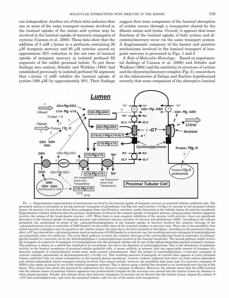
suggest that some component of the luminal absorptionof cystine occurs through a transporter shared by thedibasic amino acid lysine. Overall, it appears that somefractions of the luminal uptake of both cystine and di-cysteinylmercury occur via the same transport system.A diagrammatic summary of the known and putativemechanisms involved in the luminal transport of inor-ganic mercury is presented in Figs. 1 and 2.
6. Role of Molecular Homology. Based on experimen-tal findings of Cannon et al. (2000) and Schafer andWatkins (1984) and the similarity in structure of cystineand the dicysteinylmercury complex (Fig. 3), researchersat the laboratories of Zalups and Barfuss hypothesizedrecently that some component of the absorptive luminal
FIG. 1. Diagrammatic representation of mechanisms involved in the luminal uptake of inorganic mercury in proximal tubular epithelial cells. Thepresented scheme is premised on having mercuric conjugates of glutathione (GSH-Hg-GSH) and cysteine (CYS-Hg-CYS) present in the proximal tubularlumen. At present, it is not clear where these conjugates form, but filtration, secretion, and/or formation in the tubular lumen all must be considered.Experimental evidence indicates that the primary mechanism involved in the luminal uptake of inorganic mercury along proximal tubular segmentsinvolves the actions of the brush-border enzyme, g-GT. When there is near-complete inhibition of the enzyme (with acivicin), there are significantdecreases in the luminal uptake of inorganic mercury and enhanced urinary excretion of mercury and glutathione (GSH). According to the schemepresented, the mechanism of action of the g-glutamyltranspeptidase in the luminal uptake of mercury involves the catalytic cleavage of theg-glutamylcysteine bond on molecules of GSH bonded (via the sulfur atom of the cysteinyl residue) to mercuric ions. There also is the possibility thatmixed mercuric conjugates may be present in the tubular lumen, but these have not been included in this figure. According to the presented scheme,after g-GT has cleaved the g-glutamylcysteine bond on molecules of GSH bonded to a mercuric ion, the resulting mercuric conjugate of cysteinylglycinecan potentially enter two pathways. The most likely pathway involves the catalytic cleavage of the cysteinylglycinyl bond on molecules of cysteinyl-glycine bonded to a mercuric ion by the dehydropeptidase-1 cysteinylglycinase located on the luminal membrane. The second pathway might involvethe transport of a mercuric S-conjugate of cysteinylglycine into the proximal tubular cell by one of the sodium-dependent peptide transport systems.This pathway is shown as a dotted line attached to an arrowhead, just above the depiction of cysteinylglycinase. Due to the abundance of peptidaseactivity on the luminal membrane of proximal tubular epithelial cells, it seems unlikely, at present, that any appreciable amount of transport of amercuric conjugate of cysteinylglycine would occur under normal circumstances. After the actions of cysteinylglycinase, a mercuric conjugate ofcysteine remains, presumably as dicysteinylmercury (CYS-Hg-CYS). This resulting mercuric S-conjugate of cysteine then appears to enter proximaltubular epithelial cells via amino transporters in the luminal plasma membrane. Current evidence indicates that there are both sodium-dependentand sodium-independent amino transport systems involved. One cannot exclude, however, the possibility that some type of a mercuric conjugate ofGSH is also taken up intact by one of the luminal transport systems. This is shown using a dotted line attached to an arrowhead near the top of thefigure. This scheme also shows the potential for endocytosis of a mercuric conjugate of albumin. Moreover, the scheme shows that the GSH secretedinto the tubular lumen of proximal tubular segments may preferentially compete for the mercuric ions carried into the tubular lumen by albumin orother plasma proteins. Finally, this scheme shows that mercuric conjugates of cysteine can be filtered into the tubular lumen, bypass the actions ofg-GT and cysteinylglycinase, and enter the proximal tubular cells via one of the amino acid transporters.
MOLECULAR INTERACTIONS WITH MERCURY IN THE KIDNEY 119
transport of dicysteinylmercury occurs by a mechanisminvolving molecular homology (or “mimicry”). They pos-tulate that dicysteinylmercury may act as a molecularhomolog, or “mimic,” of the amino acid cystine at the siteof one or more transporter responsible for the luminaluptake of cystine (Cannon et al., 2000).
Molecular homology, or what some refer to as molec-ular mimicry, is not a novel concept. In 1993, Clarksondiscussed the concept that mercury, and other metals,form complexes with biological molecules that mimicstructurally endogenous molecules. For example, thecomplex formed between methylmercury and cysteine isthought to “mimic” the amino acid methionine, as ameans to gain entry into the central nervous system viaspecific amino acid transporters. Evidence supportingthis hypothesis comes from studies on the uptake and/ortransport of methylmercury by astrocytes (Aschner etal., 1990) and the endothelial cells lining the blood-brainbarrier (Aschner and Clarkson, 1989, Kerper et al.,1992). Another potential transportable molecular ho-molog may occur when inorganic mercury or methylmer-cury binds to glutathione. The complex formed when twomolecules of glutathione bind to a single mercuric ionmay also prove to be a functional molecular homolog of
glutathione disulfide. The implication of a dicystei-nylmercury complex being homologous to, or “mimick-ing,” the amino acid cystine does, however, appear to bea novel addition to the purported species of moleculesthat are involved in mimicry during the process of trans-porting a toxic metal into an epithelial cell.
D. Mechanisms of Basolateral Uptake of Mercury
1. Role of Organic Anion Transport System. In addi-tion to the large body of evidence indicating that mercu-ric ions are taken up at the luminal membrane of prox-imal tubular cells, there is substantial evidenceindicating that mercuric ions are also taken up at thebasolateral membrane of these cells. Approximately 40%of the dose of inorganic mercury is normally taken by thetotal renal mass of rats during the initial hour after thei.v. injection of a nontoxic dose of mercuric chloride(Zalups and Diamond, 1987a; Zalups and Lash, 1994;Zalups and Barfuss, 1995a, 1998a,b; Zalups, 1996,1997). Current evidence indicates that approximately 40to 60% of this renal burden of mercury can be attributedto a basolateral mechanism (Zalups, 1995, 1997,1998b,c; Zalups and Barfuss, 1995, 1998a,b; Zalups andMinor, 1995). It should be stressed that this applies only
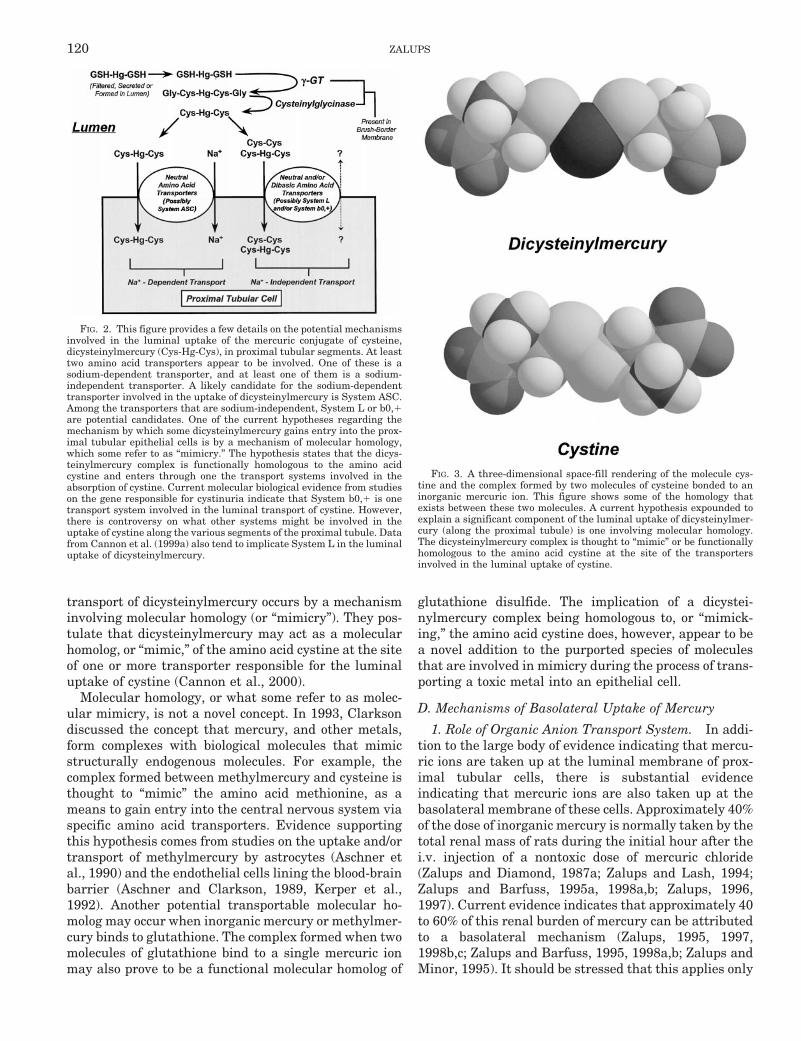
FIG. 2. This figure provides a few details on the potential mechanismsinvolved in the luminal uptake of the mercuric conjugate of cysteine,dicysteinylmercury (Cys-Hg-Cys), in proximal tubular segments. At leasttwo amino acid transporters appear to be involved. One of these is asodium-dependent transporter, and at least one of them is a sodium-independent transporter. A likely candidate for the sodium-dependenttransporter involved in the uptake of dicysteinylmercury is System ASC.Among the transporters that are sodium-independent, System L or b0,1are potential candidates. One of the current hypotheses regarding themechanism by which some dicysteinylmercury gains entry into the prox-imal tubular epithelial cells is by a mechanism of molecular homology,which some refer to as “mimicry.” The hypothesis states that the dicys-teinylmercury complex is functionally homologous to the amino acidcystine and enters through one the transport systems involved in theabsorption of cystine. Current molecular biological evidence from studieson the gene responsible for cystinuria indicate that System b0,1 is onetransport system involved in the luminal transport of cystine. However,there is controversy on what other systems might be involved in theuptake of cystine along the various segments of the proximal tubule. Datafrom Cannon et al. (1999a) also tend to implicate System L in the luminaluptake of dicysteinylmercury.
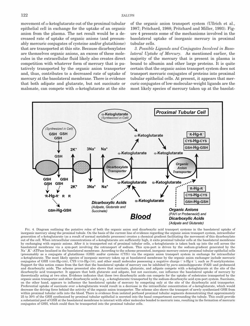
FIG. 3. A three-dimensional space-fill rendering of the molecule cys-tine and the complex formed by two molecules of cysteine bonded to aninorganic mercuric ion. This figure shows some of the homology thatexists between these two molecules. A current hypothesis expounded toexplain a significant component of the luminal uptake of dicysteinylmer-cury (along the proximal tubule) is one involving molecular homology.The dicysteinylmercury complex is thought to “mimic” or be functionallyhomologous to the amino acid cystine at the site of the transportersinvolved in the luminal uptake of cystine.
120 ZALUPS

to doses of inorganic mercury that are non-nephrotoxic.Under conditions where the dose is increased to levelsthat induce renal tubular injury, the percentage of thedose found in the kidneys (at various times after expo-sure) decreases. This is due in part to necrosis of tubularepithelial cells and the subsequent release and excretionof cytosolic mercury. (Zalups and Diamond, 1987b; Za-lups et al., 1988).
One of the first lines of substantial evidence implicat-ing a basolateral mechanism in the renal tubular uptakeof inorganic mercury comes from a recent study by Za-lups and Minor (1995). In this study, the uptake anddisposition of administered inorganic mercury wereevaluated in rats in which glomerular filtration hadbeen reduced to negligible levels in one or both kidneysthrough pretreatment with mannitol in combinationwith ureteral ligation (Zalups and Minor, 1995). It wasdemonstrated that induction of “stop-flow” conditions bythese pretreatments caused an approximately 40% de-crease in the net uptake and accumulation of inorganicmercury during the initial 1 h after the administrationof a 0.5 mmol/kg i.v. dose of mercuric chloride. Thesefindings indicate that a major fraction of the renal tu-bular uptake of inorganic mercury occurred via a baso-lateral mechanism. They also demonstrated that pre-treatment with para-aminohippurate, which is a specificcompetitive substrate for the renal organic anion trans-porter (Shimomura et al., 1981; Ferrier et al., 1983;Ullrich et al. 1987a,b); Pritchard, 1988; Roch-Ramel etal., 1992), caused significant reductions in the acuterenal tubular uptake and accumulation of inorganicmercury in normal animals and in animals that had oneor both ureters ligated. In fact, the combination of ure-teral ligation and pretreatment with para-aminohippu-rate caused an approximately 85% reduction in the netuptake and accumulation of inorganic mercury duringthe first hour after the injection of mercuric chloride.These findings suggest that the majority of the basolat-eral uptake of inorganic mercury was being inhibited bypara-aminohippurate, which implicates the organic an-ion transporter as the primary mechanism in the baso-lateral uptake of inorganic mercury. Data from otherrecent studies have confirmed that basolateral uptake ofinorganic mercury does occur in the kidney and that theprimary mechanism involved is linked to the activity ofthe organic anion transport system (Zalups and Lash,1994; Zalups, 1995, 1997, 1998a,b; Zalups and Barfuss,1995a, 1998a,b; Zalups et al., 1998).
There also are data implicating the activity of theorganic anion transporter in the basolateral uptake oforganic mercuric compounds. These data show that therenal uptake and/or accumulation (Tanaka et al., 1992)and toxicity (Ban and de Ceaurriz, 1988) of methylmer-cury are reduced significantly in mice pretreated withprobenecid, which is another competitive substrate andinhibitor of the organic anion transporter in renal prox-
imal tubules (Shimomura et al., 1981; Roch-Ramel et al.,1992).
2. Role of Dicarboxylate Transporter. In an earlystudy, Clarkson and Magos (1967) demonstrated thatpretreatment with the dicarboxylate maleate causeddose-dependent reductions in the net renal accumula-tion of inorganic mercury when it was given as a cys-teine-mercury complex (100 mg Hg/kg). Unfortunately, itis not clear from this study whether the changes in therenal disposition of mercury were due to the inhibitoryeffects of maleate on renal cellular metabolism (Rogulskiand Angielski, 1963) or whether they were due to directeffects at the site of a transporter of mercury. Interest-ingly, they found that fumarate (an isomer of maleate)did not have the same effects as maleate, which suggestsisomer specificity.
More recently, Zalups and Barfuss (1998b) demon-strated that pretreatment with small (four- to six-car-bon) aliphatic dicarboxylates, such as succinate, glut-arate, or adipate (but not malonate), inhibited the renal(basolateral) uptake of i.v. administered inorganic mer-cury in a dose-dependent manner in both normal ratsand in rats that had their ureters ligated. Putativemechanisms for the inhibitory effects of certain dicar-boxylates on the renal tubular uptake, transport, andaccumulation of inorganic mercury have been hypothe-sized by Zalups and Barfuss (1998b). Some of the detailsof these hypotheses are provided here.
Current evidence indicates that the organic aniontransporter is driven by an organic anion/dicarboxylicacid (dicarboxylate) exchange (reviewed by Pritchardand Miller, 1993, and Dantzler, 1996). It appears thatintracellular generation of a-ketoglutarate (from normalmetabolic processes) contributes to the creation of anintracellular chemical gradient favoring the movementof this dicarboxylate out of the cell. When the gradientbecomes sufficiently great, a-ketoglutarate is trans-ported out of proximal tubular cells at the basolateralmembrane via exchange with organic anions at the siteof the organic anion exchanger. There is evidence indi-cating that a significant fraction of the a-ketoglutarate(and other dicarboxylic acids) that exits proximal tubu-lar cells at the organic exchanger enters back into thecells across the basolateral membrane via a sodium-dicarboxylic acid cotransporter (Pritchard, 1988). Thiscotransport system is driven by the sodium-gradientgenerated by Na1,K1-stimulated ATPase. Although it isnot exactly clear via which mechanisms succinate, glut-arate, or adipate inhibits the renal tubular uptake ofinorganic mercury, it seems likely that an excess of anyof these dicarboxylates in the extracellular compartmentcreates competition for the sodium-dependent entry ofa-ketoglutarate at the site of the dicarboxylic acid co-transporter. Reduction in the basolateral uptake of a-ke-toglutarate would likely cause a decrease in the intra-cellular concentration of this dicarboxylate. This in turnwould decrease the chemical gradient favoring the
MOLECULAR INTERACTIONS WITH MERCURY IN THE KIDNEY 121
movement of a-ketoglutarate out of the proximal tubularepithelial cell in exchange for the uptake of an organicanion from the plasma. The net result would be a de-creased rate of uptake of organic anions (and presum-ably mercuric conjugates of cysteine and/or glutathione)that are transported at this site. Because dicarboxylatesare themselves organic anions, an excess of these mole-cules in the extracellular fluid likely also creates directcompetition with whatever form of mercury that is pu-tatively transported by the organic anion transporterand, thus, contributes to a decreased rate of uptake ofmercury at the basolateral membrane. There is evidencethat both adipate and glutarate, but not succinate ormalonate, can compete with a-ketoglutarate at the site
of the organic anion transport system (Ullrich et al.,1987; Pritchard, 1988; Pritchard and Miller, 1993). Fig-ure 4 presents some of the mechanisms involved in thebasolateral uptake of inorganic mercury in proximaltubular cells.
3. Possible Ligands and Conjugates Involved in Baso-lateral Uptake of Mercury. As mentioned earlier, themajority of the mercury that is present in plasma isbound to albumin and other large proteins. It is quitecertain that the organic anion transport system does nottransport mercuric conjugates of proteins into proximaltubular epithelial cells. At present, it appears that mer-curic conjugates of low-molecular-weight ligands are themost likely species of mercury taken up at the basolat-
FIG. 4. Diagram outlining the putative roles of both the organic anion and dicarboxylic acid transport systems in the basolateral uptake ofinorganic mercury along the proximal tubule. On the basis of the current line of evidence regarding the organic anion transport system, intracellulargeneration of a-ketoglutarate (as a result of normal metabolic processes) creates a chemical gradient facilitating the movement of this dicarboxylateout of the cell. When intracellular concentrations of a-ketoglutarate are sufficiently high, it exits proximal tubular cells at the basolateral membraneby exchanging with organic anions. After it is transported out of proximal tubular cells, a-ketoglutarate is taken back up into the cell across thebasolateral membrane via a sym-port involving the cotransport of sodium. This sym-port is driven by the sodium-gradient generated by theNa1,K1-ATPase localized in the basolateral membrane. According to the scheme presented, inorganic mercury enters proximal tubular epithelial cells(presumably as a conjugate of glutathione (GSH) and/or cysteine (CYS)) via the organic anion transport system in exchange for intracellulara-ketoglutarate. The most likely species of inorganic mercury taken up at basolateral membrane by the organic anion exchanger include mercuricconjugates of GSH (GSH-Hg-GSH), CYS (CYS-Hg-CYS), and other small molecules possessing a negative charge (2R-Hg-R2), such as N-acetylcysteine.Support for this notion comes from the fact that the basolateral uptake of mercury can be inhibited by para-aminohippurate (PAH) and probenecidand dicarboxylic acids. The scheme presented also shows that succinate, glutarate, and adipate compete with a-ketoglutarate at the site of thedicarboxylic acid transporter. It appears that both glutarate and adipate, but not succinate, can influence the basolateral uptake of mercury bytheoretically acting at two sites. Evidence indicates that these two dicarboxylic acids can compete for the uptake of substrates transported by theorganic anion transporter and other dicarboxylic acids (e.g., a-ketoglutarate) transported by the sodium-dicarboxylic acid sym-port system. Succinate,on the other hand, appears to influence the basolateral uptake of mercury by competing only at the site of the dicarboxylic acid transporter.Preferential uptake of succinate over a-ketoglutarate would result in a decrease in the intracellular concentration of a-ketoglutarate, which woulddecrease the driving force behind the activity of the organic anion transporter. The scheme also shows the transport of newly synthesized GSH fromwithin proximal tubular cells into the blood. There is evidence from isolated perfused proximal tubular segments demonstrating that approximately25 to 30% of the GSH synthesized by proximal tubular epithelial is secreted into the basal compartment surrounding the tubule. This could providea substantial pool of GSH at the basolateral membrane to interact with other molecules bonded to mercuric ions, resulting in the formation of mercuricconjugates of GSH, which could then be transported into the proximal tubular cells.
122 ZALUPS

eral membrane by the organic anion transporter. Two ofthe conjugates that have been implicated in the basolat-eral transport of mercury are mercuric conjugates ofglutathione and/or cysteine (Zalups, 1998b).
4. Mercuric Conjugates of Glutathione as Transport-able Forms of Mercury at Basolateral Membrane. Mol-ecules of glutathione have a net negative charge at phys-iological pH. Because of this charge and its size,glutathione has been postulated to be substrate at thesite of the organic anion transporter. Support for thiscomes in part from the studies of Lash and Jones (1983,1984), who demonstrated transport of glutathione (as anintact tripeptide) in basolateral membrane vesicles (iso-lated from the renal cortex of rats) via a mechanism thatwas sodium-dependent and that could be blocked byprobenecid. They also demonstrated basolateral trans-port of certain organic S-conjugates of glutathione, suchas S-(1,2-dichlorovinyl)glutathione, into proximal tubu-lar epithelial cells by a probenecid-sensitive mechanism(Lash and Jones, 1985b).
Because both glutathione and certain S-conjugates ofglutathione appear to be transported across the basolat-eral membrane of proximal tubular cells by the organicanion transport system, it seems plausible that mercuricS-conjugates of glutathione may also be transportedacross the basolateral membrane by this same transportsystem. There are some findings from a recent study, inwhich mercuric conjugates of glutathione were adminis-tered to rats that had undergone bilateral ureteral liga-tion, that support this contention (Zalups, 1998b). Thedata show that the basolateral uptake of inorganic mer-cury was greater when it was administered in the formof a mercuric conjugate of glutathione than when it wasadministered as mercuric chloride.
5. Mercuric Conjugates of Cysteine as TransportableForms of Mercury at Basolateral Membrane. Despitethe fact that cysteine has a net neutral charge at phys-iological pH, it has become highly relevant to considerthat inorganic or organic mercuric conjugates of cysteineare transportable species at the site of the organic aniontransporter. The relevance for this consideration comesfrom studies in which organic S-conjugates of cysteinehave been shown to be taken up at the basolateral mem-brane of proximal tubular cells by a mechanism consis-tent with the activity of the organic anion transporter.For example, Lash and Anders (1989) demonstratedthat organic S-conjugates of cysteine [e.g., S-(1,2-dichlo-rovinyl)-L-cysteine] were taken up by isolated proximaltubular epithelial cells from rats by a sodium-dependentand probenecid- and para-aminohippurate-sensitivetransport system. More recently, Dantzler et al. (1995),using isolated proximal tubules from rabbits, also dem-onstrated that certain organic S-conjugates of cysteinewere taken up at the basolateral membrane by a probe-necid- and p-aminohippurate-sensitive transport mech-anism.
Based on these aforementioned findings, it seems log-ical to hypothesize that mercuric conjugates of cysteinemay also be transported into proximal tubular epithelialcells at the basolateral membrane by the organic aniontransport system. Several sets of recent data tend tosupport this hypothesis. For example, one set of datashows that rates of association and transport of inor-ganic mercury in basolateral membrane vesicles (isolat-ed from the kidneys of rats) tends to be greater when thevesicles are exposed to mercuric conjugates of cysteinethan when they are exposed to mercuric chloride (Zalupsand Lash, 1997a). Additional support for this hypothesiscomes indirectly from a recent in vivo study (Zalups,1998b). First, the data from this study show that bilat-eral ureteral ligation caused an approximately one-halfreduction in the net renal accumulation of mercury incontrol rats treated with a low 0.5-mmol/kg dose of mer-curic chloride and in the rats coadministered this dose ofinorganic mercury with a 4-fold greater (2.0 mmol/kg)amount of L-cysteine. More importantly, the findingsalso show that the net renal accumulation of mercurywas greater in the animals treated with inorganic mer-cury plus cysteine than in the animals treated withmercuric chloride, whereas the relative intrarenal dis-tribution of mercury was similar in both groups of rats.Second, pretreatment with para-aminohippurate wasshown to cause a significant decrease in the renal up-take of mercury in the rats that had their ureters ligatedand that were administered inorganic mercury plus cys-teine (Zalups, 1998c). The most reasonable explanationfor these findings is that by injecting mercuric conju-gates of cysteine in animals that have had their uretersligated, more of these conjugates than are formed nor-mally when inorganic mercury is administered as mer-curic chloride are made available at the site of the or-ganic anion transporter (and possibly other basolateraltransporters, such as basolateral amino acid transport-ers) to promote the uptake of mercury.
6. Other Mercuric Conjugates as Transportable Formsof Mercury at Basolateral Membrane. Although cur-rent experimental evidence tends to point to mercuricconjugates of cysteine and glutathione being primarilyinvolved in the luminal and basolateral uptake of inor-ganic mercury along the proximal tubule (after exposureto mercuric chloride), it is clear that other thiols, espe-cially homologues of cysteine, such as homocysteine andN-acetylcysteine, can significantly influence the mannerin which inorganic mercury is being handled in thekidneys (Zalups, 1998c; Zalups and Barfuss, 1998b).This point is exemplified in the recent studies of Zalupsand Barfuss (1998b) and Zalups (1998c), who studiedand compared in rats the mechanisms involved in therenal tubular uptake of inorganic mercury when it wascoadministered with cysteine, homocysteine, or N-ace-tylcysteine. When inorganic mercury was administeredwith cysteine or as mercuric chloride, the levels of lumi-nal and basolateral uptake of mercury in the kidneys
MOLECULAR INTERACTIONS WITH MERCURY IN THE KIDNEY 123

were similar. In contrast to this pattern of uptake, wheninorganic mercury was administered with homocys-teine, a much lower level of uptake of mercury occurredat the luminal membrane relative to that which oc-curred at the basolateral membrane. Even greater dif-ferences in the levels of luminal uptake versus basolat-eral uptake of mercury were detected when rats weretreated with inorganic mercury and N-acetylcysteine.When inorganic mercury was administered with thisnegatively charged molecule, virtually all of the renaltubular uptake of mercury occurred at the basolateralmembrane, and the majority of this uptake could beinhibited by pretreatment with para-aminohippurate.In fact, regardless of how inorganic mercury was admin-istered, the majority of the basolateral uptake of mer-cury was inhibited by pretreatment with para-amin-ohippurate, which implicates the activity of the organicanion transport system in the basolateral uptake of in-organic mercury under all of the experimental condi-tions studied.
In addition to the high level of basolateral uptake ofmercury in the kidneys of the animals treated withinorganic mercury and N-acetylcysteine, the amount ofmercury excreted in 24 h was at least 45 to 50% greaterthan that in any of the other groups of rats. The overallfindings from these rats indicate that the negativecharge on N-acetylcysteine likely promotes the rapidtransport of mercuric conjugates of N-acetylcysteine intoproximal tubular cells at the site of the organic aniontransporter, whereas it prevents or impedes the uptakeof these mercuric conjugates at the luminal plasmamembrane, which promotes the urinary excretion ofmercury.
E. Role of Liver in Renal Tubular Uptake of Mercury
It appears that some aspects of hepatic function playa role in at least a component of the renal uptake andtransport of mercury. Evidence for this hypothesiscomes from recent dispositional studies. In one study,specific depletion of hepatic glutathione with 1,2-di-chloro-4-nitrobenzene before the administration of inor-ganic mercury was shown to cause a significant diminu-tion in the renal uptake and/or accumulation ofinorganic mercury in mice (Tanaka et al., 1990). In otherstudies, it has been demonstrated that biliary ligation orcannulation before the administration of inorganic mer-cury caused a decrease in the renal tubular uptake andaccumulation of inorganic mercury in rats (Zalups andBarfuss, 1996a, Zalups, 1998a; Zalups et al., 1999a,b,c).Taken together, these findings indicate that some as-pects of hepatic function are linked to a component inthe renal tubular uptake and/or accumulation of inor-ganic mercury. Hepatic synthesis and secretion of glu-tathione represent a possible candidate. Additionalstudies are necessary to better determine the role of theliver in the renal tubular uptake of mercury.
F. Intracellular Distribution of Mercury
Once inorganic mercuric ions gain entry in proximaltubular cells, it appears that they distribute throughoutall intracellular pools (Madsen, 1980; Omata et al., 1980;Baggett and Berndt, 1985; Houser and Berndt, 1988).Cellular fractionation studies using the renal cortexfrom rats treated acutely or chronically with mercuricchloride indicate that mercury distributes in nuclear,lysosomal, mitochondrial, brush-border, and superna-tant fractions, with the nuclear fraction containing thegreatest amount of mercury among the organelle frac-tions (Madsen, 1980; Madsen and Hansen, 1980). Simi-lar findings have also been obtained in other studiesusing homogenates of the renal cortex from normal anduninephrectomized rats treated with mercuric chloride(Baggett and Berndt, 1985; Houser and Berndt, 1988).In these studies, however, the cytosolic fraction wasfound to contain the greatest content of mercury.
Interestingly, the relative specific content of mercurywas shown to increase to the greatest extent in thelysosomal fraction when rats were made proteinuricwith an aminoglycoside (Madsen, 1980) or when ratswere treated chronically with mercuric chloride (Madsenand Hansen, 1980). Increases in the lysosomal content ofmercury may reflect the fusion of primary lysosomeswith endocytotic or cytosolic vesicles containing com-plexes of inorganic mercury bound to proteins.
III. Urinary Excretion of Mercury
Urinary and fecal excretion of mercury are the prin-cipal means by which humans and other mammals elim-inate the different forms of mercury from the body. Un-der most circumstances, a greater fraction of a dose ofmercury is excreted in the feces than in the urine earlyafter exposure (Rothstein and Hayes, 1960; Magos andClarkson, 1977; Zalups et al., 1987, 1988, 1991a, 1992,1993; World Health Organization, 1991; Zalups et al.,1987, 1988, 1991a, 1992, 1993; Zalups and Lash, 1994).In rats, it has been shown that more than twice as muchinorganic mercury is excreted in the feces than in theurine during the initial days after exposure to a non-nephrotoxic dose of mercuric chloride (Rothstein andHayes, 1960; Zalups et al., 1987, 1988; Zalups and Lash,1994). Less than 10% of the administered dose is ex-creted in the urine during this time. In one study, ratsinjected i.v. with a non-nephrotoxic dose of mercuricchloride had excreted about 20% of the dose in the urineand 30% of the dose in the feces during the initial 54days after injection (Rothstein and Hayes, 1960). Thelow level in the urinary excretion of mercury is due totwo principal factors, the avid uptake of mercuric ionsand the retention of accumulated mercuric ions, in prox-imal tubular segments.
After exposure to organic forms of mercury, even lessmercury is excreted in the urine than after exposure toinorganic mercury. For example, it was demonstrated
124 ZALUPS

recently that both normal and uninephrectomized ratsexcreted only about 3% of the dose of mercury in theurine by the end of the initial 7 days after the i.v.injection of a low dose (5 mg/kg) of methylmercury (Za-lups et al., 1992). By contrast, more than 15% of theadministered dose was excreted in the feces during thesame period of time. In a recent study in which sevenadult men received a tracer amount of 203Hg-labeledmethylmercury i.v., the cumulative fecal excretion ofmercury over 70 days was much greater than the cumu-lative urinary excretion of mercury (Smith et al., 1994).More specifically, about 30% of the dose was excreted inthe feces, whereas only about 4% of the dose was ex-creted in the urine.
Early reports (Mambourg and Raynaud, 1965; Vostal,1966) had claimed that mercury appeared in the urinebefore inulin (which is filtered and not absorbed or se-creted along the nephron). This was interpreted by some(Clarkson and Magos, 1967) to indicate that urinarymercury represented a pool of mercury that had beensecreted from the blood into the tubular lumen by atransepithelial mechanism. This was a reasonable viewconsidering there was a published report claiming thatapproximately 99% of the mercury in plasma was notfilterable (Berlin and Gibson, 1963). Based on recentdata, however, it appears that much more than 1% onthe mercury in plasma is filtered into the proximal tu-bule lumen (Madsen, 1980; Zalups and Minor, 1995;Zalups, 1997, 1998b,c; Zalups and Barfuss, 1998a,b) andthat the mechanisms involved in the urinary excretion ofmercury are less clear than once thought.
It should be emphasized that although 95 to 99%(depending on animal species and experimental condi-tions) of the mercury in plasma is bound to albumin (andother plasma proteins), a significant fraction of albuminis filtered at the glomerulus. Thus, substantial amountsof mercury could theoretically gain access to the luminalcompartment of proximal tubules by filtration of a mer-cury-albumin complex. There is some indirect in vivoevidence supporting this notion. Madsen (1980) demon-strated in rats made proteinuric by gentamicin (presum-ably by decreasing the absorptive capacity of the proxi-mal tubular epithelium by cellular necrosis) that muchof the administered mercury excreted in the urine wasassociated with albumin. A fundamental assumption inwith these findings, however, is that the preponderanceof the albumin associated with the mercury in the urinecame from glomerular filtration rather than intercellu-lar leak. In contrast to the findings of Madsen (1980),Clarkson and Magos (1967) found that about 70% of themercury excreted in urine by rats treated with sodiummaleate, subsequent to the exposure of inorganic mer-cury, was not bound to protein. This finding is actuallynot that surprising, because much of the mercury ex-creted in the urine probably originated from cellularstores, and thus was likely bound to low-molecular-weight thiols, such as glutathione.
Some insight into mechanisms involved in the urinaryexcretion of mercury has been gained through experi-mental maneuvers that cause the urinary excretion ofmercury to increase. In most cases, the increased uri-nary excretion of mercury is associated with decreasedluminal absorption of mercury and/or the luminal elim-ination or extraction of accumulated mercury along theproximal tubule (and/or other segments of the nephron).Some examples of these maneuvers are listed below.
In an early study by Clarkson and Magos (1967),pretreatment of female rats with sodium maleate, beforethe injection of a low 100 mg/kg dose of mercury in theform of mercuric chloride or a mercury-cysteine complex,was shown to cause the urinary excretion of mercury toincrease and the renal accumulation of mercury to de-crease. Sodium maleate was used because it caused “pro-found metabolic disturbances in renal cells.” The au-thors also found that the administration of sodiummaleate after treatment with mercury caused the renalcontent of mercury to decrease and the urinary excretionof mercury to increase.
As mentioned earlier, the urinary excretion of mer-cury also increases dramatically when renal g-GT isinhibited before the administration of inorganic mercury(Berndt et al., 1985; Zalups, 1995; Zalups et al., 1999b,c).Much of the mercury excreted in urine after the inhibi-tion of g-GT appears to be associated with glutathione,which implicates the presence of mercuric conjugates ofglutathione in the proximal tubular lumen (Baggett andBerndt, 1986). Current evidence indicates that the in-creased urinary excretion of mercury associated with theinhibition g-GT is due mainly to decreased luminal ab-sorption and transport of mercury along the proximaltubule (Berndt et al., 1985; Tanaka et al., 1990; Tanaka-Kagawa et al., 1993; de Ceaurriz et al., 1994; Kim et al.,1995; Zalups, 1995; Cannon et al., 2000).
When inorganic mercury is applied to the luminalmembrane of proximal tubular epithelial cells as a mer-curic conjugate of N-acetylcysteine (Zalups and Barfuss,1998b), DMPS (Zalups et al., 1998), DMSA (Zalups,1993c), or metallothionein (Zalups et al., 1993a), urinaryexcretion of mercury increases greatly due to the lack ofluminal uptake of these mercuric conjugates. In general,it appears that when mercuric ions are bound to organicligands possessing a net negative charge, the mercuricconjugates of these molecules are not taken up readily atthe luminal membrane and in turn are excreted in theurine. When DMPS is administered after exposure tomercury, the urinary excretion of mercury also increasesgreatly (Zalups, 1993c). Recent evidence (obtained fromisolated perfused proximal tubular segments) indicatesthat the increased urinary excretion of mercury thatoccurs under these conditions results from unidirec-tional extraction of mercury from within or on proximaltubular epithelial cells into the tubular lumen (Zalups etal., 1998). It is likely that increased urinary excretion of
MOLECULAR INTERACTIONS WITH MERCURY IN THE KIDNEY 125

mercury induced by treatment with DMSA (Zalups,1993c) occurs by a similar mechanism.
After proximal tubular necrosis is induced by mercuryor other agents (Clarkson and Magos, 1967; Magos andStoychev, 1969; Trojanowska et al., 1971), the urinaryexcretion of mercury increases. This is due largely tomercury being released from, or not being absorbed by,necrotic or degenerating proximal tubular epithelialcells (Madsen, 1980; Zalups and Diamond, 1987b; Za-lups et al., 1988). More studies are needed to betterdefine the factors and mechanisms involved in the uri-nary excretion of mercury and mercury-containing com-pounds.
Despite all the studies that have been carried out todate, very little is really known about the mechanismsinvolved in the urinary excretion of inorganic and or-ganic forms of mercury. The major questions that stillneed to be addressed include the following. 1) What arethe magnitudes and rates at which mercury is filtered atthe glomerulus? 2) To what extent is filtered mercurytaken up by proximal tubular epithelial cells? Alterna-tively, to what extent is filtered mercury excreted in theurine? 3) What is the chemical form or forms of mercuryexcreted in the urine? 4) Is some of the mercury that isexcreted in the urine added to the luminal fluid by atrans-epithelial secretory mechanism (as has been sug-gested previously by Clarkson and Magos, 1967;Foulkes, 1974; Zalups and Barfuss, 1993a, 1995a,1998b; Zalups, 1995, 1997, 1998b; Zalups and Minor,1995; Zalups and Lash, 1997a)?
IV. Molecular Interactions and Effects ofMercury in Renal Epithelial Cells
A. Effects of Mercury on Intracellular ThiolMetabolism
A major intracellular effect of mercury consists of theinduction of and binding to metallothionein (Piotrowskiet al., 1974). Metallothioneins are a group of small in-tracellular proteins with an approximate molecularweight of 6000 to 7000 Da. They contain numerous cys-teinyl residues and have the capacity to bind variousmetals, including inorganic mercury, cadmium, zinc,copper, silver, and platinum. The administration of asingle, daily, nontoxic dose of mercuric chloride overseveral days has been shown to cause a near doubling inthe concentration of metallothionein in the renal cortexor outer stripe of the outer medulla in rats (Zalups andCherian, 1992a). Induction of the synthesis of metallo-thionein in kidney has also been demonstrated in ratsexposed to elemental mercury vapor over the course ofseveral days (Cherian and Clarkson, 1976). The increaseappears to be tissue-selective, as changes in hepaticmetallothionein synthesis have not been demonstrated.Mercury vapor is converted into inorganic mercury,which is recovered predominantly (approximately 98%)in the kidney, suggesting that the induction of metallo-
thionein in the kidneys after exposure to elemental mer-cury may actually be mediated by inorganic mercury.The induction of metallothioneins in the kidney by inor-ganic mercury likely involves increased transcription ofmetallothionein-1 (MT-1) and metallothionein-2 (MT-2)genes via the interaction of zinc-dependent metal tran-scription factors and cis-acting DNA elements termedmetal-responsive elements resident in the promoter re-gion of the metallothionein genes. Sebsequently, there ispost-transcriptional control of translation of new mRNAinto metallothionein I and II protein (Koropatnick andZalups, 2000).
Some new insights into the relationships between thecellular content of mercury and the expression of metal-lothionein in both the kidneys and liver have been pro-vided in a recent study by Zalups and Koropatnick(manuscript submitted). They obtained evidence thatthe retention of inorganic mercury by renal tubular ep-ithelial cells is associated with the continual induction ofmetallothionein. More specifically, their data indicatethat in rats administered a single 0.5 mmol/kg i.v. dose ofmercuric chloride, the rate of transcription of MT-1 andMT-2 genes was as great at 2 weeks after treatmentwith mercury as it was 1 day after treatment. In addi-tion, renal levels of metallothionein-1 and -2 proteinremained elevated throughout the 2 weeks of study,during which the renal burden of mercury decreased byonly about 26% and the cumulative urinary excretion ofmercury was equal to about 24% of the dose of mercury.By contrast, they demonstrated that hepatic levels ofmercury and metallothionein protein and rates of tran-scription of MT-1 and MT-2 genes decreased continuallyover the initial 2 weeks after treatment. It was alsodemonstrated that the rates of transcription of metallo-thionein genes in the liver correlated highly with theamount of metallothionein protein in the liver. However,in the kidneys, there was no correlation between therates of transcription of metallothionein genes and me-tallothionein protein, which suggests that post-tran-scriptional events are involved in the expression of me-tallothionein protein in the kidneys after a singleexposure to inorganic mercury.
Inorganic and organic forms of mercury also have agreat influence on intracellular glutathione metabolismin the kidneys. These effects are observed acutely aftershort-term, single treatments and are concentration-de-pendent. Several sets of in vivo and in vitro data dem-onstrate increases in intracellular contents of glutathi-one in renal tubular epithelial cells after theadministration of relatively low toxic or nontoxic dosesof either methylmercury (Woods et al., 1992) or inor-ganic mercury (Fukino et al., 1986; Siegers et al., 1987;Zalups and Lash, 1990; Chung et al., 1982; Lash andZalups, 1992, 1993). At higher doses of inorganic mer-cury, decreases in renal content of glutathione (whichare often substantial) are observed (Fukino et al., 1984;Zalups and Lash, 1990; Lash and Zalups, 1992, 1993).
126 ZALUPS

Dose-dependent effects of inorganic mercury on renalglutathione metabolism have been demonstrated inmale Sprague-Dawley rats that received one of severalnontoxic or nephrotoxic i.v. doses of mercuric chloride(Zalups and Lash, 1990). At the level of the whole kidneyor in samples derived from the renal cortex or the outerstripe of the outer medulla, the nontoxic (0.5 mmol/kg) orthe moderately nephrotoxic (2 mmol/kg) dose of mercuricchloride induced significant increases in the renal con-centration of glutathione. This effect was most markedin the outer stripe of the outer medulla, where the con-centration of glutathione increased by as much as 85%.The toxicological significance of this finding relates tothe fact that the outer stripe of the outer medulla is oneof the primary zones in which proximal tubular injuryinduced by mercury occurs. At the highest nephrotoxicdose of mercuric chloride (3 mmol/kg), the concentrationsof glutathione in the renal cortex and outer stripe of theouter medulla were similar to those in controls (Zalupsand Lash, 1990).
Because the cellular content of glutathione is underfeedback control, the large increases in the renal contentof glutathione observed after treatment with inorganicmercury suggest that subtoxic or moderately toxic dosesof inorganic mercury induce the synthesis of glutathionevia g-glutamylcysteine synthetase (GCS), which is therate-limiting enzyme involved in the biosynthesis of glu-tathione. Data from Lash and Zalups (1993) support thishypothesis. They found that the activity of g-glutamyl-cysteine synthetase was increased in renal proximaltubular epithelial cells isolated from rats treated withinorganic mercury (relative to that in proximal tubularcells isolated from control rats). Further support for thehypothesis that mercuric ions induce GCS in renal epi-thelial cells comes from a study by Woods et al. (1992).They showed that the mRNA for g-glutamylcysteinesynthetase increased (by 4.4-fold) in the kidneys of maleFischer 344 rats treated with methylmercury hydroxidefor 3 weeks. Thus, at non-nephrotoxic doses, both inor-ganic and organic forms of mercury appear to induce thesynthesis of glutathione via the activity of GCS.
In addition to causing up-regulation of GCS, inorganicmercury also alters, in a dose-dependent manner, theactivity of other glutathione-dependent enzymes. Theeffects of inorganic mercury on these enzymes differdepending on whether a nontoxic, a moderately toxic, ora highly toxic dose is administered. Nontoxic doses ofmercuric chloride apparently cause increases in activi-ties of glutathione disulfide reductase and glutathioneperoxidase in isolated epithelial cells from both proximaltubular and distal tubular regions of the rat nephron(Lash and Zalups, 1993). In contrast, one group of inves-tigators (Addya et al., 1984) observed marked decreasesin the activities of renal glutathione disulfide reductaseand glutathione peroxidase in male rats treated chron-ically (15 days) with a relatively high dose of mercuricchloride (5 mg HgCl2/day per os). They also found ap-
parent adaptive increases in catalase activity. Similarly,others have found significant decreases in the activity ofglutathione disulfide reductase after the administrationof highly nephrotoxic doses of mercuric chloride [10mmol HgCl2/kg s.c. (Chung et al., 1982); 15 mmolHgCl2/kg s.c. (Fukino et al., 1984); 4 mg HgCl2/kg s.c.(Gstraunthaler et al., 1983)]. Although two groups ofinvestigators found small (20–35%), but statistically sig-nificant, decreases in the activity of glutathione peroxi-dase (Chung et al., 1982; Gstraunthaler et al., 1983),another group did not detect any change in the activityof this enzyme (Fukino et al., 1984). It is important tokeep in mind, however, that it becomes nearly impossi-ble to interpret in vivo data obtained from renal tissue inwhich there has been extensive cellular injury anddeath. When there is extensive renal tubular necrosis,decreases in the content of an enzyme or a molecule ofinterest (in samples of renal tissue) can be accounted forsimply by the release and excretion of the cytoplasmiccontents from dead epithelial cells.
B. Role of Lipid Peroxidation and Oxidative Stress inMercury-Induced Renal Cellular Injury
Findings from several studies suggest that an impor-tant mechanism involved in renal cellular injury in-duced by either in vivo or in vitro exposure to inorganicor organic forms of mercury involves the induction ofoxidative stress. The high affinity of mercuric ions forbinding to thiols naturally suggests that the ensuingdepletion of intracellular thiols (especially glutathione)either directly causes, or predisposes, proximal tubularcells to oxidative stress. Furthermore, other cellular an-tioxidants, including ascorbic acid and vitamin E, havebeen reported to be depleted in the kidneys of ratstreated with mercuric chloride (Fukino et al., 1984). Theactivity of several antioxidant enzymes also appears tobe markedly diminished after in vivo exposure of rats tonephrotoxic doses of mercuric chloride. For example, ithas been reported that the administration of mercuricchloride to male Sprague-Dawley rats caused markeddecreases in the activity of superoxide dismutase, cata-lase, glutathione peroxidase, and glutathione disulfidereductase in the renal cortex (Gstraunthaler et al.,1983).
Decreases in the activities of these protective enzymeswould be expected to enhance the susceptibility of renalepithelial cells to oxidative injury. There has been somedisagreement as to whether mercury itself causes oxida-tive injury or whether it merely makes renal epithelialcells more sensitive to agents that produce oxidativestress. Fukino et al. (1984) found that thiobarbituratereactants, which indicate occurrence of lipid peroxida-tion, were markedly increased in renal cortical homoge-nates from rats 12 h after a s.c. injection of a nephrotoxic(15 mmol/kg) dose of mercuric chloride. Gstraunthaler etal. (1983) observed increases in the formation of malon-dialdehyde in renal cortical homogenates obtained from
MOLECULAR INTERACTIONS WITH MERCURY IN THE KIDNEY 127

mercuric chloride-treated rats (relative to homogenatesgenerated from control rats treated with only cumenehydroperoxide). Because the two groups of rats wereadministered similar doses of mercuric chloride, concen-tration dependence cannot be invoked to explain thedifference in observed responses. Based on these find-ings, it appears that inorganic mercury can enhance theability of other agents to induced lipid peroxidation.
There are close relationships among maintenance ofnormal renal function, renal cellular content of glutathi-one, cellular redox status, and the generation of ATP inmitochondria. These relationships served as an impetusfor Lund et al. (1991) to investigate the role of mercury-induced oxidative stress in mitochondria of renal epithe-lial cells as a mechanism for mercury-induced renalcellular injury. More specifically, they investigated theeffects of inorganic mercury on the production of hydro-gen peroxide by renal cortical mitochondria isolatedfrom rats. Depending on the supply and coupling sitespecificity of respiratory substrates, variable increasesin the formation of hydrogen peroxide were observed;incubation of isolated mitochondria with 30 nmol mer-curic chloride/mg protein increased the formation of hy-drogen peroxide by 4-fold at the ubiquinone-cytochromeb region and 2-fold at the NADH dehydrogenase region.In addition, iron-dependent lipid peroxidation was in-creased 3.5-fold at the NADH dehydrogenase region andby 25% at the ubiquinone-cytochrome b region. Intrami-tochondrial glutathione was decreased in a time- andconcentration-dependent manner by mercuric chloride.In fact, at a concentration of 12 nmol mercury/mg pro-tein, the content of glutathione in mitochondria wasdepleted completely within 30 min, suggesting that tar-geting of mitochondrial glutathione by mercury may beresponsible for the intramitochondrial oxidative stress.Lund et al. (1993) also demonstrated that production ofhydrogen peroxide, depletion of glutathione, and lipidperoxidation increased in mitochondria (isolated fromrenal cortical homogenates of rats treated in vivo withmercuric chloride) after the addition of an appropriaterespiratory substrate. These findings support in vitrodata and lead one to suggest that mercury-induced oxi-dative stress within mitochondria is an important mech-anism involved in renal tubular injury induced by mer-cury.
C. Effects of Mercury on Renal Mitochondrial Function
As described earlier, Lund et al. (1991, 1993) demon-strated that inorganic mercury interferes with mito-chondrial respiratory function, causing increased pro-duction of hydrogen peroxide in the mitochondria,particularly at coupling site II of the electron transportchain. Their findings indicate that an oxidative stresslocalized in the mitochondria may be responsible formercury-induced inhibition of various energy-dependentprocesses in renal epithelial cells.
In an earlier series of studies, Weinberg et al.(1982a,b) compared the effects of mercuric chloride onmitochondrial function in vitro after either in vivo or invitro treatment with mercuric chloride. When mitochon-dria were isolated from male Sprague-Dawley rats andthen treated in vitro with inorganic mercury (Weinberget al., 1982a), a marked uncoupling of respiration (i.e.,increase in state 4 rate of oxygen consumption) and asignificant decrease in the rate of substrate-stimulatedrespiration (i.e., state 3 respiration) were observed. Inaddition, uptake of atractyloside-insensitive ADP andthe activities of both basal- and Mg21-activated oligomy-cin-sensitive ATPase were markedly increased by inor-ganic mercury. These in vitro effects occurred with athreshold concentration of mercuric chloride of 2nmol/mg protein. Similarly, when renal cortical mito-chondria were isolated from rats treated in vivo withmercuric chloride (5 mg/kg s.c.), the most prominenteffects detected were inhibition of ADP uptake and de-creases in the rates of state 3 and uncoupler-stimulatedrespiration (Weinberg et al., 1982b). These effects werenot attributed to interaction of mercury with mitochon-dria during the isolation procedure. However, with bothin vivo and in vitro treatment, inorganic mercury wasnot readily washed out of mitochondria, suggestingbinding between mercuric ions and thiol-containing mol-ecules in the mitochondria.
Chavez and Holguin (1988) and Chavez et al. (1991)also reported uncoupling of mitochondrial respirationafter either in vivo or in vitro treatment of male Wistarrats with mercuric chloride. Consistent with this find-ing, they found that inorganic mercury induced calciumefflux from mitochondria, oxidation of pyridine nucleo-tides, and a collapse of the membrane potential. Chavezand Holguin (1988) found that inorganic mercurybonded to mitochondrial protein in a concentration-de-pendent manner, with saturation at approximately 9nmol Hg21/mg protein. The finding supports the notionthat a mechanism by which mercury induces mitochon-drial injury is the formation of complexes between mer-curic ions and mitochondrial sulfhydryl groups.
Jung et al. (1989) used ATP depletion by differentchemical agents in microdissected nephron segments tolocalize the nephron site specificity of injury. They foundthat 1 mM mercuric chloride produced a significant de-pletion of intracellular ATP exclusively in S2 segments;nephron segments derived from the other regions of theproximal tubule (i.e., S1 or S3) or distal nephron (e.g.,the distal convoluted tubule or the medullary thick as-cending limb of the loop of Henle) were not as sensitiveto ATP depletion after incubation with inorganic mer-cury. This pattern agrees with histopathological data,which demonstrate that the pars recta of the proximaltubule is the primary target of inorganic mercury, al-though the S3 segment is also part of the pars recta andbecomes intoxicated by mercury in vivo. These data tendto support the conclusion that renal mitochondria are
128 ZALUPS
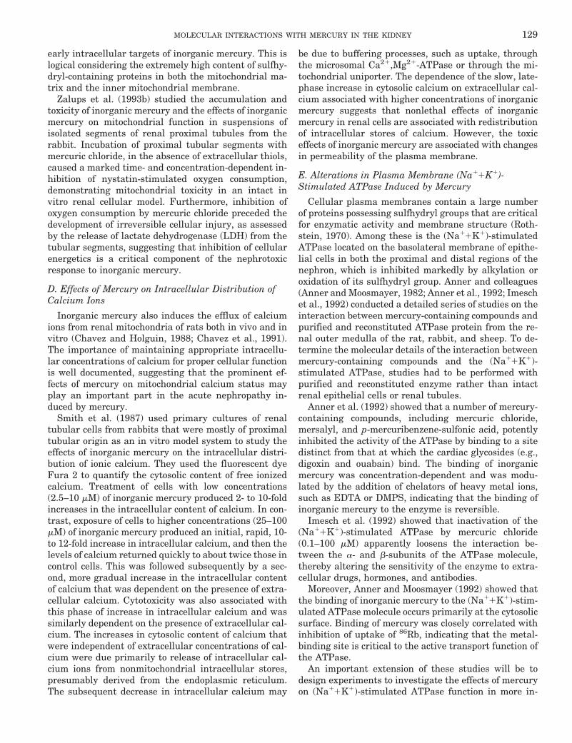
early intracellular targets of inorganic mercury. This islogical considering the extremely high content of sulfhy-dryl-containing proteins in both the mitochondrial ma-trix and the inner mitochondrial membrane.
Zalups et al. (1993b) studied the accumulation andtoxicity of inorganic mercury and the effects of inorganicmercury on mitochondrial function in suspensions ofisolated segments of renal proximal tubules from therabbit. Incubation of proximal tubular segments withmercuric chloride, in the absence of extracellular thiols,caused a marked time- and concentration-dependent in-hibition of nystatin-stimulated oxygen consumption,demonstrating mitochondrial toxicity in an intact invitro renal cellular model. Furthermore, inhibition ofoxygen consumption by mercuric chloride preceded thedevelopment of irreversible cellular injury, as assessedby the release of lactate dehydrogenase (LDH) from thetubular segments, suggesting that inhibition of cellularenergetics is a critical component of the nephrotoxicresponse to inorganic mercury.
D. Effects of Mercury on Intracellular Distribution ofCalcium Ions
Inorganic mercury also induces the efflux of calciumions from renal mitochondria of rats both in vivo and invitro (Chavez and Holguin, 1988; Chavez et al., 1991).The importance of maintaining appropriate intracellu-lar concentrations of calcium for proper cellular functionis well documented, suggesting that the prominent ef-fects of mercury on mitochondrial calcium status mayplay an important part in the acute nephropathy in-duced by mercury.
Smith et al. (1987) used primary cultures of renaltubular cells from rabbits that were mostly of proximaltubular origin as an in vitro model system to study theeffects of inorganic mercury on the intracellular distri-bution of ionic calcium. They used the fluorescent dyeFura 2 to quantify the cytosolic content of free ionizedcalcium. Treatment of cells with low concentrations(2.5–10 mM) of inorganic mercury produced 2- to 10-foldincreases in the intracellular content of calcium. In con-trast, exposure of cells to higher concentrations (25–100mM) of inorganic mercury produced an initial, rapid, 10-to 12-fold increase in intracellular calcium, and then thelevels of calcium returned quickly to about twice those incontrol cells. This was followed subsequently by a sec-ond, more gradual increase in the intracellular contentof calcium that was dependent on the presence of extra-cellular calcium. Cytotoxicity was also associated withthis phase of increase in intracellular calcium and wassimilarly dependent on the presence of extracellular cal-cium. The increases in cytosolic content of calcium thatwere independent of extracellular concentrations of cal-cium were due primarily to release of intracellular cal-cium ions from nonmitochondrial intracellular stores,presumably derived from the endoplasmic reticulum.The subsequent decrease in intracellular calcium may
be due to buffering processes, such as uptake, throughthe microsomal Ca21,Mg21-ATPase or through the mi-tochondrial uniporter. The dependence of the slow, late-phase increase in cytosolic calcium on extracellular cal-cium associated with higher concentrations of inorganicmercury suggests that nonlethal effects of inorganicmercury in renal cells are associated with redistributionof intracellular stores of calcium. However, the toxiceffects of inorganic mercury are associated with changesin permeability of the plasma membrane.
E. Alterations in Plasma Membrane (Na11K1)-Stimulated ATPase Induced by Mercury
Cellular plasma membranes contain a large numberof proteins possessing sulfhydryl groups that are criticalfor enzymatic activity and membrane structure (Roth-stein, 1970). Among these is the (Na11K1)-stimulatedATPase located on the basolateral membrane of epithe-lial cells in both the proximal and distal regions of thenephron, which is inhibited markedly by alkylation oroxidation of its sulfhydryl group. Anner and colleagues(Anner and Moosmayer, 1982; Anner et al., 1992; Imeschet al., 1992) conducted a detailed series of studies on theinteraction between mercury-containing compounds andpurified and reconstituted ATPase protein from the re-nal outer medulla of the rat, rabbit, and sheep. To de-termine the molecular details of the interaction betweenmercury-containing compounds and the (Na11K1)-stimulated ATPase, studies had to be performed withpurified and reconstituted enzyme rather than intactrenal epithelial cells or renal tubules.
Anner et al. (1992) showed that a number of mercury-containing compounds, including mercuric chloride,mersalyl, and p-mercuribenzene-sulfonic acid, potentlyinhibited the activity of the ATPase by binding to a sitedistinct from that at which the cardiac glycosides (e.g.,digoxin and ouabain) bind. The binding of inorganicmercury was concentration-dependent and was modu-lated by the addition of chelators of heavy metal ions,such as EDTA or DMPS, indicating that the binding ofinorganic mercury to the enzyme is reversible.
Imesch et al. (1992) showed that inactivation of the(Na11K1)-stimulated ATPase by mercuric chloride(0.1–100 mM) apparently loosens the interaction be-tween the a- and b-subunits of the ATPase molecule,thereby altering the sensitivity of the enzyme to extra-cellular drugs, hormones, and antibodies.
Moreover, Anner and Moosmayer (1992) showed thatthe binding of inorganic mercury to the (Na11K1)-stim-ulated ATPase molecule occurs primarily at the cytosolicsurface. Binding of mercury was closely correlated withinhibition of uptake of 86Rb, indicating that the metal-binding site is critical to the active transport function ofthe ATPase.
An important extension of these studies will be todesign experiments to investigate the effects of mercuryon (Na11K1)-stimulated ATPase function in more in-
MOLECULAR INTERACTIONS WITH MERCURY IN THE KIDNEY 129
tact renal systems, such as isolated perfused tubularsegments or isolated cells. Considering the potency ofthe interaction and the fact that the plasma membraneis a very early target site for mercury, it is likely thatthis interaction will be important in the mechanism ofmercury-induced renal cellular injury. It is also likelythat sulfhydryl groups on other membrane proteins, par-ticularly those in the epithelial cells lining the proximaltubule, interact with mercury and may play a role in thenephropathy induced by mercury.
F. Molecular Interactions between Mercuric Ions andAquaporins (Water Channels)
Of the aquaporins found in mammals, all except AQP4have been shown to be sensitive to the actions of mer-cury (Verkman, 1992). AQP1 is present in the proximaltubule, thin descending limb of the loop of Henle andvasa recta; AQP2 (which is the vasopressin-regulatedwater channel), AQP3, and AQP4 are found in the col-lecting duct; AQP6 is found in the papilla; and AQP7 isfound in the proximal tubule (Verkman, 1999). The bind-ing of mercuric ions to these aquaporins results in theblockade of their function. This blockade is likely one ofthe mechanisms by which mercuric compounds, includ-ing mercurial diuretics, induce polyurea or diuresis. Ithad been hypothesized by Levy et al. (1958) that mer-curial diuretics functioned by having the mercuric ion inthe diuretic molecule bond to critical sulfhydryl andother nucleophilic groups on the tubular epithelial cellsafter the cleavage of the mercury-carbon bond in thediuretic molecule. A likely target is one of the cysteinylresidues in one or more of the different types of aqua-porin molecules.
It is currently thought that the blockade of waterchannel function is indeed due to a critical change in theconformation of the protein, which results after the bind-ing of mercury to the sulfhydryl group of one or morecysteinyl residues in that protein. Recent data collectedusing site-directed mutagenesis in Xenopus oocytes pro-vide evidence that cysteine-11 is the mercury-sensitiveresidue in AQP3 (Kuwahara et al., 1997), which is foundin the basolateral membrane of the collecting duct, andis involved in the transport of water and small moleculeslike urea. The effects of mercury on water channel func-tion has been shown to be reversible using chelatorssuch as 2-mercaptoethanol (Verkman, 1992). It is cur-rently unclear, however, whether the molecular interac-tions that occur between mercuric ions and aquaporinsplay a mechanistic role in the nephropathy induced bymercury-containing compounds.
G. Influence of Mercury on Heme Metabolism
Exposure to mercury in vivo has also been shown toinduce porphyrinuria (Woods et al., 1990a). The porphy-rinogenic properties of mercury-containing compoundswere initially attributed to metal-induced alterations inthe regulation of enzymes involved in heme biosynthesis
or degradation in target cells. However, because themagnitude of porphyrin excretion during prolonged ex-posure to either methylmercury or inorganic mercury isgreater than can be accounted for by changes in hememetabolism alone, Woods et al. (1990a,b) invoked alter-native biochemical mechanisms to explain their find-ings. They showed that mercuric ions promoted freeradical-mediated oxidation of reduced porphyrins. Themechanism involved the depletion or interference of nor-mal antioxidants in renal epithelial cells, such as endog-enous thiols like glutathione. Furthermore, the ability ofinorganic mercury and glutathione to react with endog-enously produced reactive oxygen metabolites, fromboth hepatic and renal mitochondria of rats, was corre-lated with porphyrinogen oxidation.
An important clinical application of this effect of mer-cury is illustrated in a study by Bowers et al. (1992), whoevaluated patterns of urinary excretion of porphyrin inmale Fischer 344 rats as a diagnostic tool to assessexposure to inorganic mercury or methyl mercury. Eval-uation of the urinary excretion of porphyrins is a nonin-vasive method that can be applied to human populationssuspected of being exposed to mercury-containing com-pounds (Woods et al., 1993).
H. Expression of Stress Proteins after Exposure toMercury
Various environmental stimuli, including toxic chem-icals, increase the synthesis of a class of proteins knownas stress proteins. Goering et al. (1992) evaluated theeffect of a nephrotoxic dose of mercuric chloride (1 mg/kg) on patterns of protein synthesis in the kidneys ofmale Sprague-Dawley rats. Enhanced de novo synthesisof 70- and 90-kDa molecular mass proteins were de-tected as early as 2 h after exposure to inorganic mer-cury, and maximal increases in protein levels were ob-served at 4 to 8 h post-treatment. By 16 h postinjection,rates of synthesis of the stress proteins decreased backtoward basal levels. Changes in protein expression alsooccurred in liver but were of smaller magnitudes andwere not observed until 16 to 24 h postinjection.
Goering et al. (1992) concluded that alterations inexpression of stress proteins precede overt renal injuryand are target organ-specific, suggesting that they mayserve as biomarkers of renal injury. Furthermore, oncethe biological functions of these proteins are identified, amore complete understanding of the early effects of mer-cury can be obtained.
More recently, Hernandez-Pando et al. (1995) pro-vided data on the localization of 65- and 70-kDa heatshock proteins in the kidneys of rats in which acutetubular necrosis had been induced by mercuric chloride.In control rats, they found that the 65-kDa heat shockproteins were present in the cytoplasm of podocytes andproximal convoluted tubules, and the 70-kDa heat shockproteins were found in the cytoplasm and nuclei of podo-cytes, cortical convoluted tubules, and collecting ducts.
130 ZALUPS

They demonstrated, using immunoelectron microscopy,increased expression of the 65-kDa protein in mitochon-dria, nuclear chromatin, and nucleoli and an overexpres-sion of 70-kDa heat shock proteins in the cytoplasm,mitochondria, lysosomes, cytoskeleton, nuclear chroma-tin, and nucleoli in cortical tubular epithelial cells. Dur-ing the postregenerative phase, the level of expression ofthe 65- and 70-kDa heat shock proteins was similar tothat found in control animals. These findings indicatethe induction of 65- and 70-kDa heat shock proteins area significant component of the nephropathy induced byinorganic mercury.
I. Interactions Between Mercury and Cytoskeleton
Very little is known about the interactions betweeninorganic or organic mercuric ions and the cytoskeletonin renal epithelial cells. However, there are data fromnonrenal cells indicating that mercury can have a sig-nificant effect on the cytoskeleton. Miura et al. (1984)demonstrated that inorganic mercury and methylmer-cury inhibit in vitro polymerization of tubulin. Theyhave also demonstrated, in mouse glioma cells, thatmethylmercury disrupts the microtubular network at anearly stage of growth inhibition. Sager and Syversen(1984) also demonstrated that disruption of microtu-bules occurs in the neuroblastoma, glioma, and fibro-blast cell lines when they are exposed to methylmercury.Neuroblastoma cells appear to be particularly sensitiveto the microtubular disruption induced by methylmer-cury. Microtubular damage has also been reported inlymphocytes exposed to methylmercury (Brown et al.,1988). It was suggested by Vogel et al. (1985) that meth-ylmercury inhibits microtubular assembly by binding tofree sulfhydryl groups on the ends and surface of themicrotubules. The addition of the chelator DMSA ap-pears to promote reassembly of microtubules in cellsexposed to methylmercury (Sager and Syversen, 1984),presumably be removing mercuric ions from critical sulf-hydryl groups.
The potential for the various forms of mercury medi-ating some form of toxic effect in renal epithelial cells viainteractions with cytoskeletal elements remains a pos-sibility. Inasmuch as there are numerous homeostaticfunctions, in addition to providing structural integrity tocells, that are carried out by various cytoskeletal com-ponents, one must consider the potential effects of mer-cury on the cytoskeleton when evaluating the mecha-nisms involved in the nephropathy induced by mercury.
V. Renal Toxicity of Mercury
All forms of mercury are nephrotoxic (Cuppage andTate, 1967; Gritzka and Trump, 1968; Fowler, 1972;Klein et al., 1973; Ganote et al., 1974; McDowell et al.,1976; Zalme et al., 1976; Magos and Clarkson, 1977;Zalups and Diamond, 1987a; Zalups et al., 1988, 1991b;Zalups and Lash, 1990, 1994; Zalups, 1991b; Zalups and
Barfuss, 1996b), although the inorganic forms of mer-cury are far more acutely nephrotoxic. With organicmercuric compounds, multiple exposures to relativelylarge doses are generally required to induce renal injury(Chang et al., 1973; Magos et al., 1985; McNeil et al.,1988). Renal injury induced by inorganic mercury isgenerally expressed fully during the initial 24 h afterexposure and can be induced in rats with a single dose aslow as 1.5 mmol Hg/kg (Zalups and Diamond, 1987b;Zalups et al., 1988). It should be pointed out, however,that rats tend to be more vulnerable to the nephrotoxiceffects of inorganic mercury than New Zealand Whiterabbits or several strains of mice (R. K. Zalups, unpub-lished observations). Some strain differences in the se-verity of the nephropathy induced by inorganic mercuryin rats also appear to exist (R. K. Zalups, unpublishedobservations).
In rats, the oral LD50 for inorganic mercury, in theform of mercuric chloride, has been reported to be in therange of 25.9 to 77.7 mg/kg (Kostial et al., 1978). A lowerrange of doses of inorganic mercury (10–42 mg/kg), inthe form of mercuric chloride, has been estimated to befatal in humans (Gleason et al., 1957). In one study ofhuman poisoning with mercuric chloride, nine patientsdied after ingesting a single dose of inorganic mercuryranging from 29 to more than 50 mg/kg (Troen et al.,1951; World Health Organization, 1991). It should bepointed out that death due to the ingestion of a singledose of inorganic mercury is generally due to multipleeffects. In addition to acute renal failure, cardiovascularcollapse, shock, and severe gastrointestinal damage andbleeding are contributing causes of death.
A. Site of Tubular Injury Induced by Mercury
It is well established that the pars recta (straightsegment) of the proximal tubule (particularly the por-tion at the junction of the cortex and outer medulla) isthe segment of the nephron that is most vulnerable tothe toxic effects of both inorganic and organic forms ofmercury (Rodin and Crowson, 1962; Cuppage and Tate,1967; Gritzka and Trump, 1968; Verity and Brown,1970; Cuppage et al., 1972; Fowler, 1972; Klein et al.,1973; Ganote et al., 1974; McDowell et al., 1976; Zalmeet al., 1976; Zalups and Diamond 1987a,b; Zalups et al.,1988, 1991b; Zalups and Lash, 1990; Zalups, 1991b;Zalups and Barfuss, 1996b). Depending on the severityof the nephropathy induced by mercury, cellular injuryand necrosis can occur along the entire length of the parsrecta, from just underneath the capsule to the junctionof the outer and inner stripes of the outer medulla.
The toxic effects of inorganic forms of mercury areelicited very rapidly in the kidneys. Degenerativechanges have been detected along portions of the proxi-mal tubule of rats as early as 1 h after exposure to a veryhigh (100 mg HgCl2/kg) dose of mercuric chloride (Rodinand Crowson, 1962). At lower doses of inorganic mercury(1–5 mg HgCl2/kg), significant pathological changes are
MOLECULAR INTERACTIONS WITH MERCURY IN THE KIDNEY 131

generally not detected with light microscopy until about6 to 8 h after exposure (Rodin and Crowson, 1962;Ganote et al., 1975). At the electron microscopic level,however, cellular pathology in proximal straight tubuleshas been observed in rats in as little as 3 h after s.c.treatment with a 4-mg/kg dose of mercuric chloride(Gritzka and Trump, 1968). Some of the pathologicalfeatures detected include mitochondrial matrix swellingwith loss of matrix granules, dilation of cisternae ofrough endoplasmic reticulum, loss of ribosomes from therough endoplasmic reticulum, dispersion of ribosomes,increase in number and size of the cisternae of thesmooth endoplasmic reticulum, and single membrane-limited inclusion bodies. By the end of the initial 12 hafter exposure to nephrotoxic doses of inorganic mer-cury, cellular necrosis along the pars recta of the proxi-mal tubule is prominent at both light and electron mi-croscopic levels (Rodin and Crowley, 1962; Gritzka andTrump, 1968).
Convoluted portions of proximal tubules and some-times distal segments of the nephron can be involvedwhen the nephropathy is very severe (Rodin and Crow-son, 1962; Gritzka and Trump, 1968). The involvementof segments of the nephron distal to the proximal tubulemay represent secondary effects elicited by the severedamage to the pars recta of proximal tubules. However,until there are more definitive data on the direct, in vivo,toxic effects of mercuric compounds on segments of thenephron distal to the proximal tubule, the cause of distalinjury in the nephron remains speculative.
If the exposure to a nephrotoxic dose of inorganicmercury is not fatal, the proximal tubular epitheliumusually regenerates completely during the initial 2weeks after the induction of tubular pathology. For ex-ample, complete relining of the proximal tubular epithe-lium has been demonstrated in rats as early as 4 daysafter receiving a 1.5 mg/kg i.v. dose of mercuric chloride(Cuppage et al., 1972).
It is interesting that in contrast to the effects of mer-cury in vivo, all three segments (S1, S2, and S3) of theproximal tubule (of the rabbit) become intoxicated witheither inorganic mercury or methylmercury when themercury-containing compounds are perfused throughthe lumen of these segments in vitro (Barfuss et al.,1990; Zalups et al., 1991a; Zalups and Barfuss, 1993a).The differences between the in vivo and in vitro findingsare somewhat perplexing because all segments of theproximal tubule accumulate mercury under both exper-imental conditions. Another interesting difference be-tween the in vivo and in vitro situation is that in vitro,organic mercury (specifically methylmercury) is moretoxic to proximal tubular epithelial cells than inorganicmercury. This has been demonstrated in primary cul-tures of proximal tubular epithelial cells (Aleo et al.,1987; 1992) and in isolated perfused segments of theproximal tubule (Zalups and Barfuss, 1993a).
B. Markers of Renal Cellular Injury and ImpairedRenal Function Induced by Mercury
A number of methods have been used to detect renaltubular injury induced by mercury. One noninvasivemethod that has been used frequently is to measure theurinary excretion of a number of cellular enzymes (Elliset al., 1973; Planas-Bohne, 1977; Stroo and Hook, 1977;Kirschbaum, 1979; Buchet et al., 1980; Price, 1982; Sto-nard et al., 1983; Gottelli et al., 1985; Zalups and Dia-mond, 1987b). The rationale for using the urinary excre-tion of cellular enzymes as an indicator of renal tubularinjury is based on the close association between renalcellular necrosis and enzymuria. After renal epithelialcells have undergone cellular necrosis, most, if not all, ofthe contents of the necrotic epithelial cells, includingnumerous cellular enzymes, are released into the tubu-lar lumen and are excreted in the urine. The usefulnessof any particular cellular enzyme as a marker of renalcellular injury or necrosis depends on the stability of theenzyme in urine, whether the enzyme or the activity ofthe enzyme is greatly influenced by the toxicant that isbeing studied, and the subcellular localization of theenzyme relative to the subcellular site of injury.
During the early stages of the nephropathy induced bymercury, before tubular necrosis, cells along the proxi-mal tubule undergo a number of degenerative changesand begin to lose some of their luminal (brush-border)membrane (Zalme et al., 1976). Evidence from severalstudies shows that the urinary excretion of the brush-border enzymes, alkaline phosphatase and g-GT, in-creases during the nephropathy induced by mercury-containing compounds (Price, 1982; Gotelli et al., 1985;Zalups et al., 1988, 1991b). When tubular injury be-comes severe and necrosis of tubular epithelial cells isapparent, the urinary excretion of a number of intracel-lular enzymes, such as LDH, aspartate aminotransfer-ase, alanine aminotransferase, and N-acetyl-b-D-glu-cosaminidase, increases (Planas-Bohne, 1977; Zalupsand Diamond, 1987b; Zalups et al., 1988, 1991b; WorldHealth Organization, 1991; Agency for Toxic Substanceand Disease Registry, 1994).
After a significant number of proximal tubules havebecome functionally compromised by the toxic effects ofmercury, the capacity for the reabsorption of filteredplasma solutes and water is greatly diminished. As aconsequence of this diminished absorptive capacity,there is increased urinary excretion of both water and anumber of plasma solutes, such as glucose, amino acids,albumin, and other plasma proteins (Price, 1982; Zalupsand Diamond, 1987b; Diamond, 1988; Zalups et al.,1988). In a recent study of workers exposed to mercuryvapor, it was demonstrated that increased urinary ex-cretion of Tamm-Horsfall glycoprotein and tubular an-tigens and decreased urinary excretion of prostaglan-dins E2 and F2a and thromboxane B2 can also be used as
132 ZALUPS
indices of renal pathology induced by mercury (Carde-nas et al., 1993).
In two reports, the urinary excretion of mercury (fac-tored by the total renal mass) was demonstrated (innormal and uninephrectomized rats) to correlate veryclosely with the level of injury in pars recta segments ofproximal tubules during the acute nephropathy inducedby low toxic doses of inorganic mercury (Zalups andDiamond, 1987b; Zalups et al., 1988). In these reports,the urinary excretion of mercury was shown to correlatewith the histopathological scoring of injury to the parsrecta of proximal tubules and increased urinary excre-tion of albumin, total protein, and the cellular enzymesLDH, g-GT, and N-acetyl-b-D-glucosaminidase (Zalupsand Diamond, 1987b; Zalups et al., 1988). Overall, itappears that as the level of renal injury increases in thekidneys, there is a corresponding increase in the urinaryexcretion of mercury. Other nephrotoxic agents havealso been shown to decrease the retention of mercury inthe kidney and to increase the excretion of mercury inthe urine (Clarkson and Magos, 1967; Magos and Stoy-chev, 1969; Trojanowska et al., 1971), presumably bycausing the release of mercury from, and decreased lu-minal uptake by, renal epithelial cells undergoing ne-crosis. Although the urinary excretion of mercury ap-pears to correlate well with the level of acute renalinjury induced by mercuric chloride, there does not ap-pear to be a close correlation between the severity ofrenal injury and the renal concentration or content ofmercury (Zalups and Diamond, 1987b; Zalups et al.,1988).
When renal tubular injury becomes severe during thenephropathy induced by mercury, the concentration ofcreatinine in plasma increases due to a decrease in glo-merular filtration rate (GFR) (Barenberg et al., 1968;McDowell et al., 1976; Zalups et al., 1991b). The mech-anisms responsible for the decreased GFR are notknown at the present but are likely complex and involvea number of factors. In addition to causing decreases inGFR, mercury causes the fractional excretion of sodiumand potassium to increase (McDowell et al., 1976). Thesefunctional changes likely reflect a significant decrease inthe number of functioning nephrons, inasmuch as simi-lar changes occur in rats and mice when their total renalmass has been reduced surgically by approximately 75%(Zalups et al., 1985; Zalups, 1989; Zalups and Hender-son, 1992). As part of the severe nephropathy induced bymercury, blood urea nitrogen (BUN) also increases asplasma creatinine increases, due to the significant de-creases in GFR. Thus, the measure of plasma creatinineand/or BUN may be used as an indicator of impairedrenal function induced by mercury (McDowell et al.,1976). However, it is preferable to use the clearance ofcreatinine or inulin over the measurement of BUN as anindex of renal function. BUN can be elevated by morenonrenal causes than creatinine and therefore is not assensitive an indicator of renal function. After exposure
to high doses of mercury, an oliguric or anuric acuterenal failure ensues. The factors that lead to acute renalfailure are complex, involving multiple systems. Clearly,further research is needed to better understand themechanisms involved in the induction of acute renalfailure induced by exposure to mercury.
C. Mercury-Induced Renal Autoimmunity
There is evidence from studies with rabbits (Roman-Franco et al., 1978), inbred Brown-Norway rats (Druetet al., 1978), and a cross between Brown-Norway andLewis rats (Bigazzi, 1988, 1992) that multiple exposuresto inorganic mercury can lead to the production of anti-bodies against the glomerular basement membrane andresults in an immunologically mediated membranousglomerular nephritis. This glomerular nephropathy ischaracterized by the binding of antibodies to the glomer-ular basement membrane, followed by the deposition ofimmune complexes in the glomerulus (Sapin et al., 1977;Druet et al., 1978; Roman-Franco et al., 1978). Therealso is evidence from studies implementing severalstrains of both mice and rats that repeated exposures toinorganic mercury can lead to the deposition of immunecomplexes in the mesangium and glomerular basal lam-ina, which leads to an immune complex glomerulone-phritis (Weening et al., 1981; Enestrom and Hultman,1984; Bigazzi, 1988; Hultman and Enestrom, 1992).Whether mercury can induce an autoimmune glomeru-lonephritis in humans is not clear at the present. Itshould be pointed out that a majority of the cases ofglomerulonephritis (of an immunological origin) in hu-mans is classified as idiopathic. Thus, until researchproves otherwise, it remains possible that some forms ofglomerulonephritis could be induced by exposure to mer-cury or other environmental or occupational toxicants.
The autoimmunity induced by mercury likely reflectssome complex effects of mercuric ions on cell-signalingand gene expression events in immune cells, such as inmonocytes and lymphocytes. For example, Koropatnickand Zalups (1997) recently demonstrated that the expo-sure of human monocytes to low, nontoxic doses of theinorganic mercury causes a rapid suppression of activa-tion signaling events that are normally induced in thesecells by lipopolysaccharide or phorbol ester.
VI. Factors that Modify Renal Toxicity ofMercury
A. Influence of Intracellular Thiols on RenalAccumulation and Toxicity of Mercury
Two major intracellular thiols, glutathione and metal-lothionein, appear to be important in regulating therenal accumulation of mercury and, ultimately, the sus-ceptibility to mercury-induced renal cellular injury. It islikely that other molecules within cells, including thelarge supply of nonmetallothionein, protein sulfhydryls,
MOLECULAR INTERACTIONS WITH MERCURY IN THE KIDNEY 133

play some role in the renal cellular accumulation andtoxicity of mercury.
Intracellular concentrations of glutathione can be ma-nipulated readily within a relatively brief period in time.Several investigators have used diethyl maleate to con-jugate glutathione, thereby lowering the amount of in-tracellular glutathione available to interact with mercu-ric ions. Johnson (1982), Berndt et al. (1985), Baggettand Berndt (1986), Zalups and Lash (1997), and Zalupset al. (1999a,b,c) demonstrated that the depletion ofintracellular glutathione or nonprotein thiols is accom-panied by decreases in the renal accumulation of inor-ganic mercury in animals treated with mercuric chlo-ride. In the studies by Berndt and colleagues (Berndt etal., 1985; Baggett and Berndt, 1986), the depletion ofintracellular glutathione appeared to increase the sever-ity of renal injury induced by treatment with mercuricchloride. Zalups and Lash (1990) also found a close cor-relation between intrarenal concentrations of glutathi-one and the accumulation of inorganic mercury. Thereare some conflicting findings on the effects of diethylmaleate from the laboratory of Girardi and Elias (1991),who reported increases in renal accumulation of inor-ganic mercury in mice treated with this compound. Re-cently, Zalups and Lash (1997b) and Zalups et al.(1999a,b,c) demonstrated in rats that the acute deple-tion of glutathione in the kidneys and liver by treatmentwith diethyl maleate caused significant decreases in therenal uptake and accumulation of mercury during theinitial hour after the administration of low nontoxic doseof mercuric chloride. Interestingly, although the renalaccumulation of mercury decreased after treatment withdiethyl maleate, the net hepatic accumulation of mer-cury increased. Thus, the depletion of renal and hepaticglutathione has mixed effects on the disposition of mer-cury.
In other experiments, Tanaka-Kagawa et al. (1993)lowered the intracellular content of glutathione in thekidneys of mice by administering buthionine sulfoxi-mine (which is a potent inhibitor of g-GCS, which is therate-limiting enzyme in the intracellular synthesis ofglutathione), and then inhibited extracellular degrada-tion of glutathione by g-GT using acivicin. They ob-served no changes in the accumulation of either inor-ganic mercury or methylmercury compared with controlanimals. Zalups and Lash (1997b) and Zalups et al.(1999a) have also shown in rats that the acute depletionof renal glutathione with buthionine sulfoximine doesnot affect the early aspects of the accumulation of inor-ganic mercury in the kidneys. By contrast, Zalups et al.(1999b,c) demonstrated that pretreatment with buthi-onine sulfoximine did cause significant decreases in thenet renal content of mercury 24 h after treatment withinorganic mercury (Zalups et al., 1999b,c). These find-ings indicate that there are significant temporal factorswith respect to the effects of buthionine sulfoximine onthe renal disposition of mercury.
In studies in which acivicin was used to inhibit g-GT,Berndt et al. (1985) and Zalups (1995) showed in ratsand Tanaka et al. (1990) showed in mice that the uri-nary excretion of glutathione and inorganic mercuryincreased after the inhibition of glutathione degrada-tion. Tanaka-Kagawa et al. (1993) also found that theurinary excretion of inorganic mercury increased,whereas the renal accumulation of either inorganic mer-cury or methylmercury decreased.
Tanaka et al. (1990) also found that when mice werepretreated with 1,2-dichloro-4-nitrobenzene, to depletethe hepatic content of glutathione (before injection ofmercuric chloride), there was a marked reduction in therenal accumulation of mercury and a significant de-crease in the level of renal cellular injury induced byinorganic mercury. These findings tend to suggest thathepatically synthesized glutathione and the activity ofg-GT are involved in the renal uptake of mercury. Addi-tional findings from a set of recent studies in which bileflow was either diverted or prevented from entering thesmall intestine of rats demonstrate that some aspect ofhepatic function is linked to a component of the renaluptake and accumulation of mercury (Zalups and Bar-fuss, 1996a; Zalups, 1998a).
Increases in the intracellular contents of glutathioneand other nonprotein thiols can be achieved by severalmeans. Girardi and Elias (1991, 1993) reported that thetreatment of mice with N-acetylcysteine caused de-creased intracellular accumulation of inorganic mercuryin both the kidneys and liver. Inasmuch as hepatictransport of inorganic mercury with glutathione hasbeen established in liver, higher intracellular contents ofglutathione would be expected to provide increasednumbers of ligands for binding to inorganic mercury.The seemingly paradoxical results of Girardi and Elias(1991) and the discrepancies described earlier suggestthat the intrarenal disposition of mercury-containingcompounds must be regulated by a more complex arrayof factors than the availability of reduced glutathione.
Acute biliary ligation has also been shown to causesignificant increases in the renal and hepatic content ofglutathione in rats (Zalups et al., 1999c). Zalups et al.(1999c) suggested that the observed increased renal con-centration of glutathione induced by biliary ligation wasdue to a hepatic mechanism. They believed that as theconcentration of glutathione in the biliary canaliculi in-creased (after biliary ligation), the transport of glutathi-one out of the hepatocytes was redirected down a con-centration gradient into the sinusoidal blood. They alsobelieved that as glutathione was continually added tothe blood, plasma concentrations of this thiol increased,which provided more glutathione to be taken up at theluminal and basolateral membranes of proximal tubularepithelial cells in the kidneys. Interestingly, biliary li-gation was shown to cause the net accumulation of mer-cury in the liver to increase and the net accumulation ofmercury in the kidneys to decrease during the initial
134 ZALUPS

24 h after the i.v. injection of 0.5 mmol HgCl2/kg. Whatmakes these findings interesting is that the renal accu-mulation of mercury was decreased despite an increasedrenal cellular content of glutathione, which is contraryto what one might expect. It was postulated that thedecreased renal accumulation of mercury in animalsthat had undergone biliary ligation was not due to thecontent of glutathione in the kidney but rather the con-tent of glutathione in the liver, where the accumulationof mercury had increased. These findings also confirmthat some aspects of hepatic function play a role in therenal disposition of mercury.
Additional experiments by Tanaka-Kagawa et al.(1993), in which intracellular levels of metallothioneinwere modulated, may provide some clarification of thecontradictory reports on the effects of glutathione deple-tion on the renal accumulation of mercury. These inves-tigators found that induction of renal metallothioneinwith Bi(NO3)3 diminished the ability of acivicin to de-crease intrarenal accumulation of either inorganic mer-cury or methylmercury. They interpreted this as indi-cating that inorganic mercury or methylmercury that isbound to ligands other than metallothionein in renalcells can be secreted readily into the tubular lumen withintracellular glutathione. Other studies (Fukino et al.,1984, 1986; Zalups and Cherian, 1992a,b) documentedthat the induction of renal metallothionein is associatedwith increased intrarenal accumulation of mercury anddecreased severity of the nephropathy induced by eitherorganic or inorganic mercury. Thus, it appears there is acomplex interplay between protein and nonprotein thi-ols in the renal disposition mercury.
B. Modulation of Renal Accumulation and Toxicity ofMercury by Extracellular Thiols
Although manipulation of intracellular thiols is some-times used therapeutically to alter the accumulation ofmercury and to modulate effects of mercury once it en-ters target sites, the administration of thiol-containingcompounds can be applied before or simultaneously withmercury-containing compounds to alter the pharmaco-kinetics and pharmacodynamics of mercury. BothDMPS and DMSA are becoming two of the metal chela-tors more commonly used as antidotes for mercury poi-soning, and their chemical and pharmacological proper-ties have been reviewed by Aposhian (1983) andAposhian and Aposhian (1990). Examples of some oftheir most distinguishing features are that in contrast tothe earlier chelator dimercaprol (also known as BritishAnti-Lewisite), DMPS and DMSA are fairly nontoxic,are very water soluble, are not very lipid soluble, and areeffective if administered orally. The two compounds arequite versatile, being capable of chelating arsenic, lead,cadmium, and mercury. However, they differ in potencyand specificity; for example, DMPS is generally moreeffective of the two in chelating inorganic forms of mer-cury (Planas-Bohne, 1981; Zalups, 1993b). Additional
extracellular thiol reagents that have been used clini-cally for the removal of methylmercury are D-penicilla-mine and N-acetyl-DL-penicillamine (Aposhian, 1983).Some of the reported variability in effectiveness andpotency of the various chelators of mercury may be at-tributed to species differences, routes of administration,and doses of chelators given.
Zalups et al. (1991b) demonstrated dose-dependentprotection with DMPS in rats from the nephropathyinduced by inorganic mercury. Their data suggest thatthe protective effects of DMPS are attributed to de-creases in the renal burden of mercury and increases inthe urinary excretion of mercury. Furthermore,Maiorino et al. (1991) demonstrated a high correlationbetween the effectiveness of DMPS and urinary excre-tion of both inorganic mercury and DMPS in humans. Ina recent study (Zalups, 1993b), the same dose of DMPSor DMSA, when administered to rats 24 h after theanimals had received an i.v. nontoxic dose of mercuricchloride, was shown to reduce the renal burden of mer-cury significantly during the subsequent 24 h aftertreatment with the respective chelator. Treatment withDMPS caused a reduction in the renal burden of mer-cury by more than 80%, whereas DMSA caused a reduc-tion in the renal burden of mercury by about 50%. Thesefindings indicate that DMPS is more effective (on aper-mole basis) in reducing the renal burden of mercurywhen administered after an exposure to inorganic mer-cury. The kinetics involved in the rapid reduction of therenal burden of mercury, after treatment with DMPS orDMSA, appear to indicate that transport of both of thesechelating agents by the epithelial cells along the proxi-mal tubule is involved in the reduction in the renaltubular burden of mercury. It is well established thatboth organic anions, such as sulfonates, and dicarboxylicacids, such as succinic acid, are transported by proximaltubular epithelial cells.
In a recent mechanistic study using isolated perfusedproximal tubular segments, Zalups et al. (1998) providedata indicating that DMPS is taken up rapidly at thebasolateral membrane by the para-aminohippurate-de-pendent organic anion transport system. The findingsalso show that once inorganic mercury binds to DMPS,the mercuric conjugates are not taken up readily ateither the luminal or basolateral membranes. These par-ticular findings are contrary to the commonly held pre-sumption that mercuric conjugates of DMPS might betransported by the organic anion transport system (Za-lups, 1993b). Perhaps the most important findings fromthis study are those indicating that DMPS can extractaccumulated inorganic mercury from proximal tubularcells while it is being transported in a secretory mannerfrom the basolateral to the luminal side of proximaltubular epithelial cells. Figure 5 illustrates the mecha-nisms involved in the renal cellular transport of DMPSand the mechanisms by which DMPS reduces the renaltubular burden of mercury.
MOLECULAR INTERACTIONS WITH MERCURY IN THE KIDNEY 135

Additional support for the hypothesis that transportof DMPS and intracellular chelation of mercury occuralong segments of the proximal tubule after treatmentwith DMPS comes from the study by Klotzbach andDiamond (1988). Using isolated perfused kidneys frommale Long-Evans rats, they showed that DMPS under-goes net tubular secretion by a kinetically saturableprocess that is inhibited by para-aminohippurate andprobenecid. They also found that DMPS produced adose-dependent decrease in the retention of inorganicmercury and an increase in urinary excretion of inor-ganic mercury. Furthermore, both effects were blockedby probenecid, suggesting that the mechanism of protec-tion by DMPS is via chelation of inorganic mercurywithin proximal tubular cells. Many investigators have
observed that DMPS is readily oxidized in perfusates orin plasma to the disulfide form. To enable interactionwith metals, DMPS is reduced back to the dithiol formwithin proximal tubular cells by a glutathione-depen-dent thiol-disulfide exchange reaction (Klotzbach andDiamond, 1988; Stewart and Diamond, 1988).
Other low-molecular-weight thiols have been used ex-perimentally to modulate the nephrotoxicity of mercury.Because of its prominence as the primary intracellular,nonprotein thiol, exogenous glutathione is a logicalchoice to try as a modulatory agent. Work by Jones andcolleagues (Aw et al., 1991) demonstrated that the oraladministration of glutathione can significantly increasethe content of glutathione in the lung, kidney, heart,brain, small intestine, and skin, but not in liver under
FIG. 5. DMPS is a very effective chelating agent for the removal of inorganic mercury from the kidneys. What is of interest is that the primary sightof action of DMPS is also the primary sight where both inorganic and organic forms of mercury accumulate, namely along the proximal tubule.However, the mechanisms by which DMPS reduces the renal tubular burden of mercury has been elucidated only recently. After systemic treatmentwith DMPS, both DMPS and mercuric conjugates of DMPS (DMPS-Hg, 2 DMPS-2 Hg, and/or other forms) are present in the blood. These compoundsare filtered readily at the glomerulus and delivered to the luminal compartment of proximal tubules. They are also delivered to the basal compartmentof proximal tubules via blood flow. Because of the polar negative charge associated with the sulfonate group of DMPS, it appears that neither DMPSnor mercuric conjugates of DMPS are transported readily at either the luminal or basolateral plasma membrane of proximal tubular cells. It is notsurprising that mercuric conjugates of DMPS are not transported at the luminal plasma membrane because of their negative charge. However, it issurprising that mercuric conjugates of DMPS are not readily transported by the organic anion transporter, despite the fact that DMPS itself is readilytransported into proximal tubular cells by this transport system (details of which are described in the legend for Fig. 4). Current evidence indicatesthat the therapeutic actions of DMPS involves the following steps: 1) DMPS (in a reduced and/or oxidized state) is taken up avidly at the basolateralmembrane by the organic anion transporter. 2) The reduced form of DMPS interacts with, competes for, and then removes mercuric ions bonded toa host of potential molecules, which include proteins, metallothioneins (MT), glutathione (GSH), cysteine (Cys), and others (R-Hg-R). 3) Once a mercuricion becomes bonded to DMPS and a sufficient intracellular concentration of mercuric conjugates of DMPS have formed to generate a gradient favoringthe outward movement of these complexes, the conjugate is transported, likely in a facilitative manner, into the proximal tubular lumen (perhaps byMRP2). 4) Finally, the conjugates are excreted into the urine because they cannot be taken up by any segment of the nephron or collecting duct.
136 ZALUPS
conditions where glutathione is depleted. This suggeststhat glutathione taken orally may supplement cellularglutathione in some tissues under certain toxicologicalor pathological conditions. A large body of data fromboth in vivo and in vitro systems indicate that exogenousglutathione can protect against mercury-induced renalinjury. Zalups et al. (1991a) perfused isolated rabbitproximal tubules with 18.4 mM mercuric chloride andvarious thiols, including glutathione or cysteine. Boththiols, when present in the perfusate at a 4-fold higherconcentration than inorganic mercury, either preventedor significantly decreased the extent of acute tubularinjury induced by unbound mercuric ions. An ultrafil-trate of rabbit plasma was similarly protective. Themechanism of protection by glutathione, cysteine, orplasma ultrafiltrate appeared to involve decreased up-take of inorganic mercury across the luminal membraneand subsequent accumulation. Houser and Berndt(1988) administered glutathione monoethyl ester to ratsand found that both renal cortical accumulation of inor-ganic mercury and the severity of mercury-induced re-nal injury were diminished.
The protective effects of exogenous glutathione andDMPS have also been demonstrated in suspensions ofisolated proximal tubular cells from rats (Lash and Za-lups, 1992). Proximal tubular cells were first incubatedfor 15 min in an extracellular buffer containing bovineserum albumin and various concentrations of glutathi-one or DMPS. They were then incubated for an addi-tional 1 h in the presence of 250 mM mercuric chloride,which was found to be the threshold concentration ofinorganic mercury that produced cellular injury underthe incubation conditions being studied. Glutathioneprovided concentration-dependent protection from mer-cury-induced cytotoxicity, as assessed by decreases inthe activity of total cellular lactate dehydrogenase. Aglutathione concentration of 500 mM, or twice that ofinorganic mercury, was required to completely protectproximal tubular epithelial cells. DMPS, in contrast,provided complete protection against 250 mM mercuricchloride at a concentration (175 mM) that was less thanthat of inorganic mercury. Differences in the level ofprotection afforded by glutathione and DMPS likelyarise from differences in the chemistry and renal han-dling of the two compounds. Additional findings ob-tained from isolated proximal tubular epithelial cellsfrom both normal and uninephrectomized rats have re-cently confirmed the protective effects of both glutathi-one and DMPS against the cytotoxic effects of inorganicmercury in vitro (Lash et al., 1999).
In contrast to the in vitro data described above,Tanaka et al. (1990) found that the coadministration ofglutathione and mercuric chloride to mice caused therenal content of mercury to increase relative to that inmice that received mercuric chloride alone. These inves-tigators concluded that the transport of inorganic mer-cury to the kidney may occur as a mercury-glutathione
complex and that the simultaneous presence of glutathi-one enhances uptake of mercury. Zalups and Barfuss(1995b,c) observed similar effects in rats coadministereda nontoxic dose of inorganic mercury with glutathione orcysteine. Consistent with these findings, Miller andWoods (1993) showed recently that complexes of gluta-thione and Hg21 or glutathione disulfide and Hg1 pro-moted uroporphyrinogen oxidation and catalyzed de-composition of hydrogen peroxide, indicating thatmercury-glutathione (or other thiol) complexes likelycontribute to mercury-induced toxicity. Some of theseresults have also been confirmed in rats by R. K. Zalups(unpublished observations). Zalups and Barfuss (1996b)have very recent data from work on rats indicating thatwhen a toxic 2.0 mmol/kg dose of mercuric chloride iscoadministered with cysteine, the nephropathy inducedby the inorganic mercury is made more severe. Resolu-tion of the marked contrast between these findings andin vitro findings described earlier will require a detailedmechanistic description of the renal transport of inor-ganic mercury. Although advances have been made inthe understanding of mechanisms of renal transport ofmercury, the role of thiols in the renal cellular uptake ofmercury is still somewhat unclear.
In contrast to the highly effective protective effects ofDMPS and DMSA against mercury-induced renal cellu-lar injury, less definitive results have been obtainedwith two other dithiols, such dithioerythritol and dithio-threitol. On the one hand, Barnes et al. (1980) observed,in rats, evidence of protection against morphologicallesions and losses of activities of key marker enzymes forplasma membrane and mitochondria induced by mer-cury with dithiothreitol. Weinberg et al. (1982a) pro-vided evidence of protection for isolated renal mitochon-dria from mercuric chloride-induced dysfunction bydithioerythritol but only if the dithiol was added in vitrosimultaneously with mercuric chloride; when the dithiolagent was added in vitro after the rats had been treatedwith mercuric chloride in vivo, no protection or reversalof toxicity was observed. To complicate further the un-derstanding of how dithiols interact with mercury-con-taining compounds in biological systems, Chavez andHolguin (1988) reported that the addition of dithiothre-itol to renal mitochondria isolated from the rat that hadbeen treated with inorganic mercury actually increasedthe degree of mitochondrial injury induced by mercury.They suggested that the dithiol made additional sulfhy-dryl-sensitive sites available for interaction with mer-cury, thereby enhancing the toxic response. In the samestudy, the investigators also reported that the monothiol2-mercaptoethanol also enhanced mercuric chloride-in-duced mitochondrial injury, although higher concentra-tions than those of the dithiol were required to repro-duce the effect.
Chavez et al. (1991) also reported that the angioten-sin-converting enzyme inhibitor captopril [1-(3-mer-capto-2-methyl-1-oxopropyl)-1-proline] was an effective
MOLECULAR INTERACTIONS WITH MERCURY IN THE KIDNEY 137
protective agent both in vivo and in vitro against mer-curic chloride-induced mitochondrial injury and mor-phological damage.
Although the administration of inorganic mercurycomplexed to the small sulfhydryl-containing proteinmetallothionein has not been shown to provide protec-tion against the toxicity induced by inorganic mercury, ithas been shown to alter the renal site of injury (Chan etal., 1992). The primary target of renal injury induced bymercuric chloride is the pars recta (S2 and S3 segments)of the proximal tubule, but the primary target of renalinjury induced by mercury-metallothionein appears tobe the pars convoluta and early pars recta (S1 and S2segments) of the proximal tubule. Intrarenal accumula-tion and urinary excretion of inorganic mercury in ratshas also been demonstrated to be greater when mercurywas administered with metallothionein than when mer-cury was administered alone (Zalups et al., 1993a).
C. Effects of Reduced Nephron Number andCompensatory Tubular Hypertrophy on RenalDisposition and Toxicity of Mercury
Reduction in the number of functioning nephrons,which can occur as a consequence of aging, renal disease,or surgical removal of renal tissue, has profound effectson renal cellular function and consequently, on the renalhandling of exogenous chemicals, and on the suscepti-bility of renal tissue to chemically induced injury (Meyeret al., 1991). After a significant loss of renal mass, theremnant renal tissue undergoes compensatory growth,which is due predominantly (i.e., .85%) to cellular hy-pertrophy (rather than cellular hyperplasia), particu-larly in segments of the proximal tubule. One of themore prominent changes in renal function that occur asa result of compensatory renal growth includes markedincreases in mitochondrial metabolism, which may leadto an enhanced susceptibility of renal tissue to oxidativestress (Nath et al., 1990).
Numerous animal studies have shown that rats thathave undergone a significant reduction in renal mass,such as unilateral nephrectomy, are more susceptible tothe nephropathy induced by inorganic mercury than arerats with two normal kidneys (Houser and Berndt, 1986,1988; Zalups et al., 1988; Zalups and Lash, 1990; Lashand Zalups, 1992, 1994). The biochemical changes thatoccur as a consequence of reduced renal mass and com-pensatory renal growth are retained in vitro when prox-imal tubular cells are isolated from rats (Lash and Za-lups, 1992, 1993). Furthermore, the enhancedsusceptibility of hypertrophied proximal tubular cells tothe toxic effects of inorganic mercury is also retained invitro. In the absence of exogenous thiols in the extracel-lular incubation medium, proximal tubular cells isolatedfrom unilaterally nephrectomized (NPX) rats, in whichcompensatory renal growth had occurred, exhibited ir-reversible cellular injury at significantly lower concen-
trations of mercuric chloride than proximal tubular cellsisolated from sham-operated rats.
Although the mechanism or mechanisms for the en-hanced susceptibility of proximal tubular cells from NPXrats to injury induced by mercury are not well charac-terized, it appears that enhanced accumulation of mer-cury is a contributing factor. Findings from studies withboth mercuric chloride (Zalups and Diamond, 1987b;Zalups et al., 1988; Zalups and Lash, 1990; Zalups,1991c) and methylmercuric chloride (Zalups et al., 1992)indicate that greater amounts of mercury, on a per-gramtissue basis, accumulate in the remnant kidney of NPXrats than in the kidneys of sham-operated or controlrats. Moreover, the findings indicate that greatest in-crease in the accumulation of mercury occurs in theouter stripe of the outer medulla, specifically in parsrecta segments of proximal tubules (Zalups, 1991b),which coincides with the site at which the toxicity ofmercury is expressed in the kidney. Other factors, suchas changes in intrarenal handling of mercury, are alsoprobably involved in changing the cellular response tomercury exposure. Some of the altered accumulation ofmercury that occurs in the remnant kidney is probablyrelated to alteration in the renal concentrations of intra-cellular thiols. Recent findings show that the intracellu-lar metabolism of both glutathione (Zalups and Velt-man, 1988; Zalups and Lash, 1990) and metallothionein(Zalups and Cherian, 1992a,b; Zalups et al., 1995) arealtered significantly after renal mass is reduced afterunilateral nephrectomy and compensatory renal growth.Zalups and Lash (1990) have shown that the cellularcontent of glutathione in the remnant kidney increasesafter uninephrectomy, especially in the outer stripe ofthe outer medulla. This increase in renal cellular gluta-thione has been shown recently to be linked to increasedactivity of g-GCS (Lash and Zalups, 1994), which is therate-limiting enzyme involved in the intracellular syn-thesis of glutathione. With respect to metallothionein,recent molecular biological data indicate clearly that theincreased renal cellular contents of metallothionein thatoccur after uninephrectomy are linked directly to in-creased transcription of the genes for metallothionein-1and -2 (Zalups et al., 1995).
Despite the significant progress that has been made indefining the biochemical and physiological changes thatoccur during compensatory renal growth, much moreresearch is needed to understand the precise mecha-nisms responsible for the increased proximal tubularuptake of, and susceptibility of renal injury to, inorganicmercury that occur when renal mass has been reducedsignificantly.
VII. Summary
Toxicology of heavy metals encompasses a large fieldof research and is of interest to many because of thewidespread environmental distribution of these toxi-
138 ZALUPS

cants. This is particularly true of mercury-containingcompounds. Significant advances have been made dur-ing the past decade, in part due to the development andvalidation of various in vitro biological preparations,including isolated and perfused microdissected seg-ments of the nephron and isolated cellular suspensionsand culture techniques. Principal areas that were dis-cussed include renal accumulation and transport of mer-cury, molecular interaction of mercury in renal epithe-lial cells, renal excretion and toxicity of mercury, andfactors that influence the renal toxicity of mercury.
A. Renal Accumulation and Transport of Mercury
In summary, the kidneys are one of the primary sitesfor the accumulation of various forms of mercury. Inor-ganic and organic forms of mercury accumulate primar-ily in the renal cortex and outer stripe of the outermedulla. Most of the accumulation of mercury in thecortex and outer stripe of the outer medulla occursmainly along the three segments (S1, S2, and S3) of theproximal tubule. At present, there appears to be at leasttwo primary mechanisms involved in the uptake of mer-curic ions by proximal tubular epithelial cells. One of themechanisms is localized on the luminal plasma mem-brane and involves the activity of the g-GT. Mercuricconjugates of cysteine, in particular dicysteinylmercury,appear to be the primary species of mercury that aretaken up most avidly at the luminal plasma membrane.It appears that at least two amino acids transport sys-tems are involved in the luminal uptake of mercuricconjugates of cysteine: one is a sodium-dependent trans-port system, and the other is a sodium-independent sys-tem. At least some component of the luminal uptake ofdicysteinylmercury appears to occur through the trans-porter or transporters involved in the luminal absorp-tion of cystine, via a mechanism involving molecularhomology. At the basolateral membrane, uptake of mer-cury involves the dicarboxylate and organic anion trans-port systems. Likely species of mercury that are trans-ported at the basolateral membrane include mercuricconjugates of glutathione, cysteine, homocysteine, andN-acetylcysteine. It is not known at the present whetherother segments of the nephron and collecting duct play asignificant role in the renal uptake, transport, accumu-lation, and excretion of mercury.
To understand the mechanisms involved in the tubu-lar uptake of mercury, one must consider and under-stand the molecular interactions that occur between thevarious forms of mercury and sulfhydryl-containing mol-ecules that are present in various compartments in thebody. To further emphasize this point, one only needs toview the current body of evidence on the renal tubulartransport of mercury, which shows overwhelmingly thatmercury is likely cotransported into renal tubular (prox-imal) epithelial cells with thiol-containing compounds.However, further work is needed to establish the rela-tionship between the renal cellular uptake of mercury
and the interactions and relationships between mercu-rous and mercuric ions and cysteine, glutathione, metal-lothioneins, albumin, and other ligands (containing sulf-hydryl groups) in the different compartments of thebody.
B. Molecular Interactions with Mercury in RenalEpithelial Cells
Although many of the biological effects of mercury-containing compounds in renal tissues can be attributedto the binding of mercury to plasma membrane or intra-cellular thiols, many findings are inconsistent with thisbeing the sole mechanism of action. It has been estab-lished that through alterations in intracellular thiol me-tabolism, mercury can promote oxidative stress, lipidperoxidation, mitochondrial dysfunction, and changes inheme metabolism.
Conner and Fowler (1993) attempted to explain someof the biological effects of mercury-containing com-pounds in renal tissue. In their scheme, after mercuricor methylmercuric ions enter proximal tubular epithe-lial cells via transport across either the brush-border orbasolateral membrane or membranes, or both, they in-teract with thiol-containing compounds, principally glu-tathione and metallothionein. They proposed that theearly effects of mercury include alterations in membranepermeability to calcium ions and inhibition of mitochon-drial function. Moreover, they propose that through un-known signaling mechanisms, mercury induces the syn-thesis of glutathione, various glutathione-dependentenzymes, metallothionein, and several stress proteins(in kidneys and liver). It is important to realize that theinductive effects of mercuric or methylmercuric ions oc-cur primarily at nontoxic to moderately toxic doses ofmercury; at higher doses, cellular injury occurs and bio-synthetic processes are inhibited. Although this modelaccounts for some of the data in the literature, littleinformation regarding the mechanisms of transport ofmercury across luminal and basolateral membranes ofrenal tubular epithelial cells or certain biochemical ef-fects of mercury in renal tubular epithelial cells, such asoxidative stress, is provided. Hence, although this modelexplains some effects of mercury and illustrates howsome of these processes may interact to produce renalcellular injury, a more thorough integrated model isneeded to explain the biochemical mechanisms involvedin mercury-induced renal cellular injury.
C. Renal Toxicity of Mercury
Despite compelling histopathological data implicatingthe pars recta of the proximal tubule as the primarytarget site that is adversely affected by mercury, othersegments of the nephron may also be involved in thenephropathy induced by mercurials. To date, however,little work has been performed on in vitro systems de-rived from nephron segments other than the proximaltubules, so it is not clear how susceptible other renal
MOLECULAR INTERACTIONS WITH MERCURY IN THE KIDNEY 139

tubular cell populations are to direct exposure to mer-cury or what role injury in these segments plays in theoverall toxic response in the kidney.
Although intrarenal accumulation of mercury per sewould logically seem to be the parameter that one wouldcharacterize and expect to correlate with the severity ofrenal injury induced by mercury, it appears that urinaryexcretion of mercury correlates much more closely to theseverity of renal injury. This is an important observationbecause it can potentially provide a noninvasive meansfor assessment of the severity of the acute nephropathyinduced by mercury. An additional noninvasive mecha-nism to monitor exposure to mercury and severity ofrenal injury induced by mercury is measurement of theurinary excretion of a number of plasma solutes andrenal cellular enzymes.
D. Factors That Influence Renal Toxicity of Mercury
As a means to both better understand the mechanismof action of mercury-containing compounds and developantidotes and treatments for mercury-poisoning, fac-tors, both intracellular and extracellular, have been in-vestigated as tools to alter the disposition and metabo-lism of mercury. The intracellular content of glutathioneor metallothionein can be manipulated by a number oftechniques to alter the intracellular distribution of mer-cury-containing compounds. Similarly, exogenous gluta-thione or metallothionein can influence the renal accu-mulation and toxicity of mercury. In addition,therapeutic metal chelators, most notably DMPS andDMSA, have been used clinically as antidotes to eitherreverse or prevent the toxic effects of mercury-contain-ing compounds.
Physiological or pathological processes can dramati-cally alter the renal handling of, and cellular responsesto, mercury. One such process that has received consid-erable attention is reduced renal mass and compensa-tory renal growth. Justification for the study of thismodel lies in the fact that there is a large population ofindividuals in our society who have reduced renal mass(caused by a whole host of factors) and these individualsmay be at a greater risk of becoming intoxicated bymercury-containing or other nephrotoxic agents. Hence,an understanding of how compensatory renal growthand progressive losses in the number of functioningnephrons modify the handling and toxicity of mercury inthe remnant renal tissue is a very relevant and impor-tant issue.
Acknowledgments. This work was supported by National Instituteof Environmental Health Science Grants ES05157 and ES05980.
REFERENCES
Adam KR (1951) The effects of dithiols on the distribution of mercury in rabbits. Br JPharmacol 6:483–491.
Addya A, Chakravarti K, Basu A, Santra M, Haldar S and Chatterjee GC (1984)Effects of mercuric chloride on several scavenging enzymes in rat kidney andinfluence of vitamin E supplementation. Acta Vitaminol Enzymol 6:103–107.
Agency for Toxic Substance and Disease Registry (1999) Toxicological Profile forMercury US Department of Health and Human Services, Public Health Service,
Agency for Toxic Substance and Disease Registry, Bethesda, MD, publication no.TP-93/10.
Aleo MD, Taub ML and Kostyniak PJ (1992) Primary cultures of rabbit renalproximal tubule cells. III. Comparative cytotoxicity of inorganic and organic mer-cury. Toxicol Appl Pharmacol 112:310–317.
Aleo MD, Taub ML, Olson JR, Nickerson PA and Kostyniak PJ (1987) Cellularuptake and response of primary cultures of rabbit renal proximal tubule cellsexposed to mercuric chloride and methylmercury chloride, in In Vitro Toxicology:Approaches to Validation (Goldberg AM ed) pp 211–226, Mary Ann Liebert, Inc,New York.
Anner BM and Moosmayer M (1992) Mercury inhibits Na-K-ATPase primarily at thecytosolic side. Am J Physiol 262:F843–F848.
Anner BM, Moosmayer M and Imesch E (1992) Mercury blocks Na-K-ATPase by aligand-dependent and reversible mechanism. Am J Physiol 262:F830–F836.
Aposhian HV (1983) DMSA and DMPS: Water soluble antidotes for heavy metalpoisoning. Annu Rev Pharmacol Toxicol 23:193–215.
Aposhian HV and Aposhian MM (1990) meso-2,3-Dimercaptosuccinic acid: Chemical,pharmacological and toxicological properties of an orally effective metal chelatingagent. Annu Rev Pharmacol Toxicol 30:279–306.
Ashe WF, Largent EJ, Dutra FR, Hubbard DM and Blackstone M (1953) Behavior ofmercury in the animal organism following inhalation. Arch Ind Hygiene 7:19–43.
Aschner M and Clarkson TW (1989) Methyl mercury uptake across bovine braincapillary endothelial cells in vitro: The role of amino acids. Pharmacol Toxicol64:293–297.
Aschner M, Eberle NB, Goderie S and Kimelberg HK (1990) Methylmercury uptakein rat primary astrocyte cultures: The role of the neutral amino acid transportsystem. Brain Res 521:221–228.
Aw TY, Wierzbicka G and Jones DP (1991) Oral glutathione increases tissue gluta-thione in vivo. Chem-Biol Interact 80:89–97.
Baggett J McC and Berndt WO (1985) The effect of potassium dichromate andmercuric chloride on urinary excretion and organ and subcellular distribution of[203Hg]mercuric chloride in rats. Toxicol Lett 29:115–121.
Baggett J McC and Berndt WO (1986) The effect of depletion of nonprotein sulfhy-dryls by diethyl maleate plus buthionine sulfoximine on renal uptake of mercuryin the rat. Toxicol Appl Pharmacol 83:556–562.
Ban M and de Ceaurriz J (1988) Probenecid-induced protection against hexachloro-1,3-butadiene and methylmercury toxicity to the mouse kidney. Toxicol Lett 40:71–76.
Barenberg RL, Solomon S, Papper S and Anderson R (1968) Clearance and mi-cropuncture study of renal function in mercuric chloride treated rats. J Lab ClinMed 72:473–484.
Barfuss DW, Robinson MK and Zalups RK (1990) Inorganic mercury transport in theproximal tubule of the rabbit. J Am Soc Nephrol 1:910–917.
Barnes JL, McDowell EM, McNeil JS, Flamenbaum W and Trump BF (1980) Studieson the pathophysiology of acute renal failure. IV. Protective effect of dithiothreitolfollowing administration of mercuric chloride in the rat. Virchows Arch B CellPathol 32:201–232.
Bergstrand A, Friberg L, Mendel L and Odeblad E (1959) The localization of subcu-taneously administered radioactive mercury in the rat kidney. J Ultrastruct Res3:238–239.
Berlin M (1963) Accumulation and retention of mercury in the mouse. III. Anautoradiographic comparison of methylmercuric dicyandiamide with inorganicmercury. Arch Environ Health 6:610–616.
Berlin M and Gibson S (1963) Renal uptake, excretion, and retention of mercury. I.A study in the rabbit during infusion of mercuric chloride. Arch Environ Health6:617–625.
Berlin M and Ullberg S (1963a) Accumulation and retention of mercury in the mouse.I. An autoradiographic study after a single intravenous injection of mercuricchloride. Arch Environ Health 6:582–601.
Berlin M and Ullberg S (1963b) Accumulation and retention of mercury in the mouse.II. An autoradiographic comparison of phenylmercuric acetate with inorganicmercury. Arch Environ Health 6:602–609.
Berndt WO, Baggett J McC, Blacker A and Houser M (1985) Renal glutathione andmercury uptake by kidney. Fundam Appl Toxicol 5:832–839.
Bigazzi PE (1988) Autoimmunity induced by chemicals. Clin Toxicol 26:125–156.Bigazzi PE (1992) Lessons from animal models: The scope of mercury-induced
autoimmunity. Clin Immunol Immunopathol 65:81–84.Bowers MA, Aicher LD, Davis HA and Woods JS (1992) Quantitative determination
of porphyrins in rat and human urine and evaluation of urinary porphyrin profilesduring mercury and lead exposures. J Lab Clin Med 120:272–281.
Brown DL, Reuhl KR, Bormann S and Little JE (1988) Effects of methyl mercury onthe microtubule system of mouse lymphocytes. Toxicol Appl Pharmacol 94:66–75.
Brown JR and Shockley P (1982) Serum albumin: Structure and characterization ofits ligand binding sites, in Lipid-Protein Interactions (Jost PC and Griffith eds) vol1, pp 25–68, John Wiley & Sons, Inc, New York.
Buchet JP, Roels H, Bernard A and Lauwreys R (1980) Assessment of renal functionof workers exposed to inorganic lead, cadmium or mercury vapor. J Occup Med22:741–750.
Cannon VT, Barfuss DW and Zalups RK (1998a) The role of g-glutamyltransferase(g-GT) on the disappearance of mercury from the lumen of proximal tubularsegments perfused with GSH and mercury. Toxicol Sci 42:377.
Cannon VT, Zalups RK and Barfuss DW (1998) Mechanisms involved in the luminaldisappearance of GSH2-Hg, CYS-GLY2-Hg, AND CYS2-Hg in the proximal tubule.J Am Soc Nephrol 9:592A.
Cannon VT, Zalups RK and Barfuss DW (2000) Molecular homology and the luminaltransport of Hg11 in the renal proximal tubule. J Am Soc Nephrol, in press.
Cannon VT, Zalups RK and Barfuss DW (1999) Luminal transport of dicysteinylmer-cury ((CYS)2-Hg) in the rabbit proximal tubule. Toxicologist 48:332.
Cardenas A, Roels H, Bernard AM, Barbon R, Buchet JP, Lauwreys RR, Rosello J,Hotter G, Mutti A, Franchini I, et al. (1993) Markers of early renal changes
140 ZALUPS
induced by industrial pollutants. I. Application to workers exposed to mercuryvapour. Br J Ind Med 50:17–27.
Cember H, Gallagher P and Faulkner A (1968) Distribution of mercury among bloodfractions and serum proteins. Am Ind Hyg Assoc J 29:233–237.
Chan HM, Satoh M, Zalups RK and Cherian MG (1992) Exogenous metallothioneinand renal toxicity of cadmium and mercury in rats. Toxicology 76:15–26.
Chang LW, Ware RA and Desnoyers PA (1973) A histochemical study on someenzymes changes in the kidney, liver and brain after chronic mercury intoxicationin the rat. Food Cos Toxicol 11:283–286.
Chavez E and Holguin JA (1988) Mitochondrial calcium release induced by Hg21.J Biol Chem 283:3582–3587.
Chavez E, Zazueta C, Osornio A, Holguin JA and Miranda ME (1991) Protectivebehavior of captopril on Hg11-induced toxicity on kidney mitochondria: In vivoand in vitro experiments. J Pharmacol Exp Ther 256:385–390.
Cherian MG and Clarkson TW (1976) Biochemical changes in rat kidney on exposureto elemental mercury vapor: Effect on biosynthesis of metallothionein. Chem-BiolInteract 12:109–120.
Chung A-S, Maines MD and Reynolds WA (1982) Inhibition of the enzymes ofglutathione metabolism by mercuric chloride in the rat kidney: Reversal by sele-nium. Biochem Pharmacol 31:3093–3100.
Clarkson TW (1972) The pharmacology of mercury compounds. Annu Rev PharmacolToxicol 12:375–406.
Clarkson TW (1993) Molecular and ionic mimicry of toxic metals. Annu Rev Phar-macol Toxicol 32:545–571.
Clarkson TW and Magos L (1967) The effect of sodium maleate on the renal dispo-sition and excretion of mercury. Br J Pharmacol Chemother 31:560–567.
Conner EA and Fowler BA (1993) Mechanisms of metal-induced nephropathy, inToxicity of the Kidney, 2nd ed (Hook JB and Goldstein RS eds) pp 437–457, RavenPress, New York.
Cuppage PE, Chiga M and Tate A (1972) Cell cycle studies in the regenerating ratnephron following injury with mercuric chloride. Lab Invest 26:122–126.
Cuppage PE and Tate A (1967) Repair of the nephron following injury with mercuricchloride. Am J Pathol 51:405–429.
Danscher G, Horsted-Bindslev P and Rungby J (1990) Traces of mercury in organsfrom primates with amalgam fillings. Exp Mol Pathol 52:291–299.
Dantzler WH (1996) Comparative aspects of renal organic anion transport. CellPhysiol Biochem 6:28–38.
Dantzler WH, Evans KK and Wright SH (1995) Kinetics of interactions of para-aminohippurate, probenecid, cysteine conjugates and N-acetylcysteine conjugateswith basolateral organic anion transporter in isolated rabbit proximal renal tu-bules. J Pharmacol Exp Ther 272:663–672.
de Ceaurriz J, Payan JP, Morel G and Brondeau MT (1994) Role of extracellularglutathione and g-glutamyltranspeptidase in the disposition and kidney toxicity ofinorganic mercury in rats. J Appl Toxicol 14:201–206.
Diamond GL (1988) Biological monitoring of urine for exposure to toxic metals, inBiological Monitoring of Toxic Metals (Clarkson TW, Friberg L, Nordberg GF andSager PR eds) pp 515–529, Plenum Publishing Corporation, New York.
Druet P, Druet E, Potdevin F and Sapin C (1978) Immune type glomerulonephritisinduced by HgCl2 in the Brown-Norway rat. Ann Immunol (Institute Pasteur)129:C777–C792.
Dunn JD and Clarkson TW (1980) Does mercury exhalation signal demethylation ofmethylmercury? Health Phys 38:411–414.
Ellis BG, Price RG and Topham JC (1973) The effect of tubular damage by mercuricchloride on kidney function and some urinary enzymes in the dog. Chem-BiolInteract 7:101–113.
Enestrom S and Hultman P (1984) Immune-mediated glomerulonephritis induced bymercuric chloride in mice. Experientia 40:1234–1240.
Ferrier B, Martin M and Roch-Ramel F (1983) Effects of p-aminohippurate andpyrazinoate on the renal excretion of salicylate in the rat: A micropuncture study.J Pharmacol Exp Ther 224:451–458.
Fitzgerald WF and Clarkson TW (1991) Mercury and monomethylmercury: Presentand future concerns. Environ Health Perspectives 96:159–166.
Foulkes EC (1974) Excretion and retention of cadmium, zinc, and mercury by rabbitkidney. Am J Physiol 227:1356–1360.
Fowler BA (1972) The morphological effects of dieldrin and methyl mercuric chlorideon pars recta segments of the rat kidney proximal tubules. Am J Pathol 69:163–178.
Friberg L (1956) Studies on the accumulation, metabolism and excretion of inorganicmercury (203Hg) after prolonged subcutaneous administration to rats. Acta Phar-macol Toxicol 12:411–427.
Friberg L (1959) Studies on the metabolism of mercuric chloride and methyl mercurydicyandiamide: Experiments on rats given subcutaneous injections with radioac-tive mercury (203Hg). AMA Arch Ind Health 20:42–49.
Friberg L, Odeblad E and Forssman S (1957) Distribution of 2 mercury compoundsin rabbits after a single subcutaneous injection. AMA Arch Ind Health 16:163–168.
Friedman HL (1957) Relationship between chemical structure and biological activityin mercurial compounds. Ann NY Acad Sci 65:461–470.
Fuhr BJ and Rabenstein DL (1973) Nuclear magnetic-resonance studies of solutionchemistry of metal-complexes. 9. Binding of cadmium, zinc, lead and mercury byglutathione. J Am Chem Soc 95:6944–6950.
Fukino H, Hirai M, HsuehYM, Moriyasu S and Yamane Y (1986) Mechanism ofprotection by zinc against mercuric chloride toxicity in rats: Effects of zinc andmercury on glutathione metabolism. J Toxicol Environ Health 19:75–89.
Fukino H, Hirai M, Hsueh YM and Yamane Y (1984) Effect of zinc pretreatment onmercuric chloride-induced lipid peroxidation in the rat kidney. Toxicol Appl Phar-macol 73:395–401.
Gage JC (1964) Distribution and excretion of methyl and phenylmercury salts. Br JInd Med 21:197–202.
Ganote CE, Reimer KA and Jennings RB (1974) Acute mercuric chloride nephrotox-icity: An electron microscopic and metabolic study. Lab Invest 31:633–647.
Girardi G and Elias MM (1991) Effectiveness of N-acetylcysteine in protectingagainst mercuric chloride-induced nephrotoxicity. Toxicology 67:155–164.
Girardi G and Elias MM (1993) Effect of different renal glutathione levels on renalmercury disposition and excretion in the rat. Toxicology 81:57–67.
Gleason MN, Gosselin RE and Hodge DC (1957) Clinical Toxicology of CommercialProducts p 154, Williams & Wilkins, Baltimore.
Goering PL, Fisher BR, Chaudhary PP and Dick CA (1992) Relationship betweenstress protein induction in rat kidney by mercuric chloride and nephrotoxicity.Toxicol Appl Pharmacol 113:184–191.
Gottelli CA, Astolfi E, Cox C, Cernichiari E and Clarkson TW (1985) Early biochem-ical effects of organic mercury fungicide on infants: “Dose makes the poison.”Science (Wash DC) 227:638–640.
Gritzka TL and Trump BF (1968) Renal tubular lesions caused by mercuric chloride:Electron microscopic observations: Degeneration of the pars recta. Am J Pathol52:1225–1277.
Gstraunthaler G, Pfaller W and Kotanko P (1983) Glutathione depletion and in vitrolipid peroxidation in mercury or maleate-induced acute renal failure. BiochemPharmacol 32:2969–2972.
Hahn LJ, Kloiber R, Leininger RW, Vimy MJ and Lorscheider FL (1990) Whole-bodyimaging of the distribution of mercury released from dental fillings into monkeytissues. FASEB J 4:3256–3260.
Hahn LJ, Kloiber R, Vimy MJ, Takahashi Y and Lorscheider FL (1989) Dental“silver” tooth fillings: A source of mercury exposure revealed by whole-body imagescan and tissue analysis. FASEB J 3:2641–2646.
Hernandez-Pando R, Pedraza-Chaverri J, Orozco-Estevez H, Silva-Serna P, MorenoI, Rondan-Zarate A, Elinos M, Correa-Rotter R and Larriva-Sahd J (1995) Histo-logical and subcellular distribution of 65 and 70 kD heat shock proteins in exper-imental nephrotoxic injury. Exp Toxicol Pathol 47:501–508.
Houser MT and Berndt WO (1986) The effect of unilateral nephrectomy on thenephrotoxicity of mercuric chloride in the rat. Toxicol Appl Pharmacol 83:506–515.
Houser MT and Berndt WO (1988) Unilateral nephrectomy in the rat: Effects onmercury handling and renal cortical subcellular distribution. Toxicol Appl Phar-macol 93:187–194.
Hultman P and Enestrom S (1986) Localization of mercury in the kidney duringexperimental acute tubular necrosis studied by the cytochemical silver amplifica-tion method. Br J Exp Pathol 67:493–503.
Hultman P and Enestrom S (1992) Dose-response studies in murine mercury-induced autoimmunity and immune-complex disease. Toxicol Appl Pharmacol113:199–208.
Hultman P, Enestrom S and von Schenck H (1985) Renal handling of inorganicmercury in mice: The early excretion phase following a single intravenous injectionof mercuric chloride studied by the silver amplification method. Virchows Arch[Cell Pathol] 49:209–224.
Imesch E, Moosmayer M and Anner BM (1992) Mercury weakens membrane an-choring of Na-K-ATPase. Am J Physiol 262:F837–F842.
Johnson DR (1982) Role of renal cortical sulfhydryl groups in development of mer-cury-induced renal toxicity. J Toxicol Environ Health 9:119–126.
Jung KY, Uchida S and Endou H (1989) Nephrotoxicity assessment by measuringcellular ATP content. I. Substrate specificities in the maintenance of ATP contentin isolated rat nephron segments. Toxicol Appl Pharmacol 100:369–382.
Keppler D, Leier I, Jedlitschky G and Konig J (1998) ATP-dependent transport ofglutathione S-conjugates by the multidrug resistance protein MRP1 and its apicalisoform MRP2. Chem-Biol Interact 111:153–161.
Kerper LE, Ballatori N and Clarkson TW (1992) Methylmercury transport across theblood brain barrier by an amino acid carrier. Am J Physiol 262:R761–R765.
Kim CY, Watanabe C, Kasanuma Y and Satoh H (1995) Inhibition of gamma-glutamyltranspeptidase decreases renal deposition of mercury after mercury va-por exposure. Arch Toxicol 69:722–724.
Kirschbaum BB (1979) Alanine aminopeptidase excretion after mercuric chloriderenal failure. Biochem Med 21:220–225.
Klein R, Herman SP, Bullock BC and Talley FA (1973) Methyl mercury intoxicationin rat kidneys. Arch Pathol 96:83–90.
Koropatnick JD and Zalups RK (1997) Effect of non-toxic mercury, zinc or cadmiumpretreatment on the capacity of human monocytes to under lipopolysaccharide-induced activation. Br J Pharmacol 120:797–806.
Koropatnick JD and Zalups RK (2000) Effect of toxic and essential metals on cellularresponsiveness to cell signals, in Molecular Biology and Toxicology of Metals(Zalups RK and Koropatnick DJ eds) Taylor and Francis, London, in press.
Klotzbach JM and Diamond GL (1988) Complexing activity and excretion of 2,3-dimercapto-1-propane sulfonate in rat kidney. Am J Physiol 254:F871–F878.
Kostial K, Kello D and Jugo S (1978) Influence of age on metal metabolism andtoxicity. Environ Health Perspect 25:81–86.
Kuwahara M, Gu Y, Ishibashi K, Marumo F and Sasaki S (1997) Mercury-sensitiveresidues and pore site in AQP3 water channel. Biochemistry 36:13973–13978.
Lash LH and Anders MW (1989) Uptake of nephrotoxic S-conjugates by isolated ratrenal proximal tubular cells. J Pharmacol Exp Ther 248:531–537.
Lash LH and Jones DP (1983) Transport of glutathione by renal basal-lateralmembrane vesicles. Biochem Biophys Res Commun 112:55–60.
Lash LH and Jones DP (1984) Renal glutathione transport: Characteristics of thesodium-dependent system in the basal-lateral membrane. J Biol Chem 259:14508–14514.
Lash LH and Jones DP (1985a) Distribution of oxidized and reduced forms ofglutathione and cysteine in rat plasma. Arch Biochem Biophys 240:583–592.
Lash LH and Jones DP (1985b) Uptake of the glutathione conjugate S-(1,2-dichlorovinyl)glutathione by renal basal-lateral membrane vesicles and isolatedkidney cells. Mol Pharmacol 28:278–282.
Lash LH, Putt DA and Zalups RK (1998) Role of extracellular thiols in uptake anddistribution of inorganic mercury in rat renal proximal and distal tubular cells.J Pharmacol Exp Ther 285:1039–1050.
Lash LH, Putt Da and Zalups RK (1999) Influence of exogenous thiols on inorganic
MOLECULAR INTERACTIONS WITH MERCURY IN THE KIDNEY 141
mercury-induced injury in renal proximal and distal tubular cells from normal anduninephrectomized rats. J Pharmacol Exp Ther 291:492–502.
Lash LH and Zalups RK (1992) Mercuric chloride-induced cytotoxicity and compen-satory hypertrophy in rat kidney proximal tubular cells. J Pharmacol Exp Ther261:819–829.
Lash LH and Zalups RK (1994) Activities of enzymes involved in renal cellularglutathione metabolism after uninephrectomy in the rat. Arch Biochem Biophys309:129–138.
Lau S and Sarkar B (1979) Inorganic mercury (II)-binding components in normalhuman blood serum. J Toxicol Environ Health 5:907–916.
Levy RI, Weiner IM and Mudge GH (1958) The effects of acid-base balance ondiuresis produced by organic and inorganic mercurials. J Clin Invest 37:1016–1018.
Lund B-O, Miller DM and Woods JS (1991) Mercury-induced H2O2 production andlipid peroxidation in vitro in rat kidney mitochondria. Biochem Pharmacol 42(Suppl):S181–S187.
Lund B-O, Miller DM and Woods JS (1993) Studies on Hg(II)-induced H2O2 forma-tion and oxidative stress in vivo and in vitro in rat kidney mitochondria. BiochemPharmacol 45:2017–2024.
Madsen KM (1980) Mercury accumulation in kidney lysosomes of proteinuric rats.Kidney Int 18:445–453.
Madsen KM and Hansen JC (1980) Subcellular distribution of mercury in the ratkidney cortex after exposure to mercuric chloride. Toxicol Appl Pharmacol 54:443–453.
Magos L, Brown AW, Sparrow S, Bailey E, Snowden RT and Skipp WR (1985) Thecomparative toxicology of ethyl- and methyl-mercury. Arch Toxicol 57:260–267.
Magos L and Butler WH (1976) The kinetics of methylmercury administered repeat-edly to rats. Arch Toxicol 35:25–39.
Magos L and Clarkson TW (1977) Renal injury and urinary excretion, in Handbookof Physiology, Section 9, Renal Physiology (Lee DHK ed) pp 503–512, AmericanPhysiological Society, Washington, DC.
Magos L, Peristianis GG, Clarkson TW, Brown A, Preston S and Snowden RT (1981)Comparative study of the sensitivity of male and female rats to methylmercury.Arch Toxicol 48:11–20.
Magos L and Stoychev T (1969) Combined effect of sodium maleate and some thiolcompounds on mercury excretion and redistribution in rats. Br J Pharmacol35:121–126.
Maiorino RM, Dart RC, Carter DE and Aposhian HV (1991) Determination andmetabolism of dithiol chelating agents. XII. Metabolism and pharmacokinetics ofsodium 2,3-dimercaptopropane-1-sulfonate in humans. J Pharmacol Exp Ther259:808–814.
Mambourg AM and Raynaud C (1965) Etude a I’aide d’isotopes radioactifs dumecanisme de I’excretion urinaire du mereure chez le lapin. Revue Fr Etud ClinBiol 10:414–418.
McDowell EM, Nagle RB, Zalme RC, McNeil JS, Flamenbaum W and Trump BF(1976) Studies on the pathophysiology of acute renal failure. I. Correlation ofultrastructure and function in the proximal tubule of the rat following adminis-tration of mercuric chloride. Virchows Arch [Cell Pathol] 22:173–196.
McNeil SI, Bhatnagar MK and Turner CJ (1988) Combined toxicity of ethanol andmethylmercury in rat. Toxicology 53:345–363.
Meyer TW, Scholey JV and Brenner BM (1991) Nephron adaptation to renal injury,in The Kidney, 4th ed (Brenner BM and Rector FC Jr eds) pp 1871–1908, WBSaunders, Philadelphia.
Miller DW and Woods JS (1993) Redox activities of mercury-thiol complexes: Impli-cations for mercury induced porphyria and toxicity. Chem-Biol Interact 88:23–35.
Miura K, Inokawa M and Imura N (1984) Effects of methylmercury and some metalions on microtubule networks in mouse glioma cells and in vitro tubulin polymer-ization. Toxicol Appl Pharmacol 73:218–231.
Mussini E (1958) Bonds of mercurial diuretics to blood proteins. Boll Soc Ital BiolSper 34:1588–1590.
Naganuma A, Oda-Urano N, Tanaka T and Imura N (1988) Possible role of hepaticglutathione in transport of methylmercury into mouse kidney. Biochem Pharmacol37:291–296.
Nath KA, Croatt AJ and Hostetter TH (1990) Oxygen consumption and oxidantstress in surviving nephrons. Am J Physiol 258:F1354–F1362.
Norseth T and Clarkson TW (1970a) Studies on the biotransformation of 203Hg-labeled methylmercury chloride in rats. Arch Environ Health 21:717–727.
Norseth T and Clarkson TW (1970b) Biotransformation of methylmercury salts inthe rat studied by specific determination of inorganic mercury. Biochem Pharma-col 19:2775–2783.
Omata S, Sato M, Sakimura K and Sugano H (1980) Time-dependent accumulationof inorganic mercury in subcellular fractions of kidney, liver, and brain or ratsexposed to methylmercury. Arch Toxicol 44:231–241.
Parks LD, Zalups RK and Barfuss DW (1998) Heterogeneity of glutathione synthesisand secretion in the proximal tubule of the rabbit. Am J Physiol 274:F924–F931.
Parks LD, Zalups RK and Barfuss DW (2000) Heterogeneity of glutathione transportin the proximal tubule of the rabbit. J Am Soc Nephrol, in press.
Piotrowski JK, Trojanowska B, Wisniewska-Knypl JM and Bolanowska W (1974)Mercury binding in the kidney and liver of rats repeatedly exposed to mercuricchloride. Toxicol Appl Pharmacol 27:11–19.
Planas-Bohne F (1977) The effect of mercuric chloride on the excretion of two urinaryenzymes in the rat. Arch Toxicol 37:219–225.
Planas-Bohne F (1981) The effect of 2,3-dimercaptopropane-1-sulfonate and dimer-captosuccinic acid on the distribution and excretion of mercury in rats. Toxicology19:275–278.
Pritchard JB (1988) Coupled transport of p-aminohippurate by rat kidney basolat-eral membrane vesicles. Am J Physiol 255:F597–F604.
Pritchard JB and Miller DS (1993) Mechanisms mediating renal secretion of organicanions and cations. Physiol Rev 73:765–788.
Price RG (1982) Urinary enzymes, nephrotoxicity and renal disease. Toxicology23:99–134.
Prickett CS, Laug EP and Kunze FM (1950) Distribution of mercury in rats followingoral and intravenous administration of mercuric acetate and phenylmercuric ac-etate. Proc Soc Exp Biol Med 73:585–588.
Rabenstein DL (1989) Metal complexes of glutathione and their biological signifi-cance, in Glutathione: Chemical, Biochemical and Medical Aspects, Vol 3, Coen-zymes and Cofactors (Dophin D, Auramovibc O and Poulson R eds) pp 147–186.Wiley, New York.
Roch-Ramel F, Besseghir K and Murer H (1992) Renal excretion and tubular trans-port of organic anions and cations, in Handbook of Physiology, Section 8, RenalPhysiology (Windhager EE ed) pp 2189–2262, Oxford University Press, New York.
Rodier PM, Kates B and Simons R (1988) Mercury localization in mouse kidney overtime: Autoradiography versus silver staining. Toxicol Appl Pharmacol 257:235–245.
Rodin AE and Crowson CN (1962) Mercury nephrotoxicity in the rat. I. Factorsinfluencing the localization of tubular lesions. Am J Pathol 41:297–313.
Rogulski J and Angielski S (1963) Effect of maleic acid on the kidney. Acta BiochemPol 10:133–139.
Roman-Franco AA, Turiello M, Albini B, Ossi E, Milgrom F and Andres GA (1978)Anti-basement membrane antibodies and antigen-antibody complexes in rabbitsinjected with mercuric chloride. Clin Immunol Immunopathol 9:464–481.
Rothstein A (1970) Sulfhydryl groups in membrane structure and function. Curr TopMembr Transm 1:135–176.
Rothstein A and Hayes AD (1960) The metabolism of mercury in the rat studied byisotope techniques. J Pharmacol Exp Ther 130:166–176.
Ruegg CE, Gandolfi AJ, Nagle RB and Brendel K (1987) Differential patterns ofinjury to the proximal tubule of renal cortical slices following in vitro exposure tomercuric chloride, potassium dichromate, or hypoxic conditions. Toxicol ApplPharmacol 90:261–273.
Sager PR and Syversen TL (1984) Differential responses to methylmercury exposureand recovery in neuroblastoma and glioma cells and fibroblasts. Exp Neurol85:371–382.
Sapin C, Dreut E and Dreut P (1977) Induction of anti-glomerular basement mem-brane antibodies in the brown Norway rat by mercuric chloride. Clin Exp Immunol28:173–179.
Schafer JA and Watkins ML (1984) Transport of L-cystine in isolated perfusedproximal straight tubules. Pfluegers Arch 401:143–151.
Schaub TP, Kartenbeck J, Konig J, Spring H, Dorsam J, Staehler G, Storkel S, ThonWF and Keppler D (1999) Expression of the MRP2 gene-encoded conjugate exportpump in human kidney proximal tubules and in renal cell carcinoma. J Am SocNephrol 10:1159–1169.
Schaub TP, Kartenbeck J, Konig J, Vogel O, Witzgall R, Kriz W and Keppler D (1997)Expression of the conjugate export pump encoded by the mrp2 gene in the apicalmembrane of kidney proximal tubules. J Am Soc Nephrol 8:1213–1221.
Shimomura A, Chonko AM and Grantham JJ (1981) Basis for heterogeneity forpara-aminohippurate secretion in rabbit proximal tubules. Am J Physiol 241:F430–F436.
Siegers C-P, Schenke M and Younes M (1987) Influence of cadmium chloride,mercuric chloride, and sodium vanadate on the glutathione-conjugating enzymesystem in liver, kidney, and brain of mice. J Toxicol Environ Health 22:141–148.
Silbernagl S (1992) Tubular transport of amino acids and small peptides, in Hand-book of Physiology, Section 8, Renal Physiology (Windhager EE ed) pp 1938–1976,Oxford University Press, New York.
Smith JC, Allen PV, Turner MD, Most B, Fisher HL and Hall LL (1994) The kineticsof intravenously administered methyl mercury in man. Toxicol Appl Pharmacol128:251–256.
Smith MA, Acosta D and Bruckner JV (1986) Development of a primary culturesystem of rat kidney cortical cells to evaluate the nephrotoxicity of xenobiotics.Food Chem Toxicol 24:551–556.
Smith MW, Ambudkar IS, Phelps PC, Regec AL and Trump BF (1987) HgCl2-induced changes in cytosolic Ca21 of cultured rabbit renal tubular cells. BiochimBiophys Acta 931:130–142.
Stewart JR and Diamond GL (1988) In vivo tubular secretion and metabolism of thedisulfide of 2,3-dimercaptopropane-1-sulfonate. Drug Metab Dispos 16:189–195.
Stonard MD, Chater BV, Duffield DB, Nevitt AL, O’Sullivan JJ and Steele GT (1983)An evaluation of renal function in workers occupationally exposed to mercuryvapor. Int Arch Occup Environ Health 52:177–189.
Stroo WE and Hook JB (1977) Enzymes of renal origin in urine as indicators ofnephrotoxicity. Toxicol Appl Pharmacol 39:423–434.
Swensson A and Ulfvarson U (1968) Distribution and excretion of mercury com-pounds in rats over a long period after a single injection. Acta Pharmacol Toxicol26:273–283.
Tanaka T, Naganuma A and Imura N (1990) Role of g-glutamyltranspeptidase inrenal uptake and toxicity of inorganic mercury in mice. Toxicology 60:187–198.
Tanaka T, Naganuma A and Imura N (1992) Routes for renal transport of methyl-mercury in mice. Eur J Pharmacol 228:9–14.
Tanaka-Kagawa T, Naganuma A and Imura N (1993) Tubular secretion and reab-sorption of mercury compounds in mouse kidney. J Pharmacol Exp Ther 264:776–782.
Taugner R, Winkel K and Iravani J (1966) Zur Lokalization der Sublimatan-reicherung in der Rattenneire. Virchows Arch Pathol Anat Physiol 340:369–383.
Troen P, Kaufman SA and Katz KH (1951) Mercuric bichloride poisoning. New EnglJ Med 244:459–463.
Trojanowska B, Piotrowski JK and Szendzikowski S (1971) The influence of thioac-etamide on the excretion of mercury in rats. Toxicol Appl Pharmacol 18:374–386.
Ullrich KJ, Rumrich G, Fasold G and Kloss S (1987a) Contraluminal para-aminohippurate (PAH) transport in the proximal tubule of the rat kidney. I.Kinetics, influence of cations, anions, and capillary preperfusion. Pfluegers Arch409:229–235.
Ullrich KJ, Rumrich G, Fasold G and Kloss S (1987b) Contraluminal para-aminohippurate (PAH) transport in the proximal tubule of the rat kidney. II.Specificity: Aliphatiic dicarboxylic acids. Pfluegers Arch 408:38–45.
142 ZALUPS

Verity MA and Brown WJ (1970) Hg21-induced kidney necrosis: Subcellular local-ization and structure-linked lysosomal enzyme changes. Am J Pathol 61:57–74.
Verkman AS (1992) Water channels in cell membranes. Annu Rev Physiol 54:97–108.
Verkman AS (1999) Lessons on renal physiology from transgenic mice lackingaquaporin water channels. J Am Soc Nephrol 10:1126–1135.
Vogel DG, Margolis RL and Mottet NK (1985) The effects of methyl mercury bindingto microtubules. Toxicol Appl Pharmacol 80:473–486.
Vostal J (1966) Study of the renal excretatory mechanisms of heavy metals. Proceed-ings of the 15th International Congress on Occupational Health, Vienna 3:61–64.
Weening JJ, Hoedemaeker PJ and Bakker WW (1981) Immuno-regulation andanti-nuclear antibodies in mercury-induced glomerulopathy in the rat. Clin ExpImmunol 45:64–71.
Weinberg JM, Harding PG and Humes HD (1982a) Mitochondrial bioenergeticsduring the initiation of mercuric chloride-induced renal injury. I. Direct effects ofin vitro mercuric chloride on renal cortical mitochondrial function. J Biol Chem257:60–67.
Weinberg JM, Harding PG and Humes HD (1982b) Mitochondrial bioenergeticsduring the initiation of mercuric chloride-induced renal injury. II. Functionalalterations of renal cortical mitochondria isolated after mercuric chloride treat-ment. J Biol Chem 257:68–74.
Woods JS, Calas CA and Aicher LD (1990b) Stimulation of porphyrinogen oxidationby mercuric ion: Promotion of oxidation from the interaction of mercuric ion,glutathione, and mitochondria-generated hydrogen peroxide. Mol Pharmacol 38:261–266.
Woods JS, Calas CA, Aicher LD, Robinson BH and Mailer C (1990a) Stimulation ofporphyrinogen oxidation by mercuric ion. I. Evidence of free radical formation inthe presence of thiols and hydrogen peroxide. Mol Pharmacol 38:253–260.
Woods JS, Davis HA and Baer RP (1992) Enhancement of g-glutamylcysteine syn-thetase mRNA in rat kidney by methyl mercury. Arch Biochem Biophys 296:350–353.
Woods JS, Martin MD, Naleway CA and Echeverria D (1993) Urinary porphyrinprofiles as a biomarker of mercury exposure: Studies on dentists with occupationalexposure to mercury vapor. J Toxicol Environ Health 40:239–250.
World Health Organization (1991) Environmental Health Criteria 118: InorganicMercury. World Health Organization, Geneva.
Zalme RC, McDowell FM, Nagle RB, McNeil JS, Flamenbaum W and Trump BF(1976) Studies on the pathophysiology of acute renal failure. II. A histochemicalstudy of the proximal tubule of the rat following administration of mercuricchloride. Virchows Arch [Cell Pathol] 22:197–216.
Zalups RK (1989) Effect of dietary K1 and 75% nephrectomy on the morphology ofprincipal cells in CCDs. Am J Physiol 257:F387—F396.
Zalups RK (1991a) Autometallographic localization of inorganic mercury in thekidneys of rats: Effect of unilateral nephrectomy and compensatory renal growth.Exp Mol Pathol 54:10–21.
Zalups RK (1991b) Method for studying the in vivo accumulation of inorganicmercury in segments of the nephron in the kidneys of rats treated with mercuricchloride. J Pharmacol Methods 26:89–104.
Zalups RK (1991c) Renal accumulation and intrarenal distribution of inorganicmercury in the rabbit: Effect of unilateral nephrectomy and dose of mercuricchloride. J Toxicol Environ Health 33:213–228.
Zalups RK (1993a) Early aspects of the intrarenal distribution of mercury after theintravenous administration of mercuric chloride. Toxicology 79:215–228.
Zalups RK (1993b) Influence of 2,3-dimercaptopropane-1-sulfonate (DMPS) andmeso-2,3-dimercaptosuccinic acid (DMSA) on the renal disposition of mercury innormal and uninephrectomized rats exposed to inorganic mercury. J PharmacolExp Ther 267:791–800.
Zalups RK (1995) Organic anion transport and action of g-glutamyltranspeptidase inkidney linked mechanistically to renal tubular uptake of inorganic mercury. Toxi-col Appl Pharmacol 132:289–298.
Zalups RK (1996) Enhanced renal outer medullary uptake of mercury associatedwith uninephrectomy: Implication of a luminal mechanism. J Toxicol EnvironHealth 50:173–194.
Zalups RK (1997) Enhanced renal outer medullary uptake of mercury associatedwith uninephrectomy: Implication of a luminal mechanism. J Toxicol EnvironHealth 50:173–194.
Zalups RK (1998a) Intestinal handling of mercury in the rat: Implication of intesti-nal secretion of inorganic mercury following biliary ligation or cannulation. JToxicol Environ Health 53:615–636.
Zalups RK (1998b) Basolateral uptake of inorganic mercury in the kidney. ToxicolAppl Pharmacol 150:1–8.
Zalups RK (1998c) Basolateral uptake of mercuric conjugates of N-acetylcysteine andcysteine in the kidney involves the organic anion transport system. J ToxicolEnviron Health 54:101–117.
Zalups RK and Barfuss DW (1990) Accumulation of inorganic mercury along therenal proximal tubule of the rabbit. Toxicol Appl Pharmacol 106:245–253.
Zalups RK and Barfuss DW (1993a) Transport and toxicity of methylmercury alongthe proximal tubule of the rabbit. Toxicol Appl Pharmacol 121:176–185.
Zalups RK and Barfuss DW (1993b) Intrarenal distribution of inorganic mercury andalbumin after coadministration. Toxicol Appl Pharmacol 40:77–103.
Zalups RK and Barfuss DW (1995a) Pretreatment with p-aminohippurate inhibitsthe renal uptake and accumulation of injected inorganic mercury in the rat.Toxicology 103:23–35.
Zalups RK and Barfuss DW (1995b) Accumulation and handling of inorganic mer-cury in the kidney after co-administration with glutathione. J Toxicol EnvironHealth 44:385–399.
Zalups RK and Barfuss DW (1995c) Renal disposition of mercury in rats afterintravenous injection of inorganic mercury and cysteine. J Toxicol Environ Health44:401–413.
Zalups RK and Barfuss DW (1996a) Diversion or prevention of biliary outflow fromthe liver diminishes the renal uptake of injected inorganic mercury. Drug MetabDispos 24:480–486.
Zalups RK and Barfuss DW (1996b) Nephrotoxicity of inorganic mercury co-administered with L-cysteine. Toxicology 109:15–29.
Zalups RK and Barfuss DW (1998a) Small aliphatic dicarboxylic acids inhibit renaluptake of administered mercury. Toxicol Appl Pharmacol 148:183–193.
Zalups RK and Barfuss DW (1998b) Participation of mercuric conjugates of cysteine,homocysteine, and N-acetylcysteine in mechanisms involved in the renal tubularuptake of inorganic mercury. J Am Soc Nephrol 9:551–561.
Zalups RK, Barfuss DW and Kostyniak PJ (1992) Altered intrarenal accumulation ofmercury in uninephrectomized rats treated with methylmercury chloride. ToxicolAppl Pharmacol 115:174–182.
Zalups RK, Barfuss DW and Lash LH (1999a) Disposition of inorganic mercuryfollowing biliary obstruction and chemically-induced glutathione depletion: Dispo-sitional changes 1 h after the intravenous administration of mercuric chloride.Toxicol Appl Pharmacol 154:135–144.
Zalups RK, Barfuss DW and Lash LH (1999b) Effects of biliary ligation and modu-lation of GSH-status on the renal and hepatic disposition of inorganic mercury inrats. Toxicologist 48:330–331.
Zalups RK, Barfuss DW and Lash LH (1999c) Relationships between alterations inglutathione metabolism and the disposition of inorganic mercury in rats: Effects ofbiliary ligation and chemically induced modulation of glutathione status. Chem-Biol Interact 123:171–195.
Zalups RK and Cherian MG (1992a) Renal metallothionein metabolism after areduction of renal mass. I. Effect of unilateral nephrectomy and compensatoryrenal growth on basal and metal-induced renal metallothionein metabolism. Tox-icology 71:83–102.
Zalups RK and Cherian MG (1992b) Renal metallothionein metabolism after areduction of renal mass. II. Effect of zinc pretreatment on the renal toxicity andintrarenal accumulation of inorganic mercury. Toxicology 71:103–117.
Zalups RK, Cherian MG and Barfuss DW (1993a) Mercury-metallothionein and therenal accumulation and handling of mercury. Toxicology 83:61–78.
Zalups RK, Cox C and Diamond GL (1988) Histological and urinalysis assessment ofnephrotoxicity induced by mercuric chloride in normal and uninephrectomizedrats, in Biological Monitoring of Toxic Metals (Clarkson TW, Friberg L, NordbergGF and Sager PR eds) pp 531–545, Plenum Publishing Corporation, New York.
Zalups RK and Diamond GL (1987a) Intrarenal distribution of mercury in the rat:Effect of administered dose of mercuric chloride. Bull Environ Contam Toxicol38:67–72.
Zalups RK and Diamond GL (1987b) Mercuric chloride-induced nephrotoxicity in therat following unilateral nephrectomy and compensatory renal growth. VirchowsArch B [Cell Pathol] 53:336–346.
Zalups RK, Frazier J and Koropatnick J (1995) Enhanced transcription of metallo-thionein genes in the kidney of the rat: Effect of uninephrectomy and compensa-tory renal growth. Am J Physiol 268:F643–F650.
Zalups RK, Gelein RM and Cernichiari E (1991b) DMPS as a rescue agent for thenephropathy induced by mercuric chloride. J Pharmacol Exp Ther 256:1–10.
Zalups RK and Henderson DA (1992) Cellular morphology in outer medullary col-lecting duct: Effect of 75% nephrectomy and K1 depletion. Am J Physiol 263:F1119–F1127.
Zalups RK, Klotzbach JM and Diamond GL (1987) Enhanced accumulation of inor-ganic mercury in renal outer medulla after unilateral nephrectomy. Toxicol ApplPharmacol 89:226–236.
Zalups RK, Knutson KL and Schnellmann RG (1993b) In vitro analysis of theaccumulation and toxicity of inorganic mercury in segments of the proximal tubuleisolated from the rabbit kidney. Toxicol Appl Pharmacol 119:221–227.
Zalups RK and Lash LH (1990) Effects of uninephrectomy and mercuric chloride onrenal glutathione homeostasis. J Pharmacol Exp Ther 254:962–970.
Zalups RK and Lash LH (1994) Advances in understanding the renal transport andtoxicity of mercury. J Toxicol Environ Health 42:1–44.
Zalups RK and Lash LH (1997a) Binding of mercury in renal brush-border andbasolateral membrane-vesicles: Implication of a cysteine conjugate of mercuryinvolved in the luminal uptake of inorganic mercury. Biochem Pharmacol 53:1889–1900.
Zalups RK and Lash LH (1997b) Depletion of glutathione in the kidney and the renaldisposition of administered inorganic mercury. Drug Metab Dispos 25:516–523.
Zalups RK and Minor KH (1995) Luminal and basolateral mechanisms involved inthe renal tubular uptake of inorganic mercury. J Toxicol Environ Health 46:73–100.
Zalups RK, Parks L, Cannon VT and Barfuss DW (1998) Mechanisms of action of2,3-dimercaptopropane-1-sulfonate and the transport, disposition, and toxicity ofinorganic mercury in isolated perfused segments of rabbit proximal tubules. MolPharmacol 54:353–363.
Zalups RK, Robinson MK and Barfuss DW (1991a) Factors affecting inorganicmercury transport and toxicity in the isolated perfused proximal tubule. J Am SocNephrol 2:866–878.
Zalups RK, Stanton BA, Wade JB and Giebisch G (1985) Structural adaptation ininitial collecting tubule following reduction in renal mass. Kidney Int 27:636–642.
Zalups RK and Veltman JC (1988) Renal glutathione homeostasis in compensatoryrenal growth. Life Sci 42:2171–2176.
MOLECULAR INTERACTIONS WITH MERCURY IN THE KIDNEY 143