Molecular Epidemiology of Burkitt’s Lymphoma From … · exodintron region, and further 3‘ of...
7
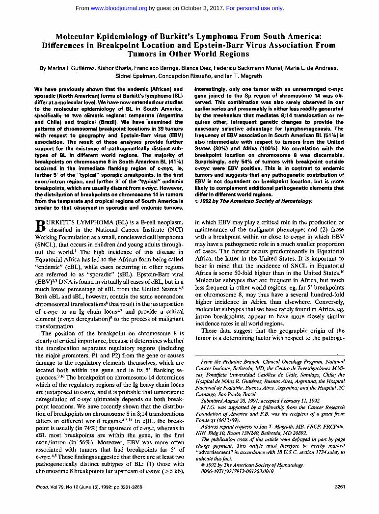
Molecular Epidemiology of Burkitt’s Lymphoma From South America: Differences in Breakpoint Location and Epstein-Barr Virus Association From Tumors in Other World Regions By Marina I. Gutierrez, Kishor Bhatia, Francisco Barriga, Blanca Diez, Federico Sackmann Muriel, Maria L. de Andreas, Sidnei Epelman, Concepcion Risueiio, and Ian T. Magrath We have previously shown that the endemic (African) and sporadic (North American) forms of Burkitt’s lymphoma (BL) differ at a molecular level. We have now extended our studies to the molecular epidemiology of BL in South America, specifically t o two climatic regions: temperate (Argentina and Chile) and tropical (Brazil). We have examined the patterns of chromosomal breakpoint locations in 39 tumors with respect to geography and Epstein-Barr virus (EBV) association. The result of these analyses provide further support for the existence of pathogenetically distinct sub- types of BL in different world regions. The majority of breakpoints on chromosome 8 in South American BL (41%) occurred in the immediate flanking region of c-myc, ie, further 5’ of the “typical” sporadic breakpoints, in the first exodintron region, and further 3‘ of the “typical” endemic breakpoints, which are usually distant from c-myc. However, the distribution of breakpointson chromosome 14 in tumors from the temperate and tropical regions of South America is similar to that observed in sporadic and endemic tumors. URKITT’S LYMPHOMA (BL) is a B-cell neoplasm, B classified in the National Cancer Institute (NCI) Working Formulation as a small, noncleaved cell lymphoma (SNCL), that occurs in children and young adults through- out the world.’ The high incidence of this disease in Equatorial Africa has led to the African form being called “endemic” (eBL), while cases occurring in other regions are referred to as “sporadic” (sBL). Epstein-Barr viral (EBV)2s3 DNA is found in virtually all cases of eBL, but in a much lower percentage of sBL from the United States.4~~ Both eBL and sBL, however, contain the same nonrandom chromosomal translocations6 that result in the juxtaposition of c-myc to an Ig chain 10cus~~~ and provide a critical element (c-myc deregulation)s to the process of malignant transformation. The position of the breakpoint on chromosome 8 is clearly of critical importance, because it determines whether the translocation separates regulatory regions (including the major promoters, P1 and P2) from the gene or causes damage to the regulatory elements themselves, which are located both within the gene and in its 5’ flanking se- quence~.~,~~ The breakpoint on chromosome 14 determines which of the regulatory regions of the Ig heavy chain locus are juxtaposed to c-myc, and it is probable that tumorigenic deregulation of c-myc ultimately depends on both break- point locations. We have recently shown that the distribu- tion of breakpoints on chromosome 8 in 8;14 translocations differs in different world regi0ns.~,5Jl In eBL, the break- point is usually (in 74%) far upstream of c-myc, whereas in sBL most breakpoints are within the gene, in the first exonlintron (in 56%). Moreover, EBV was more often associated with tumors that had breakpoints far 5’ of c-my~.~.~ These findings suggested that there are at least two pathogenetically distinct subtypes of BL (1) those with chromosome 8 breakpoints far upstream of c-myc (> 5 kb), Interestingly, only one tumor with an unrearranged c-myc gene joined to the Sp region of chromosome 14 was ob- served. This combination was also rarely observed in our earlier series and presumably is either less readily generated by the mechanism that mediates 8;14 translocation or re- quires other, infrequent genetic changes to provide the necessary selective advantage for lymphomagenesis. The frequency of E8V association in South American BL (51%) is also intermediate with respect t o tumors from the United States (30%) and Africa (100%). No conelation with the breakpoint location on chromosome 8 was discernable. Surprisingly, only 54% of tumors with breakpoint outside c-myc were EBV positive. This is in contrast to endemic tumors and suggests that any pathogenetic contribution of EBV is not dependent on breakpoint location, but is more likely to complement additional pathogenetic elements that differ in different world regions. 8 1992 by The American Society of Hematology. in which EBV may play a critical role in the production or maintenance of the malignant phenotype; and (2) those with a breakpoint within or close to c-myc in which EBV may have a pathogenetic role in a much smaller proportion of cases. The former occurs predominantly in Equatorial Africa, the latter in the United States. It is important to bear in mind that the incidence of SNCL in Equatorial Africa is some 50-fold higher than in the United Stateslo Molecular subtypes that are frequent in Africa, but much less frequent in other world regions, eg, far 5’ breakpoints on chromosome 8, may thus have a several hundred-fold higher incidence in Africa than elsewhere. Conversely, molecular subtypes that we have rarely found in Africa, eg, intron breakpoints, appear to have more closely similar incidence rates in all world regions. These data suggest that the geographic origin of the tumor is a determining factor with respect to the pathoge- From the Pediatric Branch, Clinical Oncology Program, National Cancer Institute, Bethesda, MD; the Centro de Investigaciones Mkdi- cas, Pontijicia Universidad Catdica de Chile, Santiago, Chile; the Hospital de Nirios R. Gutiirrez, Buenos Aires, Argentinind; the Hospital NacionaI de Pediam’a, Buenos Aires, Argentina; and the Hospital AC Camargo, Sa0 Paulo, Brazil. Submitted August 28,1991; accepted February 11,1992. M.I.G. was supported by a fellowship from the Cancer Research Foundation of America and F.B. was the recipient of a grant from Fondecyt (0612189). Address reprint requests to Ian T. Magrath, MB, FRCP, FRCPath, NIH, Bldg 10, Room 13N240, Bethesda, MD 20892. The publication costs of this article were defrayed in part by page charge payment. This article must therefore be hereby marked “advertisement” in accordance with 18 U.S.C. section 1734 sole4 to indicate this fact. 6 1992 by The American Society of Hematology. 0006-4971I921 7912-OO12$3.oO/0 Blood, Vol79, No 12 (June 15). 1992: pp3261-3266 3261 For personal use only. on October 3, 2017. by guest www.bloodjournal.org From
Transcript of Molecular Epidemiology of Burkitt’s Lymphoma From … · exodintron region, and further 3‘ of...
Molecular Epidemiology of Burkitt’s Lymphoma From South America: Differences in Breakpoint Location and Epstein-Barr Virus Association From
Tumors in Other World Regions
By Marina I. Gutierrez, Kishor Bhatia, Francisco Barriga, Blanca Diez, Federico Sackmann Muriel, Maria L. de Andreas, Sidnei Epelman, Concepcion Risueiio, and Ian T. Magrath
We have previously shown that the endemic (African) and sporadic (North American) forms of Burkitt’s lymphoma (BL) differ at a molecular level. We have now extended our studies to the molecular epidemiology of BL in South America, specifically to two climatic regions: temperate (Argentina and Chile) and tropical (Brazil). We have examined the patterns of chromosomal breakpoint locations in 39 tumors with respect to geography and Epstein-Barr virus (EBV) association. The result of these analyses provide further support for the existence of pathogenetically distinct sub- types of BL in different world regions. The majority of breakpoints on chromosome 8 in South American BL (41%) occurred in the immediate flanking region of c-myc, ie, further 5’ of the “typical” sporadic breakpoints, in the first exodintron region, and further 3‘ of the “typical” endemic breakpoints, which are usually distant from c-myc. However, the distribution of breakpoints on chromosome 14 in tumors from the temperate and tropical regions of South America is similar to that observed in sporadic and endemic tumors.
URKITT’S LYMPHOMA (BL) is a B-cell neoplasm, B classified in the National Cancer Institute (NCI) Working Formulation as a small, noncleaved cell lymphoma (SNCL), that occurs in children and young adults through- out the world.’ The high incidence of this disease in Equatorial Africa has led to the African form being called “endemic” (eBL), while cases occurring in other regions are referred to as “sporadic” (sBL). Epstein-Barr viral (EBV)2s3 DNA is found in virtually all cases of eBL, but in a much lower percentage of sBL from the United States.4~~ Both eBL and sBL, however, contain the same nonrandom chromosomal translocations6 that result in the juxtaposition of c-myc to an Ig chain 1 0 c u s ~ ~ ~ and provide a critical element (c-myc deregulation)s to the process of malignant transformation.
The position of the breakpoint on chromosome 8 is clearly of critical importance, because it determines whether the translocation separates regulatory regions (including the major promoters, P1 and P2) from the gene or causes damage to the regulatory elements themselves, which are located both within the gene and in its 5’ flanking se- q u e n c e ~ . ~ , ~ ~ The breakpoint on chromosome 14 determines which of the regulatory regions of the Ig heavy chain locus are juxtaposed to c-myc, and it is probable that tumorigenic deregulation of c-myc ultimately depends on both break- point locations. We have recently shown that the distribu- tion of breakpoints on chromosome 8 in 8;14 translocations differs in different world regi0ns.~,5Jl In eBL, the break- point is usually (in 74%) far upstream of c-myc, whereas in sBL most breakpoints are within the gene, in the first exonlintron (in 56%). Moreover, EBV was more often associated with tumors that had breakpoints far 5’ of c - m y ~ . ~ . ~ These findings suggested that there are at least two pathogenetically distinct subtypes of B L (1) those with chromosome 8 breakpoints far upstream of c-myc (> 5 kb),
Interestingly, only one tumor with an unrearranged c-myc gene joined to the Sp region of chromosome 14 was ob- served. This combination was also rarely observed in our earlier series and presumably is either less readily generated by the mechanism that mediates 8;14 translocation or re- quires other, infrequent genetic changes to provide the necessary selective advantage for lymphomagenesis. The frequency of E8V association in South American BL (51%) is also intermediate with respect t o tumors from the United States (30%) and Africa (100%). No conelation with the breakpoint location on chromosome 8 was discernable. Surprisingly, only 54% of tumors with breakpoint outside c-myc were EBV positive. This is in contrast to endemic tumors and suggests that any pathogenetic contribution of EBV is not dependent on breakpoint location, but is more likely to complement additional pathogenetic elements that differ in different world regions. 8 1992 by The American Society of Hematology.
in which EBV may play a critical role in the production or maintenance of the malignant phenotype; and (2) those with a breakpoint within or close to c-myc in which EBV may have a pathogenetic role in a much smaller proportion of cases. The former occurs predominantly in Equatorial Africa, the latter in the United States. It is important to bear in mind that the incidence of SNCL in Equatorial Africa is some 50-fold higher than in the United Stateslo Molecular subtypes that are frequent in Africa, but much less frequent in other world regions, eg, far 5’ breakpoints on chromosome 8, may thus have a several hundred-fold higher incidence in Africa than elsewhere. Conversely, molecular subtypes that we have rarely found in Africa, eg, intron breakpoints, appear to have more closely similar incidence rates in all world regions.
These data suggest that the geographic origin of the tumor is a determining factor with respect to the pathoge-
From the Pediatric Branch, Clinical Oncology Program, National Cancer Institute, Bethesda, MD; the Centro de Investigaciones Mkdi- cas, Pontijicia Universidad Catdica de Chile, Santiago, Chile; the Hospital de Nirios R. Gutiirrez, Buenos Aires, Argentinind; the Hospital NacionaI de Pediam’a, Buenos Aires, Argentina; and the Hospital AC Camargo, Sa0 Paulo, Brazil.
Submitted August 28,1991; accepted February 11,1992. M.I.G. was supported by a fellowship from the Cancer Research
Foundation of America and F.B. was the recipient of a grant from Fondecyt (0612189).
Address reprint requests to Ian T. Magrath, MB, FRCP, FRCPath, NIH, Bldg 10, Room 13N240, Bethesda, MD 20892.
The publication costs of this article were defrayed in part by page charge payment. This article must therefore be hereby marked “advertisement” in accordance with 18 U.S.C. section 1734 sole4 to indicate this fact. 6 1992 by The American Society of Hematology. 0006-4971 I921 7912-OO12$3.oO/0
Blood, Vol79, No 12 (June 15). 1992: pp3261-3266 3261
For personal use only.on October 3, 2017. by guest www.bloodjournal.orgFrom
3262 GUTIERREZ ET AL
netic mechanism. We decided, therefore, to study BL from other world regions to determine whether the patterns of chromosomal breakpoints are similar to either eBL, or &L, or have a unique pattern. South h e r i c a is of particular interest in this regard, because it encompasses both tropical and temperate regions, reproducing the climatic conditions in which eBL and sBL are found, although differing in other important aspects, eg, socioeconomicall~ and ethnicah’, from both Africa and the United States. We report here the results of an examination of the molecular characteristics and EBV association of 39 tumors from Argentina, Chile, and Brazil.
de Nifios “Ricardo GutiCrrez” and Hospital Nacional de Pediatria “Juan Garrahan,” both in Buenos fires, Argentina (17 tumors); the Hospital de Nifios Roberto del Rio and Hospital Infantil Luis Calvo Mackenna in Santiago, Chile (10 tumors); the Hospital AC Camargo, Sao Paulo, Brazil, and CIIH Domingos Boldrini, Campi- nas, Brazil (12 tumors). The samples were frozen at -70°C immediately after surgery. A diagnosis was made in each center according to standard histopathologic criteria.
High molecular weight DNA was prepared from each tumor by cell lysis, proteinase K digestion, phenol- chloroform extractions, and ethanol precipitation as described by Maniatis et a1.12
DNAs were digested with appropriate restriction enzymes (BamHI, EcoRI, HindIII, Pst I, Pvu 11, and Sma I) fractionated in 0.8% agarose gels and transferred to nylon membranes according to standard Southern blotting procedures. The blots were hybrid- ized to radiolabeled 32P-probes, using methods previously de- scribed.12
Molecular anafysis.
MATERIALS AND METHODS
We have studied 39 SNCLs from South America (Table 1). The biopsies were obtained from a number of institutions: the Hospital
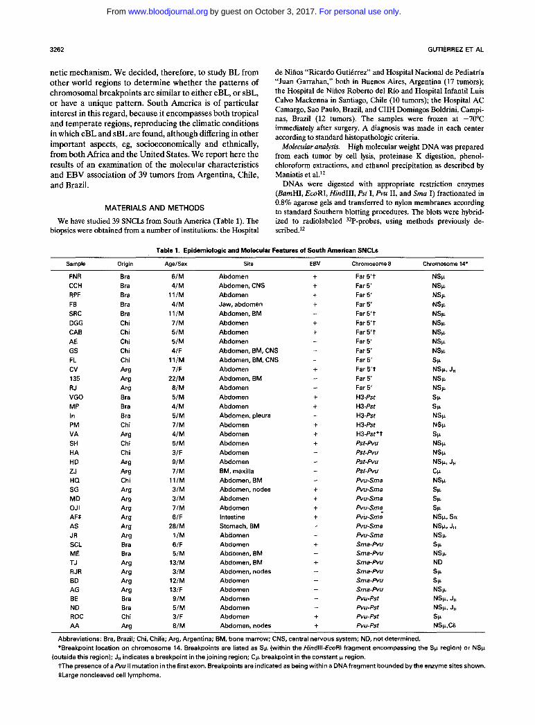
Table 1. Epidemiologic and Molecular Features of South American SNCLs
Sample Origin AgelSex Site EBV Chromosome 8 Chromosome 14’
FNR Bra 6/M Abdomen + Far 5 ’ t NSP CCH Bra 4/M Abdomen, CNS + Far 5‘ NSlr RPF Bra 11/M Abdomen + Far 5‘ NSk FB Bra 4/M Jaw, abdomen + Far 5‘ NSP SRC Bra 11/M Abdomen, BM Far 5’ t NSP DGG Chi JIM Abdomen + Far 5‘t NSP CAB Chi 5/M Abdomen + Far 5‘t NSF AE Chi 5/M Abdomen Far 5‘ NS~L GS Chi 4/F Abdomen, BM, CNS Far 5‘ NSP FL Chi 11/M Abdomen, BM, CNS Far 5‘ SP cv Arg J/F Abdomen + Far 5’ t NSP, JH 135 Arg 22/M Abdomen, BM Far 5 NSP RJ Arg 8/M Abdomen Far 5’ NSP VGO Bra 5/M Abdomen + H 3 - f ~ SP MP Bra 4/M Abdomen -t H3-Pst SP In Bra 5/M Abdomen, pleura H 3 A t NSF PM Chi JIM Abdomen + H 3 A t NSF VA Arg 4/M Abdomen + H3-P~t’t SP SH Chi 5/M Abdomen i- Pst-Pvu NSP HA Chi 3/F Abdomen Pst-Pvu NSP HD Arg ZJ Arg 7/M BM, maxilla Pst-Pvu CIL HQ Chi 11/M Abdomen, BM Pvu-Sma NSF SG Arg 3/M Abdomen, nodes + Pvu-Sma SP MD Arg 3/M Abdomen + Pvu-Sma SP OJI Arg JIM Abdomen i- Pvu-Sma SP
AS Arg JR Arg 1/M Abdomen Pvu-Sma NSF
SP SCL Bra 6/F Abdomen + ME Bra 5/M Abdomen, BM Sma-Pvu NSF TJ Arg 13/M Abdomen, BM + Sma-Pvu ND RJR Arg 3/M Abdomen, nodes Sma-Pvu SP
SP BD Arg 12/M Abdomen AG Arg 13/F Abdomen Sma-Pvu NSp BE Bra 9/M Abdomen - Pvu-Pst NSp. JH
ND Bra 5/M Abdomen - Pvu-Pst NSp, JH
ROC Chi 3/F Abdomen -t Pvu-Pst SP AA Arg 8/M Abdomen, nodes + Pvu-Pst NSp,CG
-
- - -
- -
-
- 9/M Abdomen - Pst-Pvu NSp, JH
- -
AFS Arg 6/F Intestine + Pvu-Sma NSp, Sa 28/M Stomach, BM - Pvu-Sma NSP, JH
- Sma-Pvu
-
- - Sma-Pvu -
Abbreviations: Bra, Brazil; Chi, Chile; Arg, Argentina; BM, bone marrow; CNS, central nervous system; ND, not determined. *Breakpoint location on chromosome 14. Breakpoints are listed as Sc (within the Hmdlll-EcoRI fragment encompassing the Sp region) or NSp
tThe presence of a Pvu II mutation in the first exon. Breakpoints are indicated as being within a DNAfragment bounded by the enzyme sites shown. *Large noncleaved cell lymphoma.
(outside this region); JH indicates a breakpoint in the joining region; C p breakpoint in the constant p region.
For personal use only.on October 3, 2017. by guest www.bloodjournal.orgFrom

MOLECULAR PATTERN OF SNCL IN SOUTH AMERICA 3263
The myc (first and third exon), IgH (JH, Sk, and Ck) (Fig l), and EBV (terminal repeats) probes used, as well as the Southern blotting strategy used to determine breakpoint locations, have also been previously reported by ~ 9 . 5 7 ~ ~
All tumor samples were examined for mutations in the first exon by restriction analysis. Thus, only mutations resulting in a loss of the Pvu I1 site in its 3' region would be detected.
TEMPERATE
& 9" P V t -----I1 8 8
TROPICAL RESULTS
Twenty-seven of the tumors analyzed were from the temperate climatic region (17 from Argentina and 10 from Chile) while 12 were from the tropical region (specifically Sao Paulo, Brazil). The origin, age, and sex of the patients as well as the anatomical site and a summary of the molecular characterization of their tumors are shown in Table 1. The age range was 1 to 28 years and all patients presented with abdominal lymphoma, except for two who presented with jaw tumors.
Because both Argentina and Chile are climatically simi- lar, for the purposes of the present analysis we have combined the tumors from these countries into a single group-
Although there are no cytogenetic data available, 32 of 39 tumors (82%) showed structural rearrangements that have been previously described in t(8;14) and 20 of these had definite t(8;14) at a molecular level. However, we cannot rule out the possibility of variant translocations in the remaining seven tumors that showed a non-SF non-JH breakpoint on chromosome 14 and an unaltered c-myc gene on chromosome 8.
The pattern of chromo- somal breakpoint locations differed with the geographic origin of the tumor (Fig 2). Forty-one percent (11 of 27) of tumors from the temperate region (Argentina and Chile) had breakpoints in the immediate 5' region of c-myc in a Pst I-Sma I fragment (Figs 1 and 2). In contrast, no tumors
Chromosome 8 breakpoints.
Fig 2. Distribution of breakpoint location on chromosome 8 in relation to geographic origin. Tumors from the temperate and tropical regions from South America are shown above and below the c-myc map. The (0) presence or (0) absence of EBV in each tumor is also depicted. The regions on chromosome 8 are: outside Hindlll (H) (usually far 5' but could also be far 3' in some cases); Hindlll-Pst I (Ps); Pst ISma I (S); Sma I-Pvu II (Pvl; Pvu Il-Pst 1.
from the tropical region displayed chromosomal break- points within this fragment; the majority (5 of 12 [67%]) had breakpoints further 5'.
Among the 13 tumors with a breakpoint outside the HindIII fragment encompassing c-myc, all except one were also rearranged outside the 5' EcoRI site encompassing c-myc. The exceptional tumor (RPF, from Brazil) had a breakpoint between the 5' EcoRI and HindIII sites, only a short distance upstream of myc.
Because tumors in which c-myc is not rearranged often show mutations within the c-myc locus, sometimes observed as a loss of the Pvu I1 site in the 3' region of the first exon, we examined, by restriction enzyme analysis, the South American lymphomas for the presence of such a mutation. Five of the 13 tumors (38%) with breakpoints outside HindIII carried a Pvu I1 mutation characteristic of many eBL. One additional case (VA) showed this mutation in association with a breakpoint between HindIII and Pst I.
Of the 26 tumors that had breakpoints within the HindIII
c-myc
H
1 2 3
1 Kb
H B H E B H
1 Kb
Fig 1. Restriction maps of the human c-myc and Ig heavy chain genes. Restriction endonuclease sites are shown. H, Hindlll; Ps, Pstl; Pv, Pvu II; S, Sma I; E, EcoRI; B, &"I.
For personal use only.on October 3, 2017. by guest www.bloodjournal.orgFrom
3264 GUTIERREZ ET AL
fragment encompassing the c-myc gene, 62% (16 of 26) had breakpoints in the 5' flanking region (between the HindIII and the Sma I sites) (Fig 2). Thus, these tumors retained the capacity to transcribe c-myc messenger R N A (mRNA) from either the P1 or P2 promoter.
Determination of the loca- tion of breakpoints on chromosome 14 (Fig l) was possible in all but one tumor (Table 1). Breakpoints outside the Sp region are almost twice as frequent as Sp breaks (in the HindIII-EcoRI segment). The proportions of tumors with Sp. breakpoints from the temperate and tropical regions were 38% and 25%, respectively.
Analyses of the combination of breakpoint locations on chromosomes 8 and 14 are shown in Fig 3 and Table 2. Of the four main breakpoint combinations, R/Sp., R/NSp, and U / N S p were equally frequently observed. Only a single tumor with a transloca- tion that joined an unrearranged c-myc with a Sp. region (U/Sp) was observed.
EBV association. Overall, the frequency of EBV- positive lymphomas in South America was 51%. Tumors from either the temperate or the tropical region did not significantly differ in their association with EBV (48% and 58%, respectively), although the numbers are too small for a valid comparison between these groups to be made. As shown in Fig 4, the EBV genomes present in these tumors appeared to be monoclonal when probed with the EBV terminal repeat region. This result clearly shows that the virus was associated with the tumor cells rather than with normal cells present in the tumor sample.
Chromosome 14 breakpoints.
Breakpoint combinations.
Hind 111
1 E
%
1 myc
-11 Kb
3 myc CP
Eco RI CCH F%L MP BD
U - M
JH Sp JH Sp JH Sp JH Sp
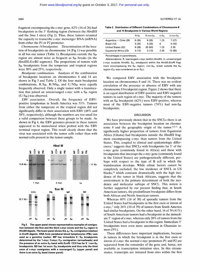
Fig 3. The upper panel shows representative examples of comigra- tion between the first and the third c-myc exons and the Cp region in Hhdlll digests. The lower panel shows the J,/Sp comigration (dashes) in EcoRl digests. DNA from peripheral blood lymphocytes (PBL) was used as a germline marker. MP has immediate 5'/Sp breakpoints determined by the l-myc/3-myc/Cp comigration (arrowheads) and the presence of an extra Sp band with €coRI. CCH has far B'/non-Sp breakpoints. BD has 1st exon/+ breakpoints and thus only the third exon of c-myc comigrates with a rearranged Cp (upper panel) and there is an extra Sp band (lower panel).
Table 2. Distribution of Different Combinations of Chromosome 8 and 14 Breakpoints in Various World Regions
RlSp Rlnon-Sp UlSp Ulnon-Sp
Argentina + Chile (26) 9 (35) 9 (35) 1 (3) 7 (27) Brazil (12) 3 (25) 4 (33) 0 5 (42) United States (32) 9 (28) 20 (63) 1 (3) 2 (6) Equatorial Africa (23) 3 (13) 3 (13) 2 (9) 15 (65)
Percentages in parentheses. Abbreviations: R, rearranged c-myc (within Hindlll); U, unrearranged
c-myc (outside Hindlll); Sp, breakpoint within the Hindlll-EcoRI frag- ment encompassing the Sp region; non-Sp, break outside the Sp region (Cp was considered as an Sp).
We compared EBV association with the breakpoint location on chromosomes 8 and 14. There was no evident correlation of the presence or absence of EBV with any chromosome 8 breakpoint region. Figure 2 shows that there is an equal distribution of EBV-positive and EBV-negative tumors in each region of c-myc. The majority of the tumors with an Sp breakpoint (62%) were EBV-positive, whereas most of the EBV-negative tumors (74%) had non-Sp. breakpoints.
DISCUSSION
We have previously shown that in the SNCLs there is an association between the breakpoint location on chromo- some 8 and the geographic origin of the tumor.'"J' A significantly higher proportion of tumors from Equatorial Africa (Ghana) had breakpoints outside the HindIII frag- ment encompassing c-myc than tumors from the United States. This, coupled to clinical and epidemiologic differ- ences,' suggests that SNCLs with breakpoints far 5' of the c-myc gene (commonly found in Africa) and those with breakpoints that interrupt the c-myc gene (commonly found in the United States) are pathogenetically different, per- haps with respect to the type of B cell in which the translocation develops. While ethnic factors cannot be completely excluded, the low incidence of SNCL in US blacks,14 which contrasts dramatically with the high inci- dence of the tumor in black Africans, suggests that the environment is the primary determinant of both the inci- dence and molecular subtype of SNCL. This notion is further supported by our present finding that, in South American tumors, the predom'inant breakpoint differs from both African and North American tumors.
Whereas 60% (18 of 30) of sporadic tumors from the United States had breakpoints in the first exon or intron of c-myc,s only 26% (10 of 39) of tumors from South America had similar breakpoints. O n the other hand, 16 of 39 (41%) of South American tumors had a breakpoint in the immedi- ate 5' region of c-myc, whereas only 28% of tumors from the United States had a breakpoint in this region. Immediate 5' breakpoints were even more uncommon in Ghanaian tu- mors (9%).
These differences have important implications, because in tumors in which the breakpoint is in the first exon or intron of c-myc the normal c-myc promoters P1 and P2 are separated from the remainder of the gene and, hence, not available as transcription initiation sites. In this circum- stance, transcripts are initiated from sites within the first
For personal use only.on October 3, 2017. by guest www.bloodjournal.orgFrom

MOLECULAR PAlTERN OF SNCL IN SOUTH AMERICA 3265
Fig 4. Representative South- ern blots showing EBV mono- clonality. DNAs were digested with BamHl and hybridized with the viral terminal repeat probe. DNA from peripheral blood lym- phocytes (PBL) sewed as a nega- tive control.
> 11 Kb -
intron.lS Moreover, in such tumors, the bulk of the regula- tory region of the gene is deleted by the translocation. As many as 60% of North American SNCLs fall into this molecular category. In contrast, our analysis shows that the majority of tumors from South America (74%) have re- tained the normal P1 and P2 promoters. Breakpoints outside the HindIII fragment that encompasses c-myc (and, hence, outside the known regulatory region of c-myc) were observed primarily in Africa (74%), but also quite fre- quently in South America (33%). Such breakpoints were uncommonly observed in tumors from the United States (9%). These data suggest that there may be three main molecular subtypes of SNCL, and that it is probable that each of the major breakpoint regions (far S', immediate 5', and first exon/intron, which predominate in Ghana, South America, and the United States, respectively) results in a different mechanism of c-myc deregulation. Presumably, the environmental conditions in each region are particu- larly conducive to the development of the molecular sub- type of SNCL that predominates there.
The distribution of breakpoints on chromosome 14 ob- served in South American lymphomas is similar to that previously described for sporadic tumors? 30% of breaks are within the Sp region. The breakpoint on chromosome 14 may be of considerable importance to an understanding of the mechanism of deregulation of c-myc, because break- points are either upstream or downstream of the 5' intronic enhancer within the heavy chain locus, such that this regulatory sequence may or may not be juxtaposed to c-myc. Indeed, it is likely that it is the combination of breakpoint locations on chromosomes 8 and 14 that is crucial to the deregulation of c-myc.
SNCL can be subdivided on the basis of the four simplest combinations of breakpoints on chromosomes 8 and 14 (Table 2). From our data it appears that the combination of an unrearranged myc gene (within HindIII) with a switch breakpoint on chromosome 14 is very uncommon, occurring in only 4 tumors (2 African, 1 North American and 1 Chilean) among 93 tumors (4%) that we have examined to date. This may result from concomitant structural con- straints with respect to the occurrence of this combination, or the necessity of other, rare genetic changes, to cooperate with this combination in providing the necessary growth advantage to the cell. Two of the other combinations examined (Ulnon-Sp and Rlnon-Sp) appear to occur at high frequency in African and North American tumors, respectively. However, in the South American lymphomas, these breakpoint combinations appear equally distributed.
The reason for the association of breakpoint locations,
and particularly of specific breakpoint combinations, with geographic regions is unknown. It seems most probable that environmental factors, acting via the immune system, are responsible for causing expansions of slightly different B-cell populations (presumably with respect to the stage of differentiation), each more or less susceptible to the devel- opment of chromosome breaks at specific locations. How- ever, an alternative explanation is that different c-myc-Ig translocations must cooperate with different genetic abnor- malities, which are environmentally determined in bringing about deregulated growth.
In addition to differences in chromosomal breakpoint location among the regions examined, the proportion of tumors associated with EBV also differs. As is the case for breakpoint location, EBV association is also intermediate in frequency in South America. We have previously drawn attention to an apparent pattern in the breakpoint location on chromosome 8 and EBV as~ociation.~.~ Tumors with breakpoint locations outside the HindIII fragment are nearly always EBV associated, and such tumors are more frequent in Equatorial Africa. Breakpoints within the transcriptional unit of the gene, however, which occur predominantly in the United States, are equally likely to be EBV negative or positive. In South America, we have not found an association between EBV and a specific break- point location (Fig 2). Interestingly, a much higher propor- tion of tumors with a breakpoint far 5' of c-myc are EBV negative in South America (6 of 13) than in Africa (0 of 17). This raises the possibility that there are two types of SNCL with breakpoints far outside the c-myc gene, distinguished on the basis of EBV association. Moreover, South Ameri- can tumors with a breakpoint in the small region immedi- ately 5' of the c-myc promoters can be subdivided into those associated with EBV and EBV-negative tumors, which contrasts with the North American tumors, in which imme- diate 5' breaks (Pst I-Sma I) were invariably EBV negative? These findings indicate that, if there is an association of EBV with a breakpoint region, it is not a simple relation- ship. Perhaps the function subserved by EBV can be replaced by another factor or perhaps the precise break- point on chromosome 14 is very important; most of the EBV-negative tumors in South America (4 of 19 [76%]) and in the United States (13 of 20 [65%]) have non-SF break- points and probably, therefore, retain the intronic enhancer.
However, there does appear to be a gradient of EBV association extending from Africa through South America to the United States-a gradient that also corresponds to socioeconomic level and such factors as the frequency of infections in children.16
For personal use only.on October 3, 2017. by guest www.bloodjournal.orgFrom

3266 GUTIERREZ ET AL
Our findings strongly support the likelihood that there are several molecular subtypes of SNCL, each of which is more or less likely to occur in the geographic regions studied to date. It is interesting that the increased incidence of Ghanaian SNCL compared with SNCL in sporadic regions is largely, if not entirely, accounted for by tumors with far 5’ breakpoints on chromosome 8. It will be
important to determine the incidence of SNCL in various regions of South America, because this will permit the incidence of the molecular subtypes to be determined.
ACKNOWLEDGMENT
We thank the Fogarty International Center for developing this collaboration.
REFERENCES 1. Magrath I T Burkitt’s lymphoma as a human tumor model:
New concepts in etiology and pathogenesis, in Pochedly C (ed): Pediatric Hem/Oncology Reviews. New York, NY, Praeger, 1985,
2. Epstein MA, Achong BG, Barr YM: Virus particles in cultured lymphoblasts from Burkitt’s lymphoma. Lancet 1:702, 1964
3. Neri A, Barriga F, Inghirami G, Knowles DM, Neequaye J, Magrath IT, Dalla-Favera R: Epstein-Barr virus infection precedes clonal expansion in Burkitt’s and acquired immunodeficiency syndrome-associated lymphoma. Blood 771092,1991
4. Barriga F, Kiwanuka J, Alvarez-Mon M, Shiramizu B, Huber B, Levine P, Magrath I T Significance of chromosome 8 breakpoint location in Burkitt’s lymphoma: Correlation with geographical origin and association with Epstein-Barr virus. Curr Top Microbiol Immunol141:128,1988
5. Shiramizu B, Barriga F, Neequaye J, Jafri A, Dalla-Favera R, Neri A, GutiCrrez MI, Levine P, Magrath I T Patterns of chromo- somal breakpoint locations in Burkitt’s lymphoma: Relevance to geography and Epstein-Barr virus association. Blood 77:1516,1991
6. Zech L, Haglund U, Nilsson K, Klein G: Characteristic chromosomal abnormalities in biopsies and lymphoid cell lines from patients with Burkitt’s and non-Burkitt’s lymphomas. Int J Cancer 1747,1976
7. Dalla-Favera R, Martinotti S, Gallo RC, Erickson J, Croce CM: Translocation and rearrangements of the c-myc oncogene
P l
locus in human undifferentiated B-cell lymphomas. Science 219: 963,1983
8. Croce CM, Nowell PC: Molecular basis of human B-cell neoplasia. Blood 652,1985
9. Hay N, Bishop JM, Levens D: Regulatory elements that modulate expression of human c-myc. Genes Dev 1:659,1987
10. Magrath I T The pathogenesis of Burkitt’s lymphoma. Adv Cancer Res 53:133,1990
11. Pellici PG, Knowles DM, Magrath IT, Dalla-Favera R: Chromosomal breakpoints and structural alteration of the c-myc locus differ in endemic and sporadic forms of Burkitt’s lymphoma. Proc Natl Acad Sci USA 83:2984,1986
12. Maniatis T, Fritsch EF, Sambrook J: Molecular Cloning: A Laboratory Manual. Cold Spring Harbor, NY, Cold Spring Harbor Laboratory, 1989, p 9.16
13. GutiCrrez MI, Barriga F, Diez B, Larripa I, Magrath I T Analisis molecular en pacientes argentinos con linfoma de Burkitt. Sangre (Barc) 3510,1990
14. Magrath I T African Burkitt’s lymphoma. Am J Pediatr Hematol Oncol13:222,1991
15. ar-Rushdi A, Nishikura K, Erikson J, Watt R, Rovera G, Croce CM: Differential expression of the translocated and the untranslocated c-myc oncogene in Burkitt lymphoma. Science 222:390,1983
16. Magrath IT: Malignant non-Hodgkin’s lymphomas, in Pizzo P, Poplack D (eds): Pediatric Oncology. Philadelphia, PA, Lippin- cott, 1989, p 415
For personal use only.on October 3, 2017. by guest www.bloodjournal.orgFrom
1992 79: 3261-3266
IT MagrathMI Gutierrez, K Bhatia, F Barriga, B Diez, FS Muriel, ML de Andreas, S Epelman, C Risueno and from tumors in other world regionsdifferences in breakpoint location and Epstein-Barr virus association Molecular epidemiology of Burkitt's lymphoma from South America:
http://www.bloodjournal.org/content/79/12/3261.full.htmlUpdated information and services can be found at:
Articles on similar topics can be found in the following Blood collections
http://www.bloodjournal.org/site/misc/rights.xhtml#repub_requestsInformation about reproducing this article in parts or in its entirety may be found online at:
http://www.bloodjournal.org/site/misc/rights.xhtml#reprintsInformation about ordering reprints may be found online at:
http://www.bloodjournal.org/site/subscriptions/index.xhtmlInformation about subscriptions and ASH membership may be found online at:
Copyright 2011 by The American Society of Hematology; all rights reserved.Society of Hematology, 2021 L St, NW, Suite 900, Washington DC 20036.Blood (print ISSN 0006-4971, online ISSN 1528-0020), is published weekly by the American
For personal use only.on October 3, 2017. by guest www.bloodjournal.orgFrom