MOLECULAR DYNAMIC SIMULATION STUDIES OF THREE...
47
MOLECULAR DYNAMIC SIMULATION STUDIES OF THREE NOVEL GERSTMANN-STRAUSSLER-SCHEINKER AND CREUTZFELDT-JACOB PRION MUTATIONS ASHRAF FADHIL JOMAH UNIVERSITI TEKNOLOGI MALAYSIA
Transcript of MOLECULAR DYNAMIC SIMULATION STUDIES OF THREE...
1
MOLECULAR DYNAMIC SIMULATION STUDIES OF THREE
NOVEL GERSTMANN-STRAUSSLER-SCHEINKER AND
CREUTZFELDT-JACOB PRION MUTATIONS
ASHRAF FADHIL JOMAH
UNIVERSITI TEKNOLOGI MALAYSIA
4
MOLECULAR DYNAMIC SIMULATION STUDIES OF THREE
NOVEL GERSTMANN-STRAUSSLER-SCHEINKER
AND CREUTZFELDT-JACOB PRION MUTATIONS
ASHRAF FADHIL JOMAH
A thesis submitted in partial fulfilment of the
requirements for the award of the degree of
Doctor of Philosophy (Bioscience)
Faculty of Biosciences and Medical Engineering
Universiti Teknologi Malaysia
FEBRUARY 2016
iii
To my wonderful parents and my sister, brother
A constant source of inspiration,
The object of my love and admiration,
And the driving force behind my motivation.
No words can express my extreme gratitude
for your unconditional love, unwavering support,
and infinite patience.
Thank you for everything you have done for me,
you are the role models I aspire to be.
iv
ACKNOWLEDGEMENT
Studying with Associate Prof DR MOHD SHAHIR SHAMSIR OMAR has
been a true learning experience, and I hope to continue applying the skills I learned
from him in my future career. I am deeply grateful DR MOHD SHAHIR that you
have introduced me to the world of prions and computational biology, and opened
my eyes to the bioinformatic research potential in these fields. Thank you for your
continuous encouragement and guidance as I explored different projects and learned
new skills. Your patience, dedication, and willingness to support me, scientifically
and financially, have helped me grow as a bioinformatician. I am deeply indebted to
your mentorship.
I also wish to extend my full appreciation to DR SALEHHUDDIN
HAMDAN as co-supervisor for the supporting and motivation.
I am also grateful for the Universiti Teknologi Malaysia for providing the
financial support for this research, beside to their supporting by the international
doctorate fellowship (IDF) during this research. My thanks are also to the staff,
faculty members, and technicians of the faculty of bioscience and biomedical
engineering, Universiti Teknologi Malaysia, who contributed in this research.
I am most grateful to my family and close friends for their infinite support,
patience, and encouragements during these years.
v
ABSTRACT
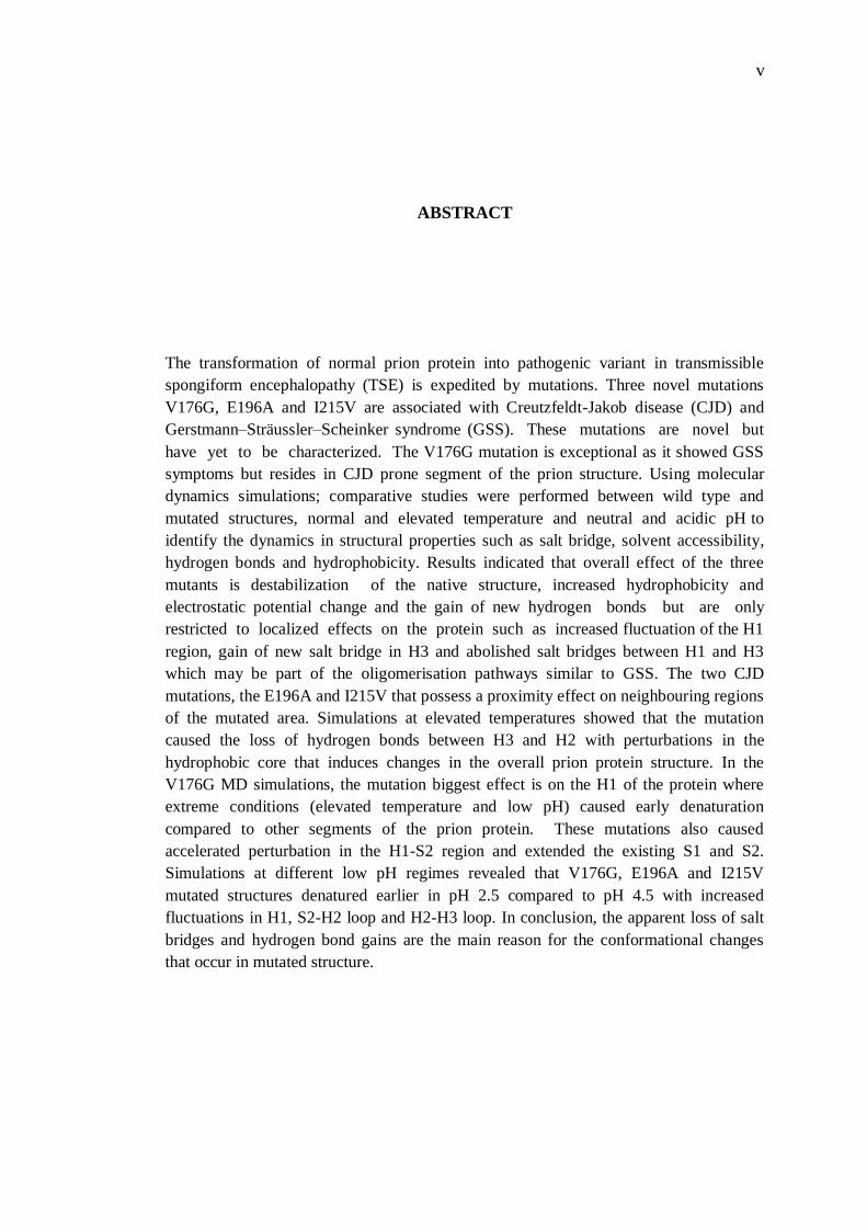
The transformation of normal prion protein into pathogenic variant in transmissible
spongiform encephalopathy (TSE) is expedited by mutations. Three novel mutations
V176G, E196A and I215V are associated with Creutzfeldt-Jakob disease (CJD) and
Gerstmann–Sträussler–Scheinker syndrome (GSS). These mutations are novel but
have yet to be characterized. The V176G mutation is exceptional as it showed GSS
symptoms but resides in CJD prone segment of the prion structure. Using molecular
dynamics simulations; comparative studies were performed between wild type and
mutated structures, normal and elevated temperature and neutral and acidic pH to
identify the dynamics in structural properties such as salt bridge, solvent accessibility,
hydrogen bonds and hydrophobicity. Results indicated that overall effect of the three
mutants is destabilization of the native structure, increased hydrophobicity and
electrostatic potential change and the gain of new hydrogen bonds but are only
restricted to localized effects on the protein such as increased fluctuation of the H1
region, gain of new salt bridge in H3 and abolished salt bridges between H1 and H3
which may be part of the oligomerisation pathways similar to GSS. The two CJD
mutations, the E196A and I215V that possess a proximity effect on neighbouring regions
of the mutated area. Simulations at elevated temperatures showed that the mutation
caused the loss of hydrogen bonds between H3 and H2 with perturbations in the
hydrophobic core that induces changes in the overall prion protein structure. In the
V176G MD simulations, the mutation biggest effect is on the H1 of the protein where
extreme conditions (elevated temperature and low pH) caused early denaturation
compared to other segments of the prion protein. These mutations also caused
accelerated perturbation in the H1-S2 region and extended the existing S1 and S2.
Simulations at different low pH regimes revealed that V176G, E196A and I215V
mutated structures denatured earlier in pH 2.5 compared to pH 4.5 with increased
fluctuations in H1, S2-H2 loop and H2-H3 loop. In conclusion, the apparent loss of salt
bridges and hydrogen bond gains are the main reason for the conformational changes
that occur in mutated structure.
vi
ABSTRAK
Transformasi protein prion yang normal ke varian yang patogenik di dalam
penyakit berjangkit seperti spongiform encephalopathy (TSE) disebabkan oleh
mutasi.Tiga mutasi baru V176G, E196A dan I215V telah dikaitkan dengan penyakit
Creutzfeldt-Jakob (CJD) dan sindrom Gerstmann-Sträussler-Scheinker (GSS). Ketiga-
tiga mutasi ini adalah unik dan masih belum di perincikan setakat ini. Mutasi V176G
adalah luar biasa kerana ia menunjukkan tanda-tanda penyakit GSS tetapi kedudukan
mutasi berada di dalam bahagian protin yang menyebabkan penyakit CJD. Menggunakan
simulasi molekul dinamik; kajian perbandingan telah dijalankan antara struktur
normal dan struktur bermutasi, pada suhu normal dan tinggi dan pada pH neutral dan
berasid untuk mengenal pasti perubahan dinamik pada sifat-sifat struktur seperti
jambatan garam, akses pelarut, ikatan hidrogen dan sifat kehidrofobikan.Keputusan
menunjukkan bahawa kesan tiga mutan tersebut adalah ketidakstabilan struktur
asal, meningkat kehidrofobikan dan perubahan potensi elektrostatik dan
pertambahan ikatan hidrogen baru. Walau bagaimanapun, mutasi ini tidak
menyebabkan perubahan yang ketara kepada struktur secara keseluruhan.Ketidakstabilan
yang disebabkan oleh mutasi ini mungkin telah menyebabkan kesan setempat pada
struktur protein seperti peningkatan gerakan dinamik di bahagian H1, pertambahan
jambatan garam baru di H3 dan pemansuhan jambatan garam antara H1 dan H3 yang
mungkin boleh menjadi sebahagian daripada laluan penwujudan oligomer yang
menentukan profil patologi yang unik pada penyakit GSS. Kedua- dua mutasi, E196A
dan I215V telah menghasilkan manifestasi simtom klinikal yang serupa dengan CJD
telah terbukti mempunyai kesan setempat di sekita jujukan asid amino berdekatan
kawasan mutasi. Simulasi pada suhu tinggi menunjukkan bahawa mutasi telah
menyebabkan kehilangan ikatan hidrogen antara H3 dan H2 beserta gangguan di dalam
teras hidrofobik yang mendorong perubahan di dalam struktur protein prion secara
keseluruhan. Sebahagian dari S2-H2 dan H2-H3 telah diketahui sebagai sangat mudah
dipengaruhi oleh perubahan asid amino, oleh itu mutasi ini telah menyebabkan kesan
yang besar ke atas struktur. Jambatan garam baru juga dibentuk di dalam teras
hidrofobik. Dalam simulasi V176G, kesan mutasi yang paling besar adalah pada helik
H1 di mana keadaan yang melampau (suhu tinggi dan pH rendah) telah menyebabkan
penyahaslian struktur lebih awal berbanding segmen lain protein tersebut. Mutasi ini
juga menyebabkan gangguan dipermudahkan di bahagian H1-S2 dan memanjangan
jujukan S1 dan S2 yang sedia ada. Simulasi pada keadaan pH yang rendah menunjukkan
bahawa V176G, E196A dan I215V akan didenaturkan lebih mudah pada pH 2.5
berbanding pH 4.5. Ini akan meningkatkan dinamik H1, bahagian S2-H2 dan gelung
H2-H3.Kesimpulannya,kehilangan ketara jambatan garam dan perubahan ikatan
hydrogen adalah sebab utama kepada peningkatan dinamik dan perubahan struktur
mutasi.
vii
TABLE OF CONTENTS
CHAPTER TITLE PAGE
DECLARATION ii
DEDICATION iii
ACKNOWLEDGEMENT iv
ABSTRACT v
ABSTRAK vi
TABLE OF CONTENTS vii
LIST OF TABLES xi
LIST OF FIGURES xii
LIST OF ABBREVIATIONS xx
LIST OF SYMBOLS xxii
LIST OF APPENDICES xxiii
1 INTRODUCTION 1
1.1 Overview 1
1.2 Problem Statement 3
1.3 Objectives of the Research 4
1.4 Scope of the Research 5
2 LITERATURE REVIEW 6
2.1 The Prion-Only Hypothesis 6
2.2 Toxicity 7
2.3 Structural and Physical Properties 8
2.4 Pathogenic Mutations 10
viii
2.4.1 Disease-linked Mutations 10
2.4.2 Pathology 11
2.4.3 Location of the Mutated Residues in PrPC
Structure 12
2.4.4 Mutations in the Flexible N-terminal
Region 12
2.4.5 Mutations in the Globular Domain 13
2.4.6 Mutations cause GSS 13
2.4.7 Mutations cause CJD 15
2.5 Novel mutations (V176G, E196A and I215V) 17
2.6 Molecular Dynamics Simulation in Biology 18
2.7 Principles 19
2.8 Scope, Limitations, and Variations 20
2.9 GROMACS Overview 22
2.10 Molecular Dynamics of Prion Proteins 23
2.11 Elevated Temperature 23
2.12 Influence of pH 24
2.13 Solvent Accessible Surface Area 25
2.14 Salt Bridges 27
3 METHODOLOGY 30
3.1 Flowchart of the Process 30
3.2 Sequence Retrieval and Selection of Wild-Type
Structure 31
3.3 Mutant Structure Construction 31
3.4 Mutant Structure Construction in Low pH 32
3.5 Molecular Dynamics Simulation 33
3.5.1 Conformational Changes and Overall
Dynamics Behaviour 34
3.5.2 Evolution of Secondary Structure 34
3.5.3 Hydrogen Bond 35
3.5.4 Salt Bridge 35
ix
3.5.5 Hydrophobicity and Solvent Accessible
Surface Area (SASA) 36
3.5.6 Visualization and Analysis 36
3.6 Summary of Software and Databases 37
4 RESULTS AND DISCUSSION 38
4.1 MD Simulation of All Structural Variants at 300K 38
4.1.1 Conformational Changes and Overall
Dynamics Behaviour 38
4.1.2 Evolution of Secondary Structure 41
4.1.3 Hydrogen Bond 43
4.1.4 Salt Bridges 47
4.1.5 Hydrophobicity and Solvent Accessible
Surface Area (SASA) 49
4.2 MD Simulations of E196A & I215V at Elevated
Temperature 52
4.2.1 Conformational Changes and Overall
Dynamics Behaviour 52
4.2.2 Evolution of Secondary Structures 57
4.2.3 Salt Bridges 60
4.2.4 Hydrophobicity and Solvent Accessible
Surface Area (SASA) 63
4.3 MD Simulations of V176G at Elevated
Temperature 64
4.3.1 Conformational Changes and Overall
Dynamics 64
4.3.2 Evolution of Secondary Structure 68
4.3.3 Percentage of Secondary Structure 70
4.3.4 Salt Bridges 71
4.3.5 Hydrophobicity and Solvent Accessible
Surface Area (SASA) 74
4.4 MD Simulations of V176G in Low pH 76
x
4.4.1 Conformational Changes and Overall
Dynamics Behaviour 76
4.4.2 Evolution of Secondary Structures 81
4.4.3 Percentage of Secondary Structures 82
4.4.4 Residue Interactions Surrounding
Histidine 86
4.4.4.1 Residue His140 87
4.4.4.2 Residue His155 87
4.4.4.3 Residue His177 88
4.4.4.4 Residue His187 88
4.4.5 Hydrophobicity and Solvent Accessible
Surface Area (SASA) 89
4.5 MD Simulations of E196A and I215V in Low pH 92
4.5.1 Conformational Changes and Overall
Dynamics Behaviour 92
4.5.2 Evolution of Secondary Structures 95
4.5.3 Residue Interactions Surrounding
Histidine 99
4.5.3.1 Residue His140 100
4.5.3.2 Residue His155 100
4.5.3.3 Residue His177 101
4.5.3.4 Residue His187 101
4.5.4 Hydrophobicity and Solvent Accessible
Surface Area (SASA) 101
5 CONCLUSIONS AND FUTURE WORKS 104
5.1 Conclusions 104
5.2 Future Works 105
REFERENCES 106
Appendices A-B
125-134
xi
LIST OF TABLES
TABLE NO TITLE PAGE
3.1 Server, Software and Tools 37
4.1 The SASA values of 1st, 2nd and 3rd run for WILD
TYPE and V176G mutant during the MD
trajectories 51
4.2 The formation of salt bridges throughout the
simulation for 300K (top) and 500K (bottom) 62
4.3 The values for the SASA for WILD TYPE, E196A
and I215V at 500K simulation 64
4.4 Values of the secondary structure content for the
WILD TYPE and V176G mutation in percentage
(%) 70
4.5 The formation of salt bridges throughout the
simulation for 300K (top) and 500K (bottom) 73
4.6 The average values for the SASA of the MD
trajectories for WILD TYPE and V176G mutation 76
4.7 The average values for the SASA of the MD
trajectories for WILD TYPE, V176G mutation 92
4.8 The SASA values (nm2) of 1st, 2nd and 3rd run for
WILD TYPE, E196A and I215V mutant during the
MD trajectories 103
xii
LIST OF FIGURES
FIGURE NO TITLE PAGE
2.1 The structure of the prion protein (green) with the
location of GSS associated substitution mutation in
red; A131V, S132I, A133V, Y145S, Q160S,
V176G, F198S, D202N and E211D Created by
PyMOL. H1-α-helix 3, H2-α-helix 2 and H3-α-
helix 3. S1-β-sheet 1, S2-β-sheet 2. 14
2.2 The structure of the prion protein (green) with the
location of CJD associated substitution mutation in
blue; R148H, D178N, V180I, T183A, H187R,
T188K, T193I, E196K, E200K, V203I, R208H,
V210I, E211Q, I215V Created by PyMOL. 17
2.3 Location CJD, GSS and FFI mutation on the prion
sequence. The CJD is coloured blue, GSS in
maroon and FFI in green. The location of the three
novel mutations V176G, E196A and I215V is
coloured bright red at the bottom of the diagram.
The diagram is modified from (Rossetti et al.,
2011) 18
2.4 The structure of the prion protein (1QLX) (green),
showing the location of Histidine residues that are
protonated at acidic pH; His140, His155 and
His187 H1-α-helix 3, H2-α-helix 2 and H3-α-helix
3. S1-β-sheet 1, S2-β-sheet 2. 25
xiii
2.5
Secondary structure of the prion with the
hydrophobic core (a) hydrophobic core at the loop
(b) hydrophobic core at the H2 (c) hydrophobic
core at the H3 27
2.6 The location of selected SBs on PrP; E211-R208
(SB1), E207-K204 (SB2), E207-R208 (SB3),
E200-K204(SB4), E196-R156(SB5), E146-K204
(SB6) and D178-R164(SB7) on human prion (PDB
ID: 1QLX) 28
3.1 The flowchart showing the summary of the process
of the research 30
4.1 The Cα RMSD of the different variants for residues
128–225 for (a) wild type, (b) V176G, (c) E196A
and (d) I215V 39
4.2 The Cα RMSF obtained from the molecular
dynamics simulation of the globular domain
(residues 0–103) at 300K; the lines are assigned
with black for WILD TYPE, red for V176G, green
for E196A and blue for I215V. The blue bands
represent the position of the α-helices while the red
bands represent β-sheets within the conformation
relative to the amino acid sequence 40
4.3 Secondary structure analysis based on the DSSP
algorithm for (a) WILD TYPE, (b) PrPSc mutant
V176G, (c) PrPSc mutant E196A and (d) PrPSc
mutant I215V 42
4.4 Secondary structure helices (blue) and β-sheets
(red) as a function of simulation time as
determined with DSSP. Plot (a) represents WILD
TYPE, Plot (b) represents V176G, and Plot (c)
represents E196A and Plot (d) represents I215V. 43
4.5 A cartoon representation of the prion structure
containing the V176G mutation (blue) with the
xiv
hydrogen bonds gains between residues S132-
Q217, Y149-D202 and Y162-T183 in red and
S170-Y218 and Y163-T221 loss bonds in magenta.
44
4.6 The prion structure of E196A mutation (blue) with
the hydrogen bonds gains between residues G131-
V161, H155-D202 and Y162-T183 in red and
S170-Y218 and T193-A196 loss bonds in magenta 45
4.7 The prion structure of I215V mutation (blue) with
the hydrogen bonds gain in L125-P165, Y157-
D202, M166-S222, D167-R228 in red and R136-
Y150, T193-A196 and Y128-D178 loss bonds in
magenta 45
4.8 The location of selected SBs on PrPC; E211-R208
(SB1), E207-K204 (SB2), E207-R208 (SB3),
E200-K204(SB4), E196-R156(SB5), E146-K204
(SB6) and D178-R164(SB7) on human prion (PDB
ID: 1QLX) 47
4.9 Selected SB1 (E211-R208), SB2 (E207-K204),
SB3 (E207-R208), SB4 (E200-K204), SB5 (E196-
R156), SB6 (E146-K204), SB7 (D178-R164)
involving helices H1, H2 and H3 are plotted over
simulation time for all runs. The presence of SBs is
indicated as white slits via VMD 48
4.10 The secondary structure of the prion with the
hydrophobic core (a) hydrophobic core at the loop
(b) hydrophobic core at the H2 (c) hydrophobic
core at the H3 49
4.11 The electrostatic potential surface of all mutants
obtained created using PyMOL software. 52
4.12 The RMSD of wild-type and mutants of the
structure of the globular domain (residues 128–
225) (A) at 500K for WILD TYPE (Red), E196A
xv
(Green) and I215V (Blue) (B) at 300K WILD
TYPE (Red) E196A (Green) and I215V (Blue)
53
4.13 The RMSF of the Cα backbone residues 0–103
throughout the simulation with WILD TYPE in
black, E196A in green, I215V in blue (A) at
500K (B) at 300K with β-sheets (S) and α helices
(H) represented as red and blue bands respectively 54
4.14 A cartoon representation of prion protein showing
the location of the mutated residue E196A (Green)
and location of residues that forms new hydrogen
bonds; the G131 in S1 with V161 and Y162 in S2
as well as H155 in L2 (Blue) 55
4.15 A cartoon representation of prion protein showing
the location of the mutated residue I215V (Blue)
on H3 and the opposite residue V176 on H2
(Orange) 56
4.16 Secondary structure helices (Blue) and β-sheets
(Red) as a function of simulation time determined
with DSSP: (A) WILD TYPE (B) PrPSc mutant
E196A and (C) PrPSc mutant I215V 58
4.17 Secondary structure analysis based on the DSSP
algorithm for (A) WILD TYPE, (B) PrPSc mutant
E196A and (C) PrPSc mutant I215V 59
4.18 A cartoon representation showing the location of
the seven SBs on PrPC on human prion protein
(PDB ID: 1QLX) 60
4.19 Selected SBs the E211-R208 (1), E207-K204 (2),
E207-R208 (3), E200-K204 (4), E196-R156 (5),
E146-K204 (6) and D202-R156 (7) involving
helices H1, H2 and H3 are plotted over simulation
time for all runs. The presence of SBs is indicated
as white slits via VMD 62
xvi
4.20 Secondary structure of the prion with the
hydrophobic core (A) hydrophobic core at the loop
(B) hydrophobic core at the H2 (C) hydrophobic
core at the H3 63
4.21 Location CJD, GSS and FFI mutation on the prion
sequence 65
4.22 The RMSD of Cα of the globular domain (residues
128–225) for PrPSc mutants (a) wild type, (b)
V176G during the simulation. The 1st run is black,
2nd is red and 3rd is green 66
4.23 The Cα RMSF obtained from the structure of the
globular domain (residues 128–225) for PrPSc
mutants (a) wild type, (b) V176G. 68
4.24 Secondary structure analysis based on the DSSP
algorithm for (a) WILD TYPE, (b) PrPSc mutant
V176G 69
4.25 The percentage of secondary structure of (a) WILD
TYPE, (b) V176G with the α-helices (blue) and β-
sheets (red) as a function of simulation time as
determined with the DSSP software 71
4.26 The 7 selected salt bridges numbered on the left
from top to bottom; E211-R208 SB1 (No 1), E207-
K204 SB2 (No 2), E207-R208 SB3 (No 3), E200-
K204 SB4 (No 4), E196-R156 SB5 (No 5), E146-
K204 SB6 (No 6) and D202- R156 SB7 (No 7)
which lies within the helical structures H1, H2 and
H3 are plotted over simulation time. The presence
of SBs is indicated as white slits via VMD and the
wildtype is (a) WILD TYPE and the PrPSc mutant
V176G is (b). 72
xvii
4.27
Location of 7 selected SBs on PrPC (PDB ID:
1QLX) designated as E211-R208 SB1 (No 1),
E207-K204 SB2 (No 2), E207-R208 SB3 (No 3),
E200-K204 SB4 (No 4), E196-R156 SB5 (No 5),
E146-K204 SB6 (No 6) and D202-R156 SB7 (No
7) 73
4.28 Secondary structure of the prion with the
hydrophobic core (a) hydrophobic core at the loop
(b) hydrophobic core at the H2 (c) hydrophobic
core at the H3 75
4.29 The Cα RMSD values obtained from the structure
of the globular domain (residues 128–225) for
PrPC WILD TYPE and V176G mutants (a) pH 2.5,
(b) pH 4.5 77
4.30 The Cα RMSF obtained from the structure of the
globular domain (residues 128–225) for wildtype
PrPC (black) and V176G mutant (red) at different
pH (a) pH 2.5 , (b) pH 4.5. 79
4.31 Secondary structure analysis based on the DSSP
algorithm for (left) WILD TYPE, (right) PrPSc
mutant V176G at pH 2.5 81
4.32 Secondary structure analysis based on the DSSP
algorithm for (left) WILD TYPE, (right) PrPSc
mutant V176G at pH 4.5 82
4.33 Secondary structure helices (blue) and β-sheets
(red) as a function of simulation time determined
with DSSP for WILD TYPE and V176G mutant at
low pH 83
4.34 Secondary structure helices (blue) and β-sheets
(red) as a function of simulation time determined
with DSSP to WILD TYPE, V176G at middle pH 85
4.35 Secondary structure of the prion with the
xviii
hydrophobic core (a) hydrophobic core at the loop
(b) hydrophobic core at the H2 (c) hydrophobic
core at the H3
90
4.36 The Solvent Accessible Surface Area (SASA) of
WILD TYPE and V176G mutant over time at three
different runs in pH 2.5 91
4.37 The Solvent Accessible Surface Area (SASA) of
WILD TYPE and V176G mutant over time at three
different runs in pH 4.5 91
4.38 The RMSD of the Cα backbone obtained from the
structure of the globular domain (residues 128–
225) for PrPSc mutants (a) low pH, (b) middle pH 93
4.39 The Cα RMSF obtained from the structure of the
globular domain (residues 128–225) for wild type
PrPC (black), I215V mutant (blue) and E196A
mutant (green) at different pH (a) pH 2.5, (b) pH
4.5. 95
4.40 The secondary structure evolution analysis result
based on the DSSP algorithm at pH 2.5. The result
for the WILD TYPE is on the left, PrPSc mutant
E196A in the middle and I215V on the right 96
4.41 The secondary structure evolution analysis result
based on the DSSP algorithm at pH 4.5. The result
for the WILD TYPE is on the left, PrPSc mutant
E196A in the middle and I215V on the right 97
4.42 Secondary structure helices (blue) and β-sheets
(red) as a function of simulation time determined
with DSSP to WILD TYPE, E196A mutation and
I215V mutation at low pH 97
4.43 Secondary structure helices (blue) and β-sheets
(red) as a function of simulation time determined
with DSSP to WILD TYPE, E196A mutation and
I215V mutation at middle pH 99
xix
4.44 Secondary structure of the prion with the
hydrophobic core (a) hydrophobic core at the loop
(b) hydrophobic core at the H2 (c) hydrophobic
core at the H3 102
4.45 The SASA of the hydrophobic core calculated as
the side chain SASA to WILD TYPE, E196A
mutation and I215V mutation at pH 2.5 103
4.46 The SASA of the hydrophobic core calculated as
the side chain SASA to WILD TYPE, E196A
mutation and I215V mutation at middle pH 103
xx
LIST OF ABBREVIATIONS
BSE - Bovine Spongiform Encephalopathy
CJD - Creutzfeldt-Jakob disease
CWD - Chronic Wasting Disease
DSSP - Dictionary of secondary structure of proteins
EM - Energy minimization
ER - Endoplasmatic Reticulum
fCJD - Familial Creutzfeldt-Jakob disease
FFI - Fatal familial insomnia
GSS - Gerstmann-Sträussler-Scheinker syndrome
H1 - α-helix 1
H2 - α-helix 2
H3 - α-helix 3
HB - Hydrogen bond
MD - Molecular dynamics
NMR - Nuclear Magnetic Resonance
PDB - Protein Data Bank
PME - Particle mesh Ewald
PRPC - Prion protein
PRPSC - Prion protein scrapie
RMSD - Root mean square deviations
RMSF - Root mean square fluctuations
S1 - β-sheet 1
S2 - β-sheet 2
SASA - Solvent accessible surface area
SB - Salt bridge
xxi
SC - Scrapie
TSE - Transmissible Spongiform Encephalopathies
WT - Wild type
xxii
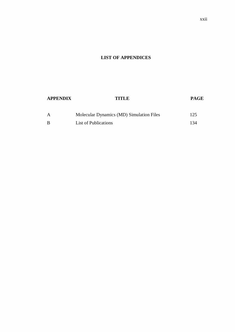
LIST OF APPENDICES
APPENDIX TITLE PAGE
A
B
Molecular Dynamics (MD) Simulation Files
List of Publications
125
134
1
CHAPTER 1
INTRODUCTION
1.1 Overview
Prions diseases are diseases that are caused by proteinacious infectious
agent (Prusiner, 1998). The term ―prion‖ was originally coined to denote the
causative agent for a group of progressive neurodegenerative disorders called
transmissible spongiform encephalopathies (TSEs), such as Creutzfeldt-Jakob
disease (CJD) in humans, bovine spongiform encephalopathy (BSE) in cattle,
chronic wasting disease (CWD) in deer and elk, and scrapie in sheep (Colby
and Prusiner, 2011). The majority of human prion diseases are sporadic with
undetermined origins while 15% are caused by heritable mutations; the
(Gerstmann-Sträussler-Scheinker syndrome (GSS), fatal familial insomnia
(FFI) and familial CJD). Prion diseases are unique as they are infectious and
can be caused by exposure to contaminated material, such as prion-infected
beef. Examples of infectious prion diseases include kuru, variant CJD and
iatrogenic CJD (iCJD). Pathological characteristics include relatively long
incubation periods, extreme vacuolation in specific regions of the brains and
general neuronal degeneration. So far, all prion associated diseases are fatal
and no cure has been found.
2
Prion studies have utilized various techniques, in-vitro, and in-vivo but
most importantly, it is studied post-mortem. The delayed onset of the disease
and invasive nature of tests have forced researcher to adopt techniques such as
molecular dynamics (MD) simulations. MD simulation studies are appropriate
as the lack of covalent modification in PrPC to PrPSc conversion. The
conformational rearrangement between the two structures provides a chance to
study purely from the structural dynamics point of view. The transition from
PrPC to PrPSc cannot be studied experimentally, especially the unfolding
transition between these two states. The hydrophobic nature of PrPSc also
prevents effective and correct structure determination by X-ray
crystallography, or even using NMR spectroscopy. Hence, MD simulation
techniques have become a viable method for studying PrPC to PrPSc
conversion. Amino acid changes that form the body of evidence of
destabilizing mutations that predisposes the organism to prion diseases also
make studying the dynamics between native and mutant PrPC a promising
approach.
Molecular dynamics have utilized extreme condition to mainly study
the effect of prion structural stabilities. Two of the main strategies used to
achieve structural changes are thermal and low pH denaturation. In thermal
denaturation, wild type and mutant PrP are simulated at elevated temperature
of 500 K (El-Bastawissy et al., 2001, Sekijima et al., 2003a, Sekijima et al.,
2003d). By elevating the temperature, the protein unfolding can be accelerated
without changing the pathway of unfolding and this enables the determination
of the unfolding pathway at minimal computational expense (Day et al., 2002).
In low pH induced denaturation, experimental evidence have showed that
acidic pH is capable to induce PrPC to PrPSc (Alonso et al., 2002, Alonso et
al., 2001, DeMarco and Daggett, 2004, DeMarco and Daggett, 2007, Gu et al.,
2003, Kuwata et al., 2003, Langella et al., 2004, Sekijima et al., 2003a,
Sekijima et al., 2003d). The hypothesis of using low pH conditions in MD
simulations is that the protein will be stable at the normal pH but will undergo
a conformational change at the lower pH due to the altered protonated states.
This will disrupt local interactions enabling the peptide to adopt conformations
3
that are unlikely at neutral pH. The decreased structural stability at acidic pH
resulted in increased flexibility and dynamics, which might provide a starting
point for the processes that ultimately, leads to conformational transformation.
In these two extreme condition MD simulations, the β sheets have been
implicated as the changes have included elongation of existing β sheets and
formation of β-sheets in the highly disordered region of the N terminus of PrP
(Alonso et al., 2002, Alonso et al., 2001, DeMarco and Daggett, 2004). This
occurrence may play a seeding role during the initial stages of PrP to PrPSc
conversion (Hornemann and Glockshuber, 1998, Jackson et al., 1999a).
1.2 Problem Statement
While the Human familial prion diseases related to PrP originated from 40
points mutations and most of the mutations occur in the protein globular
domain (Guo et al., 2012a, Rossetti et al., 2011). The role in protein
misfolding which is central to prion disease development has not been
explored in full detail these mutations are responsible for spontaneous
generation of PrPSc in the brain (Prusiner, 1994). The mutation‘s effect of the
dynamic instability in PrPC enhances the misfolding kinetics of PrP WILD
TYPE (Liemann and Glockshuber, 1999, Swietnicki et al., 1998) and the
stability of partially folded intermediate species such as PrPSc precursors
(Apetri et al., 2004, Horiuchi and Caughey, 1999, Rossetti et al., 2011,
Surewicz et al., 2006). The analysis of mutations carrying PrP provides
significant insights into factors that increase the likelihood of misfolding and
induces different types of spontaneous prion diseases even in the absence of
infection from exogenous sources (Biasini et al., 2008, Jeffrey et al., 2009).
The aim in this study was to test in vitro, three novel mutations (V176G,
E196A and I215V) were studied using molecular dynamics simulations.
These mutations are only recently reported and are unusual in their co-
occurrence in single patient or within the same family. The (V176G) is a novel
4
PRNP mutation associated with a neuropathologically condition identified in a
single patient. Post mortem analysis revealed unique neuropathological profile
consisting of high amyloid plaque encumbers, neurofibrillary degeneration and
the brain spongiform alterations similar to Gerstmann-Sträussler-Scheinker
(GSS). However, the V176G is located on the area known for CJD mutations
(Rossetti et al., 2011). The mutations E196A and I215V are novel PRNP
mutations that has been shown to possess distinct clinical phenotypes and
overlapping of clinicopathological features related with CJD. The mutations
V176G, E196A and I215V as well as their wild type (WILD TYPE) are
simulated to study their dynamics and physicochemical properties such as salt
bridge, solvent accessibility and hydrophobicity. This study will improve
current understanding of the misfolding process and the factors that influence
this process with novel mutations.
1.3 Objectives of the Research
1. To study the effect of Creutzfeldt-Jakob disease mutation E196A, I215V
on prion protein.
2. To study of effect of V176G Gerstmann-Sträussler-Scheinker mutation
that possess Creutzfeldt-Jakob disease symptom.
3. To investigate conformational changes of mutant protein using molecular
dynamics.
4. To investigate conformational changes of mutant protein at low pH and
elevated temperature.
5
1.4 Scope of the Research
The scope of the study is purely bioinformatics and computational
analysis where all the data are derived from primary data bases and analysed
using computational tools.
105
5.2 Future Works
Currently, there are as many as 40 different mutation associated with
prion diseases. The existing limitations such as lack of experimentally
determined structure, post-mortem detection, sporadic onset and computing
processing have limited majority of the work to be theoretical in nature.
Future work should focus on testing the aetiology of the V176G, E196A and
I215V mutations and looking to discover the separate symptom pathology
form a molecular level using in-vitro method. However, this requires
extensive animal model work and genetic screening to detect presence of
mutated prion. The long term commitment of resources would limit the focus
on established and well documented mutations.
106
REFERENCES
Adcock, S. A. and McCammon, J. A. (2006). Molecular dynamics: survey of
methods for simulating the activity of proteins. Chemical reviews, 106, 1589-
1615.
Adrover, M., Pauwels, K., Prigent, S., de Chiara, C., Xu, Z., Chapuis, C., Pastore, A.
and Rezaei, H. (2010). Prion fibrillization is mediated by a native structural
element that comprises helices H2 and H3. Journal of Biological Chemistry,
285, 21004-21012.
Aguzzi, A., Baumann, F. and Bremer, J. (2008). The prion's elusive reason for being.
Annu. Rev. Neurosci., 31, 439-477.
Aguzzi, A. and Calella, A. M. (2009). Prions: protein aggregation and infectious
diseases. Physiological reviews, 89, 1105-1152.
Aguzzi, A. and O'Connor, T. (2010). Protein aggregation diseases: pathogenicity and
therapeutic perspectives. Nature reviews Drug discovery, 9, 237-248.
Akiyama, Y., Sekijima, M., Motono, C., Noguchi, T. and Kaneko, K. (2003). The
Molecular Dynamics Simulations of the Conformational Transition of Prion
Protein from its Cellular Form to the Anomalous Form using the Earth
Simulator. Annual Report of the Earth Simulator Center April, 2004.
Alonso, D. O., An, C. and Daggett, V. (2002). Simulations of biomolecules:
characterization of the early steps in the pH-induced conformational
conversion of the hamster, bovine and human forms of the prion protein.
Philosophical Transactions of the Royal Society of London A: Mathematical,
Physical and Engineering Sciences, 360, 1165-1178.
Alonso, D. O. and Daggett, V. (2001). Simulations and computational analyses of
prion protein conformations. Advances in protein chemistry, 57, 107-138.
107
Alonso, D. O., DeArmond, S. J., Cohen, F. E. and Daggett, V. (2001). Mapping the
early steps in the pH-induced conformational conversion of the prion protein.
Proceedings of the National Academy of Sciences, 98, 2985-2989.
Andrew, R. L. (2001). Molecular modelling: principles and applications. Prentice
Hall.
Apetri, A. C., Maki, K., Roder, H. and Surewicz, W. K. (2006). Early intermediate in
human prion protein folding as evidenced by ultrarapid mixing experiments.
Journal of the American chemical society, 128, 11673-11678.
Apetri, A. C., Surewicz, K. and Surewicz, W. K. (2004). The effect of disease-
associated mutations on the folding pathway of human prion protein. Journal
of Biological Chemistry, 279, 18008-18014.
Ashok, A. and Hegde, R. S. (2009). Selective processing and metabolism of disease-
causing mutant prion proteins.
Auer, S., Meersman, F., Dobson, C. M. and Vendruscolo, M. (2008). A generic
mechanism of emergence of amyloid protofilaments from disordered
oligomeric aggregates. PLoS Comput Biol, 4, e1000222.
Bamdad, K. and Naderi-Manesh, H. (2007). Contribution of a putative salt bridge
and backbone dynamics in the structural instability of human prion protein
upon R208H mutation. Biochemical and biophysical research
communications, 364, 719-724.
Barducci, A., Chelli, R., Procacci, P. and Schettino, V. (2005). Misfolding pathways
of the prion protein probed by molecular dynamics simulations. Biophysical
journal, 88, 1334-1343.
Beck, D. A. and Daggett, V. (2004). Methods for molecular dynamics simulations of
protein folding/unfolding in solution. Methods, 34, 112-120.
Behmard, E., Abdolmaleki, P., Asadabadi, E. B. and Jahandideh, S. (2011). Prevalent
mutations of human prion protein: A molecular modeling and molecular
dynamics study. Journal of Biomolecular Structure and Dynamics, 29, 379-
389.
Billeter, M., Riek, R., Wider, G., Hornemann, S., Glockshuber, R. and Wüthrich, K.
(1997). Prion protein NMR structure and species barrier for prion diseases.
Proceedings of the National Academy of Sciences, 94, 7281-7285.
108
Blinov, N., Berjanskii, M., Wishart, D. and Stepanova, M. (2009). Structural
Domains and Main-Chain Flexibility in Prion Proteins†. Biochemistry, 48,
1488-1497.
Bosshard, H. R., Marti, D. N. and Jelesarov, I. (2004). Protein stabilization by salt
bridges: concepts, experimental approaches and clarification of some
misunderstandings. Journal of Molecular Recognition, 17, 1-16.
Brooks, B. R., Brooks, C. L., MacKerell, A. D., Nilsson, L., Petrella, R. J., Roux, B.,
Won, Y., Archontis, G., Bartels, C. and Boresch, S. (2009). CHARMM: the
biomolecular simulation program. Journal of computational chemistry, 30,
1545-1614.
Brown, P., Cervenáková, L., Boellaard, J., Stavrou, D., Goldfarb, L. and Gajdusek,
D. C. (1994). Identification of a PRNP gene mutation in Jakob's original
Creutzfeldt-Jakob disease family. The Lancet, 344, 130-131.
Büeler, H., Aguzzi, A., Sailer, A., Greiner, R.-A., Autenried, P., Aguet, M. and
Weissmann, C. (1993). Mice devoid of PrP are resistant to scrapie. Cell, 73,
1339-1347.
Calzolai, L., Lysek, D. A., Güntert, P., von Schroetter, C., Riek, R., Zahn, R. and
Wüthrich, K. (2000). NMR structures of three single-residue variants of the
human prion protein. Proceedings of the National Academy of Sciences, 97,
8340-8345.
Calzolai, L. and Zahn, R. (2003). Influence of pH on NMR structure and stability of
the human prion protein globular domain. Journal of Biological Chemistry,
278, 35592-35596.
Campos, S. R., Machuqueiro, M. and Baptista, A. M. (2010). Constant-pH molecular
dynamics simulations reveal a β-rich form of the human prion protein. The
Journal of Physical Chemistry B, 114, 12692-12700.
Carrell, R. W. and Lomas, D. A. (1997). Conformational disease. The Lancet, 350,
134-138.
Case, D. A., Cheatham, T. E., Darden, T., Gohlke, H., Luo, R., Merz, K. M.,
Onufriev, A., Simmerling, C., Wang, B. and Woods, R. J. (2005). The Amber
biomolecular simulation programs. Journal of computational chemistry, 26,
1668-1688.
109
Caughey, B. and Lansbury Jr, P. T. (2003). Protofibrils, pores, fibrils, and
neurodegeneration: Separating the responsible protein aggregates from the
innocent bystanders*. Annual review of neuroscience, 26, 267-298.
Chakroun, N., Prigent, S., Dreiss, C. A., Noinville, S., Chapuis, C., Fraternali, F. and
Rezaei, H. (2010). The oligomerization properties of prion protein are
restricted to the H2H3 domain. The FASEB Journal, 24, 3222-3231.
Chaudhury, S. and Gray, J. J. (2008). Conformer selection and induced fit in flexible
backbone protein–protein docking using computational and NMR ensembles.
Journal of molecular biology, 381, 1068-1087.
Chen, X., Duan, D., Zhu, S. and Zhang, J. (2013). Molecular dynamics simulation of
temperature induced unfolding of animal prion protein. Journal of molecular
modeling, 19, 4433-4441.
Cheng, C. J. and Daggett, V. (2014). Different misfolding mechanisms converge on
common conformational changes: Human prion protein pathogenic mutants
Y218N and E196K. Prion, 8, 0--1.
Chong, S.-H., Lee, C., Kang, G., Park, M. and Ham, S. (2011). Structural and
thermodynamic investigations on the aggregation and folding of
acylphosphatase by molecular dynamics simulations and solvation free
energy analysis. Journal of the American Chemical Society, 133, 7075-7083.
Christen, B., Hornemann, S., Damberger, F. F. and Wüthrich, K. (2009). Prion
protein NMR structure from tammar wallaby (Macropus eugenii) shows that
the β2–α2 loop is modulated by long-range sequence effects. Journal of
molecular biology, 389, 833-845.
Colby, D. W. and Prusiner, S. B. (2011). Prions. Cold Spring Harbor perspectives in
biology, 3, a006833.
Collinge, J. (2001). Prion diseases of humans and animals: their causes and
molecular basis. Annual review of neuroscience, 24, 519-550.
Colucci, M., Moleres, F. J., Xie, Z.-L., Ray-Chaudhury, A., Gutti, S., Butefisch, C.
M., Cervenakova, L., Wang, W., Goldfarb, L. G. and Kong, Q. (2006).
Gerstmann-Sträussler-Scheinker: a new phenotype with'curly'PrP deposits.
Journal of Neuropathology & Experimental Neurology, 65, 642-651.
Corsaro, A., Thellung, S., Bucciarelli, T., Scotti, L., Chiovitti, K., Villa, V.,
D‘Arrigo, C., Aceto, A. and Florio, T. (2011). High hydrophobic amino acid
exposure is responsible of the neurotoxic effects induced by E200K or
110
D202N disease-related mutations of the human prion protein. The
international journal of biochemistry & cell biology, 43, 372-382.
Darden, T., York, D. and Pedersen, L. (1993). Particle mesh Ewald: An N⋅ log (N)
method for Ewald sums in large systems. The Journal of chemical physics,
98, 10089-10092.
Day, R., Bennion, B. J., Ham, S. and Daggett, V. (2002). Increasing temperature
accelerates protein unfolding without changing the pathway of unfolding.
Journal of molecular biology, 322, 189-203.
Dearmond, S. J., McKinley, M. P., Barry, R. A., Braunfeld, M. B., McColloch, J. R.
and Prusinert, S. B. (1985). Identification of prion amyloid filaments in
scrapie-infected brain. Cell, 41, 221-235.
DeLano, W. L. (2002). The PyMOL molecular graphics system.
DeMarco, M. L. and Daggett, V. (2004). From conversion to aggregation: protofibril
formation of the prion protein. Proceedings of the National Academy of
Sciences of the United States of America, 101, 2293-2298.
DeMarco, M. L. and Daggett, V. (2007). Molecular mechanism for low pH triggered
misfolding of the human prion protein. Biochemistry, 46, 3045-3054.
DeMarco, M. L. and Daggett, V. (2009). Characterization of cell‐surface prion
protein relative to its recombinant analogue: insights from molecular
dynamics simulations of diglycosylated, membrane‐bound human prion
protein. Journal of neurochemistry, 109, 60-73.
DeMarco, M. L., Silveira, J., Caughey, B. and Daggett, V. (2006). Structural
properties of prion protein protofibrils and fibrils: an experimental assessment
of atomic models. Biochemistry, 45, 15573-15582.
Desiraju, G. R. and Steiner, T. (2001). The weak hydrogen bond: in structural
chemistry and biology. Oxford university press.
Diaz-Espinoza, R. and Soto, C. (2012). High-resolution structure of infectious prion
protein: the final frontier. Nature structural & molecular biology, 19, 370-
377.
Dima, R. I. and Thirumalai, D. (2004). Probing the instabilities in the dynamics of
helical fragments from mouse PrPC. Proceedings of the National Academy of
Sciences of the United States of America, 101, 15335-15340.
111
Dolinsky, T. J., Nielsen, J. E., McCammon, J. A. and Baker, N. A. (2004).
PDB2PQR: an automated pipeline for the setup of Poisson–Boltzmann
electrostatics calculations. Nucleic acids research, 32, W665-W667.
Donald, J. E., Kulp, D. W. and DeGrado, W. F. (2011). Salt bridges: Geometrically
specific, designable interactions. Proteins: Structure, Function, and
Bioinformatics, 79, 898-915.
Doss, C. G. P., Rajith, B., Rajasekaran, R., Srajan, J., Nagasundaram, N. and
Debajyoti, C. (2013). In Silico Analysis of Prion Protein Mutants: A
Comparative Study by Molecular Dynamics Approach. Cell biochemistry and
biophysics, 67, 1307-1318.
Dror, R. O., Jensen, M. Ø., Borhani, D. W. and Shaw, D. E. (2010). Exploring
atomic resolution physiology on a femtosecond to millisecond timescale
using molecular dynamics simulations. The Journal of general physiology,
135, 555-562.
El-Bastawissy, E., Knaggs, M. H. and Gilbert, I. H. (2001). Molecular dynamics
simulations of wild-type and point mutation human prion protein at normal
and elevated temperature. Journal of Molecular Graphics and Modelling, 20,
145-154.
Forloni, G., Angeretti, N., Chiesa, R., Monzani, E., Salmona, M., Bugiani, O. and
Tagliavini, F. (1993). Neurotoxicity of a prion protein fragment. NATURE-
LONDON-, 362, 543-543.
Gallo, M., Paludi, D., Cicero, D., Chiovitti, K., Millo, E., Salis, A., Damonte, G.,
Corsaro, A., Thellung, S. and Schettini, G. (2005). Identification of a
conserved N-capping box important for the structural autonomy of the prion
α3-helix: the disease associated D202N mutation destabilizes the helical
conformation. International journal of immunopathology and pharmacology,
18, 95-112.
García, F. L., Zahn, R., Riek, R. and Wüthrich, K. (2000). NMR structure of the
bovine prion protein. Proceedings of the National Academy of Sciences, 97,
8334-8339.
Gerber, R., Tahiri‐Alaoui, A., Hore, P. and James, W. (2008). Conformational pH
dependence of intermediate states during oligomerization of the human prion
protein. Protein Science, 17, 537-544.
112
Goldfarb, L. G., Brown, P., Haltia, M., Cathala, F., McCombie, . R., Kovanen, J.,
erven kov , L., Goldin, L., Nieto, A. and Godec, M. S. (1992).
Creutzfeldt‐Jakob disease cosegregates with the codon 178AsnPRNP
mutation in families of European origin. Annals of neurology, 31, 274-281.
Gossert, A. D., Bonjour, S., Lysek, D. A., Fiorito, F. and Wüthrich, K. (2005). Prion
protein NMR structures of elk and of mouse/elk hybrids. Proceedings of the
National Academy of Sciences of the United States of America, 102, 646-650.
Gu, W., Wang, T., Zhu, J., Shi, Y. and Liu, H. (2003). Molecular dynamics
simulation of the unfolding of the human prion protein domain under low pH
and high temperature conditions. Biophysical chemistry, 104, 79-94.
Gu, Y. and Singh, N. (2004). Doxycycline and protein folding agents rescue the
abnormal phenotype of familial CJD H187R in a cell model. Molecular brain
research, 123, 37-44.
Gu, Y., Verghese, S., Bose, S., Mohan, M. and Singh, N. (2007). Mutant prion
protein D202N associated with familial prion disease is retained in the
endoplasmic reticulum and forms ‗curly‘intracellular aggregates. Journal of
molecular neuroscience, 32, 90-96.
Guest, W. C., Cashman, N. R. and Plotkin, S. S. (2010). Electrostatics in the stability
and misfolding of the prion protein: salt bridges, self energy, and solvation
This paper is one of a selection of papers published in this special issue
entitled ―Canadian Society of Biochemistry, Molecular & Cellular Biology
52nd Annual Meeting-Protein Folding: Principles and Diseases‖ and has
undergone the Journal's usual peer review process. Biochemistry and Cell
Biology, 88, 371-381.
Guilbert, C., Ricard, F. and Smith, J. C. (2000). Dynamic simulation of the mouse
prion protein. Biopolymers, 54, 406-415.
Guo, J., Ning, L., Ren, H., Liu, H. and Yao, X. (2012a). Influence of the pathogenic
mutations T188K/R/A on the structural stability and misfolding of human
prion protein: Insight from molecular dynamics simulations. Biochimica et
Biophysica Acta (BBA)-General Subjects, 1820, 116-123.
Guo, J., Ren, H., Ning, L., Liu, H. and Yao, X. (2012h). Exploring structural and
thermodynamic stabilities of human prion protein pathogenic mutants
D202N, E211Q and Q217R. Journal of structural biology, 178, 225-232.
113
Hainfellner, J. A., Brantner‐Inthaler, S., Cervenáková, L., Brown, P., Kitamoto, T.,
Tateishi, J., Diringer, H., Liberski, P. P., Regele, H. and Feucht, M. (1995).
The Original Gerstmann‐Sträussler‐Scheinker Family of Austria: Divergent
Clinicopathological Phenotypes but Constant PrP Genotype. Brain
Pathology, 5, 201-211.
Harold, D., Abraham, R., Hollingworth, P., Sims, R., Gerrish, A., Hamshere, M. L.,
Pahwa, J. S., Moskvina, V., Dowzell, K. and Williams, A. (2009). Genome-
wide association study identifies variants at CLU and PICALM associated
with Alzheimer's disease. Nature genetics, 41, 1088-1093.
Henzler-Wildman, K. and Kern, D. (2007). Dynamic personalities of proteins.
Nature, 450, 964-972.
Heske, J., Heller, U., Winklhofer, K. F. and Tatzelt, J. (2004). The C-terminal
globular domain of the prion protein is necessary and sufficient for import
into the endoplasmic reticulum. Journal of Biological Chemistry, 279, 5435-
5443.
Hess, B., Bekker, H., Berendsen, H. J. and Fraaije, J. G. (1997). LINCS: a linear
constraint solver for molecular simulations. Journal of computational
chemistry, 18, 1463-1472.
Hess, B., Kutzner, C., Van Der Spoel, D. and Lindahl, E. (2008). GROMACS 4:
Algorithms for highly efficient, load-balanced, and scalable molecular
simulation. Journal of chemical theory and computation, 4, 435-447.
Hirschberger, T., Stork, M., Schropp, B., Winklhofer, K. F., Tatzelt, J. and Tavan, P.
(2006). Structural instability of the prion protein upon M205S/R mutations
revealed by molecular dynamics simulations. Biophysical journal, 90, 3908-
3918.
Hornemann, S. and Glockshuber, R. (1998). A scrapie-like unfolding intermediate of
the prion protein domain PrP (121–231) induced by acidic pH. Proceedings
of the National Academy of Sciences, 95, 6010-6014.
Humphrey, W., Dalke, A. and Schulten, K. (1996). VMD: visual molecular
dynamics. Journal of molecular graphics, 14, 33-38.
Ilc, G., Giachin, G., Jaremko, M., Jaremko, L., Benetti, F., Plavec, J., Zhukov, I. and
Legname, G. (2010). NMR structure of the human prion protein with the
pathological Q212P mutation reveals unique structural features. PLoS One, 5,
e11715.

114
Jackson, G., Hosszu, L., Power, A., Hill, A., Kenney, J., Saibil, H., Craven, C.,
Waltho, J., Clarke, A. and Collinge, J. (1999a). Reversible conversion of
monomeric human prion protein between native and fibrilogenic
conformations. Science, 283, 1935-1937.
Jackson, G. S., Hill, A. F., Joseph, C., Hosszu, L., Power, A., Waltho, J. P., Clarke,
A. R. and Collinge, J. (1999b). Multiple folding pathways for heterologously
expressed human prion protein. Biochimica et Biophysica Acta (BBA)-Protein
Structure and Molecular Enzymology, 1431, 1-13.
James, T. L., Liu, H., Ulyanov, N. B., Farr-Jones, S., Zhang, H., Donne, D. G.,
Kaneko, K., Groth, D., Mehlhorn, I. and Prusiner, S. B. (1997). Solution
structure of a 142-residue recombinant prion protein corresponding to the
infectious fragment of the scrapie isoform. Proceedings of the National
Academy of Sciences, 94, 10086-10091.
Ji, H.-F. and Zhang, H.-Y. (2007). A comparative molecular dynamics study on
thermostability of human and chicken prion proteins. Biochemical and
biophysical research communications, 359, 790-794.
Julien, O., Graether, S. P. and Sykes, B. D. (2009). Monitoring prion protein stability
by NMR. Journal of Toxicology and Environmental Health, Part A, 72, 1069-
1074.
Jung Cheng, C. and Daggett, V. (2014). Different misfolding mechanisms converge
on common conformational changes: human prion protein pathogenic
mutants Y218N and E196K. Prion, 8, 125-135.
Kabsch, W. and Sander, C. (1983). Dictionary of protein secondary structure: pattern
recognition of hydrogen‐bonded and geometrical features. Biopolymers, 22,
2577-2637.
Kachel, N., Kremer, W., Zahn, R. and Kalbitzer, H. R. (2006). Observation of
intermediate states of the human prion protein by high pressure NMR
spectroscopy. BMC structural biology, 6, 16.
Kallberg, Y., Gustafsson, M., Persson, B., Thyberg, J. and Johansson, J. (2001).
Prediction of amyloid fibril-forming proteins. Journal of Biological
Chemistry, 276, 12945-12950.
Kaminski, G. A., Friesner, R. A., Tirado-Rives, J. and Jorgensen, W. L. (2001).
Evaluation and reparametrization of the OPLS-AA force field for proteins via
115
comparison with accurate quantum chemical calculations on peptides. The
Journal of Physical Chemistry B, 105, 6474-6487.
Karplus, M. and Petsko, G. A. (1990). Molecular dynamics simulations in biology.
Nature, 347, 631-639.
Kayed, R., Head, E., Thompson, J. L., McIntire, T. M., Milton, S. C., Cotman, C. W.
and Glabe, C. G. (2003). Common structure of soluble amyloid oligomers
implies common mechanism of pathogenesis. Science, 300, 486-489.
Kimura, T., Hosokawa-Muto, J., Kamatari, Y. O. and Kuwata, K. (2011). Synthesis
of GN8 derivatives and evaluation of their antiprion activity in TSE-infected
cells. Bioorganic & medicinal chemistry letters, 21, 1502-1507.
Kovács, G. G., Trabattoni, G., Hainfellner, J. A., Ironside, J. W., Knight, R. S. and
Budka, H. (2002). Mutations of the prion protein gene. Journal of neurology,
249, 1567-1582.
Kundu, B., Maiti, N. R., Jones, E. M., Surewicz, K. A., Vanik, D. L. and Surewicz,
W. K. (2003). Nucleation-dependent conformational conversion of the
Y145Stop variant of human prion protein: structural clues for prion
propagation. Proceedings of the National Academy of Sciences, 100, 12069-
12074.
Kurt, T. D., Bett, C., Fernández-Borges, N., Joshi-Barr, S., Hornemann, S., Rülicke,
T., Castilla, J., Wüthrich, K., Aguzzi, A. and Sigurdson, C. J. (2014). Prion
transmission prevented by modifying the β2-α2 loop structure of host PrPC.
The Journal of Neuroscience, 34, 1022-1027.
Kuwata, K., Li, H., Yamada, H., Legname, G., Prusiner, S. B., Akasaka, K. and
James, T. L. (2002). Locally disordered conformer of the hamster prion
protein: a crucial intermediate to PrPSc? Biochemistry, 41, 12277-12283.
Kuwata, K., Matumoto, T., Cheng, H., Nagayama, K., James, T. L. and Roder, H.
(2003). NMR-detected hydrogen exchange and molecular dynamics
simulations provide structural insight into fibril formation of prion protein
fragment 106–126. Proceedings of the National Academy of Sciences, 100,
14790-14795.
Laio, A. and Parrinello, M. (2002). Escaping free-energy minima. Proceedings of the
National Academy of Sciences, 99, 12562-12566.
Langella, E., Improta, R. and Barone, V. (2004). Checking the pH-induced
conformational transition of prion protein by molecular dynamics
116
simulations: effect of protonation of histidine residues. Biophysical journal,
87, 3623-3632.
Langella, E., Improta, R., Crescenzi, O. and Barone, V. (2006). Assessing the acid–
base and conformational properties of histidine residues in human prion
protein (125–228) by means of pKa calculations and molecular dynamics
simulations. Proteins: Structure, Function, and Bioinformatics, 64, 167-177.
Leone, V., Lattanzi, G., Molteni, C. and Carloni, P. (2009). Mechanism of action of
cyclophilin a explored by metadynamics simulations.
Levitt, M., Hirshberg, M., Sharon, R. and Daggett, V. (1995). Potential energy
function and parameters for simulations of the molecular dynamics of
proteins and nucleic acids in solution. Computer physics communications, 91,
215-231.
Liemann, S. and Glockshuber, R. (1999). Influence of amino acid substitutions
related to inherited human prion diseases on the thermodynamic stability of
the cellular prion protein. Biochemistry, 38, 3258-3267.
Lugaresi, E., Medori, R., Montagna, P., Baruzzi, A., Cortelli, P., Lugaresi, A.,
Tinuper, P., Zucconi, M. and Gambetti, P. (1986). Fatal familial insomnia and
dysautonomia with selective degeneration of thalamic nuclei. The New
England journal of medicine.
Ma, B. and Nussinov, R. (2002). Molecular dynamics simulations of alanine rich
β‐sheet oligomers: Insight into amyloid formation. Protein Science, 11, 2335-
2350.
Mackerell, A. D. (2004). Empirical force fields for biological macromolecules:
overview and issues. Journal of computational chemistry, 25, 1584-1604.
Marinelli, F., Pietrucci, F., Laio, A. and Piana, S. (2009). A kinetic model of trp-cage
folding from multiple biased molecular dynamics simulations. PLoS Comput.
Biol, 5, e1000452.
Marsh, J. A. and Teichmann, S. A. (2011). Relative solvent accessible surface area
predicts protein conformational changes upon binding. Structure, 19, 859-
867.
McKinley, M., Meyer, R., Kenaga, L., Rahbar, F., Cotter, R., Serban, A. and
Prusiner, S. (1991). Scrapie prion rod formation in vitro requires both
detergent extraction and limited proteolysis. Journal of Virology, 65, 1340-
1351.
117
Mead, S. (2006). Prion disease genetics. European Journal of Human Genetics, 14,
273-281.
Megy, S., Bertho, G., Kozin, S. A., Debey, P., Hui Bon Hoa, G. and Girault, J. P.
(2004). Possible role of region 152–156 in the structural duality of a peptide
fragment from sheep prion protein. Protein science, 13, 3151-3160.
Meli, M., Gasset, M. and Colombo, G. (2011). Dynamic diagnosis of familial prion
diseases supports the β2-α2 loop as a universal interference target. PLoS One,
6, e19093.
Mishra, R. S., Bose, S., Gu, Y., Li, R. and Singh, N. (2003). Aggresome formation
by mutant prion proteins: the unfolding role of proteasomes in familial prion
disorders. Journal of Alzheimer's disease: JAD, 5, 15-23.
Muñoz-Nieto, M., Ramonet, N., López-Gastón, J. I., Cuadrado-Corrales, N., Calero,
O., Díaz-Hurtado, M., Ipiens, J. R., y Cajal, S. R., de Pedro-Cuesta, J. and
Calero, M. (2013). A novel mutation I215V in the PRNP gene associated
with Creutzfeldt–Jakob and Alzheimer‘s diseases in three patients with
divergent clinical phenotypes. Journal of neurology, 260, 77-84.
Naslavsky, N., Stein, R., Yanai, A., Friedlander, G. and Taraboulos, A. (1997).
Characterization of detergent-insoluble complexes containing the cellular
prion protein and its scrapie isoform. Journal of Biological Chemistry, 272,
6324-6331.
Nazabal, A., Hornemann, S., Aguzzi, A. and Zenobi, R. (2009). Hydrogen/deuterium
exchange mass spectrometry identifies two highly protected regions in
recombinant full‐length prion protein amyloid fibrils. Journal of mass
spectrometry, 44, 965-977.
Nicholson, E. M., Mo, H., Prusiner, S. B., Cohen, F. E. and Marqusee, S. (2002).
Differences between the prion protein and its homolog Doppel: a partially
structured state with implications for scrapie formation. Journal of molecular
biology, 316, 807-815.
Norstrom, E. M. and Mastrianni, J. A. (2006). The charge structure of helix 1 in the
prion protein regulates conversion to pathogenic PrPSc. Journal of virology,
80, 8521-8529.
O'Sullivan, D. B., Jones, C. E., Abdelraheim, S. R., Brazier, M. W., Toms, H.,
Brown, D. R. and Viles, J. H. (2009). Dynamics of a truncated prion protein,
PrP (113–231), from 15N NMR relaxation: Order parameters calculated and
118
slow conformational fluctuations localized to a distinct region. Protein
Science, 18, 410-423.
Oesch, B., Westaway, D., Wälchli, M., McKinley, M. P., Kent, S. B., Aebersold, R.,
Barry, R. A., Tempst, P., Teplow, D. B. and Hood, L. E. (1985). A cellular
gene encodes scrapie PrP 27-30 protein. Cell, 40, 735-746.
Okimoto, N., Yamanaka, K., Suenaga, A., Hata, M. and Hoshino, T. (2002).
Computational studies on prion proteins: effect of Ala 117→ Val mutation.
Biophysical journal, 82, 2746-2757.
Okimoto, N., Yamanaka, K., Suenaga, A., Hirano, Y., Futatsugi, N., Narumi, T.,
Yasuoka, K., Susukita, R., Koishi, T. and Furusawa, H. (2003). Molecular
dynamics simulations of prion proteins—effect of Ala117→ Val mutation.
Chem-Bio Informatics Journal, 3, 1-11.
Owen, F., Poulter, M., Collinge, J. and Crow, T. (1990). Codon 129 changes in the
prion protein gene in Caucasians. American journal of human genetics, 46,
1215.
Pan, K.-M., Baldwin, M., Nguyen, J., Gasset, M., Serban, A., Groth, D., Mehlhorn,
I., Huang, Z., Fletterick, R. J. and Cohen, F. E. (1993). Conversion of alpha-
helices into beta-sheets features in the formation of the scrapie prion proteins.
Proceedings of the National Academy of Sciences, 90, 10962-10966.
Parchment, O. G. and Essex, J. W. (2000). Molecular dynamics of mouse and Syrian
hamster PrP: implications for activity. Proteins: Structure, Function, and
Bioinformatics, 38, 327-340.
Pastore, M., Chin, S. S., Bell, K. L., Dong, Z., Yang, Q., Yang, L., Yuan, J., Chen, S.
G., Gambetti, P. and Zou, W.-Q. (2005). Creutzfeldt-Jakob disease (CJD)
with a mutation at codon 148 of prion protein gene: relationship with
sporadic CJD. The American journal of pathology, 167, 1729-1738.
Pearlman, D. A., Case, D. A., Caldwell, J. W., Ross, W. S., Cheatham, T. E., DeBolt,
S., Ferguson, D., Seibel, G. and Kollman, P. (1995). AMBER, a package of
computer programs for applying molecular mechanics, normal mode analysis,
molecular dynamics and free energy calculations to simulate the structural
and energetic properties of molecules. Computer Physics Communications,
91, 1-41.
Peoc'h, K., Levavasseur, E., Delmont, E., De Simone, A., Laffont-Proust, I., Privat,
N., Chebaro, Y., Chapuis, C., Bedoucha, P. and Brandel, J.-P. (2012).
119
Substitutions at residue 211 in the prion protein drive a switch between CJD
and GSS syndrome, a new mechanism governing inherited neurodegenerative
disorders. Human molecular genetics, dds377.
Prusiner, S. B. (1982). Novel proteinaceous infectious particles cause scrapie.
Science, 216, 136-144.
Prusiner, S. B. (1991). Molecular biology of prion diseases. Science, 252, 1515-1522.
Prusiner, S. B. (1998). Prions. Proceedings of the National Academy of Sciences, 95,
13363-13383.
Puoti, G., Rossi, G., Giaccone, G., Awan, T., Lievens, P. M. J., Defanti, C. A.,
Tagliavini, F. and Bugiani, O. (2000). Polymorphism at codon 129 of PRNP
affects the phenotypic expression of Creutzfeldt‐Jakob disease linked to
E200K mutation. Annals of neurology, 48, 269-270.
Raeberi, A., Fischert, M., Saileri, A., Kobayashit, Y. and Marino, S. (1996). Normal
host prion protein necessary for scrapie-induced neurotoxicity. Nature, 379,
339-343.
Reik, R., Hornemann, S., Wider, G., Billeter, M., Glockshuber, R. and Wuthrich, K.
(1996). NMR structure of the mouse prion protein domain PrP (121-321).
Nature, 382, 180-182.
Riek, R., Wider, G., Billeter, M., Hornemann, S., Glockshuber, R. and Wüthrich, K.
(1998). Prion protein NMR structure and familial human spongiform
encephalopathies. Proceedings of the National Academy of Sciences, 95,
11667-11672.
Rossetti, G., Bongarzone, S. and Carloni, P. (2013). Computational studies on the
prion protein. Current topics in medicinal chemistry, 13, 2419-2431.
Rossetti, G., Cong, X., Caliandro, R., Legname, G. and Carloni, P. (2011). Common
structural traits across pathogenic mutants of the human prion protein and
their implications for familial prion diseases. Journal of molecular biology,
411, 700-712.
Rossetti, G., Giachin, G., Legname, G. and Carloni, P. (2010). Structural facets of
disease-linked human prion protein mutants: A molecular dynamic study.
Proteins: Structure, Function, and Bioinformatics, 78, 3270-3280.
Rost, B. and O'Donoghue, S. (1997). Sisyphus and prediction of protein structure.
Computer applications in the biosciences: CABIOS, 13, 345-356.
120
Safar, J., Roller, P. P., Gajdusek, D. C. and Gibbs Jr, C. J. (1993). Thermal stability
and conformational transitions of scrapie amyloid (prion) protein correlate
with infectivity. Protein science: a publication of the Protein Society, 2,
2206.
Santini, S. and Derreumaux, P. (2004). Helix H1 of the prion protein is rather stable
against environmental perturbations: molecular dynamics of mutation and
deletion variants of PrP (90–231). Cellular and Molecular Life Sciences
CMLS, 61, 951-960.
Schelzke, G., Eigenbrod, S., Romero, C., Varges, D., Breithaupt, M., Taratuto, A. L.,
Kretzschmar, H. A. and Zerr, I. (2011). Genetic prion disease with codon
196< i> PRNP</i> mutation: clinical and pathological findings.
Neurobiology of aging, 32, 756. e1-756. e9.
Schiff, E., Campana, V., Tivodar, S., Lebreton, S., Gousset, K. and Zurzolo, C.
(2008). Coexpression of Wild‐type and Mutant Prion Proteins Alters Their
Cellular Localization and Partitioning into Detergent‐resistant Membranes.
Traffic, 9, 1101-1115.
Sekijima, M., Motono, C., Yamasaki, S., Kaneko, K. and Akiyama, Y. (2003a).
Molecular dynamics simulation of dimeric and monomeric forms of human
prion protein: insight into dynamics and properties. Biophysical journal, 85,
1176-1185.
Sekijima, M., Motono, C., Yamasaki, S., Kaneko, K. and Akiyama, Y. (Year).
Molecular dynamics simulation of prion protein by large scale cluster
computing. High Performance Computing, 2003d. Springer, 476-485.
Shamsir, M. S. and Dalby, A. R. (2005). One gene, two diseases and three
conformations: molecular dynamics simulations of mutants of human prion
protein at room temperature and elevated temperatures. Proteins: Structure,
Function, and Bioinformatics, 59, 275-290.
Shamsir, M. S. and Dalby, A. R. (2007). β-Sheet Containment by Flanking Prolines:
Molecular Dynamic Simulations of the Inhibition of β-Sheet Elongation by
Proline Residues in Human Prion Protein. Biophysical Journal, 92, 2080-
2089.
Shmerling, D., Hegyi, I., Fischer, M., Blättler, T., Brandner, S., Götz, J., Rülicke, T.,
Flechsig, E., Cozzio, A. and von Mering, C. (1998). Expression of amino-
121
terminally truncated PrP in the mouse leading to ataxia and specific cerebellar
lesions. Cell, 93, 203-214.
Showalter, S. A. and Brüschweiler, R. (2007). Validation of molecular dynamics
simulations of biomolecules using NMR spin relaxation as benchmarks:
application to the AMBER99SB force field. Journal of Chemical Theory and
Computation, 3, 961-975.
Sigurdson, C. J., Nilsson, K. P. R., Hornemann, S., Manco, G., Fernández-Borges,
N., Schwarz, P., Castilla, J., Wüthrich, K. and Aguzzi, A. (2010). A
molecular switch controls interspecies prion disease transmission in mice.
The Journal of clinical investigation, 120, 2590.
Silveira, J. R., Raymond, G. J., Hughson, A. G., Race, R. E., Sim, V. L., Hayes, S. F.
and Caughey, B. (2005). The most infectious prion protein particles. Nature,
437, 257-261.
Sim, V. L. and Caughey, B. (2009). Ultrastructures and strain comparison of under-
glycosylated scrapie prion fibrils. Neurobiology of aging, 30, 2031-2042.
Simpson, M., Johanssen, V., Boyd, A., Klug, G., Masters, C. L., Li, Q.-X.,
Pamphlett, R., McLean, C., Lewis, V. and Collins, S. J. (2013). Unusual
Clinical and Molecular-Pathological Profile of Gerstmann-Sträussler-
Scheinker Disease Associated With a Novel PRNP Mutation (V176G). JAMA
neurology, 70, 1180-1185.
Speare, J. O., Rush, T. S., Bloom, M. E. and Caughey, B. (2003). The role of helix 1
aspartates and salt bridges in the stability and conversion of prion protein.
Journal of Biological Chemistry, 278, 12522-12529.
Stahl, N., Baldwin, M. A., Teplow, D. B., Hood, L., Gibson, B. W., Burlingame, A.
L. and Prusiner, S. B. (1993). Structural studies of the scrapie prion protein
using mass spectrometry and amino acid sequencing. Biochemistry, 32, 1991-
2002.
Stöhr, J., Weinmann, N., Wille, H., Kaimann, T., Nagel-Steger, L., Birkmann, E.,
Panza, G., Prusiner, S. B., Eigen, M. and Riesner, D. (2008). Mechanisms of
prion protein assembly into amyloid. Proceedings of the National Academy of
Sciences, 105, 2409-2414.
Sugita, Y. and Okamoto, Y. (1999). Replica-exchange molecular dynamics method
for protein folding. Chemical physics letters, 314, 141-151.
122
Surewicz, W. K., Jones, E. M. and Apetri, A. C. (2006). The emerging principles of
mammalian prion propagation and transmissibility barriers: Insight from
studies in vitro. Accounts of chemical research, 39, 654-662.
Swietnicki, W., Petersen, R., Gambetti, P. and Surewicz, W. K. (1997). pH-
dependent stability and conformation of the recombinant human prion protein
PrP (90–231). Journal of Biological Chemistry, 272, 27517-27520.
Swietnicki, W., Petersen, R. B., Gambetti, P. and Surewicz, W. K. (1998). Familial
mutations and the thermodynamic stability of the recombinant human prion
protein. Journal of Biological Chemistry, 273, 31048-31052.
Tagliavini, F., Prelli, F., Verga, L., Giaccone, G., Sarma, R., Gorevic, P., Ghetti, B.,
Passerini, F., Ghibaudi, E. and Forloni, G. (1993). Synthetic peptides
homologous to prion protein residues 106-147 form amyloid-like fibrils in
vitro. Proceedings of the National Academy of Sciences, 90, 9678-9682.
Tartaglia, G. G., Cavalli, A. and Vendruscolo, M. (2007). Prediction of local
structural stabilities of proteins from their amino acid sequences. Structure,
15, 139-143.
Tartaglia, G. G., Pawar, A. P., Campioni, S., Dobson, C. M., Chiti, F. and
Vendruscolo, M. (2008). Prediction of aggregation-prone regions in
structured proteins. Journal of molecular biology, 380, 425-436.
Thomas, A. S. and Elcock, A. H. (2004). Molecular simulations suggest protein salt
bridges are uniquely suited to life at high temperatures. Journal of the
American Chemical Society, 126, 2208-2214.
Van der Kamp, M. W. and Daggett, V. (2009). The consequences of pathogenic
mutations to the human prion protein. Protein Engineering Design and
Selection, gzp039.
Van der Kamp, M. W. and Daggett, V. (2010a). Influence of pH on the human prion
protein: insights into the early steps of misfolding. Biophysical journal, 99,
2289-2298.
van der Kamp, M. W. and Daggett, V. (2010q). Pathogenic mutations in the
hydrophobic core of the human prion protein can promote structural
instability and misfolding. Journal of molecular biology, 404, 732-748.
van der Kamp, M. W. and Daggett, V. (2011). Molecular dynamics as an approach to
study prion protein misfolding and the effect of pathogenic mutations. Prion
Proteins. Springer. 169-197.
123
Van Der Spoel, D., Lindahl, E., Hess, B., Groenhof, G., Mark, A. E. and Berendsen,
H. J. (2005). GROMACS: fast, flexible, and free. Journal of computational
chemistry, 26, 1701-1718.
Viles, J. H., Cohen, F. E., Prusiner, S. B., Goodin, D. B., Wright, P. E. and Dyson, H.
J. (1999). Copper binding to the prion protein: structural implications of four
identical cooperative binding sites. Proceedings of the National Academy of
Sciences, 96, 2042-2047.
Viles, J. H., Donne, D., Kroon, G., Prusiner, S. B., Cohen, F. E., Dyson, H. J. and
Wright, P. E. (2001). Local structural plasticity of the prion protein. Analysis
of NMR relaxation dynamics. Biochemistry, 40, 2743-2753.
Wadsworth, J. D. and Collinge, J. (2011). Molecular pathology of human prion
disease. Acta neuropathologica, 121, 69-77.
Watanabe, Y., Inanami, O., Horiuchi, M., Hiraoka, W., Shimoyama, Y., Inagaki, F.
and Kuwabara, M. (2006). Identification of pH-sensitive regions in the mouse
prion by the cysteine-scanning spin-labeling ESR technique. Biochemical and
biophysical research communications, 350, 549-556.
Weissmann, C. (1996). Molecular biology of transmissible spongiform
encephalopathies. FEBS letters, 389, 3-11.
Wille, H., Michelitsch, M. D., Guénebaut, V., Supattapone, S., Serban, A., Cohen, F.
E., Agard, D. A. and Prusiner, S. B. (2002). Structural studies of the scrapie
prion protein by electron crystallography. Proceedings of the National
Academy of Sciences, 99, 3563-3568.
Zahn, R., Liu, A., Lührs, T., Riek, R., von Schroetter, C., García, F. L., Billeter, M.,
Calzolai, L., Wider, G. and Wüthrich, K. (2000). NMR solution structure of
the human prion protein. Proceedings of the National Academy of Sciences,
97, 145-150.
Zhang, H., Stöckel, J., Mehlhorn, I., Groth, D., Baldwin, M. A., Prusiner, S. B.,
James, T. L. and Cohen, F. E. (1997). Physical studies of conformational
plasticity in a recombinant prion protein. Biochemistry, 36, 3543-3553.
Zhang, H., Wang, M., Wu, L., Zhang, H., Jin, T., Wu, J. and Sun, L. (2014). Novel
prion protein gene mutation at codon 196 (E196A) in a septuagenarian with
Creutzfeldt–Jakob disease. Journal of Clinical Neuroscience, 21, 175-178.
124
Zhang, J. (2009). Studies on the structural stability of rabbit prion probed by
molecular dynamics simulations. Journal of Biomolecular Structure and
Dynamics, 27, 159-162.
Zhang, Y., Dai, L., Iwamoto, M. and Ou-Yang, Z.-c. (2006). Molecular dynamics
study on the conformational transition of prion induced by the point mutation:
F198S. Thin solid films, 499, 224-228.
Zhang, Y., Swietnicki, W., Zagorski, M. G., Surewicz, W. K. and Sönnichsen, F. D.
(2000). Solution Structure of the E200K Variant of Human Prion Protein
IMPLICATIONS FOR THE MECHANISM OF PATHOGENESIS IN
FAMILIAL PRION DISEASES. Journal of Biological Chemistry, 275,
33650-33654.
Zhong, L. and Xie, J. (2009). Investigation of the effect of glycosylation on human
prion protein by molecular dynamics. Journal of Biomolecular Structure and
Dynamics, 26, 525-533.
Zuegg, J. and Gready, J. (1999). Molecular dynamics simulations of human prion
protein: importance of correct treatment of electrostatic interactions.
Biochemistry, 38, 13862-13876.
Zuegg, J. and Gready, J. E. (2000). Molecular dynamics simulation of human prion
protein including both N-linked oligosaccharides and the GPI anchor.
Glycobiology, 10, 959-974.