Molecular Cell Biology 6/e
45
Lodish • Berk • Kaiser • Krieger • scott • Bretscher • Ploegh • Matsudaira MOLECULAR CELL BIOLOGY SEVENTH EDITION CHAPTER 13 Moving Proteins into Membranes and Organelles Copyright © 2013 by W. H. Freeman and Company 1
Transcript of Molecular Cell Biology 6/e

Lodish • Berk • Kaiser • Krieger • scott • Bretscher • Ploegh • Matsudaira
MOLECULAR CELL BIOLOGY SEVENTH EDITION
CHAPTER 13 Moving Proteins into Membranes
and Organelles
Copyr ight © 2013 by W. H. Freeman and Company
1

Transport of proteins into mitochondria and chloroplasts
4

Figure 12-21 Molecular Biology of the Cell (© Garland Science 2008)

Mitochondria and chloroplasts Besides being bounded by two membranes, both mitochondria and chloroplasts also contain similar types of electron-transport proteins and use an F-class ATPase to synthesize ATP. Remarkably, gram-negative bacteria also exhibit these characteristics. Also like bacterial cells, mitochondria and chloroplasts contain their own DNA, which encodes organelle rRNAs, tRNAs, and some proteins. Moreover, growth and division of mitochondria and chloroplasts are not coupled to nuclear division. Proteins encoded by mitochondrial DNA or chloroplast DNA are synthesized on ribosomes within the organelles and directed to the correct subcompartment immediately after synthesis. The majority of proteins located in mitochondria and chloroplasts, however, are encoded by genes in the nucleus and are imported into the organelles after their synthesis in the cytosol. Apparently over eons of evolution much of the genetic information from the ancestral bacterial DNA in these endosymbiotic organelles moved, by an unknown mechanism, to the nucleus (Figure 12-21 Molecular Biology of the Cell).

Molecular Cell Biology (5th ed)

mitochondrial DNA Besides being bounded by two membranes, both mitochondria and chloroplasts also contain similar types of electron-transport proteins and use an F-class ATPase to synthesize ATP. Remarkably, gram-negative bacteria also exhibit these characteristics. Also like bacterial cells, mitochondria and chloroplasts contain their own DNA, which encodes organelle rRNAs, tRNAs, and some proteins. Moreover, growth and division of mitochondria and chloroplasts are not coupled to nuclear division. Proteins encoded by mitochondrial DNA or chloroplast DNA are synthesized on ribosomes within the organelles and directed to the correct subcompartment immediately after synthesis. The majority of proteins located in mitochondria and chloroplasts, however, are encoded by genes in the nucleus and are imported into the organelles after their synthesis in the cytosol. Apparently over eons of evolution much of the genetic information from the ancestral bacterial DNA in these endosymbiotic organelles moved, by an unknown mechanism, to the nucleus (Figure 12-21 Molecular Biology of the Cell). Human mtDNA, a circular molecule that has been completely sequenced, is among the smallest known mtDNAs, containing 16,569 base pairs. It encodes the two rRNAs found in mitochondrial ribosomes and the 22 tRNAs used to translate mitochondrial mRNAs. Human mtDNA has 13 sequences that begin with an ATG (methionine) codon, end with a stop codon, and are long enough to encode a polypeptide of more than 50 amino acids; all the possible proteins encoded by these open reading frames have been identified (Figure 10-37, 5th ed).
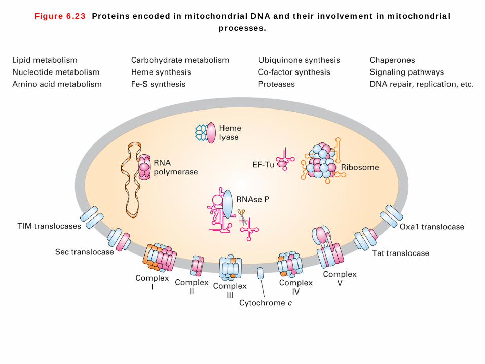
Figure 6.23 Proteins encoded in mitochondrial DNA and their involvement in mitochondrial processes.

mitochondrial DNA The mitochondrial genomes from a number of different metazoan organisms (i.e., multicellular animals) have now been cloned and sequenced, and mtDNAs from all these sources encode essential mitochondrial proteins (Figure 6-23). All proteins encoded by mtDNA are synthesized on mitochondrial ribosomes. Most mitochondrially synthesized polypeptides identified thus far are subunits of multimeric complexes used in electron transport, ATP synthesis, or insertion of proteins into the inner mitochondrial membrane or inter membrane space. However, most of the proteins localized in mitochondria, such as those involved in the processes listed at the top of Fiugre 6-23, are encoded by nuclear genes, synthesized on cytosolic ribosomes, and imported into the organelle by processes.


Sorting of proteins to mitochondria Precursor proteins synthesized in the cytosol that are destined for the matrix of mitochondria or the equivalent space, the stroma, of chloroplasts usually contain specific N-terminal uptake-targeting sequences that specify binding to receptor proteins on the organelle surface. Generally, this sequence is cleaved once it reaches the matrix or stroma. Clearly, these uptake-targeting sequences are similar in their location and general function to the signal sequences that direct nascent proteins to the ER lumen. Although the three types of signals share some common sequence features, their specific sequences differ considerably, as summarized in Table 13-1.


Figure 12-22 Molecular Biology of the Cell (© Garland Science 2008)

A signal sequence for mitochondrial protein import (A) The first 18 amino acids of the precursor to subunit IV of this enzyme serve as a signal sequence for import of the subunit into the mitochondrion. (B) When the signal sequence is folded as an α helix, the positively charged residues (red) are seen to be clustered on one face of the helix, while the nonpolar residues (yellow) are clustered primarily on the opposite face. Mitochondrial matrix-targeting sequences always have the potential to form such anamphipathic α helix, which is recognized by specific receptor proteins on the mitochondrial surface (Figure 12-22 Molecular Biology of the Cell).


Experimental Figure 13.23 Import of mitochondrial precursor proteins is assayed in a cell-free system.
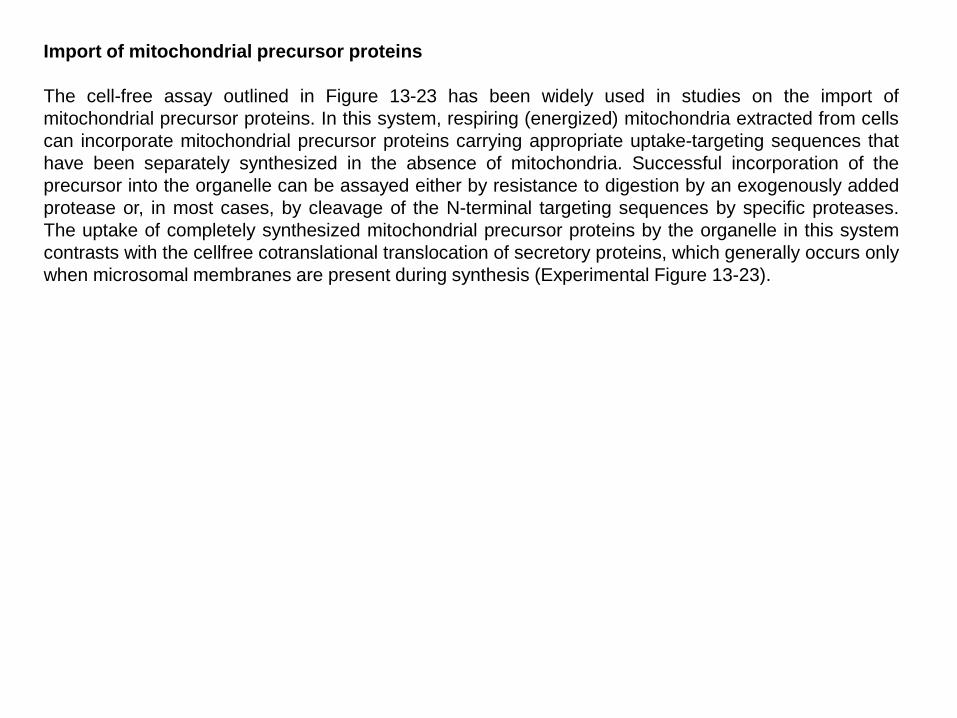
Import of mitochondrial precursor proteins The cell-free assay outlined in Figure 13-23 has been widely used in studies on the import of mitochondrial precursor proteins. In this system, respiring (energized) mitochondria extracted from cells can incorporate mitochondrial precursor proteins carrying appropriate uptake-targeting sequences that have been separately synthesized in the absence of mitochondria. Successful incorporation of the precursor into the organelle can be assayed either by resistance to digestion by an exogenously added protease or, in most cases, by cleavage of the N-terminal targeting sequences by specific proteases. The uptake of completely synthesized mitochondrial precursor proteins by the organelle in this system contrasts with the cellfree cotranslational translocation of secretory proteins, which generally occurs only when microsomal membranes are present during synthesis (Experimental Figure 13-23).


Figure 12-23 Molecular Biology of the Cell (© Garland Science 2008)

Protein translocators in the mitochondrial membranes Protein translocation across mitochondrial membranes is mediated by multi-subunit protein complexes that function as protein translocators (Figure 12-23 Molecular Biology of the Cell):
1. the TOM complex functions across the outer membrane, 2. and two TIM complexes, the TIM23 and TIM22 complexes, function across the inner membrane.
TOM and TIM stand for translocase of the outer and inner mitochondrial membranes, respectively. These complexes contain some components that act as receptors for mitochondrial precursor proteins and other components that form the translocation channel. The TOM complexis required for the import of all nucleus-encoded mitochondrial proteins. It initially transports their signal sequences into the intermembrane space and helps to insert transmembrane proteins into the outer membrane. The TIM23 complex then transports some of these proteins into the matrix space, while helping to insert transmembrane proteins into the inner membrane. The TIM22 complex mediates the insertion of a subclass of inner membrane proteins, including the carrier protein that transports ADP, ATP, and phosphate.
3. A third protein translocator in the inner mitochondrial membrane, the OXA complex, mediates the insertion of inner membrane proteins that are synthesized within the mitochondria. It also helps to insert some proteins that are initially transported into the matrix by the TOM and TIM complexes.


Figure 13.24 Protein import into the mitochondrial matrix.

Protein import into the mitochondrial matrix. 1. Figure 13-24 presents an overview of protein import from the cytosol into the mitochondrial matrix, the route into the mitochondrion followed by most imported proteins. ① After synthesis in the cytosol, the soluble precursors of mitochondrial proteins (including hydrophobic integral
membrane proteins) interact directly with the mitochondrial membrane. In general, only unfolded proteins can be imported into the mitochondrion. Chaperone proteins such as cytosolic Hsc70 keep nascent and newly made proteins in an unfolded state, so that they can be taken up by mitochondria.
② Import of an unfolded mitochondrial precursor is initiated by the binding of a mitochondrial targeting sequence to an import receptor in the outer mitochondrial membrane. For example, N-terminal matrix targeting sequences are recognized by Tom20 and Tom22.
③ The import receptors subsequently transfer the precursor proteins to an import channel in the outer membrane. ④ This channel, composed mainly of the Tom40 protein, is known as the general import pore because all known
mitochondrial precursor proteins gain access to the interior compartments of the mitochondrion through this channel.
⑤ The general import pore forms a largely passive channel through the outer mitochondrial membrane, and the driving force for unidirectional transport into mitochondria comes from within the mitochondrion. In the case of precursors destined for the mitochondrial matrix, transfer through the outer membrane occurs simultaneously with transfer through an innermembrane channel composed of the Tim23 and Tim17 proteins. Translocation into the matrix thus occurs at “contact sites” where the outer and inner membranes are in close proximity.
⑥ Soon after the N-terminal matrix-targeting sequence of a protein enters the mitochondrial matrix, it is removed by a protease that resides within the matrix. The emerging protein also is bound by matrix Hsc70, a chaperone that is localized to the translocation channels in the inner mitochondrial membrane by interacting with Tim44. This interaction stimulates ATP hydrolysis by matrix Hsc70, and together these two proteins are thought to power translocation of proteins into the matrix.
⑦ Some imported proteins can fold into their final, active conformation without further assistance. Final folding of many matrix proteins, however, requires a chaperonin. Chaperonin proteins actively facilitate protein folding in a process that depends on ATP.


Experimental Figure 13.25 Experiments with chimeric proteins elucidate mitochondrial protein import.

Important features of mitochondrial import 1. Dramatic evidence for the ability of mitochondrial matrix targeting sequences to direct import was obtained with chimeric proteins produced by recombinant DNA techniques (Experimental Figure 13-25). ① For example, the matrix-targeting sequence of alcohol dehydrogenase can be fused to the N-
terminus of dihydrofolate reductase (DHFR), which normally resides in the cytosol. In the presence of chaperones, which prevent the C-terminal DHFR segment from folding in the cytosol, cell free translocation assays show that the chimeric protein is transported into the matrix.
② The inhibitor methotrexate, which binds tightly to the active site of DHFR and greatly stabilizes its folded conformation, renders the chimeric protein resistant to unfolding by cytosolic chaperones. When translocation assays are performed in the presence of methotrexate, the chimeric protein does not completely enter the matrix. This finding demonstrates that a precursor must be unfolded in order to traverse the import pores in the mitochondrial membranes. Additional studies revealed that if a sufficiently long spacer sequence separates the N-terminal matrix-targeting sequence and DHFR portion of the chimeric protein, then a stable translocation intermediate forms in the presence of methotrexate. In order for such a stable translocation intermediate to form, the spacer sequence must be long enough to span both membranes. If the chimera contains a shorter spacer no stable translocation intermediate is obtained because the spacer cannot span both membranes. These observations provide further evidence that translocated proteins can span both inner and outer mitochondrial membranes and traverse these membranes in an unfolded state.
③ Microscopic studies of stable translocation intermediates show that they accumulate at sites where the inner and outer mitochondrial membranes are close together, evidence that precursor proteins enter only at such sites.


Figure 13.26 Targeting sequences in imported mitochondrial proteins. 5
Targeting sequences in imported mitochondrial proteins. Unlike targeting to the matrix, targeting of proteins to the intermembrane space, inner membrane, and outer membrane of mitochondria generally requires more than one targeting sequence and occurs via one of several pathways. Figure 13-26 summarizes the organization of targeting sequences in proteins sorted to different mitochondrial locations.


Mitochondrial matrix-targeting sequences The most extensively studied sequences for localizing proteins to mitochondria are the matrix-targeting sequences. These sequences, located at the N-terminus, are usually 20–50 amino acids in length. They are rich in hydrophobic amino acids, positively charged basic amino acids (arginine and lysine), and hydroxylated ones (serine and threonine), but tend to lack negatively charged acidic residues (aspartate and glutamate). Mitochondrial matrix-targeting sequences are thought to assume an α -helical conformation in which positively charged amino acids predominate on one side of the helix and hydrophobic amino acids predominate on the other side; thus these sequences are amphipathic.


Figure 13.27 Three pathways to the inner mitochondrial membrane from the cytosol.

Three pathways to the inner mitochondrial membrane from the cytosol. 1. One pathway makes use of the same machinery that is used for targeting of matrix proteins (path A). A cytochrome oxidase subunit called CoxVa is a typical protein transported by this pathway. The precursor form of CoxVa, which contains an N-terminal matrix-targeting sequence recognized by the Tom20/22 import receptor, is transferred through the general import pore of the outer membrane and the inner-membrane Tim23/17 translocation complex. In addition to the matrix-targeting sequence, which is cleaved during import, CoxVa contains a hydrophobic stop-transfer sequence. As the protein passes through the Tim23/17 channel, the stop-transfer sequence blocks translocation of the C-terminus across the inner membrane. The membrane-anchored intermediate is then transferred laterally into the bilayer of the inner membrane much as type I integral membrane proteins are incorporated into the ER membrane. 2. A second pathway to the inner membrane is followed by proteins (e.g., ATP synthase subunit 9) whose precursors contain both a matrix-targeting sequence and internal hydrophobic domains recognized by an inner-membrane protein termed Oxa1. This pathway is thought to involve translocation of at least a portion of the precursor into the matrix via the Tom20/22 and Tim23/17 channels. After cleavage of the matrix-targeting sequence, the protein is inserted into the inner membrane by a process that requires interaction with Oxa1 and perhaps other inner-membrane proteins (path B). Oxa1 is related to a bacterial protein involved in inserting some inner-membrane proteins in bacteria. This relatedness suggests that Oxa1 may have descended from the translocation machinery in the endosymbiotic bacterium that eventually became the mitochondrion. However, the proteins forming the innermembrane channels in mitochondria are not related to the SecY protein in bacterial translocons. Oxa1 also participates in the inner-membrane insertion of certain proteins (e.g., subunit II of cytochrome oxidase) that are encoded by mitochondrial DNA and synthesized in the matrix by mitochondrial ribosomes. 3. The final pathway for insertion in the inner mitochondrial membrane is followed by multipass proteins that contain six membrane-spanning domains, such as the ADP/ATP antiporter. These proteins, which lack the usual N-terminal matrix-targeting sequence, contain multiple internal mitochondrial targeting sequences. After the internal sequences are recognized by Tom70, a second import receptor located in the outer membrane, the imported protein passes through the outer membrane through the general import pore (path C). The protein then is transferred to a second translocation complex in the inner membrane composed of the Tim22 and Tim54 proteins. Transfer to the Tim22/54 complex depends on a multimeric complex of two small proteins, Tim9 and Tim10, that reside in the intermembrane space. These may act as chaperones to guide imported proteins from the general import pore to the Tim22/54 complex in the inner membrane. Ultimately the Tim22/54 complex is responsible for incorporating the multiple hydrophobic segments of the imported protein into the inner membrane (Figure 13-27).


Figure 13.28 Two pathways to the mitochondrial intermembrane space.

Two pathways to the mitochondrial intermembrane space 1. The major pathway is followed by proteins, such as cytochrome b2, whose precursors carry two different N-terminal targeting sequences, both of which ultimately are cleaved. The most N-terminal of the two sequences is a matrix-targeting sequence, which is removed by the matrix protease. The second targeting sequence is a hydrophobic segment that blocks complete translocation of the protein across the inner membrane (path A). After the resulting membrane-embedded intermediate diffuses laterally away from the Tim23/17 translocation channel, a protease in the membrane cleaves the protein near the hydrophobic transmembrane segment, releasing the mature protein in a soluble form into the intermembrane space. Except for the second proteolytic cleavage, this pathway is similar to that of inner-membrane proteins such as CoxVa. 2. Cytochrome c heme lyase, the enzyme responsible for the covalent attachment of heme to cytochrome c, illustrates a second pathway for targeting to the intermembrane space. In this pathway, the imported protein is delivered directly to the intermembrane space via the general import pore without involvement of any inner-membrane translocation factors (path B). Since translocation through the Tom40 general import pore does not seem to be coupled to any energetically favorable process such as hydrolysis of ATP or GTP, the mechanism that drives unidirectional translocation through the outer membrane is unclear. One possibility is that cytochrome c heme lyase passively diffuses through the outer membrane and then is trapped within the intermembrane space by binding to another protein that is delivered to that location by one of the translocation mechanisms discussed previously (Figure 13-28).

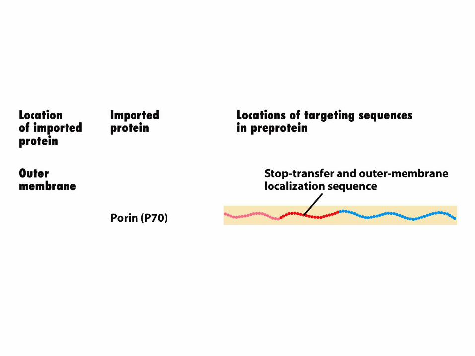

Mitochondrial outer membrane-targeting sequences Experiments with mitochondrial porin (P70) provide clues about how proteins are targeted to the outer mitochondrial membrane. A short matrix-targeting sequence at the N-terminus of P70 is followed by a long stretch of hydrophobic amino acids. If the hydrophobic sequence is experimentally deleted from P70, the protein accumulates in the matrix space with its matrix-targeting sequence still attached. This finding suggests that the long hydrophobic sequence functions as a stop-transfer sequence that both prevents transfer of the protein into the matrix and anchors it as an integral protein in the outer membrane. Normally, neither the matrix-targeting nor stop-transfer sequence is cleaved from the anchored protein. The source of energy to drive outer membrane proteins through the general import pore has not yet been identified.


Figure 13.29 Transporting proteins to chloroplast thylakoids.

Transporting proteins to chloroplast thylakoids 1. In addition to the double membrane that surrounds them, chloroplasts contain a series of internal interconnected membranous sacs, the thylakoids. Proteins localized to the thylakoid membrane or lumen carry out photosynthesis. Many of these proteins are synthesized in the cytosol as precursors containing multiple targeting sequences. 2. Four separate pathways for transporting proteins from the stroma into the thylakoid have been identified. All four pathways have been found to be closely related to analogous transport mechanisms in bacteria, illustrating the close evolutionary relationship between the stromal membrane and the bacterial inner membrane (Figure 13-29). ① Transport of plastocyanin and related proteins into the thylakoid lumen occurs by an SRP-
dependent pathway (path A). ② A second pathway for transporting proteins into the thylakoid lumen utilizes a protein related to
bacterial SecA. ③ A third pathway, which targets proteins to the thylakoid membrane, depends on a protein related to
the mitochondrial Oxa1 protein and the homologous bacterial protein (path B). Some proteins encoded by chloroplast DNA and synthesized in the stroma or transported into the stroma from the cytosol are inserted into the thylakoid membrane via this pathway.
④ Finally, thylakoid proteins that bind metal-containing cofactors follow another pathway into the thylakoid lumen (pH pathway). The unfolded precursors of these proteins are first targeted to the stroma, where the N-terminal stromal-import sequence is cleaved off and the protein then folds and binds its cofactor. A set of thylakoid-membrane proteins assists in translocating the folded protein and bound cofactor into the thylakoid lumen, a process powered by the pH gradient normally maintained across the thylakoid membrane. The thylakoid-targeting sequence that directs a protein to this pH-dependent pathway includes two closely spaced arginine residues that are crucial for recognition. Bacterial cells also have a mechanism for translocating folded proteins with a similar arginine-containing sequence across the inner membrane.