Molecular basis of cooperativity in pH-triggered ... pdfs/114_Li... · Molecular basis of...
9
ARTICLE Received 23 May 2016 | Accepted 13 Sep 2016 | Published 27 Oct 2016 Molecular basis of cooperativity in pH-triggered supramolecular self-assembly Yang Li 1 , Tian Zhao 1 , Chensu Wang 1,2 , Zhiqiang Lin 1 , Gang Huang 1 , Baran D. Sumer 3 & Jinming Gao 1 Supramolecular self-assembly offers a powerful strategy to produce high-performance, stimuli-responsive nanomaterials. However, lack of molecular understanding of stimulated responses frequently hampers our ability to rationally design nanomaterials with sharp responses. Here we elucidated the molecular pathway of pH-triggered supramolecular self-assembly of a series of ultra-pH sensitive (UPS) block copolymers. Hydrophobic micellization drove divergent proton distribution in either highly protonated unimer or neutral micelle states along the majority of the titration coordinate unlike conventional small mole- cular or polymeric bases. This all-or-nothing two-state solution is a hallmark of positive cooperativity. Integrated modelling and experimental validation yielded a Hill coefficient of 51 in pH cooperativity for a representative UPS block copolymer, by far the largest reported in the literature. These data suggest hydrophobic micellization and resulting positive coopera- tivity offer a versatile strategy to convert responsive nanomaterials into binary on/off switchable systems for chemical and biological sensing, as demonstrated in an additional anion sensing model. DOI: 10.1038/ncomms13214 OPEN 1 Department of Pharmacology, Simmons Comprehensive Cancer Center, University of Texas Southwestern Medical Center, 5323 Harry Hines Blvd., Dallas, Texas 75390, USA. 2 Department of Cell Biology, University of Texas Southwestern Medical Center, 5323 Harry Hines Blvd., Dallas, Texas 75390, USA. 3 Department of Otolaryngology, University of Texas Southwestern Medical Center, 5323 Harry Hines Blvd., Dallas, Texas 75390, USA. Correspondence and requests for materials should be addressed to J.G. (email: [email protected]). NATURE COMMUNICATIONS | 7:13214 | DOI: 10.1038/ncomms13214 | www.nature.com/naturecommunications 1
-
Upload
duongkhuong -
Category
Documents
-
view
219 -
download
0
Transcript of Molecular basis of cooperativity in pH-triggered ... pdfs/114_Li... · Molecular basis of...

ARTICLE
Received 23 May 2016 | Accepted 13 Sep 2016 | Published 27 Oct 2016
Molecular basis of cooperativity in pH-triggeredsupramolecular self-assemblyYang Li1, Tian Zhao1, Chensu Wang1,2, Zhiqiang Lin1, Gang Huang1, Baran D. Sumer3 & Jinming Gao1
Supramolecular self-assembly offers a powerful strategy to produce high-performance,
stimuli-responsive nanomaterials. However, lack of molecular understanding of stimulated
responses frequently hampers our ability to rationally design nanomaterials with sharp
responses. Here we elucidated the molecular pathway of pH-triggered supramolecular
self-assembly of a series of ultra-pH sensitive (UPS) block copolymers. Hydrophobic
micellization drove divergent proton distribution in either highly protonated unimer or neutral
micelle states along the majority of the titration coordinate unlike conventional small mole-
cular or polymeric bases. This all-or-nothing two-state solution is a hallmark of positive
cooperativity. Integrated modelling and experimental validation yielded a Hill coefficient of 51
in pH cooperativity for a representative UPS block copolymer, by far the largest reported in
the literature. These data suggest hydrophobic micellization and resulting positive coopera-
tivity offer a versatile strategy to convert responsive nanomaterials into binary on/off
switchable systems for chemical and biological sensing, as demonstrated in an additional
anion sensing model.
DOI: 10.1038/ncomms13214 OPEN
1 Department of Pharmacology, Simmons Comprehensive Cancer Center, University of Texas Southwestern Medical Center, 5323 Harry Hines Blvd., Dallas,Texas 75390, USA. 2 Department of Cell Biology, University of Texas Southwestern Medical Center, 5323 Harry Hines Blvd., Dallas, Texas 75390, USA.3 Department of Otolaryngology, University of Texas Southwestern Medical Center, 5323 Harry Hines Blvd., Dallas, Texas 75390, USA. Correspondence andrequests for materials should be addressed to J.G. (email: [email protected]).
NATURE COMMUNICATIONS | 7:13214 | DOI: 10.1038/ncomms13214 | www.nature.com/naturecommunications 1

High-performance, stimuli-responsive nanomaterials thatcan sharply respond to and amplify biological signals arerapidly developed for disease diagnosis and therapy1–7.
Synthetic approaches employing traditional covalent bondchemistry may be limited in achieving highly complexnanostructures of over 106 Da in molecular weight. In contrast,non-covalent supramolecular self-assembly offers a versatile andmodular strategy in generating nanoscale structures andarchitectures (106–109 Da and 10–100 nm) that often displaycooperative behaviours, which arises from subtle interplay of amultitude of non-covalent interactions (for example, hydrogenbonding, hydrophobic and electrostatic interactions)8–10. Thesystem as a whole behaves quite differently from the sum ofindividual parts acting in isolation. Despite the great promise,incorporation of self-assembly principles in the design ofresponsive nanomaterials is challenging due to the lack offundamental understanding of cooperativity at the molecularlevel.
pH is an important physiological parameter that plays a criticalrole in cellular and tissue homeostasis11. Dysregulated pHhas been recognized as a hallmark of cancer12. pH-sensitivenanoparticles have been widely used for tumour imaging, studyof endosome/lysosome biology and cancer-targeted drugdelivery13–15. Conventional small molecular sensors16, pHLIPpeptides17 or photoelectron transfer nanoprobes18 offercontinuous pH responses, therefore cannot convert the subtledifferences in physiological pH into a discrete signal withoutintroducing noise.
In this study, we investigated the molecular basis ofcooperativity in pH-triggered supramolecular self-assembly of a
series of ultra-pH sensitive (UPS) copolymers. When PR segmentreached above a hydrophobic threshold, nanophase separationdrove cooperative deprotonation of charged polymers, asdemonstrated by divergent proton distribution in either theprotonated unimers or neutral micelles. A combination oftheoretical modeling and experimental validation confirmedthe micellization-induced pH cooperativity and resulting sharpfluorescent transitions. Our studies suggest hydrophobicnanophase separation may serve as a versatile strategy to convertresponsive nanomaterials into binary on/off switchable systemsfor chemical and biological sensing.
ResultsUPS nanoprobes. Lysosensor Green is a commonly used smallmolecular pH sensor. It exhibits 10-fold change in fluorescenceintensity over a 2 pH span (Fig. 1a,c). The continuous change offluorescence intensity hampers its ability to differentiate small pHvariations between pathological pH (for example, acidic tumourpH, 6.5–6.9 (ref. 12)) and normal pH (7.4). Recently, we haveestablished a library of UPS nanoprobes with sharp pHtransitions that are finely tunable in a broad range ofphysiological pH (4–8; ref. 19). UPS nanoprobes displayed asharp on/off pH response (see a specific example of PEO-b-PDBA(poly(ethylene oxide)-b-poly(2-(dibutylamino) ethyl methacrylate))in Fig. 1b,c), which was used to amplify tumour micro-environmental signals for the robust detection of a broad range oftumours20. The UPS nanoparticles consist of amphiphilic blockcopolymers, where PEO is poly(ethylene oxide) and PR ishydrophobic block with multiple ionizable tertiary amines(Supplementary Methods; Supplementary Fig. 1). At low pH,
pH > pHtpH < pHt
OFFON
pH
OO O
113
O
OO
N
n
R = Pr , PEO-b-PDPABu , PEO-b-PDBAPe , PEO-b-PD5A
RR
OO
N
m
c
b
d
pH
1.0
0.8
0.6
0.4
0.2
0.0
3 4pH
RF
U
5 6 7
Lysosensor green
UPS nanoprobe
pH > pKa
pH < pKa
a
N NO
NH2CH2CH2NH O
N NO
NH2CH2CH2N O
+H+
–H+
++
+
Figure 1 | Ultra-pH sensitive (UPS) nanoprobes with unique binary on/off response to pH. (a) Structure and fluorescence images of a small molecular
pH sensor, Lysosensor Green in aqueous solution at different pH. (b) Structure and fluorescence images of a UPS nanoprobe, Rhodamine Green-conjugated
PEO-b-PDBA block copolymers in aqueous solution at different pH. (c) Relative fluorescence intensity as a function of pH for Lysosensor Green and
PEO-b-PDBA-RhoG nanoprobe. (d) Schematic illustration of pH-triggered binary on/off transition of UPS nanoprobes.
ARTICLE NATURE COMMUNICATIONS | DOI: 10.1038/ncomms13214
2 NATURE COMMUNICATIONS | 7:13214 | DOI: 10.1038/ncomms13214 | www.nature.com/naturecommunications

micelles dissociate into cationic unimers with protonatedammonium groups (Fig. 1d). When pH increases, neutralizedPR segments become hydrophobic and self-assemble into core–shell micelles.
Micellization is critical for sharp pH transition. First,we compared the pH responsive behaviours of several UPScopolymers with small molecular and polymeric bases (Fig. 2a;Supplementary Fig. 2). NH4Cl (pKa¼ 10.5) and chloroquine (pKa
values¼ 8.3 and 10.8), commonly used lysosomotropic agents tomanipulate the pH of endocytic organelles, showed typical broadpH response in the range of pH 7–11. pH titration of dipropy-laminoethanol (DPA, building block of UPS copolymer
poly(ethylene oxide)-b-poly(2-(dipropylamino) ethyl methacry-late) PEO-b-PDPA) showed similar broad pH response (Fig. 2b)as predicted by the Henderson–Hasselbalch equation for mono-meric species21. pH titrations of several extensively investigatedpolybases (polyethylenimine (PEI)22, poly(L-Lysine)23, chitosan24
and poly(L-Histidine)25) showed different degrees of broad pHresponse (Supplementary Fig. 2). Among these, PEI had thebroadest pH response from pH 3–11. In contrast, pH titration ofthree UPS nanoprobes (PEO-b-PDPA, PEO-b-PDBA and PEO-b-PD5A) displayed remarkable pH plateaus within a narrow pHrange (Supplementary Fig. 2; Supplementary Table 1), indicatingstrong buffer effect26. For polybases with multiple ionizable sites,the apparent pKa values were determined as the pH where 50% ofall the ionizable amines are protonated27–29.
DPA
ΔpH
10%
–90%
PEI
PEO-b-PDMAPEO-b-PDPA
PEO-b-PDMA &PEO-b-PDEA
cb
d e
PD
= 9
5%P
D =
85%
CMPD
HON
Dipropylaminoethanol (DPA)
12 6
5
4
3
Polybases
Small molecular base
2
1
0–4 –3 –2 –1 0 1 2 3 4
10
8
8
7
6
pH
5
1.0 0.8 0.6 0.4
Protonation degree
0.2 0.0
30
25
20
Dia
met
er (
nm)
15
10
5
pH
6
4
0 20 40 60 80
V of NaOH (μl) LogP
OO
O113
O
OO
N
n
R R
NNH
NN
HN
NH2 NH
NH2
NNH2H2N
[ ]n
Polyethyleneimine (PEI) PEO-b -PR
UPS nanoprobes
R = Me, PEO-b -PDMAPr, PEO-b -PDPA
a
PEO-b-PDPA
Figure 2 | Hydrophobic phase separation drives sharp pH response of UPS copolymers. (a) Structures of small molecular base dipropylaminoethanol
(DPA), polymeric bases of PEI, PEO-b-PDMA and PEO-b-PDPA. (b) pH titration curves of DPA, PEI, PEO-b-PDMA and PEO-b-PDPA. (c) Plot of pH
transition sharpness (DpH10–90%) as a function of octanol–water partition coefficient (LogP) of small molecular bases (NH4Cl, Chloroquine and DPA) or
repeating unit (neutral monomer) of commonly used polybases (poly(ethyleneimine), polylysine, chitosan, polyhistidine) and PEO-b-PR block copolymers.
(d) Change of hydrodynamic diameter of UPS nanoprobe PEO-b-PDPA along pH titration coordinate. Significant increase of size indicated the formation of
micelles. (e) TEM images of PEO-b-PDPA before (protonation degree at 95%) and after (protonation degree at 85%) critical micelle protonation degree
(CMPD¼90%). Micelle formation (yellow arrows) was observed when protonation degree was below CMPD. Scale bars, 100 nm.
NATURE COMMUNICATIONS | DOI: 10.1038/ncomms13214 ARTICLE
NATURE COMMUNICATIONS | 7:13214 | DOI: 10.1038/ncomms13214 | www.nature.com/naturecommunications 3

We calculated the octanol–water partition coefficients (LogP)of small molecular bases or repeating unit (neutral monomers)from polybases and used them as a quantitative measurement ofmolecular hydrophobicity. We plotted the sharpness of pHtransition as defined by DpH10–90% (the pH span between 10 and90% ionization of amino groups) as a function of LogP (Fig. 2c).Data indicate that pH sensors with higher molecularhydrophobicity displayed sharper pH responses. Furthermore,a hydrophobic threshold (LogPB2.5) appears to correlate withthe ultra-pH response (where we define DpH10–90%o0.5).
For further investigation, we performed pH titrations ofanother two PEO-b-PR copolymers with the same methacrylatebackbone but less hydrophobic aminoalkyl side chains:methyl and ethyl groups in PEO-b-PDMA (LogP¼ 1.18) andPEO-b-PDEA (LogP¼ 1.85), respectively. PEO-b-PDMA dis-played a broad pH response from 6.5 to 9.0 (Fig. 2b), whereno micelles were formed throughout the titration course.PEO-b-PDEA showed a divergent behaviour (SupplementaryFig. 3), where broad pH response and ultra-pH sensitivity wereobserved when protonation degree was above and below 60%,respectively. The combination of titration and dynamic lightscattering (DLS) results showed that the sharp pH response ofPEO-b-PDEA did not start until the formation of micelles,demonstrating correlations between hydrophobic phase separa-tion and ultra-pH sensitivity (Supplementary Fig. 4). Unlike smallmolecular sensors whose pKa values are mostly controlled byelectron withdrawing groups30,31, the apparent pKa values of UPScopolymers are controlled by the hydrophobicity of PR segment(Supplementary Fig. 5).
DLS method was applied to monitor the formation of micellesalong the pH titration coordinate (Fig. 2d; Supplementary Fig. 6).Surprisingly, micelle formation was found as early as 90% ofprotonation degree and the micelle diameter (B28 nm) remained
relatively constant below 90%. The count rate increased slightlybefore 92% protonation and increased almost linearly forthe remainder of titration. The formation of micelles whenprotonation degree decreased below 90% was confirmed bytransmission electron microscopy (Fig. 2e). Similarly, micelle-induced homoFRET fluorescence quenching did not start untilprotonation degree reduced to 89% (Supplementary Fig. 7).We define the protonation degree below which the copolymersself-assemble into micelles as the critical micellizationprotonation degree (CMPD). The CMPD values of PEO-b-PDPAcopolymer from different methods showed good consistency.
Characterization of nanocomplexes along pH titration coordinate.Intuitively, pH-triggered micellization of PEO-b-PDPA copoly-mers may undergo two opposite thermodynamic pathways(Supplementary Fig. 8). In the graduate model, below CMPD,majority of PEO-b-PDPA copolymers may first self-assemble intopositively charged loose aggregates and upon neutralization, theloose aggregates are gradually deprotonated, shrinking in size andfinally turning into neutral, mature micelles. In the divergentmodel, at any given protonation degree below CMPD, the PEO-b-PDPA copolymers exist in either protonated unimers or neutral,mature micelles with different population distributions. Theinitial DLS and fluorescence data suggest that pH-triggered self-assembly undergoes the second pathway as indicated by therelatively unchanged nanoparticle size and linearly increasedcount rate and decreased fluorescence intensity to protonationdegree.
To verify the coexistence of protonated unimers and neutralmicelles along the pH titration coordinate of PEO-b-PDPA blockcopolymers, we first applied a dialysis method to separateunimers (22 kD) from micelles (16,000 kD)20 at different
c
Centrifugation
100 kDamembrane
Micelle > 16,000 kDaUnimer ~20 kDa
Filtrate
Residual
layer
a b
d e
DLS / pH titrationquantification
Pol
ymer
dis
trib
utio
n
Unimer
Mice
lle0.0
0.2
0.4
0.6
0.8
1.0
Cha
rge
stat
e
PEI0
200
400
600
Cou
nt r
ate
(kcp
s)
Residual layer
Filtrate
Residual layer
Filtrate
0.0
0.5
1.0
0.0
0.5
1.0
Sta
tistic
alav
erag
e c
harg
e st
ate
PEO-b-
PDPA
PEO-b-
PDMA
PEI
PEO-b-
PDPA
PEO-b-
PDMA PEI
PEO-b-
PDPA
PEO-b-
PDMA
Residual layer
Filtrate
Figure 3 | Divergent proton distribution between unimer and micelle state of PEO-b-PDPA copolymers. (a) Schematic illustration of dialysis
experiments where unimers (22 kD) were separated from micelles (16,000 kD) using a semi-permeable membrane with a molecular weight cutoff of
100 kD. PEO-b-PDMA and PEI were used as negative controls without nanophase separation. Light scattering count rates (b), polymer mass distribution
(c) and charges states (d) of different polymers are shown at the protonation degree of 50%. (e) Quantification of unimer and micelle charge states of
PEO-b-PDPA at protonation degree of 50%. Protons distributed divergently where unimers were highly charged (B90%) and micelles were mostly neutral.
The experiments were repeated five times, and data are shown in mean±s.d.
ARTICLE NATURE COMMUNICATIONS | DOI: 10.1038/ncomms13214
4 NATURE COMMUNICATIONS | 7:13214 | DOI: 10.1038/ncomms13214 | www.nature.com/naturecommunications

protonation degrees (Fig. 3a). Two non-aggregating polybaseswith broad pH response (PEO-b-PDMA and PEI) were used forcomparison. Neither PEO-b-PDMA nor PEI showed changes insize and scattering count rate along its entire titration course(Fig. 3b; Supplementary Fig. 9). Quantitative analysis showed thatPEO-b-PDMA and PEI were distributed equally in the residualand filtrate layers (Fig. 3c,d). In contrast, higher amount ofPEO-b-PDPA copolymers were retained in the residual layer(Fig. 3c; Supplementary Figs 10 and 11) due to the retention ofmicelles (16,000 kD, which is above the cutoff molecular weight(100 kD) of the dialysis membrane). pH titration of residual and
filtrate samples showed that PEO-b-PDPA copolymers werehighly charged with 90% of tertiary amines protonated in thefiltrate sample whereas PEO-b-PDPA copolymers in the micellestate (after subtracting the protonated unimers) in the residuallayer were almost neutral (Fig. 3e).
To further confirm the divergent charge state in unimers andmicelles, we employed 1H NMR to study the proton distributionof PEO-b-PDPA along the pH titration path. Data revealed astriking difference in the proton distribution of non-aggregatingPEO-b-PDMA versus aggregating PEO-b-PDPA. The PEO-b-PDMA copolymer showed a continuous change of chemical
e
d
0%
20%
40%
60%
80%
0%
20%
40%
60%
80%
PEO-b-PDMA
PEO-b-PDMA
PEO-b-PDPA
PEO-b-PDPA
PEO-b-PDPA
b
100% 100%
3.2
3.5
3.4
3.3
3.2
3.1
3.0
3.0
2.8
Che
mic
al s
hift
(�)
Che
mic
al s
hift
(�)
2.6
2.4
2.21.0 0.8
Protonation degree
Protonation degree
Inte
grat
ion
Inte
grat
ion
0.6 0.4 0.2 0.0
1.0 0.8 0.6 0.4 0.2
1.0
1.0
0.8
0.6
0.4
0.2
0.0
0.8
0.6
0.4
0.2
ca
Proton
Protonation degree
50% 0%
Protonation degree
Proton
PEO-b-PDMA
–H+
–H+
–H+–H+
–H+
2.
23.
4
� (p.p.m.) � (p.p.m.)
3.2
3.0
2.8
2.6
2.4
2.2
3.4 3.
23.
02.
82.
6 2
.4
100%
50% 0%100% 90%
Figure 4 | Molecular pathway of pH-triggered self-assembly of PEO-b-PDPA copolymers. (a,b) 1H NMR spectra (in D2O) of methylene protons of
PEO-b-PDPA and methyl protons of PEO-b-PDMA adjacent to nitrogen atoms at different protonation degrees, respectively. PEO-b-PDMA was used as
negative control without nanophase separation. (c,d) Quantification of chemical shift and peak integration of chosen methyl and methylene protons in
PEO-b-PDMA and PEO-b-PDPA, respectively. Integrations were calculated using polyethylene oxide (PEO) segments as internal reference. (e) Schematic
illustration of two distinctive deprotonation pathways. Deprotonation of PEO-b-PDMA ammonium groups was gradual along the entire pH titration course.
Deprotonation of PEO-b-PDPA ammonium groups displayed a binary copolymer populations consisting of highly charged unimers in solution and neutral
copolymers inside micelles.
NATURE COMMUNICATIONS | DOI: 10.1038/ncomms13214 ARTICLE
NATURE COMMUNICATIONS | 7:13214 | DOI: 10.1038/ncomms13214 | www.nature.com/naturecommunications 5

shifts of methyl protons along the titration course (Fig. 4a,c;Supplementary Fig. 12). The peak area did not change at differentprotonation degree (Fig. 4c), suggesting a single phase solutiondeprotonation process (Fig. 4e). The water soluble smallmolecular base DPA showed similar results (SupplementaryFig. 13). In contrast, the PEO-b-PDPA copolymer displayeddivergent proton distribution along the majority of the pHtitration coordinate. The unimer peak did not change its chemicalshift but only showed linear decrease in integration (Fig. 4b,d;Supplementary Fig. 14; proton peaks in micelle cores were notvisible due to fast T2 relaxation), in accordance with the linearincrease in scattering count rate and decrease in fluorescenceintensity shown previously.
Based on these results, we constructed a molecular pathway ofpH-triggered supramolecular self-assembly of PEO-b-PDPA copo-lymer (Fig. 4e). Upon addition of NaOH, the block copolymers arehomogeneously deprotonated until reaching CMPD. Below CMPD,further addition of base results in formation of mature micelleswhere the copolymers inside micelles are neutral. Soluble unimersremain highly protonated at a charge state of CMPD in water. The
unique pH-induced supramolecular self-assembly pathwayappeared to pertain microscopic reversibility (SupplementaryFig. 14). It should be noted that the above molecular pathwaydescribes the thermodynamically stable states of polymers atspecific protonation degree as measured by steady-state analyticalmethods (for example, DLS, 1H NMR). The kinetic process ofprotonated unimer conversion to neutral micelles may still involveloose aggregates as transient intermediates and needs to be furtherinvestigated.
Model description of pH cooperativity. The all or nothingproton distribution characteristics10 suggest a cooperativedeprotonation process from fully protonated unimers to thecompletely neutralized micelles. We hypothesize that rapiddeprotonation is driven by hydrophobic phase separationthrough formation of micelles. We adopted an allosteric modelto evaluate the cooperative strength of deprotonation (Fig. 5a;Supplementary Discussion). The progressive neutralization offully protonated unimers can be characterized by a series of
a
Tertiary amine Proton
1.0 1.0
1.0
0.5
0.50.0
1.0
1.0
0.5
0.50.0
–0.5
0.0
0.0
–0.5
–1.0
1.0 –0.5
pKa-pH
pKa-pH
0.5
0.0
–1.0 –0.5 0.0
0.6
0.5
0.0
pKa-pH0.5 0.0
–1.0 –0.5
–1.0
–1.0
–0.5
0.0pKa-pH
0.5 0.0
n
+
n
(2)
(1)
K =
�i = Ki+1 / Ki
(4) Log( )≈n (pKa - pH)�A
1 –�A
K1 K2 K3••••Kn
(3)= �n–1K1
DPAPEI
PEO-b-PDPAPEO-b-PDMA
51020 60100
b c
d e
Number oftertiary amines per PEO-b-PDPApolymer chain
�A
�A Log [�A/(1–�A)]
Log[�A/(1–�A)]
nK
nMicelle core
Figure 5 | Model construction to quantify the pH cooperativity. (a) Micelle-driven deprotonation model with key equations. These equations allow the
theoretical-experimental correlation between the microscopic cooperative parameter a and macroscopically measurable pKa. (b,c) Cooperativity analysis
based on Hill plot of small molecular base DPA, polymeric bases of PEI, PEO-b-PDMA and PEO-b-PDPA. DPA and PEO-b-PDMA showed non-pH
cooperativity. PEI displayed negative pH cooperativity and PEO-b-PDPA showed strong positive cooperativity. (d,e) Cooperativity analysis of PEO-b-PDPA
copolymers with different number of repeating units in the hydrophobic segment. Increase of hydrophobic chain length led to stronger positive
cooperativity and sharper pH response.
ARTICLE NATURE COMMUNICATIONS | DOI: 10.1038/ncomms13214
6 NATURE COMMUNICATIONS | 7:13214 | DOI: 10.1038/ncomms13214 | www.nature.com/naturecommunications
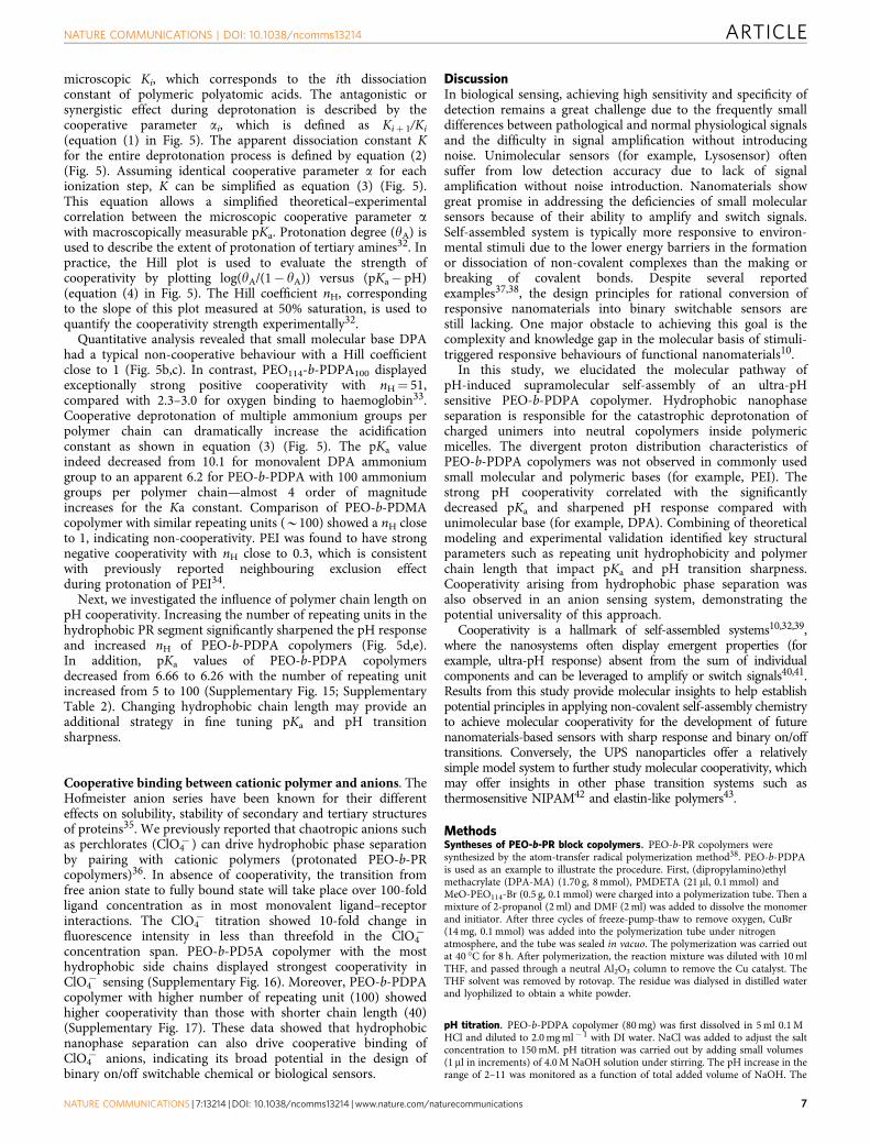
microscopic Ki, which corresponds to the ith dissociationconstant of polymeric polyatomic acids. The antagonistic orsynergistic effect during deprotonation is described by thecooperative parameter ai, which is defined as Kiþ 1/Ki
(equation (1) in Fig. 5). The apparent dissociation constant Kfor the entire deprotonation process is defined by equation (2)(Fig. 5). Assuming identical cooperative parameter a for eachionization step, K can be simplified as equation (3) (Fig. 5).This equation allows a simplified theoretical–experimentalcorrelation between the microscopic cooperative parameter awith macroscopically measurable pKa. Protonation degree (yA) isused to describe the extent of protonation of tertiary amines32. Inpractice, the Hill plot is used to evaluate the strength ofcooperativity by plotting log(yA/(1� yA)) versus (pKa� pH)(equation (4) in Fig. 5). The Hill coefficient nH, correspondingto the slope of this plot measured at 50% saturation, is used toquantify the cooperativity strength experimentally32.
Quantitative analysis revealed that small molecular base DPAhad a typical non-cooperative behaviour with a Hill coefficientclose to 1 (Fig. 5b,c). In contrast, PEO114-b-PDPA100 displayedexceptionally strong positive cooperativity with nH¼ 51,compared with 2.3–3.0 for oxygen binding to haemoglobin33.Cooperative deprotonation of multiple ammonium groups perpolymer chain can dramatically increase the acidificationconstant as shown in equation (3) (Fig. 5). The pKa valueindeed decreased from 10.1 for monovalent DPA ammoniumgroup to an apparent 6.2 for PEO-b-PDPA with 100 ammoniumgroups per polymer chain—almost 4 order of magnitudeincreases for the Ka constant. Comparison of PEO-b-PDMAcopolymer with similar repeating units (B100) showed a nH closeto 1, indicating non-cooperativity. PEI was found to have strongnegative cooperativity with nH close to 0.3, which is consistentwith previously reported neighbouring exclusion effectduring protonation of PEI34.
Next, we investigated the influence of polymer chain length onpH cooperativity. Increasing the number of repeating units in thehydrophobic PR segment significantly sharpened the pH responseand increased nH of PEO-b-PDPA copolymers (Fig. 5d,e).In addition, pKa values of PEO-b-PDPA copolymersdecreased from 6.66 to 6.26 with the number of repeating unitincreased from 5 to 100 (Supplementary Fig. 15; SupplementaryTable 2). Changing hydrophobic chain length may provide anadditional strategy in fine tuning pKa and pH transitionsharpness.
Cooperative binding between cationic polymer and anions. TheHofmeister anion series have been known for their differenteffects on solubility, stability of secondary and tertiary structuresof proteins35. We previously reported that chaotropic anions suchas perchlorates (ClO4
� ) can drive hydrophobic phase separationby pairing with cationic polymers (protonated PEO-b-PRcopolymers)36. In absence of cooperativity, the transition fromfree anion state to fully bound state will take place over 100-foldligand concentration as in most monovalent ligand–receptorinteractions. The ClO4
� titration showed 10-fold change influorescence intensity in less than threefold in the ClO4
�
concentration span. PEO-b-PD5A copolymer with the mosthydrophobic side chains displayed strongest cooperativity inClO4
� sensing (Supplementary Fig. 16). Moreover, PEO-b-PDPAcopolymer with higher number of repeating unit (100) showedhigher cooperativity than those with shorter chain length (40)(Supplementary Fig. 17). These data showed that hydrophobicnanophase separation can also drive cooperative binding ofClO4
� anions, indicating its broad potential in the design ofbinary on/off switchable chemical or biological sensors.
DiscussionIn biological sensing, achieving high sensitivity and specificity ofdetection remains a great challenge due to the frequently smalldifferences between pathological and normal physiological signalsand the difficulty in signal amplification without introducingnoise. Unimolecular sensors (for example, Lysosensor) oftensuffer from low detection accuracy due to lack of signalamplification without noise introduction. Nanomaterials showgreat promise in addressing the deficiencies of small molecularsensors because of their ability to amplify and switch signals.Self-assembled system is typically more responsive to environ-mental stimuli due to the lower energy barriers in the formationor dissociation of non-covalent complexes than the making orbreaking of covalent bonds. Despite several reportedexamples37,38, the design principles for rational conversion ofresponsive nanomaterials into binary switchable sensors arestill lacking. One major obstacle to achieving this goal is thecomplexity and knowledge gap in the molecular basis of stimuli-triggered responsive behaviours of functional nanomaterials10.
In this study, we elucidated the molecular pathway ofpH-induced supramolecular self-assembly of an ultra-pHsensitive PEO-b-PDPA copolymer. Hydrophobic nanophaseseparation is responsible for the catastrophic deprotonation ofcharged unimers into neutral copolymers inside polymericmicelles. The divergent proton distribution characteristics ofPEO-b-PDPA copolymers was not observed in commonly usedsmall molecular and polymeric bases (for example, PEI). Thestrong pH cooperativity correlated with the significantlydecreased pKa and sharpened pH response compared withunimolecular base (for example, DPA). Combining of theoreticalmodeling and experimental validation identified key structuralparameters such as repeating unit hydrophobicity and polymerchain length that impact pKa and pH transition sharpness.Cooperativity arising from hydrophobic phase separation wasalso observed in an anion sensing system, demonstrating thepotential universality of this approach.
Cooperativity is a hallmark of self-assembled systems10,32,39,where the nanosystems often display emergent properties (forexample, ultra-pH response) absent from the sum of individualcomponents and can be leveraged to amplify or switch signals40,41.Results from this study provide molecular insights to help establishpotential principles in applying non-covalent self-assembly chemistryto achieve molecular cooperativity for the development of futurenanomaterials-based sensors with sharp response and binary on/offtransitions. Conversely, the UPS nanoparticles offer a relativelysimple model system to further study molecular cooperativity, whichmay offer insights in other phase transition systems such asthermosensitive NIPAM42 and elastin-like polymers43.
MethodsSyntheses of PEO-b-PR block copolymers. PEO-b-PR copolymers weresynthesized by the atom-transfer radical polymerization method38. PEO-b-PDPAis used as an example to illustrate the procedure. First, (dipropylamino)ethylmethacrylate (DPA-MA) (1.70 g, 8 mmol), PMDETA (21 ml, 0.1 mmol) andMeO-PEO114-Br (0.5 g, 0.1 mmol) were charged into a polymerization tube. Then amixture of 2-propanol (2 ml) and DMF (2 ml) was added to dissolve the monomerand initiator. After three cycles of freeze-pump-thaw to remove oxygen, CuBr(14 mg, 0.1 mmol) was added into the polymerization tube under nitrogenatmosphere, and the tube was sealed in vacuo. The polymerization was carried outat 40 �C for 8 h. After polymerization, the reaction mixture was diluted with 10 mlTHF, and passed through a neutral Al2O3 column to remove the Cu catalyst. TheTHF solvent was removed by rotovap. The residue was dialysed in distilled waterand lyophilized to obtain a white powder.
pH titration. PEO-b-PDPA copolymer (80 mg) was first dissolved in 5 ml 0.1 MHCl and diluted to 2.0 mg ml� 1 with DI water. NaCl was added to adjust the saltconcentration to 150 mM. pH titration was carried out by adding small volumes(1 ml in increments) of 4.0 M NaOH solution under stirring. The pH increase in therange of 2–11 was monitored as a function of total added volume of NaOH. The
NATURE COMMUNICATIONS | DOI: 10.1038/ncomms13214 ARTICLE
NATURE COMMUNICATIONS | 7:13214 | DOI: 10.1038/ncomms13214 | www.nature.com/naturecommunications 7

fully protonated state and complete deprotonation states (protonation degreeequaled 100 and 0%) were determined by the two extreme value points of pHtitration curves’ 1st derivation. The pH values were measured using a MettlerToledo pH meter with a microelectrode. Titration of other pH sensitive polymersfollowed similar procedures using the same amine molar concentration.
Dialysis. PEO-b-PDPA copolymers (40 mg) was first dissolved in 2.5 ml 0.1 M HCland diluted to 2.0 mg ml� 1 with DI water. PEO-b-PDPA solutions withprotonation degree at 0, 25, 50, 75 and 100% were obtained by addingcorresponding volumes of 4.0 M NaOH. At each protonation degree, 10 mlpolymer solution was centrifuged using ultra-centrifugation tube with a molecularweight cutting-off at 100 kDa to B5 ml filtrated sample. pH titrations wereperformed to quantify the amount of polymers and degree of protonation in bothresidual and filtrate layers. PEO-b-PDMA and PEI were used as control samplesand followed similar procedures using the same amine molar concentration.We repeated the experiments five times and data were shown in mean±s.d.
NMR analysis. PEO-b-PDPA copolymer (10 mg) was first dissolved in 0.1 M DClsolution in D2O and diluted to 2.0 mg ml� 1 with D2O. NaCl was added to a finalconcentration of 150 mM. pH titration was performed by adding small volumes(1ml in increments) of 4.0 M NaOD solution under stirring. Following similarprocedures as described above, the volume of NaOD needed to adjust protonationdegree to 0, 20, 40, 60, 80 and 100% were calculated based on titration. The NMRspectra were obtained on a Varian 400 MHz 1H NMR Spectrometer at roomtemperature. NMR analysis of PEO-b-PDMA followed a similar procedure usingthe same amine molar concentration.
Data availability. The authors declare that all other data supporting the findingsof this study are available within the article and its Supplementary InformationFiles, or from the corresponding author upon request.
References1. Søndergaard, R. V. et al. Facing the design challenges of particle-based
nanosensors for metabolite quantification in living cells. Chem. Rev. 115,8344–8378 (2015).
2. Rosi, N. L. & Mirkin, C. A. Nanostructures in biodiagnostics. Chem. Rev. 105,1547–1562 (2005).
3. Hyun, D. C., Levinson, N. S., Jeong, U. & Xia, Y. Emerging applications ofphase—change materials (PCMs): teaching an old dog new tricks. Angew.Chem. Int. Ed. Engl. 53, 3780–3795 (2014).
4. Bae, Y., Fukushima, S., Harada, A. & Kataoka, K. Design of environment—sensitive supramolecular assemblies for intracellular drug delivery: polymericmicelles that are responsive to intracellular pH change. Angew. Chem. Int. Ed.Engl. 42, 4640–4643 (2003).
5. Stuart, M. A. C. et al. Emerging applications of stimuli-responsive polymermaterials. Nat. Mater. 9, 101–113 (2010).
6. Webber, M. J., Appel, E. A., Meijer, E. & Langer, R. Supramolecularbiomaterials. Nat. Mater. 15, 13–26 (2016).
7. Elsabahy, M., Heo, G. S., Lim, S.-M., Sun, G. & Wooley, K. L. Polymericnanostructures for imaging and therapy. Chem. Rev. 115, 10967–11011 (2015).
8. Williamson, J. R. Cooperativity in macromolecular assembly. Nat. Chem. Biol.4, 458–465 (2008).
9. Lehn, J.-M. Supramolecular chemistry—scope and perspectives: molecules—supermolecules—molecular devices. J. Incl. Phenom. 6, 351–396 (1988).
10. Whitesides, G., Mathias, J. & Seto, C. Molecular self-assembly andnanochemistry: a chemical strategy for the synthesis of nanostructures. Science254, 1312–1319 (1991).
11. Casey, J. R., Grinstein, S. & Orlowski, J. Sensors and regulators of intracellularpH. Nat. Rev. Mol. Cell Biol. 11, 50–61 (2010).
12. Webb, B. A., Chimenti, M., Jacobson, M. P. & Barber, D. L. Dysregulated pH:a perfect storm for cancer progression. Nat. Rev. Cancer 11, 671–677 (2011).
13. Schmaljohann, D. Thermo-and pH-responsive polymers in drug delivery. Adv.Drug Delivery Rev. 58, 1655–1670 (2006).
14. Lynn, D. M., Amiji, M. M. & Langer, R. pH-responsive polymer microspheres:rapid release of encapsulated material within the range of intracellular pH.Angew. Chem. Int. Ed. Engl. 40, 1707–1710 (2001).
15. Hu, J., Zhang, G., Ge, Z. & Liu, S. Stimuli-responsive tertiary aminemethacrylate-based block copolymers: synthesis, supramolecular self-assemblyand functional applications. Prog. Polym. Sci. 39, 1096–1143 (2014).
16. Urano, Y. et al. Selective molecular imaging of viable cancer cells withpH-activatable fluorescence probes. Nat. Med. 15, 104–109 (2009).
17. Weerakkody, D. et al. Family of pH (low) insertion peptides for tumortargeting. Proc. Natl Acad. Sci. USA 110, 5834–5839 (2013).
18. Diaz-Fernandez, Y. et al. Micelles for the self-assembly of ‘Off-On-Off’fluorescent sensors for pH windows. Chemistry 12, 921–930 (2006).
19. Ma, X. et al. Ultra-pH-sensitive nanoprobe library with broad pH tunabilityand fluorescence emissions. J. Am. Chem. Soc. 136, 11085–11092 (2014).
20. Wang, Y. et al. A nanoparticle-based strategy for the imaging of a broad rangeof tumours by nonlinear amplification of microenvironment signals. Nat.Mater. 13, 204–212 (2014).
21. Po, H. N. & Senozan, N. The Henderson-Hasselbalch equation: its history andlimitations. J. Chem. Educ. 78, 1499 (2001).
22. Boussif, O. et al. A versatile vector for gene and oligonucleotide transfer intocells in culture and in vivo: polyethylenimine. Proc. Natl Acad. Sci. USA 92,7297–7301 (1995).
23. Choi, Y. H. et al. Polyethylene glycol-grafted poly-L-lysine as polymeric genecarrier. J. Control. Release 54, 39–48 (1998).
24. Roy, K., Mao, H.-Q., Huang, S.-K. & Leong, K. W. Oral gene delivery withchitosan–DNA nanoparticles generates immunologic protection in a murinemodel of peanut allergy. Nat. Med. 5, 387–391 (1999).
25. Lee, E. S., Shin, H. J., Na, K. & Bae, Y. H. Poly (l-histidine)–PEG block copolymermicelles and pH-induced destabilization. J. Control. Release 90, 363–374 (2003).
26. Wang, C. et al. A nanobuffer reporter library for fine-scale imaging andperturbation of endocytic organelles. Nat.Commun. 6, 8524 (2015).
27. Smits, R., Koper, G. & Mandel, M. The influence of nearest-andnext-nearest-neighbor interactions on the potentiometric titration of linear poly(ethylenimine). J. Phys. Chem. 97, 5745–5751 (1993).
28. Borkovec, M., Koper, G. J. & Piguet, C. Ion binding to polyelectrolytes. Curr.Opin. Colloid Interface Sci. 11, 280–289 (2006).
29. Kokufuta, E., Terada, T., Tamura, M., Suzuki, S. & Harada, K. Potentiometrictitration behavior of polylysine and copolymer of lysine with alanine preparedby thermal polycondensation. Arch. Biochem. Biophys. 196, 23–32 (1979).
30. Pauling, L. The Nature of the Chemical Bond and the Structure of Molecules andCrystals: An Introduction to Modern Structural Chemistry Vol. 18 (CornellUniversity Press, 1960).
31. Hansch, C., Leo, A. & Taft, R. A survey of Hammett substituent constants andresonance and field parameters. Chem. Rev. 91, 165–195 (1991).
32. Hunter, C. A. & Anderson, H. L. What is cooperativity? Angew. Chem. Int. Ed.Engl. 48, 7488–7499 (2009).
33. Perutz, M. F. Mechanisms of cooperativity and allosteric regulation in proteins.Q. Rev. Biophys. 22, 139–237 (1989).
34. Koper, G. J. & Borkovec, M. Proton binding by linear, branched, andhyperbranched polyelectrolytes. Polymer 51, 5649–5662 (2010).
35. Jungwirth, P. & Cremer, P. S. Beyond hofmeister. Nat. Chem. 6, 261–263(2014).
36. Li, Y. et al. Chaotropic-anion-induced supramolecular self-assembly of ionicpolymeric micelles. Angew. Chem. Int. Ed. Engl. 53, 8074–8078 (2014).
37. Mirkin, C. A., Letsinger, R. L., Mucic, R. C. & Storhoff, J. J. A DNA-basedmethod for rationally assembling nanoparticles into macroscopic materials.Nature 382, 607–609 (1996).
38. Zhou, K. et al. Tunable, ultrasensitive pH-responsive nanoparticles targetingspecific endocytic organelles in living cells. Angew. Chem. Int. Ed. Engl. 50,6109–6114 (2011).
39. Whitty, A. Cooperativity and biological complexity. Nat. Chem. Biol. 4,435–439 (2008).
40. Lei, J. & Ju, H. Signal amplification using functional nanomaterials forbiosensing. Chem. Soc. Rev. 41, 2122–2134 (2012).
41. Elghanian, R., Storhoff, J. J., Mucic, R. C., Letsinger, R. L. & Mirkin, C. A.Selective colorimetric detection of polynucleotides based on thedistance-dependent optical properties of gold nanoparticles. Science 277,1078–1081 (1997).
42. Schild, H. G. Poly (N-isopropylacrylamide): experiment, theory andapplication. Prog. Polym. Sci. 17, 163–249 (1992).
43. Quiroz, F. G. & Chilkoti, A. Sequence heuristics to encode phase behaviour inintrinsically disordered protein polymers. Nat. Mater. 14, 1164–1171 (2015).
AcknowledgementsWe thank Dr Feng Lin from the NMR Facility at UT Southwestern for help with NMRexperiment and all the other members of the Gao lab for thoughtful comments. Wethank Dr Daniel Siegwart and Dr Xuewu Zhang for thoughtful discussions. This work issupported by the National Institutes of Health (R01CA192221) and Cancer Preventionand Research Institute of Texas (RP140140).
Author contributionsY.L. and J.G. are responsible for all phases of the research. Y.L. synthesized the UPSpolymers and performed the pH titration, fluorescence, dialysis, NMR and TEMcharacterizations. Y.L. also developed the allosteric binding model and performedcooperativity analysis. T.Z., C.W., G.H. and B.D.S assisted with experiment design anddata analysis. Z.L. helped with the maestro imaging and dialysis experiments. Y.L. wrotethe initial draft. J.G. revised the final draft.
Additional informationSupplementary Information accompanies this paper at http://www.nature.com/naturecommunications
ARTICLE NATURE COMMUNICATIONS | DOI: 10.1038/ncomms13214
8 NATURE COMMUNICATIONS | 7:13214 | DOI: 10.1038/ncomms13214 | www.nature.com/naturecommunications

Competing financial interests: The authors declare no competing financialinterests.
Reprints and permission information is available online at http://npg.nature.com/reprintsandpermissions/
How to cite this article: Li, Y. et al. Molecular basis of cooperativity in pH-triggeredsupramolecular self-assembly. Nat. Commun. 7, 13214 doi: 10.1038/ncomms13214(2016).
This work is licensed under a Creative Commons Attribution 4.0International License. The images or other third party material in this
article are included in the article’s Creative Commons license, unless indicated otherwisein the credit line; if the material is not included under the Creative Commons license,users will need to obtain permission from the license holder to reproduce the material.To view a copy of this license, visit http://creativecommons.org/licenses/by/4.0/
r The Author(s) 2016
NATURE COMMUNICATIONS | DOI: 10.1038/ncomms13214 ARTICLE
NATURE COMMUNICATIONS | 7:13214 | DOI: 10.1038/ncomms13214 | www.nature.com/naturecommunications 9