Molecular Basis for Lytic Bacteriophage Resistance in ... · the evolution of phage resistance in...
12
Molecular Basis for Lytic Bacteriophage Resistance in Enterococci Breck A. Duerkop, a Wenwen Huo, c Pooja Bhardwaj, c Kelli L. Palmer, c Lora V. Hooper a,b Department of Immunology, University of Texas Southwestern Medical Center, Dallas, Texas, USA a ; Howard Hughes Medical Institute, University of Texas Southwestern Medical Center, Dallas, Texas, USA b ; Department of Biological Sciences, University of Texas at Dallas, Richardson, Texas, USA c ABSTRACT The human intestine harbors diverse communities of bacteria and bacteriophages. Given the specificity of phages for their bacterial hosts, there is growing interest in using phage therapies to combat the rising incidence of multidrug-resistant bacterial infections. A significant barrier to such therapies is the rapid development of phage-resistant bacteria, highlighting the need to understand how bacteria acquire phage resistance in vivo. Here we identify novel lytic phages in municipal raw sewage that kill Enterococcus faecalis, a Gram-positive opportunistic pathogen that resides in the human intestine. We show that phage infection of E. faecalis requires a predicted integral membrane protein that we have named PIP EF (for phage infection protein from E. faecalis). We find that PIP EF is conserved in E. faecalis and harbors a 160-amino-acid hypervariable region that deter- mines phage tropism for distinct enterococcal strains. Finally, we use a gnotobiotic mouse model of in vivo phage predation to show that the sewage phages temporarily reduce E. faecalis colonization of the intestine but that E. faecalis acquires phage resis- tance through mutations in PIP EF . Our findings define the molecular basis for an evolutionary arms race between E. faecalis and the lytic phages that prey on them. They also suggest approaches for engineering E. faecalis phages that have altered host speci- ficity and that can subvert phage resistance in the host bacteria. IMPORTANCE Bacteriophage therapy has received renewed attention as a potential solution to the rise in antibiotic- resistant bacterial infections. However, bacteria can acquire phage resistance, posing a major barrier to phage therapy. To overcome this problem, it is necessary to understand phage resistance mechanisms in bacteria. We have unraveled one such resistance mechanism in Enterococcus faecalis, a Gram-positive natural resident of the human intestine that has ac- quired antibiotic resistance and can cause opportunistic infections. We have identified a cell wall protein hypervariable region that specifies phage tropism in E. faecalis. Using a gnotobiotic mouse model of in vivo phage predation, we show that E. faecalis acquires phage resistance through mutations in this cell wall protein. Our findings define the molecular basis for lytic phage resistance in E. faecalis. They also suggest opportunities for engineering E. faecalis phages that cir- cumvent the problem of bacterial phage resistance. Received 20 July 2016 Accepted 1 August 2016 Published 30 August 2016 Citation Duerkop BA, Huo W, Bhardwaj P, Palmer KL, Hooper LV. 2016. Molecular basis for lytic bacteriophage resistance in enterococci. mBio 7(4):e01304-16. doi:10.1128/ mBio.01304-16. Editor Jeff F. Miller, UCLA School of Medicine Copyright © 2016 Duerkop et al. This is an open-access article distributed under the terms of the Creative Commons Attribution 4.0 International license. Address correspondence to Kelli L. Palmer, [email protected], or Lora V. Hooper, [email protected]. E nterococcus faecalis is a Gram-positive bacterium that is a nat- ural resident of the mammalian gastrointestinal tract (1). In addition to living a commensal lifestyle, E. faecalis is an oppor- tunistic pathogen that causes intestinal dysbiosis and blood- stream infections (2, 3). The enterococci, including E. faecalis, have emerged as prevalent hospital-acquired pathogens and have increasingly acquired pathogenic and antibiotic resistance traits (4, 5). Antibiotic resistance in E. faecalis is especially troubling considering the emergence of organisms that are re- sistant to last resort antibiotics such as vancomycin and dap- tomycin (6–8). In addition, E. faecalis can be a conduit for the horizontal transfer of DNA between other opportunistic pathogens such as Clostridium difficile and Staphylococcus au- reus (9, 10). Therefore, new therapeutic strategies are needed to control enterococcal populations both in native intestinal hab- itats and during hospital outbreaks. Bacteriophages (phages), viruses that infect and kill bacte- ria, have long held promise as potential therapeutics (11). With the critical need for novel therapeutics in the fight against multidrug-resistant bacteria, phages are receiving renewed in- terest for their use as bactericidal agents. Phage therapy has several advantages over broad-spectrum antibiotics. First, phages are highly specific and can be tailored to target a narrow spectrum of bacteria. Second, phage replication is restricted to the environment where the host bacterium resides, and thus phages are self-resolving upon exhaustion of their host reser- voir (12). Third, obligate lytic phages cannot integrate into the host bacterial genome as prophages, which limits the chance of introducing phage-carried virulence factors and antibiotic re- sistance genes into the bacterial genome. A number of phages infect E. faecalis and Enterococcus fae- cium and include both obligate lytic phages and prophages (13). The E. faecalis genome harbors multiple prophages that have been implicated in virulence, interspecies competition, and biofilm dispersal (14–16). There are also numerous obli- gate lytic phages that infect E. faecalis and rapidly kill their bacterial hosts (13). These phages show a remarkable degree of specificity for certain E. faecalis strains. This specificity suggests RESEARCH ARTICLE crossmark July/August 2016 Volume 7 Issue 4 e01304-16 ® mbio.asm.org 1 on May 26, 2020 by guest http://mbio.asm.org/ Downloaded from
Transcript of Molecular Basis for Lytic Bacteriophage Resistance in ... · the evolution of phage resistance in...
Molecular Basis for Lytic Bacteriophage Resistance in Enterococci
Breck A. Duerkop,a Wenwen Huo,c Pooja Bhardwaj,c Kelli L. Palmer,c Lora V. Hoopera,b
Department of Immunology, University of Texas Southwestern Medical Center, Dallas, Texas, USAa; Howard Hughes Medical Institute, University of Texas SouthwesternMedical Center, Dallas, Texas, USAb; Department of Biological Sciences, University of Texas at Dallas, Richardson, Texas, USAc
ABSTRACT The human intestine harbors diverse communities of bacteria and bacteriophages. Given the specificity of phagesfor their bacterial hosts, there is growing interest in using phage therapies to combat the rising incidence of multidrug-resistantbacterial infections. A significant barrier to such therapies is the rapid development of phage-resistant bacteria, highlighting theneed to understand how bacteria acquire phage resistance in vivo. Here we identify novel lytic phages in municipal raw sewagethat kill Enterococcus faecalis, a Gram-positive opportunistic pathogen that resides in the human intestine. We show that phageinfection of E. faecalis requires a predicted integral membrane protein that we have named PIPEF (for phage infection proteinfrom E. faecalis). We find that PIPEF is conserved in E. faecalis and harbors a 160-amino-acid hypervariable region that deter-mines phage tropism for distinct enterococcal strains. Finally, we use a gnotobiotic mouse model of in vivo phage predation toshow that the sewage phages temporarily reduce E. faecalis colonization of the intestine but that E. faecalis acquires phage resis-tance through mutations in PIPEF. Our findings define the molecular basis for an evolutionary arms race between E. faecalis andthe lytic phages that prey on them. They also suggest approaches for engineering E. faecalis phages that have altered host speci-ficity and that can subvert phage resistance in the host bacteria.
IMPORTANCE Bacteriophage therapy has received renewed attention as a potential solution to the rise in antibiotic-resistant bacterial infections. However, bacteria can acquire phage resistance, posing a major barrier to phage therapy. Toovercome this problem, it is necessary to understand phage resistance mechanisms in bacteria. We have unraveled onesuch resistance mechanism in Enterococcus faecalis, a Gram-positive natural resident of the human intestine that has ac-quired antibiotic resistance and can cause opportunistic infections. We have identified a cell wall protein hypervariableregion that specifies phage tropism in E. faecalis. Using a gnotobiotic mouse model of in vivo phage predation, we showthat E. faecalis acquires phage resistance through mutations in this cell wall protein. Our findings define the molecularbasis for lytic phage resistance in E. faecalis. They also suggest opportunities for engineering E. faecalis phages that cir-cumvent the problem of bacterial phage resistance.
Received 20 July 2016 Accepted 1 August 2016 Published 30 August 2016
Citation Duerkop BA, Huo W, Bhardwaj P, Palmer KL, Hooper LV. 2016. Molecular basis for lytic bacteriophage resistance in enterococci. mBio 7(4):e01304-16. doi:10.1128/mBio.01304-16.
Editor Jeff F. Miller, UCLA School of Medicine
Copyright © 2016 Duerkop et al. This is an open-access article distributed under the terms of the Creative Commons Attribution 4.0 International license.
Address correspondence to Kelli L. Palmer, [email protected], or Lora V. Hooper, [email protected].
Enterococcus faecalis is a Gram-positive bacterium that is a nat-ural resident of the mammalian gastrointestinal tract (1). In
addition to living a commensal lifestyle, E. faecalis is an oppor-tunistic pathogen that causes intestinal dysbiosis and blood-stream infections (2, 3). The enterococci, including E. faecalis,have emerged as prevalent hospital-acquired pathogens andhave increasingly acquired pathogenic and antibiotic resistancetraits (4, 5). Antibiotic resistance in E. faecalis is especiallytroubling considering the emergence of organisms that are re-sistant to last resort antibiotics such as vancomycin and dap-tomycin (6–8). In addition, E. faecalis can be a conduit for thehorizontal transfer of DNA between other opportunisticpathogens such as Clostridium difficile and Staphylococcus au-reus (9, 10). Therefore, new therapeutic strategies are needed tocontrol enterococcal populations both in native intestinal hab-itats and during hospital outbreaks.
Bacteriophages (phages), viruses that infect and kill bacte-ria, have long held promise as potential therapeutics (11). Withthe critical need for novel therapeutics in the fight against
multidrug-resistant bacteria, phages are receiving renewed in-terest for their use as bactericidal agents. Phage therapy hasseveral advantages over broad-spectrum antibiotics. First,phages are highly specific and can be tailored to target a narrowspectrum of bacteria. Second, phage replication is restricted tothe environment where the host bacterium resides, and thusphages are self-resolving upon exhaustion of their host reser-voir (12). Third, obligate lytic phages cannot integrate into thehost bacterial genome as prophages, which limits the chance ofintroducing phage-carried virulence factors and antibiotic re-sistance genes into the bacterial genome.
A number of phages infect E. faecalis and Enterococcus fae-cium and include both obligate lytic phages and prophages(13). The E. faecalis genome harbors multiple prophages thathave been implicated in virulence, interspecies competition,and biofilm dispersal (14–16). There are also numerous obli-gate lytic phages that infect E. faecalis and rapidly kill theirbacterial hosts (13). These phages show a remarkable degree ofspecificity for certain E. faecalis strains. This specificity suggests
RESEARCH ARTICLE
crossmark
July/August 2016 Volume 7 Issue 4 e01304-16 ® mbio.asm.org 1
on May 26, 2020 by guest
http://mbio.asm
.org/D
ownloaded from

that there is an evolutionary “arms race” between E. faecalisand its lytic phages, with underlying mechanisms that promotethe evolution of phage resistance in the targeted strains and acorresponding ability of the phage to evolve new host strainspecificities.
Despite the therapeutic promise of E. faecalis phages (17, 18),little is known about E. faecalis phage receptors, the molecularbasis for phage strain specificity, or how E. faecalis develops phageresistance. Phage resistance is an especially formidable barrier forphage therapy, and thus, it is imperative to understand resistancemechanisms in order to develop phage therapies that sidestep thisproblem. Here we have isolated novel lytic phages from municipalraw sewage that infect E. faecalis. We have used these phages toidentify an E. faecalis membrane protein named PIPEF (for phageinfection protein from E. faecalis) that promotes lytic phage infec-tion. We find that a variable region in PIPEF specifies phage tro-pism for distinct E. faecalis strains and that mutations in this vari-able region confer E. faecalis phage resistance. We also find thatthe PIPEF variable region in enterococci from raw sewage is diver-sified, suggesting that phage-bacterium interactions drive the ac-cumulation of PIPEF variation in environmental E. faecalis. Last,we use a gnotobiotic mouse model of phage predation to showthat E. faecalis acquires phage resistance in vivo through mutationsin PIPEF. Our findings define the molecular basis for an evolution-ary arms race between E. faecalis and its lytic phages that leads toE. faecalis phage resistance.
RESULTSGenome sequence analysis of novel lytic E. faecalis bacterio-phages. Municipal raw sewage was screened for phages thatformed plaques on E. faecalis V583. Two phages were isolated and
clonally purified by successive agar overlays. These phages formedclear plaques on E. faecalis V583. Transmission electron micros-copy revealed that the phage morphologies were consistent withthe Siphoviridae family of noncontractile tailed phages (Fig. 1)(19). We designated these phages �VPE25 and �VFW, where Vstands for E. faecalis strain V583, on which these phages wereisolated, followed by the source of raw sewage (PE25 for primaryeffluent pump 25 and FW for flocculated water).
The �VPE25 and �VFW genomes are double-stranded DNAconsisting of 86,524 bp and 85,865 bp, respectively. Each genomeassembled into one large contig with multiple ambiguous nucle-otide assignments and low read mapping coverage at the 5= and 3=ends suggesting no clear edges at the ends of the contigs. This wasconsistent with a circularly permuted genome that is terminallyredundant. The two genomes are highly congruent, sharing ~95%nucleotide identity, suggesting that these phages recently di-verged. A comparative analysis was performed to determine thedegree of �VPE25 and �VFW genomic DNA similarity to sevenrecently characterized siphophages that infect E. faecalis (seeFig. S1A in the supplemental material) (20–23). �VPE25 and�VFW have short regions of similarity to these phages but arelargely dissimilar at the nucleotide level. The genomes of �VPE25and �VFW were then compared to the NCBI nonredundant nu-cleotide database using BLASTn (24). This confirmed that�VPE25 and �VFW have short regions of nucleotide identity toenterococcal phages AUEF3, IME-EF3, EfaCPT1, and EFAP1 andalso revealed regions of similarity to Lactococcus lactis phage KSY1and Bacillus sp. plasmid pBUYP1 (Fig. S1B) (25). These compar-ative analyses show that although some nucleotide similarity isobserved between �VPE25 and �VFW and other enterococcal
VPE25VFW
VPE25VFW
VPE25VFW
200 nm
200 nm
Pairwise Nucleotide Sequence Identity
100% >30% but <100% <30% Consensus
Protein Coding Sequence Annotation
Replication/Biosynthesis DNA restriction modification Host cell lysis Structural morphogenesis DNA packaging Hypothetical
ΦVPE25 ΦVFW
FIG 1 Genome organization of lytic phages �VPE25 and �VFW. Whole-genome alignments were performed using MAFTT version 1.3 (61). Open readingframes for �VPE25 and �VFW were determined using RAST version 2.0, and the resulting data were imported into Geneious 6.0.6. Modular gene organizationbased on predicted function is color coded. Vertical lines indicate regions with a high degree of nucleotide heterogeneity between �VPE25 and �VFW.Transmission electron microscopy revealed that �VPE25 and �VFW are noncontractile tailed siphophages.
Duerkop et al.
2 ® mbio.asm.org July/August 2016 Volume 7 Issue 4 e01304-16
on May 26, 2020 by guest
http://mbio.asm
.org/D
ownloaded from

siphophages, they are mostly composed of previously unidentifiedDNA sequence.
Open reading frames (ORFs) were identified and annotated byrapid annotation using subsystem technology (RAST) andBLASTp (24, 26). Approximately 130 ORFs were predicted forboth phage genomes. Thirty-seven percent of the ORFs were ei-ther assigned a function or were related to other predicted phagegenes. The remaining 63% of the ORFs were categorized as hypo-thetical (see Table S1 in the supplemental material). Similar toother siphophages, the genomes of �VPE25 and �VFW are mod-ular, consisting of genes organized by predicted function (27).These include genes involved in nucleotide biosynthesis and mod-ification, phage particle morphogenesis and DNA packaging, andhost cell lysis (Fig. 1 and Table S1). Analysis of these phage geneclusters revealed the presence of a putative recombinase with ho-mology to phage integrase protein family members (Pfam,PF00589). However, integrated phage genomes were not detectedin the genomic DNA of E. faecalis V583 that had evolved resistanceto �VPE25 and �VFW as determined by Southern blotting(Fig. S2). This suggests that �VPE25 and �VFW are obligate lyticphages that are incapable of lysogeny. However, it is possible thatthese phages emerged from a common prophage ancestor.
The �VPE25 and �VFW genomes are modified to avoid re-striction digestion. Phages frequently deploy strategies that limitrestriction digestion of their genomes by host bacterial endonu-cleases. This includes chemical modification of specific genomicsequences. Analysis of the �VPE25 and �VFW genomes revealedtwo ORFs predicted to encode phage DNA-modifying proteins.Both ORFs encode proteins resembling nucleotide sugarsynthetase-like enzymes, including a �-glucosyltransferase.
Phage-encoded �-glucosyltransferases modify hydroxymethy-lated DNA (28). Cytosine residues are first converted to5-methylcytosine (5mC) followed by hydroxylation of the methylgroup to 5-hydroxymethylcytosine (5hmC). �-Glucosyl-transferase then adds a glucose moiety from uridine diphosphog-lucose to 5hmC, creating 5-glucose-hydroxymethylcytosine(5ghmC) (Fig. 2A) (29–31). In Escherichia coli T-even phages,�-glucosyltransferase activity protects cytoplasmic phage DNAfrom destruction by restriction endonucleases (32).
To determine whether �VPE25 and �VFW chromosomalDNA is modified, we treated phage and E. faecalis genomic DNAwith sodium bisulfite which deaminates cytosine, converting it touracil. However, modified cytosines are protected from conver-sion (33). After sodium bisulfite treatment, the converted cytosineresidues appear as thymine in Sanger sequencing reactions. Both�VPE25 and �VFW genomic DNAs were protected from sodiumbisulfite conversion, suggesting that the phage DNAs are modifiedat cytosine residues (Fig. 2B). Both �VPE25 and �VFW DNAswere resistant to digestion with the restriction enzyme EcoRI, fur-ther suggesting that the phage DNAs are modified (Fig. 2C).
To determine whether the DNA modification was methylationor glycosylation, we performed restriction digestions using en-zymes that recognize methylated and glycosylated DNA. MspIcleaves unmethylated DNA or DNA containing 5mC and 5hmCbut not 5ghmC and was unable to digest the phage DNAs, consis-tent with the presence of glycosylated cytosine (Fig. 2C). Con-versely, PvuRts1I, which can cleave 5ghmC-containing DNA (34),digested both �VPE25 and �VFW DNAs but not the E. faecalisgenomic DNA control (Fig. 2C). These data suggest that the phage
DNAs are modified by glycosylation, likely in the form of glucosemoieties.
The protein EF0858 is essential for phage infection ofE. faecalis. We next sought to identify E. faecalis genes involved inlytic phage infection. We added �VPE25 and �VFW to E. faecalisV583 and assessed infection using a confluent lysis agar overlayassay. The emergence of colonies within zones of lysis suggestedthat these bacteria were phage resistant. This was confirmed bycross streaking pure cultures of the E. faecalis isolates against both�VPE25 and �VFW (see Table S2 in the supplemental material).To determine the nature of the phage resistance, we sequenced thegenomes of nine isolates using Illumina HiSeq. The sequencingreads of these nine isolates were mapped to the E. faecalis V583reference genome to identify potential polymorphisms. The mu-tations in each of the nine isolates mapped to the open readingframe EF0858.
EF0858 encodes an 888-amino-acid protein that is a predictedtransmembrane protein in E. faecalis V583. EF0858 is 68% iden-tical and 81% similar to the Lactococcus lactis subsp. lactis Il1403and L. lactis subsp. cremoris MG1363 phage infection protein(PIP), and the N terminus is distantly related (21% identity, 42%similarity) to the Bacillus subtilis 168 protein YueB, both of which
φφVPE25
φVFW
OG1RF
A
Unc
ut
EcoR
I
Msp
I
PvuR
ts1I
Unc
ut
EcoR
I
Msp
I
PvuR
ts1I
Unc
ut
EcoR
I
Msp
I
PvuR
ts1I
φVPE25 φVFW V583
N
N
NH2
OR
5mC
N
N
NH2
OR
5hmC
OH
N
N
NH2
OR5ghmC
OO
HOHO
OH
OH
B
C
FIG 2 �VPE25 and �VFW DNA is modified at cytosine residues. (A) Struc-ture of methylation and glycosylation modifications that occur at cytosineresidues in DNA. Cytosine can be methylated in the form of a single methylgroup (5mC) or hydroxyl-methylated (5hmC). 5hmC can be converted to aglucose-linked cytosine (5ghmC) by glucosyltransferase. (B) DNA sequencinganalysis of sodium bisulfite-treated E. faecalis OG1RF genomic DNA or�VPE25 and �VFW genomic DNA. Unmodified cytosine in E. faecalisgenomic DNA is converted to uracil after bisulfite treatment and when se-quenced appears as thymidine. �VPE25 and �VFW genomic DNA resistsbisulfite conversion, confirming cytosine modification. Dots indicate thatthese nucleotides match those in the consensus sequence. (C) Restriction en-donuclease digestion of genomic DNA from �VPE25, �VFW, and E. faecalisV583. Incomplete digestion by PvuRts1I may be due to a minimum number ofglycosylation sites or to inefficient DNA cleavage.
Bacteriophage Infection of E. faecalis
July/August 2016 Volume 7 Issue 4 e01304-16 ® mbio.asm.org 3
on May 26, 2020 by guest
http://mbio.asm
.org/D
ownloaded from
are integral membrane proteins involved in phage adsorption andinfection (35–37). Due to the high similarity of EF0858 to theL. lactis phage infection protein, we will refer to EF0858 as PIPEF
for phage infection protein from E. faecalis. Various mutationtypes were observed in PIPEF, including deletion or insertion poly-morphisms (DIPs), single nucleotide polymorphisms (SNPs), andan IS256 insertion element positioned in the 5= region of the PIPEF
coding sequence (see Table S2 in the supplemental material) (38).In the case of the DIPs and SNPs, each of these mutations pro-duced a frameshift or a nonsense mutation in the form of a pre-mature stop codon (Table S2).
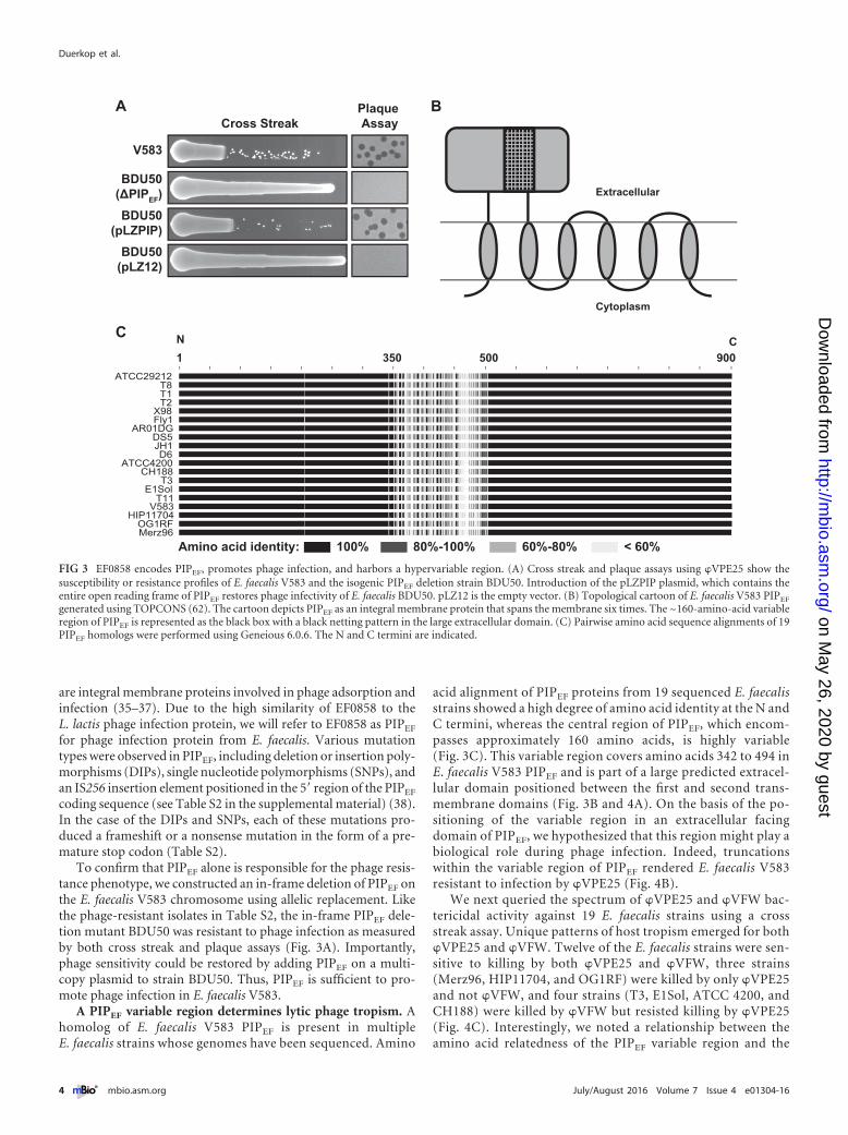
To confirm that PIPEF alone is responsible for the phage resis-tance phenotype, we constructed an in-frame deletion of PIPEF onthe E. faecalis V583 chromosome using allelic replacement. Likethe phage-resistant isolates in Table S2, the in-frame PIPEF dele-tion mutant BDU50 was resistant to phage infection as measuredby both cross streak and plaque assays (Fig. 3A). Importantly,phage sensitivity could be restored by adding PIPEF on a multi-copy plasmid to strain BDU50. Thus, PIPEF is sufficient to pro-mote phage infection in E. faecalis V583.
A PIPEF variable region determines lytic phage tropism. Ahomolog of E. faecalis V583 PIPEF is present in multipleE. faecalis strains whose genomes have been sequenced. Amino
acid alignment of PIPEF proteins from 19 sequenced E. faecalisstrains showed a high degree of amino acid identity at the N andC termini, whereas the central region of PIPEF, which encom-passes approximately 160 amino acids, is highly variable(Fig. 3C). This variable region covers amino acids 342 to 494 inE. faecalis V583 PIPEF and is part of a large predicted extracel-lular domain positioned between the first and second trans-membrane domains (Fig. 3B and 4A). On the basis of the po-sitioning of the variable region in an extracellular facingdomain of PIPEF, we hypothesized that this region might play abiological role during phage infection. Indeed, truncationswithin the variable region of PIPEF rendered E. faecalis V583resistant to infection by �VPE25 (Fig. 4B).
We next queried the spectrum of �VPE25 and �VFW bac-tericidal activity against 19 E. faecalis strains using a crossstreak assay. Unique patterns of host tropism emerged for both�VPE25 and �VFW. Twelve of the E. faecalis strains were sen-sitive to killing by both �VPE25 and �VFW, three strains(Merz96, HIP11704, and OG1RF) were killed by only �VPE25and not �VFW, and four strains (T3, E1Sol, ATCC 4200, andCH188) were killed by �VFW but resisted killing by �VPE25(Fig. 4C). Interestingly, we noted a relationship between theamino acid relatedness of the PIPEF variable region and the
Amino acid identity: 100% 80%-100% 60%-80% < 60%
1 350 500 900N C
BCross Streak
Plaque Assay
V583
BDU50(ΔPIPEF)BDU50
(pLZPIP)BDU50
(pLZ12)
A
Extracellular
Cytoplasm
C
FIG 3 EF0858 encodes PIPEF, promotes phage infection, and harbors a hypervariable region. (A) Cross streak and plaque assays using �VPE25 show thesusceptibility or resistance profiles of E. faecalis V583 and the isogenic PIPEF deletion strain BDU50. Introduction of the pLZPIP plasmid, which contains theentire open reading frame of PIPEF restores phage infectivity of E. faecalis BDU50. pLZ12 is the empty vector. (B) Topological cartoon of E. faecalis V583 PIPEF
generated using TOPCONS (62). The cartoon depicts PIPEF as an integral membrane protein that spans the membrane six times. The ~160-amino-acid variableregion of PIPEF is represented as the black box with a black netting pattern in the large extracellular domain. (C) Pairwise amino acid sequence alignments of 19PIPEF homologs were performed using Geneious 6.0.6. The N and C termini are indicated.
Duerkop et al.
4 ® mbio.asm.org July/August 2016 Volume 7 Issue 4 e01304-16
on May 26, 2020 by guest
http://mbio.asm
.org/D
ownloaded from
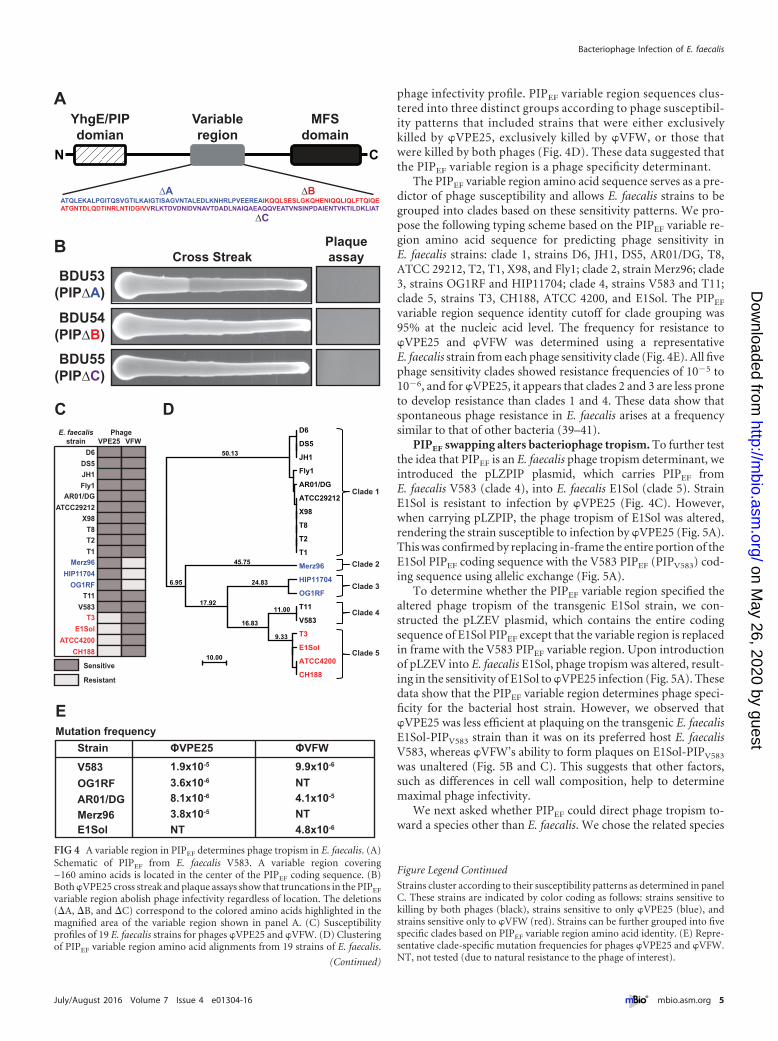
phage infectivity profile. PIPEF variable region sequences clus-tered into three distinct groups according to phage susceptibil-ity patterns that included strains that were either exclusivelykilled by �VPE25, exclusively killed by �VFW, or those thatwere killed by both phages (Fig. 4D). These data suggested thatthe PIPEF variable region is a phage specificity determinant.
The PIPEF variable region amino acid sequence serves as a pre-dictor of phage susceptibility and allows E. faecalis strains to begrouped into clades based on these sensitivity patterns. We pro-pose the following typing scheme based on the PIPEF variable re-gion amino acid sequence for predicting phage sensitivity inE. faecalis strains: clade 1, strains D6, JH1, DS5, AR01/DG, T8,ATCC 29212, T2, T1, X98, and Fly1; clade 2, strain Merz96; clade3, strains OG1RF and HIP11704; clade 4, strains V583 and T11;clade 5, strains T3, CH188, ATCC 4200, and E1Sol. The PIPEF
variable region sequence identity cutoff for clade grouping was95% at the nucleic acid level. The frequency for resistance to�VPE25 and �VFW was determined using a representativeE. faecalis strain from each phage sensitivity clade (Fig. 4E). All fivephage sensitivity clades showed resistance frequencies of 10�5 to10�6, and for �VPE25, it appears that clades 2 and 3 are less proneto develop resistance than clades 1 and 4. These data show thatspontaneous phage resistance in E. faecalis arises at a frequencysimilar to that of other bacteria (39–41).
PIPEF swapping alters bacteriophage tropism. To further testthe idea that PIPEF is an E. faecalis phage tropism determinant, weintroduced the pLZPIP plasmid, which carries PIPEF fromE. faecalis V583 (clade 4), into E. faecalis E1Sol (clade 5). StrainE1Sol is resistant to infection by �VPE25 (Fig. 4C). However,when carrying pLZPIP, the phage tropism of E1Sol was altered,rendering the strain susceptible to infection by �VPE25 (Fig. 5A).This was confirmed by replacing in-frame the entire portion of theE1Sol PIPEF coding sequence with the V583 PIPEF (PIPV583) cod-ing sequence using allelic exchange (Fig. 5A).
To determine whether the PIPEF variable region specified thealtered phage tropism of the transgenic E1Sol strain, we con-structed the pLZEV plasmid, which contains the entire codingsequence of E1Sol PIPEF except that the variable region is replacedin frame with the V583 PIPEF variable region. Upon introductionof pLZEV into E. faecalis E1Sol, phage tropism was altered, result-ing in the sensitivity of E1Sol to �VPE25 infection (Fig. 5A). Thesedata show that the PIPEF variable region determines phage speci-ficity for the bacterial host strain. However, we observed that�VPE25 was less efficient at plaquing on the transgenic E. faecalisE1Sol-PIPV583 strain than it was on its preferred host E. faecalisV583, whereas �VFW’s ability to form plaques on E1Sol-PIPV583
was unaltered (Fig. 5B and C). This suggests that other factors,such as differences in cell wall composition, help to determinemaximal phage infectivity.
We next asked whether PIPEF could direct phage tropism to-ward a species other than E. faecalis. We chose the related species
C D
A
ATQLEKALPGITQSVGTILKAIGTISAGVNTALEDLKNHRLPVEEREAIKQQLSESLGKQHENIQQLIQLFTQIQEATGNTDLQDTINRLNTIDGIVVRLKTDVDNIDVNAVTDADLNAIQAEAQQVEATVNSINPDAIENTVKTILDKLIAT
YhgE/PIPdomian
MFSdomain
N C
Variableregion
∆A ∆B
∆C
BBDU53
(PIP∆A)
BDU54(PIP∆B)
BDU55(PIP∆C)
Cross StreakPlaqueassay
V583
AR01/DGFly1
ATCC29212
DS5D6
T1T2T8
T11
X98
OG1RF
Merz96
JH1
HIP11704
T3
CH188
E1SolATCC4200
E. faecalisstrain
PhageVPE25 VFW
Sensitive
Resistant
D6
DS5
JH1
Fly1
AR01/DG
ATCC29212
X98
T8
T2
T1
Merz96
HIP11704
OG1RF
T11
V583
T3
E1Sol
ATCC4200
CH188
10.00
50.13
6.95
45.75
17.92
24.83
16.83
11.00
9.33
Clade 1
Clade 2
Clade 3
Clade 4
Clade 5
Mutation frequencyStrain ΦVPE25 ΦVFW
V583OG1RFAR01/DGMerz96E1Sol
1.9x10⁻⁵
3.6x10⁻⁶
8.1x10⁻⁶
3.8x10⁻⁵
4.8x10⁻⁶
NT
NT
NT
E
9.9x10⁻⁶
4.1x10⁻⁵
FIG 4 A variable region in PIPEF determines phage tropism in E. faecalis. (A)Schematic of PIPEF from E. faecalis V583. A variable region covering~160 amino acids is located in the center of the PIPEF coding sequence. (B)Both �VPE25 cross streak and plaque assays show that truncations in the PIPEF
variable region abolish phage infectivity regardless of location. The deletions(�A, �B, and �C) correspond to the colored amino acids highlighted in themagnified area of the variable region shown in panel A. (C) Susceptibilityprofiles of 19 E. faecalis strains for phages �VPE25 and �VFW. (D) Clusteringof PIPEF variable region amino acid alignments from 19 strains of E. faecalis.
(Continued)
Figure Legend Continued
Strains cluster according to their susceptibility patterns as determined in panelC. These strains are indicated by color coding as follows: strains sensitive tokilling by both phages (black), strains sensitive to only �VPE25 (blue), andstrains sensitive only to �VFW (red). Strains can be further grouped into fivespecific clades based on PIPEF variable region amino acid identity. (E) Repre-sentative clade-specific mutation frequencies for phages �VPE25 and �VFW.NT, not tested (due to natural resistance to the phage of interest).
Bacteriophage Infection of E. faecalis
July/August 2016 Volume 7 Issue 4 e01304-16 ® mbio.asm.org 5
on May 26, 2020 by guest
http://mbio.asm
.org/D
ownloaded from

E. faecium, focusing on three strains, Com12, Com15, and1,141,733, that each encode a PIP homolog that is ~65% identicalto V583 PIPEF. All three strains are resistant to infection by�VPE25 (Fig. 5D). To test whether the tropism of �VPE25 could
be redirected by introducing E. faecalis V583 PIPEF into thesestrains, we created the pPBPIP plasmid, which contains the entirecoding sequence of V583 PIPEF controlled by its native promoter.Expression of E. faecalis V583 PIPEF in E. faecium Com12 and1,141,733 conferred sensitivity to �VPE25, albeit to a lesser extentthan that observed when V583 PIPEF was expressed in E. faecalisE1Sol. This finding was reinforced by the observation of a zone ofclearance in the high phage titer region of cross streak plates forstrains Com12 and 1,141,733 (Fig. 5D). Expression of V583 PIPEF
in E. faecium Com15 did not render this strain sensitive to�VPE25 infection (Fig. 5D). These data show that PIP swapping inthe related species E. faecium can alter the tropism of lytic entero-coccal phages.
Ectopic expression of E. faecalis V583 PIPEF in E. faecium al-tered �VPE25 tropism, yet we did not observe visible plaquesduring the infection when performing an agar overlay experi-ment. Using E. faecium 1,141,733 carrying pPBPIP, we confirmedthat the addition of �VPE25 to logarithmically growing bacterialcells retarded growth, suggesting that these phages successfullyinfect the bacteria (see Fig. S3A in the supplemental material).However, after 2 h of growth in the presence of �VPE25, an accu-mulation of �VPE25 particles in the culture fluid was not ob-served (Fig. S3B). We detected phage transcripts after infection inboth wild-type and transgenic PIPEF E. faecium 1,141,733, sug-gesting that replicated phage particles were trapped within thebacteria due to inefficient host cell lysis (Fig. S3C). To test fortrapped viable phage particles, we treated the cells with lysozymeand sonication. Indeed, both E. faecium strains released viablephage particles upon sonication, whereas the control strainE. faecalis E1Sol did not (Fig. S3D). These data show that �VPE25infects E. faecium 1,141,733 and to a greater extent if the bacteriumis expressing E. faecalis V583 PIPEF from plasmid pPBPIP; how-ever, the phages cannot lyse the E. faecium cells.
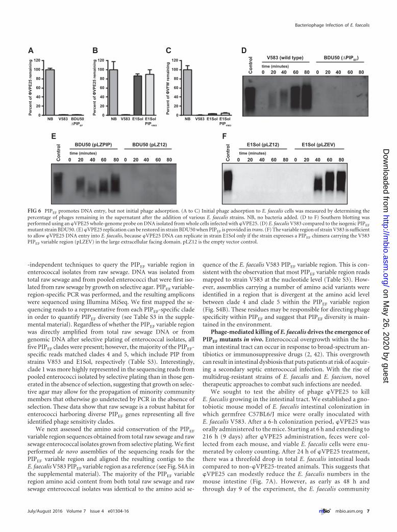
PIPEF is dispensable for initial phage attachment and is im-plicated in DNA entry. To determine the role of PIPEF duringphage adsorption, we used whole bacterial cell pulldown assays totest the ability of phages �VPE25 and �VFW to bind to theE. faecalis cell surface. �VPE25 bound to wild-type E. faecalis V583and the isogenic PIPEF mutant BDU50 equally well (Fig. 6A).However, �VPE25 did not bind to the surface of E. faecalis E1Sol,even when E1Sol had its PIPEF replaced with E. faecalis V583 PIPEF
on the chromosome (Fig. 6B). This is in contrast to �VFW whichadsorbed to E1Sol, transgenic PIPV583 E1Sol, and V583 similarly(Fig. 6C). These data suggest that PIPEF is not required for initialphage adsorption to E. faecalis.
We next sought to determine whether PIPEF is involved inphage DNA entry into E. faecalis cells. We infected E. faecalisstrains with �VPE25 and monitored intracellular phage replica-tion over time. As expected, �VPE25 replication was observed inE. faecalis V583 but not the PIPEF mutant strain BDU50 (Fig. 6D).�VPE25 replication could be restored in strain BDU50 byplasmid-encoded PIPEF (Fig. 6E). However, �VPE25 could repli-cate in E. faecalis E1Sol only when a plasmid-encoded chimericPIPEF was introduced into the strain (Fig. 6F). This chimeric PIPEF
was comprised of the E1Sol PIPEF sequence except for the variableregion, which was from V583 PIPEF. These data support the con-clusion that the PIPEF variable region facilitates phage DNA entry.
An environmental reservoir harbors E. faecalis with PIPEF
diversity. To characterize PIPEF diversity in a natural environ-ment where E. faecalis is endemic, we used culture-dependent and
E1Sol
E1Sol(pLZPIP)
E1Sol(pLZ12)
E1Sol(PIPV583)
E1Sol(pLZEV)
A
B
733
733(pPBPIP)
Com12
Com12(pPBPIP)
0.8
1.2
0.4
0.0Effic
ienc
y of
pla
quin
g (φφ
VPE2
5)
V583 E1Sol(PIPV583)
*
1.5
1.0
0.5
0.0Effic
ienc
y of
pla
quin
g (φ
VFW
)
V583 E1Sol(PIPV583)
E1Sol
C
D
Com15
Com15(pPBPIP)
Cross StreakPlaque Assay
Cross Streak
FIG 5 PIPEF swapping alters phage tropism. (A) Using the E. faecalis strainE1Sol, which is naturally resistant to infection by �VPE25, both cross streakand plaque assays showed that E1Sol can acquire �VPE25 susceptibility byexpressing the E. faecalis V583 PIPEF gene (pLZPIP). Single-copy replacementof the E. faecalis E1Sol PIPEF homolog with V583 PIPEF in the E1Sol chromo-some also confers �VPE25 sensitivity (PIPV583). A chimera of E. faecalis E1SolPIPEF and the variable region of V583 PIPEF show that the PIPEF variableregion determines phage tropism (pLZEV). (B and C) Plaquing efficiency of�VPE25 (B) and �VFW (C) on E. faecalis V583, E1Sol, and PIPV583 transgenicE1Sol strains. The value that was significantly different (P � 0.01) by Student’st test from the value for E. faecalis V583 is indicated by an asterisk. (D) Crossstreak assay showing that expression of E. faecalis V583 PIPEF from plasmidpPBPIP can confer �VPE25 sensitivity on E. faecium strains 1,141,733 andCom12, but not strain Com15.
Duerkop et al.
6 ® mbio.asm.org July/August 2016 Volume 7 Issue 4 e01304-16
on May 26, 2020 by guest
http://mbio.asm
.org/D
ownloaded from
-independent techniques to query the PIPEF variable region inenterococcal isolates from raw sewage. DNA was isolated fromtotal raw sewage and from pooled enterococci that were first iso-lated from raw sewage by growth on selective agar. PIPEF variable-region-specific PCR was performed, and the resulting ampliconswere sequenced using Illumina MiSeq. We first mapped the se-quencing reads to a representative from each PIPEF-specific cladein order to quantify PIPEF diversity (see Table S3 in the supple-mental material). Regardless of whether the PIPEF variable regionwas directly amplified from total raw sewage DNA or fromgenomic DNA after selective plating of enterococcal isolates, allfive PIPEF clades were present; however, the majority of the PIPEF-specific reads matched clades 4 and 5, which include PIP fromstrains V853 and E1Sol, respectively (Table S3). Interestingly,clade 1 was more highly represented in the sequencing reads frompooled enterococci isolated by selective plating than in those gen-erated in the absence of selection, suggesting that growth on selec-tive agar may allow for the propagation of minority communitymembers that otherwise go undetected by PCR in the absence ofselection. These data show that raw sewage is a robust habitat forenterococci harboring diverse PIPEF genes representing all fiveidentified phage sensitivity clades.
We next assessed the amino acid conservation of the PIPEF
variable region sequences obtained from total raw sewage and rawsewage enterococcal isolates grown from selective plating. We firstperformed de novo assemblies of the sequencing reads for thePIPEF variable region and aligned the resulting contigs to theE. faecalis V583 PIPEF variable region as a reference (see Fig. S4A inthe supplemental material). The majority of the PIPEF variableregion amino acid content from both total raw sewage and rawsewage enterococcal isolates was identical to the amino acid se-
quence of the E. faecalis V583 PIPEF variable region. This is con-sistent with the observation that most PIPEF variable region readsmapped to strain V583 at the nucleotide level (Table S3). How-ever, assemblies carrying a number of amino acid variants wereidentified in a region that is divergent at the amino acid levelbetween clade 4 and clade 5 within the PIPEF variable region(Fig. S4B). These residues may be responsible for directing phagespecificity within PIPEF and suggest that PIPEF diversity is main-tained in the environment.
Phage-mediated killing of E. faecalis drives the emergence ofPIPEF mutants in vivo. Enterococcal overgrowth within the hu-man intestinal tract can occur in response to broad-spectrum an-tibiotics or immunosuppressive drugs (2, 42). This overgrowthcan result in intestinal dysbiosis that puts patients at risk of acquir-ing a secondary septic enterococcal infection. With the rise ofmultidrug-resistant strains of E. faecalis and E. faecium, noveltherapeutic approaches to combat such infections are needed.
We sought to test the ability of phage �VPE25 to killE. faecalis growing in the intestinal tract. We established a gno-tobiotic mouse model of E. faecalis intestinal colonization inwhich germfree C57BL6/J mice were orally inoculated withE. faecalis V583. After a 6-h colonization period, �VPE25 wasorally administered to the mice. Starting at 6 h and extending to216 h (9 days) after �VPE25 administration, feces were col-lected from each mouse, and viable E. faecalis cells were enu-merated by colony counting. After 24 h of �VPE25 treatment,there was a threefold drop in total E. faecalis intestinal loadscompared to non-�VPE25-treated animals. This suggests that�VPE25 can modestly reduce the E. faecalis numbers in themouse intestine (Fig. 7A). However, as early as 48 h andthrough day 9 of the experiment, the E. faecalis community
A B C D
E F
Con
trol
0 20 40 60 80 0 20 40 60 80
V583 (wild type) BDU50 (∆PIPEF)C
ontr
ol
0 20 40 60 80 0 20 40 60 80
BDU50 (pLZPIP) BDU50 (pLZ12)time (minutes)
Perc
ent o
f ΦVP
E25
rem
aini
ng
0
20
40
60
80
100
120
NB V583 BDU50∆PIPEF
Perc
ent o
f ΦVP
E25
rem
aini
ng
NB V583 E1Sol E1SolPIPV583
0
20
40
60
80
100
120
NB V583 E1Sol E1SolPIPV583
Perc
ent o
f ΦVF
W re
mai
ning
0
20
40
60
80
100
120time (minutes)
Con
trol
0 20 40 60 80 0 20 40 60 80
E1Sol (pLZ12) E1Sol (pLZEV)time (minutes)
FIG 6 PIPEF promotes DNA entry, but not initial phage adsorption. (A to C) Initial phage adsorption to E. faecalis cells was measured by determining thepercentage of phages remaining in the supernatant after the addition of various E. faecalis strains. NB, no bacteria added. (D to F) Southern blotting wasperformed using an �VPE25 whole-genome probe on DNA isolated from whole cells infected with �VPE25. (D) E. faecalis V583 compared to the isogenic PIPEF
mutant strain BDU50. (E) �VPE25 replication can be restored in strain BDU50 when PIPEF is provided in trans. (F) The variable region of strain V583 is sufficientto allow �VPE25 DNA entry into E. faecalis, because �VPE25 DNA can replicate in strain E1Sol only if the strain expresses a PIPEF chimera carrying the V583PIPEF variable region (pLZEV) in the large extracellular facing domain. pLZ12 is the empty vector control.
Bacteriophage Infection of E. faecalis
July/August 2016 Volume 7 Issue 4 e01304-16 ® mbio.asm.org 7
on May 26, 2020 by guest
http://mbio.asm
.org/D
ownloaded from
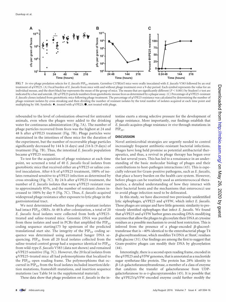
rebounded to the level of colonization observed for untreatedanimals, even when the phages were added to the drinkingwater for continuous administration (Fig. 7A). The number ofphage particles recovered from feces was the highest at 24 and48 h after �VPE25 treatment (Fig. 7B). Phage particles weremaintained in the intestines of these mice for the duration ofthe experiment, but the number of recoverable phage particlessignificantly decreased by 144 h (6 days) and 216 h (9 days) oftreatment (Fig. 7B). Thus, the intestinal E. faecalis populationbecame �VPE25 resistant.
To test for the acquisition of phage resistance at each timepoint, we screened a total of 40 E. faecalis fecal isolates fromgnotobiotic mice that received either an �VPE25 or saline con-trol inoculation. After 6 h of �VPE25 treatment, 100% of iso-lates remained sensitive to �VPE25 infection as determined bycross streaking (Fig. 7C). By 24 h after �VPE25 treatment, thenumber of E. faecalis isolates that were �VPE25 resistant roseto approximately 85%, and the number of resistant clones in-creased to 100% by day 9 (Fig. 7C). Thus, E. faecalis acquiredwidespread phage resistance after exposure to lytic phage in thegastrointestinal tract.
We next determined whether these phage-resistant isolateshad intact PIPEF ORFs. At 48 h after colonization, a total of 20E. faecalis fecal isolates were collected from both �VPE25-treated and saline-treated mice. Genomic DNA was purifiedfrom these isolates and used in PCRs that amplified the PIPEF
coding sequence starting175 bp upstream of the predictedtranslational start site. The integrity of the PIPEF coding se-quence was determined using automated Sanger DNA se-quencing. PIPEF from all 20 fecal isolates collected from thesaline-treated control group had a sequence identical to PIPEF
from wild-type E. faecalis V583 (data not shown) and remained�VPE25 sensitive (Fig. 7C). However, the 20 fecal isolates from�VPE25-treated mice all had polymorphisms that localized tothe PIPEF open reading frame. The polymorphisms that oc-curred in PIPEF from the fecal isolates included insertion/dele-tion mutations, frameshift mutations, and insertion sequencemutations (see Table S4 in the supplemental material).
These data show that phage predation on E. faecalis in the in-
testine exerts a strong selective pressure for the development ofphage resistance. More importantly, our findings establish thatE. faecalis acquires phage resistance in vivo through mutations inPIPEF.
DISCUSSION
Novel antimicrobial strategies are urgently needed to controlincreasingly frequent antibiotic-resistant bacterial infections.Phages have long held promise as potential antibacterial ther-apeutics, and thus, a revival in phage therapy has begun overthe last several years. This has led to a renaissance in an under-standing of the basic molecular biology of phages and theircontributions to host-pathogen interactions (43). This is espe-cially relevant for Gram-positive pathogens, such as E. faecalis,that place a heavy burden on the health care system. However,before phages can be routinely used as antienterococcal thera-peutics, a detailed understanding of how they interact withtheir bacterial hosts and the mechanisms that enterococci useto avoid phage infection need to be delineated.
In this study, we have discovered two previously unidentifiedlytic siphophages, �VPE25 and �VFW, which infect E. faecalis.These phages are unique and have little genomic similarity to pre-viously identified siphophages that infect E. faecalis. We foundthat �VPE25 and �VFW harbor genes encoding DNA-modifyingenzymes that allow the phages to glycosylate their DNA at cytosineresidues as a possible mechanism to avoid host restriction. This isinferred from the presence of a phage-encoded �-glucosyl-transferase that is ~40% identical to the enterobacterial phage T4�-glucosyltransferase, which modifies T4 DNA at 5hmC residueswith glucose (31). Our findings are among the first to suggest thatGram-positive phages can modify their DNA by glycosylation(44).
Interestingly, there is a second open reading frame, encoded inthe �VPE25 and �VFW genomes, that is annotated as a nucleotidesugar synthetase-like protein. The protein has 28% identity to�-1,6-galactofuranosyltransferase, a member of a class of enzymethat catalyzes the transfer of galactofuranose from UDP-galactofuranose to �-D-glucopyranosides (45). It is possible thatthe �VPE25/�VFW-encoded enzyme could further modify the
107
108
109
1010
6 24 48 144 216
*
Time (hours)
cfu
per g
ram
of f
eces
A
105
106
107
108
109
1010
Time (hours)24 48 144 216
pfu
per g
ram
of f
eces
Time (hours)
Perc
ent Φ
VPE2
5 re
sist
ance
B C
246 48 144 216
0
25
50
75
100
FIG 7 In vivo phage predation selects for E. faecalis PIPEF mutants. Germfree C57BL6/J mice were orally inoculated with E. faecalis V583 followed by an oraltreatment of �VPE25. (A) Fecal burden of E. faecalis from mice with and without phage treatment over a 9-day period. Each symbol represents the value for anindividual mouse, and the short black bar represents the mean of the group of mice. The means that are significantly different (P � 0.001) by Student’s t test areindicated by a bar and asterisk. (B) �VPE25 particle numbers from gnotobiotic mouse feces as determined by a plaque assay. (C) Percentage of �VPE25-resistantE. faecalis clones isolated from gnotobiotic mice following phage treatment. The percentage of �VPE25 resistance was calculated by determining the number ofphage-resistant isolates by cross streaking and then dividing the number of resistant isolates by the total number of isolates acquired at each time point andmultiplying by 100. Symbols: �, treated with �VPE25; �, not treated with phage.
Duerkop et al.
8 ® mbio.asm.org July/August 2016 Volume 7 Issue 4 e01304-16
on May 26, 2020 by guest
http://mbio.asm
.org/D
ownloaded from
glycosylated phage DNA. Genome sequence analysis did not un-cover any putative virulence factors or antibiotic resistance genes,suggesting that �VPE25 and �VFW should be considered candi-date phages to be further studied and possibly modified for ther-apeutic applications.
Our studies of �VPE25 and �VFW led us to identify PIPEF asan E. faecalis integral membrane protein that is essential for phageinfection. We used the phages to select for phage-resistant mu-tants of E. faecalis V583 and found that genomic mutations asso-ciated with resistance were clustered in the PIPEF-encoding read-ing frame EF0858. PIPEF is orthologous to L. lactis PIP, whichpromotes phage binding and infection (35, 36). PIPEF harbors twodistinctive domains: an N-terminal YhgE/PIP domain that is con-served in all Firmicutes that harbor a PIP ortholog and lacks aknown function and a C-terminal major facilitator superfamily(MFS) domain that might play a role in small-molecule transport.
PIPEF is conserved among E. faecalis strains, suggesting thatPIPEF performs an important biological function in E. faecalis.Interestingly, a second protein containing the YhgE/PIP domainwas identified in 8 of the 19 E. faecalis strains used in this study.This protein has 21% sequence identity to the N-terminal YhgE/PIP domain of PIPEF and resides in a cluster of genes annotated tofunction as a type VIIb secretion system (46). This protein is anortholog of the S. aureus EsaA type VIIb secretion system proteinand has a similar predicted topology of six transmembrane do-mains and a C-terminal MFS domain as observed for PIPEF (47).We therefore speculate that PIPEF may be involved in the trans-port of a small molecule or function in concert with a type VIIbsecretion system to secrete effector proteins across the bacterialmembrane.
PIPEF harbors a 160-amino-acid region with marked sequencediversity. This protein region is centrally located within the firstpredicted extracellular domain of PIPEF and can be used to groupE. faecalis strains based on their phage susceptibility profile. Wefound that this variable region is both necessary and sufficient todrive phage tropism for specific host bacteria. Raw sewage har-bored E. faecalis with diverse PIPEF variable regions, suggestingthat sewage phages coevolved with diverse E. faecalis strains. Thechanging abundance of such strains due to phage predation maydrive the emergence of phages with altered tropisms for PIPEF
variants.Variable regions of cell wall-embedded proteins have been im-
plicated in phage specificity for Gram-negative bacteria (48–50).In Gram-positive bacteria, the study of how variation inbacterium-encoded phage receptors impacts phage tropism hasbeen limited to cell wall-associated polysaccharides (51, 52). Toour knowledge, our findings constitute the first description of adiversifying region of a bacterial membrane protein phage recep-tor in Gram-positive bacteria.
A key remaining question is the identity of the phage antire-ceptor that interacts with PIPEF. Known phage antireceptors arecomponents of the phage tail (53–55). Several phage tail genes areclustered together in the �VPE25 and �VFW genomes. One par-ticular gene, represented by orf_112 in �VPE25 and orf_110 in�VFW, is a candidate for the antireceptor as it is orthologous toStreptococcus thermophilus phage antireceptors with hypervariableregions that specify phage tropism (56).
The PIPEF variable region sequences allowed clustering ofE. faecalis strains into clades based on PIPEF homology and phagesensitivity. With the continued classification of lytic phages that
target E. faecalis through a PIPEF-dependent mechanism and theirassignment to the infection of specific clades, we envision the useof PIPEF variable region sequencing as a precursor for the selectionof potential therapeutic phages that could be used to selectivelykill E. faecalis outbreak strains.
Using a gnotobiotic mouse model of in vivo phage predation,we found that E. faecalis acquires phage resistance through muta-tions in PIPEF. Lytic phages can only modestly reduce E. faecalisnumbers in the intestines of gnotobiotic mice, and E. faecalisnumbers rebound due to the selective overgrowth of PIPEF mu-tants. Thus, prophylactic phage administration provides a strongselective pressure for the emergence of PIPEF mutations inE. faecalis.
The rapid development of phage resistance is just one of severalbarriers to deploying phages therapeutically (57). Although thesephages are specific killers of E. faecalis, their exquisite selectivityfor a target host enables the development of phage infection resis-tance and highlights a potential barrier to phage monotherapiesagainst E. faecalis. We suggest that our identification of a keydriver of phage resistance in E. faecalis could facilitate the engi-neering of phages with altered tropism. Likewise, once identified,the phage antireceptor specific for PIPEF could be modified bysite-directed mutagenesis in hopes of loosening its specificity forcell wall targets. Chemical mutagenesis could also be performedon the phage genomes to create phages with altered tropisms.These approaches could enable the development of phage cock-tails that circumvent the problem of resistance and could thus beused to treat E. faecalis infections.
MATERIALS AND METHODSBacterial strains and bacteriophages. A complete list of bacterial strainsand phages used in this study can be found in Table S5 in the supplementalmaterial. E. faecalis and E. faecium were grown statically in brain heartinfusion (BHI) broth or on BHI agar at 37°C. Escherichia coli was grown inLennox L broth (LB) with shaking or on LB agar at 37°C. Chloramphen-icol was added at 15 �g/ml for E. faecalis and E. faecium or 8 �g/ml forE. coli when needed. Enterococci from municipal raw sewage were isolatedusing Enterococcosel agar (Becton Dickinson). Growth conditions for thegeneration of mutant strains of E. faecalis by allelic exchange were asspecified by Thurlow et al. (58). For a more detailed description of bacte-rial growth conditions and for plasmid construction, see Text S1 andTable S5 in the supplemental material, respectively.
Phages �VPE25 and �VFW were isolated from untreated raw sewageobtained from a Dallas-Fort Worth water reclamation facility in Texas.Fifty milliliters of raw sewage was centrifuged at 3,220 � g for 10 min atroom temperature (RT) to sediment large particles. The supernatant wasdecanted and passed through a 0.45-�m filter. One hundred microlitersof clarified sewage was mixed with 130 �l of a 1:10 dilution of an overnight(O/N) culture of E. faecalis V583 and then added to Todd-Hewitt broth(THB) top agar (0.35% agar) and poured over a 1.5% agar THB plate.Both top agar and base agar were supplemented with 10 mM MgSO4.After O/N growth at 37°C, the resulting plaques were recovered using asterile Pasteur pipette, and phages were eluted from the agar plugs in500 �l of SM-plus buffer (100 mM NaCl, 50 mM Tris-HCl, 8 mM MgSO4,5 mM CaCl2 [pH 7.4]) O/N at 4°C. Phages were replaqued on E. faecalisV583 two more times to ensure that the phages were clonal isolates.
High-titer phage stocks were propagated by infecting 300 ml of loga-rithmically growing E. faecalis V583 at a multiplicity of infection of 0.1 inBHI broth containing 10 mM MgSO4. Lysis was allowed to proceed for 4 hat 37°C with shaking. The remaining bacterial cells and debris were pel-leted at 4,400 � g for 10 min at RT. The culture supernatant was filteredthrough a 0.45-�m membrane and treated with 5 �g/ml each of DNaseand RNase at RT for 1 h, and phages were precipitated by adding 1 M NaCl
Bacteriophage Infection of E. faecalis
July/August 2016 Volume 7 Issue 4 e01304-16 ® mbio.asm.org 9
on May 26, 2020 by guest
http://mbio.asm
.org/D
ownloaded from
and 10% (wt/vol) polyethylene glycol 8000 (PEG 8000) and incubated onice O/N at 4°C. Phages were pelleted by centrifugation at 11,270 � g andresuspended in 2 ml of SM-plus buffer. One-third volume of chloroformwas added with shaking, and the phases were separated by centrifugationat 16,300 � g. The aqueous phase containing the phages was subjected tofurther purification using cesium chloride centrifugation as describedpreviously (59). Phages were enumerated using the THB agar overlayplaque assay described above.
Phage cross streak assays. We used cross streaking to determine thesensitivity of various E. faecalis and E. faecium strains to phages �VPE25and �VFW. A total of 109 to 1010 phage particles were streaked down thecenter of a THB agar plate containing 10 mM MgSO4 with or without15 �g/ml chloramphenicol when necessary. Ten microliters of an O/Nbacterial culture was spread horizontally across the phage streak. Theplates were incubated at 37°C O/N, and bacterial strain sensitivity to aparticular phage was indicated by limited to no bacterial growth withinand beyond the phage streak area.
Whole-genome sequencing. The genomes of phages �VPE25 and�VFW and E. faecalis phage-resistant isolates were sequenced by TuftsUniversity Core Facility (TUCF) Genomics, Tufts University. The phagegenomes were sequenced using Illumina MiSeq paired-end 250-bp DNAsequencing with an average coverage depth of 881� for �VPE25 and903� for �VFW. The E. faecalis phage-resistant isolates were sequencedusing Illumina HiSeq2000 single-end 100-bp DNA sequencing. Variantcoverage information for these strains can be found in Table S2. Librarieswere prepared with the Nextera XT library preparation kit. All assemblieswere performed using CLC Workbench (Qiagen). For additional infor-mation about DNA sequencing and for explanations of bioinformaticapplications, refer to Text S1.
Animals. Germfree C57BL6/J mice were reared at University of Texas(UT) Southwestern Medical Center under sterile conditions as previouslydescribed (60). Gnotobiotic E. faecalis-colonized mice were established byorally inoculating male C57BL6/J mice with 5 � 107 CFU of E. faecalisV583. Intestinal colonization levels were determined by homogenizingfresh fecal pellets in 1 ml of sterile phosphate-buffered saline (PBS) andperforming colony counting on Enterococcosel agar. When appropriate,�VPE25 was administered by orally gavaging mice with 1 � 1010 PFU andby administering phage in drinking water at a concentration of 5 �108 PFU/ml. All animal protocols were approved by the Institutional An-imal Care and Use Committee of UT Southwestern Medical Center.
Accession numbers. All sequences generated for this study have beendeposited in the European Nucleotide Archive. The following accessionnumbers have been assigned: PRJEB13004 (�VPE25 assembled genome),PRJEB13155 (�VFW assembled genome), PRJEB13005 (E. faecalis phage-resistant isolates), and PRJEB13161 (PIPEF raw sewage amplicons).
SUPPLEMENTAL MATERIALSupplemental material for this article may be found at http://mbio.asm.org/lookup/suppl/doi:10.1128/mBio.01304-16/-/DCSupplemental.
Text S1, PDF file, 0.1 MB.Figure S1, PDF file, 1.4 MB.Figure S2, PDF file, 0.8 MB.Figure S3, PDF file, 0.1 MB.Figure S4, PDF file, 1.4 MB.Table S1, PDF file, 0.2 MB.Table S2, PDF file, 0.04 MB.Table S3, PDF file, 0.04 MB.Table S4, PDF file, 0.04 MB.Table S5, PDF file, 0.1 MB.
ACKNOWLEDGMENTS
We thank C. Boyd, B. Hassell, and T. Leal for assistance with gnotobioticanimal husbandry, H. Zhang for assistance with electron microscopy, B.Zhang for preliminary screening of PIPEF mutants and phage host tro-pism, and D. Propheter for providing nucleotide art shown in Fig. 2A.
FUNDING INFORMATIONThis work, including the efforts of Kelli L Palmer, was funded by HHS |NIH | National Institute of Allergy and Infectious Diseases (NIAID)(R01AI116610 and K22AI099088). This work, including the efforts ofLora V Hooper, was funded by HHS | NIH | National Institute of Diabetesand Digestive and Kidney Diseases (NIDDK) (R01DK070855). This work,including the efforts of Breck A Duerkop, was funded by HHS | NIH |National Institute of Diabetes and Digestive and Kidney Diseases (NI-DDK) (K01DK102436). This work, including the efforts of Lora VHooper, was funded by Howard Hughes Medical Institute (HHMI). Thiswork, including the efforts of Lora V Hooper, was funded by a BurroughsWellcome Foundation New Investigators in the Pathogenesis of Infec-tious Diseases Award.
REFERENCES1. Lebreton F, Willems RJL, Gilmore MS. 2014. Enterococcus diversity,
origins in nature, and gut colonization. In Gilmore MS, Clewell DB, Ike Y,Shankar N (ed), Enterococci: from commensals to leading causes of drugresistant infection. Massachusetts Eye and Ear Infirmary, Boston, MA.
2. Ubeda C, Taur Y, Jenq RR, Equinda MJ, Son T, Samstein M, Viale A,Socci ND, van den Brink MR, Kamboj M, Pamer EG. 2010.Vancomycin-resistant Enterococcus domination of intestinal microbiota isenabled by antibiotic treatment in mice and precedes bloodstream inva-sion in humans. J Clin Invest 120:4332– 4341. http://dx.doi.org/10.1172/JCI43918.
3. Wisplinghoff H, Bischoff T, Tallent SM, Seifert H, Wenzel RP, EdmondMB. 2004. Nosocomial bloodstream infections in US hospitals: analysis of24,179 cases from a prospective nationwide surveillance study. Clin InfectDis 39:309 –317. http://dx.doi.org/10.1086/421946.
4. Sievert DM, Ricks P, Edwards JR, Schneider A, Patel J, Srinivasan A,Kallen A, Limbago B, Fridkin S, National Healthcare Safety Network(NHSN) Team and Participating NHSN Facilities. 2013. Antimicrobial-resistant pathogens associated with healthcare-associated infections: sum-mary of data reported to the National Healthcare Safety Network at theCenters for Disease Control and Prevention, 2009 –2010. Infect ControlHosp Epidemiol 34:1–14. http://dx.doi.org/10.1086/668770.
5. Agudelo Higuita NI, Huycke MM. 2014. Enterococcal disease, epidemi-ology, and implications for treatment. In Gilmore MS, Clewell DB, Ike Y,Shankar N (ed), Enterococci: from commensals to leading causes of drugresistant infection. Massachusetts Eye and Ear Infirmary, Boston, MA.
6. Cattoir V, Leclercq R. 2013. Twenty-five years of shared life withvancomycin-resistant enterococci: is it time to divorce? J Antimicrob Che-mother 68:731–742. http://dx.doi.org/10.1093/jac/dks469.
7. Arias CA, Panesso D, McGrath DM, Qin X, Mojica MF, Miller C, DiazL, Tran TT, Rincon S, Barbu EM, Reyes J, Roh JH, Lobos E, SodergrenE, Pasqualini R, Arap W, Quinn JP, Shamoo Y, Murray BE, WeinstockGM. 2011. Genetic basis for in vivo daptomycin resistance in enterococci.N Engl J Med 365:892–900. http://dx.doi.org/10.1056/NEJMoa1011138.
8. Palmer KL, Daniel A, Hardy C, Silverman J, Gilmore MS. 2011. Geneticbasis for daptomycin resistance in enterococci. Antimicrob Agents Che-mother 55:3345–3356. http://dx.doi.org/10.1128/AAC.00207-11.
9. Jasni AS, Mullany P, Hussain H, Roberts AP. 2010. Demonstration ofconjugative transposon (Tn5397)-mediated horizontal gene transfer be-tween Clostridium difficile and Enterococcus faecalis. Antimicrob AgentsChemother 54:4924 – 4926. http://dx.doi.org/10.1128/AAC.00496-10.
10. Weigel LM, Clewell DB, Gill SR, Clark NC, McDougal LK, FlannaganSE, Kolonay JF, Shetty J, Killgore GE, Tenover FC. 2003. Geneticanalysis of a high-level vancomycin-resistant isolate of Staphylococcus au-reus. Science 302:1569 –1571. http://dx.doi.org/10.1126/science.1090956.
11. Sulakvelidze A, Alavidze Z, Morris JG, Jr. 2001. Bacteriophage therapy.Antimicrob Agents Chemother 45:649 – 659. http://dx.doi.org/10.1128/AAC.45.3.649-659.2001.
12. Nobrega FL, Costa AR, Kluskens LD, Azeredo J. 2015. Revisiting phagetherapy: new applications for old resources. Trends Microbiol 23:185–191. http://dx.doi.org/10.1016/j.tim.2015.01.006.
13. Duerkop BA, Palmer KL, Horsburgh MJ. 2014. Enterococcal bacterio-phages and genome defense. In Gilmore MS, Clewell DB, Ike Y, Shankar N(ed), Enterococci: from commensals to leading causes of drug resistantinfection. Massachusetts Eye and Ear Infirmary, Boston, MA.
14. Matos RC, Lapaque N, Rigottier-Gois L, Debarbieux L, Meylheuc T,Gonzalez-Zorn B, Repoila F, Lopes MDF, Serror P. 2013. Enterococcus
Duerkop et al.
10 ® mbio.asm.org July/August 2016 Volume 7 Issue 4 e01304-16
on May 26, 2020 by guest
http://mbio.asm
.org/D
ownloaded from
faecalis prophage dynamics and contributions to pathogenic traits. PLoSGenet 9:e1003539. http://dx.doi.org/10.1371/journal.pgen.1003539.
15. Rossmann FS, Racek T, Wobser D, Puchalka J, Rabener EM, Reiger M,Hendrickx AP, Diederich AK, Jung K, Klein C, Huebner J. 2015.Phage-mediated dispersal of biofilm and distribution of bacterial viru-lence genes is induced by quorum sensing. PLoS Pathog 11:e1004653.http://dx.doi.org/10.1371/journal.ppat.1004653.
16. Duerkop BA, Clements CV, Rollins D, Rodrigues JL, Hooper LV. 2012.A composite bacteriophage alters colonization by an intestinal commensalbacterium. Proc Natl Acad Sci U S A 109:17621–17626. http://dx.doi.org/10.1073/pnas.1206136109.
17. Khalifa L, Brosh Y, Gelman D, Coppenhagen-Glazer S, Beyth S,Poradosu-Cohen R, Que YA, Beyth N, Hazan R. 2015. Targeting En-terococcus faecalis biofilms with phage therapy. Appl Environ Microbiol81:2696 –2705. http://dx.doi.org/10.1128/AEM.00096-15.
18. Letkiewicz S, Miedzybrodzki R, Fortuna W, Weber-Dabrowska B, Gor-ski A. 2009. Eradication of Enterococcus faecalis by phage therapy inchronic bacterial prostatitis— case report. Folia Microbiol (Praha) 54:457– 461. http://dx.doi.org/10.1007/s12223-009-0064-z.
19. Ackermann HW. 2007. 5500 phages examined in the electron microscope.Arch Virol 152:227–243. http://dx.doi.org/10.1007/s00705-006-0849-1.
20. Li X, Ding P, Han C, Fan H, Wang Y, Mi Z, Feng F, Tong Y. 2014.Genome analysis of Enterococcus faecalis bacteriophage IME-EF3 harbor-ing a putative metallo-beta-lactamase gene. Virus Genes 49:145–151.http://dx.doi.org/10.1007/s11262-014-1079-3.
21. Lee YD, Park JH. 2012. Complete genome sequence of enterococcal bac-teriophage SAP6. J Virol 86:5402–5403. http://dx.doi.org/10.1128/JVI.00321-12.
22. Son JS, Jun SY, Kim EB, Park JE, Paik HR, Yoon SJ, Kang SH, Choi YJ.2010. Complete genome sequence of a newly isolated lytic bacteriophage,EFAP-1 of Enterococcus faecalis, and antibacterial activity of its endolysinEFAL-1. J Appl Microbiol 108:1769 –1779. http://dx.doi.org/10.1111/j.1365-2672.2009.04576.x.
23. Fard RM, Barton MD, Arthur JL, Heuzenroeder MW. 2010. Whole-genome sequencing and gene mapping of a newly isolated lytic enterococ-cal bacteriophage EFRM31. Arch Virol 155:1887–1891. http://dx.doi.org/10.1007/s00705-010-0800-3.
24. Altschul SF, Gish W, Miller W, Myers EW, Lipman DJ. 1990. Basic localalignment search tool. J Mol Biol 215:403– 410. http://dx.doi.org/10.1016/S0022-2836(05)80360-2.
25. Chopin A, Deveau H, Ehrlich SD, Moineau S, Chopin MC. 2007. KSY1,a lactococcal phage with a T7-like transcription. Virology 365:1–9. http://dx.doi.org/10.1016/j.virol.2007.03.044.
26. Aziz RK, Bartels D, Best AA, DeJongh M, Disz T, Edwards RA,Formsma K, Gerdes S, Glass EM, Kubal M, Meyer F, Olsen GJ, OlsonR, Osterman AL, Overbeek RA, McNeil LK, Paarmann D, Paczian T,Parrello B, Pusch GD, Reich C, Stevens R, Vassieva O, Vonstein V,Wilke A, Zagnitko O. 2008. The RAST server: rapid annotations usingsubsystems technology. BMC Genomics 9:75. http://dx.doi.org/10.1186/1471-2164-9-75.
27. Brüssow H, Canchaya C, Hardt WD. 2004. Phages and the evolution ofbacterial pathogens: from genomic rearrangements to lysogenic conver-sion. Microbiol Mol Biol Rev 68:560 – 602. http://dx.doi.org/10.1128/MMBR.68.3.560-602.2004.
28. Volkin E. 1954. The linkage of glucose in coliphage nucleic acids. J AmChem Soc 76:5892–5893. http://dx.doi.org/10.1021/ja01651a117.
29. Josse J, Kornberg A. 1962. Glucosylation of deoxyribonucleic acid. III. �-and �-glucosyl transferases from T4-infected Escherichia coli. J Biol Chem237:1968 –1976.
30. Jesaitis MA. 1956. Differences in the chemical composition of the phagenucleic acids. Nature 178:637. http://dx.doi.org/10.1038/178637a0.
31. Morera S, Imberty A, Aschke-Sonnenborn U, Ruger W, Freemont PS.1999. T4 phage �-glucosyltransferase: substrate binding and proposedcatalytic mechanism. J Mol Biol 292:717–730.
32. Huang LH, Farnet CM, Ehrlich KC, Ehrlich M. 1982. Digestion of highlymodified bacteriophage DNA by restriction endonucleases. Nucleic AcidsRes 10:1579 –1591. http://dx.doi.org/10.1093/nar/10.5.1579.
33. Huang Y, Pastor WA, Shen Y, Tahiliani M, Liu DR, Rao A. 2010. Thebehaviour of 5-hydroxymethylcytosine in bisulfite sequencing. PLoS One5:e8888. http://dx.doi.org/10.1371/journal.pone.0008888.
34. Janosi L, Yonemitsu H, Hong H, Kaji A. 1994. Molecular cloning andexpression of a novel hydroxymethylcytosine-specific restriction enzyme
(PvuRts1I) modulated by glucosylation of DNA. J Mol Biol 242:45– 61.http://dx.doi.org/10.1006/jmbi.1994.1556.
35. Geller BL, Ivey RG, Trempy JE, Hettinger-Smith B. 1993. Cloning of achromosomal gene required for phage infection of Lactococcus lactissubsp. lactis C2. J Bacteriol 175:5510 –5519.
36. Monteville MR, Ardestani B, Geller BL. 1994. Lactococcal bacterio-phages require a host cell wall carbohydrate and a plasma membrane pro-tein for adsorption and ejection of DNA. Appl Environ Microbiol 60:3204 –3211.
37. São-José C, Baptista C, Santos MA. 2004. Bacillus subtilis operon encod-ing a membrane receptor for bacteriophage SPP1. J Bacteriol 186:8337– 8346. http://dx.doi.org/10.1128/JB.186.24.8337-8346.2004.
38. Shankar N, Baghdayan AS, Gilmore MS. 2002. Modulation of virulencewithin a pathogenicity island in vancomycin-resistant Enterococcus faeca-lis. Nature 417:746 –750. http://dx.doi.org/10.1038/nature00802.
39. Demerec M, Fano U. 1945. Bacteriophage-resistant mutants in Esche-richia coli. Genetics 30:119 –136.
40. Le S, Yao X, Lu S, Tan Y, Rao X, Li M, Jin X, Wang J, Zhao Y, Wu NC,Lux R, He X, Shi W, Hu F. 2014. Chromosomal DNA deletion confersphage resistance to Pseudomonas aeruginosa. Sci Rep 4:4738. http://dx.doi.org/10.1038/srep04738.
41. King WR, Collins EB, Barrett EL. 1983. Frequencies of bacteriophage-resistant and slow acid-producing variants of Streptococcus cremoris. ApplEnviron Microbiol 45:1481–1485.
42. Donskey CJ, Chowdhry TK, Hecker MT, Hoyen CK, Hanrahan JA,Hujer AM, Hutton-Thomas RA, Whalen CC, Bonomo RA, Rice LB.2000. Effect of antibiotic therapy on the density of vancomycin-resistantenterococci in the stool of colonized patients. N Engl J Med 343:1925–1932. http://dx.doi.org/10.1056/NEJM200012283432604.
43. Young R, Gill JJ. 2015. Phage therapy redux what is to be done? Science350:1163–1164. http://dx.doi.org/10.1126/science.aad6791.
44. Rosenberg E. 1965. D-Mannose as a constituent of the DNA of a mutantstrain of bacteriophage Sp8. Proc Natl Acad Sci U S A 53:836 – 841. http://dx.doi.org/10.1073/pnas.53.4.836.
45. Wing C, Errey JC, Mukhopadhyay B, Blanchard JS, Field RA. 2006.Expression and initial characterization of WbbI, a putative D-Galf:�-D-Glc�-1,6-galactofuranosyltransferase from Escherichia coli K-12. Org BiomolChem 4:3945–3950. http://dx.doi.org/10.1039/b609455d.
46. Abdallah AM, Gey van Pittius NC, Champion PA, Cox J, Luirink J,Vandenbroucke-Grauls CM, Appelmelk BJ, Bitter W. 2007. Type VIIsecretion mycobacteria show the way. Nat Rev Microbiol 5:883– 891.http://dx.doi.org/10.1038/nrmicro1773.
47. Burts ML, Williams WA, DeBord K, Missiakas DM. 2005. EsxA andEsxB are secreted by an ESAT-6-like system that is required for the patho-genesis of Staphylococcus aureus infections. Proc Natl Acad Sci U S A102:1169 –1174. http://dx.doi.org/10.1073/pnas.0405620102.
48. Rabsch W, Ma L, Wiley G, Najar FZ, Kaserer W, Schuerch DW, KlebbaJE, Roe BA, Laverde Gomez JA, Schallmey M, Newton SM, Klebba PE.2007. FepA- and TonB-dependent bacteriophage H8: receptor bindingand genomic sequence. J Bacteriol 189:5658 –5674. http://dx.doi.org/10.1128/JB.00437-07.
49. German GJ, Misra R. 2001. The TolC protein of Escherichia coli serves asa cell-surface receptor for the newly characterized TLS bacteriophage. JMol Biol 308:579 –585. http://dx.doi.org/10.1006/jmbi.2001.4578.
50. Nieweg A, Bremer E. 1997. The nucleoside-specific Tsx channel from theouter membrane of Salmonella typhimurium, Klebsiella pneumoniae andEnterobacter aerogenes: functional characterization and DNA sequenceanalysis of the tsx genes. Microbiology 143:603– 615. http://dx.doi.org/10.1099/00221287-143-2-603.
51. Ainsworth S, Sadovskaya I, Vinogradov E, Courtin P, Guerardel Y,Mahony J, Grard T, Cambillau C, Chapot-Chartier MP, van SinderenD. 2014. Differences in lactococcal cell wall polysaccharide structure aremajor determining factors in bacteriophage sensitivity. mBio 5:e00880-14. http://dx.doi.org/10.1128/mBio.00880-14.
52. Eugster MR, Morax LS, Hüls VJ, Huwiler SG, Leclercq A, Lecuit M,Loessner MJ. 2015. Bacteriophage predation promotes serovar diversifi-cation in Listeria monocytogenes. Mol Microbiol 97:33– 46. http://dx.doi.org/10.1111/mmi.13009.
53. Hashemolhosseini S, Holmes Z, Mutschler B, Henning U. 1994. Alter-ations of receptor specificities of coliphages of the T2 family. J Mol Biol240:105–110. http://dx.doi.org/10.1006/jmbi.1994.1424.
54. Vegge CS, Vogensen FK, McGrath S, Neve H, van Sinderen D, Brønd-
Bacteriophage Infection of E. faecalis
July/August 2016 Volume 7 Issue 4 e01304-16 ® mbio.asm.org 11
on May 26, 2020 by guest
http://mbio.asm
.org/D
ownloaded from
sted L. 2006. Identification of the lower baseplate protein as the antirecep-tor of the temperate lactococcal bacteriophages TP901-1 and Tuc2009. JBacteriol 188:55– 63. http://dx.doi.org/10.1128/JB.188.1.55-63.2006.
55. Stuer-Lauridsen B, Janzen T, Schnabl J, Johansen E. 2003. Identificationof the host determinant of two prolate-headed phages infecting Lactococ-cus lactis. Virology 309:10 –17. http://dx.doi.org/10.1016/S0042-6822(03)00012-6.
56. Duplessis M, Moineau S. 2001. Identification of a genetic determinantresponsible for host specificity in Streptococcus thermophilus bacterio-phages. Mol Microbiol 41:325–336. http://dx.doi.org/10.1046/j.1365-2958.2001.02521.x.
57. Henein A. 2013. What are the limitations on the wider therapeutic use ofphage? Bacteriophage 3:e24872. http://dx.doi.org/10.4161/bact.24872.
58. Thurlow LR, Thomas VC, Hancock LE. 2009. Capsular polysaccharideproduction in Enterococcus faecalis and contribution of CpsF to capsule
serospecificity. J Bacteriol 191:6203– 6210. http://dx.doi.org/10.1128/JB.00592-09.
59. Sambrook J, Fritsch EF, Maniatis T. 1989. Bacteriophage� vectors, p2.1–2.125. In Nolan C (ed), Molecular cloning: a laboratory manual, vol 1,2nd ed. Cold Spring Harbor Laboratory Press, Cold Spring Harbor, NY.
60. Cash HL, Whitham CV, Behrendt CL, Hooper LV. 2006. Symbioticbacteria direct expression of an intestinal bactericidal lectin. Science 313:1126 –1130. http://dx.doi.org/10.1126/science.1127119.
61. Katoh K, Misawa K, Kuma K, Miyata T. 2002. MAFFT: a novel methodfor rapid multiple sequence alignment based on fast Fourier transform.Nucleic Acids Res 30:3059 –3066. http://dx.doi.org/10.1093/nar/gkf436.
62. Tsirigos KD, Peters C, Shu N, Käll L, Elofsson A. 2015. The TOPCONSweb server for consensus prediction of membrane protein topology andsignal peptides. Nucleic Acids Res 43:W401–W407. http://dx.doi.org/10.1093/nar/gkv485.
Duerkop et al.
12 ® mbio.asm.org July/August 2016 Volume 7 Issue 4 e01304-16
on May 26, 2020 by guest
http://mbio.asm
.org/D
ownloaded from