MODULE (NOTES) SCIENCE FORM 2
48
Science Module Form 2 – Chapter 1 Prepared by : Abiana Bt. Ja’afar (GCSC) e-mail : [email protected].
-
Upload
nur-hafizah-alwi -
Category
Documents
-
view
382 -
download
20
description
NOTES SCIENCE FORM 2CHAPTER 1 -10
Transcript of MODULE (NOTES) SCIENCE FORM 2

Science Module Form 2 – Chapter 1
Prepared by : Abiana Bt. Ja’afar (GCSC) e-mail : [email protected].
Science Module Form 2 – Chapter 1
Prepared by : Abiana Bt. Ja’afar (GCSC) e-mail : [email protected].
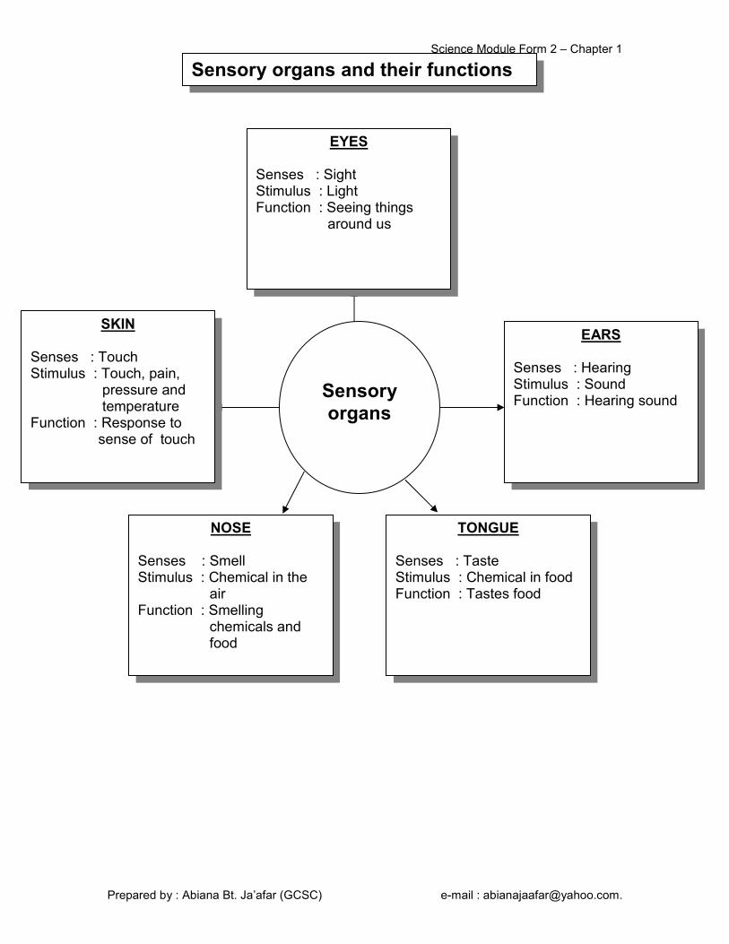
Sensory
organs
EYES
Senses : Sight Stimulus : Light Function : Seeing things around us
EARS
Senses : Hearing Stimulus : Sound Function : Hearing sound
TONGUE
Senses : Taste Stimulus : Chemical in food Function : Tastes food
SKIN
Senses : Touch Stimulus : Touch, pain, pressure and temperature Function : Response to sense of touch
NOSE
Senses : Smell Stimulus : Chemical in the air Function : Smelling chemicals and food
Sensory organs and their functions

Science Module Form 2 – Chapter 1
Prepared by : Abiana Bt. Ja’afar (GCSC) e-mail : [email protected].
The
1. The following table shows the stimuli detected by the various sensory organ. Complete the table.
Sensory organ Stimulus
Nose
Chemical substances in food
Eye
Sound
Skin
2. The following shows the pathways from stimulus to response. Fill in the
blanks.
Stimulus Nerves Response 3. Complete the following table.
Stimulus Sense Sensory organ
(a) Ticking a clock
(b) Colour of traffic light
( c) Sourness of lime juice
(d) Watching a movie
(e) Coldness of ice cubes
(f) Sharpness of a pencil point
Exercise 1.1

Science Module Form 2 – Chapter 1
Prepared by : Abiana Bt. Ja’afar (GCSC) e-mail : [email protected].
Parts of the ear Function
Vibrate when sound waves hit on it.
Amplify sound vibrations
Changes sound vibrations to nerve impulses
Send nerve impulses to the brain for interpretation.
Equalizes the air pressure in both sides of the ears.
Control the balance of the body.
Sensory Organ : ________________________________________ Senses : ________________________________________ Stimulus : ________________________________________ Name of Receptor : ________________________________________
Ossicles Ear pinna Oval window Cochlea Semicircular canal Ear canal Eardrum Auditory nerve Eustachian tube
How do we hear ?
Auditory canal
Oval window
Brain
2 structures not involved in hearing mechanism

Science Module Form 2 – Chapter 1
Prepared by : Abiana Bt. Ja’afar (GCSC) e-mail : [email protected].
1. Diagram 1 shows a cross section of the human ear.
Diagram 1
(b) Name the labeled structures using the following terms. (i) P : ____________________________________________________ (ii) Q : ____________________________________________________ (iii) R : ____________________________________________________
(c) (i) What is structure S? ________________________________________________________ (ii) State the function of structure S? ________________________________________________________
2.
Exercise 1.4
Eardrum Auditory canal Cochlea
Diagram 2

Science Module Form 2 – Chapter 1
Prepared by : Abiana Bt. Ja’afar (GCSC) e-mail : [email protected].
Diagram 2 shows the structure of human ear.
(a) Name parts P, Q, R and S. (i) P : ____________________________________________________ (ii) Q : ____________________________________________________ (iii) R : ____________________________________________________ (iv) S : ____________________________________________________
(b) Name the part that amplifies sound vibrations. ___________________________________________________________
(c) Which part converts the sound vibration to nerve impulses? ___________________________________________________________
(d) Name two parts in the ear that do not involve in hearing.
(i) ________________________________________________________ (ii) ________________________________________________________
3. Diagram 3 below shows the structure of the human ear.
Diagram 3
Science Module Form 2 – Chapter 1
Prepared by : Abiana Bt. Ja’afar (GCSC) e-mail : [email protected].
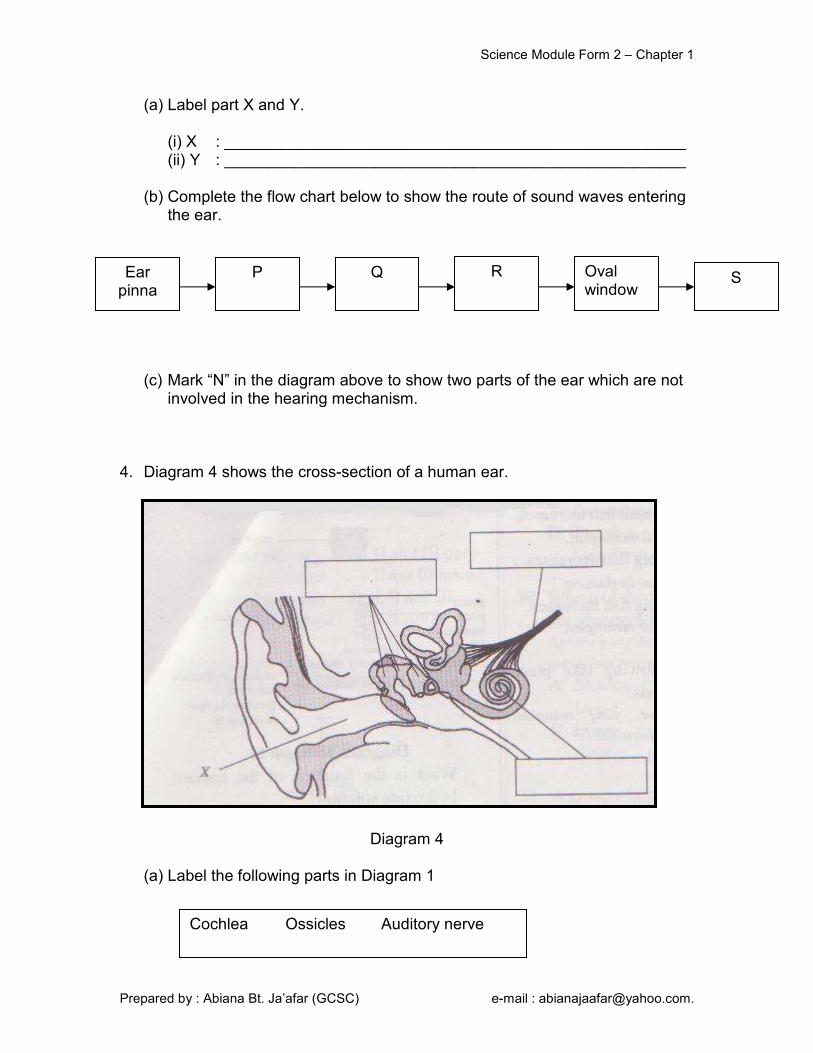
(a) Label part X and Y.
(i) X : ____________________________________________________ (ii) Y : ____________________________________________________
(b) Complete the flow chart below to show the route of sound waves entering the ear.
(c) Mark “N” in the diagram above to show two parts of the ear which are not
involved in the hearing mechanism.
4. Diagram 4 shows the cross-section of a human ear.
Diagram 4
(a) Label the following parts in Diagram 1
Ear pinna
P Q R Oval window
S
Cochlea Ossicles Auditory nerve

Science Module Form 2 – Chapter 1
Prepared by : Abiana Bt. Ja’afar (GCSC) e-mail : [email protected].
(b) State one function of X. ___________________________________________________________
(c) What is the frequency range of sound that can be detected by our ears? ___________________________________________________________
5. The diagram below shows a structure of the ear. (a) Name the parts of the ear shown in the diagram.

Science Module Form 2 – Chapter 1
Prepared by : Abiana Bt. Ja’afar (GCSC) e-mail : [email protected].
Parts of the eye Function
Protects the eye
Allows light to enter the eye
Controls the size of pupil
Sends nerve impulses to the brain for interpretation
Detects light stimulus
Maintains the shape of the eye
Focuses light onto the retina
Sensory Organ : ________________________________________ Senses : ________________________________________ Stimulus : ________________________________________ Name of Receptor : ________________________________________ Location of Receptor : ________________________________________
Aqueous humour Cornea Retina Optic Nerve Sclera Chroid Lens Vitreous humour Pupil Conjunctiva Yellow spot Blind spot Iris
How do we see ?
Aqueous humour
Vitreous humour
Brain
Science Module Form 2 – Chapter 1
Prepared by : Abiana Bt. Ja’afar (GCSC) e-mail : [email protected].
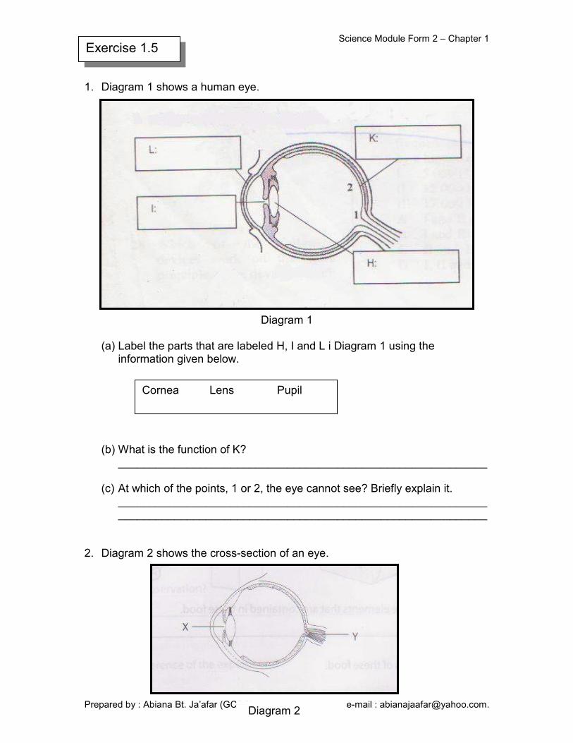
1. Diagram 1 shows a human eye.
Diagram 1
(a) Label the parts that are labeled H, I and L i Diagram 1 using the information given below.
(b) What is the function of K?
___________________________________________________________
(c) At which of the points, 1 or 2, the eye cannot see? Briefly explain it. ______________________________________________________________________________________________________________________
2. Diagram 2 shows the cross-section of an eye.
Exercise 1.5
Cornea Lens Pupil
Diagram 2
Science Module Form 2 – Chapter 1
Prepared by : Abiana Bt. Ja’afar (GCSC) e-mail : [email protected].
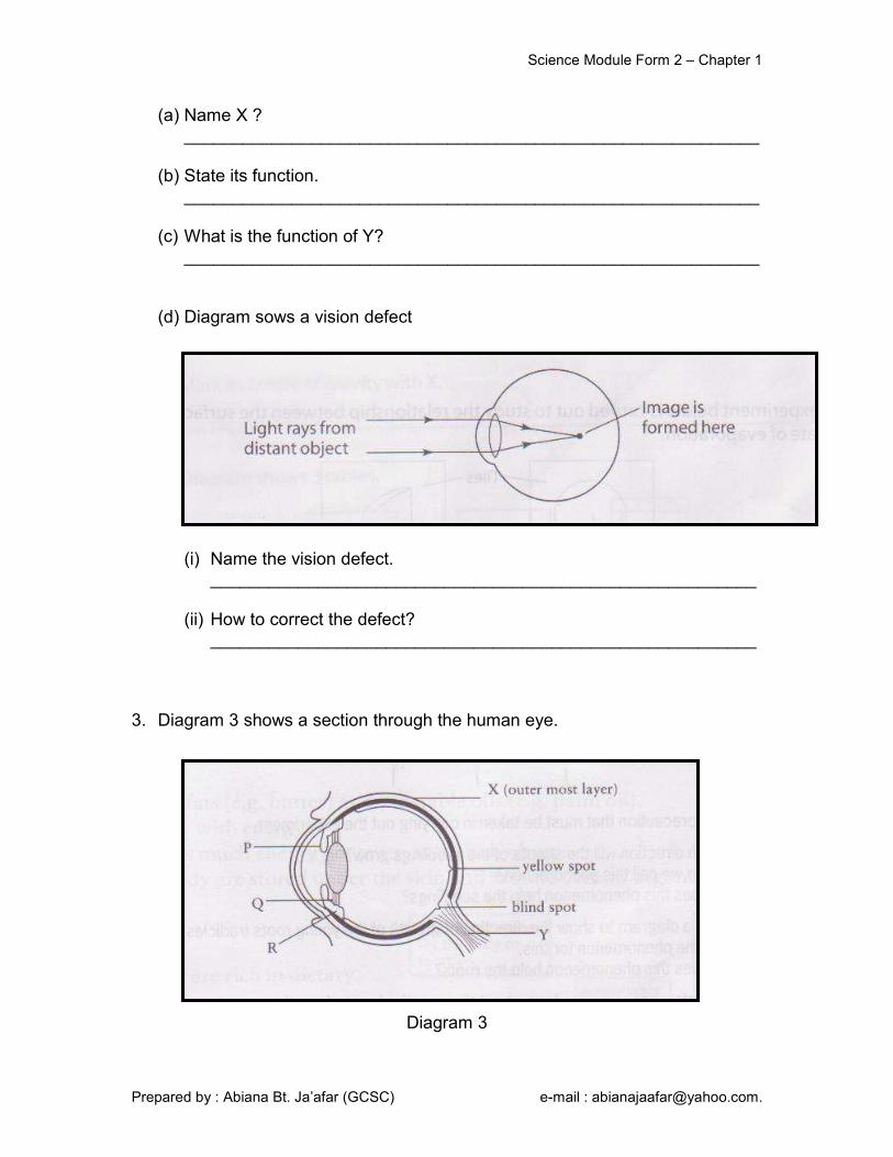
(a) Name X ? ___________________________________________________________
(b) State its function. ___________________________________________________________
(c) What is the function of Y? ___________________________________________________________
(d) Diagram sows a vision defect
(i) Name the vision defect. ________________________________________________________
(ii) How to correct the defect? ________________________________________________________
3. Diagram 3 shows a section through the human eye.
Diagram 3

Science Module Form 2 – Chapter 1
Prepared by : Abiana Bt. Ja’afar (GCSC) e-mail : [email protected].
(a) Name the parts labeled P, Q and R by using the following terms. P : ____________________________________________________ Q : ____________________________________________________ R : ____________________________________________________ (b) State the functions of X and y.
X : ____________________________________________________ Y : ____________________________________________________
(c) State one difference between the blind spot and the yellow spot. ___________________________________________________________
4. Diagram 4 shows the cross-section of the human eye.
Diagram 4
Suspensory ligament Iris Ciliary muscle

Science Module Form 2 – Chapter 1
Prepared by : Abiana Bt. Ja’afar (GCSC) e-mail : [email protected].
(a) The flow chart below shows the flow of light in the sight mechanism. Label the parts E, F and G in the above diagram. E : ____________________________________________________ F : ____________________________________________________ G : ____________________________________________________ (b) Serena suffers from an eye defect due to the part labeled Q being too thin.
At which part of the eye the image seem to appear? ___________________________________________________________
(c) What is the term used for this type of eye defect? ___________________________________________________________
(d) What type of lens can be used to correct her eye defect? ___________________________________________________________
5. Diagram 5 below shows a cross-section of the human eye.
Diagram 5
Cornea E F Eye lens Vitreous humour
Retina Brain G
Science Module Form 2 – Chapter 1
Prepared by : Abiana Bt. Ja’afar (GCSC) e-mail : [email protected].
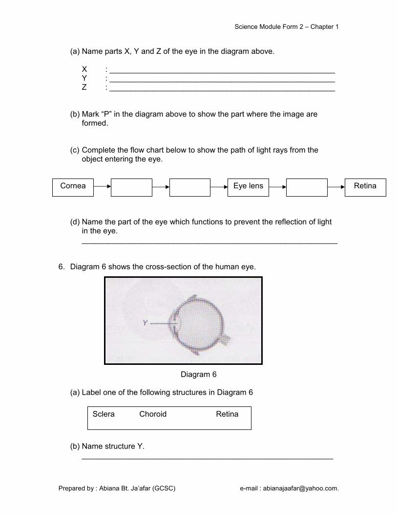
(a) Name parts X, Y and Z of the eye in the diagram above.
X : ____________________________________________________ Y : ____________________________________________________ Z : ____________________________________________________
(b) Mark “P” in the diagram above to show the part where the image are formed.
(c) Complete the flow chart below to show the path of light rays from the
object entering the eye. (d) Name the part of the eye which functions to prevent the reflection of light
in the eye. ___________________________________________________________
6. Diagram 6 shows the cross-section of the human eye.
Diagram 6 (a) Label one of the following structures in Diagram 6
(b) Name structure Y. __________________________________________________________
Cornea Eye lens Retina
Sclera Choroid Retina
Science Module Form 2 – Chapter 1
Prepared by : Abiana Bt. Ja’afar (GCSC) e-mail : [email protected].
Examples parts of body
More Sensitive Less Sensitive
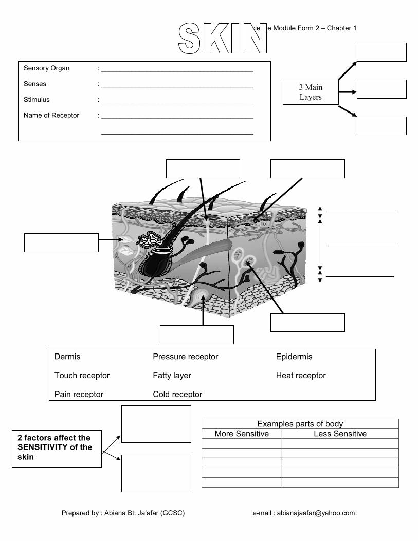
Sensory Organ : ________________________________________ Senses : ________________________________________ Stimulus : ________________________________________ Name of Receptor : ________________________________________ ________________________________________
Dermis Pressure receptor Epidermis Touch receptor Fatty layer Heat receptor Pain receptor Cold receptor
2 factors affect the SENSITIVITY of the
skin
3 Main
Layers

Science Module Form 2 – Chapter 1
Prepared by : Abiana Bt. Ja’afar (GCSC) e-mail : [email protected].
1.
Figure 1
Figure 1 shows the cross- section of human skin. (a) Label the above figure with the following information. (b) Nazri accidentally drops a coin on the floor. It rolls under a sofa. He put his
hand under the sofa and retrieves the coin. What senses are involved in this sequence of action? ___________________________________________________________
2. Figure 2 shows a cross –section of the human skin.
Exercise 1.2
Pain receptor cold receptor sweat gland
Figure 2
Science Module Form 2 – Chapter 1
Prepared by : Abiana Bt. Ja’afar (GCSC) e-mail : [email protected].
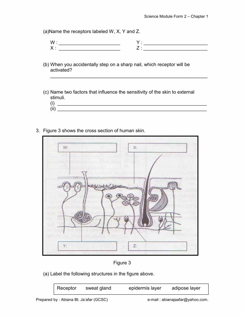
(a)Name the receptors labeled W, X, Y and Z.
W : _______________________ Y : ________________________ X : _______________________ Z : ________________________
(b) When you accidentally step on a sharp nail, which receptor will be activated? ___________________________________________________________
(c) Name two factors that influence the sensitivity of the skin to external stimuli. (i) ________________________________________________________ (ii) ________________________________________________________
3. Figure 3 shows the cross section of human skin.
Figure 3
(a) Label the following structures in the figure above.
Receptor sweat gland epidermis layer adipose layer

Science Module Form 2 – Chapter 1
Prepared by : Abiana Bt. Ja’afar (GCSC) e-mail : [email protected].
(b) State three receptors that can be found in the second layer of our skin.
(i) ________________________________________________________ (ii) ________________________________________________________ (iii) ________________________________________________________
(c) Based on the given statement above, make a hypothesis about the sensitivity of our skin. ___________________________________________________________
4. Figure 4 shows a cross-section of the human skin.
Figure 4
(a) Name the labeled structures using the following terms. (b) Name two areas of the body which are
(i) more sensitive : ___________________________________________ (ii) less sensitive : ___________________________________________
( c) State two factors that affect the sensitivity of the skin.
(i) ________________________________________________________ (ii) ________________________________________________________
Pain receptor Touch receptor Heat receptor Cold receptor Pressure receptor Epidermis Dermis

Science Module Form 2 – Chapter 1
Prepared by : Abiana Bt. Ja’afar (GCSC) e-mail : [email protected].
5. The cross-section of the human skin is shown in the diagram.
Figure 5
(a) Liza is holding a glass of orange juice. She can feel that the glass is cold. Which receptor is stimulated when she is holding the glass? ________________________________________________________
(b) Label the above diagram with these information.
Touch receptor Q Heat receptor R
Pressure receptor S Pain receptor P
(c) Name the part that actively produces sweat when you do physical
exercise. ________________________________________________________
Science Module Form 2 – Chapter 1
Prepared by : Abiana Bt. Ja’afar (GCSC) e-mail : [email protected].
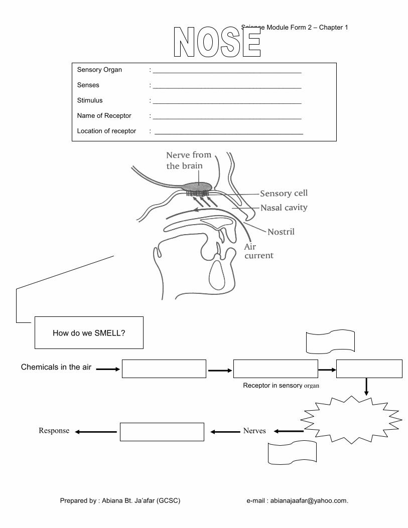
Sensory Organ : ________________________________________ Senses : ________________________________________ Stimulus : ________________________________________ Name of Receptor : ________________________________________ Location of receptor : ________________________________________
How do we SMELL?
Chemicals in the air
Response
Receptor in sensory organ
Nerves
How do we SMELL?
Science Module Form 2 – Chapter 1
Prepared by : Abiana Bt. Ja’afar (GCSC) e-mail : [email protected].
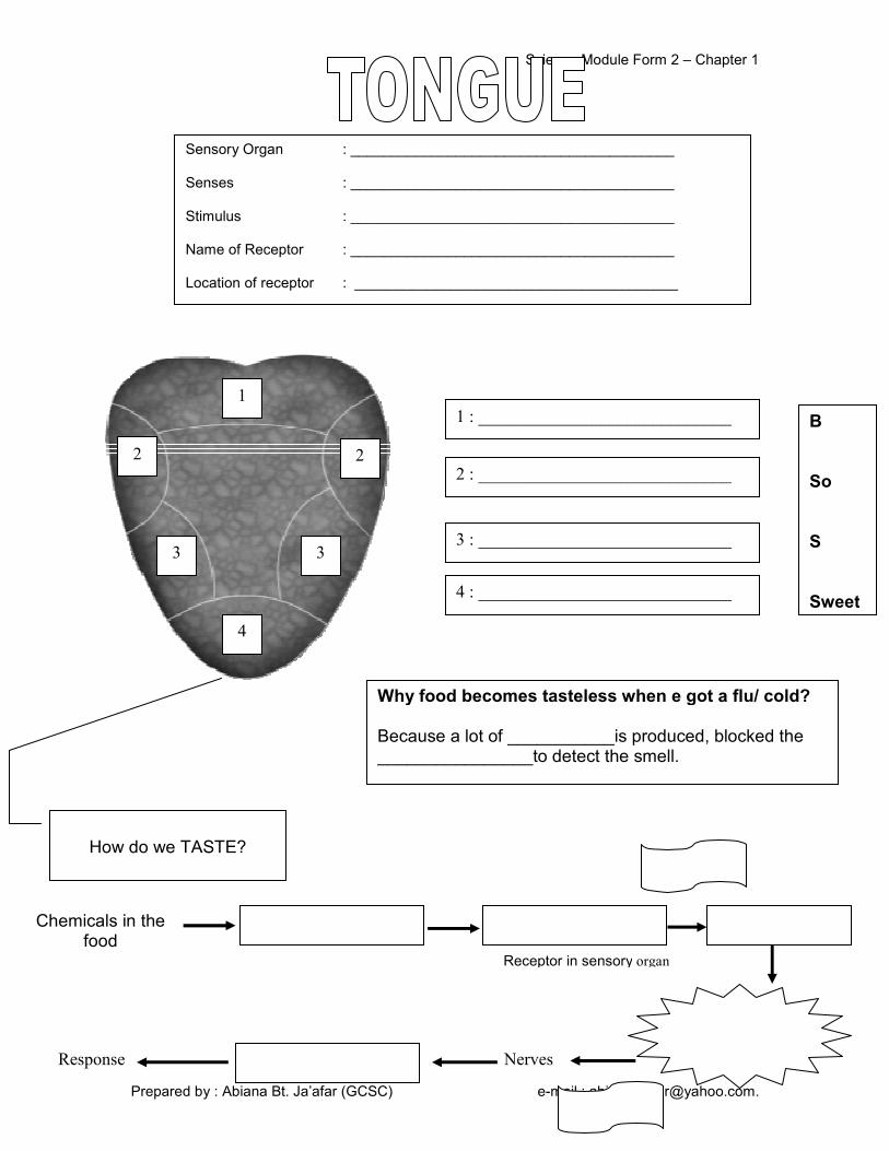
Sensory Organ : ________________________________________ Senses : ________________________________________ Stimulus : ________________________________________ Name of Receptor : ________________________________________ Location of receptor : ________________________________________
1
2 2
3 3
4
1 : _____________________________
3 : _____________________________
4 : _____________________________
2 : _____________________________
B So S Sweet
How do we SMELL?
Chemicals in the food
Response
Receptor in sensory organ
Nerves
How do we TASTE?
Why food becomes tasteless when e got a flu/ cold? Because a lot of ___________is produced, blocked the ________________to detect the smell.

Science Module Form 2 – Chapter 1
Prepared by : Abiana Bt. Ja’afar (GCSC) e-mail : [email protected].
1. Diagram 1 shows the areas P, Q, R and S on the tongue that are sensitive to
taste.
Diagram 1
(a) What taste can be detected in the following areas?
P : ___________________ Q : ____________________ R : ___________________ S : ____________________
(b) Give an example of a type of food that can be detected in areas R and S.
P : ____________________________________________________ S : ____________________________________________________
(c) What is the taste of food when the nose is closed? ___________________________________________________________
(d) Give a reason for your answer. ___________________________________________________________
Exercise 1.3

Science Module Form 2 – Chapter 1
Prepared by : Abiana Bt. Ja’afar (GCSC) e-mail : [email protected].
2. The diagram shows the surface of human tongue.
(a) Write the word bitter and sweet to show the parts of the tongue that are sensitive to the two tastes.
(b) State the taste of the following food
(i) Unripe mangoes - _________________________________________ (ii) Honey - _________________________________________
3. Diagram 2 shows a tongue. Diagram 2
(a) Can the part labeled S detect sour tastes? ___________________________________________________________
(b) Give one reason for your answer in a(i). ___________________________________________________________

Science Module Form 2 – Chapter 1
Prepared by : Abiana Bt. Ja’afar (GCSC) e-mail : [email protected].
(c) State all the different tastes that can be detected by the tongue. ___________________________________________________________
(d) Name the sensory cells in the tongue that can detect various tastes. ___________________________________________________________
(e) Name the fluid that dissolves chemical substances in food so that it can be detected by the tongue. ___________________________________________________________
4. The diagram below shows the areas of the tongue which are sensitive to
various tastes.
(a) Write in the boxes provided to show the location of the areas of the tongue that is sensitive to the following food.
(i) Salted egg (v) Bitter gourd (ii) Panadol (vi) Cocoa powder (iii) Honey (vii) Vinegar (iv) Natural Yogurt (viii) Ice-cream
Science Module Form 2 – Chapter 1
Prepared by : Abiana Bt. Ja’afar (GCSC) e-mail : [email protected].
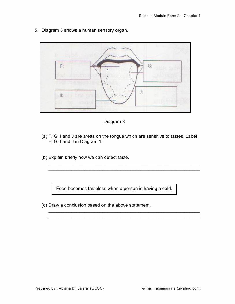
5. Diagram 3 shows a human sensory organ.
(a) F, G, I and J are areas on the tongue which are sensitive to tastes. Label F, G, I and J in Diagram 1.
(b) Explain briefly how we can detect taste.
______________________________________________________________________________________________________________________
(c) Draw a conclusion based on the above statement. ______________________________________________________________________________________________________________________
Food becomes tasteless when a person is having a cold.
Diagram 3
Science Module Form 2 – Chapter 1
Prepared by : Abiana Bt. Ja’afar (GCSC) e-mail : [email protected].
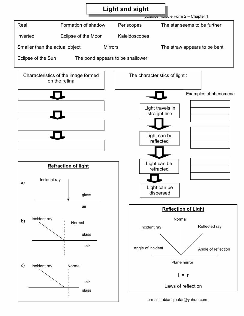
Real Formation of shadow Periscopes The star seems to be further inverted Eclipse of the Moon Kaleidoscopes Smaller than the actual object Mirrors The straw appears to be bent Eclipse of the Sun The pond appears to be shallower
Characteristics of the image formed on the retina
The characteristics of light :
Light travels in straight line
Light can be reflected
Light can be refracted
Light can be dispersed
Examples of phenomena
Reflection of Light
i = r
Laws of reflection
Refraction of light
a)
b)
c)
Incident ray Reflected ray
Normal
Angle of reflection Angle of incident
Plane mirror
Incident ray
glass
air
Incident ray
Incident ray
Normal
glass
glass
Normal
air
air
Light and sight
Science Module Form 2 – Chapter 1
Prepared by : Abiana Bt. Ja’afar (GCSC) e-mail : [email protected].
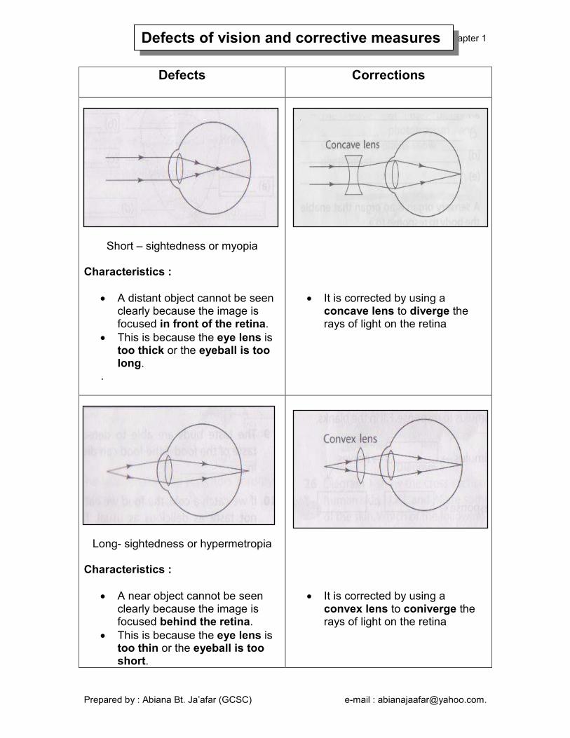
Defects Corrections
Short – sightedness or myopia Characteristics :
• A distant object cannot be seen clearly because the image is focused in front of the retina.
• This is because the eye lens is too thick or the eyeball is too long.
.
• It is corrected by using a concave lens to diverge the rays of light on the retina
Long- sightedness or hypermetropia Characteristics :
• A near object cannot be seen clearly because the image is focused behind the retina.
• This is because the eye lens is too thin or the eyeball is too short.
• It is corrected by using a convex lens to coniverge the rays of light on the retina
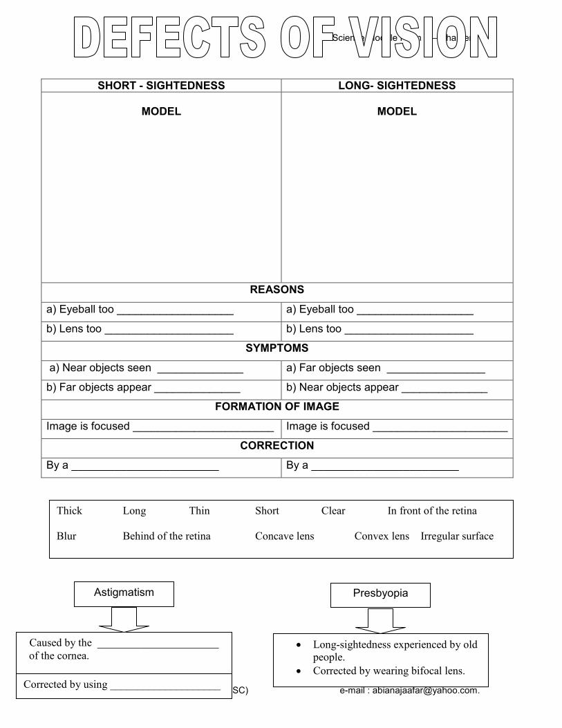
Defects of vision and corrective measures
Science Module Form 2 – Chapter 1
Prepared by : Abiana Bt. Ja’afar (GCSC) e-mail : [email protected].
SHORT - SIGHTEDNESS LONG- SIGHTEDNESS
MODEL
MODEL
REASONS
a) Eyeball too ___________________ a) Eyeball too ___________________
b) Lens too _____________________ b) Lens too _____________________
SYMPTOMS
a) Near objects seen ______________ a) Far objects seen ________________
b) Far objects appear ______________ b) Near objects appear ______________
FORMATION OF IMAGE
Image is focused _______________________ Image is focused ______________________
CORRECTION
By a ________________________ By a ________________________
Thick Long Thin Short Clear In front of the retina
Blur Behind of the retina Concave lens Convex lens Irregular surface
Astigmatism
Caused by the ______________________
of the cornea.
Presbyopia
• Long-sightedness experienced by old
people.
• Corrected by wearing bifocal lens.
Corrected by using ____________________

Science Module Form 2 – Chapter 1
Prepared by : Abiana Bt. Ja’afar (GCSC) e-mail : [email protected].
1. Fill in the blanks.
(a) What is the vision defect shown in the above diagram? ___________________________________________________________
(b) The image from a ________________________ object is focused _____________________ of the retina.
(c) This defect is caused by the lens that is too ___________________ or
the eyeball is too _____________.
(d) This defect can be corrected by wearing glasses with _____________________.
2.
(a) This vision defect shown in above diagram is ______________________. (b) A person who has this defective vision sees ___________________
objects clearly.
(c) This defect is caused by ___________________ that is too thin or if the _____________________ is too short.
(d) This defect can be corrected by wearing glasses with ________________.
Exercise 1.6

Science Module Form 2 – Chapter 1
Prepared by : Abiana Bt. Ja’afar (GCSC) e-mail : [email protected].
3.
Diagram 1 Diagram 1 shows the structure of a human eye.
(a) Mark (A) to represent the yellow spot and (B) to represent the blind spot in the diagram above.
(b) State the function of each of the parts labeled X, Y and Z.
i. X : ____________________________________________________ ii. Y : ____________________________________________________ iii. Z : ____________________________________________________
(c) i. Give two causes of the eye defect mentioned. ________________________________________________________ ________________________________________________________
ii. Name the defect mentioned in (c)i.. ________________________________________________________
iii. Suggest one way to correct the defect ________________________________________________________
A man cannot see distant objects clearly.
Science Module Form 2 – Chapter 1
Prepared by : Abiana Bt. Ja’afar (GCSC) e-mail : [email protected].
4.
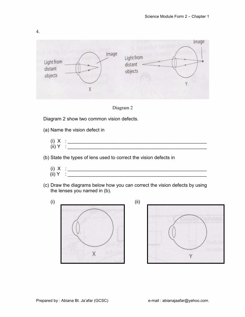
Diagram 2
Diagram 2 show two common vision defects.
(a) Name the vision defect in
(i) X : ____________________________________________________ (ii) Y : ____________________________________________________
(b) State the types of lens used to correct the vision defects in
(i) X : ____________________________________________________ (ii) Y : ____________________________________________________ (c) Draw the diagrams below how you can correct the vision defects by using
the lenses you named in (b).
(i) (ii)
Science Module Form 2 – Chapter 1
Prepared by : Abiana Bt. Ja’afar (GCSC) e-mail : [email protected].
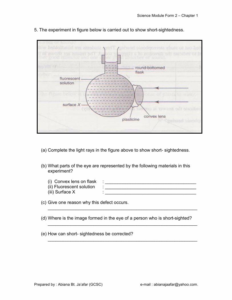
5. The experiment in figure below is carried out to show short-sightedness.
(a) Complete the light rays in the figure above to show short- sightedness. (b) What parts of the eye are represented by the following materials in this
experiment?
(i) Convex lens on flask : ____________________________________ (ii) Fluorescent solution : ____________________________________ (iii) Surface X : ____________________________________
(c) Give one reason why this defect occurs. ___________________________________________________________
(d) Where is the image formed in the eye of a person who is short-sighted? ___________________________________________________________
(e) How can short- sightedness be corrected? ___________________________________________________________

Science Module Form 2 – Chapter 1
Prepared by : Abiana Bt. Ja’afar (GCSC) e-mail : [email protected].
6. Diagram 3 shows an eye with a vision defect.
Diagram 3
(a) Name the visual defect? ___________________________________________________________
(b) Give two conditions of the eyes that caused the defect? ___________________________________________________________ ___________________________________________________________
(c) How can the defect be corrected? ___________________________________________________________
(d) On the diagram, draw the correction to the vision defect.
Science Module Form 2 – Chapter 1
Prepared by : Abiana Bt. Ja’afar (GCSC) e-mail : [email protected].
Picture 566.jpg
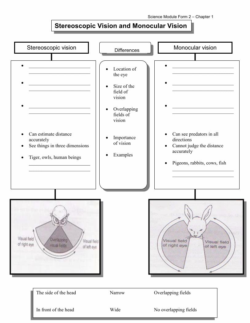
Stereoscopic vision
• _________________________
_________________________
• _________________________
_________________________
• _________________________
_________________________
• Can estimate distance
accurately
• See things in three dimensions
• Tiger, owls, human beings
_________________________
_________________________
Monocular vision
• _________________________
_________________________
• _________________________
_________________________
• _________________________
_________________________
• Can see predators in all
directions
• Cannot judge the distance
accurately
• Pigeons, rabbits, cows, fish
_________________________
_________________________
• Location of
the eye
• Size of the
field of
vision
• Overlapping
fields of
vision
• Importance
of vision
• Examples
Differences
Stereoscopic Vision and Monocular Vision
The side of the head Narrow Overlapping fields
In front of the head Wide No overlapping fields

Science Module Form 2 – Chapter 1
Prepared by : Abiana Bt. Ja’afar (GCSC) e-mail : [email protected].
1. Classify the animals according to type of vision.
Exercise 1.7
Tiger Monkey Rat Chicken Cow Lion Cat Deer Owl Goat Goose Eagle Fish Dog Human
Type of vision
Stereoscopic Monocular
The predator possesses __________________ vision whereas the prey possesses __________________ vision.
Science Module Form 2 – Chapter 1
Prepared by : Abiana Bt. Ja’afar (GCSC) e-mail : [email protected].
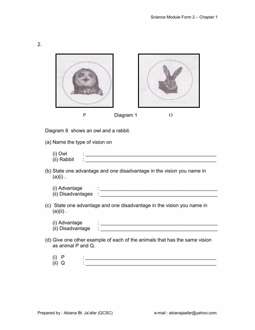
2. Diagram 8 shows an owl and a rabbit.
(a) Name the type of vision on
(i) Owl : _______________________________________________ (ii) Rabbit : _______________________________________________
(b) State one advantage and one disadvantage in the vision you name in (a)(i) .
(i) Advantage : __________________________________________ (ii) Disadvantages : __________________________________________
(c) State one advantage and one disadvantage in the vision you name in (a)(ii) .
(i) Advantage : __________________________________________ (ii) Disadvantage : __________________________________________
(d) Give one other example of each of the animals that has the same vision as animal P and Q. (i) P : _______________________________________________ (ii) Q : _______________________________________________
Diagram 1 P Q
Science Module Form 2 – Chapter 1
Prepared by : Abiana Bt. Ja’afar (GCSC) e-mail : [email protected].
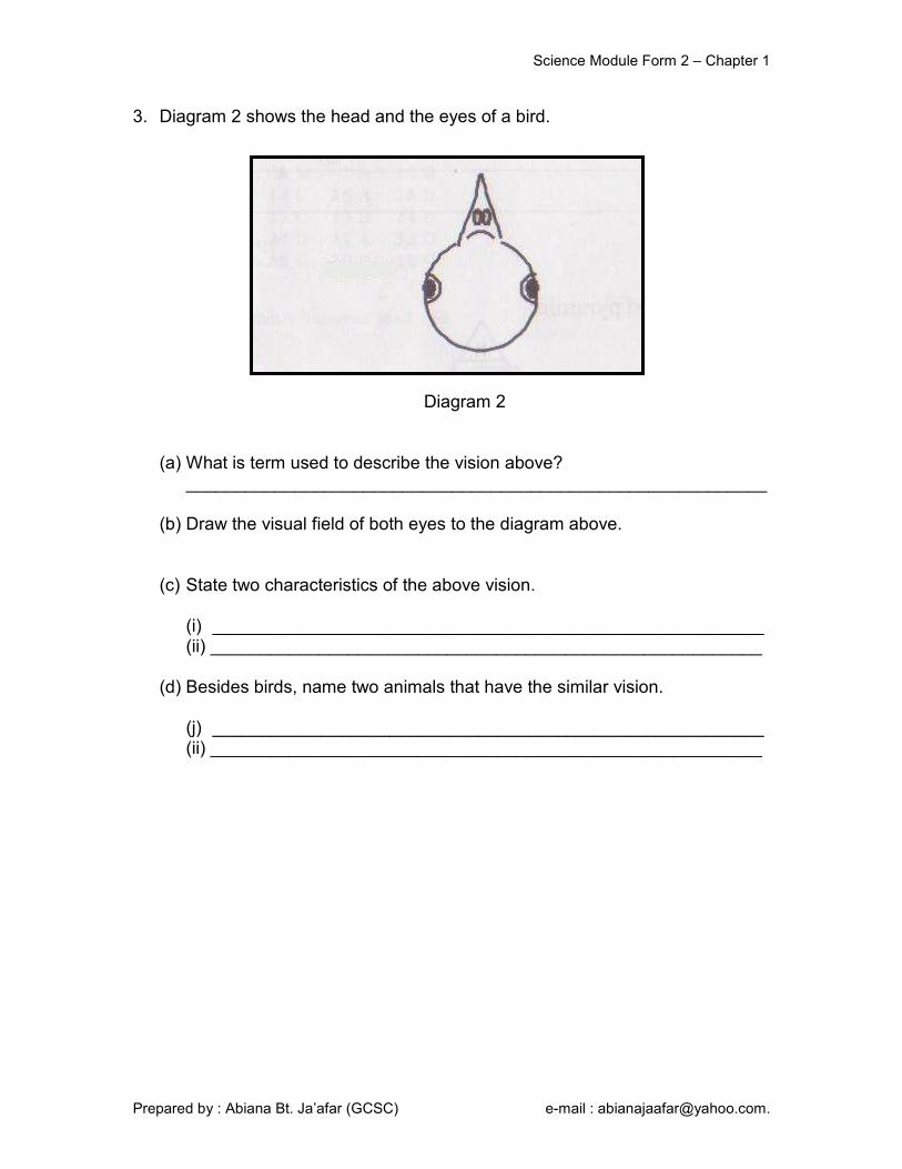
3. Diagram 2 shows the head and the eyes of a bird.
Diagram 2
(a) What is term used to describe the vision above?
___________________________________________________________
(b) Draw the visual field of both eyes to the diagram above. (c) State two characteristics of the above vision.
(i) ________________________________________________________ (ii) ________________________________________________________
(d) Besides birds, name two animals that have the similar vision.
(j) ________________________________________________________ (ii) ________________________________________________________
Science Module Form 2 – Chapter 1
Prepared by : Abiana Bt. Ja’afar (GCSC) e-mail : [email protected].
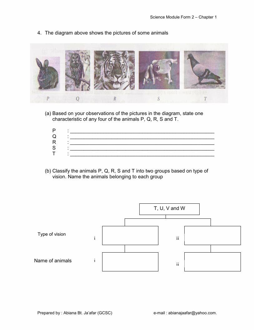
4. The diagram above shows the pictures of some animals
(a) Based on your observations of the pictures in the diagram, state one
characteristic of any four of the animals P, Q, R, S and T.
P : ____________________________________________________ Q : ____________________________________________________ R : ____________________________________________________ S : ____________________________________________________ T : ____________________________________________________
(b) Classify the animals P, Q, R, S and T into two groups based on type of
vision. Name the animals belonging to each group
T, U, V and W
Type of vision
Name of animals
i
i
ii
ii

Science Module Form 2 – Chapter 1
Prepared by : Abiana Bt. Ja’afar (GCSC) e-mail : [email protected].
Type of tropism
Definition Stimulus Part of plant involved
Phototropism Plant response to light
Light Shoot
Geotropism Plant response to gravity
Gravity Root
Hydrotropism Plant response to water
Water Root
Thigmotropism Movement made by a plant in
response to touch or contact
Touch Tendrils
Nastic movements
Movement made by a plant in response to
external stimuli
Touch Leaves
Stimuli and Responses in Plants
Sound and Hearing
Sound is produced by vibration
Properties of sound
Soft and rough surfaces are good sound absorbers
Sound needs a medium to travel
Hard and smooth surfaces are good sound reflectors

Science Module Form 2 – Chapter 1
Prepared by : Abiana Bt. Ja’afar (GCSC) e-mail : [email protected].
1. Complete the table below
2. Complete the following chart about the response of plants to stimuli and their
functions.
Tropism Response
Phototropism
Gravity
Water
Touch or contact
Exercise 1.8
Response of plants to
Light Water Gravity
Types
Functions
Shoots Roots Shoots Roots Shoots Roots
Negative Phototropism

Science Module Form 2 – Chapter 1
Prepared by : Abiana Bt. Ja’afar (GCSC) e-mail : [email protected].
3. Figure 1 shows reactions of a plant to external stimuli.
Figure 1
(a) Write phototropism, geotropism and thigmotropism in the appropriate box in Figure 1.
(b) Which part of the plant performs
(i) Negative hydrotropism : ____________________________________ (ii) Positive geotropism : ____________________________________ (iii) Positive phototropism : ____________________________________
(c) Name two examples of plants that perform nastic movements.
(i) ________________________________________________________ (ii) ________________________________________________________
Science Module Form 2 – Chapter 1
Prepared by : Abiana Bt. Ja’afar (GCSC) e-mail : [email protected].
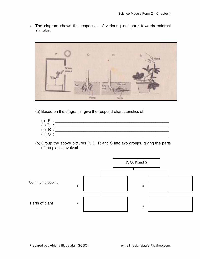
4. The diagram shows the responses of various plant parts towards external stimulus.
(a) Based on the diagrams, give the respond characteristics of (i) P : ____________________________________________________ (ii) Q : ____________________________________________________ (ii) R : ____________________________________________________ (iii) S : ____________________________________________________
(b) Group the above pictures P, Q, R and S into two groups, giving the parts
of the plants involved.
5. gjhihkj
P, Q, R and S
Common grouping
Parts of plant
i
i
ii
ii
Science Module Form 2 – Chapter 1
Prepared by : Abiana Bt. Ja’afar (GCSC) e-mail : [email protected].
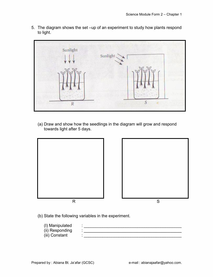
5. The diagram shows the set –up of an experiment to study how plants respond to light.
(a) Draw and show how the seedlings in the diagram will grow and respond towards light after 5 days.
R S (b) State the following variables in the experiment. (I) Manipulated : __________________________________________ (ii) Responding : __________________________________________ (iii) Constant : __________________________________________
Science Module Form 2 – Chapter 1
Prepared by : Abiana Bt. Ja’afar (GCSC) e-mail : [email protected].
(c) Which part of the seedlings responds towards light? ___________________________________________________________
(d) What is the name of the response of the shoots of plants towards light? __________________________________________________________
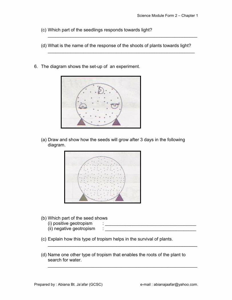
6. The diagram shows the set-up of an experiment.
(a) Draw and show how the seeds will grow after 3 days in the following diagram.
(b) Which part of the seed shows
(i) positive geotropism : ____________________________________ (ii) negative geotropism : ____________________________________
(c) Explain how this type of tropism helps in the survival of plants. ___________________________________________________________ (d) Name one other type of tropism that enables the roots of the plant to
search for water. ___________________________________________________________

Science Module Form 2 – Chapter 1
Prepared by : Abiana Bt. Ja’afar (GCSC) e-mail : [email protected].
7. The experiment in the figure below is carried out to study the response of plants to water.
(a) What variables are involved in this experiment?
(i) Manipulated : __________________________________________ (ii) Responding : __________________________________________ (iii) Constant : __________________________________________
(b) Suggest the relationship between the variable which is manipulated and the variable which responds. ___________________________________________________________
(c) Draw the response of the roots of seedlings in the figure below at the end of experiment.
(d) What is the name given to the response of the roots to water?
___________________________________________________________
(e) What is the importance of the roots responding in (d) to plants? _________________________________________________________

Science Module Form 2 – Chapter 1
Prepared by : Abiana Bt. Ja’afar (GCSC) e-mail : [email protected].
8. The diagram shows the set-up of an experiment.
(a) What is the purpose of conducting the above experiment? ___________________________________________________________
(b) Describe briefly what will happen to the roots of seedlings in beaker Q after five days. ___________________________________________________________
(c) Draw a diagram to show what will happen to the seedlings in beaker P after five days.
(d) Name the response that is shown by the roots in beaker P and beaker Q. ___________________________________________________________
(e) Complete the following table.
Tropism Stimulus
Light
Geotropism

Science Module Form 2 – Chapter 1
Prepared by : Abiana Bt. Ja’afar (GCSC) e-mail : [email protected].
9.
(a) Explain the hypothesis of the experiment shown in the above figure. ___________________________________________________________
(b) Give the variable that is
(i) Constant : __________________________________________ (ii) Manipulated : __________________________________________ (iii) Responding : __________________________________________
(c) What is the use of anhydrous calcium chloride? ___________________________________________________________
(d) What is the result after the experiment is left for three days? ___________________________________________________________
(e) Draw what you will see one week after the rooting of the seedlings. (f) What conclusion can you make?
___________________________________________________________
Science Module Form 2 – Chapter 1
Prepared by : Abiana Bt. Ja’afar (GCSC) e-mail : [email protected].
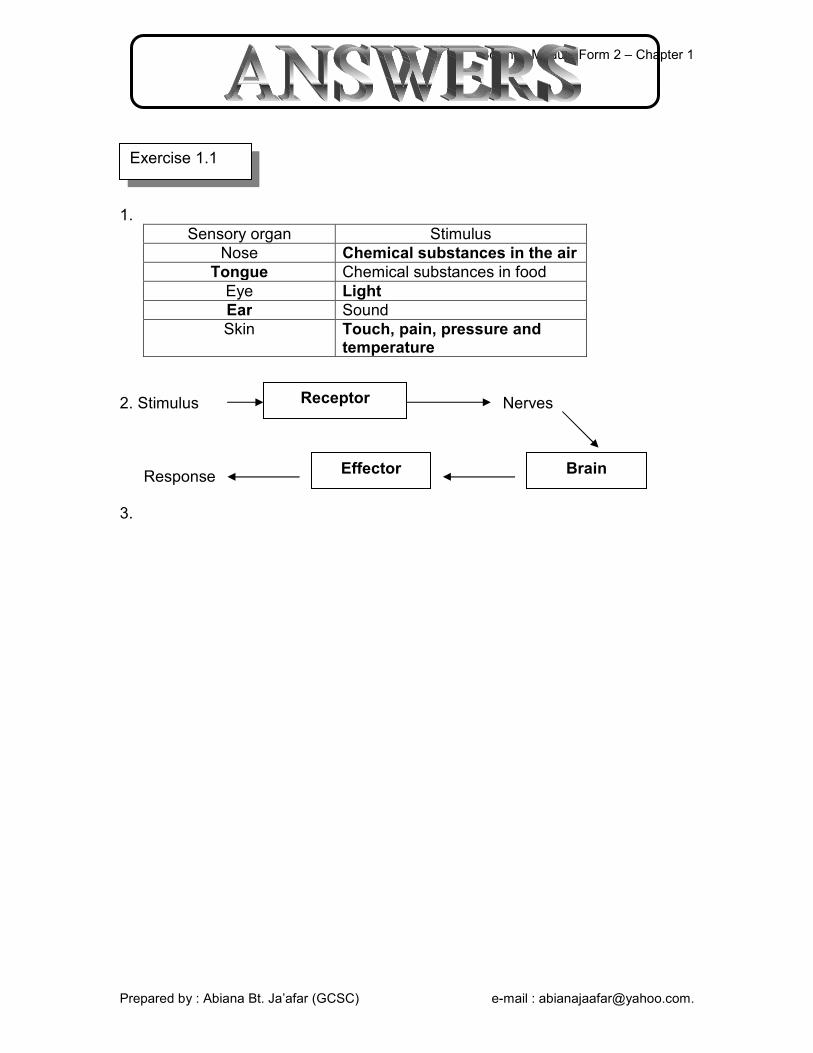
1.
Sensory organ Stimulus
Nose Chemical substances in the air
Tongue Chemical substances in food
Eye Light
Ear Sound
Skin Touch, pain, pressure and temperature
2. Stimulus Nerves Response 3.
Exercise 1.1
Brain Effector
Receptor