Module: gastro intestinal system Histology of the major...
19
Jeanne Adiwinata pawitan - Bagian Histologi FKUI 1 Module: gastro intestinal system Histology of the major glands: The liver - 2008 Jeanne Adiwinata Pawitan Dept. of Histology FMUI
Transcript of Module: gastro intestinal system Histology of the major...

Jeanne Adiwinata pawitan - Bagian Histologi FKUI1
Module: gastro intestinal systemHistology of the major glands: The liver - 2008
Jeanne Adiwinata PawitanDept. of Histology FMUI

Jeanne Adiwinata pawitan - Bagian Histologi FKUI2
Major glands of the digestive system
They end up in the digestive tract:• Liver • Pancreas
• Gall bladder (Vesica fellea) – not a gland

Jeanne Adiwinata pawitan - Bagian Histologi FKUI3
Liver • inferior surface – transverse fissure =
porta hepatis° Portal vein° Hepatic a.° Ductus hepaticus (bile duct)
• Fibrous capsule = Glisson’s capsule° Septa → lobes → lobules° Certain region - peritoneum

Jeanne Adiwinata pawitan - Bagian Histologi FKUI4
Liver – microscopic structure• Cells → plates/sheets (1-2
cells thick) – branching –anastomosing
• Sheets – radial towards central vein –hexagonal prism/cylinder ≅ classical liver lobules
• Between sheets – sinusoids - blood → structure – endocrine gland
• Prism – corner – portal tract = portal canal = portal area = ∆ Kiernan

Jeanne Adiwinata pawitan - Bagian Histologi FKUI5
Portal tract• Connective tissue– contains branches of
° Portal vein° Hepatic artery° Ductus biliaris (bile duct)° Lymph vessel
• around portal tract - liver cells ( < the cells at the center) – 1 layer – limiting plate –pierced by branches of hepatic a., portal v., ductus biliaris

Jeanne Adiwinata pawitan - Bagian Histologi FKUI6
Units in the liver
• Classical lobule (lobulus hepatis)° Anatomical point of view ° Polyhedral
prism/cylinder, axis – central vein, portal tract - corners
• Portal lobule • Hepatic (liver) acinus
Jeanne Adiwinata pawitan - Bagian Histologi FKUI7
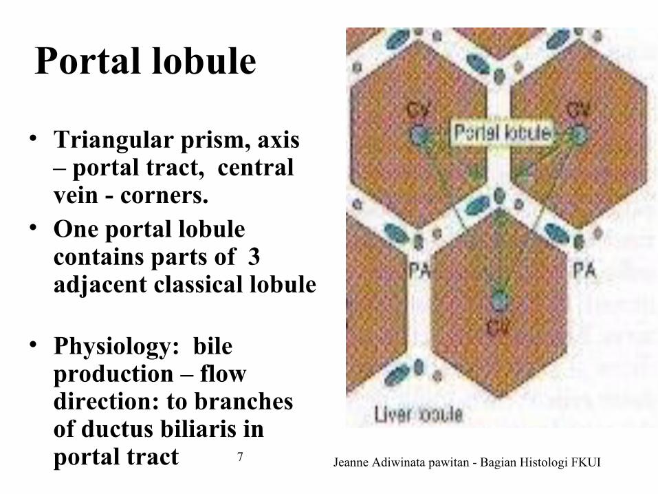
Portal lobule
• Triangular prism, axis – portal tract, central vein - corners.
• One portal lobule contains parts of 3 adjacent classical lobule
• Physiology: bile production – flow direction: to branches of ductus biliaris in portal tract

Jeanne Adiwinata pawitan - Bagian Histologi FKUI8
Liver acinus – portal acinus of Rappaport
• Irregular, ovoid-diamond shape, central vein – both ends, axis: connective tissue (periphery-classical lobule) – interlobular a. and vein (branches of the blood vessels in portal tract)
• Physiology: smallest unit of blood supply

Jeanne Adiwinata pawitan - Bagian Histologi FKUI9
Parenchyma – liver cells (hepatocytes)
• cell - polygonal – 20-35 µm° Surface ≥ 6° Nucleus 1, sometimes 2
(25%)• Size – highly variable• Round/ovoid – vesicular –
chromatin granules: few - distinct - scattered
• nucleolus 1/more - prominent
° Cytoplasm - variable ≅ functional activity
• glycogen/fat storage – routine HE – dissolved – irregular empty spaces (glycogen), round vacuoles (fat)

Jeanne Adiwinata pawitan - Bagian Histologi FKUI10
Liver cells (hepatocytes)• Cytoplasm ° Mitochondria: small
– numerous° Golgi apparatus
• Adjacent to nucleus• Periphery • Adjacent to canaliculi
biliaris (bile canaliculi)
° Clumps of basophilic materials → sometimes – cytoplasm - basophilic
• Cell membrane - distinct
Jeanne Adiwinata pawitan - Bagian Histologi FKUI11
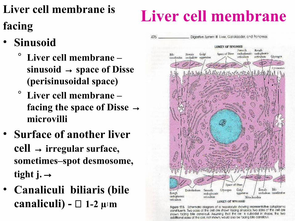
Liver cell membraneLiver cell membrane is facing• Sinusoid
° Liver cell membrane – sinusoid → space of Disse (perisinusoidal space)
° Liver cell membrane – facing the space of Disse → microvilli
• Surface of another liver cell → irregular surface, sometimes–spot desmosome, tight j.→
• Canaliculi biliaris (bile canaliculi) - ∅ 1-2 µ m

Jeanne Adiwinata pawitan - Bagian Histologi FKUI12
Liver (hepatic)sinusoid∀ ≠ blood capillary - ∅
bigger → 9-12 µm• Lining cells ≠
ordinary endothelium° Endothelial cell,
sinusoidal lining cells° Phagocyte (stellate,
Kupffer) cells° (Intermediate cells)
• Basal lamina in continuous

Jeanne Adiwinata pawitan - Bagian Histologi FKUI13
Endothelial cells – sinusoidal lining cells
• Nucleus – small - flattened – dark• Cytoplasm - thin, overlapping – ME
° Thin part – pores ∅ ± 100 nm, diaphragm (-) → ≈ sieve
° In the cytoplasm• << small organelles• >> pinocytotic vesicles

Jeanne Adiwinata pawitan - Bagian Histologi FKUI14
Phagocyte (stellate, Kupffer) cells• Active - phagocytic • Number can ↑ - origin? diff –endothelial cells
(more primitive)• Nucleus – larger, pale• Cytoplasm
° > abundant, branching, traversing the sinusoid° Often contain
• Degenerated - erythrocytes • Iron containing - pigment granules

Jeanne Adiwinata pawitan - Bagian Histologi FKUI15
Perisinusoidal space (space of Disse)• Borders of perisinusoidal
space° Liver cell membrane
• Long microvilli –increase the surface for absorption, secretion
• Cytoplasm beneath the membrane – vacuoles, vesicles
° Sinusoidal wall (basal lamina, endothelial cells)
• Equivalent to interstitial space° Contains reticular,
collagen fiber° Fluid circulates freely

Jeanne Adiwinata pawitan - Bagian Histologi FKUI16
Perisinusoidal space (space of Disse)• Contains
° Mesenchymal cells = pericytes = fat storing (Ito) cell = lipocytes = extra vascular reticular cells
• Located near reticular/collagen fiber• Is believed to: form the fiber, store vitamin A• Might have a role in lymph production
° Pit cells = NK cells?• Sinusoidal wall ≠ barrier
° Basal lamina - in continuous° Endothelial cells - in continuous° → blood plasma (sinusoid) – perisinusoidal space –
liver cells → metabolite exchange: blood – liver cells

Jeanne Adiwinata pawitan - Bagian Histologi FKUI17
Bile flow – in the liver• Bile canaliculi
° Liver cell membrane • Ductulus (canal) of Hering
° Small-clear cells, on basal lamina, nucleus-dark, incomplete organelles
• Bile duct in portal tract° Small duct – simple cuboidal epithelium° Large duct – columnar epithelium, outside –
connective tissue• (Hepatic duct)

Jeanne Adiwinata pawitan - Bagian Histologi FKUI18
Blood flow in the liverPortal vein (75%), hepatic a. (25%) – porta hepatisBranches of portal v., hepatic a. – portal tract
Interlobular v., interlobular a.Inlet v., inlet a.Liver sinusoid
↓ central veinSublobular veinCollecting vein Hepatic vein → v. cava inferior

Jeanne Adiwinata pawitan - Bagian Histologi FKUI19
The end
Thank you