MODULE 2 MICROBIAL CELL STRUCTURE AND - … 2 MICROBIAL CELL STRUCTURE AND - FUNCTION Lecture 1: The...
24
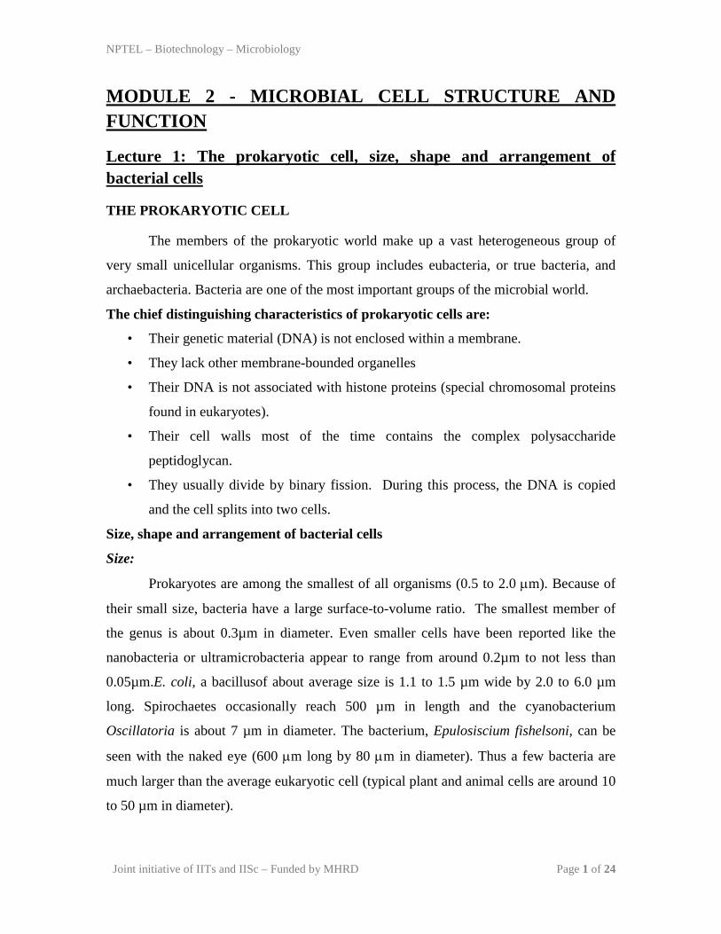
NPTEL – Biotechnology – Microbiology Joint initiative of IITs and IISc – Funded by MHRD Page 1 of 24 MODULE 2 - MICROBIAL CELL STRUCTURE AND FUNCTION Lecture 1: The prokaryotic cell, size, shape and arrangement of bacterial cells THE PROKARYOTIC CELL The members of the prokaryotic world make up a vast heterogeneous group of very small unicellular organisms. This group includes eubacteria, or true bacteria, and archaebacteria. Bacteria are one of the most important groups of the microbial world. The chief distinguishing characteristics of prokaryotic cells are: • Their genetic material (DNA) is not enclosed within a membrane. • They lack other membrane-bounded organelles • Their DNA is not associated with histone proteins (special chromosomal proteins found in eukaryotes). • Their cell walls most of the time contains the complex polysaccharide peptidoglycan. • They usually divide by binary fission. During this process, the DNA is copied and the cell splits into two cells. Size, shape and arrangement of bacterial cells Size: Prokaryotes are among the smallest of all organisms (0.5 to 2.0 µm). Because of their small size, bacteria have a large surface-to-volume ratio. The smallest member of the genus is about 0.3μm in diameter. Even smaller cells have been reported like the nanobacteria or ultramicrobacteria appear to range from around 0.2μm to not less than 0.05μm.E. coli, a bacillusof about average size is 1.1 to 1.5 μm wide by 2.0 to 6.0 μm long. Spirochaetes occasionally reach 500 μm in length and the cyanobacterium Oscillatoria is about 7 μm in diameter. The bacterium, Epulosiscium fishelsoni, can be seen with the naked eye (600 µm long by 80 µm in diameter). Thus a few bacteria are much larger than the average eukaryotic cell (typical plant and animal cells are around 10 to 50 μm in diameter).
Transcript of MODULE 2 MICROBIAL CELL STRUCTURE AND - … 2 MICROBIAL CELL STRUCTURE AND - FUNCTION Lecture 1: The...
NPTEL – Biotechnology – Microbiology
Joint initiative of IITs and IISc – Funded by MHRD Page 1 of 24
MODULE 2 - MICROBIAL CELL STRUCTURE AND FUNCTION
Lecture 1: The prokaryotic cell, size, shape and arrangement of bacterial cells
THE PROKARYOTIC CELL
The members of the prokaryotic world make up a vast heterogeneous group of
very small unicellular organisms. This group includes eubacteria, or true bacteria, and
archaebacteria. Bacteria are one of the most important groups of the microbial world.
The chief distinguishing characteristics of prokaryotic cells are:
• Their genetic material (DNA) is not enclosed within a membrane.
• They lack other membrane-bounded organelles
• Their DNA is not associated with histone proteins (special chromosomal proteins
found in eukaryotes).
• Their cell walls most of the time contains the complex polysaccharide
peptidoglycan.
• They usually divide by binary fission. During this process, the DNA is copied
and the cell splits into two cells.
Size, shape and arrangement of bacterial cells
Size:
Prokaryotes are among the smallest of all organisms (0.5 to 2.0 µm). Because of
their small size, bacteria have a large surface-to-volume ratio. The smallest member of
the genus is about 0.3µm in diameter. Even smaller cells have been reported like the
nanobacteria or ultramicrobacteria appear to range from around 0.2µm to not less than
0.05µm.E. coli, a bacillusof about average size is 1.1 to 1.5 µm wide by 2.0 to 6.0 µm
long. Spirochaetes occasionally reach 500 µm in length and the cyanobacterium
Oscillatoria is about 7 µm in diameter. The bacterium, Epulosiscium fishelsoni, can be
seen with the naked eye (600 µm long by 80 µm in diameter). Thus a few bacteria are
much larger than the average eukaryotic cell (typical plant and animal cells are around 10
to 50 µm in diameter).
NPTEL – Biotechnology – Microbiology
Joint initiative of IITs and IISc – Funded by MHRD Page 2 of 24
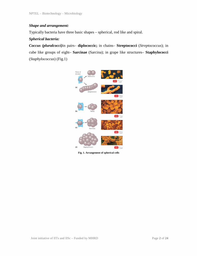
Shape and arrangement:
Typically bacteria have three basic shapes – spherical, rod like and spiral.
Spherical bacteria:
Coccus (pluralcocci)in pairs– diplococcic; in chains– Streptococci (Streptococcus); in
cube like groups of eight– Sarcinae (Sarcina); in grape like structures– Staphylococci
(Staphylococcus) (Fig.1)
Fig. 1. Arrangement of spherical cells

NPTEL – Biotechnology – Microbiology
Joint initiative of IITs and IISc – Funded by MHRD Page 3 of 24
Rod like bacteria:
Bacillus(pluralbacilli)in pairs –diplococci; in chains – streptococci. Still others look
like cocci and are called coccobacilli(Fig. 2).
Fig. 2.Arrangement of rod like cells
Spiral bacteria
Comma shaped – Vibrio (Vibrio); Helical, long and curved – Spirilla (Rhodospirillium);
Helical and flexible – Sprirochetes (Treponema and Borrelia) movement by axial
filaments (Fig. 3).
Fig. 3.Sprial bacteria
NPTEL – Biotechnology – Microbiology
Joint initiative of IITs and IISc – Funded by MHRD Page 4 of 24
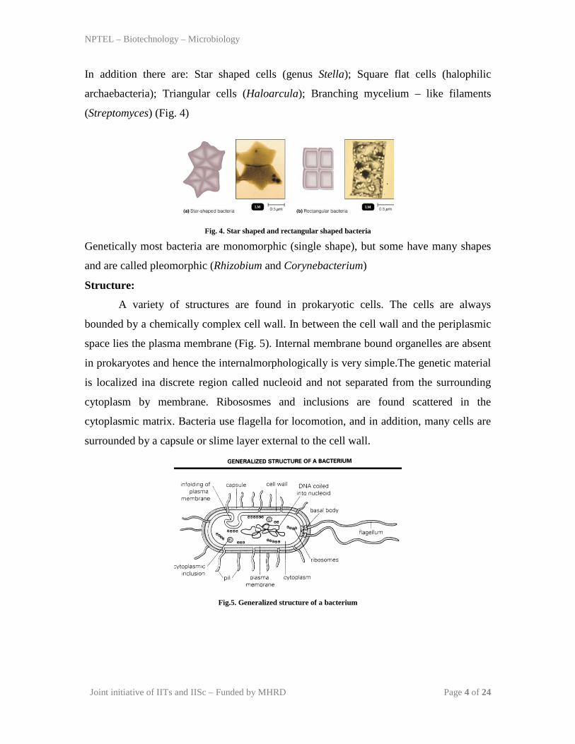
In addition there are: Star shaped cells (genus Stella); Square flat cells (halophilic
archaebacteria); Triangular cells (Haloarcula); Branching mycelium – like filaments
(Streptomyces) (Fig. 4)
Fig. 4. Star shaped and rectangular shaped bacteria
Genetically most bacteria are monomorphic (single shape), but some have many shapes
and are called pleomorphic (Rhizobium and Corynebacterium)
Structure:
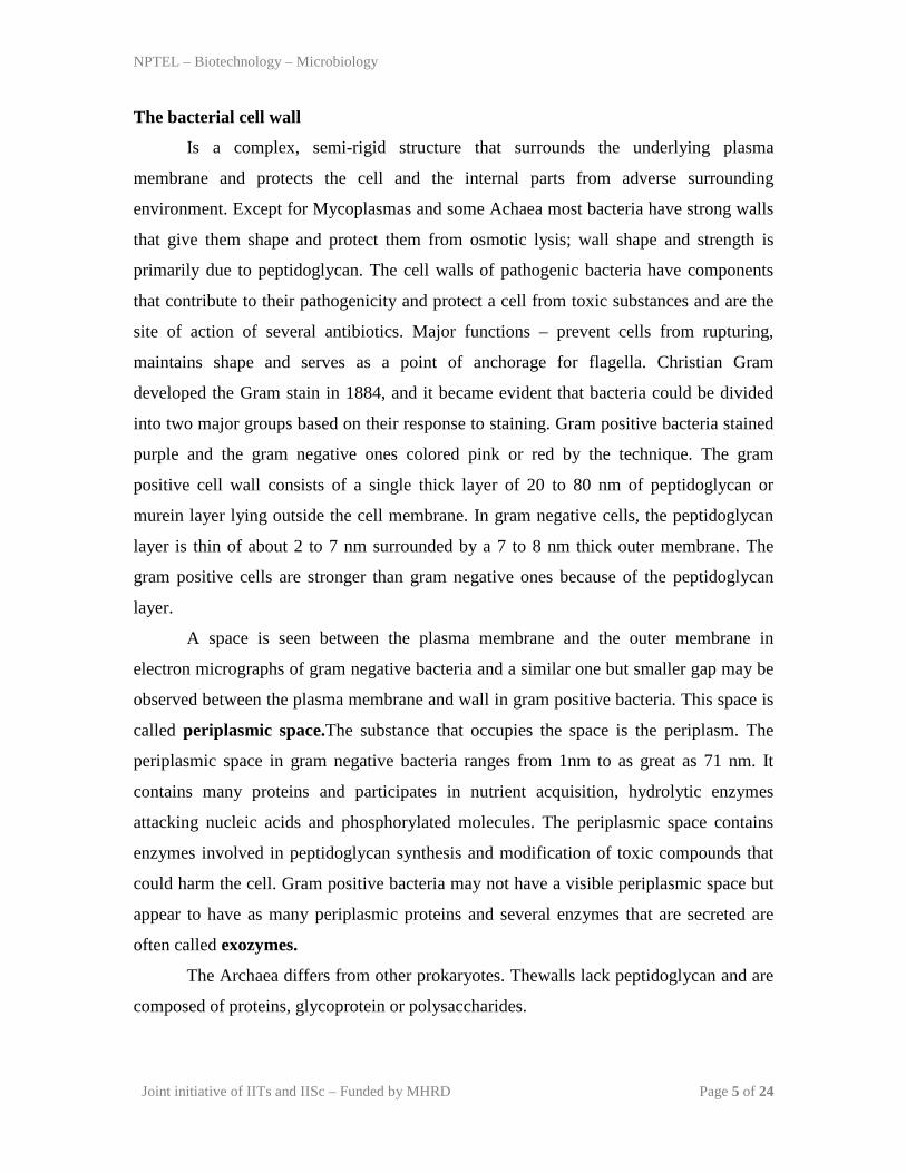
A variety of structures are found in prokaryotic cells. The cells are always
bounded by a chemically complex cell wall. In between the cell wall and the periplasmic
space lies the plasma membrane (Fig. 5). Internal membrane bound organelles are absent
in prokaryotes and hence the internalmorphologically is very simple.The genetic material
is localized ina discrete region called nucleoid and not separated from the surrounding
cytoplasm by membrane. Ribososmes and inclusions are found scattered in the
cytoplasmic matrix. Bacteria use flagella for locomotion, and in addition, many cells are
surrounded by a capsule or slime layer external to the cell wall.
Fig.5. Generalized structure of a bacterium
NPTEL – Biotechnology – Microbiology
Joint initiative of IITs and IISc – Funded by MHRD Page 5 of 24
The bacterial cell wall
Is a complex, semi-rigid structure that surrounds the underlying plasma
membrane and protects the cell and the internal parts from adverse surrounding
environment. Except for Mycoplasmas and some Achaea most bacteria have strong walls
that give them shape and protect them from osmotic lysis; wall shape and strength is
primarily due to peptidoglycan. The cell walls of pathogenic bacteria have components
that contribute to their pathogenicity and protect a cell from toxic substances and are the
site of action of several antibiotics. Major functions – prevent cells from rupturing,
maintains shape and serves as a point of anchorage for flagella. Christian Gram
developed the Gram stain in 1884, and it became evident that bacteria could be divided
into two major groups based on their response to staining. Gram positive bacteria stained
purple and the gram negative ones colored pink or red by the technique. The gram
positive cell wall consists of a single thick layer of 20 to 80 nm of peptidoglycan or
murein layer lying outside the cell membrane. In gram negative cells, the peptidoglycan
layer is thin of about 2 to 7 nm surrounded by a 7 to 8 nm thick outer membrane. The
gram positive cells are stronger than gram negative ones because of the peptidoglycan
layer.
A space is seen between the plasma membrane and the outer membrane in
electron micrographs of gram negative bacteria and a similar one but smaller gap may be
observed between the plasma membrane and wall in gram positive bacteria. This space is
called periplasmic space.The substance that occupies the space is the periplasm. The
periplasmic space in gram negative bacteria ranges from 1nm to as great as 71 nm. It
contains many proteins and participates in nutrient acquisition, hydrolytic enzymes
attacking nucleic acids and phosphorylated molecules. The periplasmic space contains
enzymes involved in peptidoglycan synthesis and modification of toxic compounds that
could harm the cell. Gram positive bacteria may not have a visible periplasmic space but
appear to have as many periplasmic proteins and several enzymes that are secreted are
often called exozymes.
The Archaea differs from other prokaryotes. Thewalls lack peptidoglycan and are
composed of proteins, glycoprotein or polysaccharides.
NPTEL – Biotechnology – Microbiology
Joint initiative of IITs and IISc – Funded by MHRD Page 6 of 24
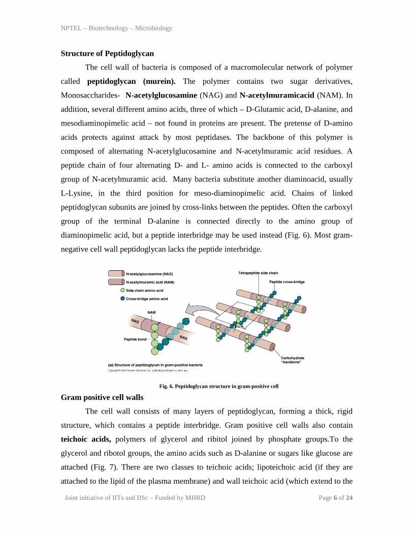
Structure of Peptidoglycan
The cell wall of bacteria is composed of a macromolecular network of polymer
called peptidoglycan (murein). The polymer contains two sugar derivatives,
Monosaccharides- N-acetylglucosamine (NAG) and N-acetylmuramicacid (NAM). In
addition, several different amino acids, three of which – D-Glutamic acid, D-alanine, and
mesodiaminopimelic acid – not found in proteins are present. The pretense of D-amino
acids protects against attack by most peptidases. The backbone of this polymer is
composed of alternating N-acetylglucosamine and N-acetylmuramic acid residues. A
peptide chain of four alternating D- and L- amino acids is connected to the carboxyl
group of N-acetylmuramic acid. Many bacteria substitute another diaminoacid, usually
L-Lysine, in the third position for meso-diaminopimelic acid. Chains of linked
peptidoglycan subunits are joined by cross-links between the peptides. Often the carboxyl
group of the terminal D-alanine is connected directly to the amino group of
diaminopimelic acid, but a peptide interbridge may be used instead (Fig. 6). Most gram-
negative cell wall peptidoglycan lacks the peptide interbridge.
Fig. 6. Peptidoglycan structure in gram-positive cell
Gram positive cell walls
The cell wall consists of many layers of peptidoglycan, forming a thick, rigid
structure, which contains a peptide interbridge. Gram positive cell walls also contain
teichoic acids, polymers of glycerol and ribitol joined by phosphate groups.To the
glycerol and ribotol groups, the amino acids such as D-alanine or sugars like glucose are
attached (Fig. 7). There are two classes to teichoic acids; lipoteichoic acid (if they are
attached to the lipid of the plasma membrane) and wall teichoic acid (which extend to the
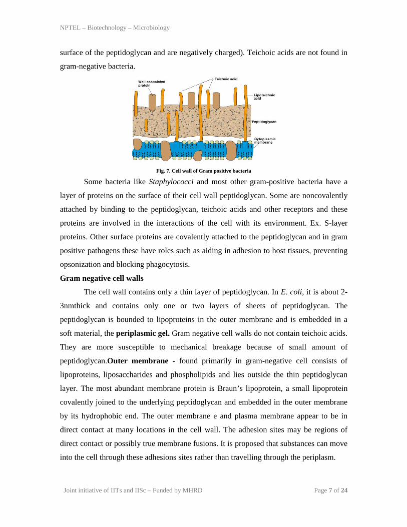
NPTEL – Biotechnology – Microbiology
Joint initiative of IITs and IISc – Funded by MHRD Page 7 of 24
surface of the peptidoglycan and are negatively charged). Teichoic acids are not found in
gram-negative bacteria.
Fig. 7. Cell wall of Gram positive bacteria
Some bacteria like Staphylococci and most other gram-positive bacteria have a
layer of proteins on the surface of their cell wall peptidoglycan. Some are noncovalently
attached by binding to the peptidoglycan, teichoic acids and other receptors and these
proteins are involved in the interactions of the cell with its environment. Ex. S-layer
proteins. Other surface proteins are covalently attached to the peptidoglycan and in gram
positive pathogens these have roles such as aiding in adhesion to host tissues, preventing
opsonization and blocking phagocytosis.
Gram negative cell walls
The cell wall contains only a thin layer of peptidoglycan. In E. coli, it is about 2-
3nmthick and contains only one or two layers of sheets of peptidoglycan. The
peptidoglycan is bounded to lipoproteins in the outer membrane and is embedded in a
soft material, the periplasmic gel. Gram negative cell walls do not contain teichoic acids.
They are more susceptible to mechanical breakage because of small amount of
peptidoglycan.Outer membrane - found primarily in gram-negative cell consists of
lipoproteins, liposaccharides and phospholipids and lies outside the thin peptidoglycan
layer. The most abundant membrane protein is Braun’s lipoprotein, a small lipoprotein
covalently joined to the underlying peptidoglycan and embedded in the outer membrane
by its hydrophobic end. The outer membrane e and plasma membrane appear to be in
direct contact at many locations in the cell wall. The adhesion sites may be regions of
direct contact or possibly true membrane fusions. It is proposed that substances can move
into the cell through these adhesions sites rather than travelling through the periplasm.
NPTEL – Biotechnology – Microbiology
Joint initiative of IITs and IISc – Funded by MHRD Page 8 of 24
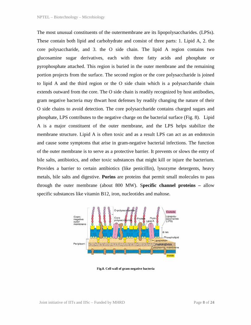
The most unusual constituents of the outermembrane are its lipopolysaccharides. (LPSs).
These contain both lipid and carbohydrate and consist of three parts: 1. Lipid A, 2. the
core polysaccharide, and 3. the O side chain. The lipid A region contains two
glucosamine sugar derivatives, each with three fatty acids and phosphate or
pyrophosphate attached. This region is buried in the outer membrane and the remaining
portion projects from the surface. The second region or the core polysaccharide is joined
to lipid A and the third region or the O side chain which is a polysaccharide chain
extends outward from the core. The O side chain is readily recognized by host antibodies,
gram negative bacteria may thwart host defenses by readily changing the nature of their
O side chains to avoid detection. The core polysaccharide contains charged sugars and
phosphate, LPS contributes to the negative charge on the bacterial surface (Fig. 8). Lipid
A is a major constituent of the outer membrane, and the LPS helps stabilize the
membrane structure. Lipid A is often toxic and as a result LPS can act as an endotoxin
and cause some symptoms that arise in gram-negative bacterial infections. The function
of the outer membrane is to serve as a protective barrier. It prevents or slows the entry of
bile salts, antibiotics, and other toxic substances that might kill or injure the bacterium.
Provides a barrier to certain antibiotics (like penicillin), lysozyme detergents, heavy
metals, bile salts and digestive. Porins are proteins that permit small molecules to pass
through the outer membrane (about 800 MW). Specific channel proteins – allow
specific substances like vitamin B12, iron, nucleotides and maltose.
Fig.8. Cell wall of gram negative bacteria
NPTEL – Biotechnology – Microbiology
Joint initiative of IITs and IISc – Funded by MHRD Page 9 of 24
Atypical cell walls:
• Mycoplasma – is a bacterial genus that naturally lacks cell walls. Their plasma
membranes have lipids called sterols, which protect them from osmotic lysis.
• Archaeabacteria – have pseudomurein (N-acetylalosaminuronic acid) but no
peptidoglycan.
• L forms - are mutant bacteria with defective cell walls.
Damage to the cell wall
The presence of cell wall is essential to protect bacteria against destruction by
osmotic pressure. The bacterial cytoplasm is much more concentrated with solutes than in
most microbial habitats which are hypotonic. During osmosis, water moves across
selectively permeable membranes such as the plasma membrane from dilute solutions
(higher water concentration) to more concentrated solutions (lower water concentration).
Usually water generally enters the bacterial cells and the osmotic pressure may reach 20
atmospheres. Plasma membrane cannot resist such high pressures and the cell will swell
and be physically disrupted and destroyed, a process called lysis. In hypertonic habitats,
the water flows outward, and the cytoplasm shrivels up and pulls away from the cell wall.
This phenomenon is called plamolysis and is useful in food preservation because many
microorganisms cannot grow in dried foods and jellies as they cannot avoid plasmolysis.
The importance of the cell wall in protecting bacteria can be demonstrated by treatment
with lysozyme (naturally occurs in eukaryotic cells and is a constituent of tears, mucus
and saliva), which attacks the peptidoglycan by hydrolyzing the bond that connects NAM
and NAG units. Penicillin inhibits peptidoglycan synthesis. Gram positive cell walls are
destroyed and the remaining cellular contents are referred to as protoplast.Gram negative
cells are not completely destroyed and the remaining cellular contents are referred to as
spheroplast. Protoplast and spheroplast are subject to osmotic lysis. Antibiotics such as
penicillin destroy bacteria by interfering with the formation of the peptide cross bridges
of peptidoglycan and ultimately cell wall synthesis.
NPTEL – Biotechnology – Microbiology
Joint initiative of IITs and IISc – Funded by MHRD Page 10 of 24
REFERENCES:
Text Books:
1. Jeffery C. Pommerville. Alcamo’s Fundamentals of Microbiology (Tenth Edition).
Jones and Bartlett Student edition.
2. Gerard J. Tortora, Berdell R. Funke, Christine L. Case. Pearson - Microbiology: An
Introduction. Benjamin Cummings.
Reference Books:
1. Lansing M. Prescott, John P. Harley and Donald A. Klein. Microbiology. Mc Graw
Hill companies.
NPTEL – Biotechnology – Microbiology
Joint initiative of IITs and IISc – Funded by MHRD Page 11 of 24
MODULE 2 - MICROBIAL CELL STRUCTURE AND FUNCTION
Lecture 2: Structures internal to the cell wall
In this lecture we shall be dealing with the structures internal to the cell wall of a
bacterial cell. They include plasma membrane, organelles in the cytoplasm like nuclear
area, ribosomes, inclusion bodies and endospores.
Plasma (cytoplasmic) membrane
Membranes are absolute requirement of all living organisms. It is the chief point
of contact with the cell’s environment and thus is responsible for much of its relationship
with the outside world.Plasma membrane – encloses the cytoplasm and consists of
phospholipids and proteins (fluid mosaic model).Most membrane-associated lipids are
structurally asymmetric with polar and nonpolar ends. The polar ends interact with water
and are hydrophilic and the nonpolar hydrophobic ends are insoluble in water. The lipid
composition of bacterial membranes varies with environmental temperature in such a
way that the membrane remains fluid during growth. Bacterial membranes usually differ
from eukaryotic membranes in lacking sterols such as cholesterol and they contain
pentacyclic sterol-like molecules called hopanoids and these are said to stabilize the
bacterial membranes. Cell membranes are very thin structures about 5 to 10 nm thick and
can be seen only with electron microscope. Plasma membranes have a complex internal
structure; the small globular particles seen in these membranes are thought to be
membrane proteins that lie within the membrane lipid bilayer (Fig. 9).
The most widely accepted current model for membrane structure is the fluid
mosaic model of S. Jonathan Singer and Garth Nicholson. Two types of membrane
proteins are seen, Peripheral proteins - which are loosely connected to the membrane
and can be easily removed and are soluble in aqueous solutions and make up about 20 to
30% of total membrane protein. About 70 to 80% of membrane proteins are integral
proteins. These cannot be easily extracted from membranes and are insoluble in aqueous
solutions when freed of lipids. Integral proteins, like membrane lipids are amphipathic;
their hydrophobic regions are buried in the lipid while the hydrophilic portions project
from the membrane surface. The plasma membrane retains the cytoplasm, particularly in

NPTEL – Biotechnology – Microbiology
Joint initiative of IITs and IISc – Funded by MHRD Page 12 of 24
cells without cell walls, and separates it from the surroundings. Plasma membranes serve
as a selectively permeable barrier; it allows particular ions and molecules to pass, either
into or out of the cell, while preventing the movement of others. Transport systems can
be used for such tasks as nutrient uptake, waste excretion, and protein secretion. The
plasma membrane also is the location of a variety of crucial metabolic processes;
respiration, photosynthesis, the synthesis of lipids and cell wall constituents, and
probably chromosome segregation.
The bacterial plasma membrane can be destroyed by alcohols and polymixins
which cause leakage of intracellular contents and subsequent cell death of the organism.
Fig. 9. Plasma membrane structure
Internal membrane systems:
Prokaryotes do not contain complex membrane systems as present in eukaryotes
like chloroplast and mitochondria. They contain membranous structures like the one
observed most is mesosome. Mesosomes – irregular infoldings or invaginations of the
plasma membrane in the shape of vesicles, tubules, or lamellae. They can be seen in both
gram positive and gram-negative bacteria. These are often found next to the septa or
cross-walls in dividing bacteria and sometimes seems attached to the bacterial
chromosome. Thus they seem to be involved in cell wall formation during division or
play a role in chromosome replication and distribution to daughter cells.
Some bacteria have internal membrane systems quite different from the
mesosomes. The infoldings of the plasma membrane can become extensive and complex
in photosynthetic bacteria such as the cyanobacteria and purple bacteria or in bacteria
with very high respiratory activity like the nitrifying bacteria. They may be aggregates of
NPTEL – Biotechnology – Microbiology
Joint initiative of IITs and IISc – Funded by MHRD Page 13 of 24
spherical vesicles, flattened vesicles, or tubular membranes. Their function may be to
provide a larger membrane surface for greater metabolic activity.
Cytoplasm
Is the fluid component inside the plasma membrane. Cytoplasm is about 80%
water and contains primarily proteins (enzymes), carbohydrates, lipids, inorganic ions
and many low MW compounds. Major structures in the cytoplasm are DNA, ribosomes
and inclusions.
Nuclear area
The striking difference between prokaryotic and eukaryotic systems is the way in
which their genetic material is packaged. Prokaryotes lack a membrane-delimited
nucleus. Contains a single long circular molecule of double-stranded DNA bacterial
chromosomes do not include histones and are not surrounded by a nuclear envelope and
located in an irregularly shaped region called nucleiod. The nuclear area can be
spherical, elongated, or dumb-bell shaped. It is attached to the plasma membrane and
proteins of plasma membrane are believed to be responsible for replication of the DNA.
It has been discovered recently that Vibrio cholera has more than one chromosome.
Electron microscope studies have shown the nucleiod in contact with either the
mesosome or the plasma membrane and hence evidence that the membranes may be
involved in the separation of DNA into daughter cells during division. Chemical analysis
reveals that they nucleoids are composed to about 60% DNA, 30% RNA and 10%
protein. E. coli, which is about 2 to 6um long, the closed DNA circle measures
approximately 1400um. Hence, it is evident that the DNA is efficiently packaged to fit
within the nucleoid and the DA is looped and coiled extensively (Fig. 10).
NPTEL – Biotechnology – Microbiology
Joint initiative of IITs and IISc – Funded by MHRD Page 14 of 24
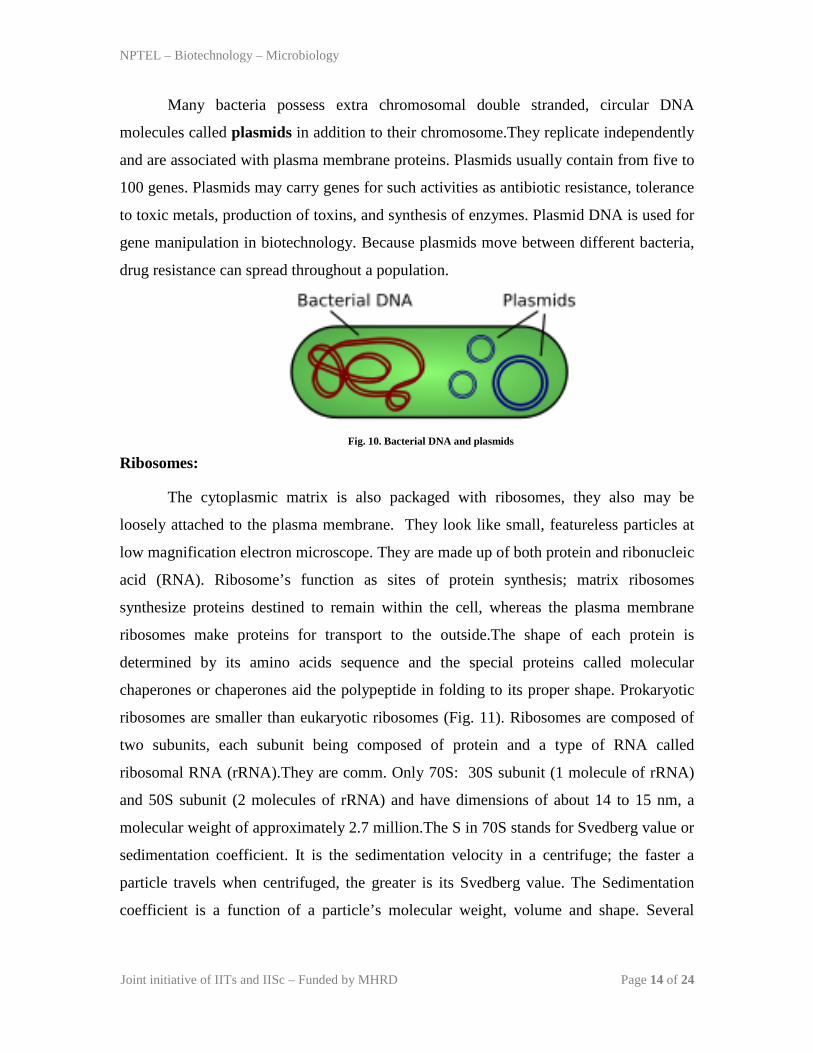
Many bacteria possess extra chromosomal double stranded, circular DNA
molecules called plasmids in addition to their chromosome.They replicate independently
and are associated with plasma membrane proteins. Plasmids usually contain from five to
100 genes. Plasmids may carry genes for such activities as antibiotic resistance, tolerance
to toxic metals, production of toxins, and synthesis of enzymes. Plasmid DNA is used for
gene manipulation in biotechnology. Because plasmids move between different bacteria,
drug resistance can spread throughout a population.
Fig. 10. Bacterial DNA and plasmids
Ribosomes:
The cytoplasmic matrix is also packaged with ribosomes, they also may be
loosely attached to the plasma membrane. They look like small, featureless particles at
low magnification electron microscope. They are made up of both protein and ribonucleic
acid (RNA). Ribosome’s function as sites of protein synthesis; matrix ribosomes
synthesize proteins destined to remain within the cell, whereas the plasma membrane
ribosomes make proteins for transport to the outside.The shape of each protein is
determined by its amino acids sequence and the special proteins called molecular
chaperones or chaperones aid the polypeptide in folding to its proper shape. Prokaryotic
ribosomes are smaller than eukaryotic ribosomes (Fig. 11). Ribosomes are composed of
two subunits, each subunit being composed of protein and a type of RNA called
ribosomal RNA (rRNA).They are comm. Only 70S: 30S subunit (1 molecule of rRNA)
and 50S subunit (2 molecules of rRNA) and have dimensions of about 14 to 15 nm, a
molecular weight of approximately 2.7 million.The S in 70S stands for Svedberg value or
sedimentation coefficient. It is the sedimentation velocity in a centrifuge; the faster a
particle travels when centrifuged, the greater is its Svedberg value. The Sedimentation
coefficient is a function of a particle’s molecular weight, volume and shape. Several

NPTEL – Biotechnology – Microbiology
Joint initiative of IITs and IISc – Funded by MHRD Page 15 of 24
antibiotics, such as streptomycin, neomycin and tetracyclines, exert their antimicrobial
effects by inhibiting protein synthesis on ribosomes.
Fig. 11. Ribosmes in bacteria
Inclusions:
Inclusion bodies can be divided into two types:
Inclusion bodies not bounded by a membrane and lie free in the cytoplasm. Ex.
Polyphosphate granules, cyanophycingranules and some glycogen granules.
Inclusion bodies enclosed by a membrane about 2-4nm thick. Ex.PolyB-
hydroxybutyrate granules, some glycogen and sulfur granules, carboxysomes and gas
vacuoles.
Organic inclusion bodies:
Glycogen:
Polymer of glucose units composed of long chains formed by alpha (1-4)
glycosidic bonds and branching chains connected to themby alpha (1-6)glycosidic bonds.
Ex. glycogen and starch, and their presence can be demonstrated when iodine is applied
to the cells (glycogen granules appear reddish brown and starch granules appear blue).
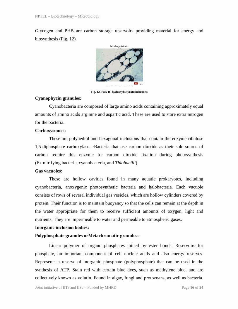
Poly B- hydroxybutyrate:
Contains beta-hydroxybutyrate molecules joined by ester bonds between the
carboxyl and hydroxyl groups of adjacent molecules. Appear in various species of
Mycobacterium, Bacillus, Azotobacter, Spirillum and other genera. Lipid inclusions are
revealed by use of fat-soluble dyes, such as Sudan dyes.
NPTEL – Biotechnology – Microbiology
Joint initiative of IITs and IISc – Funded by MHRD Page 16 of 24
Glycogen and PHB are carbon storage reservoirs providing material for energy and
biosynthesis (Fig. 12).
Fig. 12. Poly B- hydroxybutyrateinclusions
Cyanophycin granules:
Cyanobacteria are composed of large amino acids containing approximately equal
amounts of amino acids arginine and aspartic acid. These are used to store extra nitrogen
for the bacteria.
Carboxysomes:
These are polyhedral and hexagonal inclusions that contain the enzyme ribulose
1,5-diphosphate carboxylase. ·Bacteria that use carbon dioxide as their sole source of
carbon require this enzyme for carbon dioxide fixation during photosynthesis
(Ex.nitrifying bacteria, cyanobacteria, and Thiobacilli).
Gas vacuoles:
These are hollow cavities found in many aquatic prokaryotes, including
cyanobacteria, anoxygenic photosynthetic bacteria and halobacteria. Each vacuole
consists of rows of several individual gas vesicles, which are hollow cylinders covered by
protein. Their function is to maintain buoyancy so that the cells can remain at the depth in
the water appropriate for them to receive sufficient amounts of oxygen, light and
nutrients. They are impermeable to water and permeable to atmospheric gases.
Inorganic inclusion bodies:
Polyphosphate granules orMetachromatic granules:
Linear polymer of organo phosphates joined by ester bonds. Reservoirs for
phosphate, an important component of cell nucleic acids and also energy reserves.
Represents a reserve of inorganic phosphate (polyphosphate) that can be used in the
synthesis of ATP. Stain red with certain blue dyes, such as methylene blue, and are
collectively known as volutin. Found in algae, fungi and protozoans, as well as bacteria.
NPTEL – Biotechnology – Microbiology
Joint initiative of IITs and IISc – Funded by MHRD Page 17 of 24
These granules are quite large and are characteristic of Corynebacterium diphtheriae, the
causative agent of diphtheria, thus they have diagnostic significance.
Sulphur granules:
Sulphur bacteria, which belong to the genus Thiobacillus, derive energy by
oxidizing sulfur and sulfur containing compounds. These bacteria may deposit sulfur
granules in the cell, where they serve as an energy reserve. Purple photosynthetic bacteria
use H2S as electron donor and accumulate resulting sulfurin either the periplasmic space
or in special cytoplasmic globules.
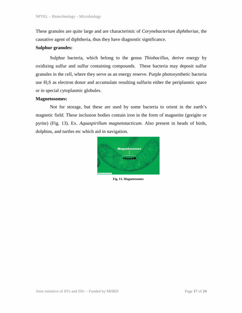
Magnetosomes:
Not for storage, but these are used by some bacteria to orient in the earth’s
magnetic field. These inclusion bodies contain iron in the form of magnetite (greigite or
pyrite) (Fig. 13). Ex. Aquaspirillum magnetotacticum. Also present in heads of birds,
dolphins, and turtles etc which aid in navigation.
Fig. 13. Magnetosomes
NPTEL – Biotechnology – Microbiology
Joint initiative of IITs and IISc – Funded by MHRD Page 18 of 24
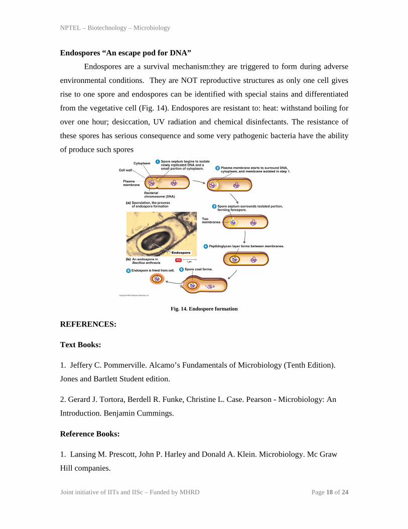
Endospores “An escape pod for DNA”
Endospores are a survival mechanism:they are triggered to form during adverse
environmental conditions. They are NOT reproductive structures as only one cell gives
rise to one spore and endospores can be identified with special stains and differentiated
from the vegetative cell (Fig. 14). Endospores are resistant to: heat: withstand boiling for
over one hour; desiccation, UV radiation and chemical disinfectants. The resistance of
these spores has serious consequence and some very pathogenic bacteria have the ability
of produce such spores
Fig. 14. Endospore formation
REFERENCES:
Text Books:
1. Jeffery C. Pommerville. Alcamo’s Fundamentals of Microbiology (Tenth Edition).
Jones and Bartlett Student edition.
2. Gerard J. Tortora, Berdell R. Funke, Christine L. Case. Pearson - Microbiology: An
Introduction. Benjamin Cummings.
Reference Books:
1. Lansing M. Prescott, John P. Harley and Donald A. Klein. Microbiology. Mc Graw
Hill companies.
NPTEL – Biotechnology – Microbiology
Joint initiative of IITs and IISc – Funded by MHRD Page 19 of 24
MODULE 2 - MICROBIAL CELL STRUCTURE AND
FUNCTION Lecture 3: Structures external to the cell wall
In this lecture we shall look into the structures external to the cell wall of bacterial cells.
This includes glycocalyx, fimbriae, pili, flagella, axial filaments
Glycocalyx (Capsules, Slime layers and S-layers)
It is a viscous (sticky), gelatinous polymer composed of polysaccharide,
polypeptide or both. If the substance is organized and is firmly attached to the cell wall,
the glycocalyx is described as a capsule (negative staining). If the substance is
unorganized and only loosely attached to the cell wall, the glycocalyx is described as a
slime layer. Capsules protect pathogenic bacteria from phagocytosis (process by which
certain white blood cells engulf and destroy microbes) and contribute to virulence.
Unencapsulated Streptomyces pneumoniae and Bacillus anthracis does not cause disease
because the cells are readily phagocytosized. This allows the bacteria to attach to various
surfaces, such as rocks in fast-moving streams, plant roots, human tooth and tissues and
even other bacteria. Capsules also contain water which prevents them from desiccation.
Other examples are Streptococcus mutans (dental caries), Klebsiella pneumoniae
(respiratory tract). These can protect a cell against dehydration. Capsules and slime layers
usually are made up of polysaccharides, but they may be constructed of othermaterial,
like Bacillus anthracis has a capsule of poly D-glutamic acid. Capsules are clearly visible
in the light microscope by using stains or special capsule stains.
A regularly structured layer called S-layer is usually seen in many gram positive
and gram negative bacteria. It consists of proteins or glycoproteins and resembles a
pattern something similar to floor tiles. The S-layer adheres directly to the outer
membrane in case of gram negative bacteria and with the peptidoglycan surface in gram
positive bacteria. These protect the bacteria against ion and pH fluctuations, osmotic
stress, enzymes, or the predacious bacterium Bdellovibrio. The S layer also helps
maintain the shape and envelope rigidity of at least bacterial cells and also promotes cell
NPTEL – Biotechnology – Microbiology
Joint initiative of IITs and IISc – Funded by MHRD Page 20 of 24
adhesion to surfaces. Sometimes, the layer also seems to protect some pathogens against
complement attack and phagocytosis, thus contributing to their virulence.
Fimbriae and Pili:
Many gram negative bacteria have hairlike appendages that are shorter, straighter
and thinner than flagella and are used for attachment rather than for motility. They are
usually called fimbriae. These structures contain a protein called pilin. Fimbriae - occur
at the poles of the bacterial cell, or they can be evenly distributed over the entire surface
of the cell. Fimbriae of Neisseria gonorrhoeae the causative agent of gonorrhea help the
microbe to colonize mucous membranes to cause the disease. At least some types of
fimbriae attach bacteria to solid surfaces such as rocks in streams and host tissues.
Pilior sex pili or pilus- usually longer than fimbriae and number only one to ten per cell.
Pili function to join bacterial cells prior to the transfer to DNA from one cell to another
(sometimes called sex pili). They are genetically determined by sex factors or conjugative
plasmids and are required for bacterial mating. Some bacterial viruses attach specifically
to receptors on sex pili at the start of their reproductive cycle.
Flagella:
Motile bacteria move by use of flagella, threadlike locomotor appendages
extending outward from the plasma membrane and cell wall. They are slender, rigid
structures, about 20 nm across and up to 15 or 20 µm long. Bacterial species often differ
distinctively in their patterns of flagella distribution (Fig. 15).
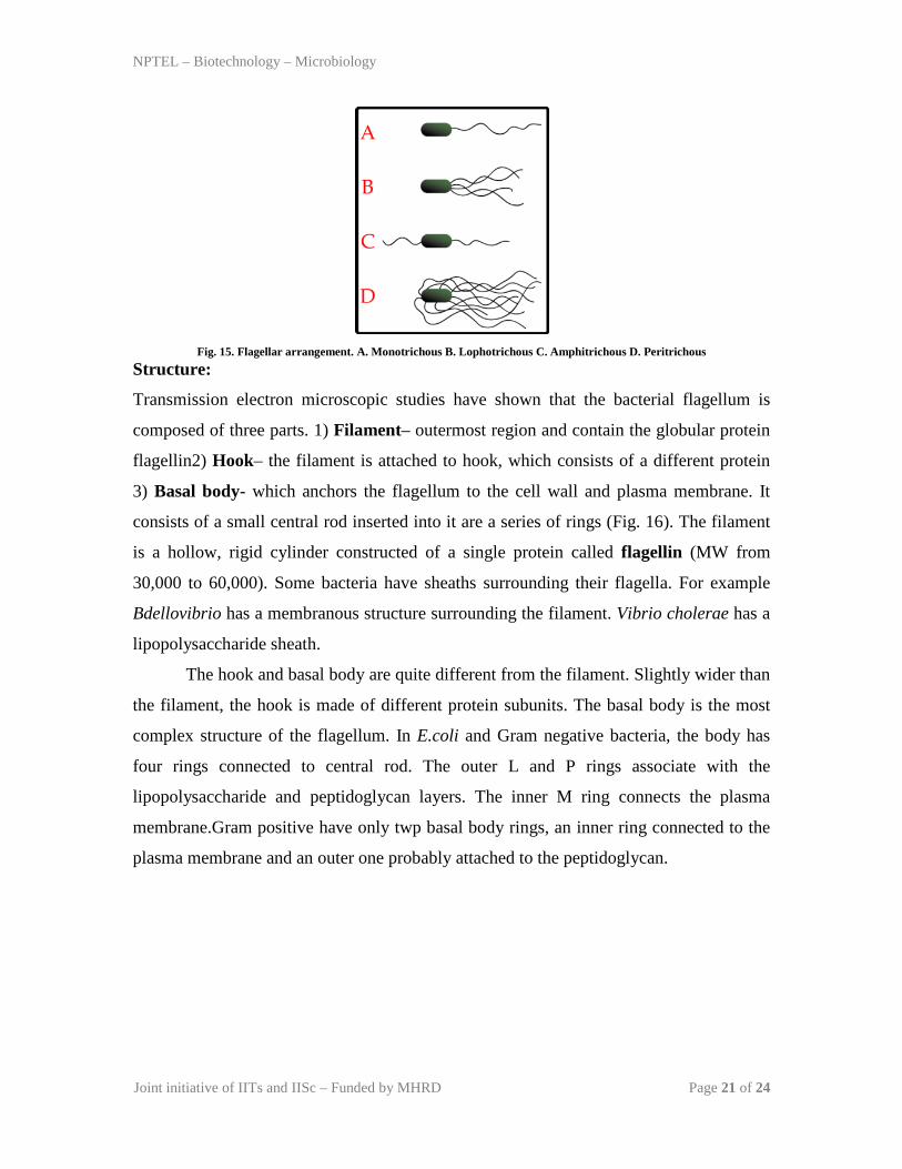
Monotrichous- single polar flagellum located at one end
Amphitrochous- With two flagella, one at each end
Lophotrichous- With two or more flagella at one or both ends
Peritrichous- flagella all over the surface
Atrichous- Bacteria without flagella (Cocci rarely have flagella)
NPTEL – Biotechnology – Microbiology
Joint initiative of IITs and IISc – Funded by MHRD Page 21 of 24
Fig. 15. Flagellar arrangement. A. Monotrichous B. Lophotrichous C. Amphitrichous D. Peritrichous
Structure:
Transmission electron microscopic studies have shown that the bacterial flagellum is
composed of three parts. 1) Filament– outermost region and contain the globular protein
flagellin2) Hook– the filament is attached to hook, which consists of a different protein
3) Basal body- which anchors the flagellum to the cell wall and plasma membrane. It
consists of a small central rod inserted into it are a series of rings (Fig. 16). The filament
is a hollow, rigid cylinder constructed of a single protein called flagellin (MW from
30,000 to 60,000). Some bacteria have sheaths surrounding their flagella. For example
Bdellovibrio has a membranous structure surrounding the filament. Vibrio cholerae has a
lipopolysaccharide sheath.
The hook and basal body are quite different from the filament. Slightly wider than
the filament, the hook is made of different protein subunits. The basal body is the most
complex structure of the flagellum. In E.coli and Gram negative bacteria, the body has
four rings connected to central rod. The outer L and P rings associate with the
lipopolysaccharide and peptidoglycan layers. The inner M ring connects the plasma
membrane.Gram positive have only twp basal body rings, an inner ring connected to the
plasma membrane and an outer one probably attached to the peptidoglycan.

NPTEL – Biotechnology – Microbiology
Joint initiative of IITs and IISc – Funded by MHRD Page 22 of 24
Fig. 16. Structure of bacterial flagella (Gram negative)
The synthesis of flagella is a complex process involving atleast 20 to 30 genes. Flagellin
subunits are transported through the filament’s hollow internal core. When they reach the
tip, the subunits spontaneously aggregate under the direction of a special filament cap so
that the filament grows at its tip rather than at the base. Filament synthesis is an excellent
example of self-assembly.
Flagellar movement:
The mechanism of flagellar movement in prokaryotes is different from
eukaryotic flagella. The bacterium moves when the helix rotates as the filament is in the
shape of rigid helix. The flagella act just like propellers on a boat. The direction of
flagellar rotation determines the nature of bacterial movement. The movement in
monotrichous bacteria stop and tumble randomly by reversing the flagellar rotation. The
polar flagella, rotate counter clockwise during normal forward movement, whereas the
cell itself rotates slowly clockwise. Peritrichous bacteria also operate in a similar way. To
move forward, the flagella rotate counter clockwise. As they do so, they bend at their
hooks to for a rotating bundle that propels them forward. Clockwise rotation of the
flagella disrupts the bundle and the cell tumbles (Fig. 17).
NPTEL – Biotechnology – Microbiology
Joint initiative of IITs and IISc – Funded by MHRD Page 23 of 24
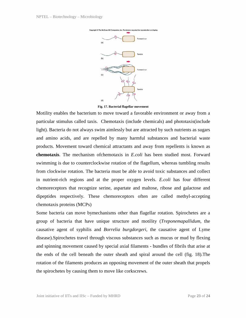
Fig. 17. Bacterial flagellar movement
Motility enables the bacterium to move toward a favorable environment or away from a
particular stimulus called taxis. Chemotaxis (include chemicals) and phototaxis(include
light). Bacteria do not always swim aimlessly but are attracted by such nutrients as sugars
and amino acids, and are repelled by many harmful substances and bacterial waste
products. Movement toward chemical attractants and away from repellents is known as
chemotaxis. The mechanism ofchemotaxis in E.coli has been studied most. Forward
swimming is due to counterclockwise rotation of the flagellum, whereas tumbling results
from clockwise rotation. The bacteria must be able to avoid toxic substances and collect
in nutrient-rich regions and at the proper oxygen levels. E.coli has four different
chemoreceptors that recognize serine, aspartate and maltose, ribose and galactose and
dipeptides respectively. These chemoreceptors often are called methyl-accepting
chemotaxis proteins (MCPs)
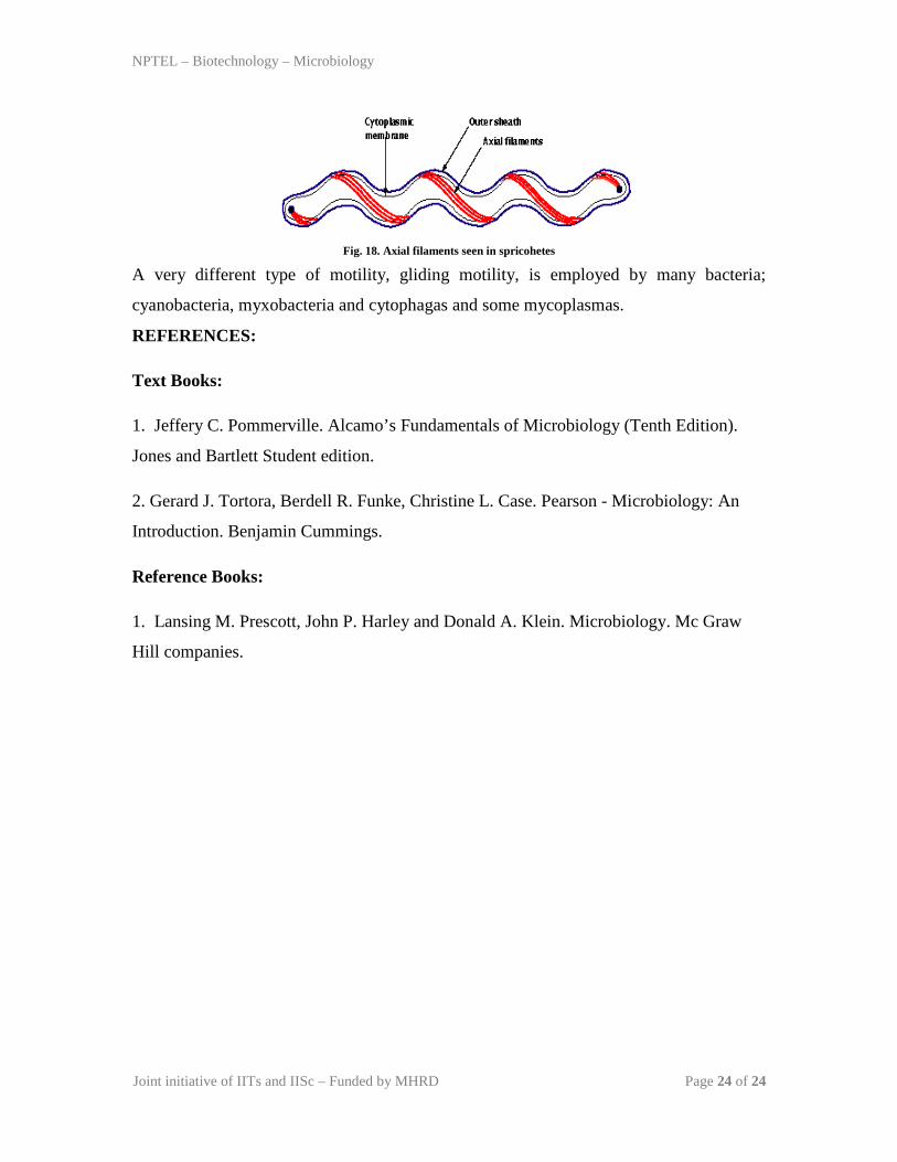
Some bacteria can move bymechanisms other than flagellar rotation. Spirochetes are a
group of bacteria that have unique structure and motility (Treponemapallidum, the
causative agent of syphilis and Borrelia burgdorgeri, the causative agent of Lyme
disease).Spirochetes travel through viscous substances such as mucus or mud by flexing
and spinning movement caused by special axial filaments - bundles of fibrils that arise at
the ends of the cell beneath the outer sheath and spiral around the cell (fig. 18).The
rotation of the filaments produces an opposing movement of the outer sheath that propels
the spirochetes by causing them to move like corkscrews.
NPTEL – Biotechnology – Microbiology
Joint initiative of IITs and IISc – Funded by MHRD Page 24 of 24
Fig. 18. Axial filaments seen in spricohetes
A very different type of motility, gliding motility, is employed by many bacteria;
cyanobacteria, myxobacteria and cytophagas and some mycoplasmas.
REFERENCES:
Text Books:
1. Jeffery C. Pommerville. Alcamo’s Fundamentals of Microbiology (Tenth Edition).
Jones and Bartlett Student edition.
2. Gerard J. Tortora, Berdell R. Funke, Christine L. Case. Pearson - Microbiology: An
Introduction. Benjamin Cummings.
Reference Books:
1. Lansing M. Prescott, John P. Harley and Donald A. Klein. Microbiology. Mc Graw
Hill companies.