MODULATORY ROLE OF AZIMA TETRACANTHA LEAVES ON CARBON …
15
www.wjpr.net Vol 6, Issue 10, 2017. 924 MODULATORY ROLE OF AZIMA TETRACANTHA LEAVES EXTRACT ON CARBON TETRACHLORIDE INDUCED OXIDATIVE STRESS IN RATS. K. Saleem*, T. Nargis Begum # and M. H. Muhammad Ilyas @ *Research Scholar, Department of Biochemistry, PRIST University, Tamil Nadu, India. # Assistant Professor, P.G. and Research Department of Biotechnology, Jamal Mohamed College (Autonomous), Tiruchirappalli - 620 020, Tamil Nadu, India. @ Visiting Professor, Department of Biotechnology, Bharathidasan University, Tiruchirappalli - 620 024, Tamil Nadu, India. ABSTRACT Current hypothesis favour the concept that lowering oxidative stress can have a health benefits. In this study modulatory effect of ethanolic extract of Azima tetracantha was investigated using carbon tetrachloride (CCl 4 )-induced oxidative stress as the experimental model. The oxidative stress rats were administered different doses (100, 200 and 400mg/ Kg BW) of Azima tetracantha leaves extract. Malondialdehyde (MDA) is a product of lipid peroxidation in CCl 4 – intoxicated rats was evidenced by a marked increment in the levels of thiobarbituric acid reactive substances (TBARS) and also a distinct diminution in glutathione (GSH) content in the liver. In CCl 4 + Azima tetracantha treated rats these biochemical parameters attained an almost normal as dose dependent manner. The decreased activity of antioxidant enzymes, such as superoxide dismutase (SOD), catalase (CAT), glutathione peroxidase (GPx) and non enzymatic antioxidant, such as glutathione (GSH), vitamin C and vitamin E in CCl 4 intoxicated rats were observed. On treatment with different doses (100, 200 and 400mg/ Kg BW) of Azima tetracantha leaves extract increased in the activity as dose dependent manner. Among the various doses, 400mg/kg has potential activity than other doses. From these results, it was suggested that lipid peroxidation and oxidative stress elicited by CCl 4 intoxication had been nullified due to the effect of Azima tetracantha. World Journal of Pharmaceutical Research SJIF Impact Factor 7.523 Volume 6, Issue 10, 924-938. Research Article ISSN 2277– 7105 *Corresponding Author K. Saleem Research Scholar, Department of Biochemistry, PRIST University, Tamil Nadu, India. Article Received on 13 July 2017, Revised on 03 August 2017, Accepted on 23 August 2017 DOI: 10.20959/wjpr201710-9394
Transcript of MODULATORY ROLE OF AZIMA TETRACANTHA LEAVES ON CARBON …

www.wjpr.net Vol 6, Issue 10, 2017.
924
Saleem et al. World Journal of Pharmaceutical Research
MODULATORY ROLE OF AZIMA TETRACANTHA LEAVES
EXTRACT ON CARBON TETRACHLORIDE INDUCED OXIDATIVE
STRESS IN RATS.
K. Saleem*, T. Nargis Begum# and M. H. Muhammad Ilyas
@
*Research Scholar, Department of Biochemistry, PRIST University, Tamil Nadu, India.
#Assistant Professor, P.G. and Research Department of Biotechnology, Jamal Mohamed
College (Autonomous), Tiruchirappalli - 620 020, Tamil Nadu, India.
@Visiting Professor, Department of Biotechnology, Bharathidasan University, Tiruchirappalli
- 620 024, Tamil Nadu, India.
ABSTRACT
Current hypothesis favour the concept that lowering oxidative stress
can have a health benefits. In this study modulatory effect of ethanolic
extract of Azima tetracantha was investigated using carbon
tetrachloride (CCl4)-induced oxidative stress as the experimental
model. The oxidative stress rats were administered different doses
(100, 200 and 400mg/ Kg BW) of Azima tetracantha leaves extract.
Malondialdehyde (MDA) is a product of lipid peroxidation in CCl4 –
intoxicated rats was evidenced by a marked increment in the levels of
thiobarbituric acid reactive substances (TBARS) and also a distinct
diminution in glutathione (GSH) content in the liver. In CCl4 + Azima
tetracantha treated rats these biochemical parameters attained an
almost normal as dose dependent manner. The decreased activity of antioxidant enzymes,
such as superoxide dismutase (SOD), catalase (CAT), glutathione peroxidase (GPx) and non
enzymatic antioxidant, such as glutathione (GSH), vitamin C and vitamin E in CCl4
intoxicated rats were observed. On treatment with different doses (100, 200 and 400mg/ Kg
BW) of Azima tetracantha leaves extract increased in the activity as dose dependent manner.
Among the various doses, 400mg/kg has potential activity than other doses. From these
results, it was suggested that lipid peroxidation and oxidative stress elicited by CCl4
intoxication had been nullified due to the effect of Azima tetracantha.
World Journal of Pharmaceutical Research SJIF Impact Factor 7.523
Volume 6, Issue 10, 924-938. Research Article ISSN 2277– 7105
*Corresponding Author
K. Saleem
Research Scholar,
Department of
Biochemistry, PRIST
University, Tamil Nadu,
India.
Article Received on
13 July 2017,
Revised on 03 August 2017,
Accepted on 23 August 2017
DOI: 10.20959/wjpr201710-9394
www.wjpr.net Vol 6, Issue 10, 2017.
925
Saleem et al. World Journal of Pharmaceutical Research
KEYWORDS: Antioxidant enzymes, Oxidative stress, Carbon tetrachloride, Lipid
peroxidation, Azima tetracantha.
INTRODUCTION
Current hypothesis favour the concept that lowering oxidative stress can have a health
benefit. Free radicals can be overproduced or the natural antioxidant system defenses
weakened, first resulting in oxidative stress and then leading to oxidative injury and disease.
Free radicals are fundamental to any biochemical process and represent an essential part of
aerobic life and our metabolism (Tiwari, 2001). The most common Reactive oxygen species
(ROS) include superoxide (O2●–
) anion, hydrogen peroxide (H2O2), peroxyl (ROO●) radicals,
and the very reactive hydroxyl (OH●) radicals. The nitrogen-derived free radicals are nitric
oxide (NO●) and peroxy nitrite anion (ONOO
●). ROS have been implicated in over a
hundreds of diseases states which range from arthritis and connective tissue disorders to
carcinogenesis, aging, physical injury, infection and acquired immunodeficiency syndrome
(Joyce, 1987).
It is well established that CCl4 induces hepatotoxicity by metabolic activation; therefore it
selectively causes toxicity in liver cells maintaining semi-normal metabolic function. CCl4 is
bio-transformed by the cytochrome P450 system in the endoplasmic reticulum to produce
trichloromethyl free radical (CCl3●). Trichloromethyl free radical then combined with cellular
lipids and proteins in the presence of oxygen to form a trichloromethyl peroxyl radical, which
may attack lipids on the membrane of endoplasmic reticulum faster than trichloromethyl free
radical. Thus, trichloromethylperoxyl free radical leads to elicit lipid peroxidation, the
destruction of Ca2+
homeostasis, and finally, results in cell death (De Groot and Noll, 1986;
Clawson, 1989; Reckengel et al., 1989). These result in changes of structures of the
endoplasmic reticulum and other membrane, loss of enzyme metabolic enzyme activation,
reduction of protein synthesis and loss of glucose-6-phosphatase activation, leading to liver
damage (Recknagel 1983; Wolf et al., 1980; Azri et al., 1992).
The harmful effect of reactive oxygen species is neutralized by a broad class of protective
agents called antioxidants, which prevents oxidative damage by reacting with free radicals
before any other molecules can become a target. The non-enzymatic antioxidants (Vitamin E,
C, reduced glutathione etc.) and antioxidant enzymes (SOD, CAT, GSHPx) play an important
role in the protection of cells and tissues against free radical mediated tissue damage (Yu,
1994; Ray and Husain, 2002). Any compound, natural or synthetic, with antioxidant

www.wjpr.net Vol 6, Issue 10, 2017.
926
Saleem et al. World Journal of Pharmaceutical Research
properties might contribute towards the partial or total alleviation of this type of damage. As
plants produce a lot of antioxidants to control the oxidative stress, they can represent a source
of new compounds with antioxidant activity. A number of plants and plant isolates have been
reported to protect free radical induced damage in various experimental models (Scartezzini
and Sproni, 2000).
Natural antioxidants strengthen the endogenous antioxidant defences from ROS ravage and
restore the optimal balance by neutralizing the reactive species. They are gaining immense
importance by virtue of their critical role in disease prevention. The medicinal value of the
chosen plant Azima tetracantha has been extensively worked out. However its therapeutic
efficacy in the state of oxidative stress has not been evaluated. Previously reported that the
presence of alkaloids, flavanoids, tannins, cardio glycosides, saponins and terpenoids like
compounds in Azima tetracantha (Abirami et al., 2015; Janardhan et al., 2014). Hence in the
present study an attempt has been made to create an animal model with oxidative stress using
CCl4 and the therapeutic efficacy of the extract of Azima tetracantha was evaluated.
MATERIALS AND METHODS
Animals
Male albino rats of Wistar strain approximately 3-4 months young rats (weighing
approximately 140-160g) and 24-26 months old rats (weighing approximately 380-410g were
used in this study. They were healthy animals procured from Sri Venkateswara enterprises,
Bangalore, India. The animals were housed in spacious polypropylene cages bedded with rice
husk. The animal room was well ventilated and maintained under standard experimental
conditions (Temperature 27±2ºC and 12 hours light / dark cycle) throughout the experimental
period. All the animals were fed with standard pellet diet (Gold Mohur, Mumbai, India) and
water ad libitum. They were acclimatization to the environment for 1 week prior to
experimental use. The experiment was carried out according to the guidelines of the
Committee (Ethical No: SAC/IAEC/BC/2016/Ph.D-005) for the Purpose of Control and
Supervision of Experiments on Animals (CPCSEA), New Delhi, India.
Chemicals: Carbon tetrachloride, Thiobarbituric acid, 2,4-Dinitro phenyl hydrazine and
glutathione were purchased from sigma chemical, Mumbai. All other reagents and chemicals
used in this study were of analytical grade with high purity.
www.wjpr.net Vol 6, Issue 10, 2017.
927
Saleem et al. World Journal of Pharmaceutical Research
Plant Material
The fresh leaves of Azima tetracantha were collected in the month of January 2015 at Melur,
Thiruchirappalli District, Tamil Nadu, South India. The leaves were identified and
authenticated by Dr. S. John Britto, The Director, the Rabinat Herbarium and centre for
molecular systematics, St. Joseph’s college Trichy-Tamil Nadu. India. A Voucher specimen
(EP001) has been deposited at the Rapinat Herbarium, St. Josephs College, Thiruchirappalli,
Tamil Nadu, India.
Preparation of Plant Extract
Fresh plant material was shade dried and powdered coarsely using electric blender. 250g of
dried plant material was soaked in Ethanol for 48 hours. After 48 hrs of soaking the solvent
was distilled off under reduced pressure at 50°C and dried in vacuum. The residue was
dissolved in isotonic saline and used for the study.
Experimental protocol
Body weights of the animals were recorded and they were divided into 4 groups of 6 animals
each as follows. Group I – Normal Rats. Group II – Negative control - Animals were
administrated orally with CCl4 (0.5 ml/150g of bw-v/v in olive oil) on 1st, 8
th and 16
th day.
Group III – Animals were administrated orally with CCl4 (0.5 ml/150 g of bw-v/v in olive oil
on 1st, 8
th and 16
th day) and treated with Azima tetracantha leaves extract (100mg/ Kg BW)
orally for 21 days. Group IV - Animals were administrated orally with CCl4 (0.5 ml/150 g of
bw-v/v in olive oil on 1st, 8
th and 16
th day) and treated with Azima tetracantha leaves extract
(200mg/ Kg BW) orally for 21 days. Group V –Animals were administrated orally with CCl4
(0.5 ml/150 g of bw-v/v in olive oil on 1st, 8
th and 16
th day) and treated with Azima
tetracantha leaves extract (400mg/ Kg BW) orally for 21 days. Group VI –Animals were
administrated orally with CCl4 (0.5 ml/150 g of bw-v/v in olive oil on 1st, 8
th and 16
th day)
and treated with Silymarin (20mg/ Kg BW) orally for 21 days.
Biochemical analysis
Lipid peroxide content was estimated by the method of Ohkawa, (1979). The activities of
antioxidant enzymes SOD, Catalase and Glutathione peroxidase were determine by the
method of Misra and Fridovich (1972), Chance and B, Maehly, (1955) and Rotruck et al.
(1973) respectively. The levels of non-enzymatic antioxidants such as GSH, Vitamin C and
Vitamin E were estimated by the method of Moron et al. (1979), Omaye et al. (1979) and
www.wjpr.net Vol 6, Issue 10, 2017.
928
Saleem et al. World Journal of Pharmaceutical Research
Baker et al. (1980) respectively. The protein content was estimated by the method of Lowry’s
et al. (1951).
Statistical analysis
Values were expressed as mean SD for six rats in the each group and statistical significant
differences between mean values were determined by one way analysis of variance
(ANOVA) followed by the Duncan’s test for multiple comparisons. The results were
considered statistically significant if the p-values were 0.05 or less (p<0.05).
RESULTS
Table 1 shows the effect of Azima tetracantha extract on LPO in control and experimental
animals. The concentration of LPO was significantly (p<0.05) higher in liver of CCl4 treated
rats, as compared to normal and Azima tetracantha extract treated animals. On treatment with
different doses (100, 200 and 400mg/ Kg BW) of Azima tetracantha leaves extract decreased
in the content of LPO as dose dependent manner. The silymarin treated rats shows did not
have significant changes.
Table 1 shows the effect of Azima tetracantha extract on antioxidant enzymes as SOD, CAT,
GPx and GST in control and experimental animals. The activities of SOD, CAT and GPx
recorded a significant (p<0.05) decline in CCl4 – administered rats, when compared with
normal controls. In CCl4 + Azima tetracantha treated rats, the activities of these enzymes
attained a near-normalcy. On treatment with different doses (100, 200 and 400mg/ Kg BW)
of Azima tetracantha leaves extract increased in the activity of SOD, CAT, GPx and GST as
dose dependent manner. The silymarin treated rats shows restored the activities of all the
enzymes.
Table 1 shows the effect of Azima tetracantha extract on non antioxidant enzymes as GSH,
Vitamin C and E in control and experimental animals. GSH, Vitamin C and E content in liver
of Group II animals showed (Table 1) a significant decline (p<0.05) when compared with
controls. On treatment with different doses (100, 200 and 400mg/ Kg BW) of Azima
tetracantha leaves extract increased in the content of GSH, Vitamin C and E as dose
dependent manner. There is no significant alterations were observed on treatment with
silymarin.

www.wjpr.net Vol 6, Issue 10, 2017.
929
Saleem et al. World Journal of Pharmaceutical Research
Table 1: Effect of plant extract on LPO, SOD, CAT, GPx, GST, GSH, Vitamin C and E of experimental animals.
Groups
LPO
(nmol MDA/g
tissue)
SOD
(U/mg
protein)
CAT
(U/mg
protein)
GPx
(U/µg
protein)
GST (µmoles of
CDNB-GSH conjugate
formed/min/mg protein,)
GSH
(μg/ g
tissue)
Vitamin E
(µg/ g
tissue)
Vitamin C
(µg/ g tissue)
Group I 87.10±1.3a 10.47±0.90
a 951.0±15.5
a 9.51±0.15 333.55±2.01 17.8±0.1
a 13.9±0.3
a 0.063±0.002
a
Group II 161.8±1.4 b 4.13±0.34
b 342.5±5.7
b 3.43±0.06 173.65±1.82 13.1±0.1
b 10.3±0.1
b 0.045±0.001
b
Group III 131.10±1.5 c 5.26±0.35
c 583.5±135.5
c 5.84±1.35 209.70±2.09 14.5±0.2
c 11.6±0.1
c 0.052±0.001
c
Group IV 110.4±1.3 d 7.06±0.55
d 746.5±5.4
d 7.47±0.05 264.00±0.41 16.4±0.4
d 12.9±0.1
d 0.056±0.001
d
Group V 89.50±1.8 a 10.44±2.07
a 912.5±9.23
a 9.13±0.01 288.95±1.26 17.4±0.20
a 13.40±0.10
a 0.060±0.002
a
Group VI 88.10±1.3 a 11.57±0.43
a 933.0±14.1
a 9.33±0.14 298.75±2.46 17.6±0.10
a 13.10±0.20
a 0.061±0.002
a
Results were expressed as Mean ± SD for six animals
Mean values within the row followed by different letters (Superscript) are significant (p< 0.05) level different from each other and same letter
are non-significant were comparison by Duncan’s multiple range test (DMRT).

www.wjpr.net Vol 6, Issue 10, 2017.
930
Saleem et al. World Journal of Pharmaceutical Research

www.wjpr.net Vol 6, Issue 10, 2017.
931
Saleem et al. World Journal of Pharmaceutical Research
www.wjpr.net Vol 6, Issue 10, 2017.
932
Saleem et al. World Journal of Pharmaceutical Research
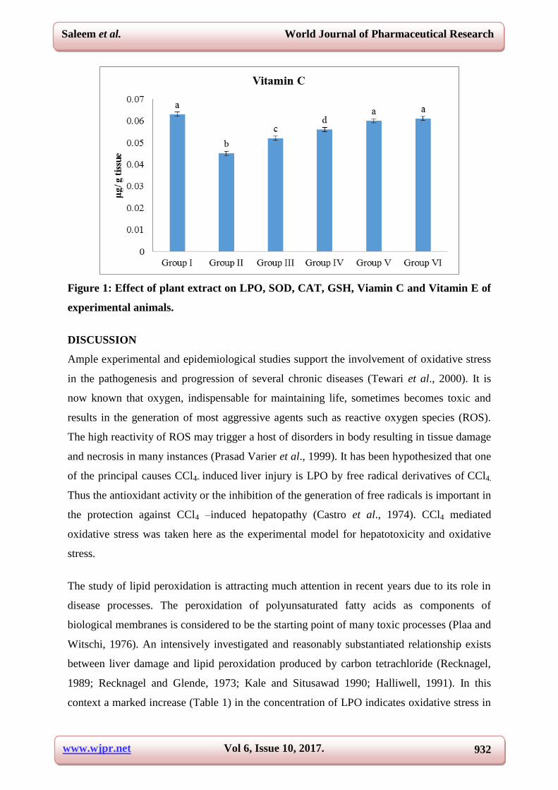
Figure 1: Effect of plant extract on LPO, SOD, CAT, GSH, Viamin C and Vitamin E of
experimental animals.
DISCUSSION
Ample experimental and epidemiological studies support the involvement of oxidative stress
in the pathogenesis and progression of several chronic diseases (Tewari et al., 2000). It is
now known that oxygen, indispensable for maintaining life, sometimes becomes toxic and
results in the generation of most aggressive agents such as reactive oxygen species (ROS).
The high reactivity of ROS may trigger a host of disorders in body resulting in tissue damage
and necrosis in many instances (Prasad Varier et al., 1999). It has been hypothesized that one
of the principal causes CCl4- induced liver injury is LPO by free radical derivatives of CCl4.
Thus the antioxidant activity or the inhibition of the generation of free radicals is important in
the protection against CCl4 –induced hepatopathy (Castro et al., 1974). CCl4 mediated
oxidative stress was taken here as the experimental model for hepatotoxicity and oxidative
stress.
The study of lipid peroxidation is attracting much attention in recent years due to its role in
disease processes. The peroxidation of polyunsaturated fatty acids as components of
biological membranes is considered to be the starting point of many toxic processes (Plaa and
Witschi, 1976). An intensively investigated and reasonably substantiated relationship exists
between liver damage and lipid peroxidation produced by carbon tetrachloride (Recknagel,
1989; Recknagel and Glende, 1973; Kale and Situsawad 1990; Halliwell, 1991). In this
context a marked increase (Table 1) in the concentration of LPO indicates oxidative stress in

www.wjpr.net Vol 6, Issue 10, 2017.
933
Saleem et al. World Journal of Pharmaceutical Research
CCl4 intoxicated rats when compared to control rats. Administration of Azima tetracantha at
a doses of at 100, 200 and 400mg/kg significantly (p<0.05) decreased in the level of LPO
demonstrate the reduction of oxidative stress in Azima tetracantha and CCl4 intoxicated rats.
The LPO content at 400mg/kg is almost similar to control rats.
Biological systems protect themselves against the damaging effects of activated species by
several means. These include free radical scavengers and chain reaction terminators; enzymes
such as SOD, CAT and GPx system (Proctor and McGinness, 1986). The SOD dismutases
superoxide radicals О2¯ into H2О2 plus О2, thus participating with other antioxidant enzymes,
in the enzymatic defense against oxygen toxicity. In this study, SOD plays an important role
in the elimination of ROS derived from the peroxidative process of xenobiotics in liver
tissues. In CCl4 intoxicated rats, the activity of SOD decreased drastically compared to that of
normal group. CCl4 administered rats treated with ATLE at 100, 200 and 400mg/kg revealed
that MDA and oxidative stress elicited by CCl4 intoxication have been nullified due to the
effect of Azima tetracantha. The SOD activity at 400mg/kg is almost similar to the activity
shown by silymarin. This observation perfectly agrees with those of Lin et al. (1998) study.
CAT is a key component of the antioxidant defense system. Inhibition of these protective
mechanisms results in enhanced sensitivity to free radical-induced cellular damage.
Excessive production of free radicals may result in alterations in the biological activity of
cellular macromolecules. Therefore, the reduction in the activity of these enzymes may result
in a number of deleterious effects due to the accumulation of superoxide radicals and
hydrogen peroxide (Caroline et al., 2008). In CCl4 intoxicated rats, the activity of CAT
decreased drastically compared to that of normal group. Administration of Azima tetracantha
increases the activities of catalase in CCl4 induced oxidative stress rats at 100, 200 and
400mg/kg of ATLE to prevent the accumulation of excessive free radicals and protects the
liver from CCl4 intoxication. The CAT activity at a dose of 400mg/kg is potential activity
than other doses and similar to the activity shown by control rats. This observation agrees
with those of Gupta et al. (2002) study.
GPx is a seleno-enzyme two third of which (in liver) is present in the cytosol and one-third in
the mitochondria. It catalyses the reaction of hydroperoxides with reduced glutathione to
form glutathione disulphide (GSSG) and the reduction product of the hydroperoxide. In CCl4
intoxicated rats, the activity of GPx decreased drastically compared to that of normal group.
The observed increase of GPx activity at 100, 200 and 400mg/kg of ATLE suggests that the
www.wjpr.net Vol 6, Issue 10, 2017.
934
Saleem et al. World Journal of Pharmaceutical Research
Azima tetracantha have efficient protective mechanism in response to ROS. And also, these
findings indicate that Azima tetracantha may be associated with decreased oxidative stress
and free radical-mediated tissue damage. This observation consisted with those of
Venukumar and Latha (2002) study.
Drug-metabolizing enzymes, such as glutathione S-transferase (GSTs) function concertedly
as the two major inducible defense systems against electrophiles and xenobiotic toxicity
(Enomoto et al. 2001). GSTs may have significant roles in the detoxification processes of
oxidatively stressed cells (Sagara et al. 1998). GSTs have endogenous substrates, such as
lipid and nucleic acid hydroperoxides and alkenals, which result from the decomposition of
lipid hydroperoxides (Coles and Ketterer 1990). In CCl4 intoxicated rats, the activity of GST
decreased drastically compared to that of normal group. The activity of GST recovered
significantly (p< 0.05) at 100, 200 and 400mg/kg of Azima tetracantha extract compared to
that of CCl4 group. In contrast, the GST activity at 400mg/kg is almost similar to the activity
shown by silymarin, a potent hepatoprotective agent. This observation agrees with those of
Guntupalli et al. (2006) study.
GSH is a major non- protein thiol in living organism, which plays a central role of co-
ordinating the body’s antioxidant defense process. It is implicated in the cellular defense
against xenobiotics and naturally occurring deleterious compounds such as free radicals.
Glutathione status is a highly sensitive indicator of cell functionality and viability.
Perturbation of GSH status of a biological system has been reported to lead to serious
consequences (Pastore, 2003). In CCl4 intoxicated rats, the content of GSH improved
significantly (p< 0.05) at 100, 200 and 400mg/kg of Azima tetracantha extract compared to
control rats and its subsequent return towards near normalcy in CCl4 and Azima tetracantha
treated rats reveal antioxidant effect of Azima tetracantha. Explanations of the possible
mechanism underlying the hepatoprotective properties of drugs include the prevention of
GSH depletion and destruction of free radicals (Fraga et al., 1987).
Ascorbate (Vitamin C) plays an important role with the lipophilic antioxidant α – tocopherol
in protecting the membrane from oxidative stress. Recycling of ascorbic acid requires GSH,
which reduces dehydroascorbate to ascorbate (Winkler, 1992). Ascorbte in turn is essential
for the recycling of tocopherol radical to tocopherol (Packer et al., 1997). In the present
study, significantly decreased level of vitamin C and α- tocopherol in CCl4 intoxicated rats,
demonstrating the increased free radical accumulation in CCl4 administered rats. The

www.wjpr.net Vol 6, Issue 10, 2017.
935
Saleem et al. World Journal of Pharmaceutical Research
observed decline in glutathione level may contribute to the decrease in ascorbate as well
tocopherol concentration in CCl4 intoxicated rats. Supplementation of Azima tetracantha to
CCl4 intoxicated rats improved vitamin C and α- tocopherol level as compared to control rats
(Table 2), which may be due to increase the GSH in Azima tetracantha treated rats improve
the recycling of vitamin C and α- tocopherol. In CCl4 intoxicated rats, the content of GSH,
vitamin C and E improved significantly (p< 0.05) at 100, 200 and 400mg/kg of Azima
tetracantha extract compared to control rats The GSH, vitamin C and α- tocopherol content
were higher at a dose of 400mg/kg of Azima tetracantha treated and similar to control rats.
In conclusion, the entire variable tested i.e., SOD. CAT, GPx, reduced glutathione, vitamin C
and vitamin E recorded a significant decline on CCl4 treatment. However, treatment with
different doses of Azima tetracantha extract (100, 200 and 400mg/kg) significantly increased
in the antioxidant activity as compared to CCl4 intoxicated, suggesting the therapeutic effect
of Azima tetracantha to counter the oxidative stress. Among the various doses, 400mg/kg has
potential activity than other doses. It can be suggested said that ethanolic extract of Azima
tetracantha exhibit against CCl4 induced oxidative stress and possessed anti-lipid
peroxidative and antioxidant activities. This indicates that the lipid peroxidation and
oxidative stress elicited by CCl4 intoxication had been nullified due to the effect of
phytochemicals present in Azima tetracantha.
REFERENCES
1. Abirami H., M. H. Muhammad Ilyas, K. Prem Kumar and T. Nargis Begum. Chemical
composition of the ethanolic extract of leaves of Azima tetracantha (Lam.) Asian Journal
of Plant Science and Research, 2015; 5(3): 1-5.
2. Azri, S., Mata, H.P., Reid, L., Gandlofi, A.J., Brendel, H.P., 1992. Further examination of
selective toxicity of CCl4 rat liver slices. Toxicology and Applied pharmacology, 112:
81-86.
3. Baker H, Frank O, De Angeles B, Feinglod S. Plasma tocopherol in man at various times
after ingesting free or acetylated tocopherol. Nutr Repor Inter, 1980; 21: 531.
4. Beers R, Sizer I, A spectrophotometric method for measuring the breakdown of hydrogen
peroxide by catalase. J Biol Chem, 1952; 195: 133.
5. Beuge JA, Aust SD. The thiobarbituric acid assay. Method Enzymol, 1978; 52: 306-307.
6. Caroline D. Matheus A.B. P., 2 Marcos R. O., Fernanda M. U., Mirian S., Joao A.P. H.,
and Jose C.F.M. (2008) Protective Effects of Purple Grape Juice on Carbon

www.wjpr.net Vol 6, Issue 10, 2017.
936
Saleem et al. World Journal of Pharmaceutical Research
Tetrachloride-Induced Oxidative Stress in Brains of Adult Wistar Rats. J Med Food,
11(1): 55–61.
7. Castro JA Ferrya GC Castro CR Sasame H Fenos OM and Gillette JR. 1974. Prevention
of carbon tetrachloride-induced necrosis by inhibitors of drug metabolism. Further studies
on the mechanism of their action. Biochem. Pharmacol. 23: 295-302.
8. Chance B, Maehly AC, 1955. Assay of catalase and peroxidase. Meth. Enzymol, 2:
764-775.
9. Clawson, G.A., 1989. Mechanism of carbon tetrachloride hepatotoxicity. Pathology and
immunology research, 8: 104–112.
10. De Groot, H., Noll., 1986 . The crucial role of low steady state oxygen partial pressure in
halo alkane free radical mediated lipid peroxidation. Biochemical pharamacology, 35:
15-19.
11. Fraga C, Leibovitz B and Tappel A. 1987. Halogenated compounds as induced of lipid
peroxidation in tissue slices. Free Rad. Biol. Med. 3: 119-123.
12. Gupta M, Mazumder UK, Sivakumar T, Gomathi P and Sampath Kumar R. 2004.
Antioxidant and hepatoprotective effects of Bauhinia racemosa against paracetamol and
carbon tetrachloride induced liver damage in rats. Iranian journal of Pharmacology and
Therapeutics. 3: 12-20.
13. Guntupalli M. Mohana Rao, Chandana V. Rao Palpu Pushpangadan, Annie Shirwaikar
(2006) Hepatoprotective effects of rubiadin, a major constituent of Rubia cordifolia Linn.
Journal of Ethnopharmacology, 2006; 103: 484–490.
14. Halliwell B. 1991. Reactive oxygen species in living systems; source, biochemistry and
role in human disease. Am. J. Med. 91: 14s.
15. Harvey J, Paige SM. The Instat Guide to choosing and interpreting statistical tests: A
manual for Graph pad Instat, Version 3. San Diego, CA USA. 1998.
16. Janardhan L, Vineetha M Shrikanth, Kiran K Mirajkar and Sunil S More. 2014 In vitro
screening and evaluation of antivenom phytochemicals from Azima tetracantha Lam.
leaves against Bungarus caeruleus and Vipera russelli. Journal of Venomous Animals and
Toxins including Tropical Diseases, 2014; 20: 12.
17. Joyce DA. (1987) Oxygen radicals in disease. Adverse Drug Reaction Bulletin. 127:
476-479.
18. Kadiiska MB, Gladen BC, Baird DD, Dikalova AE, Sohal RS, Hatch GE, Jones DP,
Mason RP and Barrett JC. 2000. Biomarkers of oxidative stress study: Are plasma
www.wjpr.net Vol 6, Issue 10, 2017.
937
Saleem et al. World Journal of Pharmaceutical Research
antioxidants markers of CCl4 poisoning? Free Radical Biology & Medicine, 28(6):
838–845.
19. Kakkar P, Das B, Viswanathan PN. A modified spectrophotometric assay of SOD. Ind J
Biochem Biophy, 1984; 21: 130-132.
20. Kale, R.K., & Sitasawad, S.L., Radiation–induced lipid peroxidation in liposomes Radiat
phys chem., 1990; 36: 361.
21. Lin CC, Yen MH, Lo TS and Lin JM. 1998. Evaluation of the hepatoprotective and
antioxidant activity of Boehmeria nivea var. nivea and B. nivea var. tenacissma. J.
Ethnopharmacol. 60: 9-17.
22. Lowry OH, Rosenbrough NJ, Farr AL, Randall RJ. Protein measurement with the Folin’s
reagent. J Biol Chem. 1951; 193: 265-276.
23. Misra, H. P., and I. Fridovich (1972). The role of superoxide anion in the autooxidation of
epinephrine and simple assay for superoxide dismutase. J. Biol. Chem. 247: 3170-3175.
24. Moron MS, DsePierre JW, Manerwik KB. Levels of glutathione, glutathione reductase
and glutathione-s-transferase activities in rat lung and liver. Biochim Biophy Acta, 1979;
582: 67-68.
25. Omaye ST, Tumball JD, Sauberlich HE. Selected methods for the determination of
ascorbic acid in animal cells, tissues and fluids. Method Enzymol, 1979; 62: 1-11.
26. Packer L., Tritschler H.J and Wessel K. (1997) Neuroprotection by the metabolic
antioxidant alpha-lipoic acid. Free Radical Biology and Medicine. 22: 359- 378.
27. Prasad Varier S, Venkatachalam SR, Ramesh Chander R and Paul Thomas R. 1999.
Dietary antioxdant natural defence against disease. Aryavaidyan. 12(3): 149-158.
28. Proctor PH and McGinness JE. 1986. The function of melanin. Arch Dermatol. 122:
507-508.
29. Ray G and Husain SH. 2002. Oxidants antioxidants and carcinogenesis. Indian J Exp
Biol. 40: 1213-1232.
30. Recknagel, R.O and Glende, EA. (1973). Carbon tetrachloride hepatotoxicity: An
example of lethal cleavage. Crit. Rev. ToxicoL, 2: 263-297.
31. Recknagel, R.O., Glende, E.A., Jr, Dolak, J.A., Waller, R.L., 1989. Mechanisms of
carbon tetrachloride toxicity. pharmacol. Ther. 43: 139–154.
32. Recknagel, RO. 1983 A new direction in the study of carbon tetrachloride hepatotoxicity.
Life Sci. 33: 401-408.
33. Reitman S, Frankel S. A colorimetric method for the determination of serum glutamate
oxaloacetic and glutamate pyruvate transaminases. Am J Clin Pathol. 1957; 28: 56-63.

www.wjpr.net Vol 6, Issue 10, 2017.
938
Saleem et al. World Journal of Pharmaceutical Research
34. Rotruck JT, Pope AL, Ganther HE, Swanson AB, Hafeman DG, Hoekstra WG. Selenium:
biochemical roles as component of glutathione peroxidase. Sci., 1973; 179: 588-590.
35. Scartezzini P and Speroni E. 2000. Review on some plants on Indian traditional medicine
with antioxdant activity. J. Ethanopharmacol. 71: 23-43.
36. Tewari S, Gupta V and Bhattacharya S. 2000. Comparative study of antioxidant potential
of tea with without additives. Indian J Physiol. Pharmacol. 44(2): 215-219.
37. Tiwari A. (2001) Imbalance in antioxidant defence and human diseases: Multiple
approach of natural antioxidants therapy. Current science, 81: 1179-1187.
38. Venukumar MR and Latha MS. 2002. Antioxidant activity of Curculigo orchioides in
carbon tetrachloride induced hepatopathy in rats. Ind. J. Clin. Biochem. 17(2): 80-87.
39. Winkler BS. (1992) Unequival evidence in support of the non-enzymatic redox coupling
between glutathione/glutathione disulfide and ascorbic acid/dehydroascorbic acid.
Biochimca Biophysics Acta. 1117: 287-290.
40. Wolf, C.R., Harrelson Jr., W.G., Nastainezyk, W.M., Philpot, R.M., Kalayanaraman, B.,
Mason, R.P., 1980. Metabolism of carbontetrachloride in hepatic microsome and
reconstitution monoxygenase systems and its relationship to lipid peroxidation. Molecular
pharmacology, 18: 558–558.
41. Yu BP. 1994. Cellular defences against damage from reactive oxygen species. Biol Rev.
74: 139-162.