
Modulation of Conduction andRefractoriness in...
8
Modulation of Conduction and Refractoriness in Atrioventricular Junctional Reentrant Circuit Effect on Reentry Initiated by Atrial Extrastimulus Rehan Mahmud, Stephen T. Denker, Patrick J. Tchou, Mohammad Jazayeri, and Masood Akhtar The Natalie and Norman Soref & Family Electrophysiology Laboratory, University of Wisconsin, Milwaukee, Wisconsin 53233; and Mount Sinai Medical Center, Milwaukee, Wisconsin 53233 Abstract The importance of activation sequence of an atrioventricular junctional reentrant (AVJRe) circuit, before delivery of an ex- trastimulus, has received little attention in studies concerned with clinical tachycardias. In this study a change in activation sequence was accomplished using bidirectional activation (V-A sequential pacing) during the basic drive (VIA1-V1A1). It was noted that, compared with an atrial extrastimulus (A2) after an atrial drive (Al-Al), earlier activation (by V1 impulse of the VIA,-VIA, drive) consistently improved conduction, or de- creased refractoriness, or both, in the anterograde as well as the retrograde pathway of the AVJRe circuit. In all patients, five with AV nodal reentry and six with Wolff-Parkinson- White syndrome, reentrant tachycardia could be prevented during V-A sequential pacing. In four of eleven patients, reen- try was prevented despite achieving the so-called critical atrio- ventricular nodal delays that had previously caused reentry during control study. This finding suggested that conduction delay necessary for reentry was related to the site of block, which in turn was affected by V-A sequential pacing. We concluded that changing the activation sequence during basic drive modulates conduction and refractoriness in AVJRe circuits, and allows the study of a wide range of electrophysical factors that prevent or permit reentry. Introduction Programmed electrical stimulation has proved to be a singu- larly useful method for study of reentrant circuits (1-18). Since the original description of this method by Schmitt and Er- langer (1); the conduction characteristics of the premature beat have remained the primary focus of studies concerning reen- trant circuits. What has received very little attention is the influence of activation sequence, during basic drive, on behav- ior of the premature beat. In a previous study, we observed the effect of bidirectional activation of the His-Purkinje system on conduction, refractoriness, and occurrence of bundle branch reentry (19, 20). Since conduction delay and conduction block also play a pivotal role in atrioventricular junctional reentrant Address reprint requests to Dr. Mahmud, Section of Cardiology, School of Medicine, East Carolina University, Greenville, NC 27858- 4354. Receivedfor publication 18 May 1987 and in revisedform 10 Au- gust 1987. (AVJRe)' tachycardias (8-12), we hypothesized that if con- duction and refractoriness were affected by changing the acti- vation sequence of the AVJRe circuit during basic drive, then a new set of relationships would emerge between conduction delay, conduction block, and coupling interval of the extra- stimulus. These observations could potentially result in a clearer understanding of electrophysiological factors that facil- itate or prevent clinical tachycardias. In this study, we describe the electrophysiologic sequelae of changing the activation sequence during basic drive, on the reentrant process, in both small (atrioventricular [AV] nodal reentry) as well as large (Wolff-Parkinson-White syndrome) clinical circuits. Methods Study population. Eleven consecutive patients (six females and five males) with recurrent AVJRe tachycardias, in whom the clinical tachy- cardia could be reproduced by atrial premature stimulation, consti- tuted the study group. Of the eleven patients, five had AV nodal reen- try and six had Wolff-Parkinson-White syndrome. The mean age ofthe patients was 46±13 yr. There was no evidence of underlying structural heart disease in any of these patients. Two patients, however, had a history of hypertension. All patients were in sinus rhythm. Right heart catheterization was done in the nonsedated, postabsorptive state. The nature ofthe electro- physiological procedure was explained, and an informed and signed consent was obtained. Electrophysiology study. Three to four quadripolar 6F catheters were introduced percutaneously through peripheral veins and fluoro- scopically positioned in the high right atrium, AV junction, coronary sinus, and right ventricular apex for local bipolar recordings (filtered at 30-500 Hz) and electrical stimulation. In addition to the bipolar elec- trograms, three surface electrocardiographic leads (I, II, and VI) and time lines were displayed simultaneously on a multichannel oscillo- scope (VR-16; Electronics for Medicine Inc., Pleasantville, NY) and recorded on magnetic tape (model 5600 C; Honeywell Inc., Medical Electronics Div., Pleasantville, NY) for subsequent retrieval on photo- graphic paper at 100 mm/s. Intracardiac stimulation was performed with a Bloom DTU 101 digital stimulator capable of delivering pre- mature stimuli after a basic drive of A-V sequential as well as ventricu- loatrial (V-A) sequential pacing with adjustable A-V (or V-A) intervals. Pacing protocol (Figs. 1-3). Initial refractory period studies were done according to previously described pacing methods (21). In gen- eral, after a basic atrial drive (Al-Al) (range, 400 to 700 ms), the diastolic interval was scanned with premature atrial beat (A2) at pro- gressively shorter Al-A2 intervals until the A2 encountered atrial mus- cle refractoriness or resulted in repetitive atrial responses (control study, Fig. 1 A). After the zone of AV junctional reentry had been defined with the control method described above, the pacing protocol was repeated with the basic atrial drive (Al-Al) substituted by a V-A 1. Abbreviations used in this paper: AV, atrioventricular; AVJRe, atrioventricular junctional reentrant; ERP, effective refractory periods. Effect ofActivation Sequence on Reentry 39 J. Clin. Invest. © The American Society for Clinical Investigation, Inc. 0021-9738/88/01/0039/08 $2.00 Volume 81, January 1988, 39-46
Transcript of Modulation of Conduction andRefractoriness in...
Modulation of Conduction and Refractoriness in AtrioventricularJunctional Reentrant CircuitEffect on Reentry Initiated by Atrial Extrastimulus
Rehan Mahmud, Stephen T. Denker, Patrick J. Tchou, MohammadJazayeri, and Masood AkhtarThe Natalie and Norman Soref & Family Electrophysiology Laboratory, University of Wisconsin, Milwaukee, Wisconsin 53233; andMount Sinai Medical Center, Milwaukee, Wisconsin 53233
Abstract
The importance of activation sequence of an atrioventricularjunctional reentrant (AVJRe) circuit, before delivery of an ex-trastimulus, has received little attention in studies concernedwith clinical tachycardias. In this study a change in activationsequence was accomplished using bidirectional activation (V-Asequential pacing) during the basic drive (VIA1-V1A1). It wasnoted that, compared with an atrial extrastimulus (A2) after anatrial drive (Al-Al), earlier activation (by V1 impulse of theVIA,-VIA, drive) consistently improved conduction, or de-creased refractoriness, or both, in the anterograde as well asthe retrograde pathway of the AVJRe circuit. In all patients,five with AV nodal reentry and six with Wolff-Parkinson-White syndrome, reentrant tachycardia could be preventedduring V-A sequential pacing. In four of eleven patients, reen-try was prevented despite achieving the so-called critical atrio-ventricular nodal delays that had previously caused reentryduring control study. This finding suggested that conductiondelay necessary for reentry was related to the site of block,which in turn was affected by V-A sequential pacing.
Weconcluded that changing the activation sequence duringbasic drive modulates conduction and refractoriness in AVJRecircuits, and allows the study of a wide range of electrophysicalfactors that prevent or permit reentry.
Introduction
Programmed electrical stimulation has proved to be a singu-larly useful method for study of reentrant circuits (1-18). Sincethe original description of this method by Schmitt and Er-langer (1); the conduction characteristics of the premature beathave remained the primary focus of studies concerning reen-trant circuits. What has received very little attention is theinfluence of activation sequence, during basic drive, on behav-ior of the premature beat. In a previous study, we observed theeffect of bidirectional activation of the His-Purkinje system onconduction, refractoriness, and occurrence of bundle branchreentry (19, 20). Since conduction delay and conduction blockalso play a pivotal role in atrioventricular junctional reentrant
Address reprint requests to Dr. Mahmud, Section of Cardiology,School of Medicine, East Carolina University, Greenville, NC27858-4354.
Receivedfor publication 18 May 1987 and in revisedform 10 Au-gust 1987.
(AVJRe)' tachycardias (8-12), we hypothesized that if con-duction and refractoriness were affected by changing the acti-vation sequence of the AVJRe circuit during basic drive, thena new set of relationships would emerge between conductiondelay, conduction block, and coupling interval of the extra-stimulus. These observations could potentially result in aclearer understanding of electrophysiological factors that facil-itate or prevent clinical tachycardias.
In this study, we describe the electrophysiologic sequelae ofchanging the activation sequence during basic drive, on thereentrant process, in both small (atrioventricular [AV] nodalreentry) as well as large (Wolff-Parkinson-White syndrome)clinical circuits.
Methods
Study population. Eleven consecutive patients (six females and fivemales) with recurrent AVJRe tachycardias, in whomthe clinical tachy-cardia could be reproduced by atrial premature stimulation, consti-tuted the study group. Of the eleven patients, five had AV nodal reen-try and six had Wolff-Parkinson-White syndrome. The mean age of thepatients was 46±13 yr. There was no evidence of underlying structuralheart disease in any of these patients. Two patients, however, had ahistory of hypertension.
All patients were in sinus rhythm. Right heart catheterization wasdone in the nonsedated, postabsorptive state. The nature of the electro-physiological procedure was explained, and an informed and signedconsent was obtained.
Electrophysiology study. Three to four quadripolar 6F catheterswere introduced percutaneously through peripheral veins and fluoro-scopically positioned in the high right atrium, AV junction, coronarysinus, and right ventricular apex for local bipolar recordings (filtered at30-500 Hz) and electrical stimulation. In addition to the bipolar elec-trograms, three surface electrocardiographic leads (I, II, and VI) andtime lines were displayed simultaneously on a multichannel oscillo-scope (VR-16; Electronics for Medicine Inc., Pleasantville, NY) andrecorded on magnetic tape (model 5600 C; Honeywell Inc., MedicalElectronics Div., Pleasantville, NY) for subsequent retrieval on photo-graphic paper at 100 mm/s. Intracardiac stimulation was performedwith a Bloom DTU 101 digital stimulator capable of delivering pre-mature stimuli after a basic drive of A-V sequential as well as ventricu-loatrial (V-A) sequential pacing with adjustable A-V (or V-A) intervals.
Pacing protocol (Figs. 1-3). Initial refractory period studies weredone according to previously described pacing methods (21). In gen-eral, after a basic atrial drive (Al-Al) (range, 400 to 700 ms), thediastolic interval was scanned with premature atrial beat (A2) at pro-gressively shorter Al-A2 intervals until the A2 encountered atrial mus-cle refractoriness or resulted in repetitive atrial responses (controlstudy, Fig. 1 A). After the zone of AV junctional reentry had beendefined with the control method described above, the pacing protocolwas repeated with the basic atrial drive (Al-Al) substituted by a V-A
1. Abbreviations used in this paper: AV, atrioventricular; AVJRe,atrioventricular junctional reentrant; ERP, effective refractory periods.
Effect of Activation Sequence on Reentry 39
J. Clin. Invest.© The American Society for Clinical Investigation, Inc.0021-9738/88/01/0039/08 $2.00Volume 81, January 1988, 39-46
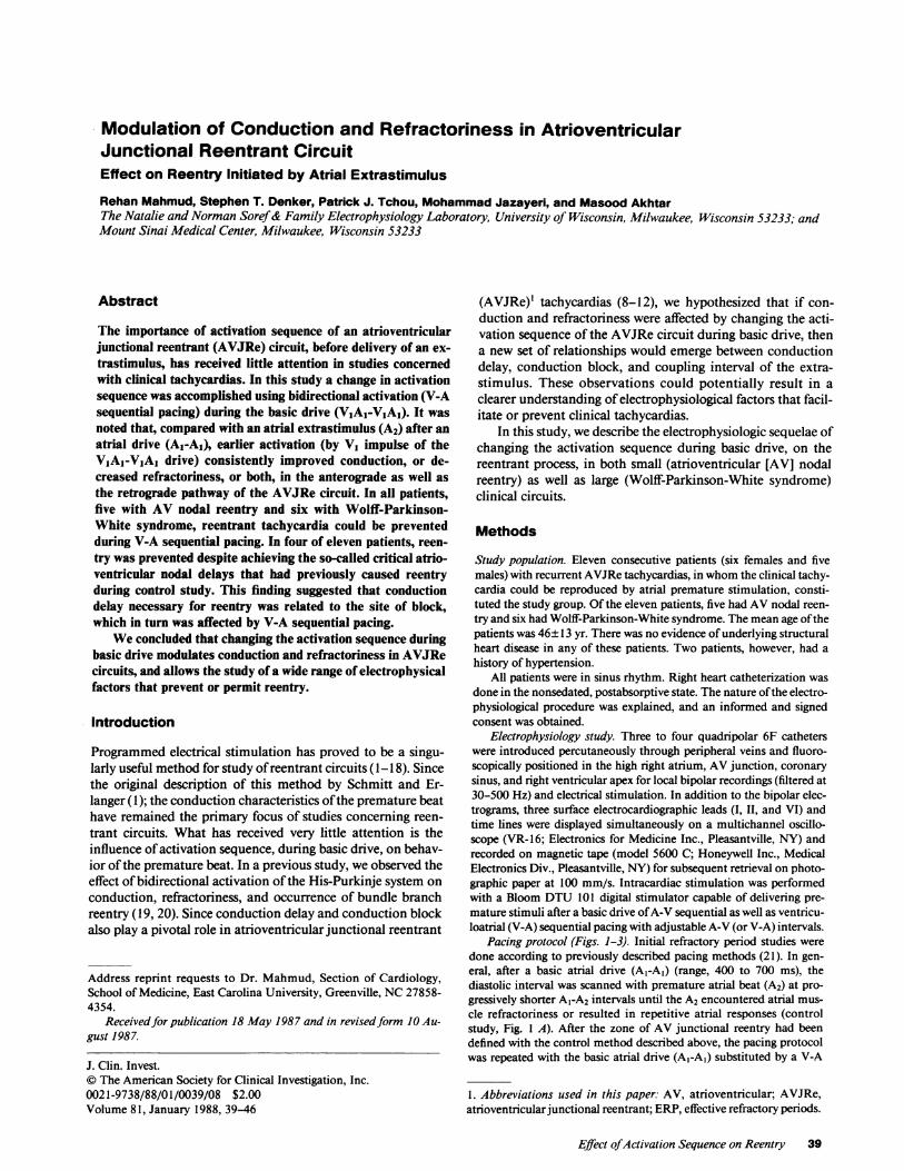
CONTROL Figure 1. Pacing protocol.A A1 Al A1 * A2 A shows the control
method, which consists es-sentially of scanning thediastole with A2 after a
PRE- EXCITATION basic drive (Al-A1). TheB Al Al Al wA2 retrograde preexcitation
protocols are depicted in Band C. The ventricle andatrium are paced sequen-
v1 V1 tially (V-A sequential pac-ing, VIA,-VIA,) and again
C Al Al Al *A2 the diastole is scanned withj A2. Al-A2 intervals andV cycle lengths of V-A se-
quential drive VIA,-VIA,V1 V1 Vl are identical to that during
control. In Cthe V-A inter-val is longer (compared with B), which results in greater retrogradepreexcitation (see text, Figs. 2 and 3).
sequential drive (VIA,-VIA,) (Figs. 1, B and C) at the same cyclelength (V-A sequential method). The initial V-A interval was arbi-trarily selected as 0 ms. This was done to cause collision of the pacedatrial and ventricular impulses in the AV node as well as in the acces-sory pathways, when present. Thus, when compared with the controlmethod, the paced ventricular impulse during VA sequential drivecaused earlier or preexcitation of segments of the reentrant circuits(i.e., AV node or accessory pathways) (see following paragraph, titledDefinition of terms, and Figs. 2 and 3). After the basic V-A sequentialdrive (VIAI-VIA1), A2 was introduced at progressively shorter couplingintervals, identical to those during control method, and the zone of AVjunctional reentry was again determined (Figs. 1-3). The pacing pro-tocol was then repeated with progressively longer (by 10-20 ins) V-Aintervals programmed to analyze the effect of greater preexcitation ofthe AVJRe circuit (Fig. 1 C) until the zone of reentry was completelyabolished.
Definition of termsAnterograde refractory periods. A-H interval was measured from theinitial low frequency deflection of the low atrial electrogram to theonset of His-bundle deflection.
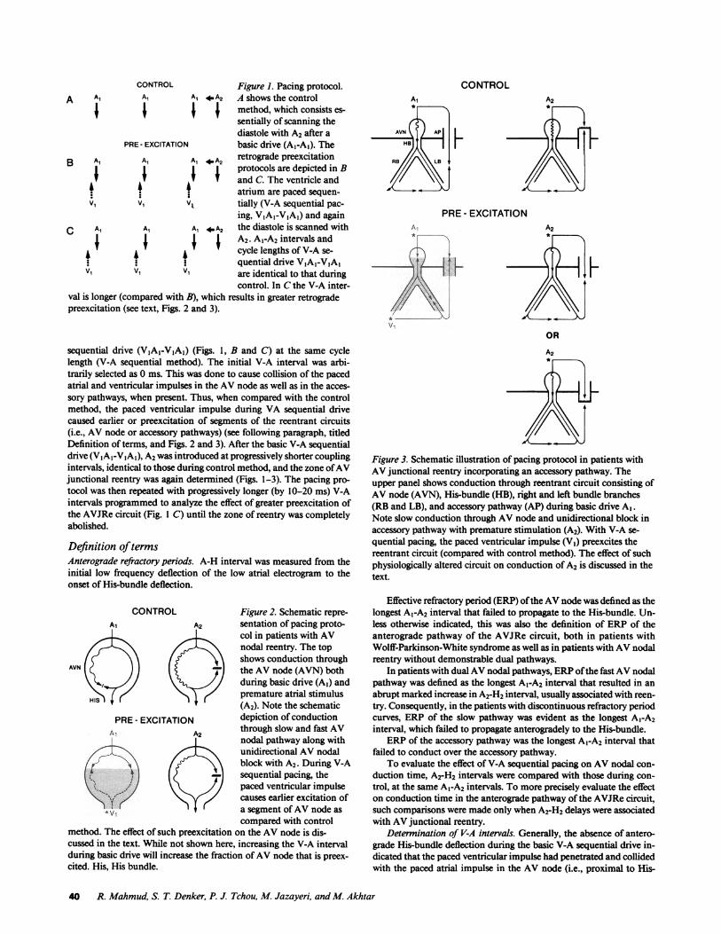
CONTROL Figure 2. Schematic repre-Ai A2 sentation of pacing proto-g-KAcol in patients with AV/<x~y tH >nodal reentry. The top
shows conduction throughAVN ( the AV node (AVN) both
during basic drive (Al) and
HIS premature atrial stimulus(A2). Note the schematic
PRE- EXCITATION depiction of conductionA1 A2 through slow and fast AV
nodal pathway along withunidirectional AV nodalblock with A2. During V-A
Wl) -j sequential pacing, thepaced ventricular impulsecauses earlier excitation of
*v1 + a segment of AVnode ascompared with control
method. The effect of such preexcitation on the AV node is dis-cussed in the text. While not shown here, increasing the V-A intervalduring basic drive will increase the fraction of AV node that is preex-cited. His, His bundle.
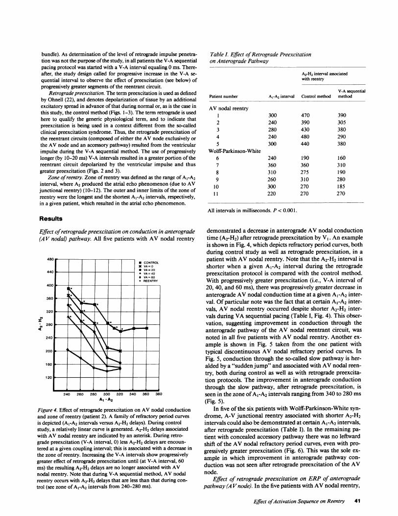
CONTROLA1 A2
PRE- EXCITATIONA1
V -
VI
[L
OR
Figure 3. Schematic illustration of pacing protocol in patients withAVjunctional reentry incorporating an accessory pathway. Theupper panel shows conduction through reentrant circuit consisting ofAV node (AVN), His-bundle (HB), right and left bundle branches(RB and LB), and accessory pathway (AP) during basic drive A1.Note slow conduction through AV node and unidirectional block inaccessory pathway with premature stimulation (A2). With V-A se-quential pacing, the paced ventricular impulse (VI) preexcites thereentrant circuit (compared with control method). The effect of suchphysiologically altered circuit on conduction of A2 is discussed in thetext.
Effective refractory period (ERP) of the AVnode was defined as thelongest A1-A2 interval that failed to propagate to the His-bundle. Un-less otherwise indicated, this was also the definition of ERPof theanterograde pathway of the AVJRe circuit, both in patients withWolff-Parkinson-White syndrome as well as in patients with AVnodalreentry without demonstrable dual pathways.
In patients with dual AVnodal pathways, ERPof the fast AVnodalpathway was defined as the longest Al-A2 interval that resulted in anabrupt marked increase in A2-H2 interval, usually associated with reen-try. Consequently, in the patients with discontinuous refractory periodcurves, ERP of the slow pathway was evident as the longest Al-A2interval, which failed to propagate anterogradely to the His-bundle.
ERPof the accessory pathway was the longest Al-A2 interval thatfailed to conduct over the accessory pathway.
To evaluate the effect of V-A sequential pacing on AV nodal con-duction time, A2-H2 intervals were compared with those during con-trol, at the same Al-A2 intervals. To more precisely evaluate the effecton conduction time in the anterograde pathway of the AVJRe circuit,such comparisons were made only when A2-H2 delays were associatedwith AVjunctional reentry.
Determination of V-A intervals. Generally, the absence of antero-grade His-bundle deflection during the basic V-A sequential drive in-dicated that the paced ventricular impulse had penetrated and collidedwith the paced atrial impulse in the AV node (i.e., proximal to His-
40 R. Mahmud, S. T. Denker, P. J. Tchou, M. Jazayeri, and M. Akhtar
bundle). As determination of the level of retrograde impulse penetra-tion was not the purpose of the study, in all patients the V-A sequentialpacing protocol was started with a V-A interval equaling 0 ms. There-after, the study design called for progressive increase in the V-A se-quential interval to observe the effect of preexcitation (see below) ofprogressively greater segments of the reentrant circuit.
Retrograde preexcitation. The term preexcitation is used as definedby Ohnell (22), and denotes depolarization of tissue by an additionalexcitatory spread in advance of that during normal or, as is the case inthis study, the control method (Figs. 1-3). The term retrograde is usedhere to qualify the generic physiological term, and to indicate thatpreexcitation is being used in a context different from the so-calledclinical preexcitation syndrome. Thus, the retrograde preexcitation ofthe reentrant circuits (composed of either the AV node exclusively orthe AV node and an accessory pathway) resulted from the ventricularimpulse during the V-A sequential method. The use of progressivelylonger (by 10-20 ms) V-A intervals resulted in a greater portion of thereentrant circuit depolarized by the ventricular impulse and thusgreater preexcitation (Figs. 2 and 3).
Zone of reentry. Zone of reentry was defined as the range of Al-A2interval, where A2 produced the atrial echo phenomenon (due to AVjunctional reentry) (10-12). The outer and inner limits of the zone ofreentry were the longest and the shortest Al-A2 intervals, respectively,in a given patient, which resulted in the atrial echo phenomenon.
Table L Effect of Retrograde Preexcitationon Anterograde Pathway
ArH2 interval associatedwith reentry
V-A sequentialPatient number A,-A2 interval Control method method
AV nodal reentry1 300 470 3902 240 390 3053 280 430 3804 240 480 2905 300 440 380
Wolff-Parkinson-White6 240 190 1607 360 360 3108 310 275 1909 260 310 280
10 300 270 18511 220 270 270
All intervals in milliseconds. P < 0.001.Results
Effect of retrograde preexcitation on conduction in anterograde(A V nodal) pathway. All five patients with AV nodal reentry
4c
I
* CONTROLX VA=0* VA= 20
_ VA=40* VA = 60* REENTRY
360
3200
160
120
240 260 280 300 320A1 - A2
340 360 380
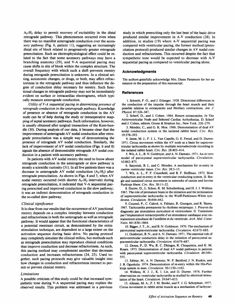
Figure 4. Effect of retrograde preexcitation on AV nodal conductionand zone of reentry (patient 2). A family of refractory period curvesis depicted (A1-A2 intervals versus A2-H2 delays). During controlstudy, a relatively linear curve is generated. A2-H2 delays associatedwith AV nodal reentry are indicated by an asterisk. During retro-grade preexcitation (V-A interval, 0) less A2-H2 delays are encoun-tered at a given coupling interval; this is associated with a decrease inthe zone of reentry. Increasing the V-A intervals show progressivelygreater effect of retrograde preexcitation until (at V-A interval, 60ms) the resulting A2-H2 delays are no longer associated with AVnodal reentry. Note that during V-A sequential method, AV nodalreentry occurs with A2-H2 delays that are less than that during con-trol (see zone of A1-A2 intervals from 240-280 ms).
demonstrated a decrease in anterograde AVnodal conductiontime (A2-H2) after retrograde preexcitation by VI. An exampleis shown in Fig. 4, which depicts refractory period curves, bothduring control study as well as retrograde preexcitation, in apatient with AV nodal reentry. Note that the A2-H2 interval isshorter when a given Al-A2 interval during the retrogradepreexcitation protocol is compared with the control method.With progressively greater preexcitation (i.e., V-A interval of20, 40, and 60 ms), there was progressively greater decrease inanterograde AV nodal conduction time at a given Al-A2 inter-val. Of particular note was the fact that at certain Al-A2 inter-vals, AV nodal reentry occurred despite shorter A2-H2 inter-vals during VA sequential pacing (Table I, Fig. 4). This obser-vation, suggesting improvement in conduction through theanterograde pathway of the AV nodal reentrant circuit, wasnoted in all five patients with AV nodal reentry. Another ex-ample is shown in Fig. 5 taken from the one patient withtypical discontinuous AV nodal refractory period curves. InFig. 5, conduction through the so-called slow pathway is her-alded by a "sudden jump" and associated with AVnodal reen-try, both during control as well as with retrograde preexcita-tion protocols. The improvement in anterograde conductionthrough the slow pathway, after retrograde preexcitation, isseen in the zone of AI-A2 intervals ranging from 340 to 280 ms
(Fig. 5).In five of the six patients with Wolff-Parkinson-White syn-
drome, A-V junctional reentry associated with shorter A2-H2intervals could also be demonstrated at certain A1-A2 intervals,after retrograde preexcitation (Table I). In the remaining pa-tient with concealed accessory pathway there was no leftwardshift of the AV nodal refractory period curves, even with pro-gressively greater preexcitation (Fig. 6). This was the sole ex-ample in which improvement in anterograde pathway con-
duction was not seen after retrograde preexcitation of the AVnode.
Effect of retrograde preexcitation on ERPof anterogradepathway (A Vnode). In the five patients with AVnodal reentry,
Effect ofActivation Sequence on Reentry 41

5ou
Cdz
Cd.4
.*
'IUUg *r
360
320
280
240
200
160
120
300 320 340Al - A2
* CONTROLX VA=O* VA=20A VA= 40* VA=60* REENTRY
Cd
Cd4(
360 380 400
Figure 5. Effect of preexcitation in a patient with dual pathways (pa-tient 3). This figure depicts another family of refractory periodcurves. During control method, a sudden jump in A2-H2 interval,characteristic of so-called dual pathways, is seen. Note how (with in-creasing preexcitation) the ERPof the fast pathway is progressivelyshortened and that AV nodal reentry (*) is always associated withconduction through slow pathway. With V-A interval of 60 ms theERPof fast pathway was less than ERPof the atrium, thus effec-tively abolishing AV nodal reentry at all coupling intervals.
AV nodal ERPwas not attained during control study becauseof the following reasons: (a) it was less than the ERPof atrium(three patients), or (b) induction of unsustained atrial arrhyth-mias (two patients) and further testing at shorter Al-A2 inter-vals was not attempted. With retrograde preexcitation, therewas no change either in the ERPof the atrium or in inductionof unsustained atrial arrhythmias. In four of six patients withWolff-Parkinson-White syndrome, ERPof the AV node wasattained during the control study. With retrograde preexcita-tion, ERPof the AVnode was achieved at shorter Al-A2 inter-vals. In other words, retrograde preexcitation resulted in adecrease in the ERPof anterograde pathway, when this param-eter could be determined during the control method.
Effect of retrograde preexcitation on ERPof 'fast "A Vnodalpathway or accessory pathway. Of the three patients withmanifest anterograde conduction over the accessory pathway,in two the ERPof the accessory pathway decreased with V-Asequential pacing (Figs. 3 and 7), and in one patient there wasno change. The decrease in ERPof the fast pathway in the solepatient with discontinuous AV nodal pathways is evident inFig. 5. In the remaining four patients with AV nodal reentry,the effect on ERPof the fast pathway could not be determined,as there was no clear distinction between the so-called fast andslow pathways at the cycle lengths tested. For the same reason,improvement in anterograde conduction time in the fast AVnodal pathway with retrograde preexcitation could only beclearly demonstrated in the patient with discontinuous AVnodal curves (Fig. 5).
290[ [11* CONTROLX VA=0* VA=20A VA=40
250 i
230 - __ _
210 I__ZONEOF REENTRY190 _ CONTR( L
*V VA= 040_ _I
* *VA =40
190I- VA=2c 0MEJ
220 240 260 280A1 A2
300 320 340
Figure 6. Refractory period curves in a patient with accessory path-way without retrograde AV nodal conduction (patient 1). In thisfigure, Al-A2 intervals are plotted against resulting AVdelays(A2-V2). Note absence of significant and consistent improvement inAV conduction with V-A sequential pacing. At some coupling inter-vals, His-Purkinje system delay seen during control study was abol-ished with V-A sequential pacing, resulting in some shortening ofAV delay. Overall, despite little change in A2-V2 delay, there wasprogressive shortening of the zone of reentry with greater preexcita-tion (see bar graph). A postulated mechanism was a distal shift in siteof block in the accessory pathway (see Fig. 3). As the site of block oc-curred more distally with increasing V-A intervals, progressivelygreater AV nodal delays were required to initiate reentry. Thus,Al-A2 intervals, at which the zone of reentry started, became progres-sively shorter with greater preexcitation. Fig. 9 shows selected panelsfrom this patient. (*) Denotes the only two Al-A2 intervals whereAV reentry occurred.
Effect of retrograde preexcitation on initiation of reentry byatrial extrastimulus. Several of the functional changes de-scribed above acted alone or in concert to prevent initiation ofreentry by A2. In all patients the zone of reentry was decreasedby V-A sequential pacing. Both AV nodal reentry as well asAVjunctional reentry incorporating an accessory pathway wasabolished at all coupling intervals by increasing the V-A inter-val during basic drive. In 10 of 11 patients there appeared to bea relationship between the improvement in anterograde con-duction and the decrease in the zone of reentry (Figs. 4 and 5).In the remaining one patient, the zone of reentry decreasedprogressively with greater retrograde preexcitation, eventhough there was no improvement in anterograde AV nodalconduction (Figs. 6 and 8). In three patients an additionalmechanism for prevention of reentry was a decrease in ERPofaccessory or fast AV nodal pathway (Fig. 7). In three otherpatients AVjunctional reentry failed to occur, even though theA2-H2 delay during retrograde preexcitation was greater thanthe A2-H2 delay associated with reentry during control method(Fig. 9).
In contrast, facilitation of reentry by retrograde preexcita-tion was also seen in three patients with Wolff-Parkinson-White syndrome. In all three the zone of reentry was limitedby ERPof the AV node during control study. Preexcitationresulted in abolition of AV nodal conduction block with de-crease in the ERPof the AV node, thus permitting reentry to
42 R. Mahmud, S. T. Denker, P. J. Tchou, M. Jazayeri, and M. Akhtar
F----l
280
i

ATRIAL PACINGCONTROL V-A SEQUENTIAL PACINGV-A=20
S Si Si S2PC SNk % -'*4 L
V,----a-r---Io ir--A- AA1 V1 Al V1 Al V1A2 V2 A V V1A1 V1A1 V1A1 V2
HB A-i ii5 0 0 500 3 0 0 500 500 280
T ' I.."
B DCONTROL ATRIAL PACING V-A SEQUENTIAL PACING
V-A=20
Si S Si 1 SA Si S2~~~~~~~~ dAl V1 Al V1 Al V, V2 A V V V vV1 A1 V1A1 V1A1 V2H H Hi A2 H2 AH AH AH0AH A
50 500 2 80 500 500 2 60
Figure 7. Effect of V-A sequential pacing on ERPof accessory path-way (patient 6). Electrograms from top to bottom show surface ECG(VI), proximal coronary sinus (PCS), and His-bundle (HB) electro-grams. T, Timing markers. A shows conduction of paced atrial im-pulse over the accessory pathway both during basic drive as well as
premature stimulus (A2). At S1-S2 interval of 280 ms (B), AVjunc-tional tachycardia is initiated as the ERPof the accessory pathway isachieved. Note occurrence of intermittent functional bundle branchblock. Cand Dshow continued conduction over accessory pathwayat shorter S1-S2 intervals during V-A sequential pacing.
ACONTROL
CATRIAL PACING
HRA A A-- 1.-~f t
LRA Ik ,I
A1 V1 A1 V1 Al V1 A2 V2 Ae
HB A0-30
600 600 300T l-l| | ---- l
V-A SEQUENTIAL PACING V-A=40
V\_< J_2
Al A- AlV1 V1 V1 A2 2
S1A 600 sli 600 S1 30 0
BCONTROL
DATRIAL PACING V-A SEQUENTIAL PACING V-A=40
Ae
S . S 1 Si S2
A1 V1 A1 V1 A1 V1A2 V2 Ae
600 600 220
Figure 8. Effect of V-A sequential pacing on reentry involving acces-
sory pathway (patient I 1). Electrograms from top to bottom showsurface ECGlead (1), high right atrial (HRA), low right atrial (LRA),and His-bundle (HB) electrograms. T, Timing markers. A and Bshow the outer (SI-S2, 300 ms) and inner (S1-S2, 220 ms) limits ofthe zone of reentry in a patient with concealed accessory pathway.Note the AV nodal delays associated with reentry. Cand D showV-A sequential pacing with SI-S2 intervals identical to A and B, re-
V1 V1 IV2
600 sl 600 Si 220
spectively. Of interest is the fact that the AV nodal delays are similar(despite preexcitation), and yet no AV reentry takes place. The ab-sence of improvement in AV nodal conduction may be explained byabsence of retrograde penetration by the preexciting ventricular im-pulse. However, the site of block (accessory pathway) is preexcited,and possibly the resultant distal shift in site of block prevents reentrydespite achievement of AVdelays similar to control method (see textand Figs. 3 and 7).
Effect of Activation Seauence on Reentrv 43
Vi
A C
I

CONTROLB
ATRIAL PACING V-A SEQUENTIAL PACING1
Ae Aee eHRA
LRA_ I %- 4-A1 V1
HC
600T [----I---l aoi
C
A1 V1 A2 Ae
380....i .....I. ...j ...I
V
A -I IKV1 A1 V1 A A2 V2 A V
600 380
V-A SEQUENTIAL PACING V-A=40
V1A1 VlA1 A2 H2 V2
600 320
Figure 9. Electrophysiologic mechanisms of prevention of AV nodalreentry (patient 1). Electrograms from top to bottom show surfaceECGlead 1, high right atrial (HRA), low right atrial (LRA), and His-bundle (HB) electrograms. T, Timing markers. A shows induction ofAV nodal reentry by atrial extrastimulus A2 at coupling intervals of380 ms. Note AV nodal delay (A2-H2, 290 ms) sufficient for onset ofAV nodal reentry. With V-A sequential pacing (V-A interval, 20 ms),A2 at same coupling interval now results in decrease in AV nodal
occur at shorter coupling intervals and extending the zone ofreentry. However, further increases in V-A intervals were fol-lowed by progressive decrease and then abolition of the zone ofreentry.
Discussion
The extrastimulus method has proved to be a potent and use-ful technique for reproducing clinical arrhythmias in the elec-trophysiology laboratory. The wake of refractoriness of thebasic drive determines the coupling interval of the extrastimu-lus which results in the necessary conduction delay and con-duction block required for reentry (10, 11, 16). In the AVnode, increase in the rate of basic drive results in greater delayat a given coupling interval (23-25). In the His-Purkinje sys-tem, an abrupt change in cycle length influences refractoriness,and thereby reentry (26). Ventricular tachycardia is morelikely to be induced by a premature ventricular beat after aventricular drive as compared with that programmed duringsinus rhythm ( 18). This suggests that both direction as well assequence of activation of the reentrant circuit may be impor-tant.
In this study, varying fractions of both large as well as smallAV junctional reentrant circuits were excited earlier from ret-rograde direction during the basic drive. This resulted in im-provement in conduction and decrease in refractoriness in
delay (B) with prevention of AV nodal reentry. This mechanism isgraphically illustrated in Fig. 2. Cdepicts the effect of preexcitationwith V-A interval of 40 ms. Note that AV nodal delay is now signifi-cantly greater than in A, yet AV nodal reentry is again prevented. Apossible explanation may be that preexcitation of fast pathway causesA2 to block more distally in the fast pathway, in turn requiringgreater delay in the slow pathway for reentry. (See text for details.)
both the anterograde as well as retrograde pathways of thereentrant circuit. While the reentrant process was facilitated insome cases, the overall effect was prevention of clinical tachy-cardia (see Results). In most instances the electrophysiologicalfactors responsible for failure of reentry were quite apparent(Figs. 4 and 7). In other instances, mechanisms (previouslyunknown) were postulated after comparing refractory periodcurves obtained during control method with those after retro-grade preexcitation.
Role of conduction delay in AVjunctional reentry: its rela-tionship to site of block. It has been reported that the mecha-nism of prevention of reentry with dual chamber pacing isprimarily the failure to achieve sufficient conduction delay inthe anterograde pathway (9, 27, 28). While mitigation of con-
duction delay may play a role, it is not the only factor thatprevents reentry. Figs. 8 and 9 demonstrate that with V-Asequential pacing, AV junctional reentry may be preventedeven when anterograde conduction delay is greater than thatwhich was sufficient for reentry during the control study. Thisphenomenon could best be explained by postulating that V-Asequential pacing produced a shift in the site of block (of A2) inthe retrograde pathway. It is not unreasonable to postulate thatV-A sequential pacing decreased refractoriness of the retro-grade pathway and caused A2 to block more distally at a given
AI-A2 interval (Fig. 3). In turn, the distal shift in the site ofblock (compared with control method) would require greater
44 R. Mahmud, S. T. Denker, P. J. Tchou, M. Jazayeri, and M. Akhta'
AV-A=-20
Sl sN& -. A --
I ---] v
A2-H2 delay to permit recovery of excitability in the distalretrograde pathway. This phenomenon occurred even whenthere was no manifest anterograde conduction over the acces-sory pathway (Fig. 6, patient 1 1), suggesting an increasinglydistal site of block related to progressively greater retrogradepreexcitation. Such an electrophysiological effect could be re-lated to the fact that some accessory pathways may have abranching anatomy (29), and V-A sequential pacing maycause shifts in site of block within the complex structure. Theoverall frequency with which such a shift prevents reentryduring retrograde preexcitation is unknown. In a clinical set-ting, autonomic changes, or drugs, or both, may affect refrac-toriness in the retrograde pathway and thus influence the de-gree of conduction delay necessary for reentry. Such func-tional changes in retrograde pathway may not be immediatelyevident on surface or intracardiac electrograms, which typi-cally measure anterograde conduction.
Utility of V-A sequential pacing in determining presence ofretrograde conduction in the anterograde pathway. Knowledgeof presence or absence of retrograde conduction in the AVnode can be of help during the study or intraoperative map-ping of septal accessory pathways. Such information, however,is usually obtained after surgical transection of the Kent-bun-dle (30). During analysis of our data, it became clear that theimprovement of anterograde AVnodal conduction after retro-grade preexcitation was a simple way of demonstrating thepresence of retrograde AV nodal conduction. Similarly, thelack of improvement of AV nodal conduction (Figs. 6 and 8)signals the absence of demonstrable retrograde AV nodal con-duction in a patient with accessory pathway.
In patients with AV nodal reentry the need to know aboutretrograde conduction in the anterograde or slow pathway ismostly a scientific curiosity (31). In all five patients there was adecrease in anterograde AV nodal conduction (A2-H2) afterretrograde preexcitation. As shown in Figs. 4 and 5, when AVnodal reentry occurred (with shorter A2-H2 intervals) duringretrograde preexcitation, it indicated that V-A sequential pac-ing preexcited and improved conduction in the slow pathway;it was an indirect demonstration of retrograde conduction inthe so-called slow pathway.
Clinical significanceIt is clear from our results that the occurrence of AVjunctionalreentry depends on a complex interplay between conductionand refractoriness in both the anterograde as well as retrogradepathway. It would appear that the functional characteristics ofa potential reentrant circuit, as demonstrated by the extra-stimulation technique, are dependent to a large extent on theactivation sequence during basic drive. No pacing protocolmay completely simulate the clinical milieu, but methods suchas retrograde preexcitation may reproduce clinical conditionsthat improve conduction and decrease refractoriness. As such,this pacing method may complement another that decreasesconduction and increases refractoriness (24, 25). Used to-gether, such pacing protocols may give valuable insight intohow changes in conduction and refractoriness interact to per-mit or prevent clinical reentry.
LimitationsA possible criticism of this study could be that increased sym-pathetic tone during V-A sequential pacing may explain theobserved results. This problem was addressed in a previous
study in which preexciting only the last beat of the basic driveproduced similar improvement in A-V conduction (28). Inaddition, in studies (19) where A-V sequential pacing wascompared with ventricular pacing, the former method (preex-citation protocol) produced similar changes in AV nodal con-duction and refractoriness. This occurred despite the fact thatsympathetic tone would be expected to decrease with A-Vsequential pacing as compared to ventricular pacing alone.
Acknowledgments
The authors gratefully acknowledge Mrs. Diane Paramore for her as-sistance in the preparation of this manuscript.
References
1. Schmitt, F. O., and J. Erlanger. 1928. Directional differences inthe conduction of the impulse through the heart muscle and theirpossible relation to extrasystolic and fibrillary contractions. Am. J.Physiol. 87:326-347.
2. Scherf, D., and J. Cohen. 1964. Return extrasystoles. In TheAtrioventricular Node and Selected Cardiac Arrhythmias. D. Scherfand J. Cohen, editors. Grune & Stratton Inc., NewYork. 226-279.
3. Mendez, C., and G. K. Moe. 1966. Demonstration of dual AVnodal conduction system in the isolated rabbit heart. Circ. Res.19:378-393.
4. Janse, M. J., F. J. L. Van Capelle, G. E. Freud, and D. Durrer.1971. Circus movement within the AV node as a basis for supraven-tricular tachycardia as shown by multiple microelectrode recording inthe isolated rabbit heart. Circ. Res. 28:403-414.
5. Witj A. L., B. N. Goldreyer, and A. N. Damato. 1971. An in vitromodel of paroxysmal supraventricular tachycardia. Circulation.43:862-875.
6. Sasyniuk, B. I., and C. Mendez. A mechanism for re-entry incanine ventricular tissue. Circ. Res. 28:3-15.
7. Wit, A. L., P. F. Cranefield, and B. F. Hoffman. 1972. Slowconduction and re-entry in the ventricular conducting system. II. Sin-gle and sustained circus movement in networks of canine and bovinePurkinje fibers. Circ. Res. 30:11-22.
8. Durrer, D., L. Schoo, R. M. Schuilenburg, and H. J. J. Wellens.1967. The role of premature beats in the initiation and the terminationof supraventricular tachycardia in the Wolff-Parkinson-White syn-drome. Circulation. 36:644-662.
9. Coumel, P., C. Cabrol, A. Fabiato, R. Gourgon, and R. Slama.1967. Tachycardia premanente by rhythme reciproque. 1. Preuves dudiagnostic par stimulation auriculaire et ventriculaire. II. Traitementpar l'implantation intracorporelle d'un stimulateur cardiaque avec en-trainement simultane de l'oreillette et du ventricule. Arch. Mal. Coeur.Vaiss. 60:1830-1864.
10. Bigger, J. T., Jr., and B. N. Goldreyer. 1970. The mechanism ofparoxysmal supraventricular tachycardia. Circulation. 42:673-683.
11. Goldreyer, B. N., and A. N. Damato. 1971. The essential role ofatrioventricular conduction delay in the initiation of paroxysmal su-praventricular tachycardia. Circulation. 43:679-687.
12. Denes, P., D. Wu, R. C. Dhingra, R. Chuquimia, and K. M.Rosen. 1973. Demonstration of dual AV nodal pathways in patientswith paroxysmal supraventricular tachycardia. Circulation. 48:549-555.
13. Akhtar, M., A. N. Damato, W. P. Batsford, J. N. Ruskin, andJ. B. Ogunkelu. 1974. Demonstration of re-entry within the His-Pur-kinje system in man. Circulation. 50:1150-1162.
14. Wellens, H. J. J., K. I. Lie, and D. Durrer. 1974. Furtherobservations on ventricular tachycardia as studied by electrical stimu-lation of the heart. Circulation. 39:647-653.
15. Allessie, M. A., F. I. M. Bonke, and F. J. G. Schopman. 1977.Circus movement in rabbit atrial muscle as a mechanism of tachycar-
Effect ofActivation Sequence on Reentry 45

dia. III. The "leading circles" concept: a new model of circus move-ment in cardiac tissue without the involvement of an anatomic obsta-cle. Circ. Res. 41:9-18.
16. Denes, P., D. Wu, F. Amat-Y.-Leon, R. Dhingra, C. R. Whyd-ham, and K. M. Rosen. 1977. The determinants of atrioventricularnodal re-entrance with premature atrial stimulation in patients withdual AV nodal pathways. Circulation. 56:253-259.
17. Gallagher, J. J., E. L. C. Pritchett, W. C. Sealy, J. Kasell, andA. G. Wallace. 1978. The pre-excitation syndromes. Prog. Cardiovasc.Dis. 20:285-327.
18. Josephson, M. E., L. N. Horowitz, A. Farshidi, and J. A. Kas-tor. 1978. Recurrent sustained ventricular tachycardia. 1. Mechanism.Circulation. 57:431-440.
19. Mahmud, R., M. Lehmann, S. Denker, C. J. Gilbert, and M.Akhtar. 1983. Atrioventricular sequential pacing: its differential effecton retrograde conduction related to level of impulse collision. Circula-tion. 68:23-32.
20. Mahmud, R., P. J. Tchou, S. T. Denker, M. H. Lehmann, andM. Akhtar. 1983. Functional characteristics of human macro-reentry:a study of"Pre-Excited" circuits by extrastimulus method. J. Am. Coll.Cardiol. 3:1488-1499.
21. Akhtar, M., A. N. Damato, W. P. Batsford, J. N. Ruskin, andJ. B. Ogunkelu. 1975. A comparative analysis of antegrade and retro-grade conduction patterns in man. Circulation. 52:766-778.
22. Ohnell, R. F. 1944. Preexcitation a cardiac abnormality. Path-ophysiological, patho-anatomical and clinical studies of an excitatoryspread phenomenon. Acta Med. Scand. 152: 14-16.
23. Batsford, W. P., M. Akhtar, A. R. Caracta, M. E. Josephson,S. F. Seides, and A. N. Damato. 1974. Effect of atrial stimulation site
on the electrophysiologic properties of the atrioventricular node inman. Circulation. 50:283-293.
24. Cagin, N. A., D. Kunstadt, P. Wolfish, and B. Levitt. 1973. Theinfluence of heart rate on the refractory period of the atrium and AVconducting system. Am. Heart. J. 85:358-366.
25. Denes, P., D. Wu, R. Dhingra, R. J. Pietras, and K. M. Rosen.1974. The effects of cycle length on cardiac refractory periods in man.Circulation. 49:32-41.
26. Denker, S. T., M. H. Lehmann, C. J. Gilbert, and M. Akhtar.1984. Facilitation of macro-reentry within the His-Purkinge systemwith abrupt changes in cycle length. Circulation. 69:26-32.
27. Coumel, P., R. Slama, and Y. Bouvrain. 1972. Les procedes destimulation cardiaque dans le traitement des tachycardies. Sem. Hop.Paris. 48:2947-2961.
28. Akhtar, M., C. J. Gilbert, M. Al-Nouri, and D. H. Schmidt.1979. Electrophysiologic mechanism for modification and abolition ofatrioventricular junctional tachycardia with simultaneous and se-quential atrial and ventricular pacing. Circulation. 60:1433-1454.
29. Jackman, W., K. Friday, J. Yeung Lai Wah, E. Aliot, and R.Iazzara. 1985. Accessory pathways: branching networks and tachycar-dia. Circulation. 72:III-270.
30. Prystowsky, E. N., E. L. C. Pritchett, W. M. Smith, A. G.Wallace, W. C. Sealy, and J. M. Gallagher. 1979. Electrophysiologicassessment of the atrioventricular conduction system after surgicalcorrection of ventricular pre-excitation. Circulation. 59:789-797.
31. Akhtar, M. 1984. Supraventricular tachycardias. Electrophysi-ologic mechanisms, diagnosis and pharmacologic therapy. In Tachy-cardias: Mechanism, Diagnosis, Treatment. M. E. Josephson andH. J. J. Wellens, editors. Lea and Febiger, Philadelphia. 137-169.
46 R. Mahmud, S. T. Denker, P. J. Tchou, M. Jazayeri, and M. Akhtar