Modulating the Hedgehog pathway - DUO
124
Modulating the Hedgehog pathway Martin Frank Strand Unit for Cell Signaling Institute of Microbiology Oslo University Hospital, Rikshospitalet Oslo, April 2011
Transcript of Modulating the Hedgehog pathway - DUO

Modulating the Hedgehog pathway
Martin Frank Strand
Unit for Cell Signaling
Institute of Microbiology
Oslo University Hospital, Rikshospitalet
Oslo, April 2011

© Martin Frank Strand, 2013 Series of dissertations submitted to the Faculty of Medicine, University of Oslo No. 1469 ISBN 978-82-8264-405-1 All rights reserved. No part of this publication may be reproduced or transmitted, in any form or by any means, without permission. Cover: Inger Sandved Anfinsen. Printed in Norway: AIT Oslo AS. Produced in co-operation with Akademika publishing. The thesis is produced by Akademika publishing merely in connection with the thesis defence. Kindly direct all inquiries regarding the thesis to the copyright holder or the unit which grants the doctorate.

Table of Contents
1. Acknowledgement
2. Abbreviations
3. Publications included
4. Introduction
4.1 History of the Hedgehog pathway
4.2 The Hedgehog pathway in development
4.3 The Hedgehog signaling pathway
4.3.1 Maturation and movement of hedgehog ligands
4.3.2 Hh signal reception; Patched, Smoothened, lipids and cilia.
4.3.3 The hedgehog signaling complex
4.3.4 Transcriptional targets of Hh signaling
4.4 Pathway interactions
4.5 Hh pathway in disease and cancer
4.6 Natural and synthetic modulators of Hh signaling
4.7 Clinical trials with Hh inhibitors
5. Aims of the study
6. Summary of results
6.1 Paper I
6.2 Paper II
6.3 Paper III
6.4 Paper IV
7. General discussion
8. Appendix I. Drug discovery, target deconvolution and chemical analysis
9. References
10. Appendix II. Papers I-IV

1. Acknowledgement
At first I would like to thank my supervisor, Stefan Krauss, for letting me become a part of the Unit for Cell Signaling. His enthusiasm for Shh and the Hh signaling pathway has been inspiring and contagious. Starting a new research project took time, and involved some rounds of trial and error, but looking back I see that I have learned important lessons. Thank you for the opportunity to work in your group, and for all the interesting discussions on biology and signaling mechanisms.
I would like to thank the Norwegian Research Council and the National Hospital of Norway for financing my PhD research.
I would also like to thank my outstanding colleagues that now work or that have worked in the Krauss group; Jo, Olga, Ondrej, Jennifer, Eline, Nina, Petter, Line, Benoit, David, Romain, Monica, Bie, Huyen, Markus, Anita, Andrej, Tore, Anders and Khahn. I would also like to thank Steven and Hanne and everybody else in Elsa Lundanes group at the analytical group at the chemistry department. The collaboration between the Krauss group and the chemistry department has been essential in the research and publications that are presented in this thesis. Steven, you rock my world, both with your music and your chemical analytics expertise. In addition I would like to thank other collaborators and co-authors for their contributions and advice.
Finally I would like to thank my family. My mother has always supported my curious nature; and my dad has shown me the value of hard work and of following your ideas. My four brothers and two sisters have all been inspirations to me. I also want to thank my wife Mina. When I started as a PhD student, we had a 5 month old baby girl at home, and halfway through my PhD we had our second child, a boy who didn’t like to sleep very much at night. Mina, your support, patience and encouragements have been invaluable.
Martin Strand

2. Abbreviations General marks: Species taxonomy names and gene names and are written in cursive. Although
there are exeptions, Human genes/proteins are generally written in capital letters (PTCH), while mouse (Ptch) and Drosophila (Ptc) genes/proteins are written with a capital letter only in the first letter of the name. Unless a species is specified or a part of the context, the mouse protein/gene name is used.
A Anterior AmphiHh Amphioxus Hh protein Arrb2 -arrestin 2 BCC Basal cell carcinoma BMP Bone morphogen protein Boc Brother of Cdo Botv Brother of Tout velu Cdo Cell adhesion molecule-
related/down-regulated by oncogenes
Ci Cubitus interruptus CiA 155 kDa transcriptional
activator form of Ci CiR 75 kDa transcriptional repressor
form of Ci CKI Casein kinase I Cos2 Costal2 D Dorsal Dhh/DHH Desert hedgehog Disp/DISP Dispatched Dlp Dally-like protein Dpp Decapentaplegic EXT1/2 Exostosin 1/2 En Engrailed F Floorplate Fgf10 Fibroblast growth factor 10 Fu Fused, a serine/threonine kinase Gas1 Growth arrest-specific gene 1 Gli1-3/GLI1-3 Glioma-associated oncogenes 1-
3 GliA Transcriptional activator form
of Gli2/3 GliR Transcriptional repressor form
of Gli2/3 Glis Gli-similar Glypican Glycosylphosphatidylinositol
linked HSPG GPCR G-protein coupled receptor GPI Glycosylphosphatidylinositol
G-protein Heterotrimeric GTP-binding regulatory proteins
GRK2 G-protein coupled receptor kinase 2
GSK3 Glycogen synthase kinase 3 Hedge N-terminal Hh fragment
(signaling component) Hh Hedgehog Hh-N N-terminal Hh fragment
(signaling component) Hh-Np Dual lipid modified Hh-N
fragment (p for processed) HPLC High performance (pressure)
liquid chromatography HIP Hedgehog interacting protein Hog C-terminal (autocatalytic)
domain of Hh HSC Hedgehog signaling complex HSPG Heparin sulfate proteglycan Ihh Indian hedgehog Ihog Interference hedgehog IP Intra peritoneal LC Liquid chromatography Lpp Lipophorin LRP2 Low density lipoprotein-related
protein 2 LRX Liver X receptor MEF Mouse embryonic fibroblast MN Motor neurons MP Micro particles MS Mass spectrometry N Notochord NMR Nuclear magnetic resonance
spectroscopy NPC1 Niemann–Pick type C1 disease
gene/protein NO Nitric oxide OHC hydroxycholesterol/oxysterol OSBP Oxysterol binding protein

ORP OSBP related protein P Posterior PI4P Phosphatidylinositol-4
phosphate PKA Protein kinase A PKC Protein kinase C Ptc/PTCH Patched Ptc-1-3 Patched gene homologs 1-3 Ptr Patched related gene/protein RND Resistance nodulation division SANT Smo antagonist SIM Selected ion monitoring Shh/SHH Sonic hedgehog Ski/Skn/SKI Skinny hedgehog Smo Smoothened
Sotv Sister of Tout velu SSD Sterol sensing domain STT4 kinase Staurosporine and temperature
sensitive PI4P kinase SuFu Supressor of Fused TGF Transforming growth factor TLC Thin layer chromatography TIC Total ion current Ttv Tout velu V Ventral V0-V3 Interneurons Wg/Wnt Wingless ZPA Zone of polarizing activity
3. Publications included
Paper I
Wilson SR, Strand MF, Krapp A, Rise F, Herstad G, Malterud KE, Krauss S. Hedgehog antagonists cyclopamine and dihydroveratramine can be mistaken for each other in Veratrum album. J. Pharm. Biomed. Anal. 2010. 53(3):497-502.
Paper II
Wilson SR, Strand MF, Krapp A, Rise F, Petersen D, Krauss S. Hedgehog antagonist cyclopamine isomerizes to less potent forms when acidified. J. Pharm. Biomed. Anal. 2010. 52(5):707-13.
Paper III
Strand MF, Wilson SR, Dembinski JL, Holsworth DD, Khvat A, Okun I, Petersen D, Krauss S. A novel synthetic smoothened antagonist transiently inhibits pancreatic adenocarcinoma xenografts in a mouse model. PLoS One. 2011;6(6):e19904. Epub 2011 Jun 15.
Paper IV
Larsen HR, Strand MF, Grimsmo A, Olsen PA, Dembinski JL, Rise F, Lundanes E, Greibrokk T, Krauss S, Wilson SR. High sensitivity measurements of active oxysterols with automated filtration/filter backflush-solid phase extraction-liquid chromatography-mass spectrometry. J Chromatogr A. 2012 Feb 28. [Epub ahead of print]


4. Introduction
Inter- and intracellular signaling is a key biological feature of all organisms, from single cell organisms like bacteria to multi-cellular organisms like vertebrates. Cellular signaling occurs in several ways; by direct contact between cells (juxtacrine signaling), by secreted ligands that act on the ligand producing cell itself (autocrine signaling), by secreted ligands that act on neighboring cells over short distances (paracrine signaling) and by secreted ligands that act over large distances (endocrine signaling). The ligands can be peptides, lipids and other molecules, and each ligand can activate a specific response by targeting a certain receptor (or receptors) on the surface or inside the receiving cell. Signal pathway activation is often followed by a signaling cascade where the signal is amplified by the use of kinases, phosphatases and secondary messengers. The activation of a signaling cascade can have multiple outcomes, including alterations in the intracellular concentration of specific ions, such as Ca2+, changes in enzymatic activity, and regulation of transcription factor activity. These changes alone or in combination control cell function and behavior. Cellular signaling plays a vital role in biological processes like development, tissue homeostasis, tissue repair, cell growth and immunity. Changes or errors in cell signaling are linked to various human diseases and disorders, and the identification of molecules that can activate (agonize) or inhibit (antagonize) signaling pathways is an important field of medical research and drug discovery.
4.1 History of the Hedgehog pathway
The Hedgehog (Hh) pathway is a highly conserved signaling pathway that plays a vital role in embryonic development, as well as being involved in several human disorders and diseases. The first member of the Hedgehog pathway was identified in 1980 [2] by Christiane Nüsslein-Volhard and Eric Wieschaus who performed a series of genetic screens on the fruit fly Drosophila melanogaster in the late 1970s and early 1980s. The fruit flies have several strengths as a genetic model organism. The small flies are easy to maintain in large numbers due to their small size, simple needs and quick generation time. The genetic screens were based on chemically induced random mutations that were linked to various phenotypes in the easily observable developmental stages of the fruit fly, especially the larval body segmentation. Based on this technique, the two researchers identified more than 50 genes that were involved in embryonic development, and thus laid the basis for much of our current understanding of development in the fruit fly as well as vertebrates. Their work was recognized in 1995

when they earned the Nobel Prize in Physiology or Medicine together with the developmental geneticist Edward B. Lewis. One of the mutations they induced resulted in a distinct phenotype of short larva where the ventral cuticular denticles formed a dense lawn instead of the distinct segmented bands in the wild type (Figure 1).
Due to the close resemblance between this stubby and “hairy” phenotype and a
hedgehog, the gene harboring the loss-of-function mutation was named Hedgehog (Hh). Early research suggested that the Hh gene encoded a protein involved in cell-cell signaling [28,29], and this was later confirmed when the Drosophila Hh gene was cloned [30–33]. It was quickly apparent that the Hh gene was highly conserved in vertebrates, as homologues was identified in Mus musculus (mouse) [34], Danio rerio (zebrafish) [35], Gallus gallus (chicken) [36], Xenopus laevis (frog) [37], Rattus rattus (rat) [38], Homo sapiens (human) [39,40] and many other species. In total three distinct Hh homologues were identified in vertebrates, contrasting the one Hh gene found in Drosophila. While two of the vertebrate homologues were named after hedgehog species, Indian Hedgehog (Ihh) and Desert hedgehog (Dhh), the third member was named Sonic Hedgehog (Shh) after the Sega videogame character Sonic the Hedgehog [34]. The gene Patched (Ptc) was identified alongside hedgehog in the Drosophila segmentation studies [2]. Ptc was later identified as a target of Hh in Drosophila segmentation, and Ingham et al. suggested early that Ptc could be the Hh receptor [29]. Still the focus shifted for a while to another gene implied in Hh signaling, called Smoothened (Smo), that was proposed as the Hh receptor [41,42]. Further studies, however, revealed that Ptc was indeed the receptor for the Hh protein ligands [43–46]. The transcription factor Cubitus interruptus (Ci) was identified
Figure 1. Ventral cuticular pattern of fixed Drosophila larva shortly after hatching, showing normal wild type larva, and a larva homozygous for hedgehog. In the Hedgehog mutant the segmentation is lost, with the resulting cuticle lawn phenotype appearing (modified from (Nüsslein-Volhard and Wieschaus 1980) [2]).
as the key response element in Hh signaling [47,48], and later three mammalian Ci-related proteins with similar functions, named Glioma-associated oncogens 1-3 (Gli1-3), were found [49,50].
4.2 The Hedgehog pathway in development
Cellular signaling is a key aspect in the developmental systems that drive the development of a fertilized egg (a zygote) into an adult organism. Important developmental systems are differentiation, growth, pattern formation, segmentation and morphogenesis. The zygote is totipotent, which entails that it has the potency to develop into every kind of cell that the adult organism contains, as well as extraembryonic tissues such as the placenta. Each cell type maintains a particular pattern of gene expression, and the development of a specific cell type is called differentiation. Gene expression is regulated by transcription factors and signaling proteins and a certain pattern of gene expression in a differentiated cell can be maintained or altered depending on the signals the cell receives. Embryogenesis in placental mammals start with the cleavage of the zygote into eight totipotent cells, and these cells form a spherical shape called a morula. The outer cells of the morula then develop into trophoblast cells that give rise to the extraembryonic tissues, while the cells inside the morula develop into the inner cell mass. The inner cell mass contains pluripotent cells that can give rise to the three germ layers, the endoderm, mesoderm and ectoderm. These cells are no longer defined as totipotent as they cannot form extraembryonic tissues. The growth of the embryo is achieved by increasing the number of cells through repeated cellular divisions, called cell proliferation, and as the embryo grows the cells differentiate into specific cell lineages and further into specific cell types. While the cells of the inner cell mass are pluripotent, they lose a part of their potency as they develop into specific cell lineages. In the adult body, cells are classified as adult stem cells, multipotent progenitor cells, unipotent precursor cells and differentiated cells. Multipotent cells have the potential to give rise to a limited number of cellular lineages, while unipotent cells only have the capacity to develop into one cell type.
Pattern formation is the process by which equivalent cells develop more complex
structures and organizations of cell fate, and is for instance important in determining which cells develop into which organs. Pattern formation is controlled by genes, and is best understood in the anterior-posterior axis formation in Drosophila larva. Pattern formation involves signaling between adjacent cells and gradients of secreted signaling ligands, called morphogens. Segmentation is the division of a body plan into a series of

repetitive segments, an example being the human spinal cord, while morphogenesis refers to the process by which a part of a developing system forms a defined shape.
The first insights into the role of the Hh protein in development were its
involvement in the segmentation of the fruit fly larva [2], and in the development of adult fly appendages [51–55]. Hh and another signal that was also identified in early Drosophila studies, Wingless (Wg) [56], establishes the parasegmental boundary between the separate segments in the fruit fly larva epidermis. While Wg is expressed by a narrow band of cells on the posterior (P) side of the segment, Hh is expressed by a narrow band of cells on the anterior (A) side, and the Wg and Hh expressing cells lie in juxtaposition on each side of the parasegmental boundary. Hh expression is maintained by Wg induced expression of Engrailed (En) and Wg expression is in turn regulated by Hh. The two ligands mutually thus exclude eachothers expression; maintaing a tight boundary.
Figure 2. Hh signaling in mouse development and tissue patterning, including a cartoon of Ptch expression during mouse embryonic development, as well as bars depicting the approximate embryonic stages when Shh, Ihh and Dhh are involved in development and patterning (modified from Varjosalo and Taipale, 2008 [3]). For refrences see [3].

An important feature of Hh signaling is that the ligands are able to both act on short distance, and act over a long range, controlling cell fates as a function of Hh concentration. In Drosophila, Hh is required for the development of the wings and legs as well as the patterning of the embryonic segments [57]. One such role is the formation of posterior/anterior axis and dorsal/ventral axis in the early embryo. In the development of the wing imaginal disc, the posterior cells express and secrete Hh proteins that act on cells in the neighboring anterior compartment to induce the expression of target genes including Decapentaplegic (Dpp). Dpp encodes a member of the transcription growth factor (TGF )/bone morphogen protein (BMP) family [51,54] that diffuses bi-directionally into both the A and P compartments, functioning as a long range morphogen that controls the growth and patterning of the cells throughout the wing [58,59] (Figure 3A). While Hh exerts an effect in the whole imaginal disk through its induction of Dpp expression, it also acts as a short rage signal that regulates the expression of Ptc and Engrailed (En) within spatially limited regions [15].
Mammalian Hh homologues have critical roles in developmental stages as well as
having important functions in the adult body. Shh has emerged as the most important of the three homologues, and is the one that has been most extensively studied. During mammalian development, Shh is involved in the patterning of several parts of the embryo, including the neural tube and midline structures in the brain, the limbs, the teeth, internal organs, the gut, and bone growth. The role of the Hh signaling in development has been reviewed in detail by Tabin et al. [60]. Figure 2 depicts the role of Shh, Ihh and Dhh in mouse tissue patterning and development, as reviewed by Varjosalo and Taipale [3]. In the patterning of the neural tube, cells in the notochord (N) and floor plate (FP) express Shh (Figure 3B). The Shh gradient that results from this expression is involved in the “programming” of different cell fates in the ventricular zone of the ventral neural tube [21,61–63]. Factors that influence the cellular response to a Hh signal include the threshold and the signal strength and durance. The spinal chord patterning by Shh involves the formation of several distinct neuronal subtypes (Figure 3B); the motor neurons (MN) and the interneurons (V0-V3). These subpopulations are defined by the strength and durance of the Shh signal. A similar mechanism is involved in the patterning of the digits in the developing limb, where a Shh gradient is involved in the patterning of the anterior-posterior axis. Shh is expressed in the posterior compartment of the limb bud that is called the Zone of Polarizing Activity (ZPA) (Figure 3C).

Figure 3. Hh signaling in development: A. Hh protein (blue) is produced by cells in the posterior (P) compartment of Drosophila wing imaginal disk, and diffuses towards the anterior (A) compartment where it induces neighboring A compartment cells to produce Dpp. Dpp (dark pink) then diffuses into both A and P compartments, controlling cell growth and patterning in the whole wing. Hh also acts as a local morphogen to specify patterning by regulating target gene expression, such as Ptc and En, within different spatial limits). (modified from Jia and Jiang, 2006 [7] and Strigini and Cohen, 1997 [15]). B. Shh protein produced by the notochord (N) and the floor plate (FP) in the ventral (V) part of the neural tube forms a gradient towards the dorsal side (D) that mediates the patterning of the neural tube, and the formation of motor neurons (MN) and interneurons (V0-V3) (modified from Jacob and Briscoe, 2003 [21]). C. In the patterning of the mouse limb Shh protein (blue) forms a gradient from the P compartment towards the A compartment, directing the patterning of the presumptive region of digits (P1-5) and the resulting digit morphological identities (D1-5) through the different developmental stages (modified from Tamura et al. 2011 [25]). D. Lung branching is mediated by the combined effect of Fgf10 and Shh signaling between the mesenchyme and epithelium. Mesenchymal cells secrete Fgf10 (red) that induce proliferation and growth of epithelial cells. Fgf10 also induces expression of Shh (blue) in the epithelial cells (left), which in turn inhibits Fgf10 expression in neighboring mesenchymal cells (middle). The continued Fgf10 expression further away on each side of the bud continues to induce growth, leading to a branching of the growth towards the two sides (right) (modified from Robb W. Glenny, 2011 [27]).

Interestingly researchers have found that if they take out a part of the tissue from the ZPA and transplant it from the posterior side to the anterior side of the limb bud, the digit development is patterned from both sides of the limb bud, resulting in a mirror image duplication of the digits [64,65]. These examples of anterior/posterior and dorsal/ventral axis formation all build on the same principle, namely the generation of a signaling gradient by Hh ligands that act as a morphogen. Shh is also required for the correct formation of the lungs [66–68] where it is involved in lung branching morphogenesis [27,69]. Lung branching is mediated through interaction between the epithelium and the distal mesenchyme. The mesenchyme secretes Fibroblast growth factor-10 (Fgf10), which in turn activates epithelial proliferation and growth, as well as expression and secretion of Shh. As the budding epithelium grows towards the mesenchyme, secreted Shh inhibits expression of Fgf10 in the neighboring mesenchyme. Further away in the mesenchyme the Fgf10 expression is retained, resulting in a branched growth of the epithelial bud towards the Fgf10 expressing regions on each side (Figure 3D).
While playing a vital role in vertebrate development, Shh signaling is also
important in the adult body. There are reports of the involvement of Shh in the maintenance of stem cells in various tissues [70], including neural [70–76], mammary [77], gastric [78], and hematopoietic stem cells [79,80], but as the term stem cells in some cases is more loosely defined, this may be more progenitor like cell populations. Shh has also been reported to be critical for regeneration of the pulmonary epithelium [81], prostate epithelium [82] and the exocrine pancreas [83]. Many of these tissues are the site for development of cancers that harbor Hh signaling, as will be detailed below. Shh is required for the proper formation of the hair follicle bulb and hair production during the postnatal hair cycle [84]. Hh signaling may have a role in regulating insulin in the murine pancreas [85], and Shh has been identified as an important factor in wound healing [86–89], possibly by promoting angiogenesis in the wounded tissue [87].
4.3 The Hedgehog signaling pathway
In brief, the key signaling components in the hedgehog pathway are the ligands, Hh and Shh, the cell membrane receptors, Ptc and Smo, lipophilic signal mediators and the signaling complex containing the transcription factors Ci/Gli1-3. In absence of Hh, Ptc acts by suppressing the function of Smo by regulating the distribution of cholesterol and oxysterols in the cells and by regulating the synthesis of the phospholipid phosphatidylinositol-4-phosphate (PI4P). In the lack of active Smo, the Ci/Gli signaling complex releases the repressor form of the Ci/Gli proteins. The repressor form of the
transcription factors is then translocated to the nucleus where it blocks the transcription of target genes. When Hh is present, it binds and blocks the function of Ptc, thus releasing Smo from Ptc mediated suppression. Activation of Smo then leads to a change in the activity of the Ci/Gli signaling complex, resulting in the release of activating Ci/Gli proteins. The activating form of the transcription factors then translocate to the nucleus to activate the transcription of target genes. While active Ptc and Smo are located in the cell membrane in Drosophila, they are localized in the primary cilium in vertebrates. The Hh pathway’s double-negative regulation, with the Hh ligand inhibiting Ptc and preventing it from inhibiting Smo, resulting in gene expression, is quite different from the “normal” scheme of cellular signaling in which a ligand activates a receptor and a signal cascade. The important role played by cholesterol, oxysterols and PI4P in mediating the Hh signal is also a unique feature of this pathway. The signaling mechanisms and protein interactions involved in the Hh signaling pathway are further detailed below.
4.3.1 Maturation and movement of hedgehog ligands
As briefly described above, the Hh proteins act as cell-cell signaling molecules. The Drosophila Hh and its mammalian counterparts are all synthesized as a pre-protein that enters the secretory pathway. This pre-protein contains three parts, a signal sequence, a C-terminal domain that has an autocatalytic activity, and an N-terminal domain that contains the signaling properties. After a removal of the signal sequence, the Hh precursor is further matured as the C-terminal domain catalyzes the cleavage of the unfinished precursor into two fragments [90–92], the N-terminal Hh fragment (Hh-N) (also named “hedge”) and the C-terminal fragment (named “hog”). The C-terminal domain also catalyzes the addition of a cholesterol moiety to the C-terminal part of Hh-N [93,94]. The Hh-N fragment is further modified by the addition of a palmitate to a cysteine residue near the N-terminal end of Hh-N by an acyltransferase called Skinny Hedgehog (Ski) (murine Skn, human SKI or HHAT) [95–97], forming a dual lipid modified Hh ligand (Hh-Np) (p for processed). Hh-Np is then transported to the cell membrane where the hydrophobic property of the cholesterol and palmitate moieties tethers it to the phospholipid bilayer. While membrane tethered Hh-Np can act on neighboring cells, the Hh gradients displayed in the developmental stages still require a soluble form of Hh. It seems illogical that a lipid-modified protein should be able to diffuse over a long range, as lipid modifications normally anchor proteins to the membrane. Paradoxically, although the non-lipid modified Hh-N diffuses much further than Hh-Np, it can only activate low level targets, probably due to a low affinity for the plasma membrane and for proteins that are implicated in Hh movement [98]. Hh-Np has

been shown to form multimeric complexes where the hydrophobic moieties cluster at the core of the complex, and these multimeric complexes are capable of long range signaling [95,99,100]. Active multimeric Hh complexes can isolated from tissue-culture-cell supernatants, and vary in size between 158 and 4.000 kDa, equivalent to 6-160 times the monomer size [101]. The release of Hh-Np is dependent on the activity of a Ptc related protein called Dispatched (Disp) [102–105]. Hh-Np co-localizes with the Apo-lipoprotein Lipophorin (Lpp) in lipoprotein particles in Drosophila, and lipoproteins may be required for Hh signaling in the fly [106,107]. It has also been demonstrated that Shh can be carried by micro particles (MP), small fragments of cell membrane that is shed from the plasma membrane of activated/apoptotic T-cells [108]. Similarly small membrane fragments, called nodal vesicular parcels have been implied in left right patterning in mouse development [109]. MP’s containing Shh may also contribute to reparative neovascularization after ischemic injury, by regulating the nitric oxide (NO) pathway and genes involved in angiogenesis [110]. In Drosophila the movement of modified Hh protein across several cells requires the expression of glycosaminoglycan transferases like Tout velu (Ttv), Sister of Tout velu (Sotv) and Brother of Tout velu (Botv), enzymes that are involved in the synthesis of heparin sulphate proteglycans (HSPG), implicating that HSPG’s are required for the movement of Hh ligands [111,112]. The exotosin (EXT1 and EXT2) proteins are homologues of Ttv and Sotv that have similar functions in mammals [113,114]. The glycosylphosphatidylinositol (GPI) linked HSPG (Glypicans) Dally and Dally-like protein (Dlp) are substrates of Ttv that facilitate movement of Hh ligands along cell membranes [115–117] by binding of Hh to the heparin sulfate moieties. Dally and Dlp also recruit Lpp to the membrane of the Hh secreting cells, mediating the interaction between Lpp and the Hh ligand [106]. It has been suggested that the mechanism of Hh release is that complexes of Hh, Lpp and glypicans are released from the cells by cleavage of the GPI anchor of Dally/Dlp [118]. In addition, metalloproteases may be involved in the release of Hh from cells [119]. Other membrane-anchored proteins that influence Hh transport and gradients have been described, and Hh gradient formation has been reviewed in detail by Torroya et al [120].
Several proteins have been identified that affect the reception of the Hh ligand in
the receiving cells, including the transmembrane proteins Interference hedgehog (Ihog) and Brother of Ihog (Boi) in Drosophila, and the vertebrate homologs “Cell adhesion molecule-related/down-regulated by oncogenes” (Cdo) and Brother of Cdo (Boc). These proteins facilitate binding of Hh ligands to the Ptc receptor in a synergistic manner [121,122], and their expression is down regulated in response to Hh signaling in mammals. The membrane protein Growth Arrest-Specific Gene 1 (Gas1) has an activity similar to Cdo/Boc, promoting Hh binding to Ptc [123,124]. Similar to Ihog/Boi and

Cdo/Boc, Gas1 is down regulated in response to Hh, serving as a negative feedback to Hh signaling.
Figure 4. Maturation, release, movement and signal reception of the Hh ligands. The maturation of the Hh protein involves signal sequence cleavage, autocatalytic cleavage and addition of cholesterol and subsequently, addition of palmitic acid. Mature Hh proteins associate with the cell membrane and are packaged into multimeric units (and lipoproteins) by Dispatched, facilitating their release. Movement of Hh across cells requires HSPG and Glypicans, and the synthesis of the heparin sulfate side chains on HSPG and Glypicans require the activity of Ttv, Botv, Sotv/EXT1-3. Megalin, Ihog/Boi/BOC/CDO and Gas1 positively influence Hh recruitment and binding to Ptc, while HIP and Ptc sequesters Hh ligands. Upon binding Ptc, the Hh signal pathway is activated. Parts of the figure are modified from Varjosalo and Taipale, 2008 [3], Jiang and Hui, 2008 [12], Wilson and Chuang , 2010 [9].

Megalin, also known as Low density lipoprotein-related protein 2 (LRP2) is a multi-ligand binding receptor in the plasma membrane of endothelial cells, that binds lipoproteins [125,126] and is involved in the uptake of a range of different molecules and proteins into cells [127]. Megalin is involved in the recovery of vitamin D in the renal tubule [128], and has been shown to bind the Hh protein as a positive regulator of the pathway [129]. Mice that lack Megalin experience defects in forebrain development, with a holoprosencephalic phenotype similar to Shh knockout mice [130]. Hh proteins that bind to Ptc is internalized and degraded, and thus the Ptc protein does not only mediate the signal reception, but also acts to sequester Hh ligands [131]. As Hh induces Ptc expression, Ptc acts in a negative feedback loop from Hh pathway activation to limit the range of diffusing Hh. Similarly, a protein called hedgehog-interacting protein (HIP) also binds and sequesters Hh ligands, limiting the range and strength of Hh signals. HIP is expressed both as a cell membrane bound and as a soluble extracellular protein [131–133]. For clarity Hh-Np will be referred to as Hh hereafter. The maturation, secretion, movement and reception of Hh ligands are detailed in Figure 4.
4.3.2 Hh signal reception; Patched, Smoothened, lipids and cilia.
There are two vertebrate homologues of Ptc, Ptch1 and Ptch2, and while Ptch1 function seems to be equivalent to Ptc, the function of Ptch2 function is not well known. Still there are indications that Ptch2 may have overlapping functions to Ptch1 in certain tissues [134–136]. Ptch is a 12-pass multimeric transmembrane domain protein [29,43,44,46,57,137–140], that have a sterol-sensing domain (SSD), and that share sequence similarity to resistance nodulation division (RND) family of proton-driven transmembrane transporter proteins, often called RND permeases [141]. Proteins carrying SSD domains are generally involved in sterol metabolism or vesicle transport [142], while RND permeases are membrane pumps that have rather broad substrate specificities. It seems that, similar to RND permeases, Ptch forms trimers in the cell membrane [143]. In addition to the protein Disp that is needed for Shh release, the closest relative of Ptch is a protein encoded by the Niemann–Pick type C1 disease gene (NPC1). NPC1 was discovered through its association with Niemann–Pick syndrome, a disease in cholesterol storage/metabolism [144,145]. Smo is a 7-pass transmembrane domain protein that is similar in structure to a family of 7-transmembrane heterotrimeric GTP-binding regulatory proteins (G-protein)-coupled receptors (GPCR). Although some reports suggest that Smo could function like other GPCR’s by activating G i-family members [146–151] this still remains to be proven [152].

While it was first believed that Ptch blocks Smo by direct binding, this model was later discarded in favor of a catalytic model, as it was discovered that Ptch can inhibit Smo at a stoichiometry of 1:50 [153]. Several lines of evidence now point towards Ptch being a sterol pump, and towards Smo being a sterol receptor [153]. Interestingly the data does not point towards one specific ligand, and the activity of Smo can be modulated by a range of molecules, from endogenous metabolites such as cholesterol [154], oxysterols [155,156] and provitamin D3 and/or vitamin D3 [157] as well as a number of synthetic small molecules that antagonize and agonize Smo activity [14,158]. Ptch has also been implied in the transport of some of these molecules. For instance, there is some evidence that Ptch acts as a pump that increases cholesterol efflux from the cell [154]. In support of this model, the binding of Shh to Ptch led to an increase in the intracellular levels of cholesterol. The addition of cholesterol also induced Hh pathway activity similarly to Shh and the Hh agonist purmorphamine [154]. Oxysterols, or hydroxycholesterols (OHC), are a group of oxygenated 27-carbon cholesterol derivatives that emerge from direct sterol oxidation or from metabolic pathways involved in the formation of sterols, steroids and bile acids. Several oxysterols have been implied in Hh signaling as factors that lead to ciliary translocation and activation of Smo [155,156], and recent data indicate that oxysterols can act as allosteric modulators of Smo [159]. An identified connection between mutations in enzymes involved in cholesterol synthesis and phenotypes associated with mutations in the Hh pathway was first interpreted as evidence that cholesterol modification of the Hh protein was essential. However, cholesterol synthesis was later shown to be essential for correct transmission of the Hh signal in the receiving cell at the level of Smo [160], supporting the idea that cholesterol and oxysterols are activators of Smo. Mutation based analysis of the Ptch protein has also shown that a functional SSD domain is essential for Smo repression [161]. It has also been reported that another sterol related molecule, pro-vitamin D3 (7-dehydrocholesterol), can be pumped out of cultured cells by Ptch, and that provitamin D3 and its metabolite, vitamin D3, are potent antagonists of Smo [157]. In addition it has been proposed that Ptc regulates Smo in Drosophila by recruiting lipoproteins and a lipoprotein derived lipophilic Smo antagonist to the endosomes that Smo is trafficking through [162]. Ptch has also been implied in the regulation of the intracellular levels of the phospholipid phosphatidylinositol-4 phosphate (PI4P), possibly through inhibition of the STT4 kinase (staurosporine and temperature sensitive PI4P kinase) [163], the kinase that forms PI4P. Smo activation was also shown to require PI4P [163].

While implied in Hh signaling, oxysterols are also involved several biological mechanisms, including cholesterol homeostasis [164], apoptosis [165–167], atherosclerotic plaque formation [168], cell differentiation [169] and signaling [170]. Several oxysterols occur naturally, including 25-OHC (cholest-5-ene-3 , 25-diol), 24S-OHC (cholest-5-ene-3 , 24-diol), and 27-OHC (cholest-5-ene-3 , 27-diol) [171]. Oxysterols have been described to excerpt their effect in the cells by acting as ligands for the nuclear receptors of the liver X receptor (LXR) family [172–174]. However oxysterols appear to have many other effects in cells in addition, and an oxysterol binding protein (OSBP) [175,176] and several OSBP related proteins (ORP’s) have been identified. The OSBP/ORP family has been characterized both in mouse [177] and human [178], and the human family consists of 12 OSBP/ORP family members, encoding at least 16 different transcripts, many of which appear in short and long splice variants [179]. The OSBP family is involved in the control of lipid metabolism, sterol transport, signal transduction, and vesicular transport [170,180–187] but have not yet been linked to the Hh pathway. The oxysterols found to activate Hh signaling are 20 -OHC, 22S-OHC, 24S-OHC and 25-OHC [155,156], and while 24S-OHC is synthesized by cholesterol-24-
Figure 5. Structure of cholesterol and Hh pathway activating hydroxycholesterol (OHC) products, 24S-OHC, 25-OHC, 22S-OHC and 20 -OHC. The enzymes or pathways involved in synthesis of the oxysterols are also indicated. The figure is modified from Olkkonen et al, 2012 [1].

hydroxylase (encoded by CYP46A1) in the CNS, and transferred into the circulation, 25-OHC is either a product of cholesterol autooxidation or created by enzymatic action of cholesterol-25-hydroxylase (CH25H) [188] or CYP27A1 [189]. 22R-OHC and 20R-OHC are formed in steroidogenic tissues as intermediates in steroid hormone synthesis [190]. Hh related oxysterols and their enzymatic synthesis are depicted in Figure 5.
When trying to understand Ptch and Smo function it is important to take their
cellular localization into account. In Drosophila, these two proteins cycle between internal endosomal compartments and the cell membrane [191] and their localization is regulated by the Hh ligand. In absence of Hh, Ptc is primarily localized in the plasma membrane where it acts by inhibiting the accumulation of Smo [137,192], by inducing conditions that favor the inward movement of Smo [193]. The binding of Hh protein to Ptc leads to an internalization of the Hh-Ptc complex through a dynamin-dependant endocytosis [194], and the complex is further targeted for degradation in lysosomes [195]. Being released from suppression by Ptc, Smo is then activated and translocated to the cell membrane. Smo has been shown to exists in both active and inactive forms, and is thought to be activated in a two step process [196–198]. In Drosophila, Smo activation involves PKA and CK1 mediated hyperphosphorylation of the cytoplasmic tail of Smo [199,200] and dimerization of Smo proteins. This dimerization is required for translocation to the membrane [201], and Smo mutants lacking the phosphorylation sites are defective in Hh signaling [199]. In contrast, vertebrate Smo lacks PKA and CK21 phosphorylation sites [201], and several reports indicate that the kinase phosphorylating Smo in vertebrates is the GPCR kinase 2 (GRK2) [202–204].
Hh signaling in vertebrates diverge from Drosophila in several ways, the most important being the requirement for the primary cilia in vertebrate Hh signaling [205–210]. The primary cilium is a structure found in most eukaryotic cells that is about 3 M long. It consists of a basal body, one of the centrioles in the cell, and a microtubule shaft, called an anoxeme, that is enclosed by the cell membrane, sticking out from the cell like an antenna. The basal body and the daughter centriole is part of a larger structure that is called the pericentriolar material or the cetrosome. Mutations in intraflagellar transport (IFT) proteins involved in anterograde ciliary transport have been reported, resulting in phenotypes similar to Hh loss of function phenotypes [209,211,212]. The role of cilia seems to be specific for vertebrates. Hh signaling is impaired with the loss of the primary cilium in mammals and zebrafish, but not in Drosophila. The protein Iguana, which was originally implicated in the regulation of the Hh pathway in zebrafish, has been shown to have a role in ciliogenesis [213], and the loss of Iguana results in a lack of ciliary pit formation of and axonemal outgrowth [214]. In the absence of Hh ligands in vertebrates, Ptch is found in the membrane, where it is enriched in the shaft and around the base of

the primary cilium [215]. Ptch inhibits the phosphorylation and activation of Smo, thus preventing Smo from being translocated to the primary cilium where it can further activate the pathway [205,210,215–217]. Similarly to Drosophila, vertebrate Ptch is internalized when binding Hh ligands [215] and further processed for degradation [192]. Upon the release from Ptch suppression, Smo is phosphorylated, changes its conformation [201] and translocates from the endosomes and into the primary cilium [198]. Activated Smo associates with B-arrestin 2 (Arrb2), leading to endocytosis via clatrin coated pits [202], and Arrb2 may regulate Smo translocation to the cilia [218]. The translocation of Smo to the cilia is not enough to activate the pathway, supporting the idea that the Smo protein can exist in several forms, possibly in an inactive, a partially active and a fully active form [196,198].
The differences in the Hh pathway between vertebrates and the invertebrate
Drosophila lead to several questions about the evolutionary origin of this pathway. By evaluating the function and presence of different parts of the Hh signaling pathway across species, more light has been shed on its evolutionary origin. The Hh protein appears to have evolved from two separate proteins, the N-terminal “Hedge” domain and the C-terminal “Hog” domain [219,220]. While evolutionary relatives of both Hedge and Hog have been identified in poriferans [219,220], cnidarians contain both a complete Hh gene and separate Hedge and Hog genes, indicating that the merge of these two genes into one Hh gene happened after the last common ancestor of these two phyla [219]. Amphioxus, which is a close relative to vertebrates contain a single Hh gene, called AmphiHh, indicating that the duplication of the Hh gene is specific to the vertebrate evolutionary tree [221]. AmphiHh is expressed in the notochord and ventral neural tube of amphioxus, similar to the expression of Shh and Shh-like proteins in vertebrates. According to one model, the Hh pathway evolved from a system that was controlling lipid homeostasis. In this system Smo is thought to have acted as a sensor of a lipid transported by Ptch, possibly cholesterol or a sterol derivative of cholesterol. Increased lipid levels could then have induced Smo mediated expression of Ptch, resulting in an increased efflux of this lipid from the cell [222]. The evolutionary fusion of the Hedge and Hog domains would further have provided the means for coupling of cholesterol to the Hedge protein, catalyzed by Hog, resulting in a Hh protein capable of blocking Ptch function. The earliest evolutionary relatives of Ptch are the RND permeases, which secrete hopanoids out of bacteria. Hopanoids are a sterol like class of molecules that are byproducts of bacterial metabolism. The nematode Caenorhabditis elegans lack homologues of Hh and Smo, but has three Ptch gene homologues, ptc-1/3 [223]. In addition as many as 24 ptc-related (ptr) genes are present. The lack of Smo indicates that the nematode Ptch homologues may have retained a more ancestral role reminiscent of its evolutionary

origin. The absence of ptc-3 in the nematode results in a late embryonic lethality due to an apparent defect in osmoregulation [223].
The immotile primary cilia in eukaryotic cells were for a long time overlooked and
believed to be a nonfunctional cellular equivalent of vestigial organs, evolutionary relics from unicellular flagellate ancestors that have lost the function that was retained in motile cilia. But while only a few cell types in the body have motile cilia, most eukaryotic cells have a primary cilium. The cilium has most likely developed as an extension of the eukaryotic cytoskeleton organized by the centrioles, and is now recognized as an important sensory organelle that communicates signals into the cell based on both mechanical and chemical stimuli in the environment. While the primary cilia are required for the hedgehog pathway in developmental processes [224], they also play an essential part in sensory cells in the adult body. Odorant receptors in the nose and fluid flow mechanosensors in the kidney localize to cilia, while gigantic modified cilia make up the photoreceptors in the eye [225]. The coupling of Smo function to the primary cilium is interesting in light of the sensory role of the primary cilia and the role of GPCR’s in mediating senses like taste, odor, vision and pain [226]. The characterization of the Hh pathway in planarians in comparison to the pathway in mammals and Drosophila has provided some insights into the merge of the Hh pathway and the cilium. In planarians, RNA interference mediated reduction of Cos2/Kif7, Fu or Iguana does not lead to measurable defects in the Hh pathway. In contrast, these proteins are essential for ciliogenesis in this organism [227]. These proteins may have been required for the assembly or function of motile cilia in an early eukaryotic ancestor. Further steps in the evolution of this pathway would then have been the association of Ptc, Smo and Gli with cilia, in addition to the loss of motility, resulting in immotile primary cilia with exclusively sensory capabilities. As a part of the ciliogenesis apparatus Cos2 and Fu may then have assumed roles in Hh signaling, and while the role of Fu have diminished in mammals, both Cos2 and Fu have been incorporated fully into the Hh pathway in Drosophila, with a following loss of primary cilia from fruit flies [228]. Drosophila Hh signaling is detailed in Figure 6, while vertebrate Hh signaling is detailed in Figure 7.

Figure 6. Hh signaling in Drosophila. A) In absence of Hh ligand, the Ptc receptor inhibits Smo from accumulating at the cell membrane by blocking STT4 kinase mediated P14P synthesis, by removing cholesterol and possibly by exposing membrane associated Smo to provitamin D3. Ptch may also inhibit Smo while cycling through the same endosome compartments as Smo. Inactive Smo is also inhibited/retained by Cos2. Cos2 acts as a scaffold for the Hh signaling complex (HSC), bringing Ci, Fu and the kinases PKA, CK1 and GSK3 together. Upon phosphorylation, Ci is targeted for cleavage and partil degradation in the proteasomes, resulting in the release of CiR that represses Hh target genes. In addition Sufu inhibits Ci by retaining it in the cytosol, and Sufu is possibly also a part of the HSC. B) Schematic of the signaling events in the absence of Hh. C) 1. Upon binding of the Hh ligand Ptc is internalized and degraded. 2. In absence of Ptch, STT4 kinase produces PI4P, and in addition cholesterol (and/or oxysterols) is allowed to accumulate, resulting in Smo activation. 3. Activated Smo alters the HSC activity, leading to the release of the uncleaved activator form of Ci, CiA. This is likely mediated by phosphorylation and activation of Fu (Fu*), which in turn phosphorylates and inactivates Cos2 (Cos2*) and Sufu, leading to a dissociation of the HSC from the microtubules. 4. In addition Smo is further activated (Smo*) by hyperphosphorylation, possibly mediated by Fu*/Cos2* complexes. 5. Smo* then accumulates at the cell membrane. 6. There it continues to recruit and activate HSC for the release of CiA. CiA is transfereed to the nucles where it induces transcription of target genes. In presence of Hh some of the activity of Smo may be due to activation of G-protein (not shown). D) Schematic of signaling events in the presence of Hh. Parts of the figure are modified from Stegman et al. 2004 [16], Aikin et al. 2008 [19], Jiang and Hui, 2008 [12], Wilson and Chuang , 2010 [9], Li et al. 2011 [11] and Goetz and Anderson , 2010 [18].

4.3.3 The hedgehog signaling complex
In both Drosophila and vertebrates the signaling downstream of Smo centers around the function of a signaling complex called the Hedgehog signaling complex (HSC). In Drosophila the HSC contains the serine/threonine kinase Fused (Fu), the microtubule or membrane tethered kinesin-like protein Costal2 (Cos2), and the transcription factor Cubitus interruptus (Ci) [229]. In absence of Hh ligand Cos2 tethers the HSC to microtubules and endosomal membranes [16], while recruiting protein kinase A (PKA), protein kinase 1 (CK1) and glycogen synthase kinase 3 (GSK3). PKA, and subsequently GSK3 and CK1 then phosphorylate C-terminal domain of Ci, targeting Ci for cleavage and partial degradation into a transcriptional repressor (CiR) that translocates to the nucleus to suppress target genes [230–235]. Phosphorylated Ci is bound by a protein called Supernumberary limbs (Slimb) [236], that is a subunit of an ubiquitin ligase complex that targets phosphorylated proteins or peptides for ubiquitination and proteosomal degradation [237]. Slimb catalyses the ubiquitination of the carboxyl terminus of Ci, targeting it for degradation by the proteasome. In absence of active Smo, the translocation of full length Ci to the nucleus is also prevented by a protein called Suppressor of fused (Sufu) that binds and sequesters Ci in the cytoplasm [238,239]. In resting cells a large portion of Ci is bound by Sufu, while the rest is bound in the HSC [240]. Some of the HSC’s in a cell may also exist as a non-microtubule-bound unit that interacts with Sufu [240,241], and binds to Smo [242]. In addition to its activity in the HSC, Cos2 is also thought to be involved in removing Smo from the plasma membrane, and sequestering Smo inside the cell [193].
Upon the release from Ptch suppression mediated by Hh proteins, two key events
occur, one being the phosphorylation, activation and translocation of Smo to the membrane, and the other being a change in the processing of Ci in the HSC. Yet several of the proteins in the Hh pathway have more than one role, and the complex interactions make it difficult to establish a defined timeline of the signaling events. Activated Smo is primarily thought to induce a change in the HSC through its interaction with Cos2 and Fu, leading to phosphorylation of Fu. Fu in turn phosphorylates and alters the activity of Cos2 and Sufu, preventing these proteins from blocking the release and nuclear translocation of CiA [240,243]. In contrast, Fu and Cos2 have also been implied in the activation of Smo [244], indicating that Cos2 could mediate an interaction between Smo and the HSC in both its inactive and active state. The release of Smo from Ptch suppression may induce a change in the interaction between Smo and Cos2 resulting in a switch of the activity of PKA and CK1 from Ci to Smo [19,242], resulting in

phosphorylation and further activation of Smo, while the uncleaved activating form of Ci (CiA) is released [240,245–247].
There are also reports that Fu phosphorylation of Cos2 attenuates Cos2-Smo interaction and induces degradation of Cos2, releasing Smo from the suppression by Cos2 [244].
Figure 7. Hh signaling in vertebrates. A) In absence of Hh ligand, the Ptc receptor inhibits Smo from accumulating in the cilia, possibly by removing a sterol like lipid from the cell or from the ciliary environment. Ptch may also affect Smo while cycling through endosomes/vesicles containing Smo. In absence of active Smo, Gli is prevented from reaching the nucleus by Sufu, and Gli associates with a HSC possibly contain Kif7. Kif7 may recruit HSC’s to the base of the primary cilia, but in the absence of active Smo, the HSC releases GliR that represses target genes. The amount of HSC’s in the cilia in absence of Hh is low. B) In the presence of Hh, Ptch function is inhibited and Ptch is removed from the cilium by internalization into endosomes. This results in activation and translocation of Smo to the primary cilium, possibly involving Arrb2 and Kif3a. The amount of HSC in the cilia increases, and in presence of active ciliary Smo the HSC releases GliA that activates transcription of target genes in the nucleus. In presence of Hh and activated Smo, the suppressor function of Sufu is also inhibited, and Sufu is ubiquinated and degraded, resulting in a lowered ability of the cell to retain Gli proteins in the cytosol. The involvement of cholesterol and other lipids like PI4P is not shown in this figure. Parts of the figure are modified from Wilson and Chuang, 2010 [9], Li et al. 2011 [11], Goetz and Anderson, 2010 [18], Scales and de Sauvage, 2009 [22] and Rubin and de Sauvage, 2006 [24].

Cos2 has been shown to have kinsein like motor function, being able to move along microtubules [248], but it remains to be shown whether Cos2 does mediates movement of Smo or the HSC along microtubule, or whether Cos2 only tethers he complexes to the microtubule. Table 1. Hedgehog pathway components in Drosophila and vertebrates (mouse) Drosophila gene Vertebrate (mouse) Function Hedgehog (Hh) Shh, Ihh, Dhh Secreted signaling ligand activating the Hh pathway Skinny hedgehog (Ski) Hhat (Skn) Acyltransferase involved in Hh ligand palmitoylation Dispatched (Disp) Disp1, Disp2 Transmembrane protein involved in Hh ligand release Smoothened (Smo) Smo Positive transmembrane transducer Patched (Ptc) Ptch1, Ptch2 Inhibits Smo translocation and activity, and possibly a sterol pump
(cholesterol, provitamin D3). Also blocks STT4 kinase activity. Interference hedgehog (Ihog), Brother Ihog (Boi)
Cdo, Boc Ptch co-receptors
Costal-2 (Cos-2) Kif 7, (Kif family) Scaffold for Ci/Gli processing, positive and negative roles Fused (Fu) Fu (Stk36) Positive transducer required for Sufu and Cos2 phosphorylation Suppressor of fused (Sufu)
Sufu Negative regulator of Ci/Gli proteins
Cubitus interruptus (Ci).
Gli1, Gli2, Gli3 Transcriptional activator and repressor of Hh target genes
Dally, Dally-like protein (Dlp)
GPC4, GPC6 Heparan-sulfate proteoglycan glypican involved in Hh movement and reception
Tout-velu (Ttv), Sister of Ttv (Sotv), Brother of Ttv (Botv)
Ext1-3 HSPG glycosylation (polymerization), involved in Hh movement
Shifted (Shf) Wif Secreted protein involved in Hh movement Hedgehog-interacting protein (HIP1)
HIP Negative regulator of Hh movement
Protein kinase A (PKA)
PKA Ci/Gli and Smo phosphorylation, positive and negative regulator
Casein kinase 1 (CkI) CK1 Ci and Smo phosphorylation Shaggy (Sgg) GSK3 Ci and Smo phosphorylation Supernumerary limbs (Slimb)
-TRCP F-box protein substrate recognition subunit of ubiquitin E3 ligase
The table is modified from Jia and Jiang 2006 [7].
There are three vertebrate homologues of Ci, namely Gli1, Gli2 and Gli3. The three homologues seem to have adopted different roles that, when combined, overlap with the regulatory properties of Ci. Gli2 and Gli3 are the primary mediators of Hh signaling [49,50,249,250]. Similarly to Ci, Gli3 and to a lesser extent Gli2 [251] undergo proteasome-mediated cleavage of the carboxyl-end to repressor forms (GliR) in the absence of Hh ligands. The primary role of Gli3 is to act as an Hh regulated repressor of transcription in absence of Hh, while the primary role of Gli2 is to act as an activator of transcription in the presence of Hh [21,252]. However, Gli2 can act as a repressor and Gli3 can act as a activator in some developmental contexts [49,251,253–255]. The third member, Gli1 acts solely as an activator [49,50], and is an important transcriptional target

of activated Gli2. Gli1 repressor function has never been observed [50,256,257]. In absence of Hh signaling Gli3 is cleaved and represses transcription of target genes while Gli2 is degraded and thus unable to activate transcription, resulting in a lack of Gli1 transcription [251]. Upon Hh pathway activation Gli3 is no longer cleaved and is thus unable to repress its targets, while Gli2 is translocated to the nucleus as an activator, leading to Gli1 transcription. Gli1 acts in a positive feedback loop, activating transcription of target genes, including itself. In support of the idea that the primary cilium is required for Hh signaling, not only Ptch and Smo, but Gli1, Gli2, Gli3 and Sufu are also trafficked through this organelle [196–198,212,215,216,258]. Like Ci in Drosophila, vertebrate Gli’s are also found in a HSC. In absence of Hh and active Smo, the Gli proteins are retained in the cytoplasm as a part of the HSC’s. In vertebrates this complex contains Fu, PKA, CK1, GSK3 and possibly the Cos2 analog Kif7 [259,260]. Gli2-3 is phosphorylated by PKA in four sites [251,261], which targets Gli2-3 for further phosphorylations by CK1 and GSK3 in a manner similar to Ci [232,234]. The hyperphosphorylated Gli proteins are then recognized by the Slimb homologue -TrCP [251,262] before ubiquitination and degradation is mediated, and the truncated N-terminal repressor form is released. While the Slimb/ -TrCP mediated ubiquitination of Ci/Gli2/3 leads to partial degradation, a E3 ubiquitin ligase system Hib/SPOP mediates ubiquitination that leads to complete Ci/Gli1 degradation [263,264]. Further a HECT family E3 ligase, Numb, promotes Itch-dependent ubiquitination and degradation of Gli1 [265]. With the requirement for cilia in Hh vertebrate Hh signaling, it is interesting to note that proteasomes localize to the base of the primary cilium. While Gli proteins that are captured by the HSC are processed to GliR in the absence of Hh, the nuclear localization of unprocessed Gli proteins is prevented by Sufu [266–268]. In addition Sufu acts as a nuclear co-repressor that removes Gli from the nucleus [269–271]. Sufu may also interact with Arrb2, Kif3a and IFT88 [216], but the cilium does not appear to be required for the Sufu mediated inhibition of Gli function [258,272]. Thus, the role of Sufu may be to prevent newly formed Gli proteins from entering the nucleus, allowing Gli to be retained in the cytosol where it can bind HSC’s and be processed to a repressor. Thus Gli has to be processed through the cilia in order for Smo to inhibit Sufu [273], in order for Gli proteins to reach the nucleus. Gli protein activation is also prevented by other proteins; PKA can retain Gli1 proteins in the cytoplasm by phosphorylation of a PKA site in the nuclear localization signal peptide of Gli1 [274]. Upon binding of Hh to Ptch and the subsequent translocation of activated Smo to the cilia, HSC’s containing Gli2 and Gli3 accumulate in the cilia. The presence of Smo leads to changes in the processing of Gli proteins in the HSC. The Gli suppressor Sufu, for instance, is ubiquitinated upon the activation of Hh signaling [266] which initiates its proteasomal degradation [275] leading to the release of the activating forms of Gli2/3 (GliA). GliA proteins translocate to the

nucleus to regulate transcription of downstream target genes including Gli1 and Ptch. The closely graded boundaries in early development also entail that the Hh pathway is not a on or off system, but that it can translate different Hh ligand concentrations and signal durations into various response levels.
While the hedgehog pathway in vertebrates and fruit flies has diverged in the requirement for the primary cilium, there are also other important differences. The phosphorylation sites for PKA, CKI and GSK3 are conserved between Ci and Gli1-3 [231,234] but in both mice and zebrafish the absence of PKA is enough to activate Hh signaling [276,277]. A recent kinase screen has identified Dyrk2 as a kinase that targets Gli2 for proteasomal degradation, while Dyrk2 in turn is modulated by MAP3K10 [278]. There are several Cos2 homologues in mammals in the family of Kif proteins, but a single Kif protein that functions like Cos2 has not been identified. While inactivation of Kif7 and Kif27 in cultured mammalian cells did not affect Hh signaling [279], in vivo studies still support that Kif7 is involved in the Hh pathway without interacting directly with Smo [259,260]. It is possible that several Kif’s overlap in function as regulators of the transport of Smo and other signaling components in the cilia, both in retrograde and anterograde ciliary transport. The roles of Fu and Sufu have also diverged between fruit flies and vertebrates. While Fu can inhibit the degradation complex [280], as well as phosphorylating and inhibiting Sufu [270], Fu knock-out mice does not display phenotypes of impaired Hh signaling in embryonic development [281,282]. Thus Fu function may be fully dispensable in vertebrates, although Fu function is essential for fly development [283]. In contrast, while Sufu knock-out has very mild phenotypes in the fruit fly [284], Sufu knock-out mice have phenotypes that correspond to full pathway activation, equal to Ptch knockout mice [285].
Additional proteins that are not detailed here but that have been shown to affect Hh signaling are; Ren, a negative regulator preventing Gli translocation to the nucleus [286], Rab23, a Rab-GTPase vesicular transport protein that acts as a negative regulator of Hh signaling [287], Tectonic, a secreted glycoprotein that is required for both activation and inhibition of Hh signaling [288], Dyrk1, a kinase has been shown to enhance the transcriptional activity of Gli1 in the nucleus [289], MIM/BEG4, a actin-binding protein that modulates Hh signaling by regulating Gli-dependent transcriptional activation [290] and Sil, a protein that may be involved in the Hh pathway downstream of Ptc [291]. Two protein families of Krüppel-like zinc finger proteins that are related to Gli transcription factors have been identified called the Zic protein family [292] and the Gli-similar (Glis) protein family [293]. While Glis proteins regulate a number of physiological processes, and localize to the primary cilium, very little is known of how these transcription factors are regulated [293]. Members of the Zic protein family are

homologues of the Drosophila protein Odd-paired, and are involved in development [292], in some contexts in conjunction with Gli proteins. In addition, there are some indications that Zic and Gli proteins interact physically [294]. Key Hh pathway components are listed in Table 1.
4.3.4 Transcriptional targets of Hh signaling
The Hh signaling response is mediated by the binding of the Ci/Gli1-3 transcription factors to a Gli-consensus binding sequence, ‘TGGGTGGTC’ [295–298], in promoter and enhancer regions of target genes. The transcription factors act as both activators and repressors on the transcription of a number of genes that vary between organisms and tissues. There are several examples of graded responses to Hh signaling, often in conjunction with other signaling factors, including the establishment of the A/P boundary in wing disks and the tight segmental boundaries in Drosophila described above. In addition several response elements and enhancers, in addition to the Gli consensus sequence, may regulate the expression of each specific gene target. In Drosophila, Hh targets genes include Dpp, Wg, Ptch, Col and En [239,299]. Vertebrate targets include components of the pathway, Ptch, Gli1 and Hip, as well as several proteins from various protein families including BMP [300] HOX [300], FGF [301], Myc [302], Cyclin [303], VEGF [304,305], Angiopoietin [305,306], and other proteins including PDGFR [307], Bcl-2 [308,309], Bmi1 [310], Wnt [311], Hes1 [312], HNF-3b [298], SPOP [263]. GliR represses target genes when Hh is absent, and while moderate levels of Hh signaling leads to the de-depression these genes, higher levels leads to transcriptional activation. In mice, a total of 42 genes have two or more Gli consensus binding sequences in the enhancer regions [313]. There are also several Gli gene targets that act in feedback mechanisms on Hh pathway activity and the Hh protein. While Gli1 mediates an important positive feedback signal, the expression of Ptch and Hip reduce the movement of Hh ligands and retrains Hh signaling in a negative feedback loop. Factors involved in movement and reception of the Hh ligands, like Ihog/Boi, Cdo/Boc and Gas1 are down regulated in response to Hh signaling, also functioning as negative feedback to Hh pathway activation.
4.4 Pathway interactions
Several of the Gli gene targets mentioned above are involved in other signaling pathways, including the secreted ligands BMP, FGF, VEGF and Wnt that can activate their corresponding signaling pathways in the Hh active or surrounding cells. Hh signaling, in turn, is regulated and modulated by a wide range of other pathways, and the

multiple pathway interactions make up a signaling network that controls cell fate and differentiation. The evolution of such pathway interactions was key to the development of multicellular organisms, in which single cells were required to cooperate with and respond to neighboring cells, while taking on different roles. Signaling pathways that affect Hh signaling can be divided into two categories. The fist category is the pathways that induce expression of Hh ligands and the second, pathways that modulate the signaling cascade and especially the Gli transcription factors. Signaling pathways and factors that have been reported to activate Hh ligand expression, including Shh, are EGF induced PI3K/Akt [314], Ras [315,316], NF B pathway [317,318], p63 overexpression [319], ROR via -catenin and p300 [320], and ER activation [321]. Hh signaling and Gli protein activity is regulated and modulated by several other signaling pathways and factors, including RAS or RAS/MEK/AKT [322–324], PI3K/Akt [325], PKC /MEK-1 [326], JUN [327], EGF/EGFR [328], Notch1 [329], and possibly Wnt/ -catenin [330]. Hh signaling is reported to downregulate Wnt signaling in gastric cancer through expression of a Wnt signaling inhibitor SFRP-1 [331–333]. GLI1 expression is activated by the transcription factor EWS-FLI1 [334,335] while the protein product of the SCL/TAL1 interrupting locus (SIL) is responsible for the de-repression of GLI1 [336]. The expression of Gli1 and Gli2 is also increased by SMAD3 mediated TGF signaling [337,338]. Tumor suppressors that negatively regulate Hh signaling are PTEN [323], p53 [339], REN [286,340] and Numb [265]. Active Hh signaling inhibits p53 accumulation [339,341]. In light of the effect oxysterols exert on Smo, it is interesting to note that though OSBP and/or ORP proteins could be involved directly in the Hh pathway, they may also modulate Hh signaling by affecting signaling pathways that interact with the Hh pathway. In addition to regulating shingomyelin synthesis [342] and modulating insulin signaling in hepatic lipogenesis [343], mammalian OSBP also acts as a sterol-responsive regulatory scaffolding protein in the cytosol through its interaction with serine/threonine phosphatase PP2A and the tyrosine phosphatase PTPPBS [182], two phosphatases that regulate the activity of the MAPK/MEK targets ERK1 and ERK2. In addition, ORP9 regulates of Akt phosphorylation [344], and the oxysterol binding protein BIP in C. elegans is involved in modulation of TGF-ß signaling [345]. Hh related pathway interactions in tissue repair and carcinogenesis are shown in Figure 7.

4.5 Hh pathway in disease and cancer
The Hh pathway and proteins required for Hh pathway function are also behind several severe birth defects, disorders and diseases. Mutations that induce loss-of-function in the human Shh gene, SHH, leads to a loss of the ventral midline and results in a disorder called holoprosencephaly (HPE) [346–349]. HPE phenotypes include cyclopia and impaired separation of the two halves of the forebrain. HPE is found in approximately 1/250 induced abortions and in 1/16,000 live births [350]. Mutations in
Figure 8. Pathway interaction in tissue repair and carcinogenesis. A schematic representation of cellular events and signaling elements involved in the regulation of SHH expression, Shh dependant and independent GLI activation, and mediation of the Hh activation-induced cellular response. Potential cellular signaling elements involved in the regulation of SHH expression are indicated, including signals activated in response to growth factors and cytokines during tissue injury and regeneration, inflammation and cancer progression. Modified from Mimeault and Batra 2010 [8]

PTCH [351] and GLI2 [352] have also been linked to HPE-like phenotypes. Mutations in GLI3 are linked to Pallister-Hall syndrome [353,354], Grieg cephalopolysyndactyly syndrome [353,355], Postaxial polydactyly type A [356], Postaxial polydactyly type A/B [357] and Preaxial polydactyly type-IV [357]. The autosomal recessive Smith-Lemli-Opitz syndrome (SLOS) [358,359] results in some phenotypes reminiscent of Hh mutations due to a defect in cholesterol biosynthesis that in turn affects Hh signaling. Bardet-Biedl syndrome [360], Joubert syndrome [361], Kartager syndrome [362] and Polycystic kidney disease [363] are tied to dysformation of cilia and also include phenotypes reminiscent of Hh pathway mutations.
In addition to the disorders resulting from alterations in Hh signaling during
development, the pathway is also involved in human disease in young and adult individuals. For instance, activation of the Hh pathway can modulate hair growth [84,364] and erectile dysfunction [365]. But the most prominent human disease involving the Hh pathway is cancer. The first link between the Hh pathway and cancer came when PTCH was identified as a tumor suppressor, based on mutations PTCH in an uncommon human disorder called nevoid basal cell carcinoma syndrome, or Gorlin syndrome [366–368]. Patients who have the Gorlin syndrome are PTCH1 heterozygotes, and suffer from birth defects such as skeletal malformations. They frequently develop neoplasia and cancer [369,370] due to mutations that inactivate the remaining wild type allele of PTCH1. The PTCH heterozygosis predisposes patients with the Gorlin syndrome to various forms of cancers, including multiple basal cell carcinomas, medulloblastomas, ovarian fibromas, and with a lower frequency of occurrence rhabdomyosarcomas, meningiomas, fibrosarcomas and cardiac fibromas [370–372]. PTCH1 mutations have also been found to be associated with a number of sporadic tumors, and are found in up to 40% or more of sporadic basal cell carcinomas (BCC) [373–376], the most common form of human cancer. BCC represents 1 out of 3 diagnosed tumors, and affects one out of six individuals in their lifetime [377]. Other sporadic cancers include medulloblastoma [376,378–380], squamous cell esophageal carcinoma [381], meningioma [382], transitional cell bladder carcinoma [383], benign skin trichoepitheliomas [384], and various noninflammatory cysts [385]. BCC is believed to be derived from stem cells in the bulge of the hair follicle, cells that have endogenous Hh signaling, and the various tumors mentioned above also arise from tissues that harbor endogenous Hh signaling. In addition to the inhibiting mutations in PTCH1, activating mutations in SMO have been found to be associated with medulloblastoma [386,387], as well as being identified in about 20% of sporadic BCC’s [386–388]. In addition, inactivating mutations in the SUFU gene have also been identified in BCC [389]. As PTCH1 and SMO mutations lead to GLI1 expression, it is not surprising that high GLI1 expression is a consistent feature of
tumors harboring such mutations [390,391]. This implies that the activity of GLI1, through its transcriptional targets, may be an important or prerequisite step for cancer development in the cells harboring PTCH1 and SMO mutations. Because GLI1 up-regulation is a consistent finding in all BCC’s, transcriptional changes would seem to be the most likely mechanism by which the pathway affects tumorigenesis. The GLI1 gene was in fact discovered due to its gene amplification and high expression in gliomas [392]. GLI1 gene amplification has been reported in some lymphomas, and sarcomas [393,394], and the degree of GLI1 gene expression in sarcomas carrying GLI1 amplification correlates with tumor pathology [395].
In addition to the named tumors where Hh signaling appears to play a central role, abnormal Hh pathway activity has been implied in a broad spectrum of other human tumors, including brain [376,378–380,392], skin [366,367], liver [396], blood (leukemia) [397], lung [81], ovarian [398,399], breast [400–402], bladder [403], prostate [404,405] and gastrointestinal cancers [406] such as oral [407], esophageal, gastric, biliary tract [406] and pancreatic cancer [408]. In many of these tumors however, the cause of Hh pathway activation is not due to pathway mutations, but rather due to over expression of Hh ligands, and especially Shh [81,406,409]. There has been some controversy whether tumor derived Hh ligands act on the tumor by autocrine signaling or on the stroma by paracrine signaling, but the current understanding is that Hh ligands expressed by tumors can act by both paracrine [410–417] and autocrine mechanisms [81,82,406,408]. The involvement of Hh signaling in carcinogenesis is well documented [399,401,402,411–413,416–419]. For instance, Shh acts at multiple stages during pancreatic carcinogenesis [420], and Hh activation is found in both early tumors and metastatic cancers in the pancreas and esophagus [410,421–423]. Transgenic mice with pancreatic specific expression of SHH or GLI2 develop pancreatic tumors [315,408], and similarly, in gastric and prostate cancers, Hh signal activation is associated with cancer progression [82,404,405,424,425]. The involvement of Hh signaling and GLI transcription factors in proliferation in a developmental context supports the idea that abnormal Hh activation may drive tumor cell proliferation. The relation between Hh activation in tumor cells and proliferation is very clear in BCC, and MB. For instance, the expression of an active Smo mutant, SmoM2 in mouse cerebellar granule neuron precursors results in formation of medulloblastoma [426]. There are also some reports that Hh signaling is involved in the maintenance of putative tumor progenitor cell proliferations [427,428] and possibly “cancer stem cells” [77,397,429–435]. In addition to its involvement in carcinogenesis, Hh signaling has also been coupled to tumor metastasis and invasiveness [82,323,425,436], and resistance to chemotherapy and radiotherapy [437–439]. One of the mechanisms for how Hh signaling is thought to be involved in chemotherapy

resistance is the stromal barrier around the tumor tissue resulting from paracrine signaling between tumor and stromal cells [437,440,441]. In support of this, a Hh inhibitor enhanced the delivery of chemotherapy in a mouse model of pancreatic cancer by modulating the vascularization of the stromal barrier [437]. Similarly, cyclopamine treatment led to increased cytotoxic effects of radiation and paclitaxel [442] and inhibition of metastasis in combination with gemcitabine [436]. Shh has also been implied in regulation of transmembrane drug transport, providing a possible explanation to the effect of combined therapy of Hh inhibition and chemotherapy [438,443]. While oxysterols have been found to play an important role in mediating Hh signaling, oxysterols have pro-oxidation and pro-inflammatory properties and may be involved in carcinogenesis [444].
While it is evident that Hh signaling has an important role in carcinogenesis and
tumor development, elucidating this role is complicated due to the involvement of additional signal pathways that affect and are affected by the Hh signaling pathway. A detailed genetic study of 24 human pancreatic cancers revealed an average of 64 genetic alterations, and most of these were point mutations. The alterations were found in a set of 12 core cellular signaling pathways and processes that were genetically altered in 67- 100% of the tumors [445]. In all of the 24 pancreatic tumors that were tested the researchers found alterations in the KRAS, Wnt/Notch, Hh and TGFß signaling pathways [445], pathways that are important in development and stem cell maintenance. In this setting it is difficult to name one of the pathways the main culprit; as the combined dysregulation of these core pathways may be a major feature of pancreatic tumorigenesis. In addition, the crosstalk between these core pathways is extensive, as discussed above, with one example being the deregulation of Ras signaling that leads to Shh expression and Hh pathway activation in pancreatic carcinogenesis [315].
4.6 Natural and synthetic modulators of Hh signaling
The first natural Hh pathway inhibitor that was identified, cyclopamine, has a very interesting history. In 1957 the United States Department of Agriculture started an investigation of a strange epidemic of cyclopic birth defects in sheep located in Idaho. Researchers found that the fatal birth defects were caused by a teratogen from the plant Veratrum californicum [446,447] (Figure 9) that had been indigested by ewes during the gestation period. Based on plant extracts, a steroid alkaloid was identified as the cause of the fatal birth defects, and it was named cyclopamine due to the cyclopia it induced in the lambs [448–451].

Evidence emerged that linked mutations in the Hh pathway genes to cyclopia [346,347,452], and shortly after the Hh pathway was identified as the biological target of cyclopamine [453,454]. In addition to cyclopamine and jervine, this plant also contains several toxic alkaloids [455,456], with veratridine as one example. It is interesting to note that V. californicum, and related plants have played a part of natural medicine among both Native Americans and medieval Europeans. The juice of the roots was used to poison arrows before combat, the powdered root was used as an insecticide [457], and plant decoctions were used by both men and woman as a contraceptive [458–460], were used to treat tumors in combination with another plant, Salvia dorii, and its teratogenic effect was known to the native Americans [457]. The plant has also been used to treat cold, infections, injuries and pains of various kinds. Among the Shoshone a decoction of the root was taken daily for three weeks to produce permanent sterility in woman [458,461,462]. The white false hellebore, Veratrum album, a European relative of V. californicum is also toxic and has been used in similar manners [463]. In China, a relative, V. unigram has been used as a last resort emetic (induction of vomiting) and
Figure 9. Veratrum californicum at Silver Lake, Wasatch Mountains, Utah, identified by locals as Skunk Cabbage. In addition to the common name Skunk Cabbage, Veratrum californicum is also commonly called “Corn Lily” and “False Hellebore”. It has also been called Veratrum tenuipetalum.

expectorants (as a mucus thinner and cough medicine), and externally as insecticides [462,464]. As the highly toxic alkaloids are produced in growing plants of the Veratrum species, the Native Americans harvested the plant during its dormant stage in the winter when using it as a medicine. The levels of toxic constituents are lowest during the winter months when the toxic alkaloids are degraded and metabolized. Cyclopamine has been used extensively for in vitro cancer research and in several in vivo xenograft models with varying results. Oral cyclopamine can block the growth of UV induced BCC’s in Ptch+/- mice by 50%, as well as inhibiting the formation of new tumors [465]. Cyclopamine also reduces medulloblastoma development in Ptch+/- mice [466], and reduces growth of many cancer cell lines in nu/nu mice [82,323,408,467]. Yet cyclopamine can also cause cell death at doses that do not inhibit Hh signaling [413]. Cyclopamine has been stated to convert to veratramine in acidic conditions [468] and is poorly soluble in water. In treatment of gastric tumors, cyclopamine had a better effect when co treatment with antacid was given [469], consistent with the idea of cyclopamine being unstable in acid.
Several derivatives of cyclopamine that have a higher potency, reduced instability and improved pharmacokinetic profiles have been identified, such as KAAD-cyclopamine [470], IPI-609 [421], IPI-926 [471] and cyclopamine tartarate salt (Cyc-T) [472]. In addition to cyclopamine, other plant derived natural inhibitors of Hh/Gli induced transcription have also been identified [6,17]. These include pentacyclic triterpenes, such as betulinic acid, that inhibit Hh/Gli signaling [17] and sesquiterpenes like zerumbone, bisindole alkaloids like staurosporinone and arcyriaflavin C, and physalin B [6]. The natural and dietary alkaloids solasodine, solanidine, tomatidine also act as Hh inhibitors, but with a lower potency than cyclopamine [23]. These compounds are found in plants of the genus Solanum, including tomato and potato plants. Natural Hh inhibitors and synthetic Cyclopamine derivatives are shown in Figure 10.
Current Hh pathway antagonists and agonists can be classified according to what level in the pathway they modulate; at the level of Hh/Ptch interaction, at the level of Smo translocation and activation and at the level of Gli activation, nuclear translocation and transcription. Modulators acting at the level of Hh/Ptch interaction are the Hh inhibitor robotnikinin [473] and the Shh neutralizing antibody 5E1 [474].

Antagonists acting at the level of Smo include cyclopamine [453], KAAD-cyclopamine [470], IPI-609 [421], IPI-926 (Saridegib) [471], provitamin D3 [157], CUR-61414 (HhAntag) [475,476], GDC-0449 (Vismodegib/Erivedge) [477–479], compound 5 and Z [480], 2-amino-thiazole [24], Sant1-4 [14], Sant-75 [481], itraconazole [482], BMS-833923 (XL-139) (http://www.exelixis.com/pipeline/xl139), PF-04449913 [483], LDE-225 (Erismodegib) [484] and desmethylveramiline [485]. In addition inhibitors of cholesterol synthesis, such as statins [160], affect signaling between Ptch and Smo. Compounds that activate the hedgehog pathway by agonistic action on Smo are SAG [14], purmorphamine [486] and certain oxysterols [155,156].
Antagonists acting at the level of the Gli transcription factors are HPI1-4 [26], GANT58 and GANT61 [20], arsenic trioxide [487,488], and the natural inhibitors including physalin B that was mentioned above [6]. There are also compounds can inhibit Hh signaling through by targeting signal pathways that modulate Hh signaling. For
Figure 10. The structures of natural Hh inhibitors cyclopamine, betulinic acid, tomatidine, solasodine, solanidine, zerumbone, staurosporinone, arcyriaflavin C and physalin are shown. The structures of the synthetic cyclopamine derivatives KAAD-cyclopamine, IPI-609 and IPI926 are also shown. Modified from Hosoya et al. 2008 [6], Arai et al. 2008 [17], Lipinksi et al. 2007 [23] and Heretsch et al. 2010 [5].

instance, the alcohol dehydrogenase 7 (Adh7) inhibitor JK-184 inhibits Gli-transcription factor activity in a dose-dependent manner [489], and Forskolin inhibits Hh signaling by activating PKA that in turn inhibits Gli transcription factors [490,491]. The TGF- inhibitor SB-431542 blocks TGF- induced Gli2 transcriptional activity. Structures of Hh pathway antagonists are shown in Figure 11 and 12.
Figure 11. Structures of Hh pathway antagonists. While robotnikinin inhibits the Hh protein, the rest of the compounds are antagonists that block Smo function. Modified from Heretsch et al. 2010 [5] and Peukert and Miller-Moslin, 2010 [13].

4.7 Clinical trials with Hh inhibitors
The Roche/Genentech compound GDC-0449, discovered by Curis, and now named Vismodegib (trade name Erivedge) represents the first Hedgehog signaling pathway antagonist to gain U.S. Food and Drug Administration (FDA) approval (January 30th 2012). Erivedge is a Smo antagonist, and is also the first drug approved for treatment of advanced metastatic BCC by the FDA. The drug is intended for patients with locally advanced BCC who are not candidates for surgery or radiation, and whose cancer has spread to other parts of the body. Erivedge has undergone a Phase I and a Phase II clinical trial for BCC treatment [478] prior to the FDA approval, and is also undergoing several clinical trials for diverse cancers, including metastatic colorectal cancer, advanced ovarian cancer, small-cell lung cancer, advanced stomach cancer, pancreatic cancer, medulloblastoma and chondrosarcoma. The phase I trial for BCC treatment included 33 patients, 18 with metastatic BCC and 15 with locally advanced BCC. Of the 18 patients, 8 had confirmed partial responses, 1 patient had an unconfirmed partial response, 7 patients had stable disease as best response and 2 patients had progressive disease, and the overall response rate was 50%. In the 15 patients, 2 had complete clinical responses, 7 patients had partial responses, 4 patients had stable disease as the best response and 2 patients had progressive disease, with an overall response rate of 60%. Erivedge displayed a favorable pharmacokinetic (PK) and pharmacodynamic (PD) profile, and no grade 5 adverse events were observed. The most common side effects in Erivedge treated patients include muscle spasms, hair loss, weight loss, nausea, diarrhea, fatigue, distorted
Figure 12. The structures of Hh pathway agonists purmorphamine and SAG and structures of Gli antagonists GANT58, GANT61 and HPI-1-4. Modified from Heretsch et al. 2010 [5], Chen et al. 2002 [14], Lauth et al. 2007 [20] and Hyman et al. 2009 [26].

sense of taste, decreased appetite, constipation, vomiting and loss of taste. The clinical data from Erivedge treatment demonstrates that Hh inhibitors can be used to improve cancer treatment. As an example a case report from the treatment of a medulloblastoma patient can be highlighted. In April 2008 a 26-year-old male patient with widespread systemic metastatic medulloblastoma that was refractory to multiple therapies began treatment with Erivedge. By June the same year the patient’s pain score had been reduced from 8 to 0 on a 10-point scale, and the tumors had regressed rapidly, despite the heavy tumor burden and the extent of metastasis at the beginning of the treatment. This regression was transient, however, with a regrowth detected in July, and after a rapid progression of the disease the patient died in September, the same year [492]. Further studies showed that a mutation causing an amino acid substitution in the Smo protein had rendered the cancer cells resistant to the Erivedge treatment [414].
The Infinity Pharmaceuticals compound IPI-926 (Saridegib) is a semi-synthetic cyclopamine derivative [471]. Saridegib is synthesized based on cyclopamine extracted from harvested V. californicum, and is currently undergoing clinical trials for the treatment of various types of cancer, including myelofibrosis and chondrosarcoma. Infinity recently ended a Phase 1b/2 clinical trial where patients with previously untreated metastatic pancreatic cancer had been treated with a combination of Saridegib and Gemcitabine. In the Phase 1b portion of the study, partial responses were observed in 5 of 16, or 31%, of patients treated with three different dose levels of Saridegib (Infinity Pharmaceuticals press release). The Phase 2 double blind, randomized, placebo controlled trial comparing a Saridegib/Gemcitabine combination to placebo/Gemcitabine treatment was ended as preliminary data showed a difference in survival favoring the placebo/Gemcitabine arm due to a higher rate of progressive disease in the Saridegib/Gemcitabine arm of the study.
Other Hh antagonists that are under evaluation in clinical trials, but that have not progressed as far as Erivedge and Saridegib, include the Novartis compound LDE-225/ Erismodegib and LEQ506, the Exelixis/Bristol-Myers Squibb’s BMS-833923 (XL139), the Millennium Pharmaceuticals’s TAK-441, and Phizer’s PF-04449913. In addition the Maastricht University Medical Center is performing a clinical trial using Vitamin D3 (from a search in March 2012; http://www.clinicaltrials.gov/ct2/results?term=Hedgehog).

5. Aims of the study
The Hh signaling pathway is a key regulatory pathway involved in human development and disease. Abnormal activation of this pathway due to mutations or overexpression has been identified in several human cancers. Many natural and synthetic inhibitors of this pathway have been identified, including the plant derived cyclopamine, and while some of these inhibitors have progressed to clinical trials, one of them have been approved for advanced BCC treatment by the FDA. While it is known that oxysterols and sterol metabolism are involved Hh signaling, the mode of action of the oxysterols in relation to the proteins Ptch and Smo are not fully understood. Reliable identification of specific oxysterols is challenging in biological samples, and especially in the small cellular samples used in standard in vitro cell based assays.
The aim of this thesis has been divided into three core aspects; a caracterization of a new Hh antagonist, a study of natural Hh antagonists and a study of Hh pathway mechanisms with a focus on oxysterols. These focus areas can be divided into these aims:
Identify cyclopamine in Veratrum album. Analyze the weakness and strengths of cyclopamine as an Hh inhibitor. Characterize a novel SMO antagonist, and investigate the in vitro and in vivo
properties of this agonist against tumor growth. Develop a reliable technique for identifying and detecting oxysterols in small
cellular samples, and use this technique to further elucidate the role of oxysterols in the Hh pathway and cancer.

6. Summary of results
6.1 Paper I
Based on the knowledge that the native population in America and Europe has used plants of the Veratrum family extensively in natural medicine, we proceeded to investigate Veratrum album, a species growing Norway, for the presence of cyclopamine. Using dried benzene extracts from the root of a subspecies of V. album (ssp. Virescens Gaudin) we investigated whether it could inhibit Hh pathway activity in vitro. In a standard Hh assay, using Shh-Light2 cells treated with Shh conditioned medium, we found that the extracts had a 50% inhibitory effect at a dilution of 1:50,000. The initial Mass spectrometry (MS) analysis revealed substantial amounts of what appeared to be cyclopamine, but some of the MS/MS fragments that were seen when analyzing control samples of commercially available cyclopamine was lacking in the analysis of the extracts. This was first believed to be due to performance variations with the MS instrumentation, but when the samples were analyzed on a shallower liquid chromatography (LC) gradient, it was evident that the compound derived from V. album ssp. viriscens had a shorter retention time than cyclopamine. To confirm these findings, the plant extracts were spiked with cyclopamine, and the analysis revealed two distinct peaks. As this unknown compound had the same mass, and a very similar retention time when compared to cyclopamine, we believed that what we had detected in the plant extracts was an isomer of cyclopamine. The shorter retention time in the reverse phase LC indicated that the potential cyclopamine isomer had slightly less hydrophobic properties when compared to cyclopamine, possibly due to the presence of an additional hydrophilic functional group. The compound was isolated for further elucidation of its structure, properties and biological activity. The compound was diluted with D2O, and analysis revealed that the deuterated compound had a 1 Da higher mass than deuterated cyclopamine, implying that the compound had an extra hydroxyl group compared to cyclopamine. 13C NMR data supported the presence of a hydroxyl group, and the presence of an aromatic ring was confirmed by UV spectroscopy. The presence of the aromatic ring corresponded with the limited MS/MS fragmentation in the isomer compared with cyclopamine. As cyclopamine has been previously reported to convert into veratramine, and as veratramine contains an aromatic ring, the 13C NMR data of veratramine and the isolated compound was compared. Only six aromatic resonances were found in the compound while eight was present in veratramine. Based on the interpretation of different NMR spectrums (one dimensional 1H, 13C APT, HSC, HMBC

and NOESY), the isolated compound was identified as dihydroveratramine (DHV). To our knowledge, DHV has not previously been reported as a natural product. We are confident that DHV was present in the plant, and that it was not a product of reduction of veratramine during sample preparation. DHV was detected by LC-MS directly in the crude benzene extract, and a reduction of the double bond in veratramine would be an energetically non-favorable process under the conditions of the isolation procedure. The isolated DHV inhibited Hh signaling with an IC50 of 8 μM in Shh treated cells and an IC50 > 10 μM in SAG treated cells. In comparison, cyclopamine had an IC50 of 0,3 μM in Shh treated cells and an IC50 of 2 μM in SAG treated cells. Approximately 4% (w/w) of the benzene extract was measured to be DHV. We have demonstrated that VA does not contain cyclopamine, but DHV, a molecule that is easily mistaken for cyclopamine. Both of these molecules have the same molecular formula and mass and similar retention times. The mass fragmentation spectra for these molecules are similar, and the main fragmentation product is the same for both molecules.
6.2 Paper II
In light of the cyclopamine isomer identified in V. album and the reported acid instability in cyclopamine, a sample of cyclopamine was exposed to an acidic environment, and selected ion monitoring (SIM) and total ion current (TIC) chromatograms of acidified cyclopamine were generated using capillary LC-TOF-MS. A steep LC gradient similar to one used for PK studies of cyclopamine was employed, and cyclopamine was eluted within a relatively short time (6.5 min) and a single peak appeared in the generated chromatogram. In order to improve the resolution a shallower LC gradient was also used, and two partially co-eluting peaks with the same m/z value as cyclopamine appeared for the acid treated cyclopamine. It was believed that the two peaks were acid induced isomers of cyclopamine. The peak area of these isomers constituted about 40% of the total peak area. It was assumed that the peak area ratio was the actual ratio, and the ratio of the two peaks was not altered within a period of 24 hours, or when the solution was neutralized or basified. In contrast cyclopamine that had not been exposed to an acidic environment appeared as a single peak using the shallower gradient. The isomerization was not seen in non-aqueous, acidic solutions. After 5 days in an aqueous environment a slow reversion was observed, and approximately 1/5 of the peak area of the isomers reverted to cyclopamine. In order to investigate if cyclopamine would isomerize when exposed to stomach acid in vivo, we analyzed plasma from mice that had received cyclopamine by PO administration. Isomers were present in the plasma 4 h after administration, demonstrating that exposure to stomach acid will cause
structural alterations in cyclopamine. It is likely that the in vivo detected isomers are the same as those detected in vitro, but an elucidation of the isomers in the plasma by NMR was not possible. The isomers were not detected in the plasma when cyclopamine was given intraperitoneally (IP), supporting the idea of a stomach acid induced isomerization. As previous reports suggest that cyclopamine converts to veratramine in an acidic environment, the mouse plasma samples were also examined for the presence of veratramine, but no peak corresponding to veratramine was detected. The isomers were extracted from an acidic cyclopamine solution using a preparative LC-UV method. In this preparative method the isomers co-eluded completely. The structure of the isomers in this mix was determined using NMR and computational techniques, and the identified isomers were named cyclopamine (S) and cyclopamine (X). Cyclopamine (X) was less abundant relative to (S) (0.67:1) in the mixture, and no regular cyclopamine was detected. Therefore, it is assumed that the smallest of the partially co-eluding peaks in the LC–MS chromatogram is cyclopamine (X). The (S) isomer has an inverted configuration at carbon atom 23 as compared to cyclopamine, which has R configuration at this position. Quantum chemical calculations confirmed the NMR signal assignment and helped to rule out other possible isomerization products by comparing the calculated and the experimental 13C chemical shift spectra. Using Shh-Light2 hedgehog reporter cells, we investigated whether cyclopamine (S) and (X), retained cyclopamine’s antagonistic effect on the Hh pathway. Cyclopamine inhibited Hh pathway activation in Shh induced Shh-Light2 cells by 75% at a dose of 1 μM. Contrasting this effect the extracted isomer mix did not display a statistically significant antagonistic effect to Hh signaling at a concentration of 1 μM. This was confirmed in a dose response assay where natural cyclopamine had a classical S shaped drop in activity and a relative IC50 of 250 nM. In contrast, a non-S shaped gradual drop of Hh pathway activity was exhibited by the combined cyclopamine isomers (S) and (X) with a relative IC50 of 2.25 μM. Thus the antagonistic activity of the isomers is an order of magnitude lower than seen with natural cyclopamine. It was not determined whether the weak activity that was detected was due to the isomers or whether it was due to a partial reversion to cyclopamine. As cyclopamine (S) and (X) does not block Hh signaling effectively, we investigated at which stage of the Hh pathway the inhibition is lost. Even through natural cyclopamine blocks Hh signaling by binding and inactivating Smo, it also induces the transport of this inactive form of Smo into the primary cilium where it is enriched. Thus, when Shh-Light-2 cells were treated with either Shh or 1 μM natural cyclopamine, increased ciliary presence of Smo was observed as expected. The isomers lacked this activity, demonstrating the absence of bioactive potential. In conclusion, pH-induced cyclopamine isomerization abolishes the potential of the molecule to inhibit Hh signaling at the level

of SMO, and provides a mechanistic understanding for the reduced efficiency of natural cyclopamine in an acidic environment.
6.3 Paper III
A compound (2-bromo-N-(4-(8-methylimidazo[1,2-a]pyridin-2-yl)phenyl)- benzamide) with an IC50 of 100 nM in Shh-L2 cells was identified and named MS-0022. In comparison, cyclopamine had an IC50 of 210 nM whereas GDC-0449 had an IC50 of 30 nM using the same conditions. The structure-activity relationship of MS-0022 was explored using two sets of closely related MS-0022 analogs. All of the analogs had a lower activity than MS-0022, and while some structural alterations led to large drops in activity, some alterations were tolerated (see Paper III for a full description). MS-0022 was shown to compete with BODIPY-Cyclopamine for binding to Smo (IC50 = 259 nM), and effectively inhibited Hh signaling in Shh-Light2 cells activated with Shh, SAG and activating oxysterols (OHC-20 and OHC-22S in combination). In addition MS-0022 reduced Gli1 mRNA expression in the mouse embryonic fibroblast (MEF) cell line Ptch-/- (based on Ptch-/- knockout mice). While cyclopamine induced translocation of inactive Smo to the cilia, MS-0022 blocked ciliary accumulation of Smo similar to GDC-0449. In contrast to cyclopamine and GDC-0449, MS-0022 reduced Gli1 mRNA expression by 50% in Sufu -/- MEF cells at a dose of 10 μM. MS-0022 did not block the activity induced by Gli1 and Gli2 overexpression, but effectively blocked the effect of Shh stimulation on Gli1 and Gli2 overexpressing cells. At a dose of 20 μM, MS-0022 did not reduce nuclear Gli1 translocation in Sufu -/- cells, but the total level of Gli1 in the cells was reduced, correlating well with the reduction in Gli1 mRNA. MS-0022 reduced growth and Gli1 mRNA expression in cancer cell lines PANC-1, SUIT-2, PC-3 and FEMX in a varying degree, and at lower doses the growth inhibition and reduction in Gli1 mRNA did not correlate for all of the cell lines. However, at a dose of 10 μM MS-0022, the reduction in Gli1 mRNA expression correlated with growth reduction across the four cell lines, compared to a control cell line. This is consistent with an additional Hh pathway inhibitory effect of MS-0022 downstream of Smo/Sufu that requires relative higher doses of the compound as compared to direct Smo inhibition. MS-0022 reduced growth from 40% to 70% in these four cell lines at 10 μM, while GDC-0449 and cyclopamine had a maximum of 30% growth inhibition at the same concentration. MS-0022 reduced the number of large and medium sized colonies in a soft agar colony assay in a dose dependent manner. While the number of large and medium size colonies decreased, the number of small colonies after MS-0022 treatment increased, indicating that the reduced growth is due to decreased proliferation rather than apoptosis. Long-term

growth assays further confirmed the efficacy of this compound. MS-0022 did not significantly block Wnt signaling or NFkB signaling at doses up to 20 μM, indicating pathway specificity towards the Hh pathway. MS-0022 led to a slight increase of NFkB signaling, but did not inhibit the activity of any of a panel of 58 Kinases (Millipore diversity panel).
The PO availability of MS-0022 as calculated to be 98% and the half-life in mouse plasma was 55-60 minutes, indicating a moderate stability. The compound was well tolerated in mice upon IP administration of 200 mg/kg for 5 days, and IP administration of 50 mg/kg for 30 days. The animals did not show any outward signs of toxicity or other side effects (weight loss, fur ruffling, hunched posture). Upon the longer exposure, the AST/ALT levels remained greater than 1. PANC-1 xenografts were established in mice and the animals were treated for 30 days with control solvent or 50 mg/kg MS-0022 IP in a 5-day injection/2-day pause scheme. At the end of the treatment there was a 38% reduction in tumor volume in MS-treated animals compared to the control, but the growth appeared to be delayed transiently as the growth curves had the same shape towards the end of the experiment. No reduction in human or mouse Gli1 mRNA expression could be detected at the end of the experiment in the treated tumors compared to the control. In mice xenografted with SUIT-2, similar data was observed. However, after only seven day of MS-0022 PO treatment, Gli1 mRNA expression was reduces in the mouse stromal cells in the tumors. After 18 days of treatment this effect on stromal Gli1 expression was not observed. In the treated SUIT-2 xenografts, similarly to the PANC-1 xenografts, a transient delay of growth was observed during the first days of treatment, followed by a recovery of growth with growth rates in the treated group becoming similar to untreated tumors. Due to substantial variability in the SUIT-2 tumor, no apparent difference between the vascularization of samples derived from treated and untreated animals was detectable. Irrespective of MS-0022 treatment, CD31 staining revealed high vascularization at the tumor edge, while vascularization in the center of the tumor was generally low. In conclusion the identified compound, MS-0022, acts at the level of Smo, blocking its ciliary transport in the nanomolar range. An additional inhibitory effect of on Hh signaling downstream of Sufu that required a dose in the 10–20 micro molar range is linked to a reduction of Gli1 protein levels. MS-0022 transiently inhibited PANC-1 and SUIT-2 tumor growth, and displayed good pharmacokinetic properties and no apparent toxicity.
6.4 Paper IV
The oxysterols that activate the Hh signaling pathway, 20 -OHC, 22S-OHC, 24S-OHC and 25-OHC have very similar structures. Reliable identification of these, and other similar oxysterols, is very challenging and requires an effective separation step. They share the same molecular mass and mass fragmentation and have very similar logPs. In addition the potential autoxidation of cholesterol present in cells may contaminate oxysterols of cellular origin. After failing to separate oxysterols isomers by a previously established method, a new method was developed that provided the selectivity necessary to separate the isomers with enough resolution for quantitative determination. Under well separating conditions, the oxysterol analytes eluted as rather broad peaks (in both samples and standards), and we speculate that the derivatized oxysterols elute as mixtures of stereoisomers, and this was supported with 13C NMR experiments. Due to their neutral charge, oxysterols are difficult to detect with ESI-MS. Therefore, a “charge-tagging” is appropriate, and this was achieved using a Girard T reagent in large excess. Several challenges were overcome in in order to improve oxysterol separation and detection in the development of the described methodology (Paper IV). Cells were lyzed/precipitated directly in ice-cold ethanol, pipetted vigorously and frozen. Samples were then evaporated to dryness, and all of the following sample preparation steps were performed in the same tube. The reaction mixture containing tagged oxysterols, excess Girard T reagent, precipitates and particulate cellular matter was injected directly onto the SPE-LC system. An automated filtration/filter backflush (AFFL) technique was used in order to avoid filter clogging, and the loss of oxysterols across the AFFL system was <1%. In comparison, upon spin filtration of the sample before loading into the separation system there was a poor recovery of oxysterols (<1%). Although large amounts of Girard T were injected, the hydrophilic reagent was not retained by the reverse phase SPE enrichment column employed, and was flushed to waste without entering the separation column and the MS instrument. When employing automated AFFL-SPE-LC, only ~10 minutes of total manual effort (i.e. pipetting) per ~20 cell lysates was needed. The method was evaluated for the analytes 22S-OHC, 24S-OHC and 25-OHC, and the range was linear for the standards and spiked cell samples (0.2-33 nM vial concentration, 8-1400 pg injected). Even though 20 -OHC was initially also monitored, it was not detected in the cell samples tested, and was not included in the final evaluation of the method. The within-day and between-day precisions, calculated as relative standard deviation, for low, medium and high levels of analytes was in the range of 5-19%, and the recovery for spiked lysate samples were 105-109%. In-vial sample intactness was good as samples produced equal peak areas 0-12 hours after first injection. Importantly, in the described methodology, autoxidation during lysis/sample preparation was not an issue for our

analytes, as oxysterol products of a heavy (13C) cholesterol control present in the lysis buffer were monitored for and were for all practical purposes absent. The retention time was constant and the SPE and LC columns held pressures of ~50 and ~150 bar after the implementation of AFFL-SPE-LC and did not need replacement during the study. As an initial sensitivity test, the oxysterol analytes were assessed in cell lines NIH3T3, Shh-Light2, Ptch-/-, Sufu-/-, HEK293 (a human embryonic kidney cell line), HCT15 and HCT116 (human colon cancer cell lines). All tested human cell lines contained clearly detectable levels of 22S-OHC, 24S-OHC and 25-OHC, while oxysterol levels in mouse fibroblast cells were significantly lower, with 24S-OHC being under the detection limit in Sufu-/- and NIH3T3 cells, and 22S-OHC being undetectable in Sufu-/- cells. Flasks, vials, solutions and medium were not found to be sources of oxysterol contamination within the method’s LOD. In order to identify whether the technique could be used to detect differences in oxysterols between small cell populations, the heterogenic pancreas adenocarcinoma cell line BxPC3 was sorted into vimentin positive (vim+) and negative (vim-), vimentin being a marker for epithelial to mesenchymal transition (EMT). Only 330,000 cells per population were tested by AFFL-SPE-LC-MS (n =2 for both vim + and vim -), and the vim+ cells showed clearly higher levels (150 ±25 pg/100,000 cells) of the oxysterol analyte 24S-OHC compared to vim- cells (58 ± 20 pg/100,000 cells). It remains to be seen whether the detected alterations of 24S-OHC in a subpopulation of BxPC3 cells are of functional relevance, however, the reliability and sensitivity of the here described AFFL-SPE-LC-MS methodology opens for a future detailed analysis of oxysterol levels in small cell populations, and thus will be able to contribute to an improved understanding of this important group of metabolites. As oxysterols are involved in Hh signaling, we exposed a colon cancer cell line, HCT-116 to an Hh antagonist, MS-0022 at 5 μM. When treated 24 hours prior to lysis, the cells showed a significant ~3-fold decrease of 24S-OHC levels. Although the underlying mechanism behind this effect is unclear, these initial observations encourage further studies.

7. General discussion
In the previous decades, the key aspects of cancer treatment have been the use of resection, radiation, chemotherapy or combinations of these to battle this deadly disease. Both chemotherapy and radiation targets one of the inherent abilities of cancer, the aspect of uncontrolled growth, proving valuable tools in cancer treatment. But where one type of cancer responds well to a certain treatment, other types quickly develop resistance, or re-grow after an initial treatment response. As our understanding of cancer has increased there has been an intensified focus on targeting the signal pathways that the cancer cells depend on for their aberrant abilities. The discovery of the plant derived teratogenic compound cyclopamine was one of several discoveries that played a part in shifting the focus of cancer therapeutics from treating uncontrolled growth, to targeting the abnormal signaling pathways that underlies the uncontrolled growth and other properties of cancer cells. As summarized in the section “aim of this study”, the research summarized in this thesis has focused on three areas related to inhibition of the Hh signal pathway. The first area of focus has been the natural plant derived Hh antagonist cyclopamine, with isomers, combining cell signaling biology with high-resolution analytic chemistry. The second area of focus was the identification and characterization of a Smo antagonist. The third area of focus was on the role of oxysterols in pathway mechanisms and in preparation of this kind of study the development and testing of a method for measuring oxysterols in small cellular samples.
We describe here (Paper I) the identification of dihydroveratramine (DHV) in V. album growing in the botanical garden in Oslo, Norway. To our knowledge DHV has not been detected as a natural product previously. However, the synthesis of DHV by reducing veratramine using H2 gas and a Pt catalyst was reported as early as in 1940 [493]. In addition, DHV was later studied for its activity as a cardiac anti-accelerator [494]. As DHV was detected by LC-MS directly in the crude benzene extract form the plant root, we are confident that this compound was present in the plant and not a product of the isolation process. Also, a reduction of the double bond in veratramine would be an energetically non-favorable process under the conditions of the isolation procedure. We were not able to detect cyclopamine in V. album contrary to a previous report [495]. Another subspecies of the plant was used in this study, so we cannot rule out differences between the subspecies of V. album. In addition DHV is very easily mistaken for cyclopamine in L-MS analysis. Both of these molecules have the same molecular formula, mass and similar reverse phase retention times. The mass fragmentation spectra for these molecules are also very similar, with the main fragmentation product being the same for both molecules. DHV possessed an in vitro Hh inhibitory effect that was about 1

log lower than the activity of cyclopamine, and failed to induce translocation of Smo to the cilium. When comparing the Hh inhibitory action of the crude benzene extracts with the activity of the isolated DHV, it is evident that there may be other compounds in this plant that affect the hedgehog pathway. As the extract inhibited Shh induced signaling stronger than SAG induced signaling, similarly to cyclopamine, the Hh inhibitory activity in the extract likely acts on Smo. A DHV precursor or one or more DHV/cyclopamine related compounds that have a higher potency towards the Hh pathway could be present in V. album. While V. album has been a part of nature medicine in the past, it is still in use in “alterative” medicine today. In light of the Hh inhibitory activity identified in this plant, the medical use of this plant is highly questionable. Yet, V. album is still found in some commercial products. One product containing V. album, along with other plants, which has been marketed as an “infantile cholic remedy” has recently (2010) been associated with apparent life threatening events among infants [496].
Issues with poor solubility in water and early reports that cyclopamine converted to veratramine in an acidic environment [497] has led to the development of different derivatives. In contrast to the previous reports we found two previously undetected isomers (S and X) of cyclopamine instead of veratramine after treating cyclopamine with acid (Paper II). The isomers can easily be mistaken for active natural cyclopamine when analyzed by standard LC–MS analysis conditions. In pharmaceutical analysis it is common to use short analysis times, and a steep solvent gradient. However, only when using a slower, shallower (and hence higher resolving separation) gradient it was possible to reveal a large degree of isomerization of natural cyclopamine as a result of acidification. A mechanism for the acid induced isomerization is proposed, including a ring opening and either a cyclization or an elimination of water, and the isomerization is believed to be reversible. The reappearance of original cyclopamine in an aqueous (S/X) solution indicates the existence of a thermodynamic equilibrium between at least two forms. From a chemical point of view the first step in this back reaction, hydratization of one of the double bonds in the conjugated double bond system, seems less likely. So our conclusion at this stage is that the R and S forms of cyclopamine do indeed freely isomerize between themselves in acidic water solution, however the kinetics of this transformation is not fast; chromatography of the isomers the same day after isolation did not show traces of regular cyclopamine. The isomerization appears to occur in vivo as isomers were detected in serum samples after per oral administration of cyclopamine in mice. IP administration of cyclopamine did not result in isomerization in vivo, supporting the idea of a stomach acid induced isomerization. During the study, our group detected only marginal/less than detectable amounts of veratramine after acidification of cyclopamine. This agrees with an earlier report [495], but is in disagreement with
Keeler [498], where the evidence for the conversion to veratramine is based on IR spectroscopy and thin layer chromatography (TLC). We speculate that the IR spectrum of cyclopamine (X) is similar to that of veratramine, since they both have additional hydroxyl groups and double bonds.
The biological activity of cyclopamine against the Hh pathway is markedly
reduced in the acid induced isomers. However, since a major portion of cyclopamine does not isomerize, the remaining “regular” cyclopamine will still be able to inhibit the Hh pathway outside the stomach, as has been confirmed by others [436,465]. On the other hand, it has been reported that cyclopamine is ineffective regarding the Hh pathway in gastric acid [469]. As cyclopamine does not isomerize more than 40% or less in acid, we speculate that it is the protonation of cyclopamine in acid rather than isomerization that causes the drop in effect in a gastric environment. The importance of protonation on a drug has been previously demonstrated [499]. It was not determined whether the weak Hh inhibiting activity displayed by isolated isomers was due to the isomers themselves or due to a partial reversion to cyclopamine when in the aqueous environment of the cell medium. The pharmacokinetics of the isomers is unknown, and whether protonation or isomerization affects uptake of cyclopamine in the digestive tract is not clear. In addition the in vivo toxicity or side effects of the isomers are not known. As these isomers have remained undetected in previously reported analysis of cyclopamine, it may be that this is the case for other pharmaceuticals that have been analyzed with similar resolutions. To our knowledge, this is one of the first reports on sterol drugs showing a decrease in activity because of isomerization due to acidification. Thus we show that high-resolution analysis with shallow gradients is critical for detecting isomers that may be overlooked using a steep gradient. The awareness of pH-induced alterations of cyclopamine into nonfunctional isomers/states has an impact on the understanding of its pharmacokinetics and may provide a further basis for the rational design of bioactive analogues. For both natural cyclopamine, and novel analogues, it will be important to detect possible isomers with altered bioactivity, especially if such isomers escape detection in standard analysis.
Our results with detection of a previously unreported isomer of cyclopamine provided an explanation to a phenomenon that was already known. Cyclopamine was less potent or unstable after passing though acid. The implications of this study however stress the importance of high-resolution analysis of drugs; especially as oral drugs are passing though the rough and highly acid environment in the stomach. Drug isomerization in itself is not a new problem, and has been highlighted before due to cases like the thalidomide disaster. Thalidomide was a drug used to treat nausea. In the late

fifties and early sixties over 10.000 children across several countries were born with deformities after thalidomide use by pregnant woman. One of the most common deformities associated with thalidomide use is Pharcomelia, where the children were born with “flipper” like limbs. A link between thalidomide and the birth defects was found in 1961 [500,501], and it was later discovered that it was an optical isomer of thalidomide that caused the teratogenicity [502]. The safe thalidomide isomer was shown to convert to its teratogenic enantiomer one in the human body. Drug isomerization is thus a very important issue, especially considering that possible isomers may be overlooked when using the steep gradients commonly used in pharmacokinetic studies in drug discovery.
The compound 2-bromo-N-(4-(8-methylimidazo[1,2-a]pyridin-2-yl)phenyl)- benzamide (MS-0022) was identified as a potent antagonist of Hh signaling (Paper III). Similar to GDC-0449 and cyclopamine, MS-0022 inhibited Hh pathway activity after induction by various agonistic factors like Shh, oxysterols and SAG. The ability of MS-0022 to compete with BODIPY-cyclopamine binding suggests that it acts by a similar mode of action as structurally related Smo antagonists, such as GDC-0449. This was further confirmed by the inhibition of Smo translocation to the primary cilium by MS-0022. MS-0022 contains a core structural motif common to other Smo antagonists. Analysis of the core structure of MS-0022 revealed that the “phenyl-amide-phenyl” portion of the molecule was required for activity, and this core structure is also present in HhAntag [33], GDC-0449 [52], Sant-2 [53] and Compound Z [54] and other structures (see grey core structure, Figure 11). Although the side groups sticking out of the core structure in these molecules differ, their properties are similar. While MS-0022 inhibited Smo in the nanomolar range, it displayed an additional effect on the Hh pathway downstream of Smo that required a higher dose. In the micromolar range, MS-0022 led to a significant reduction of Gli1 mRNA and protein levels in Sufu-/- cells. This effect was not due to an altered or inhibited translocation of Gli1 to the nucleus, indicating that MS-0022 did not reduce Gli1 levels by direct inhibition of Gli1. The precise cause of this secondary effect remains to be elucidated. As described in the introduction section of this thesis, several of the proteins in the Hh pathway have dual roles, and some of the interactions are not properly understood. It should be noted that GDC-0449 has been shown to block ABC class membrane transporters in addition to blocking Smo [503]; and although this was not investigated in this work, it could well be that this is the case for MS-0022 and other Smo antagonists that share similar structures.
As the implications of aberrant Hh signaling are getting increasingly evident in many types of cancer, new pathway antagonists will be an essential part of the toolkit for the development of future treatments. The emerging data from clinical trials with GDC-

0449 show both the benefits and possible pitfalls of a pure Smo antagonist; a clear tumor response in Hh driven tumors such as basal cell carcinoma and medulloblastoma [414,504,505], and the occurrence of a drug induced resistance caused by mutations in the Smo locus [414,504]. Our MS-0022 based research displays that Smo inhibitors are good candidates for Hh inhibition in cancer, but the development of resistance is a challenge. The mutation conferring resistance to GDC-0449 in medulloblastoma cancer cells was mapped to the central binding cavity in GPCR helix bundle of the Smo protein [414]. As several GDC-0449 related (see figure 11, compounds with a grey core structure) and non-related compounds appear to block Smo function, the best way to target Smo in cancer would likely be to exploit this diversity. By identifying Smo antagonists that bind to different parts of Smo, and using these in combined treatment, one could greatly reduce the chance of point mutations leading to treatment resistance. It was demonstrated by Dijkgraaf et al. that cyclopamine, Sant-1 and other Smo antagonists based on the GDC-0449 core structure reduced Hh signaling in tumor cells that were resistant to GDC-0449 [506]. Whether MS-0022 would be active against the GDC-0449 resistant tumor cells have not been determined. Attacking a signaling pathway at two sites at the same time makes sense when taking the high mutation rate in cancer cells into account [506,507]. Thus a mutation rendering the cell resistant to one of the drugs would not have a much greater chance of surviving than the non-resistant cells, and the development two mutations at the same time is a much less likely event. The possibility of such a dual inhibition tactic would also be relevant to consider for other signal pathways that are targeted for cancer treatment. Another possible solution to blocking Hh/GLI1 signaling in patients with a developed resistance to a Smo antagonist would be to develop drugs that act on the pathway downstream of Smo [20]. A Gli inhibitor, for instance, could be used to target tumors harboring non-canonical activation of Gli [508] or mutations that cause activation of the Hh pathway downstream of Smo, in addition to tumors that have developed a resistanse to a Smo antagonist. However, in a recent study, point mutations in Smo were detected in only 7 of 135 tumors resistant to the Smo inhibitor NVP-LDE225 [509]. The high level of tumor-stromal interactions found in advanced cancers like pancreatic adenocarcinoma poses additional challenges for developing treatment strategies based on Hh antagonists. Such strategies would require that, in addition to autocrine efficacy, paracrine interactions between tumor activated stroma and tumor cells are disrupted [413]. Furthermore, the barrier that the stromal cells create around the tumor, and the lack of compound delivery to areas of low vascularization needs to be addressed [437].
MS-0022 readily entered the body tissues after per oral administration and was
well tolerated in vivo. In addition to blocking the growth of several tumor cell lines in

various assays in vitro, MS-0022 also inhibited the growth of SUIT-2 and PANC-1 tumors in xenograft experiments. The growth inhibition appeared to be transient, and despite a strong growth inhibition in the early days of the treatment the growth recovered and eventually had the same growth pattern as the untreated tumor cells. Our in vivo results also lend support to the idea that the primary effect of Hh inhibition in pancreatic cancer treatment is due to the inhibition of Hh pathway activity in the stromal cells, although we also observe a mild initial reduction of Gli1 levels in tumor cells. The initial growth inhibition correlates with the reduction in stromal Gli1 mRNA levels after 7 days, while after 18 days the Gli1 mRNA levels and growth was no longer blocked by MS-0022. Olive et al reported that treatment with another Hh inhibitor, IPI-926, that also displayed a transient effect in the treated tumors [437]. This study was based on mice that develop pancreatic tumors that resemble human pancreatic ductal adenocarcinoma due to a conditional expression of endogenous mutant Kras and p53 alleles in pancreatic cells (KPC-mice) [316,510]. Both the SUIT-2 and PANC-1 cell lines are mutant for p53 and Kras [511–515]. Although the transient inhibition could possibly be due both to an increased stromal barrier, or be due to mutations that lead to drug resistance, another possibility should also be considered. Hh inhibition may trigger the recruitment of other core signaling pathways to take over the proliferative drive in the tumors, and Smo inhibition could also lead to an uncoupling of Gli from Smo control, allowing other pathways to take over the control of Gli proteins. For instance, upregulation of PI3K signaling has been linked to the development of resistance against a Smo antagonist in cancer, but the underlying mechanism remains to be investigated [509].
Oxysterols have emerged as molecules of interest ad have ben studied within
various fields for the last 30 years. The effect and regulation of Hh active oxysterols are little known, but are of great importance to investigate in order to understand this cancer related pathway in greater detail. As reviewed by George J. Schroepher Jr. (2000), the analysis of oxysterols is hampered by low physiological concentrations in comparison to cholesterol, autoxidation and difficulties with analytic techniques. Several separation techniques and analytic methods have been used and are in use today for detection of oxysterols. In order to detect the low amounts of oxysterol present, larger amounts of sample material are required. In addition the sample preparation and pre separation and pre-processing of the sample prior to analysis is difficult and time consuming. We report here (Paper IV) a sample preparation technique combined with an AFFL-SPE-LC-MS analysis method that allows the determination of a set of highly Hh active oxysterols using just one sample transfer step (from preparation tube to autosampler vial). Compared to established methodology, the manual sample preparation was significantly reduced. The total hands-on time pr. 20 lyzed cell-samples were about 10 minutes.

Clogging was avoided by using AFFL and excess Girard reagent did not enter the MS instrumentation as it did not bind to the SPE column and was flushed to waste before the elution of analytes. The AFFL-SPE-LC-MS methodology was validated (precision, sensitivity etc.) with good performance. The chromatographic resolution of the oxysterol isomers 22S-OHC, 24S-OHC and 25-OHC was satisfactory for our purposes, and the method was shown to be suitable for determination of the oxysterol analytes in various cell lines with a high degree of precision. The methodology was also reliable for the analysis of relatively small cell samples (330.000 cells), allowing us to study selected cell subpopulations. Thus this methodology could be used on stem cells, on cellular subpopulations or on other cells that are difficult to harvest in higher quantities. We have here focused on determination of oxysterols of known, strong potency regarding the Hedgehog pathway, and the methodology may be expanded in follow up studies to other oxysterol groups and other similar cellular lipophilic metabolites. In addition to the oxysterols involved in the cancer and stem cell related Hh pathway, other oxysterols are for instance involved in atherosclerotic plaque formation [171,516]. 24S-OHC has been suggested as a marker for neurodegenerative diseases like alzheimers disease and multiple sclerosis, as the concentration of this oxysterol is increased in the spinal fluid and decreased in the blood of patients suffering from these diseases [517,518]. One of the main reasons for the development of this methodology has been to find a way to be able to detect oxysterols of interest in cellular samples in the size-range normally used for cellular assays, as current methodology failed at this task in our hands. But we believe out technique could be a valuable tool in measuring oxysterols in body fluids, if they proov to be valid markers of disease. In addition the involvement of oxysterols in carcinogenesis [444], and the high levels of oxysterols we have detected in the colon carcinoma cell lines HCT15 and HCT116 merits further investigation to whether oxysterols could be a marker for cancer development or treatment progression.
The use of this technique have already provided us with a couple of very
interesting observations. The biological relevance of these observations needs to be validated further in follow up studies, but they have demonstrated the sensitivity and usefulness of the developed methodology. For instance we were able to measure the oxysterol content of samples from several cultured human and mouse cell lines with very little standard deviation between replicates. The ratio of different oxysterols across the analyzed cell lines varied, and was more similar between cell lines of the same species and cell type. In addition, two oxysterols (24S-OHC and 22S-OHC) present in the Ptch-/- MEF cell line were not found in the related Sufu-/- MEF cell line. It remains to be shown whether these levels are related to the presence/absence of Ptch-/- or if they are a result of differences in morphology or other cellular differences. It could also be speculated that

the Hh pathway in Sufu-/- cells is independent on oxysterols as the pathway is constitutively activated below the level of Smo. Another interesting observation is that inhibition of the Hh pathway by MS-0022 reduced the cellular levels of 24S-OHC by 3-fold after 24 hours. Whether this reduction was due to enzymatic depletion or cellular efflux of 24S-OHC remains to be shown. As Ptch has been shown by others to pump cholesterol out of the cell, it is possible that Ptch could pump oxysterols in a similar manner. Another possibility is that the efflux of cholesterol mediated by Ptch could result in lower cholesterol levels and in turn lower production of oxysterols. While some reports claim that 24S-OHC acts as an agonist of LRX [519,520], but a newer in vivo study contradics this claim [521]. 24S-OHC has primarily been identified as a carrier of cholesterol from the brain to the liver. Unlike cholesterol, 24S-OHC can pass the blood/brain barrier, and about 7 mg 24S-OHC is transferred from the brain to the liver in a 24 hour period [522]. As oxysterols can pass cell membranes and bind lipoproteins at a much higher rate than cholesterol, oxysterol formation may be a mechanism utilized by come cells to eliminate excess cholesterol in addition or in stead of cholesterol transport mediated by ABC transporters and HDL [522]. In light of the discovery that GDC-0449 blocks ABC transporters in addition to blocking Smo, the effect of MS-0022 on 24S-OHC may not be Hh pathway related, but rather related to an effect on ABC transporters. The ABC transporter ABCA1, for instance, has been shown to mediate efflux of oxysterols from fibroblast cells [523]. Still, a possible MS-0022 mediated blockage of ABC transporter efflux of cholesterol or sterols does not fit with the decreased 24S-OHC levels detected after MS-0022 treatment. An effect of MS-0022 on ABC transporters would also fail to explain how SAG would lead to an increase in 24S-OHC level in contrast to the reduction mediated by MS-0022. The vide variety of both synthetic and natural lipophilic compounds that bind and modulate Smo activity indicate that Smo might be a more general lipid sensory receptor. Likewise Ptch has been reported to modulate the levels of cholesterol [154] PI4P [163], provitamin D3 [157] and possibly oxysterols. Although it seems that oxysterols activate Smo directly as allosteric effectors [159], they may also modulate the Hh pathway through other mechanisms. Oxysterol binding proteins are involved in vesicle transport, late endosomes, Akt signaling and ERK signaling; all of which are involved in or modulate Hh signaling. It is interesting to note while cholesterol and 25-OHC regulates the OSBP/PP2A/PTPPBS complex and thereby ERK activity, PP2A has been identified as a possible Hh pathway component in an RNAi screen [524]. Although Nachtergaele et al. finds 20S-OHC as the primary oxysterol activating Smo, 22S-OHC and 25-OHC have activated the pathway similar to 20S-OHC in our hands. In our hands 24S-OHC was more potent in activating Hh signaling than the other reported HH activating oxysterols. In addition we have found that all of these oxysterols have a synergistic effect on Hh pathway activity. It could be that

the specific oxysterols target different proteins, or that the binding of different oxysterols to the two parts of a protein dimer enhances the activity. Dimerization has been reported for both OSBP and Smo. The role of PI4P in Smo activation may be to recruit OSBP or ORP proteins to the vincinity of Smo, thus facilitating the transport of oxysterols to activate Smo in absence of Ptch, but this remains to bee proven.
In conclusion this thesis has focused on the Hedgehog signaling pathway, an important developmental pathway that is abnormally activated in several forms of cancer, including types of cancer that are difficult to treat. The reported research has encompassed the natural and plant derived Hh inhibitors cyclopamine and DHV, the synthetic Hh inhibitor MS-0022 and cell-derived oxysterols. We have identified DHV in V. album, and showed that this compound is a weaker Hh antagonist than cyclopamine. We did, however, not find cyclopamine in the subspecies of V. album that we had acess to. We have identified two previously unknown isomers of cyclopamine that are formed when cyclopamine is in contact with acid, and showed that these isomers are an order of magnitude less potent antagonists of the Hh pathway that cyclopamine. These results confirm the weakness of cyclopamine in acidic environments. We have also characterized the effect of the Smo antagonist MS-0022 against cancer growth in vitro and in vivo, finding that although this compound reduced tumor growth in mice xenografts, the effect was transient. Further we have developed and effectively tested a method for detecting oxysterols, an important signal transducer in Hh signaling, in small cellular samples, making observations that merit further studies.

8. Appendix I. Drug discovery, target deconvolution and chemical analysis
The discovery and use of natural and synthetic drugs have had a huge impact in medical science and practice in the last century. In short, the term “drug discovery” encompasses the process by which drugs are identified, tested and developed. The drugs in this context are molecules that modulate biological processes, often by modulating protein activity and function. Drug discovery is an important part in the fields of medical research, biotechnology and pharmacology. The use of drugs is not a new phenomenon in human history. Various plants and herbs have been used as natural remedies in the past and are still being used today. However with the development of the fields of organic chemistry and chemical analytics coupled with a greater understanding of genetics and biochemistry is has become possible to isolate the chemical compounds in a plant that has a biological function, as well as making new compounds that are not found in nature. The number of possible organic compounds that can be made with a molecular weight below 500 Daltons is estimated to be over 1060 [525]. In comparison the earth is estimated to be made up of 1051 atoms. When looking for compounds that have a specific biological action, it is thus not feasible to synthesize and test the activity of this staggeringly high number of possible compounds. The strategy has instead been to make large libraries of compounds with as diverse chemical structures as possible, and to use a technique called “high-throughput screening” (HTS) to identify compounds that affect a specific biological process. Whereas drug discovery normally begins with HTS, it is followed by a Hit-to-Lead phase where the activity of possible drugs is confirmed, and the best candidates are chosen for further developed before animal experiments and a possible clinical trial. Although the scheme of drug discovery is well established it is not an easy process. The cost of the research and development required to produce a “new molecular entity” (a new approved drug that is not similar to a previously approved drug) was estimated to be approximately 1,8 billion USD in 2010 [526].
The use of HTS for identification of compounds that targets a spesific biological process requires the development of an assay where the activity of the biological process can be monitored in a large number of samples. Processing large numbers of samples and assays manually is very time consuming; but the use of multi-welled assay plates, automated robotics, control software, computer based data processing, liquid handling devices and sensitive detectors have speed the process up, enabling a researcher to conduct anything from hundreds to millions of chemical or biological tests in a relatively short timeframe. A wide range of detection techniques are in use, depending on the type

of assay, including various forms of light detection, such as luminescence measurements and microscopy. Computation and statistics can then be used to process the acquired data in order to identify compounds that affected the reaction or process used in the assay. The identified compounds are normally tested in additional screens and assays in order to verify their activity, and to identify the best candidates for further development. Specificity, cytotoxicity, analogging, structure-activity relationship analysis (SAR), deconvolution, target affinity, druglikeness, rational drug design and pharmacokinetic studies are important aspects of drug development and optimization. The term “druglikeness” describes a compound’s potential as a drug with respect to structural factors that influence bioavailability. There are two major approaches to HTS in drug discovery. The more classical approach, termed chemical genetics [527,528], has been to expose cells or organisms to a library of plant derived compound or synthesized chemicals, and to look for cellular or phenotypic changes in order to identify compounds that affect a biological process. As this approach yields hits that affect a system or process, it is challenging to identify the direct mode of action. In order to avoid this problem, the focus shifted to target-based drug discovery. Assays were developed where a specific reaction or a specific protein interaction was assayed against chemical libraries of compounds. The target-based drug discovery has not work as optimally as hoped [529], and the field has now shifted back towards more chemical genetics [527]. Several strategies have been developed that aim to help discover the mode of action of a potential drug that affects a biological process, but this can be a time consuming and difficult research. This determination of a compound’s mode of action is called target deconvolution. When a compound affects a cellular signaling pathway or a biological process has been identified, assays can be designed that will allow the researcher to distinguish at what level of a process or pathway the drug exerts its effect. But even when the level has been identified, target deconvolution can be a challenge. The Hh pathway is a good example where target deconvolution is difficult. Several of the interacting proteins in this signalling pathway have dual roles, and the proteins are part of several different protein complexes where the interactions are not fully understood. Yet, if the drug binds a certain protein, in theory this interaction between drug and protein should be detectable. One approach is the use of affinity chromatography, a technique primarily used for protein purification [530], in identifying cellular protein targets of small drugs [531–533]. The small compound of interest is tagged by introducing a chemical group called a linker. This linker can act to immobilize the compound by binding it to a solid material that is referred to as a matrix. The compound with its attached linker can then be incubated with a protein extract in presence of the matrix, followed by washing steps that will remove all

proteins that do not bind to the matrix-linked compound. After several washing steps the linker-matrix interaction can be disrupted to elute the bound protein for immunodetection [534] or MS analysis [535]. In order to use this technique, the drug of interest must tolerate the attachment of a linker without losing its activity. Another limitation is that a specific compound often can bind several types of proteins. Further steps may be required to distinguish which protein of a group of proteins that interact with the compound that is responsible for the biological activity, while removing “false positives”. Other techniques used in drug discovery are expression cloning technologies such as the yeast three hybrid system, phage display and mRNA display where protein abundance is artificially increased [536], protein microarray, cellular cDNA array, biochemical suppression [536] and anisotrophy based analysis. In order to discriminate between positive and false-positive compound-protein interactions, the protein binding targets of a compound can be mapped by biological function using bioinformatics; allowing the researcher to determine which of the proteins is the most likely candidate to be involved in the targeted biological process or pathway [537]. In silico computational compound-protein interaction profiles [538,539] may be generated if three-dimensional protein structures are available. Various methods can be used to confirm a physical interaction, including surface plasmon resonance, resonance acoustic profiling and nuclear magnetic resonance (NMR). In order to verify that the compound’s effect is due to an interaction with a certain protein, it is possible to investigate the cellular or biological effect of altering the protein level directly. If the compound is a process or pathway inhibitor or agonist, an RNA interference knockdown of protein expression should have the same phenotype or effect. If the compound acts as an agonist that activates or enhances a pathway or process, overexpression of the identified target protein should display the same effect.
The field of chemical analysis has provided several important tools that are
important in drug discovery as well as the research fields of biochemistry and molecular biology in general. Tools that have been important for this thesis and that are widely used in drug discovery are high performance liquid chromatography (HPLC), mass spectrometry (MS) and NMR spectroscopy [540,541]. Chromatography is widely used to separate different substituents of a mixture, both in liquid and gaseous phases. In HPLC, a mixture is pumped with the mobile phase through a column for example containing particles with a stationary phase attached. For high performance (and high resolution) separations, the particles must be small (~2 to 5 μm). This creates a high back-pressure, and that is why HPLC is often called high pressure liquid chromatography. Each of the components in the mixture will be differentially partitioned between the stationary and

the mobile phase, and this will cause them to move through the system at different speeds. This difference is used to separate the constituents of the mixture. The chemical property of the stationary phase that is selected is based on what kinds of substance that is to be separated. The most commonly used technique today is reversed phase HPLC. The more hydrophobic a component is the longer it will take to move (elute) through a reversed phase LC system. This is opposite (reversed) of the first available LC columns, where more hydrophilic compounds elute later (commonly called “normal phased” liquid chromatography). The bioavailability of drugs depends on both hydrophilic and hydrophobic properties, as the drugs have to pass through body fluids and cell membranes. Reversed phase LC is very often used to separate drugs or drug candidates according to their hydrophobic properties. The time it takes for a compound to elute is called the retention time. After separation the different constituents of a mixture can be fractioned for further analysis or be monitored/analyzed in real time by coupling the LC system to a detector. Common detection systems are based on UV absorption spectrometry or MS. MS analysis however requires that the analytes are in gas phase. This initially limited the use of MS to gas chromatography, which is limited to analysis of volatile, thermostable compounds. However, the development of the electrospray ionization (ESI) [542] technique allowed LC to be coupled more readily to MS. In brief, MS analysis is based on ionization of molecules or molecular fragments before measuring the mass-to-charge (m/z) ratio. The MS technique has several uses, including aiding the determination of a compounds structure based on observations of fragmentation, and for identification and highly selective quantification. The ion abundance versus m/z is plotted in a mass spectrum. An MS instrument contains an ionization source, a mass analyzer and a detector. Several different ionization sources and mass analyzers have been developed that have different properties. The purpose of the ionization source is to generate ions (gaseous) that are subsequently sorted by their masses in the mass analyzer by utilization of electromagnetic fields. After ionization and sorting the m/z ratio is measured in the detector. Two key ionization methods are ESI [542] and matrix-assisted laser desorption/ionization (MALDI) [543] and the efforts related to the development of these two techniques lead to a share of the 2002 Nobel Prize in Chemistry.
ESI is, as mentioned above, used to couple LC to MS instrumentation. In brief, ESI is produced when a liquid sample is passed through an electrically charged capillary. The polarity of the voltage applied to the capillary will result in an electrophoretic effect, where the eluent will have either a net positive or a net negative charge. Due to resulting repulsion within the eluent, it forms an aerosol spray of charged droplets (a “Coulomb explosion”). As the droplets pass through air or nitrogen steam the droplets shrink in size

due to evaporation and subsequent Coulomb explosions. Two different theories are used to explain the final stage of ESI, namely the event of ions entering gas phase intact [10].
According to the charge residue theory the tiny droplets formed by Coulomb
explosions are thought to undergo complete evaporation, thus releasing the charged molecules. In contrast the ion evaporation theory is based on the idea that the ions are desorbed from the surface of the tiny droplets. The gaseous ions formed are then passed on to the mass analyzer part of the MS instrument. An important advantage of ESI is that it is a soft technique that allows e.g. non-volatile, thermolabile non covalent
Figure 13. LC-MS instrumentation. A. A schematic depiction of a typical LC-MS setup. B. A figure showing the use of ESI as an ion source. Tiny droplets containing charged analytes are sprayed out of a capillary, and gas phase ions are formed due to solvent evaporation and coulomb explosions in the evaporation chamber. C. The quadropole ion trap mass analyzer utilizes a structure containing two endcap electrodes and a ring electrode. Electric fields over these electrodes are used to trap ions from the ion source and to release ions into the detector. D. The TOF mass analyzer separates accelerated ions based on m/z ratio as the ions drift through the TOF drift tube. Modified from http://www.magnet.fsu.edu/education/tutorials/tools/ionization_esi.html, Glish et al. 2003 [4] and El-Aneed et al. 2009 [10].

biomacromolecular complexes to be ionized intact [544], offering the possibility of studying protein-drug complexes and other multimolecular complexes. Several kinds of mass analyzers with different properties are currently used in MS instrumentation, divided into two classes, beam analyzers and trapping analyzers. The beam analyzers are based on ions that leave the ionization source in a beam and pass through the analyzing field and on to the detector, and include mass analyzers called time-of-flight (TOF), sector analyzers, double focusing analyzers and quadrupole mass analyzers. The mass analyzers used in this thesis are the TOF [545] and quadrupole ion trap. The TOF mass analyser uses a simple technique where ions are separated based on their velocity. In theory the ions are all formed at the same time and are then allowed to accelerate through a fixed electric potential into a TOF drift tube that typically is 0.5-2 meters long. Each ion will receive the same kinetic energy in the acceleration process, and ions with a lower m/z ratio will move faster through the drift tube than ions with a high m/z ratio. The m/z of the ions is calculated from the time of ionization until the ions strike the detector. The quadrupole ion trap utilizes a three-dimensional electric field in order to trap ions inside the instrument. The ion trap is made up of two hyperbolic metal electrodes that face each other and that have a hyperbolic ring electrode halfway between each other. The ions are trapped in the space between the three electrodes by two types of electric fields; oscillating and non-static AC and non-oscillating and static DC fields. The ions are received into the trap by electrostatic gate pulses that open and close, allowing a certain number of ions to enter. An AC field is applied to the ring electrode in order to further focus the ions in the center of the trap. The trapped ions will oscillate with a certain frequency and are released from the trap if their trajectories become unstable. Making the trajectories unstable is achieved by changing the amplitude of the voltage so that the ions are sequentially released from low to high m/z. As the released ions collide with the detector into the detector mass spectrums are generated. Ions can also be ejected by inducing resonance conditions where the frequency of the ion is matched by a frequency of an additional potential applied to the end cap electrodes. The electric fields in the ion trap can also be used to induce fragmentation. Due to the focusing of and sequential release of ion “packages” the quadrupole ion trap has a very high level of sensitivity. The number of ions that can be allowed into the trap however is limited, as too many ions will reduce the overall performance of the trap due to electric field distortions.
NMR is a research technique that exploits the ability of magnetic atomic isotopes to (in a magnetic field) absorb and re-emit electromagnetic radiation in order to determine the physical, chemical and biological properties of single atoms or molecules. NMR is for instance utilized in medical imaging techniques of e.g. tissue (magnetic resonance imaging (MRI)), but in drug discovery NMR analysis provides information about the chemical structure, dynamics, reaction state and chemical environment of

molecules. NMR measurement is made possible due to atomic isotopes such as 1H and 13C that possess a magnetic moment due to a property called nuclear spin. Each nucleus in a molecule will be encompassed by a local magnetic field induced by the currents of electrons in the molecular orbitals and atoms surrounding it. The electron distribution around a nucleus depends on the binding partners, bond lengths and bond angles, resulting in differences in the local magnetic field around each nucleus. Hence, the NMR signal of an atom depends on the properties of its neighbors. This information can be used to determine the structure of the molecule. The magnetic properties correspond to specific resonance frequencies in a magnetic field, referred to as the chemical shift. The chemical shit of a certain nucleus in NMR spectroscopy is given as the resonant frequency relative to a standard. By measuring the chemical shifts in a sample molecule, it is possible to determine structural information about a molecule. For instance, the chemical shift of a proton neighboring an electronegative atom will be ~ 4 ppm, but closer to 0 when neighboring an electropositive group.

9. References Reference List
1. Olkkonen VM, Béaslas O, Nissilä E (2012) Oxysterols and Their Cellular Effectors. Biomolecules 2: 76-103.
2. Nusslein-Volhard C, Wieschaus E (1980) Mutations affecting segment number and polarity in Drosophila. Nature 287: 795-801.
3. Varjosalo M, Taipale J (2008) Hedgehog: functions and mechanisms. Genes Dev 22: 2454-2472.
4. Glish GL, Vachet RW (2003) The basics of mass spectrometry in the twenty-first century. Nat Rev Drug Discov 2: 140-150.
5. Heretsch P, Tzagkaroulaki L, Giannis A (2010) Modulators of the hedgehog signaling pathway. Bioorg Med Chem 18: 6613-6624.
6. Hosoya T, Arai MA, Koyano T, Kowithayakorn T, Ishibashi M (2008) Naturally occurring small-molecule inhibitors of hedgehog/GLI-mediated transcription. Chembiochem 9: 1082-1092.
7. Jia J, Jiang J (2006) Decoding the Hedgehog signal in animal development. Cellular and Molecular Life Sciences 63: 1249-1265.
8. Mimeault M, Batra SK (2010) Frequent deregulations in the hedgehog signaling network and cross-talks with the epidermal growth factor receptor pathway involved in cancer progression and targeted therapies. Pharmacol Rev 62: 497-524.
9. Wilson CW, Chuang PT (2010) Mechanism and evolution of cytosolic Hedgehog signal transduction. Development 137: 2079-2094.
10. El-Aneed A, Cohen A, Banoub J (2009) Mass Spectrometry, Review of the Basics: Electrospray, MALDI, and Commonly Used Mass Analyzers. Applied Spectroscopy Reviews 44: 210-230. doi: 10.1080/05704920902717872.
11. Li C, Chi S, Xie J (2011) Hedgehog signaling in skin cancers. Cell Signal 23: 1235-1243.
12. Jiang J, Hui CC (2008) Hedgehog signaling in development and cancer. Dev Cell 15: 801-812.
13. Peukert S, Miller-Moslin K (2010) Small-molecule inhibitors of the hedgehog signaling pathway as cancer therapeutics. ChemMedChem 5: 500-512.
14. Chen JK, Taipale J, Young KE, Maiti T, Beachy PA (2002) Small molecule modulation of Smoothened activity. Proc Natl Acad Sci U S A 99: 14071-14076.
15. Strigini M, Cohen SM (1997) A hedgehog activity gradient contributes to AP axial patterning of the Drosophila wing. Development 124: 4697-4705.
16. Stegman MA, Goetz JA, Ascano M, Jr., Ogden SK, Nybakken KE, Robbins DJ (2004) The Kinesin-related protein Costal2 associates with membranes in a Hedgehog-sensitive, Smoothened-independent manner. J Biol Chem 279: 7064-7071.
17. Arai MA, Tateno C, Hosoya T, Koyano T, Kowithayakorn T, Ishibashi M (2008) Hedgehog/GLI-mediated transcriptional inhibitors from Zizyphus cambodiana. Bioorg Med Chem 16: 9420-9424.

18. Goetz SC, Anderson KV (2010) The primary cilium: a signalling centre during vertebrate development. Nat Rev Genet 11: 331-344.
19. Aikin RA, Ayers KL, Therond PP (2008) The role of kinases in the Hedgehog signalling pathway. EMBO Rep 9: 330-336.
20. Lauth M, Bergstrom A, Shimokawa T, Toftgard R (2007) Inhibition of GLI-mediated transcription and tumor cell growth by small-molecule antagonists. Proc Natl Acad Sci U S A 104: 8455-8460.
21. Jacob J, Briscoe J (2003) Gli proteins and the control of spinal-cord patterning. EMBO Rep 4: 761-765.
22. Scales SJ, de Sauvage FJ (2009) Mechanisms of Hedgehog pathway activation in cancer and implications for therapy. Trends Pharmacol Sci 30: 303-312.
23. Lipinski RJ, Dengler E, Kiehn M, Peterson RE, Bushman W (2007) Identification and characterization of several dietary alkaloids as weak inhibitors of hedgehog signaling. Toxicol Sci 100: 456-463.
24. Rubin LL, de Sauvage FJ (2006) Targeting the Hedgehog pathway in cancer. Nat Rev Drug Discov 5: 1026-1033.
25. Tamura K, Nomura N, Seki R, Yonei-Tamura S, Yokoyama H (2011) Embryological evidence identifies wing digits in birds as digits 1, 2, and 3. Science 331: 753-757.
26. Hyman JM, Firestone AJ, Heine VM, Zhao Y, Ocasio CA, Han K, Sun M, Rack PG, Sinha S, Wu JJ, Solow-Cordero DE, Jiang J, Rowitch DH, Chen JK (2009) Small-molecule inhibitors reveal multiple strategies for Hedgehog pathway blockade. Proc Natl Acad Sci U S A 106: 14132-14137.
27. Glenny RW (2011) Emergence of matched airway and vascular trees from fractal rules. Journal of Applied Physiology 110: 1119-1129.
28. Mohler J (1988) Requirements for hedgehog, a segmental polarity gene, in patterning larval and adult cuticle of Drosophila. Genetics 120: 1061-1072.
29. Ingham PW, Taylor AM, Nakano Y (1991) Role of the Drosophila patched gene in positional signalling. Nature 353: 184-187.
30. Lee JJ, von Kessler DP, Parks S, Beachy PA (1992) Secretion and localized transcription suggest a role in positional signaling for products of the segmentation gene hedgehog. Cell 71: 33-50.
31. Mohler J, Vani K (1992) Molecular organization and embryonic expression of the hedgehog gene involved in cell-cell communication in segmental patterning of Drosophila. Development 115: 957-971.
32. Tabata T, Eaton S, Kornberg TB (1992) The Drosophila hedgehog gene is expressed specifically in posterior compartment cells and is a target of engrailed regulation. Genes Dev 6: 2635-2645.
33. Tashiro S, Michiue T, Higashijima S, Zenno S, Ishimaru S, Takahashi F, Orihara M, Kojima T, Saigo K (1993) Structure and expression of hedgehog, a Drosophila segment-polarity gene required for cell-cell communication. Gene 124: 183-189.
34. Echelard Y, Epstein DJ, St-Jacques B, Shen L, Mohler J, McMahon JA, McMahon AP (1993) Sonic hedgehog, a member of a family of putative signaling molecules, is implicated in the regulation of CNS polarity. Cell 75: 1417-1430.
35. Krauss S, Concordet JP, Ingham PW (1993) A functionally conserved homolog of the Drosophila segment polarity gene hh is expressed in tissues with polarizing activity in zebrafish embryos. Cell 75: 1431-1444.

36. Riddle RD, Johnson RL, Laufer E, Tabin C (1993) Sonic hedgehog mediates the polarizing activity of the ZPA. Cell 75: 1401-1416.
37. Ekker SC, McGrew LL, Lai CJ, Lee JJ, von Kessler DP, Moon RT, Beachy PA (1995) Distinct expression and shared activities of members of the hedgehog gene family of Xenopus laevis. Development 121: 2337-2347.
38. Roelink H, Augsburger A, Heemskerk J, Korzh V, Norlin S, Altaba A, Tanabe Y, Placzek M, Edlund T, Jessell TM, . (1994) Floor plate and motor neuron induction by vhh-1, a vertebrate homolog of hedgehog expressed by the notochord. Cell 76: 761-775.
39. Marigo V, Roberts DJ, Lee SM, Tsukurov O, Levi T, Gastier JM, Epstein DJ, Gilbert DJ, Copeland NG, Seidman CE, . (1995) Cloning, expression, and chromosomal location of SHH and IHH: two human homologues of the Drosophila segment polarity gene hedgehog. Genomics 28: 44-51.
40. Chang DT, Lopez A, von Kessler DP, Chiang C, Simandl BK, Zhao R, Seldin MF, Fallon JF, Beachy PA (1994) Products, genetic linkage and limb patterning activity of a murine hedgehog gene. Development 120: 3339-3353.
41. Alcedo J, Ayzenzon M, Von OT, Noll M, Hooper JE (1996) The Drosophila smoothened gene encodes a seven-pass membrane protein, a putative receptor for the hedgehog signal. Cell 86: 221-232.
42. van den HM, Ingham PW (1996) smoothened encodes a receptor-like serpentine protein required for hedgehog signalling. Nature 382: 547-551.
43. Chen Y, Struhl G (1996) Dual roles for patched in sequestering and transducing Hedgehog. Cell 87: 553-563.
44. Marigo V, Davey RA, Zuo Y, Cunningham JM, Tabin CJ (1996) Biochemical evidence that patched is the Hedgehog receptor. Nature 384: 176-179.
45. Nusse R (1996) Patching up Hedgehog. Nature 384: 119-120.
46. Stone DM, Hynes M, Armanini M, Swanson TA, Gu Q, Johnson RL, Scott MP, Pennica D, Goddard A, Phillips H, Noll M, Hooper JE, de SF, Rosenthal A (1996) The tumour-suppressor gene patched encodes a candidate receptor for Sonic hedgehog. Nature 384: 129-134.
47. Forbes AJ, Nakano Y, Taylor AM, Ingham PW (1993) Genetic-Analysis of Hedgehog Signaling in the Drosophila Embryo. Development 115-124.
48. Alexandre C, Jacinto A, Ingham PW (1996) Transcriptional activation of hedgehog target genes in Drosophila is mediated directly by the cubitus interruptus protein, a member of the GLI family of zinc finger DNA-binding proteins. Genes & Development 10: 2003-2013.
49. Bai CB, Stephen D, Joyner AL (2004) All mouse ventral spinal cord patterning by hedgehog is Gli dependent and involves an activator function of Gli3. Dev Cell 6: 103-115.
50. Dai P, Akimaru H, Tanaka Y, Maekawa T, Nakafuku M, Ishii S (1999) Sonic Hedgehog-induced activation of the Gli1 promoter is mediated by GLI3. J Biol Chem 274: 8143-8152.
51. Basler K, Struhl G (1994) Compartment boundaries and the control of Drosophila limb pattern by hedgehog protein. Nature 368: 208-214.
52. Capdevila J, Guerrero I (1994) Targeted expression of the signaling molecule decapentaplegic induces pattern duplications and growth alterations in Drosophila wings. EMBO J 13: 4459-4468.

53. Ingham PW, Fietz MJ (1995) Quantitative effects of hedgehog and decapentaplegic activity on the patterning of the Drosophila wing. Curr Biol 5: 432-440.
54. Tabata T, Kornberg TB (1994) Hedgehog is a signaling protein with a key role in patterning Drosophila imaginal discs. Cell 76: 89-102.
55. Zecca M, Basler K, Struhl G (1995) Sequential organizing activities of engrailed, hedgehog and decapentaplegic in the Drosophila wing. Development 121: 2265-2278.
56. Sharma RP, Chopra VL (1976) Effect of the Wingless (wg1) mutation on wing and haltere development in Drosophila melanogaster. Dev Biol 48: 461-465.
57. Ingham PW, McMahon AP (2001) Hedgehog signaling in animal development: paradigms and principles. Genes Dev 15: 3059-3087.
58. Lecuit T, Brook WJ, Ng M, Calleja M, Sun H, Cohen SM (1996) Two distinct mechanisms for long-range patterning by decapentaplegic in the Drosophila wing. Nature 381: 387-393.
59. Nellen D, Burke R, Struhl G, Basler K (1996) Direct and long-range action of a DPP morphogen gradient. Cell 85: 357-368.
60. McMahon AP, Ingham PW, Tabin CJ (2003) Developmental roles and clinical significance of hedgehog signaling. Curr Top Dev Biol 53: 1-114.
61. Jessell TM (2000) Neuronal specification in the spinal cord: inductive signals and transcriptional codes. Nat Rev Genet 1: 20-29.
62. Marti E, Bovolenta P (2002) Sonic hedgehog in CNS development: one signal, multiple outputs. Trends Neurosci 25: 89-96.
63. Patten I, Placzek M (2000) The role of Sonic hedgehog in neural tube patterning. Cell Mol Life Sci 57: 1695-1708.
64. Tickle C (2006) Making digit patterns in the vertebrate limb. Nat Rev Mol Cell Biol 7: 45-53.
65. Tickle C, Summerbell D, Wolpert L (1975) Positional signalling and specification of digits in chick limb morphogenesis. Nature 254: 199-202.
66. Bellusci S, Furuta Y, Rush MG, Henderson R, Winnier G, Hogan BLM (1997) Involvement of Sonic hedgehog (Shh) in mouse embryonic lung growth and morphogenesis. Development 124: 53-63.
67. Pepicelli CV, Lewis PM, McMahon AP (1998) Sonic hedgehog regulates branching morphogenesis in the mammalian lung. Current Biology 8: 1083-1086.
68. Litingtung Y, Lei L, Westphal H, Chiang C (1998) Sonic hedgehog is essential to foregut development. Nature Genetics 20: 58-61.
69. Metzger RJ, Krasnow MA (1999) Development - Genetic control of branching morphogenesis. Science 284: 1635-1639.
70. Beachy PA, Karhadkar SS, Berman DM (2004) Tissue repair and stem cell renewal in carcinogenesis. Nature 432: 324-331.
71. Palma V, Altaba A (2004) Hedgehog-GLI signaling regulates the behavior of cells with stem cell properties in the developing neocortex. Development 131: 337-345.

72. Palma V, Lim DA, Dahmane N, Sanchez P, Brionne TC, Herzberg CD, Gitton Y, Carleton A, varez-Buylla A, Altaba A (2005) Sonic hedgehog controls stem cell behavior in the postnatal and adult brain. Development 132: 335-344.
73. Ahn S, Joyner AL (2005) In vivo analysis of quiescent adult neural stem cells responding to Sonic hedgehog. Nature 437: 894-897.
74. Lai K, Kaspar BK, Gage FH, Schaffer DV (2003) Sonic hedgehog regulates adult neural progenitor proliferation in vitro and in vivo. Nat Neurosci 6: 21-27.
75. Machold R, Hayashi S, Rutlin M, Muzumdar MD, Nery S, Corbin JG, Gritli-Linde A, Dellovade T, Porter JA, Rubin LL, Dudek H, McMahon AP, Fishell G (2003) Sonic hedgehog is required for progenitor cell maintenance in telencephalic stem cell niches. Neuron 39: 937-950.
76. Balordi F, Fishell G (2007) Hedgehog signaling in the subventricular zone is required for both the maintenance of stem cells and the migration of newborn neurons. J Neurosci 27: 5936-5947.
77. Liu S, Dontu G, Mantle ID, Patel S, Ahn NS, Jackson KW, Suri P, Wicha MS (2006) Hedgehog signaling and Bmi-1 regulate self-renewal of normal and malignant human mammary stem cells. Cancer Res 66: 6063-6071.
78. Katoh Y, Katoh M (2006) Hedgehog signaling pathway and gastrointestinal stem cell signaling network (review). Int J Mol Med 18: 1019-1023.
79. Gering M, Patient R (2005) Hedgehog signaling is required for adult blood stem cell formation in zebrafish embryos. Dev Cell 8: 389-400.
80. Bhardwaj G, Murdoch B, Wu D, Baker DP, Williams KP, Chadwick K, Ling LE, Karanu FN, Bhatia M (2001) Sonic hedgehog induces the proliferation of primitive human hematopoietic cells via BMP regulation. Nat Immunol 2: 172-180.
81. Watkins DN, Berman DM, Burkholder SG, Wang B, Beachy PA, Baylin SB (2003) Hedgehog signalling within airway epithelial progenitors and in small-cell lung cancer. Nature 422: 313-317.
82. Karhadkar SS, Bova GS, Abdallah N, Dhara S, Gardner D, Maitra A, Isaacs JT, Berman DM, Beachy PA (2004) Hedgehog signalling in prostate regeneration, neoplasia and metastasis. Nature 431: 707-712.
83. Fendrich V, Esni F, Garay MV, Feldmann G, Habbe N, Jensen JN, Dor Y, Stoffers D, Jensen J, Leach SD, Maitra A (2008) Hedgehog signaling is required for effective regeneration of exocrine pancreas. Gastroenterology 135: 621-631.
84. Paladini RD, Saleh J, Qian C, Xu GX, Rubin LL (2005) Modulation of hair growth with small molecule agonists of the hedgehog signaling pathway. J Invest Dermatol 125: 638-646.
85. Thomas MK, Rastalsky N, Lee JH, Habener JF (2000) Hedgehog signaling regulation of insulin production by pancreatic beta-cells. Diabetes 49: 2039-2047.
86. Agouni A, Mostefai HA, Porro C, Carusio N, Favre J, Richard V, Henrion D, Martinez MC, Andriantsitohaina R (2007) Sonic hedgehog carried by microparticles corrects endothelial injury through nitric oxide release. FASEB J 21: 2735-2741.
87. Asai J, Takenaka H, Kusano KF, Ii M, Luedemann C, Curry C, Eaton E, Iwakura A, Tsutsumi Y, Hamada H, Kishimoto S, Thorne T, Kishore R, Losordo DW (2006) Topical sonic hedgehog gene therapy accelerates wound healing in diabetes by enhancing endothelial progenitor cell-mediated microvascular remodeling. Circulation 113: 2413-2424.

88. Le H, Kleinerman R, Lerman OZ, Brown D, Galiano R, Gurtner GC, Warren SM, Levine JP, Saadeh PB (2008) Hedgehog signaling is essential for normal wound healing. Wound Repair Regen 16: 768-773.
89. Luo JD, Hu TP, Wang L, Chen MS, Liu SM, Chen AF (2009) Sonic hedgehog improves delayed wound healing via enhancing cutaneous nitric oxide function in diabetes. Am J Physiol Endocrinol Metab 297: E525-E531.
90. Bumcrot DA, Takada R, McMahon AP (1995) Proteolytic processing yields two secreted forms of sonic hedgehog. Mol Cell Biol 15: 2294-2303.
91. Lee JJ, Ekker SC, von Kessler DP, Porter JA, Sun BI, Beachy PA (1994) Autoproteolysis in hedgehog protein biogenesis. Science 266: 1528-1537.
92. Porter JA, von Kessler DP, Ekker SC, Young KE, Lee JJ, Moses K, Beachy PA (1995) The product of hedgehog autoproteolytic cleavage active in local and long-range signalling. Nature 374: 363-366.
93. Porter JA, Ekker SC, Park WJ, von Kessler DP, Young KE, Chen CH, Ma Y, Woods AS, Cotter RJ, Koonin EV, Beachy PA (1996) Hedgehog patterning activity: role of a lipophilic modification mediated by the carboxy-terminal autoprocessing domain. Cell 86: 21-34.
94. Porter JA, Young KE, Beachy PA (1996) Cholesterol modification of hedgehog signaling proteins in animal development. Science 274: 255-259.
95. Chen MH, Li YJ, Kawakami T, Xu SM, Chuang PT (2004) Palmitoylation is required for the production of a soluble multimeric Hedgehog protein complex and long-range signaling in vertebrates. Genes Dev 18: 641-659.
96. Pepinsky RB, Zeng C, Wen D, Rayhorn P, Baker DP, Williams KP, Bixler SA, Ambrose CM, Garber EA, Miatkowski K, Taylor FR, Wang EA, Galdes A (1998) Identification of a palmitic acid-modified form of human Sonic hedgehog. J Biol Chem 273: 14037-14045.
97. Chamoun Z, Mann RK, Nellen D, von Kessler DP, Bellotto M, Beachy PA, Basler K (2001) Skinny hedgehog, an acyltransferase required for palmitoylation and activity of the hedgehog signal. Science 293: 2080-2084.
98. Callejo A, Torroja C, Quijada L, Guerrero I (2006) Hedgehog lipid modifications are required for Hedgehog stabilization in the extracellular matrix. Development 133: 471-483.
99. Goetz JA, Singh S, Suber LM, Kull FJ, Robbins DJ (2006) A highly conserved amino-terminal region of sonic hedgehog is required for the formation of its freely diffusible multimeric form. J Biol Chem 281: 4087-4093.
100. Zeng X, Goetz JA, Suber LM, Scott WJ, Jr., Schreiner CM, Robbins DJ (2001) A freely diffusible form of Sonic hedgehog mediates long-range signalling. Nature 411: 716-720.
101. Eaton S (2008) Multiple roles for lipids in the Hedgehog signalling pathway. Nat Rev Mol Cell Biol 9: 437-445.
102. Burke R, Nellen D, Bellotto M, Hafen E, Senti KA, Dickson BJ, Basler K (1999) Dispatched, a novel sterol-sensing domain protein dedicated to the release of cholesterol-modified hedgehog from signaling cells. Cell 99: 803-815.
103. Kawakami T, Kawcak T, Li YJ, Zhang W, Hu Y, Chuang PT (2002) Mouse dispatched mutants fail to distribute hedgehog proteins and are defective in hedgehog signaling. Development 129: 5753-5765.

104. Caspary T, Garcia-Garcia MJ, Huangfu D, Eggenschwiler JT, Wyler MR, Rakeman AS, Alcorn HL, Anderson KV (2002) Mouse Dispatched homolog1 is required for long-range, but not juxtacrine, Hh signaling. Curr Biol 12: 1628-1632.
105. Ma Y, Erkner A, Gong R, Yao S, Taipale J, Basler K, Beachy PA (2002) Hedgehog-mediated patterning of the mammalian embryo requires transporter-like function of dispatched. Cell 111: 63-75.
106. Panakova D, Sprong H, Marois E, Thiele C, Eaton S (2005) Lipoprotein particles are required for Hedgehog and Wingless signalling. Nature 435: 58-65.
107. Callejo A, Culi J, Guerrero I (2008) Patched, the receptor of Hedgehog, is a lipoprotein receptor. Proc Natl Acad Sci U S A 105: 912-917.
108. Martinez MC, Larbret F, Zobairi F, Coulombe J, Debili N, Vainchenker W, Ruat M, Freyssinet JM (2006) Transfer of differentiation signal by membrane microvesicles harboring hedgehog morphogens. Blood 108: 3012-3020.
109. Tanaka Y, Okada Y, Hirokawa N (2005) FGF-induced vesicular release of Sonic hedgehog and retinoic acid in leftward nodal flow is critical for left-right determination. Nature 435: 172-177.
110. Benameur T, Soleti R, Porro C, Andriantsitohaina R, Martinez MC (2010) Microparticles carrying Sonic hedgehog favor neovascularization through the activation of nitric oxide pathway in mice. PLoS One 5: e12688.
111. Bellaiche Y, The I, Perrimon N (1998) Tout-velu is a Drosophila homologue of the putative tumour suppressor EXT-1 and is needed for Hh diffusion. Nature 394: 85-88.
112. Toyoda H, Kinoshita-Toyoda A, Fox B, Selleck SB (2000) Structural analysis of glycosaminoglycans in animals bearing mutations in sugarless, sulfateless, and tout-velu. Drosophila homologues of vertebrate genes encoding glycosaminoglycan biosynthetic enzymes. J Biol Chem 275: 21856-21861.
113. Lin X, Wei G, Shi Z, Dryer L, Esko JD, Wells DE, Matzuk MM (2000) Disruption of gastrulation and heparan sulfate biosynthesis in EXT1-deficient mice. Dev Biol 224: 299-311.
114. Koziel L, Kunath M, Kelly OG, Vortkamp A (2004) Ext1-dependent heparan sulfate regulates the range of Ihh signaling during endochondral ossification. Dev Cell 6: 801-813.
115. Han C, Belenkaya TY, Wang B, Lin X (2004) Drosophila glypicans control the cell-to-cell movement of Hedgehog by a dynamin-independent process. Development 131: 601-611.
116. Lum L, Yao S, Mozer B, Rovescalli A, Von KD, Nirenberg M, Beachy PA (2003) Identification of Hedgehog pathway components by RNAi in Drosophila cultured cells. Science 299: 2039-2045.
117. Nakato H, Futch TA, Selleck SB (1995) The division abnormally delayed (dally) gene: a putative integral membrane proteoglycan required for cell division patterning during postembryonic development of the nervous system in Drosophila. Development 121: 3687-3702.
118. Ingham PW, Nakano Y, Seger C (2011) Mechanisms and functions of Hedgehog signalling across the metazoa. Nature Reviews Genetics 12: 393-406.
119. Dierker T, Dreier R, Petersen A, Bordych C, Grobe K (2009) Heparan sulfate-modulated, metalloprotease-mediated sonic hedgehog release from producing cells. J Biol Chem 284: 8013-8022.
120. Torroja C, Gorfinkiel N, Guerrero I (2005) Mechanisms of Hedgehog gradient formation and interpretation. Journal of Neurobiology 64: 334-356.

121. Tenzen T, Allen BL, Cole F, Kang JS, Krauss RS, McMahon AP (2006) The cell surface membrane proteins Cdo and Boc are components and targets of the Hedgehog signaling pathway and feedback network in mice. Dev Cell 10: 647-656.
122. Yao S, Lum L, Beachy P (2006) The ihog cell-surface proteins bind Hedgehog and mediate pathway activation. Cell 125: 343-357.
123. Allen BL, Tenzen T, McMahon AP (2007) The Hedgehog-binding proteins Gas1 and Cdo cooperate to positively regulate Shh signaling during mouse development. Genes Dev 21: 1244-1257.
124. Martinelli DC, Fan CM (2007) Gas1 extends the range of Hedgehog action by facilitating its signaling. Genes Dev 21: 1231-1243.
125. Niemeier A, Willnow T, Dieplinger H, Jacobsen C, Meyer N, Hilpert J, Beisiegel U (1999) Identification of megalin/gp330 as a receptor for lipoprotein(a) in vitro. Arterioscler Thromb Vasc Biol 19: 552-561.
126. Barth JL, Argraves WS (2001) Cubilin and megalin: partners in lipoprotein and vitamin metabolism. Trends Cardiovasc Med 11: 26-31.
127. Marzolo MaP, Farf+ín P (2011) New Insights into the Roles of Megalin/LRP2 and the Regulation of its Functional Expression. Biological Research 44: 89-105.
128. Nykjaer A, Dragun D, Walther D, Vorum H, Jacobsen C, Herz J, Melsen F, Christensen EI, Willnow TE (1999) An endocytic pathway essential for renal uptake and activation of the steroid 25-(OH) vitamin D3. Cell 96: 507-515.
129. McCarthy RA, Barth JL, Chintalapudi MR, Knaak C, Argraves WS (2002) Megalin functions as an endocytic sonic hedgehog receptor. J Biol Chem 277: 25660-25667.
130. Willnow TE, Hilpert J, Armstrong SA, Rohlmann A, Hammer RE, Burns DK, Herz J (1996) Defective forebrain development in mice lacking gp330/megalin. Proc Natl Acad Sci U S A 93: 8460-8464.
131. Jeong J, McMahon AP (2005) Growth and pattern of the mammalian neural tube are governed by partially overlapping feedback activities of the hedgehog antagonists patched 1 and Hhip1. Development 132: 143-154.
132. Chuang PT, McMahon AP (1999) Vertebrate Hedgehog signalling modulated by induction of a Hedgehog-binding protein. Nature 397: 617-621.
133. Coulombe J, Traiffort E, Loulier K, Faure H, Ruat M (2004) Hedgehog interacting protein in the mature brain: membrane-associated and soluble forms. Molecular and Cellular Neuroscience 25: 323-333. doi: 10.1016/j.mcn.2003.10.024.
134. Motoyama J, Takabatake T, Takeshima K, Hui C (1998) Ptch2, a second mouse Patched gene is co-expressed with Sonic hedgehog. Nat Genet 18: 104-106.
135. Pearse RV, Vogan KJ, Tabin CJ (2001) Ptc1 and Ptc2 transcripts provide distinct readouts of Hedgehog signaling activity during chick embryogenesis. Dev Biol 239: 15-29.
136. Rahnama F, Toftgard R, Zaphiropoulos PG (2004) Distinct roles of PTCH2 splice variants in Hedgehog signalling. Biochem J 378: 325-334.
137. Hooper JE, Scott MP (2005) Communicating with Hedgehogs. Nat Rev Mol Cell Biol 6: 306-317.

138. Hooper JE, Scott MP (1989) The Drosophila patched gene encodes a putative membrane protein required for segmental patterning. Cell 59: 751-765.
139. Nakano Y, Guerrero I, Hidalgo A, Taylor A, Whittle JR, Ingham PW (1989) A protein with several possible membrane-spanning domains encoded by the Drosophila segment polarity gene patched. Nature 341: 508-513.
140. Fuse N, Maiti T, Wang B, Porter JA, Hall TM, Leahy DJ, Beachy PA (1999) Sonic hedgehog protein signals not as a hydrolytic enzyme but as an apparent ligand for patched. Proc Natl Acad Sci U S A 96: 10992-10999.
141. Tseng TT, Gratwick KS, Kollman J, Park D, Nies DH, Goffeau A, Saier MH, Jr. (1999) The RND permease superfamily: an ancient, ubiquitous and diverse family that includes human disease and development proteins. J Mol Microbiol Biotechnol 1: 107-125.
142. Kuwabara PE, Labouesse M (2002) The sterol-sensing domain: multiple families, a unique role? Trends Genet 18: 193-201.
143. Lu X, Liu S, Kornberg TB (2006) The C-terminal tail of the Hedgehog receptor Patched regulates both localization and turnover. Genes Dev 20: 2539-2551.
144. Carstea ED, Morris JA, Coleman KG, Loftus SK, Zhang D, Cummings C, Gu J, Rosenfeld MA, Pavan WJ, Krizman DB, Nagle J, Polymeropoulos MH, Sturley SL, Ioannou YA, Higgins ME, Comly M, Cooney A, Brown A, Kaneski CR, Blanchette-Mackie EJ, Dwyer NK, Neufeld EB, Chang TY, Liscum L, Strauss JF, III, Ohno K, Zeigler M, Carmi R, Sokol J, Markie D, O'Neill RR, van Diggelen OP, Elleder M, Patterson MC, Brady RO, Vanier MT, Pentchev PG, Tagle DA (1997) Niemann-Pick C1 disease gene: homology to mediators of cholesterol homeostasis. Science 277: 228-231.
145. Loftus SK, Morris JA, Carstea ED, Gu JZ, Cummings C, Brown A, Ellison J, Ohno K, Rosenfeld MA, Tagle DA, Pentchev PG, Pavan WJ (1997) Murine model of Niemann-Pick C disease: mutation in a cholesterol homeostasis gene. Science 277: 232-235.
146. Kasai K, Takahashi M, Osumi N, Sinnarajah S, Takeo T, Ikeda H, Kehrl JH, Itoh G, Arnheiter H (2004) The G12 family of heterotrimeric G proteins and Rho GTPase mediate Sonic hedgehog signalling. Genes Cells 9: 49-58.
147. Low WC, Wang C, Pan Y, Huang XY, Chen JK, Wang B (2008) The decoupling of Smoothened from Galphai proteins has little effect on Gli3 protein processing and Hedgehog-regulated chick neural tube patterning. Dev Biol 321: 188-196.
148. Masdeu C, Faure H, Coulombe J, Schoenfelder A, Mann A, Brabet I, Pin JP, Traiffort E, Ruat M (2006) Identification and characterization of Hedgehog modulator properties after functional coupling of Smoothened to G15. Biochem Biophys Res Commun 349: 471-479.
149. Ogden SK, Fei DL, Schilling NS, Ahmed YF, Hwa J, Robbins DJ (2008) G protein Galphai functions immediately downstream of Smoothened in Hedgehog signalling. Nature 456: 967-970.
150. Riobo NA, Saucy B, Dilizio C, Manning DR (2006) Activation of heterotrimeric G proteins by Smoothened. Proc Natl Acad Sci U S A 103: 12607-12612.
151. DeCamp DL, Thompson TM, de Sauvage FJ, Lerner MR (2000) Smoothened activates Galphai-mediated signaling in frog melanophores. J Biol Chem 275: 26322-26327.
152. Ayers KL, Therond PP (2010) Evaluating Smoothened as a G-protein-coupled receptor for Hedgehog signalling. Trends Cell Biol 20: 287-298.

153. Taipale J, Cooper MK, Maiti T, Beachy PA (2002) Patched acts catalytically to suppress the activity of Smoothened. Nature 418: 892-897.
154. Bidet M, Joubert O, Lacombe B, Ciantar M, Nehme R, Mollat P, Bretillon L, Faure H, Bittman R, Ruat M, Mus-Veteau I (2011) The hedgehog receptor patched is involved in cholesterol transport. PLoS One 6: e23834.
155. Corcoran RB, Scott MP (2006) Oxysterols stimulate Sonic hedgehog signal transduction and proliferation of medulloblastoma cells. Proc Natl Acad Sci U S A 103: 8408-8413.
156. Dwyer JR, Sever N, Carlson M, Nelson SF, Beachy PA, Parhami F (2007) Oxysterols are novel activators of the hedgehog signaling pathway in pluripotent mesenchymal cells. J Biol Chem 282: 8959-8968.
157. Bijlsma MF, Spek CA, Zivkovic D, van de WS, Rezaee F, Peppelenbosch MP (2006) Repression of smoothened by patched-dependent (pro-)vitamin D3 secretion. PLoS Biol 4: e232.
158. Frank-Kamenetsky M, Zhang XM, Bottega S, Guicherit O, Wichterle H, Dudek H, Bumcrot D, Wang FY, Jones S, Shulok J, Rubin LL, Porter JA (2002) Small-molecule modulators of Hedgehog signaling: identification and characterization of Smoothened agonists and antagonists. J Biol 1: 10.
159. Nachtergaele S, Mydock LK, Krishnan K, Rammohan J, Schlesinger PH, Covey DF, Rohatgi R (2012) Oxysterols are allosteric activators of the oncoprotein Smoothened. Nat Chem Biol 8: 211-220.
160. Cooper MK, Wassif CA, Krakowiak PA, Taipale J, Gong R, Kelley RI, Porter FD, Beachy PA (2003) A defective response to Hedgehog signaling in disorders of cholesterol biosynthesis. Nat Genet 33: 508-513.
161. Martin V, Carrillo G, Torroja C, Guerrero I (2001) The sterol-sensing domain of Patched protein seems to control Smoothened activity through Patched vesicular trafficking. Curr Biol 11: 601-607.
162. Khaliullina H, Panakova D, Eugster C, Riedel F, Carvalho M, Eaton S (2009) Patched regulates Smoothened trafficking using lipoprotein-derived lipids. Development 136: 4111-4121.
163. Yavari A, Nagaraj R, Owusu-Ansah E, Folick A, Ngo K, Hillman T, Call G, Rohatgi R, Scott MP, Banerjee U (2010) Role of lipid metabolism in smoothened derepression in hedgehog signaling. Dev Cell 19: 54-65.
164. Wolf G (1999) The role of oxysterols in cholesterol homeostasis. Nutr Rev 57: 196-198.
165. Bakos JT, Johnson BH, Thompson EB (1993) Oxysterol-induced cell death in human leukemic T-cells correlates with oxysterol binding protein occupancy and is independent of glucocorticoid-induced apoptosis. J Steroid Biochem Mol Biol 46: 415-426.
166. Rusinol AE, Yang L, Thewke D, Panini SR, Kramer MF, Sinensky MS (2000) Isolation of a somatic cell mutant resistant to the induction of apoptosis by oxidized low density lipoprotein. J Biol Chem 275: 7296-7303.
167. Thompson EB, yala-Torres S (1999) Oxysterols and apoptosis: evidence for gene regulation outside the cholesterol pathway. Crit Rev Biochem Mol Biol 34: 25-32.
168. Morin RJ, Peng SK (1989) The role of cholesterol oxidation products in the pathogenesis of atherosclerosis. Ann Clin Lab Sci 19: 225-237.

169. Hanley K, Ng DC, He SS, Lau P, Min K, Elias PM, Bikle DD, Mangelsdorf DJ, Williams ML, Feingold KR (2000) Oxysterols induce differentiation in human keratinocytes and increase Ap-1-dependent involucrin transcription. J Invest Dermatol 114: 545-553.
170. Lehto M, Olkkonen VM (2003) The OSBP-related proteins: a novel protein family involved in vesicle transport, cellular lipid metabolism, and cell signalling. Biochim Biophys Acta 1631: 1-11.
171. Bjorkhem I, Andersson O, Diczfalusy U, Sevastik B, Xiu RJ, Duan C, Lund E (1994) Atherosclerosis and sterol 27-hydroxylase: evidence for a role of this enzyme in elimination of cholesterol from human macrophages. Proc Natl Acad Sci U S A 91: 8592-8596.
172. Olkkonen VM, Lehto M (2004) Oxysterols and oxysterol binding proteins: role in lipid metabolism and atherosclerosis. Ann Med 36: 562-572.
173. Olkkonen VM, Levine TP (2004) Oxysterol binding proteins: in more than one place at one time? Biochem Cell Biol 82: 87-98.
174. Cummins CL, Mangelsdorf DJ (2006) Liver X receptors and cholesterol homoeostasis: spotlight on the adrenal gland. Biochem Soc Trans 34: 1110-1113.
175. Levanon D, Hsieh CL, Francke U, Dawson PA, Ridgway ND, Brown MS, Goldstein JL (1990) cDNA cloning of human oxysterol-binding protein and localization of the gene to human chromosome 11 and mouse chromosome 19. Genomics 7: 65-74.
176. Kandutsch AA, Taylor FR, Shown EP (1984) Different forms of the oxysterol-binding protein. Binding kinetics and stability. J Biol Chem 259: 12388-12397.
177. Anniss AM, Apostolopoulos J, Dworkin S, Purton LE, Sparrow RL (2002) An oxysterol-binding protein family identified in the mouse. DNA Cell Biol 21: 571-580.
178. Lehto M, Laitinen S, Chinetti G, Johansson M, Ehnholm C, Staels B, Ikonen E, Olkkonen VM (2001) The OSBP-related protein family in humans. J Lipid Res 42: 1203-1213.
179. Olkkonen VM (2004) Oxysterol binding protein and its homologues: new regulatory factors involved in lipid metabolism. Curr Opin Lipidol 15: 321-327.
180. Raychaudhuri S, Im YJ, Hurley JH, Prinz WA (2006) Nonvesicular sterol movement from plasma membrane to ER requires oxysterol-binding protein-related proteins and phosphoinositides. J Cell Biol 173: 107-119.
181. Im YJ, Raychaudhuri S, Prinz WA, Hurley JH (2005) Structural mechanism for sterol sensing and transport by OSBP-related proteins. Nature 437: 154-158.
182. Wang PY, Weng J, Anderson RG (2005) OSBP is a cholesterol-regulated scaffolding protein in control of ERK 1/2 activation. Science 307: 1472-1476.
183. Yan D, Lehto M, Rasilainen L, Metso J, Ehnholm C, Yla-Herttuala S, Jauhiainen M, Olkkonen VM (2007) Oxysterol binding protein induces upregulation of SREBP-1c and enhances hepatic lipogenesis. Arterioscler Thromb Vasc Biol 27: 1108-1114.
184. Kakela R, Tanhuanpaa K, Laitinen S, Somerharju P, Olkkonen VM (2005) Overexpression of OSBP-related protein 2 (ORP2) in CHO cells induces alterations of phospholipid species composition. Biochem Cell Biol 83: 677-683.

185. Hynynen R, Laitinen S, Kakela R, Tanhuanpaa K, Lusa S, Ehnholm C, Somerharju P, Ikonen E, Olkkonen VM (2005) Overexpression of OSBP-related protein 2 (ORP2) induces changes in cellular cholesterol metabolism and enhances endocytosis. Biochem J 390: 273-283.
186. Laitinen S, Lehto M, Lehtonen S, Hyvarinen K, Heino S, Lehtonen E, Ehnholm C, Ikonen E, Olkkonen VM (2002) ORP2, a homolog of oxysterol binding protein, regulates cellular cholesterol metabolism. J Lipid Res 43: 245-255.
187. Xu Y, Liu Y, Ridgway ND, McMaster CR (2001) Novel members of the human oxysterol-binding protein family bind phospholipids and regulate vesicle transport. J Biol Chem 276: 18407-18414.
188. Lund EG, Kerr TA, Sakai J, Li WP, Russell DW (1998) cDNA cloning of mouse and human cholesterol 25-hydroxylases, polytopic membrane proteins that synthesize a potent oxysterol regulator of lipid metabolism. J Biol Chem 273: 34316-34327.
189. Lund E, Bjorkhem I, Furster C, Wikvall K (1993) 24-, 25- and 27-hydroxylation of cholesterol by a purified preparation of 27-hydroxylase from pig liver. Biochim Biophys Acta 1166: 177-182.
190. Sugano S, Miura R, Morishima N (1996) Identification of intermediates in the conversion of cholesterol to pregnenolone with a reconstituted cytochrome p-450scc system: accumulation of the intermediate modulated by the adrenodoxin level. J Biochem 120: 780-787.
191. Kalderon D (2000) Transducing the hedgehog signal. Cell 103: 371-374.
192. Denef N, Neubuser D, Perez L, Cohen SM (2000) Hedgehog induces opposite changes in turnover and subcellular localization of patched and smoothened. Cell 102: 521-531.
193. Zhu AJ, Zheng L, Suyama K, Scott MP (2003) Altered localization of Drosophila Smoothened protein activates Hedgehog signal transduction. Genes Dev 17: 1240-1252.
194. Incardona JP, Lee JH, Robertson CP, Enga K, Kapur RP, Roelink H (2000) Receptor-mediated endocytosis of soluble and membrane-tethered Sonic hedgehog by Patched-1. Proc Natl Acad Sci U S A 97: 12044-12049.
195. Gallet A, Therond PP (2005) Temporal modulation of the Hedgehog morphogen gradient by a patched-dependent targeting to lysosomal compartment. Dev Biol 277: 51-62.
196. Rohatgi R, Milenkovic L, Corcoran RB, Scott MP (2009) Hedgehog signal transduction by Smoothened: pharmacologic evidence for a 2-step activation process. Proc Natl Acad Sci U S A 106: 3196-3201.
197. Wang Y, Zhou Z, Walsh CT, McMahon AP (2009) Selective translocation of intracellular Smoothened to the primary cilium in response to Hedgehog pathway modulation. Proc Natl Acad Sci U S A 106: 2623-2628.
198. Wilson CW, Chen MH, Chuang PT (2009) Smoothened adopts multiple active and inactive conformations capable of trafficking to the primary cilium. PLoS One 4: e5182.
199. Zhang C, Williams EH, Guo Y, Lum L, Beachy PA (2004) Extensive phosphorylation of Smoothened in Hedgehog pathway activation. Proc Natl Acad Sci U S A 101: 17900-17907.
200. Jia J, Tong C, Wang B, Luo L, Jiang J (2004) Hedgehog signalling activity of Smoothened requires phosphorylation by protein kinase A and casein kinase I. Nature 432: 1045-1050.
201. Zhao Y, Tong C, Jiang J (2007) Hedgehog regulates smoothened activity by inducing a conformational switch. Nature 450: 252-258.

202. Chen W, Ren XR, Nelson CD, Barak LS, Chen JK, Beachy PA, de SF, Lefkowitz RJ (2004) Activity-dependent internalization of smoothened mediated by beta-arrestin 2 and GRK2. Science 306: 2257-2260.
203. Meloni AR, Fralish GB, Kelly P, Salahpour A, Chen JK, Wechsler-Reya RJ, Lefkowitz RJ, Caron MG (2006) Smoothened signal transduction is promoted by G protein-coupled receptor kinase 2. Mol Cell Biol 26: 7550-7560.
204. Philipp M, Fralish GB, Meloni AR, Chen W, MacInnes AW, Barak LS, Caron MG (2008) Smoothened signaling in vertebrates is facilitated by a G protein-coupled receptor kinase. Mol Biol Cell 19: 5478-5489.
205. Corbit KC, Aanstad P, Singla V, Norman AR, Stainier DY, Reiter JF (2005) Vertebrate Smoothened functions at the primary cilium. Nature 437: 1018-1021.
206. May SR, Ashique AM, Karlen M, Wang B, Shen Y, Zarbalis K, Reiter J, Ericson J, Peterson AS (2005) Loss of the retrograde motor for IFT disrupts localization of Smo to cilia and prevents the expression of both activator and repressor functions of Gli. Dev Biol 287: 378-389.
207. Zhang Q, Davenport JR, Croyle MJ, Haycraft CJ, Yoder BK (2005) Disruption of IFT results in both exocrine and endocrine abnormalities in the pancreas of Tg737(orpk) mutant mice. Lab Invest 85: 45-64.
208. Hoover AN, Wynkoop A, Zeng H, Jia J, Niswander LA, Liu A (2008) C2cd3 is required for cilia formation and Hedgehog signaling in mouse. Development 135: 4049-4058.
209. Huangfu D, Liu A, Rakeman AS, Murcia NS, Niswander L, Anderson KV (2003) Hedgehog signalling in the mouse requires intraflagellar transport proteins. Nature 426: 83-87.
210. Huangfu D, Anderson KV (2005) Cilia and Hedgehog responsiveness in the mouse. Proc Natl Acad Sci U S A 102: 11325-11330.
211. Cortellino S, Wang C, Wang B, Bassi MR, Caretti E, Champeval D, Calmont A, Jarnik M, Burch J, Zaret KS, Larue L, Bellacosa A (2009) Defective ciliogenesis, embryonic lethality and severe impairment of the Sonic Hedgehog pathway caused by inactivation of the mouse complex A intraflagellar transport gene Ift122/Wdr10, partially overlapping with the DNA repair gene Med1/Mbd4. Dev Biol 325: 225-237.
212. Michaud EJ, Yoder BK (2006) The primary cilium in cell signaling and cancer. Cancer Res 66: 6463-6467.
213. Glazer AM, Wilkinson AW, Backer CB, Lapan SW, Gutzman JH, Cheeseman IM, Reddien PW (2010) The Zn finger protein Iguana impacts Hedgehog signaling by promoting ciliogenesis. Dev Biol 337: 148-156.
214. Tay SY, Yu X, Wong KN, Panse P, Ng CP, Roy S (2010) The iguana/DZIP1 protein is a novel component of the ciliogenic pathway essential for axonemal biogenesis. Dev Dyn 239: 527-534.
215. Rohatgi R, Milenkovic L, Scott MP (2007) Patched1 regulates hedgehog signaling at the primary cilium. Science 317: 372-376.
216. Haycraft CJ, Banizs B, ydin-Son Y, Zhang Q, Michaud EJ, Yoder BK (2005) Gli2 and Gli3 localize to cilia and require the intraflagellar transport protein polaris for processing and function. PLoS Genet 1: e53.
217. Huangfu D, Anderson KV (2006) Signaling from Smo to Ci/Gli: conservation and divergence of Hedgehog pathways from Drosophila to vertebrates. Development 133: 3-14.

218. Kovacs JJ, Whalen EJ, Liu R, Xiao K, Kim J, Chen M, Wang J, Chen W, Lefkowitz RJ (2008) Beta-arrestin-mediated localization of smoothened to the primary cilium. Science 320: 1777-1781.
219. Adamska M, Matus DQ, Adamski M, Green K, Rokhsar DS, Martindale MQ, Degnan BM (2007) The evolutionary origin of hedgehog proteins. Curr Biol 17: R836-R837.
220. Snell EA, Brooke NM, Taylor WR, Casane D, Philippe H, Holland PW (2006) An unusual choanoflagellate protein released by Hedgehog autocatalytic processing. Proc Biol Sci 273: 401-407.
221. Shimeld SM (1999) The evolution of the hedgehog gene family in chordates: insights from amphioxus hedgehog. Dev Genes Evol 209: 40-47.
222. Hausmann G, von MC, Basler K (2009) The hedgehog signaling pathway: where did it come from? PLoS Biol 7: e1000146.
223. Soloviev A, Gallagher J, Marnef A, Kuwabara PE (2011) C. elegans patched-3 is an essential gene implicated in osmoregulation and requiring an intact permease transporter domain. Dev Biol 351: 242-253.
224. Caspary T, Larkins CE, Anderson KV (2007) The graded response to Sonic Hedgehog depends on cilia architecture. Dev Cell 12: 767-778.
225. Singla V, Reiter JF (2006) The primary cilium as the cell's antenna: signaling at a sensory organelle. Science 313: 629-633.
226. Dong X, Han S, Zylka MJ, Simon MI, Anderson DJ (2001) A diverse family of GPCRs expressed in specific subsets of nociceptive sensory neurons. Cell 106: 619-632.
227. Rink JC, Gurley KA, Elliott SA, Sanchez AA (2009) Planarian Hh signaling regulates regeneration polarity and links Hh pathway evolution to cilia. Science 326: 1406-1410.
228. Roy S (2012) Cilia and Hedgehog: When and how was their marriage solemnized? Differentiation 83: S43-S48.
229. Robbins DJ, Nybakken KE, Kobayashi R, Sisson JC, Bishop JM, Therond PP (1997) Hedgehog elicits signal transduction by means of a large complex containing the kinesin-related protein costal2. Cell 90: 225-234.
230. Chen Y, Cardinaux JR, Goodman RH, Smolik SM (1999) Mutants of cubitus interruptus that are independent of PKA regulation are independent of hedgehog signaling. Development 126: 3607-3616.
231. Jia J, Amanai K, Wang G, Tang J, Wang B, Jiang J (2002) Shaggy/GSK3 antagonizes Hedgehog signalling by regulating Cubitus interruptus. Nature 416: 548-552.
232. Jia J, Zhang L, Zhang Q, Tong C, Wang B, Hou F, Amanai K, Jiang J (2005) Phosphorylation by double-time/CKIepsilon and CKIalpha targets cubitus interruptus for Slimb/beta-TRCP-mediated proteolytic processing. Dev Cell 9: 819-830.
233. Price MA, Kalderon D (1999) Proteolysis of cubitus interruptus in Drosophila requires phosphorylation by protein kinase A. Development 126: 4331-4339.
234. Price MA, Kalderon D (2002) Proteolysis of the Hedgehog signaling effector Cubitus interruptus requires phosphorylation by Glycogen Synthase Kinase 3 and Casein Kinase 1. Cell 108: 823-835.

235. Wang G, Wang B, Jiang J (1999) Protein kinase A antagonizes Hedgehog signaling by regulating both the activator and repressor forms of Cubitus interruptus. Genes Dev 13: 2828-2837.
236. Jiang J, Struhl G (1998) Regulation of the Hedgehog and Wingless signalling pathways by the F-box/WD40-repeat protein Slimb. Nature 391: 493-496.
237. Maniatis T (1999) A ubiquitin ligase complex essential for the NF-kappaB, Wnt/Wingless, and Hedgehog signaling pathways. Genes Dev 13: 505-510.
238. Pham A, Therond P, Alves G, Tournier FB, Busson D, Lamour-Isnard C, Bouchon BL, Preat T, Tricoire H (1995) The Suppressor of fused gene encodes a novel PEST protein involved in Drosophila segment polarity establishment. Genetics 140: 587-598.
239. Methot N, Basler K (2000) Suppressor of fused opposes hedgehog signal transduction by impeding nuclear accumulation of the activator form of Cubitus interruptus. Development 127: 4001-4010.
240. Lum L, Zhang C, Oh S, Mann RK, von Kessler DP, Taipale J, Weis-Garcia F, Gong R, Wang B, Beachy PA (2003) Hedgehog signal transduction via Smoothened association with a cytoplasmic complex scaffolded by the atypical kinesin, Costal-2. Mol Cell 12: 1261-1274.
241. Stegman MA, Vallance JE, Elangovan G, Sosinski J, Cheng Y, Robbins DJ (2000) Identification of a tetrameric hedgehog signaling complex. J Biol Chem 275: 21809-21812.
242. Ogden SK, Casso DJ, Ascano M, Jr., Yore MM, Kornberg TB, Robbins DJ (2006) Smoothened regulates activator and repressor functions of Hedgehog signaling via two distinct mechanisms. J Biol Chem 281: 7237-7243.
243. Lefers MA, Wang QT, Holmgren RA (2001) Genetic dissection of the Drosophila Cubitus interruptus signaling complex. Dev Biol 236: 411-420.
244. Liu Y, Cao X, Jiang J, Jia J (2007) Fused-Costal2 protein complex regulates Hedgehog-induced Smo phosphorylation and cell-surface accumulation. Genes Dev 21: 1949-1963.
245. Ogden SK, Ascano M, Jr., Stegman MA, Suber LM, Hooper JE, Robbins DJ (2003) Identification of a functional interaction between the transmembrane protein Smoothened and the kinesin-related protein Costal2. Curr Biol 13: 1998-2003.
246. Jia J, Tong C, Jiang J (2003) Smoothened transduces Hedgehog signal by physically interacting with Costal2/Fused complex through its C-terminal tail. Genes Dev 17: 2709-2720.
247. Ruel L, Rodriguez R, Gallet A, Lavenant-Staccini L, Therond PP (2003) Stability and association of Smoothened, Costal2 and Fused with Cubitus interruptus are regulated by Hedgehog. Nat Cell Biol 5: 907-913.
248. Farzan SF, Ascano M, Jr., Ogden SK, Sanial M, Brigui A, Plessis A, Robbins DJ (2008) Costal2 functions as a kinesin-like protein in the hedgehog signal transduction pathway. Curr Biol 18: 1215-1220.
249. Ding Q, Motoyama J, Gasca S, Mo R, Sasaki H, Rossant J, Hui CC (1998) Diminished Sonic hedgehog signaling and lack of floor plate differentiation in Gli2 mutant mice. Development 125: 2533-2543.
250. Matise MP, Epstein DJ, Park HL, Platt KA, Joyner AL (1998) Gli2 is required for induction of floor plate and adjacent cells, but not most ventral neurons in the mouse central nervous system. Development 125: 2759-2770.

251. Pan Y, Bai CB, Joyner AL, Wang B (2006) Sonic hedgehog signaling regulates Gli2 transcriptional activity by suppressing its processing and degradation. Mol Cell Biol 26: 3365-3377.
252. Altaba A, Mas C, Stecca B (2007) The Gli code: an information nexus regulating cell fate, stemness and cancer. Trends Cell Biol 17: 438-447.
253. McDermott A, Gustafsson M, Elsam T, Hui CC, Emerson CP, Jr., Borycki AG (2005) Gli2 and Gli3 have redundant and context-dependent function in skeletal muscle formation. Development 132: 345-357.
254. Motoyama J, Milenkovic L, Iwama M, Shikata Y, Scott MP, Hui CC (2003) Differential requirement for Gli2 and Gli3 in ventral neural cell fate specification. Dev Biol 259: 150-161.
255. Tyurina OV, Guner B, Popova E, Feng J, Schier AF, Kohtz JD, Karlstrom RO (2005) Zebrafish Gli3 functions as both an activator and a repressor in Hedgehog signaling. Dev Biol 277: 537-556.
256. Lee J, Platt KA, Censullo P, Altaba A (1997) Gli1 is a target of Sonic hedgehog that induces ventral neural tube development. Development 124: 2537-2552.
257. Yoon JW, Liu CZ, Yang JT, Swart R, Iannaccone P, Walterhouse D (1998) GLI activates transcription through a herpes simplex viral protein 16-like activation domain. J Biol Chem 273: 3496-3501.
258. Chen MH, Wilson CW, Li YJ, Law KK, Lu CS, Gacayan R, Zhang X, Hui CC, Chuang PT (2009) Cilium-independent regulation of Gli protein function by Sufu in Hedgehog signaling is evolutionarily conserved. Genes Dev 23: 1910-1928.
259. Cheung HO, Zhang X, Ribeiro A, Mo R, Makino S, Puviindran V, Law KK, Briscoe J, Hui CC (2009) The kinesin protein Kif7 is a critical regulator of Gli transcription factors in mammalian hedgehog signaling. Sci Signal 2: ra29.
260. Endoh-Yamagami S, Evangelista M, Wilson D, Wen X, Theunissen JW, Phamluong K, Davis M, Scales SJ, Solloway MJ, de Sauvage FJ, Peterson AS (2009) The mammalian Cos2 homolog Kif7 plays an essential role in modulating Hh signal transduction during development. Curr Biol 19: 1320-1326.
261. Wang B, Fallon JF, Beachy PA (2000) Hedgehog-regulated processing of Gli3 produces an anterior/posterior repressor gradient in the developing vertebrate limb. Cell 100: 423-434.
262. Wang B, Li Y (2006) Evidence for the direct involvement of {beta}TrCP in Gli3 protein processing. Proc Natl Acad Sci U S A 103: 33-38.
263. Zhang Q, Zhang L, Wang B, Ou CY, Chien CT, Jiang J (2006) A hedgehog-induced BTB protein modulates hedgehog signaling by degrading Ci/Gli transcription factor. Dev Cell 10: 719-729.
264. Kent D, Bush EW, Hooper JE (2006) Roadkill attenuates Hedgehog responses through degradation of Cubitus interruptus. Development 133: 2001-2010.
265. Di ML, Ferretti E, Greco A, De SE, Po A, Sico MA, Alimandi M, Giannini G, Maroder M, Screpanti I, Gulino A (2006) Numb is a suppressor of Hedgehog signalling and targets Gli1 for Itch-dependent ubiquitination. Nat Cell Biol 8: 1415-1423.
266. Cheng SY, Yue S (2008) Role and regulation of human tumor suppressor SUFU in Hedgehog signaling. Adv Cancer Res 101: 29-43.
267. Barakat MT, Scott MP (2009) Tail wags dog: primary cilia and tumorigenesis. Cancer cell 16: 276-277.

268. Wong SY, Seol AD, So PL, Ermilov AN, Bichakjian CK, Epstein EH, Jr., Dlugosz AA, Reiter JF (2009) Primary cilia can both mediate and suppress Hedgehog pathway-dependent tumorigenesis. Nat Med 15: 1055-1061.
269. Cheng SY, Bishop JM (2002) Suppressor of Fused represses Gli-mediated transcription by recruiting the SAP18-mSin3 corepressor complex. Proc Natl Acad Sci U S A 99: 5442-5447.
270. Taylor MD, Liu L, Raffel C, Hui CC, Mainprize TG, Zhang X, Agatep R, Chiappa S, Gao L, Lowrance A, Hao A, Goldstein AM, Stavrou T, Scherer SW, Dura WT, Wainwright B, Squire JA, Rutka JT, Hogg D (2002) Mutations in SUFU predispose to medulloblastoma. Nat Genet 31: 306-310.
271. Kogerman P, Grimm T, Kogerman L, Krause D, Unden AB, Sandstedt B, Toftgard R, Zaphiropoulos PG (1999) Mammalian suppressor-of-fused modulates nuclear-cytoplasmic shuttling of Gli-1. Nat Cell Biol 1: 312-319.
272. Jia J, Kolterud A, Zeng H, Hoover A, Teglund S, Toftgard R, Liu A (2009) Suppressor of Fused inhibits mammalian Hedgehog signaling in the absence of cilia. Dev Biol 330: 452-460.
273. Tukachinsky H, Lopez LV, Salic A (2010) A mechanism for vertebrate Hedgehog signaling: recruitment to cilia and dissociation of SuFu-Gli protein complexes. J Cell Biol 191: 415-428.
274. Sheng T, Chi S, Zhang X, Xie J (2006) Regulation of Gli1 localization by the cAMP/protein kinase A signaling axis through a site near the nuclear localization signal. J Biol Chem 281: 9-12.
275. Yue S, Chen Y, Cheng SY (2009) Hedgehog signaling promotes the degradation of tumor suppressor Sufu through the ubiquitin-proteasome pathway. Oncogene 28: 492-499.
276. Epstein DJ, Marti E, Scott MP, McMahon AP (1996) Antagonizing cAMP-dependent protein kinase A in the dorsal CNS activates a conserved Sonic hedgehog signaling pathway. Development 122: 2885-2894.
277. Hammerschmidt M, Bitgood MJ, McMahon AP (1996) Protein kinase A is a common negative regulator of Hedgehog signaling in the vertebrate embryo. Genes Dev 10: 647-658.
278. Varjosalo M, Bjorklund M, Cheng F, Syvanen H, Kivioja T, Kilpinen S, Sun Z, Kallioniemi O, Stunnenberg HG, He WW, Ojala P, Taipale J (2008) Application of active and kinase-deficient kinome collection for identification of kinases regulating hedgehog signaling. Cell 133: 537-548.
279. Varjosalo M, Li SP, Taipale J (2006) Divergence of hedgehog signal transduction mechanism between Drosophila and mammals. Dev Cell 10: 177-186.
280. Osterlund T, Everman DB, Betz RC, Mosca M, Nothen MM, Schwartz CE, Zaphiropoulos PG, Toftgard R (2004) The FU gene and its possible protein isoforms. BMC Genomics 5: 49.
281. Merchant M, Evangelista M, Luoh SM, Frantz GD, Chalasani S, Carano RA, van HM, Ramirez J, Ogasawara AK, McFarland LM, Filvaroff EH, French DM, de Sauvage FJ (2005) Loss of the serine/threonine kinase fused results in postnatal growth defects and lethality due to progressive hydrocephalus. Mol Cell Biol 25: 7054-7068.
282. Chen MH, Gao N, Kawakami T, Chuang PT (2005) Mice deficient in the fused homolog do not exhibit phenotypes indicative of perturbed hedgehog signaling during embryonic development. Mol Cell Biol 25: 7042-7053.
283. Preat T, Therond P, Limbourg-Bouchon B, Pham A, Tricoire H, Busson D, Lamour-Isnard C (1993) Segmental polarity in Drosophila melanogaster: genetic dissection of fused in a Suppressor of fused background reveals interaction with costal-2. Genetics 135: 1047-1062.

284. Ohlmeyer JT (1998) Hedgehog stimulates maturation of Cubitus interruptus into a labile transcriptional activator.
285. Svard J, Heby-Henricson K, Persson-Lek M, Rozell B, Lauth M, Bergstrom A, Ericson J, Toftgard R, Teglund S (2006) Genetic elimination of Suppressor of fused reveals an essential repressor function in the mammalian Hedgehog signaling pathway. Dev Cell 10: 187-197.
286. Di ML, Ferretti E, De SE, Argenti B, Mincione C, Zazzeroni F, Gallo R, Masuelli L, Napolitano M, Maroder M, Modesti A, Giangaspero F, Screpanti I, Alesse E, Gulino A (2004) REN(KCTD11) is a suppressor of Hedgehog signaling and is deleted in human medulloblastoma. Proc Natl Acad Sci U S A 101: 10833-10838.
287. Evans TM, Simpson F, Parton RG, Wicking C (2005) Characterization of Rab23, a negative regulator of sonic hedgehog signaling. Methods Enzymol 403: 759-777.
288. Reiter JF, Skarnes WC (2006) Tectonic, a novel regulator of the Hedgehog pathway required for both activation and inhibition. Genes Dev 20: 22-27.
289. Mao J, Maye P, Kogerman P, Tejedor FJ, Toftgard R, Xie W, Wu G, Wu D (2002) Regulation of Gli1 transcriptional activity in the nucleus by Dyrk1. J Biol Chem 277: 35156-35161.
290. Callahan CA, Ofstad T, Horng L, Wang JK, Zhen HH, Coulombe PA, Oro AE (2004) MIM/BEG4, a Sonic hedgehog-responsive gene that potentiates Gli-dependent transcription. Genes Dev 18: 2724-2729.
291. Izraeli S, Lowe LA, Bertness VL, Campaner S, Hahn H, Kirsch IR, Kuehn MR (2001) Genetic evidence that Sil is required for the Sonic Hedgehog response pathway. Genesis 31: 72-77.
292. Grinberg I, Millen KJ (2005) The ZIC gene family in development and disease. Clin Genet 67: 290-296.
293. Kang HS, ZeRuth G, Lichti-Kaiser K, Vasanth S, Yin Z, Kim YS, Jetten AM (2010) Gli-similar (Glis) Kruppel-like zinc finger proteins: insights into their physiological functions and critical roles in neonatal diabetes and cystic renal disease. Histol Histopathol 25: 1481-1496.
294. Koyabu Y, Nakata K, Mizugishi K, Aruga J, Mikoshiba K (2001) Physical and functional interactions between Zic and Gli proteins. J Biol Chem 276: 6889-6892.
295. Kinzler KW, Ruppert JM, Bigner SH, Vogelstein B (1988) The GLI gene is a member of the Kruppel family of zinc finger proteins. Nature 332: 371-374.
296. Ruppert JM, Kinzler KW, Wong AJ, Bigner SH, Kao FT, Law ML, Seuanez HN, O'Brien SJ, Vogelstein B (1988) The GLI-Kruppel family of human genes. Mol Cell Biol 8: 3104-3113.
297. Kinzler KW, Vogelstein B (1990) The GLI gene encodes a nuclear protein which binds specific sequences in the human genome. Mol Cell Biol 10: 634-642.
298. Sasaki H, Hui C, Nakafuku M, Kondoh H (1997) A binding site for Gli proteins is essential for HNF-3beta floor plate enhancer activity in transgenics and can respond to Shh in vitro. Development 124: 1313-1322.
299. Methot N, Basler K (1999) Hedgehog controls limb development by regulating the activities of distinct transcriptional activator and repressor forms of Cubitus interruptus. Cell 96: 819-831.
300. Roberts DJ, Johnson RL, Burke AC, Nelson CE, Morgan BA, Tabin C (1995) Sonic hedgehog is an endodermal signal inducing Bmp-4 and Hox genes during induction and regionalization of the chick hindgut. Development 121: 3163-3174.

301. Laufer E, Nelson CE, Johnson RL, Morgan BA, Tabin C (1994) Sonic hedgehog and Fgf-4 act through a signaling cascade and feedback loop to integrate growth and patterning of the developing limb bud. Cell 79: 993-1003.
302. Oliver TG, Grasfeder LL, Carroll AL, Kaiser C, Gillingham CL, Lin SM, Wickramasinghe R, Scott MP, Wechsler-Reya RJ (2003) Transcriptional profiling of the Sonic hedgehog response: a critical role for N-myc in proliferation of neuronal precursors. Proc Natl Acad Sci U S A 100: 7331-7336.
303. Duman-Scheel M, Weng L, Xin S, Du W (2002) Hedgehog regulates cell growth and proliferation by inducing Cyclin D and Cyclin E. Nature 417: 299-304.
304. Chen W, Tang T, Eastham-Anderson J, Dunlap D, Alicke B, Nannini M, Gould S, Yauch R, Modrusan Z, DuPree KJ, Darbonne WC, Plowman G, de Sauvage FJ, Callahan CA (2011) Canonical hedgehog signaling augments tumor angiogenesis by induction of VEGF-A in stromal perivascular cells. Proc Natl Acad Sci U S A 108: 9589-9594.
305. Lavine KJ, White AC, Park C, Smith CS, Choi K, Long F, Hui CC, Ornitz DM (2006) Fibroblast growth factor signals regulate a wave of Hedgehog activation that is essential for coronary vascular development. Genes Dev 20: 1651-1666.
306. Pola R, Ling LE, Silver M, Corbley MJ, Kearney M, Blake PR, Shapiro R, Taylor FR, Baker DP, Asahara T, Isner JM (2001) The morphogen Sonic hedgehog is an indirect angiogenic agent upregulating two families of angiogenic growth factors. Nat Med 7: 706-711.
307. Nery S, Wichterle H, Fishell G (2001) Sonic hedgehog contributes to oligodendrocyte specification in the mammalian forebrain. Development 128: 527-540.
308. Cayuso J, Ulloa F, Cox B, Briscoe J, Marti E (2006) The Sonic hedgehog pathway independently controls the patterning, proliferation and survival of neuroepithelial cells by regulating Gli activity. Development 133: 517-528.
309. Bigelow RL, Chari NS, Unden AB, Spurgers KB, Lee S, Roop DR, Toftgard R, McDonnell TJ (2004) Transcriptional regulation of bcl-2 mediated by the sonic hedgehog signaling pathway through gli-1. J Biol Chem 279: 1197-1205.
310. Leung C, Lingbeek M, Shakhova O, Liu J, Tanger E, Saremaslani P, Van LM, Marino S (2004) Bmi1 is essential for cerebellar development and is overexpressed in human medulloblastomas. Nature 428: 337-341.
311. Mullor JL, Dahmane N, Sun T, Altaba A (2001) Wnt signals are targets and mediators of Gli function. Curr Biol 11: 769-773.
312. Ishibashi M (2004) Molecular mechanisms for morphogenesis of the central nervous system in mammals. Anat Sci Int 79: 226-234.
313. Hallikas O, Palin K, Sinjushina N, Rautiainen R, Partanen J, Ukkonen E, Taipale J (2006) Genome-wide prediction of mammalian enhancers based on analysis of transcription-factor binding affinity. Cell 124: 47-59.
314. Stepan V, Ramamoorthy S, Nitsche H, Zavros Y, Merchant JL, Todisco A (2005) Regulation and function of the sonic hedgehog signal transduction pathway in isolated gastric parietal cells. J Biol Chem 280: 15700-15708.
315. Pasca di MM, Sekine S, Ermilov A, Ferris J, Dlugosz AA, Hebrok M (2006) Hedgehog/Ras interactions regulate early stages of pancreatic cancer. Genes Dev 20: 3161-3173.

316. Hingorani SR, Wang L, Multani AS, Combs C, Deramaudt TB, Hruban RH, Rustgi AK, Chang S, Tuveson DA (2005) Trp53R172H and KrasG12D cooperate to promote chromosomal instability and widely metastatic pancreatic ductal adenocarcinoma in mice. Cancer cell 7: 469-483.
317. Nakashima H, Nakamura M, Yamaguchi H, Yamanaka N, Akiyoshi T, Koga K, Yamaguchi K, Tsuneyoshi M, Tanaka M, Katano M (2006) Nuclear factor-kappaB contributes to hedgehog signaling pathway activation through sonic hedgehog induction in pancreatic cancer. Cancer Res 66: 7041-7049.
318. Kasperczyk H, Baumann B, Debatin KM, Fulda S (2009) Characterization of sonic hedgehog as a novel NF-kappaB target gene that promotes NF-kappaB-mediated apoptosis resistance and tumor growth in vivo. FASEB J 23: 21-33.
319. Caserta TM, Kommagani R, Yuan Z, Robbins DJ, Mercer CA, Kadakia MP (2006) p63 overexpression induces the expression of Sonic Hedgehog. Mol Cancer Res 4: 759-768.
320. Gold DA, Baek SH, Schork NJ, Rose DW, Larsen DD, Sachs BD, Rosenfeld MG, Hamilton BA (2003) RORalpha coordinates reciprocal signaling in cerebellar development through sonic hedgehog and calcium-dependent pathways. Neuron 40: 1119-1131.
321. Koga K, Nakamura M, Nakashima H, Akiyoshi T, Kubo M, Sato N, Kuroki S, Nomura M, Tanaka M, Katano M (2008) Novel link between estrogen receptor alpha and hedgehog pathway in breast cancer. Anticancer Res 28: 731-740.
322. Ji Z, Mei FC, Xie J, Cheng X (2007) Oncogenic KRAS activates hedgehog signaling pathway in pancreatic cancer cells. J Biol Chem 282: 14048-14055.
323. Stecca B, Mas C, Clement V, Zbinden M, Correa R, Piguet V, Beermann F, Ruiz IA (2007) Melanomas require HEDGEHOG-GLI signaling regulated by interactions between GLI1 and the RAS-MEK/AKT pathways. Proc Natl Acad Sci U S A 104: 5895-5900.
324. Seto M, Ohta M, Asaoka Y, Ikenoue T, Tada M, Miyabayashi K, Mohri D, Tanaka Y, Ijichi H, Tateishi K, Kanai F, Kawabe T, Omata M (2009) Regulation of the hedgehog signaling by the mitogen-activated protein kinase cascade in gastric cancer. Mol Carcinog 48: 703-712.
325. Riobo NA, Lu K, Ai X, Haines GM, Emerson CP, Jr. (2006) Phosphoinositide 3-kinase and Akt are essential for Sonic Hedgehog signaling. Proc Natl Acad Sci U S A 103: 4505-4510.
326. Riobo NA, Haines GM, Emerson CP, Jr. (2006) Protein kinase C-delta and mitogen-activated protein/extracellular signal-regulated kinase-1 control GLI activation in hedgehog signaling. Cancer Res 66: 839-845.
327. Schnidar H, Eberl M, Klingler S, Mangelberger D, Kasper M, Hauser-Kronberger C, Regl G, Kroismayr R, Moriggl R, Sibilia M, Aberger F (2009) Epidermal growth factor receptor signaling synergizes with Hedgehog/GLI in oncogenic transformation via activation of the MEK/ERK/JUN pathway. Cancer Res 69: 1284-1292.
328. Kasper M, Schnidar H, Neill GW, Hanneder M, Klingler S, Blaas L, Schmid C, Hauser-Kronberger C, Regl G, Philpott MP, Aberger F (2006) Selective modulation of Hedgehog/GLI target gene expression by epidermal growth factor signaling in human keratinocytes. Mol Cell Biol 26: 6283-6298.
329. Nicolas M, Wolfer A, Raj K, Kummer JA, Mill P, van NM, Hui CC, Clevers H, Dotto GP, Radtke F (2003) Notch1 functions as a tumor suppressor in mouse skin. Nat Genet 33: 416-421.

330. Maeda O, Kondo M, Fujita T, Usami N, Fukui T, Shimokata K, Ando T, Goto H, Sekido Y (2006) Enhancement of GLI1-transcriptional activity by beta-catenin in human cancer cells. Oncol Rep 16: 91-96.
331. He J, Sheng T, Stelter AA, Li C, Zhang X, Sinha M, Luxon BA, Xie J (2006) Suppressing Wnt signaling by the hedgehog pathway through sFRP-1. J Biol Chem 281: 35598-35602.
332. Katoh Y, Katoh M (2006) WNT antagonist, SFRP1, is Hedgehog signaling target. Int J Mol Med 17: 171-175.
333. Yanai K, Nakamura M, Akiyoshi T, Nagai S, Wada J, Koga K, Noshiro H, Nagai E, Tsuneyoshi M, Tanaka M, Katano M (2008) Crosstalk of hedgehog and Wnt pathways in gastric cancer. Cancer Lett 263: 145-156.
334. Beauchamp E, Bulut G, Abaan O, Chen K, Merchant A, Matsui W, Endo Y, Rubin JS, Toretsky J, Uren A (2009) GLI1 is a direct transcriptional target of EWS-FLI1 oncoprotein. J Biol Chem 284: 9074-9082.
335. Joo J, Christensen L, Warner K, States L, Kang HG, Vo K, Lawlor ER, May WA (2009) GLI1 is a central mediator of EWS/FLI1 signaling in Ewing tumors. PLoS One 4: e7608.
336. Kasai K, Inaguma S, Yoneyama A, Yoshikawa K, Ikeda H (2008) SCL/TAL1 interrupting locus derepresses GLI1 from the negative control of suppressor-of-fused in pancreatic cancer cell. Cancer Res 68: 7723-7729.
337. Dennler S, Andre J, Alexaki I, Li A, Magnaldo T, ten DP, Wang XJ, Verrecchia F, Mauviel A (2007) Induction of sonic hedgehog mediators by transforming growth factor-beta: Smad3-dependent activation of Gli2 and Gli1 expression in vitro and in vivo. Cancer Res 67: 6981-6986.
338. Yoo YA, Kang MH, Kim JS, Oh SC (2008) Sonic hedgehog signaling promotes motility and invasiveness of gastric cancer cells through TGF-beta-mediated activation of the ALK5-Smad 3 pathway. Carcinogenesis 29: 480-490.
339. Stecca B, Altaba A (2009) A GLI1-p53 inhibitory loop controls neural stem cell and tumour cell numbers. EMBO J 28: 663-676.
340. Argenti B, Gallo R, Di ML, Ferretti E, Napolitano M, Canterini S, De SE, Greco A, Fiorenza MT, Maroder M, Screpanti I, Alesse E, Gulino A (2005) Hedgehog antagonist REN(KCTD11) regulates proliferation and apoptosis of developing granule cell progenitors. J Neurosci 25: 8338-8346.
341. Abe Y, Oda-Sato E, Tobiume K, Kawauchi K, Taya Y, Okamoto K, Oren M, Tanaka N (2008) Hedgehog signaling overrides p53-mediated tumor suppression by activating Mdm2. Proc Natl Acad Sci U S A 105: 4838-4843.
342. Perry RJ, Ridgway ND (2006) Oxysterol-binding protein and vesicle-associated membrane protein-associated protein are required for sterol-dependent activation of the ceramide transport protein. Mol Biol Cell 17: 2604-2616.
343. Yan D, Lehto M, Rasilainen L, Metso J, Ehnholm C, Yla-Herttuala S, Jauhiainen M, Olkkonen VM (2007) Oxysterol binding protein induces upregulation of SREBP-1c and enhances hepatic lipogenesis. Arterioscler Thromb Vasc Biol 27: 1108-1114.
344. Lessmann E, Ngo M, Leitges M, Minguet S, Ridgway ND, Huber M (2007) Oxysterol-binding protein-related protein (ORP) 9 is a PDK-2 substrate and regulates Akt phosphorylation. Cell Signal 19: 384-392.

345. Sugawara K, Morita K, Ueno N, Shibuya H (2001) BIP, a BRAM-interacting protein involved in TGF-beta signalling, regulates body length in Caenorhabditis elegans. Genes Cells 6: 599-606.
346. Belloni E, Muenke M, Roessler E, Traverso G, Siegel-Bartelt J, Frumkin A, Mitchell HF, Donis-Keller H, Helms C, Hing AV, Heng HH, Koop B, Martindale D, Rommens JM, Tsui LC, Scherer SW (1996) Identification of Sonic hedgehog as a candidate gene responsible for holoprosencephaly. Nat Genet 14: 353-356.
347. Roessler E, Belloni E, Gaudenz K, Jay P, Berta P, Scherer SW, Tsui LC, Muenke M (1996) Mutations in the human Sonic Hedgehog gene cause holoprosencephaly. Nat Genet 14: 357-360.
348. Nanni L, Ming JE, Bocian M, Steinhaus K, Bianchi DW, Die-Smulders C, Giannotti A, Imaizumi K, Jones KL, Campo MD, Martin RA, Meinecke P, Pierpont ME, Robin NH, Young ID, Roessler E, Muenke M (1999) The mutational spectrum of the sonic hedgehog gene in holoprosencephaly: SHH mutations cause a significant proportion of autosomal dominant holoprosencephaly. Hum Mol Genet 8: 2479-2488.
349. Roessler E, Belloni E, Gaudenz K, Vargas F, Scherer SW, Tsui LC, Muenke M (1997) Mutations in the C-terminal domain of Sonic Hedgehog cause holoprosencephaly. Hum Mol Genet 6: 1847-1853.
350. Cohen MM, Jr. (1989) Perspectives on holoprosencephaly: Part I. Epidemiology, genetics, and syndromology. Teratology 40: 211-235.
351. Ming JE, Kaupas ME, Roessler E, Brunner HG, Golabi M, Tekin M, Stratton RF, Sujansky E, Bale SJ, Muenke M (2002) Mutations in PATCHED-1, the receptor for SONIC HEDGEHOG, are associated with holoprosencephaly. Hum Genet 110: 297-301.
352. Roessler E, Du YZ, Mullor JL, Casas E, Allen WP, Gillessen-Kaesbach G, Roeder ER, Ming JE, Altaba A, Muenke M (2003) Loss-of-function mutations in the human GLI2 gene are associated with pituitary anomalies and holoprosencephaly-like features. Proc Natl Acad Sci U S A 100: 13424-13429.
353. Biesecker LG (1997) Strike three for GLI3. Nat Genet 17: 259-260.
354. Kang S, Graham JM, Jr., Olney AH, Biesecker LG (1997) GLI3 frameshift mutations cause autosomal dominant Pallister-Hall syndrome. Nat Genet 15: 266-268.
355. Wild A, Kalff-Suske M, Vortkamp A, Bornholdt D, Konig R, Grzeschik KH (1997) Point mutations in human GLI3 cause Greig syndrome. Hum Mol Genet 6: 1979-1984.
356. Radhakrishna U, Wild A, Grzeschik KH, Antonarakis SE (1997) Mutation in GLI3 in postaxial polydactyly type A. Nat Genet 17: 269-271.
357. Radhakrishna U, Bornholdt D, Scott HS, Patel UC, Rossier C, Engel H, Bottani A, Chandal D, Blouin JL, Solanki JV, Grzeschik KH, Antonarakis SE (1999) The phenotypic spectrum of GLI3 morphopathies includes autosomal dominant preaxial polydactyly type-IV and postaxial polydactyly type-A/B; No phenotype prediction from the position of GLI3 mutations. Am J Hum Genet 65: 645-655.
358. Donnai D, Burn J, Hughes H (1987) Smith-Lemli-Opitz syndromes: do they include the Pallister-Hall syndrome? Am J Med Genet 28: 741-743.
359. Tint GS, Irons M, Elias ER, Batta AK, Frieden R, Chen TS, Salen G (1994) Defective cholesterol biosynthesis associated with the Smith-Lemli-Opitz syndrome. N Engl J Med 330: 107-113.
360. Tobin JL, Beales PL (2007) Bardet-Biedl syndrome: beyond the cilium. Pediatr Nephrol 22: 926-936.

361. Dafinger C, Liebau MC, Elsayed SM, Hellenbroich Y, Boltshauser E, Korenke GC, Fabretti F, Janecke AR, Ebermann I, Nurnberg G, Nurnberg P, Zentgraf H, Koerber F, Addicks K, Elsobky E, Benzing T, Schermer B, Bolz HJ (2011) Mutations in KIF7 link Joubert syndrome with Sonic Hedgehog signaling and microtubule dynamics. J Clin Invest 121: 2662-2667.
362. Chodhari R, Mitchison HM, Meeks M (2004) Cilia, primary ciliary dyskinesia and molecular genetics. Paediatr Respir Rev 5: 69-76.
363. Harris PC, Torres VE (2009) Polycystic kidney disease. Annu Rev Med 60: 321-337.
364. Sato N, Leopold PL, Crystal RG (2001) Effect of adenovirus-mediated expression of Sonic hedgehog gene on hair regrowth in mice with chemotherapy-induced alopecia. J Natl Cancer Inst 93: 1858-1864.
365. Podlasek CA, Meroz CL, Korolis H, Tang Y, McKenna KE, McVary KT (2005) Sonic hedgehog, the penis and erectile dysfunction: a review of sonic hedgehog signaling in the penis. Curr Pharm Des 11: 4011-4027.
366. Hahn H, Wicking C, Zaphiropoulous PG, Gailani MR, Shanley S, Chidambaram A, Vorechovsky I, Holmberg E, Unden AB, Gillies S, Negus K, Smyth I, Pressman C, Leffell DJ, Gerrard B, Goldstein AM, Dean M, Toftgard R, Chenevix-Trench G, Wainwright B, Bale AE (1996) Mutations of the human homolog of Drosophila patched in the nevoid basal cell carcinoma syndrome. Cell 85: 841-851.
367. Johnson RL, Rothman AL, Xie J, Goodrich LV, Bare JW, Bonifas JM, Quinn AG, Myers RM, Cox DR, Epstein EH, Jr., Scott MP (1996) Human homolog of patched, a candidate gene for the basal cell nevus syndrome. Science 272: 1668-1671.
368. Epstein E Jr (2001) Genetic determinants of basal cell carcinoma risk. Med Pediatr Oncol 36: 555-558.
369. Evans DG, Ladusans EJ, Rimmer S, Burnell LD, Thakker N, Farndon PA (1993) Complications of the naevoid basal cell carcinoma syndrome: results of a population based study. J Med Genet 30: 460-464.
370. Kimonis VE, Goldstein AM, Pastakia B, Yang ML, Kase R, DiGiovanna JJ, Bale AE, Bale SJ (1997) Clinical manifestations in 105 persons with nevoid basal cell carcinoma syndrome. Am J Med Genet 69: 299-308.
371. Bale SJ, Amos CI, Parry DM, Bale AE (1991) Relationship between head circumference and height in normal adults and in the nevoid basal cell carcinoma syndrome and neurofibromatosis type I. Am J Med Genet 40: 206-210.
372. Gorlin RJ (1995) Nevoid basal cell carcinoma syndrome. Dermatol Clin 13: 113-125.
373. Aszterbaum M, Rothman A, Johnson RL, Fisher M, Xie J, Bonifas JM, Zhang X, Scott MP, Epstein EH, Jr. (1998) Identification of mutations in the human PATCHED gene in sporadic basal cell carcinomas and in patients with the basal cell nevus syndrome. J Invest Dermatol 110: 885-888.
374. Gailani MR, Stahle-Backdahl M, Leffell DJ, Glynn M, Zaphiropoulos PG, Pressman C, Unden AB, Dean M, Brash DE, Bale AE, Toftgard R (1996) The role of the human homologue of Drosophila patched in sporadic basal cell carcinomas. Nat Genet 14: 78-81.
375. Unden AB, Holmberg E, Lundh-Rozell B, Stahle-Backdahl M, Zaphiropoulos PG, Toftgard R, Vorechovsky I (1996) Mutations in the human homologue of Drosophila patched (PTCH) in basal cell carcinomas and the Gorlin syndrome: different in vivo mechanisms of PTCH inactivation. Cancer Res 56: 4562-4565.
376. Wolter M, Reifenberger J, Sommer C, Ruzicka T, Reifenberger G (1997) Mutations in the human homologue of the Drosophila segment polarity gene patched (PTCH) in sporadic basal cell carcinomas of the skin and primitive neuroectodermal tumors of the central nervous system. Cancer Res 57: 2581-2585.
377. Landis SH, Murray T, Bolden S, Wingo PA (1998) Cancer statistics, 1998. CA Cancer J Clin 48: 6-29.
378. Vorechovsky I, Tingby O, Hartman M, Stromberg B, Nister M, Collins VP, Toftgard R (1997) Somatic mutations in the human homologue of Drosophila patched in primitive neuroectodermal tumours. Oncogene 15: 361-366.
379. Pietsch T, Waha A, Koch A, Kraus J, Albrecht S, Tonn J, Sorensen N, Berthold F, Henk B, Schmandt N, Wolf HK, von DA, Wainwright B, Chenevix-Trench G, Wiestler OD, Wicking C (1997) Medulloblastomas of the desmoplastic variant carry mutations of the human homologue of Drosophila patched. Cancer Res 57: 2085-2088.
380. Raffel C, Jenkins RB, Frederick L, Hebrink D, Alderete B, Fults DW, James CD (1997) Sporadic medulloblastomas contain PTCH mutations. Cancer Res 57: 842-845.
381. Maesawa C, Tamura G, Iwaya T, Ogasawara S, Ishida K, Sato N, Nishizuka S, Suzuki Y, Ikeda K, Aoki K, Saito K, Satodate R (1998) Mutations in the human homologue of the Drosophila patched gene in esophageal squamous cell carcinoma. Genes Chromosomes Cancer 21: 276-279.
382. Xie J, Johnson RL, Zhang X, Bare JW, Waldman FM, Cogen PH, Menon AG, Warren RS, Chen LC, Scott MP, Epstein EH, Jr. (1997) Mutations of the PATCHED gene in several types of sporadic extracutaneous tumors. Cancer Res 57: 2369-2372.
383. McGarvey TW, Maruta Y, Tomaszewski JE, Linnenbach AJ, Malkowicz SB (1998) PTCH gene mutations in invasive transitional cell carcinoma of the bladder. Oncogene 17: 1167-1172.
384. Vorechovsky I, Unden AB, Sandstedt B, Toftgard R, Stahle-Backdahl M (1997) Trichoepitheliomas contain somatic mutations in the overexpressed PTCH gene: support for a gatekeeper mechanism in skin tumorigenesis. Cancer Res 57: 4677-4681.
385. Levanat S, Pavelic B, Crnic I, Oreskovic S, Manojlovic S (2000) Involvement of PTCH gene in various noninflammatory cysts. J Mol Med (Berl) 78: 140-146.
386. Reifenberger J, Wolter M, Weber RG, Megahed M, Ruzicka T, Lichter P, Reifenberger G (1998) Missense mutations in SMOH in sporadic basal cell carcinomas of the skin and primitive neuroectodermal tumors of the central nervous system. Cancer Res 58: 1798-1803.
387. Xie J, Murone M, Luoh SM, Ryan A, Gu Q, Zhang C, Bonifas JM, Lam CW, Hynes M, Goddard A, Rosenthal A, Epstein EH, Jr., de Sauvage FJ (1998) Activating Smoothened mutations in sporadic basal-cell carcinoma. Nature 391: 90-92.
388. Lam CW, Xie J, To KF, Ng HK, Lee KC, Yuen NW, Lim PL, Chan LY, Tong SF, McCormick F (1999) A frequent activated smoothened mutation in sporadic basal cell carcinomas. Oncogene 18: 833-836.
389. Reifenberger J, Wolter M, Knobbe CB, Kohler B, Schonicke A, Scharwachter C, Kumar K, Blaschke B, Ruzicka T, Reifenberger G (2005) Somatic mutations in the PTCH, SMOH, SUFUH and TP53 genes in sporadic basal cell carcinomas. Br J Dermatol 152: 43-51.
390. Dahmane N, Lee J, Robins P, Heller P, Altaba A (1997) Activation of the transcription factor Gli1 and the Sonic hedgehog signalling pathway in skin tumours. Nature 389: 876-881.

391. Ghali L, Wong ST, Green J, Tidman N, Quinn AG (1999) Gli1 protein is expressed in basal cell carcinomas, outer root sheath keratinocytes and a subpopulation of mesenchymal cells in normal human skin. J Invest Dermatol 113: 595-599.
392. Kinzler KW, Bigner SH, Bigner DD, Trent JM, Law ML, O'Brien SJ, Wong AJ, Vogelstein B (1987) Identification of an amplified, highly expressed gene in a human glioma. Science 236: 70-73.
393. Roberts WM, Douglass EC, Peiper SC, Houghton PJ, Look AT (1989) Amplification of the gli gene in childhood sarcomas. Cancer Res 49: 5407-5413.
394. Werner CA, Dohner H, Joos S, Trumper LH, Baudis M, Barth TF, Ott G, Moller P, Lichter P, Bentz M (1997) High-level DNA amplifications are common genetic aberrations in B-cell neoplasms. Am J Pathol 151: 335-342.
395. Stein U, Eder C, Karsten U, Haensch W, Walther W, Schlag PM (1999) GLI gene expression in bone and soft tissue sarcomas of adult patients correlates with tumor grade. Cancer Res 59: 1890-1895.
396. Huang S, He J, Zhang X, Bian Y, Yang L, Xie G, Zhang K, Tang W, Stelter AA, Wang Q, Zhang H, Xie J (2006) Activation of the hedgehog pathway in human hepatocellular carcinomas. Carcinogenesis 27: 1334-1340.
397. Dierks C, Beigi R, Guo GR, Zirlik K, Stegert MR, Manley P, Trussell C, Schmitt-Graeff A, Landwerlin K, Veelken H, Warmuth M (2008) Expansion of Bcr-Abl-positive leukemic stem cells is dependent on Hedgehog pathway activation. Cancer cell 14: 238-249.
398. Chen X, Horiuchi A, Kikuchi N, Osada R, Yoshida J, Shiozawa T, Konishi I (2007) Hedgehog signal pathway is activated in ovarian carcinomas, correlating with cell proliferation: it's inhibition leads to growth suppression and apoptosis. Cancer Sci 98: 68-76.
399. Bhattacharya R, Kwon J, Ali B, Wang E, Patra S, Shridhar V, Mukherjee P (2008) Role of hedgehog signaling in ovarian cancer. Clin Cancer Res 14: 7659-7666.
400. Kubo M, Nakamura M, Tasaki A, Yamanaka N, Nakashima H, Nomura M, Kuroki S, Katano M (2004) Hedgehog signaling pathway is a new therapeutic target for patients with breast cancer. Cancer Res 64: 6071-6074.
401. Fiaschi M, Rozell B, Bergstrom A, Toftgard R (2009) Development of mammary tumors by conditional expression of GLI1. Cancer Res 69: 4810-4817.
402. Kasper M, Jaks V, Fiaschi M, Toftgard R (2009) Hedgehog signalling in breast cancer. Carcinogenesis 30: 903-911.
403. Hamed S, LaRue H, Hovington H, Girard J, Jeannotte L, Latulippe E, Fradet Y (2004) Accelerated induction of bladder cancer in patched heterozygous mutant mice. Cancer Res 64: 1938-1942.
404. Fan L, Pepicelli CV, Dibble CC, Catbagan W, Zarycki JL, Laciak R, Gipp J, Shaw A, Lamm ML, Munoz A, Lipinski R, Thrasher JB, Bushman W (2004) Hedgehog signaling promotes prostate xenograft tumor growth. Endocrinology 145: 3961-3970.
405. Sanchez P, Hernandez AM, Stecca B, Kahler AJ, DeGueme AM, Barrett A, Beyna M, Datta MW, Datta S, Altaba A (2004) Inhibition of prostate cancer proliferation by interference with SONIC HEDGEHOG-GLI1 signaling. Proc Natl Acad Sci U S A 101: 12561-12566.
406. Berman DM, Karhadkar SS, Maitra A, Montes De OR, Gerstenblith MR, Briggs K, Parker AR, Shimada Y, Eshleman JR, Watkins DN, Beachy PA (2003) Widespread requirement for Hedgehog ligand stimulation in growth of digestive tract tumours. Nature 425: 846-851.

407. Nishimaki H, Kasai K, Kozaki K, Takeo T, Ikeda H, Saga S, Nitta M, Itoh G (2004) A role of activated Sonic hedgehog signaling for the cellular proliferation of oral squamous cell carcinoma cell line. Biochem Biophys Res Commun 314: 313-320.
408. Thayer SP, di Magliano MP, Heiser PW, Nielsen CM, Roberts DJ, Lauwers GY, Qi YP, Gysin S, Fernandez-del CC, Yajnik V, Antoniu B, McMahon M, Warshaw AL, Hebrok M (2003) Hedgehog is an early and late mediator of pancreatic cancer tumorigenesis. Nature 425: 851-856.
409. Oro AE, Higgins KM, Hu Z, Bonifas JM, Epstein EH, Jr., Scott MP (1997) Basal cell carcinomas in mice overexpressing sonic hedgehog. Science 276: 817-821.
410. Bailey JM, Mohr AM, Hollingsworth MA (2009) Sonic hedgehog paracrine signaling regulates metastasis and lymphangiogenesis in pancreatic cancer. Oncogene 28: 3513-3525.
411. Nolan-Stevaux O, Lau J, Truitt ML, Chu GC, Hebrok M, Fernandez-Zapico ME, Hanahan D (2009) GLI1 is regulated through Smoothened-independent mechanisms in neoplastic pancreatic ducts and mediates PDAC cell survival and transformation. Genes Dev 23: 24-36.
412. Tian H, Callahan CA, DuPree KJ, Darbonne WC, Ahn CP, Scales SJ, de Sauvage FJ (2009) Hedgehog signaling is restricted to the stromal compartment during pancreatic carcinogenesis. Proc Natl Acad Sci U S A 106: 4254-4259.
413. Yauch RL, Gould SE, Scales SJ, Tang T, Tian H, Ahn CP, Marshall D, Fu L, Januario T, Kallop D, Nannini-Pepe M, Kotkow K, Marsters JC, Rubin LL, de Sauvage FJ (2008) A paracrine requirement for hedgehog signalling in cancer. Nature 455: 406-410.
414. Yauch RL, Dijkgraaf GJ, Alicke B, Januario T, Ahn CP, Holcomb T, Pujara K, Stinson J, Callahan CA, Tang T, Bazan JF, Kan Z, Seshagiri S, Hann CL, Gould SE, Low JA, Rudin CM, de Sauvage FJ (2009) Smoothened mutation confers resistance to a Hedgehog pathway inhibitor in medulloblastoma. Science 326: 572-574.
415. Theunissen JW, de Sauvage FJ (2009) Paracrine Hedgehog signaling in cancer. Cancer Res 69: 6007-6010.
416. Dierks C, Grbic J, Zirlik K, Beigi R, Englund NP, Guo GR, Veelken H, Engelhardt M, Mertelsmann R, Kelleher JF, Schultz P, Warmuth M (2007) Essential role of stromally induced hedgehog signaling in B-cell malignancies. Nat Med 13: 944-951.
417. Lindemann RK (2008) Stroma-initiated hedgehog signaling takes center stage in B-cell lymphoma. Cancer Res 68: 961-964.
418. Oldak M, Grzela T, Lazarczyk M, Malejczyk J, Skopinski P (2001) Clinical aspects of disrupted Hedgehog signaling (Review). Int J Mol Med 8: 445-452.
419. Ehtesham M, Sarangi A, Valadez JG, Chanthaphaychith S, Becher MW, Abel TW, Thompson RC, Cooper MK (2007) Ligand-dependent activation of the hedgehog pathway in glioma progenitor cells. Oncogene 26: 5752-5761.
420. Morton JP, Mongeau ME, Klimstra DS, Morris JP, Lee YC, Kawaguchi Y, Wright CV, Hebrok M, Lewis BC (2007) Sonic hedgehog acts at multiple stages during pancreatic tumorigenesis. Proc Natl Acad Sci U S A 104: 5103-5108.
421. Feldmann G, Fendrich V, McGovern K, Bedja D, Bisht S, Alvarez H, Koorstra JB, Habbe N, Karikari C, Mullendore M, Gabrielson KL, Sharma R, Matsui W, Maitra A (2008) An orally bioavailable small-molecule inhibitor of Hedgehog signaling inhibits tumor initiation and metastasis in pancreatic cancer. Mol Cancer Ther 7: 2725-2735.

422. Ma X, Sheng T, Zhang Y, Zhang X, He J, Huang S, Chen K, Sultz J, Adegboyega PA, Zhang H, Xie J (2006) Hedgehog signaling is activated in subsets of esophageal cancers. Int J Cancer 118: 139-148.
423. Pasca di MM, Hebrok M (2003) Hedgehog signalling in cancer formation and maintenance. Nat Rev Cancer 3: 903-911.
424. Ma X, Chen K, Huang S, Zhang X, Adegboyega PA, Evers BM, Zhang H, Xie J (2005) Frequent activation of the hedgehog pathway in advanced gastric adenocarcinomas. Carcinogenesis 26: 1698-1705.
425. Sheng T, Li C, Zhang X, Chi S, He N, Chen K, McCormick F, Gatalica Z, Xie J (2004) Activation of the hedgehog pathway in advanced prostate cancer. Mol Cancer 3: 29.
426. Hatton BA, Villavicencio EH, Tsuchiya KD, Pritchard JI, Ditzler S, Pullar B, Hansen S, Knoblaugh SE, Lee D, Eberhart CG, Hallahan AR, Olson JM (2008) The Smo/Smo model: hedgehog-induced medulloblastoma with 90% incidence and leptomeningeal spread. Cancer Res 68: 1768-1776.
427. Dahmane N, Altaba A (1999) Sonic hedgehog regulates the growth and patterning of the cerebellum. Development 126: 3089-3100.
428. Wechsler-Reya RJ, Scott MP (1999) Control of neuronal precursor proliferation in the cerebellum by Sonic Hedgehog. Neuron 22: 103-114.
429. Zhao C, Chen A, Jamieson CH, Fereshteh M, Abrahamsson A, Blum J, Kwon HY, Kim J, Chute JP, Rizzieri D, Munchhof M, VanArsdale T, Beachy PA, Reya T (2009) Hedgehog signalling is essential for maintenance of cancer stem cells in myeloid leukaemia. Nature 458: 776-779.
430. Peacock CD, Wang Q, Gesell GS, Corcoran-Schwartz IM, Jones E, Kim J, Devereux WL, Rhodes JT, Huff CA, Beachy PA, Watkins DN, Matsui W (2007) Hedgehog signaling maintains a tumor stem cell compartment in multiple myeloma. Proc Natl Acad Sci U S A 104: 4048-4053.
431. Read TA, Fogarty MP, Markant SL, McLendon RE, Wei Z, Ellison DW, Febbo PG, Wechsler-Reya RJ (2009) Identification of CD15 as a marker for tumor-propagating cells in a mouse model of medulloblastoma. Cancer cell 15: 135-147.
432. Bar EE, Chaudhry A, Lin A, Fan X, Schreck K, Matsui W, Piccirillo S, Vescovi AL, DiMeco F, Olivi A, Eberhart CG (2007) Cyclopamine-Mediated Hedgehog Pathway Inhibition Depletes Stem-Like Cancer Cells in Glioblastoma. STEM CELLS 25: 2524-2533. 10.1634/stemcells.2007-0166.
433. Clement V, Sanchez P, de TN, Radovanovic I, Altaba A (2007) HEDGEHOG-GLI1 signaling regulates human glioma growth, cancer stem cell self-renewal, and tumorigenicity. Curr Biol 17: 165-172.
434. Sullivan JP, Minna JD, Shay JW (2010) Evidence for self-renewing lung cancer stem cells and their implications in tumor initiation, progression, and targeted therapy. Cancer Metastasis Rev 29: 61-72.
435. Song Z, Yue W, Wei B, Wang N, Li T, Guan L, Shi S, Zeng Q, Pei X, Chen L (2011) Sonic hedgehog pathway is essential for maintenance of cancer stem-like cells in human gastric cancer. PLoS One 6: e17687.
436. Feldmann G, Dhara S, Fendrich V, Bedja D, Beaty R, Mullendore M, Karikari C, Alvarez H, Iacobuzio-Donahue C, Jimeno A, Gabrielson KL, Matsui W, Maitra A (2007) Blockade of hedgehog signaling inhibits pancreatic cancer invasion and metastases: a new paradigm for combination therapy in solid cancers. Cancer Res 67: 2187-2196.
437. Olive KP, Jacobetz MA, Davidson CJ, Gopinathan A, McIntyre D, Honess D, Madhu B, Goldgraben MA, Caldwell ME, Allard D, Frese KK, Denicola G, Feig C, Combs C, Winter SP, Ireland-Zecchini H,

Reichelt S, Howat WJ, Chang A, Dhara M, Wang L, Ruckert F, Grutzmann R, Pilarsky C, Izeradjene K, Hingorani SR, Huang P, Davies SE, Plunkett W, Egorin M, Hruban RH, Whitebread N, McGovern K, Adams J, Iacobuzio-Donahue C, Griffiths J, Tuveson DA (2009) Inhibition of Hedgehog signaling enhances delivery of chemotherapy in a mouse model of pancreatic cancer. Science 324: 1457-1461.
438. Sims-Mourtada J, Izzo JG, Apisarnthanarax S, Wu TT, Malhotra U, Luthra R, Liao Z, Komaki R, van der KA, Ajani J, Chao KS (2006) Hedgehog: an attribute to tumor regrowth after chemoradiotherapy and a target to improve radiation response. Clin Cancer Res 12: 6565-6572.
439. Yoshikawa R, Nakano Y, Tao L, Koishi K, Matsumoto T, Sasako M, Tsujimura T, Hashimoto-Tamaoki T, Fujiwara Y (2008) Hedgehog signal activation in oesophageal cancer patients undergoing neoadjuvant chemoradiotherapy. Br J Cancer 98: 1670-1674.
440. Neesse A, Michl P, Frese KK, Feig C, Cook N, Jacobetz MA, Lolkema MP, Buchholz M, Olive KP, Gress TM, Tuveson DA (2011) Stromal biology and therapy in pancreatic cancer. Gut 60: 861-868.
441. Bailey JM, Swanson BJ, Hamada T, Eggers JP, Singh PK, Caffery T, Ouellette MM, Hollingsworth MA (2008) Sonic hedgehog promotes desmoplasia in pancreatic cancer. Clin Cancer Res 14: 5995-6004.
442. Shafaee Z, Schmidt H, Du W, Posner M, Weichselbaum R (2006) Cyclopamine increases the cytotoxic effects of paclitaxel and radiation but not cisplatin and gemcitabine in Hedgehog expressing pancreatic cancer cells. Cancer Chemother Pharmacol 58: 765-770.
443. Sims-Mourtada J, Izzo JG, Ajani J, Chao KS (2007) Sonic Hedgehog promotes multiple drug resistance by regulation of drug transport. Oncogene 26: 5674-5679.
444. Jusakul A, Yongvanit P, Loilome W, Namwat N, Kuver R (2011) Mechanisms of oxysterol-induced carcinogenesis. Lipids Health Dis 10: 44.
445. Jones S, Zhang X, Parsons DW, Lin JC, Leary RJ, Angenendt P, Mankoo P, Carter H, Kamiyama H, Jimeno A, Hong SM, Fu B, Lin MT, Calhoun ES, Kamiyama M, Walter K, Nikolskaya T, Nikolsky Y, Hartigan J, Smith DR, Hidalgo M, Leach SD, Klein AP, Jaffee EM, Goggins M, Maitra A, Iacobuzio-Donahue C, Eshleman JR, Kern SE, Hruban RH, Karchin R, Papadopoulos N, Parmigiani G, Vogelstein B, Velculescu VE, Kinzler KW (2008) Core signaling pathways in human pancreatic cancers revealed by global genomic analyses. Science 321: 1801-1806.
446. BINNS W, JAMES LF, SHUPE JL, EVERETT G (1963) A CONGENITAL CYCLOPIAN-TYPE MALFORMATION IN LAMBS INDUCED BY MATERNAL INGESTION OF A RANGE PLANT, VERATRUM CALIFORNICUM. Am J Vet Res 24: 1164-1175.
447. KEELER RF, BINNS W (1964) CHEMICAL COMPOUNDS OF VERATRUM CALIFORNICUM RELATED TO CONGENITAL OVINE CYCLOPIAN MALFORMATIONS: EXTRACTION OF ACTIVE MATERIAL. Proc Soc Exp Biol Med 116: 123-127.
448. KEELER RF (1969) Teratogenic compounds of veratrum californicum (durand) - VI: The structure of cyclopamine.
449. KEELER RF, BINNS W (1966) Teratogenic compounds of Veratrum californicum (Durand). I. Preparation and characterization of fractions and alkaloids for biologic testing. Can J Biochem 44: 819-828.
450. KEELER RF, BINNS W (1966) Teratogenic compounds of Veratrum californicum (Durand). II. Production of ovine fetal cyclopia by fractions and alkaloid preparations. Can J Biochem 44: 829-838.

451. KEELER RF, BINNS W (1968) Teratogenic compounds of Veratrum californicum (Durand). V. Comparison of cyclopian effects of steroidal alkaloids from the plant and structurally related compounds from other sources. Teratology 1: 5-10.
452. Chiang C, Litingtung Y, Lee E, Young KE, Corden JL, Westphal H, Beachy PA (1996) Cyclopia and defective axial patterning in mice lacking Sonic hedgehog gene function. Nature 383: 407-413.
453. Incardona JP, Gaffield W, Kapur RP, Roelink H (1998) The teratogenic Veratrum alkaloid cyclopamine inhibits sonic hedgehog signal transduction. Development 125: 3553-3562.
454. Cooper MK, Porter JA, Young KE, Beachy PA (1998) Teratogen-mediated inhibition of target tissue response to Shh signaling. Science 280: 1603-1607.
455. Fogh A, Kulling P, Wickstrom E (1983) Veratrum alkaloids in sneezing-powder a potential danger. J Toxicol Clin Toxicol 20: 175-179.
456. Grobosch T, Binscheck T, Martens F, Lampe D (2008) Accidental intoxication with Veratrum album. J Anal Toxicol 32: 768-773.
457. Tilford, Gregory L. (1-6-1997) Edible and medicinal plants of the West. Mountain Pr; 1st edition (June 1, 1997).
458. Train, Percy, Henrichs, James R., and Archer, W. Andrew (1983) Medicinal Uses of Plants by Indian Tribes of Nevada (Bioactive Plants, V. 1). Quarterman Pubns (January 1983). 148 p.
459. Usher, G. and Zickler, F. (1974) A Dictionary of Plants Used by Man. Macmillan Pub Co (September 1974).
460. Moerman, D. E. (1998) Native American Ethnobotany. Timber Press; 1 edition (August 15, 1998).
461. Coffey, T. (1994) The History and Folklore of North American Wild Flowers. Houghton Mifflin; Reprinted edition edition (Jun 15 1994).
462. Chevallier, Andrew (1-10-1996) The Encyclopedia of Medicinal Plants: A Practical Reference Guide to over 550 Key Herbs and Their Medicinal Uses. DK ADULT (October 1, 1996).
463. Alm, T. (2002) Norwegian and Sámi ethnobotany of Veratrum album (Melanthiaceae). 619 p.
464. Bensky, Dan, Clavey, Steven, and Stoger, Erich (31-1-2004) Materica Medica, Chinese Herbal Medicine - 3rd Edition. Eastland Press (Jan. 31st, 2004.
465. Athar M, Li C, Tang X, Chi S, Zhang X, Kim AL, Tyring SK, Kopelovich L, Hebert J, Epstein EH, Jr., Bickers DR, Xie J (2004) Inhibition of smoothened signaling prevents ultraviolet B-induced basal cell carcinomas through regulation of Fas expression and apoptosis. Cancer Res 64: 7545-7552.
466. Sanchez P, Altaba A (2005) In vivo inhibition of endogenous brain tumors through systemic interference of Hedgehog signaling in mice. Mech Dev 122: 223-230.
467. Berman DM, Karhadkar SS, Hallahan AR, Pritchard JI, Eberhart CG, Watkins DN, Chen JK, Cooper MK, Taipale J, Olson JM, Beachy PA (2002) Medulloblastoma growth inhibition by hedgehog pathway blockade. Science 297: 1559-1561.
468. KEELER RF (1969) Teratogenic compounds of veratrum californicum (durand) - VI: The structure of cyclopamine.

469. El-Zaatari M, Grabowska AM, McKenzie AJ, Powe DG, Scotting PJ, Watson SA (2008) Cyclopamine inhibition of the sonic hedgehog pathway in the stomach requires concomitant acid inhibition. Regul Pept 146: 131-139.
470. Taipale J, Chen JK, Cooper MK, Wang B, Mann RK, Milenkovic L, Scott MP, Beachy PA (2000) Effects of oncogenic mutations in Smoothened and Patched can be reversed by cyclopamine. Nature 406: 1005-1009.
471. Tremblay MR, Lescarbeau A, Grogan MJ, Tan E, Lin G, Austad BC, Yu LC, Behnke ML, Nair SJ, Hagel M, White K, Conley J, Manna JD, varez-Diez TM, Hoyt J, Woodward CN, Sydor JR, Pink M, MacDougall J, Campbell MJ, Cushing J, Ferguson J, Curtis MS, McGovern K, Read MA, Palombella VJ, Adams J, Castro AC (2009) Discovery of a potent and orally active hedgehog pathway antagonist (IPI-926). J Med Chem 52: 4400-4418.
472. Xie J, Garrossian M (2009) The Board of Regents of the University of Texas System, Assignee. Cyclopamine tartrate salt and uses thereof. US patent application number WO 2009099625.
473. Stanton BZ, Peng LF, Maloof N, Nakai K, Wang X, Duffner JL, Taveras KM, Hyman JM, Lee SW, Koehler AN, Chen JK, Fox JL, Mandinova A, Schreiber SL (2009) A small molecule that binds Hedgehog and blocks its signaling in human cells. Nat Chem Biol 5: 154-156.
474. Ericson J, Morton S, Kawakami A, Roelink H, Jessell TM (1996) Two critical periods of Sonic Hedgehog signaling required for the specification of motor neuron identity. Cell 87: 661-673.
475. Williams JA, Guicherit OM, Zaharian BI, Xu Y, Chai L, Wichterle H, Kon C, Gatchalian C, Porter JA, Rubin LL, Wang FY (2003) Identification of a small molecule inhibitor of the hedgehog signaling pathway: effects on basal cell carcinoma-like lesions. Proc Natl Acad Sci U S A 100: 4616-4621.
476. Romer JT, Kimura H, Magdaleno S, Sasai K, Fuller C, Baines H, Connelly M, Stewart CF, Gould S, Rubin LL, Curran T (2004) Suppression of the Shh pathway using a small molecule inhibitor eliminates medulloblastoma in Ptc1(+/-)p53(-/-) mice. Cancer cell 6: 229-240.
477. Molckovsky A, Siu LL (2008) First-in-class, first-in-human phase I results of targeted agents: highlights of the 2008 American society of clinical oncology meeting. J Hematol Oncol 1: 20.
478. Von Hoff DD, LoRusso PM, Rudin CM, Reddy JC, Yauch RL, Tibes R, Weiss GJ, Borad MJ, Hann CL, Brahmer JR, Mackey HM, Lum BL, Darbonne WC, Marsters JC, Jr., de Sauvage FJ, Low JA (2009) Inhibition of the hedgehog pathway in advanced basal-cell carcinoma. N Engl J Med 361: 1164-1172.
479. Robarge KD, Brunton SA, Castanedo GM, Cui Y, Dina MS, Goldsmith R, Gould SE, Guichert O, Gunzner JL, Halladay J, Jia W, Khojasteh C, Koehler MF, Kotkow K, La H, Lalonde RL, Lau K, Lee L, Marshall D, Marsters JC, Jr., Murray LJ, Qian C, Rubin LL, Salphati L, Stanley MS, Stibbard JH, Sutherlin DP, Ubhayaker S, Wang S, Wong S, Xie M (2009) GDC-0449-a potent inhibitor of the hedgehog pathway. Bioorg Med Chem Lett 19: 5576-5581.
480. Borzillo GV, Lippa B (2005) The Hedgehog signaling pathway as a target for anticancer drug discovery. Curr Top Med Chem 5: 147-157.
481. Yang H, Xiang J, Wang N, Zhao Y, Hyman J, Li S, Jiang J, Chen JK, Yang Z, Lin S (2009) Converse conformational control of smoothened activity by structurally related small molecules. J Biol Chem 284: 20876-20884.
482. Kim J, Tang JY, Gong R, Kim J, Lee JJ, Clemons KV, Chong CR, Chang KS, Fereshteh M, Gardner D, Reya T, Liu JO, Epstein EH, Stevens DA, Beachy PA (2010) Itraconazole, a commonly used antifungal that inhibits Hedgehog pathway activity and cancer growth. Cancer cell 17: 388-399.

483. Munchhof MJ, Li QF, Shavnya A, Borzillo GV, Boyden TL, Jones CS, LaGreca SD, Martinez-Alsina L, Patel N, Pelletier K, Reiter LA, Robbins MD, Tkalcevic GT (2012) Discovery of PF-04449913, a Potent and Orally Bioavailable Inhibitor of Smoothened. ACS Med Chem Lett 3: 106-111.
484. Pan S, Wu X, Jiang J, Gao W, Wan Y, Cheng D, Han D, Liu J, Englund NP, Wang Y, Peukert S, Miller-Moslin K, Yuan J, Guo R, Matsumoto M, Vattay A, Jiang Y, Tsao J, Sun F, Pferdekamper AC, Dodd S, Tuntland T, Maniara W, Kelleher JF, Yao Ym, Warmuth M, Williams J, Dorsch M (2010) Discovery of NVP-LDE225, a Potent and Selective Smoothened Antagonist. ACS Med Chem Lett 1: 130-134. doi: 10.1021/ml1000307.
485. Guerlet G, Spangenberg T, Mann A, Faure H, Ruat M (2011) Synthesis and biological evaluation of desmethylveramiline, a micromolar Hedgehog inhibitor. Bioorg Med Chem Lett 21: 3608-3612.
486. Wu X, Walker J, Zhang J, Ding S, Schultz PG (2004) Purmorphamine induces osteogenesis by activation of the hedgehog signaling pathway. Chem Biol 11: 1229-1238.
487. Kim J, Lee JJ, Kim J, Gardner D, Beachy PA (2010) Arsenic antagonizes the Hedgehog pathway by preventing ciliary accumulation and reducing stability of the Gli2 transcriptional effector. Proc Natl Acad Sci U S A 107: 13432-13437.
488. Beauchamp EM, Ringer L, Bulut G, Sajwan KP, Hall MD, Lee YC, Peaceman D, Ozdemirli M, Rodriguez O, Macdonald TJ, Albanese C, Toretsky JA, Uren A (2011) Arsenic trioxide inhibits human cancer cell growth and tumor development in mice by blocking Hedgehog/GLI pathway. J Clin Invest 121: 148-160.
489. Lee J, Wu X, Pasca di MM, Peters EC, Wang Y, Hong J, Hebrok M, Ding S, Cho CY, Schultz PG (2007) A small-molecule antagonist of the hedgehog signaling pathway. Chembiochem 8: 1916-1919.
490. Yamanaka H, Oue T, Uehara S, Fukuzawa M (2011) Hedgehog signal inhibitor forskolin suppresses cell proliferation and tumor growth of human rhabdomyosarcoma xenograft. J Pediatr Surg 46: 320-325.
491. Waschek JA, cicco-Bloom E, Nicot A, Lelievre V (2006) Hedgehog signaling: new targets for GPCRs coupled to cAMP and protein kinase A. Ann N Y Acad Sci 1070: 120-128.
492. Rudin CM, Hann CL, Laterra J, Yauch RL, Callahan CA, Fu L, Holcomb T, Stinson J, Gould SE, Coleman B, LoRusso PM, Von Hoff DD, de Sauvage FJ, Low JA (2009) Treatment of medulloblastoma with hedgehog pathway inhibitor GDC-0449. N Engl J Med 361: 1173-1178.
493. Saito K (1940) Alkaloids of white hellebore. IV. Veratramine, a new alkaloid of white hellebore (Veratrum grandiflorum Loes. fil.). Bull Chem Soc Jpn 15: 22-27.
494. KRAYER O, UHLE FC, OURISSON P (1951) Studies on veratrum alkaloids. XIV. The antiaccelerator cardiac action of derivatives of veratramine and jervine and of synthetic steroid secondary alkamines obtained from pregnenolone and from sapogenins. J Pharmacol Exp Ther 102: 261-268.
495. Masamune T, Mori Y, Takasugi M, Murai A, Ohuchi S, Sato N, Katsui N (1965) 11-Deoxojervine, a new alkaloid from Veratrum species. Bull Chem Soc Jpn 38: 1374-1378.
496. Aviner S, Berkovitch M, Dalkian H, Braunstein R, Lomnicky Y, Schlesinger M (2010) Use of a homeopathic preparation for "infantile colic" and an apparent life-threatening event. Pediatrics 125: e318-e323.
497. KEELER RF (1969) Teratogenic compounds of veratrum californicum (durand) - VI: The structure of cyclopamine.

498. KEELER RF (1969) Teratogenic compounds of veratrum californicum (durand) - VI: The structure of cyclopamine.
499. Raffa RB, Stagliano GW, Spencer SD (2004) Protonation effect on drug affinity. Eur J Pharmacol 483: 323-324.
500. Mcbride WG (1961) Thalidomide and Congenital Abnormalities. Lancet 2: 1358-&.
501. Lenz W (1962) Thalidomide and Congenital Abnormalities. Lancet 1: 45-&.
502. Franks ME, Macpherson GR, Figg WD (2004) Thalidomide. Lancet 363: 1802-1811.
503. Zhang YM, Laterra J, Pomper MG (2009) Hedgehog Pathway Inhibitor HhAntag691 Is a Potent Inhibitor of ABCG2/BCRP and ABCB1/Pgp. Neoplasia 11: 96-101.
504. Kean S (2009) MEDICINE Disrupting Hedgehog May Reverse Advanced Cancer, If Only Temporarily. Science 325: 1188.
505. So PL, Tang JY, Epstein EH (2010) Novel investigational drugs for basal cell carcinoma. Expert Opinion on Investigational Drugs 19: 1099-1112.
506. Dijkgraaf GJ, Alicke B, Weinmann L, Januario T, West K, Modrusan Z, Burdick D, Goldsmith R, Robarge K, Sutherlin D, Scales SJ, Gould SE, Yauch RL, de Sauvage FJ (2011) Small molecule inhibition of GDC-0449 refractory smoothened mutants and downstream mechanisms of drug resistance. Cancer Res 71: 435-444.
507. Jackson AL, Loeb LA (1998) The mutation rate and cancer. Genetics 148: 1483-1490.
508. Lauth M, Toftgard R (2007) Non-canonical activation of GLI transcription factors - Implications for targeted anti-cancer therapy. Cell Cycle 6: 2458-2463.
509. Buonamici S, Williams J, Morrissey M, Wang AL, Guo RB, Vattay A, Hsiao K, Yuan J, Green J, Ospina B, Yu QY, Ostrom L, Fordjour P, Anderson DL, Monahan JE, Kelleher JF, Peukert S, Pan SF, Wu X, Maira SM, Garcia-Echeverria C, Briggs KJ, Watkins DN, Yao YM, Lengauer C, Warmuth M, Sellers WR, Dorsch M (2010) Interfering with Resistance to Smoothened Antagonists by Inhibition of the PI3K Pathway in Medulloblastoma. Science Translational Medicine 2.
510. Hruban RH, Adsay NV, bores-Saavedra J, Anver MR, Biankin AV, Boivin GP, Furth EE, Furukawa T, Klein A, Klimstra DS, Kloppel G, Lauwers GY, Longnecker DS, Luttges J, Maitra A, Offerhaus GJA, Perez-Gallego L, Redston M, Tuveson DA (2006) Pathology of genetically engineered mouse models of pancreatic exocrine cancer: Consensus report and recommendations. Cancer Research 66: 95-106.
511. Birnbaum DJ, Adelaide J, Mamessier E, Finetti P, Lagarde A, Monges G, Viret F, Goncalves A, Turrini O, Delpero JR, Iovanna J, Giovannini M, Birnbaum D, Chaffanet M (2011) Genome Profiling of Pancreatic Adenocarcinoma. Genes Chromosomes & Cancer 50: 456-465.
512. Butz J, Wickstrom E, Edwards J (2003) Characterization of mutations and loss of heterozygosity of p53 and K-ras2 in pancreatic cancer cell lines by immobilized polymerase chain reaction. Bmc Biotechnology 3.
513. Hidaka H, Ishiko T, Ishikawa S, Ikeda O, Mita S, Iwamura T, Chijiiwa K, Ogawa M (2005) Constitutive IL-8 expression in cancer cells is associated with mutation of p53. Journal of Experimental & Clinical Cancer Research 24: 127-133.

514. Kainuma O, Asano T, Hasegawa M, Kenmochi T, Nakagohri T, Tokoro Y, Isono K (1997) Inhibition of growth and invasive activity of human pancreatic cancer cells by a farnesyltransferase inhibitor, manumycin. Pancreas 15: 379-383.
515. Sato N, Mizumoto K, Kusumoto M, Niiyama H, Maehara N, Ogawa T, Tanaka M (1998) 9-hydroxyellipticine inhibits telomerase activity in human pancreatic cancer cells. Febs Letters 441: 318-321.
516. Brown AJ, Jessup W (1999) Oxysterols and atherosclerosis. Atherosclerosis 142: 1-28.
517. Bjorkhem I, Cedazo-Minguez A, Leoni V, Meaney S (2009) Oxysterols and neurodegenerative diseases. Mol Aspects Med 30: 171-179.
518. Leoni V (2009) Oxysterols as markers of neurological disease--a review. Scand J Clin Lab Invest 69: 22-25.
519. Janowski BA, Willy PJ, Devi TR, Falck JR, Mangelsdorf DJ (1996) An oxysterol signalling pathway mediated by the nuclear receptor LXR alpha. Nature 383: 728-731.
520. Lehmann JM, Kliewer SA, Moore LB, Smith-Oliver TA, Oliver BB, Su JL, Sundseth SS, Winegar DA, Blanchard DE, Spencer TA, Willson TM (1997) Activation of the nuclear receptor LXR by oxysterols defines a new hormone response pathway. J Biol Chem 272: 3137-3140.
521. Shafaati M, Olin M, Bavner A, Pettersson H, Rozell B, Meaney S, Parini P, Bjorkhem I (2011) Enhanced production of 24S-hydroxycholesterol is not sufficient to drive liver X receptor target genes in vivo. J Intern Med 270: 377-387.
522. Bjorkhem I (2002) Do oxysterols control cholesterol homeostasis? Journal of Clinical Investigation 110: 725-730.
523. Tam SP, Mok L, Chimini G, Vasa M, Deeley RG (2006) ABCA1 mediates high-affinity uptake of 25-hydroxycholesterol by membrane vesicles and rapid efflux of oxysterol by intact cells. Am J Physiol Cell Physiol 291: C490-C502.
524. Nybakken K, Vokes SA, Lin TY, McMahon AP, Perrimon N (2005) A genome-wide RNA interference screen in Drosophila melanogaster cells for new components of the Hh signaling pathway. Nat Genet 37: 1323-1332.
525. Bohacek RS, McMartin C, Guida WC (1996) The art and practice of structure-based drug design: a molecular modeling perspective. Med Res Rev 16: 3-50.
526. Paul SM, Mytelka DS, Dunwiddie CT, Persinger CC, Munos BH, Lindborg SR, Schacht AL (2010) How to improve R&D productivity: the pharmaceutical industry's grand challenge. Nat Rev Drug Discov 9: 203-214.
527. Butcher EC (2005) Can cell systems biology rescue drug discovery? Nat Rev Drug Discov 4: 461-467.
528. Strausberg RL, Schreiber SL (2003) From knowing to controlling: a path from genomics to drugs using small molecule probes. Science 300: 294-295.
529. Sams-Dodd F (2005) Target-based drug discovery: is something wrong? Drug Discov Today 10: 139-147.
530. Cuatrecasas P (1972) Affinity chromatography and purification of the insulin receptor of liver cell membranes. Proc Natl Acad Sci U S A 69: 1277-1281.

531. Siekierka JJ, Hung SH, Poe M, Lin CS, Sigal NH (1989) A cytosolic binding protein for the immunosuppressant FK506 has peptidyl-prolyl isomerase activity but is distinct from cyclophilin. Nature 341: 755-757.
532. Taunton J, Hassig CA, Schreiber SL (1996) A mammalian histone deacetylase related to the yeast transcriptional regulator Rpd3p. Science 272: 408-411.
533. Guiffant D, Tribouillard D, Gug F, Galons H, Meijer L, Blondel M, Bach S (2007) Identification of intracellular targets of small molecular weight chemical compounds using affinity chromatography. Biotechnol J 2: 68-75.
534. Laing P (1986) Luminescent visualization of antigens on blots. J Immunol Methods 92: 161-165.
535. Schuchardt S, Sickmann A (2007) Protein identification using mass spectrometry: a method overview. EXS 97: 141-170.
536. Terstappen GC, Schlupen C, Raggiaschi R, Gaviraghi G (2007) Target deconvolution strategies in drug discovery. Nat Rev Drug Discov 6: 891-903.
537. Ekins S, Nikolsky Y, Bugrim A, Kirillov E, Nikolskaya T (2007) Pathway mapping tools for analysis of high content data. Methods Mol Biol 356: 319-350.
538. Macchiarulo A, Nobeli I, Thornton JM (2004) Ligand selectivity and competition between enzymes in silico. Nat Biotechnol 22: 1039-1045.
539. Mueller M, Martens L, Apweiler R (2007) Annotating the human proteome: beyond establishing a parts list. Biochim Biophys Acta 1774: 175-191.
540. Lee MS, Kerns EH (1999) LC/MS applications in drug development. Mass Spectrometry Reviews 18: 187-279.
541. Kassel DB (2001) Combinatorial chemistry and mass spectrometry in the 21st century drug discovery laboratory. Chemical Reviews 101: 255-267.
542. Yamashita M, Fenn JB (1984) Electrospray Ion-Source - Another Variation on the Free-Jet Theme. Journal of Physical Chemistry 88: 4451-4459.
543. Karas M, Bachmann D, Bahr U, Hillenkamp F (1987) Matrix-Assisted Ultraviolet-Laser Desorption of Nonvolatile Compounds. International Journal of Mass Spectrometry and Ion Processes 78: 53-68.
544. Smith RD, Bruce JE, Wu QY, Lei QP (1997) New mass spectrometric methods for the study of noncovalent associations of biopolymers. Chemical Society Reviews 26: 191-202.
545. Weickhardt C, Moritz F, Grotemeyer J (1996) Time-of-flight mass spectrometry: State-of-the-art in chemical analysis and molecular science. Mass Spectrometry Reviews 15: 139-162.

10. Appendix II. Papers I-IV


I


II


III

A Novel Synthetic Smoothened Antagonist TransientlyInhibits Pancreatic Adenocarcinoma Xenografts in aMouse ModelMartin F. Strand1*, Steven R. Wilson3, Jennifer L. Dembinski1, Daniel D. Holsworth2, Alexander Khvat2,
Ilya Okun2, Dirk Petersen3, Stefan Krauss1*
1Unit for Cell Signalling, Institute for Microbiology, Oslo University Hospital, Rikshospitalet, Oslo, Norway, 2ChemDiv Inc., San Diego, California, United States of America,
3Department of Chemistry, University of Oslo, Oslo, Norway
Abstract
Background: Hedgehog (Hh) signaling is over-activated in several solid tumors where it plays a central role in cell growth,stroma recruitment and tumor progression. In the Hh signaling pathway, the Smoothened (SMO) receptor comprises aprimary drug target with experimental small molecule SMO antagonists currently being evaluated in clinical trials.
Principal Findings: Using Shh-Light II (Shh-L2) and alkaline phosphatase (AP) based screening formats on a ‘‘focuseddiversity’’ library we identified a novel small molecule inhibitor of the Hh pathway, MS-0022 (2-bromo-N-(4-(8-methylimidazo[1,2-a]pyridin-2-yl)phenyl)benzamide). MS-0022 showed effective Hh signaling pathway inhibition at thelevel of SMO in the low nM range, and Hh pathway inhibition downstream of Suppressor of fused (SUFU) in the low mMrange. MS-0022 reduced growth in the tumor cell lines PANC-1, SUIT-2, PC-3 and FEMX in vitro. MS-0022 treatment led to atransient delay of tumor growth that correlated with a reduction of stromal Gli1 levels in SUIT-2 xenografts in vivo.
Significance: We document the in vitro and in vivo efficacy and bioavailability of a novel small molecule SMO antagonist,MS-0022. Although MS-0022 primarily interferes with Hh signaling at the level of SMO, it also has a downstream inhibitoryeffect and leads to a stronger reduction of growth in several tumor cell lines when compared to related SMO antagonists.
Citation: Strand MF, Wilson SR, Dembinski JL, Holsworth DD, Khvat A, et al. (2011) A Novel Synthetic Smoothened Antagonist Transiently Inhibits PancreaticAdenocarcinoma Xenografts in a Mouse Model. PLoS ONE 6(6): e19904. doi:10.1371/journal.pone.0019904
Editor: Eric J. Bernhard, National Cancer Institute, United States of America
Received March 18, 2010; Accepted April 20, 2011; Published June 15, 2011
Copyright: � 2011 Strand et al. This is an open-access article distributed under the terms of the Creative Commons Attribution License, which permitsunrestricted use, distribution, and reproduction in any medium, provided the original author and source are credited.
Funding: This study was funded by the Research Council of Norway (http://www.forskningsradet.no/), and by ChemDiv Inc. (http://www.chemdiv.com/). TheResearch Council of Norway had no role in study design, data collection and analysis, decision to publish, or preparation of the manuscript. As indicated under thecompeting interests section, Drs. Alex Khvat, Ilya Okun and Dan Holsworth are employed by ChemDiv Inc., and ChemDiv Inc. had a partial role in the study design,execution of the experiments, data analysis, and decision to publish and in reviewing the manuscript. The MS-0022 chemotype has been patented by ChemDivInc. (PCT/IB2008/055258; WO 2009/077956 A2 and WO 2009/077956 A3).
Competing Interests: The chemotype detailed in this study is part of a patent filed by Drs. Alex Khvat, Ilya Okun and others titled: HETEROCYCLIC INHIBITORSOF AN Hh-SIGNAL CASCADE, MEDICINAL COMPOSITIONS BASED THEREON AND METHODS FOR TREATING DISEASES CAUSED BY THE ABERRANT ACTIVITY OF ANHh-SIGNAL SYSTEM (PCT/IB2008/055258; WO 2009/077956 A2 and WO 2009/077956 A3). There are currently no products arising from the work underdevelopment based on the chemotype detailed in this study. None of the authors, aside from Drs. Alex Khvat, Ilya Okun and Dan Holsworth who are employed byChemDiv Inc, have any financial relationships that could be perceived as relevant. The authors declare no other competing interests. The competing interestdetailed here does not alter the authors’ adherence to all the PLoS ONE policies on sharing data and materials.
* E-mail: [email protected] (MFS); [email protected] (SK)
Introduction
The Hedgehog (Hh) signaling pathway is one of the key
regulators in vertebrate development and is highly conserved
among species from fruit flies to humans [1–4]. It is also one of the
key pathways that regulate stem cells in the adult body [5].
Aberrant Hh signaling has been associated with a number of
human tumors where the pathway has been implicated in tumor
growth, malignancy, metastasis, and cancer stem cells [6–9]. Thus,
the Hh pathway has become a focus for drug discovery and
development [10–15].
The Hh pathway is unusual by several means, and central
aspects of its functioning remain to be explored. The morphogens
IHH, DHH and SHH interact with the 12-pass transmembrane
receptor Patched (PTCH). PTCH inhibits the physically separate
7-pass transmembrane receptor Smoothened (SMO) by gating the
movement of SMO into cilia. Evidence suggests, that upon Hh
binding, PTCH leaves the shaft of the primary cilium which allows
SMO to enter from its inactive endosomal state into cilia [16–18].
Furthermore, it has been proposed that SMO exists in an inactive
and active state [19,20] that may be regulated through a
hypothesized sterol-like small molecule [4,19,21]. SMO migration
into the primary cilium is followed by the inactivation of
Suppressor of fused (SUFU) [22]. Current data suggest that
SUFU, being a part of a multiprotein complex that also includes b-arrestin, KIF3a and IFT88, impedes the nuclear localization of
GLI proteins [16,17,22]. In addition it may act as a nuclear co-
repressor [23]. SUFU is ubiquitinated upon the activation of Hh
signaling which initiates its degradation in the proteasomes [24]
leading to the release of GLI2/3 into the nucleus where they
regulate transcription of downstream target genes including the
activating transcription factor GLI1. Although GLI1 presence in
PLoS ONE | www.plosone.org 1 June 2011 | Volume 6 | Issue 6 | e19904

the nucleus is primarily a consequence of active Hh signaling, it
can be attenuated by other signaling pathways [25].
There are several key mechanisms in tumorigenesis that may
involve Hh/GLI signaling [11,13]; first, inactivating mutations in
the negative regulators PTCH or SUFU, or activating mutations
in the positive regulator SMO cause pathway activation in a cell-
autonomous and Hh ligand independent manner [5,26–28];
secondly, ligand-dependent autocrine mechanisms in which
cancer cells both secrete and respond to Hh ligands causing cell-
autonomous pathway activation [29,30]; thirdly, paracrine
mechanisms in which stromal cells are induced by Hh producing
cancer cells [31–34]. Both autocrine and paracrine effects can lead
to heterogeneity with respect to Hh pathway activity within a
tumor [35]. Several SMO antagonists have been developed and
early data show clinical efficacy in selected tumors [36]. However,
there has been some debate whether the in vivo growth inhibition
observed for Hh antagonists is due to inhibition of autocrine or
paracrine Hh signaling. Several recent studies suggest that the
primary role of Hh inhibition in Hh secreting tumors may be due
to the inhibition of paracrine signaling involving tumor-stroma
interactions [33,37–41]. In particular, tumor derived SHH has
been shown to promote desmoplasia in pancreatic cancer [42],
where the induced stroma in combination with poor vasculariza-
tion may act as a barrier that is linked to a poor response to
chemotherapy [40,41].
Following the identification of cyclopamine as a natural SMO
inhibitor [43–45], several Hh pathway antagonists have been
reported that either act at the level of SMO [46], GLI1 [47], or
other parts of the pathway [10,13,36]. Among these inhibitors,
some have been progressed to clinical trials. One of these, GDC-
0449 [15,34,48], is currently in several phase I and phase II
clinical trials for various types of cancers, including pancreatic
cancer (trial ID: NCT01064622 and NCT00878163). Also, the
cyclopamine derivative IPI-926 [14] has been through a phase I
clinical trial in patients with non-disclosed advanced and/or
metastatic solid tumors, and is currently in a phase Ib/II clinical
trial in patients with untreated metastatic pancreatic cancer (trial
ID: NCT01130142).
Here, we describe the identification and evaluation of a novel
small molecule SMO antagonist, MS-0022. MS-0022 displays a
differential efficacy on various solid tumors in vitro and on PANC-1
and SUIT-2 xenografts in vivo. The reported findings are a further
confirmation of the potential of small molecule Hh antagonists as
anticancer agents.
Results
Identification of the novel Hh antagonist MS-0022To identify novel antagonists to Hh signaling, a focused diversity
library of 12,000 compounds (10 mM) was screened using
C3H10T1/2 cells induced by recombinant human SHH and
employing an alkaline phosphatase (AP) readout in high throughput
format [49], followed by a verification step using Shh-L2 cells. MS-
0022 (2-bromo-N-(4-(8-methylimidazo[1,2-a]pyridin-2-yl)phenyl)-
benzamide), was identified as a potent Hh pathway antagonist with
an IC50 of 100 nM in Shh-L2 cells. The structure of MS-0022 was
confirmed by 1H and 13C NMR (Table S1 and Figure S1). In order
to explore the parts of the core structure required for activity in MS-
0022, a small scale broad structural analysis was performed based
on activity inhibition in Shh-L2 cells. As shown in Table 1, a
deletion of the 2-bromophenyl or the Imidazo[1,2-a]pyridine
moiety of MS-0022 led to a substantial loss of activity (for the
structure backbone see Figure 1A). The activity was partially
retained when replacing the Imidazo[1,2-a]pyridine system with a
napthlene-2-ylcarbamoyl system (MS-0018) or a 6-morpholinopyr-
idazin-3-yl system (MS-0015). A further focused structural analysis
of a limited number of MS-0022 analogs is shown in Table 2 (for the
base structure see Figure 1B).
Changing R2 from a hydrogen to a methyl group reduced
activity 6-fold (MS-0030). Incorporation of a nitrogen atom in
position Y reduced activity 23-fold (MS-0031). Substitution of R3
with a 2-fluorophenyl group reduced activity 1.6-fold (MS-0032).
Interestingly, if R1 was replaced with a hydrogen and R3 with 4-
methoxyphenyl, the activity dropped 1.8-fold (MS-0033). If R1 of
MS-0033 was replaced with a nitrogen atom in the ring (MS-0035),
the activity dropped an additional 8-fold. In general, a nitrogen
atom in the 8-position of the imidazo[1,2-a]pyridine ring system
caused a significant reduction in activity. Furthermore, a nitrogen
atom placed in position Y impacted the activity negatively.
A dose response curve of MS-0022 in Shh activated Shh-L2
cells, is shown in Figure 1C, using cyclopamine and GDC-0449 as
a comparison. MS-0022 exhibited an IC50 of 100 nM, while
cyclopamine exhibited an IC50 of 210 nM and GDC-0449 an
IC50 of 30 nM.
To ascertain that the compounds interacted at the level of
SMO, the most potent compounds, MS-0022, MS-0032 and MS-
0033 were shown to compete with BODIPY-cyclopamine with
IC50’s of 259, 93 and 287 nM respectively (Table 2). Thus, MS-
0022 and analogs of MS-0022, as well as GDC-0449, inhibit Hh
Table 1. Activity of MS-0022 and deletion/substitution analogs (See structure in figure 1A).
ID R1 R2 R3 MW IC50 (Shh-L2, nM)
MS-0022 8-methylimidazo[1,2-a]pyridine – 2-bromophenyl 406.3 100
MS-0011 8-methylimidazo[1,2-a]pyridine – Methyl 265.3 .20,000
MS-0012 8-methylimidazo[1,2-a]pyridine – 2-propyl 293.4 .20,000
MS-0013 – O-Amide 2-bromophenyl 319.2 .20,000
MS-0014 Propyl – 2-bromophenyl 318.2 10,000
MS-0015 6-morpholinopyridazin-3-yl – 2-bromophenyl 439.3 800
MS-0016 (6-phenylpyrimidin-4-yl)amino – 2-bromophenyl 445.3 2000
MS-0017 naphthalen-2-ylcarbamoyl – 2-bromophenyl 445.3 280
MS-0018 2-benzylidenehydrazinecarbonyl – 2-bromophenyl 442.3 5000
Table depicting molecular weight and bological activity (nM IC50) of deletion or substitution analogs of MS-0022 as measured by pathway inhibition in Shh inducedShh-L2 cells.doi:10.1371/journal.pone.0019904.t001
SMO Antagonist, Transient Xenograft Inhibition
PLoS ONE | www.plosone.org 2 June 2011 | Volume 6 | Issue 6 | e19904

SMO Antagonist, Transient Xenograft Inhibition
PLoS ONE | www.plosone.org 3 June 2011 | Volume 6 | Issue 6 | e19904

signaling at the level of SMO. MS-0022 inhibited SMO with
similar affinity as cyclopamine.
MS-0022 blocks Hh signaling at the level of SMO, but hasadditional activity downstream of SUFUMS-0022 was shown to compete with BODIPY-cyclopamine,
and the observed effect on C3H10T1/2 cells and Shh-L2 cells
suggested that MS-0022 blocks Hh signaling at the level of SMO.
However, to further verify the function of MS-0022 as a SMO
blocker, the activity of MS-0022 was evaluated in experiments that
explore the various modes of Hh signaling induction in mouse cell
lines. Upstream of Gli, Hh signaling can be induced by the Hh
morphogens; by functional loss of Ptch, as present in the
constitutively active Ptch2/2 mouse embryonic fibroblast
(MEF) cell line; by a combination of the activating oxysterols
hydroxycholesterol 20-alpha and hydroxycholesterol 22S (Ac-
tOXS) and by the SMO agonist SAG and by Sufu inactivation, as
present in Sufu2/2 MEFs. As seen in Figure 1D, MS-0022
blocked Hh pathway induction in Shh, SAG and ActOXS induced
Shh-L2 cells similarly to GDC-0449 and cyclopamine. MS-0022
also reduced Gli1 mRNA expression in Ptch2/2 MEFs, similar
to GDC-0449 and cyclopamine (Figure 1E). Cyclopamine blocks
Smo function within primary cilia without blocking its transloca-
tion to the cilia. In contrast, MS-0022 effectively blocked ciliary
accumulation of Smo (Figure 1F-H), indicating an effect on the
level of Smo similar to GDC-0449, but divergent from
cyclopamine. Primary cilia in reporter cells were identified using
an antibody against the ciliary maker acetylated tubulin.
Acetylation of tubulin is found in stabilized microtubular
structures like cilia and mitotic spindles, and has effectively been
used as a marker for cilia in various studies [50,51].
As expected for a SMO antagonist, cyclopamine and GDC-
0449 exhibited little inhibitory effect on Hh signaling downstream
of Smo in Sufu2/2MEFs. In contrast, MS-0022 reduced relative
Gli1 mRNA levels Sufu2/2 MEFs by 50% at a dose of 10 mM(Figure 1I). Further downstream, at the level of Gli1 or Gli2, Hh
pathway inhibitory effect by MS-0022 was not detected as
indicated by the lack of inhibition of a forced expression of either
of the two transcription factors with 10 mMofMS-0022 (Figure 1J).
MS-0022 was able to show partial inhibition (Figure 1J) of Gli1 or
Gli2, only when the Hh signaling pathway was further activated by
Shh. In order to further elucidate the activity of MS-0022, Sufu2/
2 MEF cells that have been treated with MS-0022, cyclopamine
or GDC-0449 were stained with an antibody recognizing Gli1.
The intensity of Gli1 staining in the nucleus and the cytoplasm was
measured, and neither of the treatments resulted in a shift in the
ratio of Gli1 in the nucleus versus the cytoplasm (Figure 1K). Thus
the reduction of Gli1 mRNA levels in the Sufu2/2 MEF cells by
MS-0022 was not a consequence of Gli1 relocation in the cell.
However, while the treatment with 20 mM MS-0022 did not alter
the ratio of nuclear versus cytoplasmic Gli1, the total level of Gli1
in the cells was reduced (Figure 1 L–M), correlating well with the
Table 2. Activity analysis of MS-0022 chemotype (See structure in figure 1B).
ID R1 R2 R3 X Y MWIC50 (Shh-L2,nM)
IC50 (nM, BODIPY-cyclopamine inhib.) cLogP PSA
MS-0022 CH3 H 2-bromophenyl C C 406.3 100 259 5.44 46.4
MS-0030 CH3 CH3 2-bromophenyl C C 420.3 630 – 5.86 46.4
MS-0031 CH3 H 2-bromophenyl C N 407.3 2300 – 3.88 59.29
MS-0032 CH3 H 2-fluorophenyl C C 345.4 161 93 4.39 46.2
MS-0033 H H 4-methoxyphenyl C C 343.4 181 287 4.24 55.63
MS-0034 – H 2-methoxyphenyl N C 344.4 .10,000 – 3.21 68.5
MS-0035 – H 4-methoxyphenyl N C 344.4 877 – 3.1 68.52
MS-0036 – H 3,5-dimethylphenyl N C 342.4 3,700 – 4.06 59.29
MS-0037 – H 3-chlorophenyl N C 348.8 1,870 – 3.87 59.29
MS-0038 – H 2-trifluoromethylphenyl N C 382.3 1,150 – 2.98 59.3
Biological activity of side group analogues of MS-0022 measured by pathway inhibition in Shh induced Shh-L2 cells and by BODIPY-cyclopamine competition, includingcLogP (computed LogP, octanol/water partition coefficient) and polar surface area (PSA).doi:10.1371/journal.pone.0019904.t002
Figure 1. Identification and activity of MS-0022. A) Structure for Table 1. B) Structure for Table 2. C) Dose response curves of indicatedcompounds in Shh activated Shh-L2 cells after 48 hours treatment (n = 8). D) Shh-L2 cells activated with Shh, 200 nM SAG and ActOxs (5 mM+5 mM)treated with indicated compounds at 5 mM for 48 hours. Graph displays fold Hh reporter activity with standard deviation (SD) (n = 3). The basalactivity (DMSO) was set as = 1. E) Normalized (GADPH) Gli1 mRNA expression in Ptch2/2MEF after 48 hours of treatment with indicated compoundsat 5 mM. Graph displays fold expression compared to DMSO control, with SD (n = 3). F–H) Immunostaining of Ptch 2/2 MEF cells treated with MS-0022 or DMSO control for 24 hours (anti-smoothened (Smo) in green, anti acetylated Tubulin (aTub) in red, DAPI in blue). Treated and control cultureslides (n = 3) were stained and 3 regions on each slide was imaged and analyzed. F) Graph displaying the ratio of Smo positive cilia versus totalnumber of cilia in stained cells by eye count, with SD. G) Graph displaying the ratio of mean ciliary intensity of Smo staining versus aTub as quantifiedusing ImageJ software, with SD. I) Dose response curve of MS-0022, GDC-0449 and cyclopamine by normalized (GAPDH) Gli1 mRNA expression inSufu 2/2 MEF after 72 hours treatment (n = 3). J) NIH3T3 cells cotransfected with GliBS-Luc in combination with pBluescript (pBS), FL-GLI1-HA orGli2-GFP, were treated with DMSO, 50% Shh conditioned medium, 10 mM MS-0022 and a combination of Shh and 10 mM MS-0022 for 48 hours.Graph displays relative Hh pathway activity (reporter firefly luciferase normalized for renilla transfection control), with SD (n = 3). K–M)Immunostaining of Sufu2/2 MEF cells treated with DMSO control, MS-0022 (5, 10 and 20 mM), cyclopamine (20 mM) and GDC-0449 (20 mM) for48 hours (n = 3). K) Graph displaying ratio of nuclear Gli1 staining intensity versus cytoplasmic Gli1 staining intensity, with SD. L) Graph displayingsum of nuclear and cytoplasmic Gli1 staining intensity in treated cells, with SD.doi:10.1371/journal.pone.0019904.g001
SMO Antagonist, Transient Xenograft Inhibition
PLoS ONE | www.plosone.org 4 June 2011 | Volume 6 | Issue 6 | e19904
reduction in Gli1 mRNA. At the same dose GDC-0449 also
reduced Gli1 levels in the cells, but to a lesser extent than MS-
0022 (Figure 1L), while cyclopamine had no significant effect.
We conclude that MS-0022 acts at the level of SMO blocking its
ciliary transport in the nanomolar range. An additional inhibitory
effect of MS-0022 on Hh signaling downstream of Sufu that
required a dose in the 10–20 micro molar range is linked to a
reduction of Gli1 protein levels.
MS-0022 blocks tumor growth in pancreaticadenocarcinoma, prostate carcinoma and melanoma celllines in vitroTo test in vitro efficacy of MS-0022, we profiled the presence of
central components of the Hh signaling pathway in the pancreas
adenocarcinoma cell lines PANC-1 and SUIT-2, the prostate
cancer cell line PC-3, and the melanoma cell line FEMX by real
time PCR (Table 3). Although all cell lines expressed detectable
levels of GLI1 mRNA, the level of expression varied, as did other
components of the Hh signaling pathway. However, the clear
presence of the direct Hh downstream marker PTCH1 in all cell
lines, indicated Hh/GLI1 pathway activity.
To determine if GLI1 mRNA levels in the tumor cells could be
reduced byMS-0022, real time PCR was carried out on cells treated
with different doses of MS-0022, cyclopamine and GDC-0449 for
48 hrs (Figure 2A). In parallel, growth inhibition was measured by
MTS (Figure 2B). For the PANC-1 cell line, growth inhibition and
reduction of GLI1 mRNA levels upon treatment, correlated well at
10 mM for all three compounds. At 5 mM, however, MS-0022 and
GDC-0449 reduced growth without reducing GLI1 mRNA levels.
In the SUIT-2 cell line, MS-0022, cyclopamine and GDC-0449 all
reduced GLI1 mRNA levels, but only MS-0022 reduced growth.
For the PC-3 cell line, bothMS-0022 and GDC-0449 reduced GLI1
mRNA levels, although growth was only reduced by MS-0022. For
the FEMX cell line, the growth and GLI1 mRNA levels correlated
well at 10 mM, but not at 5 mM. In conclusion, using the SMO
antagonists GDC-0449 and cyclopamine, no correlation between
growth inhibition and reduction of GLI1 mRNA levels could be
detected in the four tumor cell lines PANC-1, SUIT-2, PC-3 and
FEMX. However, a correlation between growth inhibition and
GLI1 mRNA levels were apparent at a dose of 10 mM MS-0022
across all four tumor cell lines. The data set is consistent with an
additional Hh pathway inhibitory effect of MS-0022 downstream of
SMO/SUFU that requires relative higher doses of the compound as
compared to direct SMO inhibition. As seen in Figure 2B, both
CDG-0449 and cyclopamine did not lead to more than 30% growth
reduction in the tested cell lines PANC-1, SUIT-2, PC-3 and FEMX
during a 4 day exposure to 10 mM compound in a MTS assay. In
contrast, at the same dose, MS-0022 reduced growth from 40%–
70% in the same cell lines (Figure 2B). An immortalized,
non-tumorigenic hepatocyte cell line, THLE-2, was included as a
control, and the THLE-2 cells responded with a 25%–30% growth
reduction to 10 mM compound exposure possibly indicating a weak
Hh dependency in this control cell line (Figure 2B).
To address growth inhibition in a model relevant to xenograft
studies, PANC-1 and SUIT-2 cells were seeded in a soft agar
colony forming assay. A dose response curve was generated for
MS-0022, while using GDC-0229 and cyclopamine as controls.
For both cell lines, treatment with MS-0022 led to a reduction in
large (Figure 2C–D, and G) and medium sized colonies in a dose
dependant manner (Figure 2E–F, and G). Also upon MS-0022
treatment, an increase in the number of small colonies was
observed, indicating that the reduced growth of the small and
medium sized colonies is linked to decreased proliferation rather
than apoptosis. The cyclopamine control was excluded from the
dataset due to problems with crystallization of cyclopamine in the
soft agar.
Long term growth assays with 2–3 serial passages (5–7 days per
passage) further confirmed the efficacy of MS-0022 on the tested
PANC-1, SUIT-2, PC-3 and control THLE-2 cell lines (Figure 2H).
In the presence of 5 mM MS-0022, there was an initial growth
reduction in the first passage of the control THLE-2 cells, but by the
second passage the growth was not affected by the treatment. In
contrast, the growth was reduced by 80% in PANC-1 cells and PC-3
cells after passage 2. Serial passage growth reduction was not
significant for passage 2 and 3 in the SUIT-2 cells.
Pathway specificityTo address possible effects of MS-0022 on other central
signaling pathways, we analyzed whether MS-0022 affected Wnt
and TNF-a signaling using firefly luciferase reporter assays. As
seen in Figure 3A, MS-0022 did not significantly block L1 medium
induced Wnt signaling in HEK293 cells, nor did it block TNF-ainduced NfkB signaling in NIH3T3 cells at 10 mM and 20 mM(Figure 3B). Instead, MS-0022 led to a slight increase of NFkB
signaling (Figure 3B). In addition, MS-0022 was tested at 10 mMin a Millipore diversity panel comprised of 58 kinases. MS-0022
did not significantly inhibit the activity of any of the tested kinases
(Table 4).
Efficacy and bioavailability of MS-0022 in an in vivopancreatic adenocarcinoma xenograft modelIn preparation for in vivo xenografts, the bioavailability of MS-
0022 in mouse plasma was evaluated. The maximum concentra-
tion (Cmax) of the compound in plasma was 934 ng/mL upon a
5 mg/kg IV injection. For a 5 mg/kg IP injection, it was 378 ng/
mL and after 5 mg/kg PO administration, it was 912 ng/mL
(Figure 3D–F and Table 5). Per oral bioavailability was calculated
to be 98%. The half-life (T1/2) after MS-0022 administration was
between 55 and 60 minutes, indicating that MS-0022 has a
moderate stability in plasma. Compound concentration in mouse
liver tissue 2 hours after IP administration was 244 ng/g. Thus,
MS-0022 readily enters tissue from the plasma. Overall, the
compound exhibited good exposure and moderate stability in
plasma.
Next the in vivo tolerability of MS-0022 was examined. No
statistically significant alterations in body weight were observed in
animals treated with daily IP injections of 200 mg/kg of MS-0022
for 5 days (Figure 3G), indicating that the compound was well
tolerated. Mice did not show any outward signs of toxicity or other
side effects (weight loss, fur ruffling, hunched posture). Even
though there were no outward signs of side effects or toxicity, we
analyzed the AST/ALT levels as a measure of liver toxicity in
xenografted animals that had been treated with MS-0022 for a
Table 3. Expression profile, Tumor cell lines.
PTCH1 SMO GLI1 GLI2 GLI3 SHH HIP SUFU
PANC-1 +++ + +++ +++ + +++ +/2 +++
SUIT-2 ++ ++ ++ ++ nt +++ nt nt
PC-3 ++ ++ ++ + + +++ + +
FEMX ++ + + + 2 2 2 +
Expression analysis of tumor cell lines according to the PCR cycle whereamplification was detected, starting with 1 mg mRNA (+++=20–25, ++= 25–30,+= 30–35, 2=.35 and nt = not tested).doi:10.1371/journal.pone.0019904.t003
SMO Antagonist, Transient Xenograft Inhibition
PLoS ONE | www.plosone.org 5 June 2011 | Volume 6 | Issue 6 | e19904
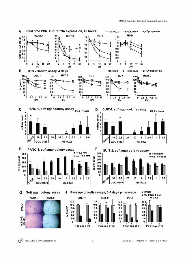
SMO Antagonist, Transient Xenograft Inhibition
PLoS ONE | www.plosone.org 6 June 2011 | Volume 6 | Issue 6 | e19904

longer time period than 5 days. As the liver normally acts as the
primary site of drug metabolism, AST/ALT serum levels is an
easily obtainable measure of toxic side effects. As seen in
Figure 3C, AST/ALT levels remained greater than 1 in mice
treated with 50 mg/kg of MS-0022 after a 30 days injection
scheme (5 day injection, 2 day pause), suggesting that at this dose
MS-0022 was well tolerated without apparent toxic effects in the
liver in comparison to the control treated mice.
In-vivo efficacy of MS-0022 was tested against two pancreatic
adenocarcinoma cell lines PANC-1 and SUIT-2. For PANC-1,
subcutaneous tumors were established in CB17/SCID mice
(n = 6). Mice were randomized and dosed with control solvent
(water with 1% Tween 80) or MS-0022 (QD, IP at 50 mg/kg) in a
5 day injection/2 day pause scheme. Tumor volumes were
evaluated throughout the treatment by measuring two perpendic-
ular diameters with calipers, and by calculating tumor volume
(mm3) using the formula V = a6b26,Pi./6. At the end of the
treatment there was a 38% reduction in tumor volume and a 33%
reduction in tumor weight compared to tumors in control solvent
treated littermates (Figure 3H–I). The growth curve (Figure 3H),
shows that the tumors in both control and treated groups
eventually reached a similar growth pattern, and the growth
inhibition by MS-0022 appeared to be transient. At the endpoint
of the experiment, no statistically relevant difference in human
(tumor) or mouse (stroma) Ptch1 or Gli1 mRNA levels could be
identified in the extracted tumor mRNA (P.0.05) (Figure 3J).
Compared to PANC-1, SUIT-2 cells displayed a stronger
responsiveness to MS-0022 in vitro, both in the reduction of
GLI1 expression and in growth (MTS assay). Therefore, the
effect of MS-0022 was also tested in vivo using SUIT-2 pancreas
adenocarcinoma cells, and subcutaneous tumors were estab-
lished in CB17/SCID mice (n = 11). Mice were randomized
and dosed daily with solvent or 50 mg/kg MS-0022 via PO
administration. The MS-0022 treatment did not result in any
outwards signs of toxicity in the mice, and animal weight
(Figure 4C) was unaffected by the treatment when compared to
the animals that received control solvent. After 7 days, 3 mice
from each treatment group were sacrificed and the tumors
were harvested for analysis, while the remaining animals (n = 8)
were sacrificed after 18 days of treatment, due to tumor
burden. In general, the SUIT-2 tumors showed a more
aggressive growth in comparison to the PANC-1 tumors. At
the endpoint of the experiment, a 27% reduction in tumor
volume and a 36% reduction in tumor weight were measured
in treated animals compared to the control group (Figure 4A–
B). Similar to PANC-1 xenografts (Figure 3D), a transient delay
of growth was observed during the first days of treatment,
followed by a recovery of growth with growth rates similar to
untreated tumors. As in PANC-1 xenografts, no statistical
relevant reduction of human or mouse Gli1, or Ptch1 mRNA
levels was detectable at the endpoint of the experiment
(Figure 4E). However, when samples from the SUIT-2
xenografts were analyzed after 7 days of treatment only,
mouse (stromal) Gli1 mRNA in the tumors was reduced
significantly (P,0.001), while both human and mouse PTCH
and human GLI1 remained unaltered (P.0.05) (Figure 4D).
There was no detectable difference in animal weight between
the control and MS-0022 treated animals (Figure 4C). As
inhibition of SMO has been linked to an increase in
vascularization in poorly vascularized tumors [41], we inves-
tigated the vascularization of the tumor tissue upon MS-0022
treatment. Samples from the harvested tumors were cryosec-
tioned, fixed and stained for the presence of the endothelial cell
marker (CD31), as a marker for microvessles in tumor tissue.
Large variations in SUIT-2 tumor shape including the
occurrence of necrotic cavities, led to substantial variations in
the vascularization of the tumors in all samples derived after 7
or 18 days of treatment and in control samples. Due to
substantial variability, no apparent difference between the
vascularization of samples derived from treated and untreated
animals was detectable. Irrespective of MS-0022 treatment,
CD31 staining revealed high vascularization at the tumor edge,
while vascularization in the center of the tumor was generally
low (Figure 4F).
Discussion
MS-0022 was identified as a potent antagonist of Hh signaling
that blocks the translocation of SMO to the cilia displaying a
transient in vivo antagonistic effect in a pancreatic adenocarcinoma
xenograft model.
MS-0022 contains a core structural motif common to other
SMO antagonists. Analysis of the core structure of MS-0022
revealed that the ‘‘phenyl-amide-phenyl’’ (Figure 1A, where R3 =
phenyl) portion of the molecule was also present in HhAntag [33],
GDC-0449 [52], Sant-2 [53] and Compound Z [54]. Deleting the
2-bromobenzene moiety of MS-0022 resulted in a total loss of
activity, confirming the importance of the identified core structure
for the activity of this class of molecules. Interestingly, although the
‘‘phenyl-amide-phenyl’’ core is identical in all of these molecules,
there are also important differences in the structures. HhAntag,
Sant-2 and Compound Z are closer to each other in structure than
MS-0022 and GDC-0449, as they share a 1H-benzoimidazo
structure coupled in the 3 position to the 4-chloro-N-phenyl.
Interestingly, when the imidazo[1,2-a]pyridine of MS-0022 was
deleted, activity was reduced. However, when it was substituted
with groups that contain nitrogen in ortho position to the N-
phenyl, partial activity was retained. Similar to GDC-0449 and
cyclopamine, MS-0022 inhibited the Hh pathway activity after
induction by various agonistic factors like Shh, oxysterols and
SAG. The ability of MS-0022 to compete with BOPIDY-
cyclopamine binding suggests that it acts by a similar mode of
action as structurally related SMO antagonists, such as GDC-
0449. This was further confirmed by the inhibition of SMO
translocation to the primary cilium by MS-0022. While MS-0022
inhibited SMO in the nanomolar range, it displayed an additional
effect on the Hh pathway downstream of SMO that required a
higher dose. In the micromolar range, MS-0022 led to a significant
Figure 2. In vitro activity in tumor cell lines and a control cell line. A) Graphs displaying dose response curves of normalized (GAPDH) Gli1mRNA expression in indicated tumor cell lines after 48 hour treatment of MS-0022, cyclopamine and GDC-0449. Graphs include standard deviation(n = 3). B) Graphs displaying 4 day dose response curves of cell growth as a % growth of DMSO control (MTS) for PANC-1, SUIT-2, PC-3, FEMX andTHLE-2 cells treated with MS-0022, GDC-0449 and cyclopamine at the indicated dose, with SD (n = 8) C-G) Soft agar colony formation assay of PANC-1(21 days) and SUIT-2 (14 days) cells treated with DMSO or MS-0022 and GDC-0449 at the indicated doses (n = 3). Each well was imaged and colonieswere counted by eye according to size. Graphs displaying the colony count of larger colonies (0.5–1 mm) are shown in C) for PANC-1 and D) for SUIT-2, with SD. Graphs displaying the colony count of medium (0.2–0.5 mm) and small (,0.2 mm) colonies are shown in E) for PANC-1 and F) for SUIT-2,with SD. H) Passage growth assays with PANC-1, SUIT-2, PC-3 and THLE-2 cell lines treated with DMSO control or 5 mM MS-0022 for 2–3 passages of5–7 days, displayed as % growth of DMSO control, with SD (n = 3).doi:10.1371/journal.pone.0019904.g002
SMO Antagonist, Transient Xenograft Inhibition
PLoS ONE | www.plosone.org 7 June 2011 | Volume 6 | Issue 6 | e19904

Figure 3. In vitro MS-0022 pathway selectivity, in vivo tolerance and distribution, and activity in a PANC-1 xenograft model. A)HEK293 cells cotransfected with SuperTop-luc (ST-Luc) and pRL-TK, were treated with control medium and L1 medium +/2 10 and 20 mM MS-0022 for24 hours. Graph displays fold reporter activity with standard deviation (n=3). B) NIH3T3 cells cotransfected with NFkB-luc and pRL-TK, treated with controlmedium and 10 ng/mL TNF-a +/2 10 and 20 mM NFkB inhibitor Bay11-7082 or 10 and 20 mMMS-0022 for 24 hours. Graph displays fold reporter activitywith SD (n=3). C) Graph depicting average AST/ALT ratio frommouse blood at end of animal treatment in the PANC-1 xenograft. D–F) Blood distributioncurves after IV, IP and PO administration of 5 mg/kg MS-0022 in mice. Graphs display mean dose (ng/mL by time) +/2 SD. G) Body weight change inanimals treated with 200 mg/kg MS-0022 over 5 days, with SD (n=5). H–J) Analysis of subcutaneous PANC-1 tumors in mice treated with solvent controlor 50 mg/kg MS-0022 for 21 days (n= 6) by IP administration. H) Growth curves of PANC-1 tumors in mice after 30 days of treatment, showing averagetumor volume (mm3) with standard error of the mean (SEM). I) Graph displaying average PANC-1 tumor weight at end of treatment, with SD. J) Graphdisplays normalized (GUSB) fold expression of mouse and human Gli1 and Ptch in treated tumors, with SD (n=3). * = P value,0.05. (n.s. = P value.0.05).doi:10.1371/journal.pone.0019904.g003
SMO Antagonist, Transient Xenograft Inhibition
PLoS ONE | www.plosone.org 8 June 2011 | Volume 6 | Issue 6 | e19904

reduction of Gli1 mRNA and protein levels in Sufu2/2 cells.
This effect was not due to an altered or inhibited translocation of
Gli1 to the nucleus, indicating that MS-0022 did not reduce Gli1
levels by direct inhibition of Gli1. The precise cause of this
secondary effect remains to be elucidated.
As the implications of aberrant Hh signaling are getting
increasingly evident in several cancers, pathway antagonists will
be an essential toolkit for development of future treatments. The
emerging data from clinical trials with GDC-0449 show both the
benefits and possible pitfalls of a pure SMO antagonist; a clear
tumor response in Hh driven tumors such as basal cell carcinoma
and medulloblastoma [34,55,56], and the occurrence of a drug
induced resistance caused by mutations in the Smo locus [34,55].
A possible solution to block Hh/GLI1signalling despite activation
mutations in the Smo locus would be to develop drugs that act on
the pathway downstream of SMO [47]. A GLI inhibitor would
also address non-canonical activation of GLI [57].
The high level of tumor-stroma interactions found in advanced
cancers like pancreatic adenocarcinoma poses additional chal-
lenges for developing treatment strategies based on Hh
antagonists. Such strategies would require that, in addition to
autocrine efficacy, paracrine interactions between tumor activat-
ed stroma and tumor cells are disrupted [33]. Furthermore, the
barrier that the stromal cells create around the tumor, and the
lack of compound delivery to areas of low vascularization need to
be addressed [41].
The physiochemical properties of MS-0022 (MW =406.3;
clogP: 5.44; PSA: 46.4 (Table 2)), its excellent per oral
bioavailability, and the initial evidence that it is well tolerated
in vivo (MTD .200mg/kg) makes MS-0022 a potential drug
candidate. In PANC-1 and SUIT-2 xenograft experiments, MS-
0022 treatment led to a partial response, where growth was
halted during the first days of treatment compared to the control.
Over time, however, both the treated and control xenograft
groups reverted to similar growth. In both SUIT-2 and PANC-1
xenografts, the growth delay in the MS-0022 treated group
resulted in a statistically relevant (P,0.05) average reduction in
tumor volume of subsequently 27% and 38%. Our in vivo results
lend support to the idea that the primary effect of Hh inhibition
in pancreatic cancer treatment is due to the inhibition of Hh
pathway activity in the stromal cells, although we also observe a
mild initial reduction of GLI1 levels in tumor cells. Both
pancreatic cancer cell lines, SUIT-2 and PANC-1, that were
xenografted as a part of this study, express high levels of Shh
mRNA (Table 4), and should be able to induce a Hh response in
stromal cells. It’s interesting to note that though both MS-0022
and GDC-0449, and to a certain degree cyclopamine, led to a
reduction of GLI1 mRNA in SUIT-2 cells in vitro, suggesting the
possibility to affect autocrine Hh signaling in this cancer cell line.
In an in vivo context, the effect of MS-0022 in vivo on SUIT-2 and
PANC-1 tumors was most likely due to an inhibition paracrine
Hh signaling since no statistically relevant reduction of either
hGLI1 or hPTCH could be measured in mRNA derived from the
tumor tissue. In contrast, mGli1 was reduced in the stroma of the
SUIT-2 tumors (P,0.05) after 7 days of treatment, but not at the
end of the treatment, while at the end of the treatment in PANC-
1 xenografts, there was a slight reduction of mGli1 (although with
a P.0.05). Reasons for the lack of a measurable reduction of
hGLI1 or hPTCH in the tumors, at the time points that were
taken, could be low levels of compound reaching the tumor, or
activation mutations that occurred within the tumors. Activation
mutations in SMO have been observed in a recent clinical trial
using CDG-0449 in a medulloblastoma patient [34,58]. Also, the
development of resistance against chemotherapeutic agents in
pancreatic cancer has been linked to both dense stromal matrix
and increased stromal barrier, which over time, may cause
resistance. Interestingly, Olive et al. found that treatment with
IPI-926 led to an increased vascularization in KPC tumors, but
similarly to our observations, the effect of this SMO inhibitor was
due to a developing resistance [41].
KPC mice develop pancreatic tumors that resemble human
pancreatic ductal adenocarcinoma due to a conditional expression
of endogenous mutant Kras and p53 alleles in pancreatic cells
[59,60]. Similarly, the SUIT-2 and PANC-1 cell lines are mutant
for p53 andKras [61–65]. Despite the differences in the twomodels,
with tumors forming in the mouse pancreas in the KPC model and
tumor cells injected under the skin in the in vivo xenograft model,
Table 4. Kinase activity in presence of 10 mM MS-0022 in akinaseprofiler diversity array.
kinase:Activity% kinase:
Activity% kinase:
Activity%
Abl(h) 96 EphB4(h) 99 NEK2(h) 127
ALK(h) 95 Fyn(h) 100 p70S6K(h) 94
AMPK(r) 98 GRK5(h) 119 PAK2(h) 93
ASK1(h) 91 GSK3b(h) 88 PDGFRb(h) 108
CaMKI(h) 106 IGF-1R(h) 105 Pim-1(h) 103
CDK1/cyclinB(h) 110 IKKa(h) 103 PKA(h) 100
CDK2/cyclinA(h) 98 IRAK4(h) 95 PKBa(h) 107
CDK7/cyclinH/MAT1(h)
103 JAK2(h) 110 PKCa(h) 106
CDK9/cyclinT1(h)
98 JNK3(h) 71 PKCh(h) 103
CHK1(h) 84 KDR(h) 103 PKG1a(h) 94
CK1c1(h) 93 LOK(h) 93 Plk3(h) 107
CK2a2(h) 109 Lyn(h) 103 PRAK(h) 71
cKit(h) 109 MAPK2(h) 101 ROCK-I(h) 103
c-RAF(h) 106 MAPKAP-K2(h)
92 Rse(h) 107
cSRC(h) 83 MEK1(h) 73 Rsk1(h) 91
DRAK1(h) 97 MKK7b(h) 109 SAPK2a(h) 102
DYRK2(h) 97 MLK1(h) 103 SRPK1(h) 102
eEF-2K(h) 99 Mnk2(h) 90 TAK1(h) 98
EGFR(h) 84 MSK2(h) 92
EphA5(h) 112 MST1(h) 94
Millipore diversity panel - percent activity of tested kinases in the presence of10 mM MS-0022.doi:10.1371/journal.pone.0019904.t004
Table 5. Pharmacokinetics of MS-0022.
Cmax,ng/ml
AUClastng*min/ml
AUCinfng*min/ml T1/2, min
Kel,min-1
IV 934.3 87683.5 93897.6 59.27 0.0117
IP 377.7 25025.0 26237.8 54.83 0.0126
PO 912.0 64177.5 68526.0 59.10 0.0117
Pharmacokinetic profile of MS-0022 by IV, IP and PO administration in mice. Thetable displays the maximum concentration (Cmax), total drug exposure(AUClast) and (AUCinf), half-life (T1/2) and terminal slope (Kel).doi:10.1371/journal.pone.0019904.t005
SMO Antagonist, Transient Xenograft Inhibition
PLoS ONE | www.plosone.org 9 June 2011 | Volume 6 | Issue 6 | e19904

both studies provide evidence that the pancreatic cancer may only
transiently be inhibited by a SMO antagonist, revealing that the
challenge of overcoming resistance is ever present.
Materials and Methods
Ethics statementAll animals were housed and treated under the approved
protocols in accordance with the National Institute of Health guide
for the care and use of laboratory animals and according to
regulations outlined in the USDA Animal Welfare Act (9 CFR Parts
1, 2 and 3) and the conditions specified in The Guide for Care and Use of
Laboratory Animals (National Academy Press, Washington, D.C.,
1996). Animal experiments performed in Norway were approved by
the Centre for Comparative Medicine at the National Hospital of
Norway according to the Regional Ethical Comity guidelines
(permit nr: 19/08-910) and at the Norwegian Institute of Public
Health (permit nr 2722), while animal experiments performed by
Chemdiv were approved by ChemDiv’s Institutional Animal Care
and Use Committee (Permit nr: 8/21.01.2008). All efforts were
made to minimize the number and suffering of animals.
Cells and culture conditionsPANC-1, PC-3, THLE-2, NIH3T3, Shh-Light 2 (Shh-L2) and
C3H10T1/2 cells were obtained from ATCC. SUIT-2 cells were
obtained from Cell Bank, RIKEN BioResource Center.
PTCH2/2 MEF [45] was a gift from P. A. Beachy and
SUFU2/2 MEF were a gift from R. Toftgard (Karolinska
Institutet). FEMX was a gift from G. E. Melandsmo. All cell lines
were cultured at 37uC in a humidified atmosphere of 5% CO2 in
the medium formulations as instructed by the suppliers.
Alkaline phosphatase assay and screening protocolC3H/10T1/2 cells were seeded 5*103 cells per well in a 96-well
format and allowed to attach for 3–5 hours, before adding serial
dilutions of compounds (from a DMSO stock). After 30 minutes
Figure 4. In vivo activity of MS-0022 in a SUIT-2 xenograft. A-F) Analysis of subcutaneous SUIT-2 tumors in mice treated with solvent controlor 50 mg/kg MS-0022 for 7 (n = 3) or 18 days (n = 8) by PO administration. A) Graph displaying average SUIT-2 tumor weight at end of treatment, withSD. B) Tumor growth curve of SUIT-2 tumors in mice after 18 days of treatment, plotted as tumor volume (mm3) with standard error of the mean(SEM). C) Average animal weight through the course of the treatment with SD (n = 8). D) and E) Graphs showing normalized (GUSB) fold expression ofmouse and human Gli1 and Ptch in treated tumors with SD. D) Shows expression in tumors harvested after 7 days (n = 3), while E) shows expression intumors harvested after 18 days (n = 8). F) Immunostaining of microvessles using anti-CD31 (endothelial cell marker) in cryosectioned tumor tissueslides. Images depict CD31 staining in the tumor edge and in the centre of the tumors. * = P value ,0.05.doi:10.1371/journal.pone.0019904.g004
SMO Antagonist, Transient Xenograft Inhibition
PLoS ONE | www.plosone.org 10 June 2011 | Volume 6 | Issue 6 | e19904

recombinant human SHH (C24II) (RnD systems (1845SH)) was
added (300 ng/mL). After 72 hours, the cells were lysed for 15
minutes using 20 mL of a 10 mM Ethanolamine Buffer (pH 8.0)
with 0.2% Triton-X100, supplemented 1:100 with Protease
Inhibitor Cocktail (EMD Bioscience). 7.5 mL of lysate was mixed
with 45 mL of CSPD Alkaline Phosphatase Substrate with
Emerald-II (Applied Biosystems) and incubated at room temper-
ature, in the dark, for 45 minutes. The plate was measured using a
Wallac VictorV reader (0.1 s Luminescence). Screening com-
pounds and MS-0022 were part of a Chemdiv chemical library.
Hh reporter assayShh-L2 cells were used as a Hh reporter cell line as described
previously [45]. Hh signaling was induced by Shh conditioned
medium, 200 nM SAG (AH-Diagnostics) and ActOXS (a
combination of 5 mM Hydroxycholesterol 20-alfa and 5 mMHydroxycholesterol 22S (Sigma-Aldrich)). For Shh conditioned
medium, Shh-PANC-1 cells were grown to confluence and
switched to fresh medium. Shh conditioned medium was harvested
after 48 hours, and was diluted to 50% in fresh DMEM before
use. The clonal Shh-PANC-1 cell line was generated by stably
transducing PANC-1 cells with virus containing medium from
HEK293T cells that were transfected using a Virapower Lentiviral
expression kit (Invitrogen). The vector was produced by removing
GFP from a pLenti 6.2-vector, and inserting the mouse Shh cDNA
sequence.
IC50 calculationIC50 values were calculated using dose response curve data
where n= 3. The calculations were performed with an online
calculator using the formula: a exp (22bx) + c at the website;
http://www.changbioscience.com/stat/ec50.html
NMR spectroscopy of MS-0022NMR spectra of MS-0022 (approx. 2 mg) were obtained from a
solution in 0.5 ml of DMSO-d6 ((CD3)2S(O), 99.9% D, Cam-
bridge Isotope Laboratories, Andover, MA) in a 5.0 mm tube
(WILMAD, WG-5 Economy). The spectra were acquired on an
Avance AV 600 MHz NMR spectrometer (Bruker BioSpin,
Rheinstetten, Germany) with a 5 mm CP-TCI (1H/13C, 15N-
2H) triple-resonance inverse cryo probe, equipped with a Z-
gradient coil. NMR assignments of MS-0022 were inferred from
examination of 1H- and 13C spectra, attached proton test (APT),
correlated spectroscopy (COSY45), total correlation spectroscopy
(TOCSY) pulsed field gradient heteronuclear single quantum
coherence (g-HSQC), pulsed field gradient heteronuclear multiple
bond correlation (g-HMBC) and pulsed field gradient hetero-
nuclear two-bond correlation (g-H2BC/g-HSQC-COSY). The
data were processed using the Bruker TOPSPIN software (version
1.3 or version 2.1 pl 2). Chemical shift values were referenced to
the residual solvent signals, i.e. CHD2S(O)CD3=2.49 ppm and
(CD3)2S(O) = 39.5 ppm, respectively. All NMR spectra were
acquired at 25uC.
BODIPY assayBODIPY assay was performed to determine competition with
cyclopamine as described previously [21,66].
Real Time PCRCells were lysed for RNA extraction after 48 or 72 hours of
treatment. Total RNA from cultured cells or tumor tissue was
isolated using the GeneElute miniprep kit (Sigma-Aldritch)
following the manufacturer’s instructions. cDNA was synthesized
from the isolated mRNA using the Retroscript kit (Ambion), and
real-time PCR was carried out using the SYBR Green PCR
master mix (Stratagene) according to the manufacturer’s
instructions with an Mx3000P cycler (Stratagene). The relative
concentrations of cDNA present in each sample were calculated
by the MxPro software (Stratagene), normalized for GAPDH
(mRNA from cells) or GUS (mRNA from tumors). For real time
PCR primer sequences see Table 6. For the tumor tissue real time
PCR, previously described human and mouse specific primers
against GUSB, PTCH1 and GLI1were used [33].
ImmunofluorescencePTCH2/2 MEF were seeded on pre-coated glass slides (1
hour coating with 0.1% Gelatin (G1393) and 0.003% Collagen
(C8919) (Sigma-Aldrich) in PBS). 80% confluent cells were
switched from 10% to 0.5% FBS in medium and cells were
treated with DMSO or 5 mM MS-0022 for 24 hours in (n = 3)
before subsequent immunofluorescent staining: Slides were fixed
with 4% paraformaldehyde (Sigma-Aldrich), permeabilized for 10
min in 0.1% v/v Triton X-100 (Sigma-Aldrich) in PBS (PBT), and
blocked with 10% w/v BSA (Saveen Werner) in PBT for 1 hour.
Primary antibodies, mouse anti-acetylated tubulin (a-Tub) (Sigma-
Aldrich (T7451), 1:1000) goat anti-Smo (Santa Cruz Biotechnol-
ogy (sc-6367), 1:1000), and goat anti-GLI-1 (RnD systems
(AF3455), 1:500) were diluted in 0.5% w/v BSA in PBT and left
on the slides over night at 4uC. Secondary antibodies donkey anti-goat IgG-Alexa 594 and donkey anti-mouse IgG-Alexa 488
(Invitrogen, both at 1:500) were added in 0.5% w/v BSA in PBT
for 1 hour. The slides were counterstained with DAPI (Roche,
Table 6. Primer sequences for quantitative real time PCR.
Primers: Primer sequence, 59 R 39 Tm,6C
hPTCH1 fwd CGA GCC CCC CTG TAC GAA GTG G 67.7
hPTCH1 rev GAC CCC CAG CAA GCC CAG AAA A 64.0
hGLI1 fwd GCG CAT CCC GAG CCC AGC 65.1
hGLI1 rev GCC CTC GGT GCA GCT GTT GGT C 67.7
hGLI2 fwd GCC TCC GAG AAG CAA GAA GCC AAA A 64.4
hGLI2 rev CCT GGT GTC GCA TGT CAA TCG GTA G 66.3
hGLI3 fwd CGG GAC GGT GTT TGC CAT GGA C 65.8
hGLI3 rev GGA GGA TGG AAG GCA GGG AAA AGA T 64.6
hSHH fwd GCC AGC GGA AGG TAT GAA GGG AAG 66.1
hSHH rev ACC GAG ATG GCC AAA GCG TTC AAC 64.4
hHIP fwd TGG GGA TGG CTC GCA ACG TCT C 65.8
hHIP rev TGG GAT GGA ATG CGA GGC TTA GC 64.2
hSUFU fwd CCC GAG GAT GAC GAG GAC AGC C 67.7
hSUFU rev CGC GTG CGA ATC AGC TCA TGG G 65.8
hSMO fwd CCA GGA GGA AGC GCA CGG CAA G 67.7
hSMO rev TCG CAC TGG CCT GAA CTG TTG AAC T 64.6
hGAPDH fwd GCC CCC TCT GCT GAT GCC CCC A 75.7
hGAPDH rev TGG GTG GCA GTG ATG GCA TGG 70.2
mGLI1 fwd AGC CAA CTT TAT GTC AGG GTC CCA GGG T 71.2
mGLI1 rev GAG CCC GCT TCT TTG TTA ATT TGA CTG 66.5
mGAPDH fwd TAT GTC GTG GAG TCT ACT GGT GTC TTC ACC 68.1
mGAPDH rev GAG TTG TCA TAT TTC TCG TGG TTC ACA CCC 66.8
Table of real time PCR primers; name, sequence and TmuC (h = human,m=mouse).doi:10.1371/journal.pone.0019904.t006
SMO Antagonist, Transient Xenograft Inhibition
PLoS ONE | www.plosone.org 11 June 2011 | Volume 6 | Issue 6 | e19904

1 mg/ml) in PBS. 3 random regions were imaged pr slide using a
confocal LSM510 microscope (Carl Zeiss MicroImaging). The
ratio of green Smo positive cilia versus the red aTub positive cilia
was quantified manually or processed and analyzed using ImageJ
to quantify the intensity of Smo and aTub for each cilium in the
images (http://rsb.info.nih.goc/ij/). Graphs display mean with
standard deviation error bars.
Frozen tumor tissue was cryosectioned (n= 8 pr group), and the
sections were fixed with 4% paraformaldehyde for 10 minutes
before permeabilization with 0.2% Triton-X in PBS for 10 minutes.
Sections were blocked for 30 minutes at room temperature with 3%
BSA in PBS and was subsequently incubated over night at 4uC with
rat anti-mouse CD31 (BD Biosciences (550274), 1:500) in 1% BSA
in PBS. The secondary antibody used was Alexa Fluor 488 goat-
anti-rat (Molecular Probes; 1/700 dilution) in 1% BSA in PBS for
1 hour at RT. Nuclei were counterstained with DAPI, in PBS for 10
minutes at RT. Images was acquired as above and electronic images
was further processed using ImageJ.
Transfection and luciferase assayVectors were obtained from the following: GliBS-Luc reporter
[67] (gift from H. Sasaki), pBluescript (pBS) (Stratagene), FL-HA-
GLI1 (Gli1) and Gli2-GFP (Gli2) (gifts from R. Toftgard),
Super8XTOPFlash [68], and NFkB-luc (Panomics). 10 ng renilla
luciferase (pRL-TK) (Promega) was used as a transfection control.
HEK293 and NIH3T3 cells were seeded at 80,000 per well in 48-
well plates on day 1, and were co-transfected on day 2 using a total
of 0.4 mg plasmid per well mixed at 1:3 in FUGENE6 according to
manufacturer’s instructions. On day 3, control medium and
activating medium (50% L1 conditioned medium, 50% Shh
conditioned medium or medium containing 10 ng/mL recombi-
nant rcrTNF-a (R&D Systems)) +/2 10–20 mM MS-0022 were
added, using DMSO as a control (the NFkB pathway inhibitor
Bay11-7082 was used as a control in the NFkB assay). Samples
were analyzed on day 4 or 5 using the Dual Luciferase assay kit
according to manufacturer’s instructions. Graphs display relative
or fold reporter activity, and are calculated from a mean ratio of
firefly reporter luminescence/pRL-TK luminescence with stan-
dard deviation error bars (n = 3). The experiments have been
repeated with similar results.
In vitro antiproliferative assayCells were plated at a density of 3000 (THLE-2 and PC-3) and
1000 (PANC-1, SUIT-2 and FEMX) cells per well (on the basis of
their growth rate) in 200 ml of medium in a 96-well plate. Medium
was changed daily, and after four days, the assay was read using a
MTS kit (Promega) according to the instructions of the
manufacturer. Graphs display average of percent growth with
standard deviation (n= 8).
Passage growth assay20,000 cells were seeded per well in 6 well plates (triplicates)
with DMSO or 5 mM MS-0022. Treatment medium was changed
on day 3–5, and after 5–7 days the cells were trypsinized, re-
suspended in medium and counted, before reseeding cells at
20,000/well for a subsequent passage. The average growth of MS-
0022 treated cells was calculated as a percentage of the growth of
the DMSO control at the end of each passage. Graphs display
average of percent growth with standard deviation bars (n = 3).
Soft agar colony formation assay2000 PANC-1 or SUIT-2 cells were suspended in 1.5 mL
growth media containing 0.35% agar (Oxoid), over a 1.5 mL
base layer containing 0.5% agar in 6-well plates. The plates
were incubated for 14 days (SUIT-2) and 21 days (PANC-1)
and then stained for 1 h with 1 ml of 0.02% crystal violet
(Sigma-Aldrich), and colonies were counted according to sizes;
,0.2 mm (small), 0.2–1 mm (medium) and .1 mm (large).
Data represent average numbers with standard deviation bars
(n = 3). The experiments have been repeated with similar
result.
Pharmacokinetics, dose tolerance, animal tumorestablishment and treatmentFor pharmacokinetic (PK) analysis, 5 mg/kg MS-0022 was given
by IV, PO and IP administration to 7–8 week old male C57BL mice
(mouse musculus), and blood was collected after 5, 15, 30, 60, 120, and
240 min, with n=3. Animals were sacrificed and blood samples
(,0.5 mL each) were collected from the abdominal aorta into
EDTA-containing tubes, centrifuged (2–8uC for ,10 minutes at
,10000 xg) and plasma harvested into single tubes for each animal
and frozen (,270uC). Blood samples were analyzed by ChemDiv’s
bioanalytical department. The maximum concentration (Cmax),
total drug exposure (the area under the curve to the last quantifiable
concentration (AUClast) and as the AUClast value extrapolated to
infinity (AUCinf), calculated as AUCinf=AUClast+C(t)last/Kel,
where C(t)last is the last measurable concentration), half-life (T1/2)
calculated as ln(2)/Kel and Kel calculated as the slope of the terminal
linear portion of the concentration/time curve. The WinNonlin
Professional 5.2 software (Pharsight Corp.) was used for the
calculation of the PK parameters.
For the analysis of uptake of MS-0022 in organs and tissue,
livers were dissected out from mice 120 min after PO adminis-
tration of MS-0022 and frozen (,270uC). The LC-MS method
used to detect MS-0022 was described previously [69], with the
main exception being that isocratic conditions were used (50%
0.1% FA (aq), 50% 0.1% FA (ACN), v/v), and that a UV detector
was placed between the LC and MS instrument.
MS-0022 tolerance was tested by daily injections of 200 mg/kg
MS-0022 for 5 days in 7–8 week old male C57BL mice. Animal
were observed daily for outward signs of toxicity (weight loss, fur
ruffling, hunched posture).
For PANC-1 xenografts, 4–6 week old CB17/SCID mice were
injected with 56106 PANC-1 (n= 6 per group) cells in 200 ml PBSsubcutaneously on day 1. IP treatment with 50 mg/kg MS-0022
began when median tumor size reached 25 mm2 (day 27). MS-
0022 was pulverized using a pestle, and was mixed into a stable
suspension in water containing 1% Tween 80 (Sigma-Aldrich).
Mice were treated with MS-0022 or solvent for a period of 30 days
after initiation in a 5 day injection 2 day pause scheme, and tumor
size was measured throughout using calipers (mm2). After 30 days
the mice were sacrificed and tumors dissected and weighed. The
PANC-1 xenograft endpoint was taken two days after the last
compound treatment.
For SUIT-2 xenografts, 4–6 week old CB17/SCID mice were
injected with 56106 SUIT-2 (n=11 per group) cells in 200 ml PBSsubcutaneously on day 1. IP treatment with 50 mg/kg MS-0022
began when median tumor size reached 35 mm2 (day 8). Mice were
treated daily with MS-0022 or solvent for a period of 18 days after
initiation, and tumor size was measured throughout using calipers
(mm2). After 8 days, three mice from each group were sacrificed and
tumors dissected, weighed and processed for analysis. The remaining
animals (n= 8) were sacrificed after 19 days of treatment, and tumors
harvested. Tumor volume (mm3) was calculated using the formula
a6b26,pi./6. End tumor measurement statistics were obtained
using two samples T-test in the Sigmaplot software.
SMO Antagonist, Transient Xenograft Inhibition
PLoS ONE | www.plosone.org 12 June 2011 | Volume 6 | Issue 6 | e19904

Liver transaminasesBlood was collected from the hepatic vein of freshly sacrificed
animals into heparinized capillary blood collection tubes
(Sarstedt), which were immediately centrifuged and the serum
was collected. AST and ALT were run on non-hemolyzed
samples using AST and ALT kits (Randox) following the
manufacturer’s instructions.
Supporting Information
Figure S1 Atom numbering of MS-0022. The atom
numbering of MS-0022 used in the 1H and 13C NMR analysis.
(See Table S1)
(TIF)
Table S1 NMR analysis of MS-0022. 1H and 13C NMR Data
for MS-0022 (DMSO-d6). For the atom numbering used see Figure
S1.
(DOC)
Acknowledgments
We thank Eline Buchman and Line Mygland for excellent technical
support.
Author Contributions
Conceived and designed the experiments: MFS SRW JLD DDH AK IO
DP SK. Performed the experiments: MFS SRW JLD AK IO DP. Analyzed
the data: MFS SRW JLD DDH AK IO DP SK. Contributed reagents/
materials/analysis tools: MFS SRW JLD DDH AK IO DP. Wrote the
paper: MFS SRW JLD DDH AK IO DP SK.
References
1. Fietz MJ, Concordet JP, Barbosa R, Johnson R, Ingham PW, et al. (1994) The
hedgehog gene family in Drosophila and vertebrate development. Dev Suppl:
43–51.
2. Jiang J, Hui CC (2008) Hedgehog signaling in development and cancer. Dev
Cell 15: 801–812.
3. Krauss S, Concordet JP, Ingham PW (1993) A functionally conserved homolog
of the Drosophila segment polarity gene hh is expressed in tissues with polarizing
activity in zebrafish embryos. Cell 75: 1431–1444.
4. Rohatgi R, Scott MP (2007) Patching the gaps in Hedgehog signalling. Nat Cell
Biol 9: 1005–1009.
5. Han YG, Spassky N, Romaguera-Ros M, Garcia-Verdugo JM, varez-Buylla A,
et al. (2008) Hedgehog signaling and primary cilia are required for the formation
of adult neural stem cells. Nat Neurosci 11: 277–284.
6. Altaba A, Mas C, Stecca B (2007) The Gli code: an information nexus regulating
cell fate, stemness and cancer. Trends Cell Biol 17: 438–447.
7. Altaba A (2008) Therapeutic inhibition of Hedgehog-GLI signaling in cancer:
epithelial, stromal, or stem cell targets? Cancer Cell 14: 281–283.
8. Bailey JM, Singh PK, Hollingsworth MA (2007) Cancer metastasis facilitated by
developmental pathways: Sonic hedgehog, Notch, and bone morphogenic
proteins. J Cell Biochem 102: 829–839.
9. Beachy PA, Karhadkar SS, Berman DM (2004) Tissue repair and stem cell
renewal in carcinogenesis. Nature 432: 324–331.
10. Hyman JM, Firestone AJ, Heine VM, Zhao Y, Chen JK, et al. (2009) Small-
molecule inhibitors reveal multiple strategies for Hedgehog pathway blockade.
Proc Natl Acad Sci U S A 106: 14132–14137.
11. Rubin LL, de Sauvage FJ (2006) Targeting the Hedgehog pathway in cancer.
Nat Rev Drug Discov 5: 1026–1033.
12. Rudin CM, Hann CL, Laterra J, Yauch RL, Low JA, et al. (2009) Treatment of
medulloblastoma with hedgehog pathway inhibitor GDC-0449. N Engl J Med
361: 1173–1178.
13. Scales SJ, de Sauvage FJ (2009) Mechanisms of Hedgehog pathway
activation in cancer and implications for therapy. Trends Pharmacol Sci
30: 303–312.
14. Tremblay MR, Lescarbeau A, Grogan MJ, Tan E, Castro AC, et al. (2009)
Discovery of a potent and orally active hedgehog pathway antagonist (IPI-926).
J Med Chem 52: 4400–4418.
15. Von Hoff DD, LoRusso PM, Rudin CM, Reddy JC, Low JA, et al. (2009)
Inhibition of the hedgehog pathway in advanced basal-cell carcinoma.
N Engl J Med 361: 1164–1172.
16. Barakat MT, Scott MP (2009) Tail wags dog: primary cilia and tumorigenesis.
Cancer Cell 16: 276–277.
17. Wong SY, Seol AD, So PL, Ermilov AN, Reiter JF, et al. (2009) Primary cilia
can both mediate and suppress Hedgehog pathway-dependent tumorigenesis.
Nat Med 15: 1055–1061.
18. Wilson CW, Chen MH, Chuang PT (2009) Smoothened adopts multiple active
and inactive conformations capable of trafficking to the primary cilium. PLoS
One 4: e5182.
19. Taipale J, Cooper MK, Maiti T, Beachy PA (2002) Patched acts catalytically to
suppress the activity of Smoothened. Nature 418: 892–897.
20. Zhao Y, Tong C, Jiang J (2007) Hedgehog regulates smoothened activity by
inducing a conformational switch. Nature 450: 252–258.
21. Dwyer JR, Sever N, Carlson M, Nelson SF, Beachy PA, et al. (2007) Oxysterols
are novel activators of the hedgehog signaling pathway in pluripotent
mesenchymal cells. J Biol Chem 282: 8959–8968.
22. Cheng SY, Yue S (2008) Role and regulation of human tumor suppressor SUFU
in Hedgehog signaling. Adv Cancer Res 101: 29–43.
23. Cheng SY, Bishop JM (2002) Suppressor of Fused represses Gli-mediated
transcription by recruiting the SAP18-mSin3 corepressor complex. Proc Natl
Acad Sci U S A 99: 5442–5447.
24. Yue S, Chen Y, Cheng SY (2009) Hedgehog signaling promotes the degradation
of tumor suppressor Sufu through the ubiquitin-proteasome pathway. Oncogene
28: 492–499.
25. Katoh Y, Katoh M (2009) Integrative genomic analyses on GLI1: positive
regulation of GLI1 by Hedgehog-GLI, TGFbeta-Smads, and RTK-PI3K-AKT
signals, and negative regulation of GLI1 by Notch-CSL-HES/HEY, and
GPCR-Gs-PKA signals. Int J Oncol 35: 187–192.
26. Epstein EH (2008) Basal cell carcinomas: attack of the hedgehog. Nat Rev
Cancer 8: 743–754.
27. Toftgard R (2009) Two sides to cilia in cancer. Nat Med 15: 994–996. 10.1038/
nm0909-994.
28. Unden AB, Holmberg E, Lundh-Rozell B, Stahle-Backdahl M, Vorechovsky I,
et al. (1996) Mutations in the human homologue of Drosophila patched (PTCH)
in basal cell carcinomas and the Gorlin syndrome: different in vivo mechanisms
of PTCH inactivation. Cancer Res 56: 4562–4565.
29. Berman DM, Karhadkar SS, Maitra A, Montes De OR, Beachy PA, et al. (2003)
Widespread requirement for Hedgehog ligand stimulation in growth of digestive
tract tumours. Nature 425: 846–851.
30. Thayer SP, di Magliano MP, Heiser PW, Nielsen CM, Hebrok M, et al. (2003)
Hedgehog is an early and late mediator of pancreatic cancer tumorigenesis.
Nature 425: 851–856.
31. Bailey JM, Mohr AM, Hollingsworth MA (2009) Sonic hedgehog paracrine
signaling regulates metastasis and lymphangiogenesis in pancreatic cancer.
Oncogene 28: 3513–3525.
32. Theunissen JW, de Sauvage FJ (2009) Paracrine Hedgehog signaling in cancer.
Cancer Res 69: 6007–6010.
33. Yauch RL, Gould SE, Scales SJ, Tang T, de Sauvage FJ, et al. (2008) A
paracrine requirement for hedgehog signalling in cancer. Nature 455: 406–410.
34. Yauch RL, Dijkgraaf GJ, Alicke B, Januario T, de Sauvage FJ, et al. (2009)
Smoothened Mutation Confers Resistance to a Hedgehog Pathway Inhibitor in
Medulloblastoma. Science.
35. Dembinski JL, Krauss S (2009) Characterization and functional analysis of a
slow cycling stem cell-like subpopulation in pancreas adenocarcinoma. Clin Exp
Metastasis.
36. Low JA, de Sauvage FJ (2010) Clinical experience with Hedgehog pathway
inhibitors. J Clin Oncol 28: 5321–5326.
37. Tian H, Callahan CA, DuPree KJ, Darbonne WC, Ahn CP, et al. (2009)
Hedgehog signaling is restricted to the stromal compartment during pancreatic
carcinogenesis. Proc Natl Acad Sci U S A 106: 4254–4259.
38. Nolan-Stevaux O, Lau J, Truitt ML, Chu GC, Hebrok M, et al. (2009) GLI1 is
regulated through Smoothened-independent mechanisms in neoplastic pancre-
atic ducts and mediates PDAC cell survival and transformation. Genes Dev 23:
24–36.
39. Walter K, Omura N, Hong SM, Griffith M, Vincent A, et al. (2010)
Overexpression of smoothened activates the sonic hedgehog signaling pathway
in pancreatic cancer-associated fibroblasts. Clin Cancer Res 16: 1781–1789.
40. Neesse A, Michl P, Frese KK, Feig C, Cook N, et al. (2010) Stromal biology and
therapy in pancreatic cancer. Gut 2010 Oct 21 doi:10.1136/gut.2010.226092.
41. Olive KP, Jacobetz MA, Davidson CJ, Gopinathan A, Tuveson DA (2009)
Inhibition of Hedgehog signaling enhances delivery of chemotherapy in a mouse
model of pancreatic cancer. Science 324: 1457–1461.
42. Bailey JM, Swanson BJ, Hamada T, Eggers JP, Singh PK, et al. (2008) Sonic
hedgehog promotes desmoplasia in pancreatic cancer. Clin Cancer Res 14:
5995–6004.
43. Cooper MK, Porter JA, Young KE, Beachy PA (1998) Teratogen-mediated
inhibition of target tissue response to Shh signaling. Science 280: 1603–1607.
44. Incardona JP, Gaffield W, Kapur RP, Roelink H (1998) The teratogenic
Veratrum alkaloid cyclopamine inhibits sonic hedgehog signal transduction.
Development 125: 3553–3562.
SMO Antagonist, Transient Xenograft Inhibition
PLoS ONE | www.plosone.org 13 June 2011 | Volume 6 | Issue 6 | e19904

45. Taipale J, Chen JK, Cooper MK, Wang B, Beachy PA, et al. (2000) Effects of
oncogenic mutations in Smoothened and Patched can be reversed bycyclopamine. Nature 406: 1005–1009.
46. Frank-Kamenetsky M, Zhang XM, Bottega S, Guicherit O, Porter JA, et al.
(2002) Small-molecule modulators of Hedgehog signaling: identification andcharacterization of Smoothened agonists and antagonists. J Biol 1: 10.
47. Lauth M, Bergstrom A, Shimokawa T, Toftgard R (2007) Inhibition of GLI-mediated transcription and tumor cell growth by small-molecule antagonists.
Proc Natl Acad Sci U S A 104: 8455–8460.
48. Dierks C (2010) GDC-0449–targeting the hedgehog signaling pathway. RecentResults Cancer Res 184: 235–238.
49. Kiselyov AS (2006) Biology-driven diversity: concept of focused diversity. ChemBiol Drug Des 68: 67–69.
50. Alieva IB, Gorgidze LA, Komarova YA, Chernobelskaya OA, Vorobjev IA(1999) Experimental model for studying the primary cilia in tissue culture cells.
Membr Cell Biol 12: 895–905.
51. Seeley ES, Nachury MV (2010) The perennial organelle: assembly anddisassembly of the primary cilium. J Cell Sci 123: 511–518.
52. Robarge KD, Brunton SA, Castanedo GM, Xie M, et al. (2009) GDC-0449-apotent inhibitor of the hedgehog pathway. Bioorg Med Chem Lett 19:
5576–5581.
53. Chen JK, Taipale J, Young KE, Maiti T, Beachy PA (2002) Small moleculemodulation of Smoothened activity. Proc Natl Acad Sci U S A 99:
14071–14076.54. Borzillo GV, Lippa B (2005) The Hedgehog signaling pathway as a target for
anticancer drug discovery. Curr Top Med Chem 5: 147–157.55. Kean S (2009) Medicine. Disrupting Hedgehog may reverse advanced cancer, if
only temporarily. Science 325: 1188.
56. So PL, Tang JY, Epstein EH (2010) Novel investigational drugs for basal cellcarcinoma. Expert Opin Investig Drugs 19: 1099–1112.
57. Lauth M, Toftgard R (2007) Non-canonical activation of GLI transcriptionfactors: implications for targeted anti-cancer therapy. Cell Cycle 6: 2458–2463.
58. Dijkgraaf GJ, Alicke B, Weinmann L, Januario T, West K, et al. (2011) Small
molecule inhibition of GDC-0449 refractory smoothened mutants anddownstream mechanisms of drug resistance. Cancer Res 71: 435–444.
59. Hingorani SR, Wang L, Multani AS, Combs C, Deramaudt TB, et al. (2005)
Trp53R172H and KrasG12D cooperate to promote chromosomal instabilityand widely metastatic pancreatic ductal adenocarcinoma in mice. Cancer Cell 7:
469–483.
60. Hruban RH, Adsay NV, bores-Saavedra J, Anver MR, Biankin AV, et al. (2006)Pathology of genetically engineered mouse models of pancreatic exocrine
cancer: consensus report and recommendations. Cancer Res 66: 95–106.61. Butz J, Wickstrom E, Edwards J (2003) Characterization of mutations and loss of
heterozygosity of p53 and K-ras2 in pancreatic cancer cell lines by immobilized
polymerase chain reaction. BMC Biotechnol 3: 11.62. Kainuma O, Asano T, Hasegawa M, Kenmochi T, Nakagohri T, et al. (1997)
Inhibition of growth and invasive activity of human pancreatic cancer cells by afarnesyltransferase inhibitor, manumycin. Pancreas 15: 379–383.
63. Birnbaum DJ, Adelaide J, Mamessier E, Finetti P, Lagarde A, et al. (2011)Genome profiling of pancreatic adenocarcinoma. Genes Chromosomes &
Cancer 50: 456–465 (2011).
64. Hidaka H, Ishiko T, Ishikawa S, Ikeda O, Mita S, et al. (2005) Constitutive IL-8expression in cancer cells is associated with mutation of p53. J Exp Clin Cancer
Res 24: 127–133.65. Sato N, Mizumoto K, Kusumoto M, Niiyama H, Maehara N, et al. (1998) 9-
Hydroxyellipticine inhibits telomerase activity in human pancreatic cancer cells.
FEBS Lett 441: 318–321.66. Chen JK, Taipale J, Cooper MK, Beachy PA (2002) Inhibition of Hedgehog
signaling by direct binding of cyclopamine to Smoothened. Genes Dev 16:2743–2748.
67. Sasaki H, Hui C, Nakafuku M, Kondoh H (1997) A binding site for Gli proteinsis essential for HNF-3beta floor plate enhancer activity in transgenics and can
respond to Shh in vitro. Development 124: 1313–1322.
68. Veeman MT, Slusarski DC, Kaykas A, Louie SH, Moon RT (2003) Zebrafishprickle, a modulator of noncanonical Wnt/Fz signaling, regulates gastrulation
movements. Curr Biol 13: 680–685.69. Wilson SR, Boix F, Holm A, Lundanes E, Greibrokk T, et al. (2005)
Determination of bradykinin and arg-bradykinin in rat muscle tissue by
microdialysis and capillary column-switching liquid chromatography with massspectrometric detection. J Sep Sci 28: 1751–1758.
SMO Antagonist, Transient Xenograft Inhibition
PLoS ONE | www.plosone.org 14 June 2011 | Volume 6 | Issue 6 | e19904

IV