MODIFICATIONS INDUCED - Plant · PDF fileroot modifications in Vicia, faba seedlings grown...
10
ROOT MODIFICATIONS INDUCED IN ZEA MAYS SEEDLINGS BY IRRADIATING DRY SEEDS WITH SOFT X-RAYS G. F. SMITH AND H. KERSTEN (WITH NINETEEN FIGURES) Introduction This investigation is in part similar to a previous one which described root modifications in Vicia, faba seedlings grown from x-rayed seeds (5). Its purpose is to repeat and supplement these data, using Zea mays, so that it might be possible to state more generally the effects of x-rays on plant roots; and also to test the opinion that one effect of x-raying seeds, which appears in the roots during germinationi, is on factors involved in cavity formation. In Zea mays, cavities of the lvsigenous type occur normally in the roots. If the radiation were active in this respect, it might be expected that the process would show variations in rate and extent as compared with controls. Methods The radiation referred to as soft x-rays was supplied bv an apparatus described in previous papers (3, 4, 5, 6). Unsoaked seeds of Zea mays were oriented with their embrvo sides toward the source of x-ray radiation, 8 cm. from the focal spot of the x-ray tube. The tube was of the gas type, having a copper target. The most intense part of its radiation consisted of the Ka line (1.54 A). The dose of radiation selected was 30 peak kv., 10 ma. with an exposure time of 100 minutes. This treatment, according to pre- liminary tests, results in growth cessation during germinationi. Any ex- posure time over 30 minutes, unider these conditions, also results in this characteristic growth cessation but to a lesser extent. Variations in growth of seedlings germinated from seeds given increasing exposures to x-ravs, are given in figure 1. Following irradiation, the seeds were germiniated in moist peat moss at 850 F. The approximate period of germination before growth cessation becomes apparent in treated seedlings is about 8 days. External ob-erva- tions were recorded for this time and histological studies were made of pri- mary roots of 3-, 5-, and 8-day-old seedlings. Fifty roots of each type were examined microscopically, usinlg standard methods of microtechiiique. The characteristic irregularitv in arrangement and extent of tissue in seedlings grown from x-raved seeds, makes it quite impossible to secure his- tological sections without some distortion. This accounts for the irregu- larity in some illustrations of the x-rayed material presented throughout the paper. 455 www.plantphysiol.org on May 25, 2018 - Published by Downloaded from Copyright © 1942 American Society of Plant Biologists. All rights reserved.
Transcript of MODIFICATIONS INDUCED - Plant · PDF fileroot modifications in Vicia, faba seedlings grown...
ROOT MODIFICATIONS INDUCED IN ZEA MAYS SEEDLINGSBY IRRADIATING DRY SEEDS WITH SOFT X-RAYS
G. F. SMITH AND H. KERSTEN
(WITH NINETEEN FIGURES)
Introduction
This investigation is in part similar to a previous one which describedroot modifications in Vicia, faba seedlings grown from x-rayed seeds (5).Its purpose is to repeat and supplement these data, using Zea mays, so thatit might be possible to state more generally the effects of x-rays on plantroots; and also to test the opinion that one effect of x-raying seeds, whichappears in the roots during germinationi, is on factors involved in cavityformation.
In Zea mays, cavities of the lvsigenous type occur normally in the roots.If the radiation were active in this respect, it might be expected that theprocess would show variations in rate and extent as compared with controls.
Methods
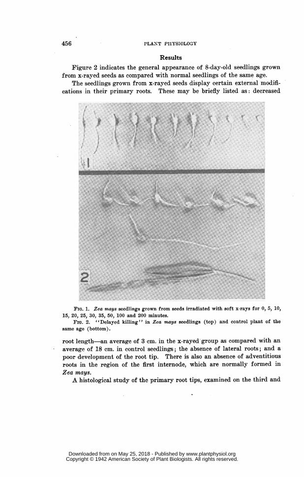
The radiation referred to as soft x-rays was supplied bv an apparatusdescribed in previous papers (3, 4, 5, 6). Unsoaked seeds of Zea mays wereoriented with their embrvo sides toward the source of x-ray radiation, 8 cm.from the focal spot of the x-ray tube. The tube was of the gas type, havinga copper target. The most intense part of its radiation consisted of theKa line (1.54 A). The dose of radiation selected was 30 peak kv., 10 ma.with an exposure time of 100 minutes. This treatment, according to pre-liminary tests, results in growth cessation during germinationi. Any ex-posure time over 30 minutes, unider these conditions, also results in thischaracteristic growth cessation but to a lesser extent. Variations in growthof seedlings germinated from seeds given increasing exposures to x-ravs,are given in figure 1.
Following irradiation, the seeds were germiniated in moist peat moss at850 F. The approximate period of germination before growth cessationbecomes apparent in treated seedlings is about 8 days. External ob-erva-tions were recorded for this time and histological studies were made of pri-mary roots of 3-, 5-, and 8-day-old seedlings. Fifty roots of each type wereexamined microscopically, usinlg standard methods of microtechiiique.
The characteristic irregularitv in arrangement and extent of tissue inseedlings grown from x-raved seeds, makes it quite impossible to secure his-tological sections without some distortion. This accounts for the irregu-larity in some illustrations of the x-rayed material presented throughout thepaper.
455
www.plantphysiol.orgon May 25, 2018 - Published by Downloaded from Copyright © 1942 American Society of Plant Biologists. All rights reserved.
456 PLANT PHYSIOLOGY
ResultsFigure 2 indicates the general appearance of 8-day-old seedlings grown
from x-rayed seeds as compared with normal seedlings of the same age.The seedlings grown from x-rayed seeds display certain external modifi-
cations in their primary roots. These may be briefly listed as: decreased
.::F .~~~~~~~~~~~~~~~~~~~~~~~~I
2:
FIG. 1. Zea mays seedlings grown from seeds irradiated with soft x-rays for 0, 5, 10,15, 20, 25, 30, 35, 50, 100 and 200 minutes.
Fi. 2. "Delayed killing" in Zea mays seedlings (top) and control plant of thesame age (bottom).
root length-an average of 3 cm. in the x-rayed group as compared with anaverage of 18 cm. in control seedlings; the absence of lateral roots; and apoor development of the root tip. There is also an absence of adventitiousroots in the region of the first internode, which are normally formed inZea mays.A histological study of the primary root tips, examined on the third and
www.plantphysiol.orgon May 25, 2018 - Published by Downloaded from Copyright © 1942 American Society of Plant Biologists. All rights reserved.
SMITH AND KERSTEN: ROOT MODIFICATIONS INDUCED BY X-RAYS 457
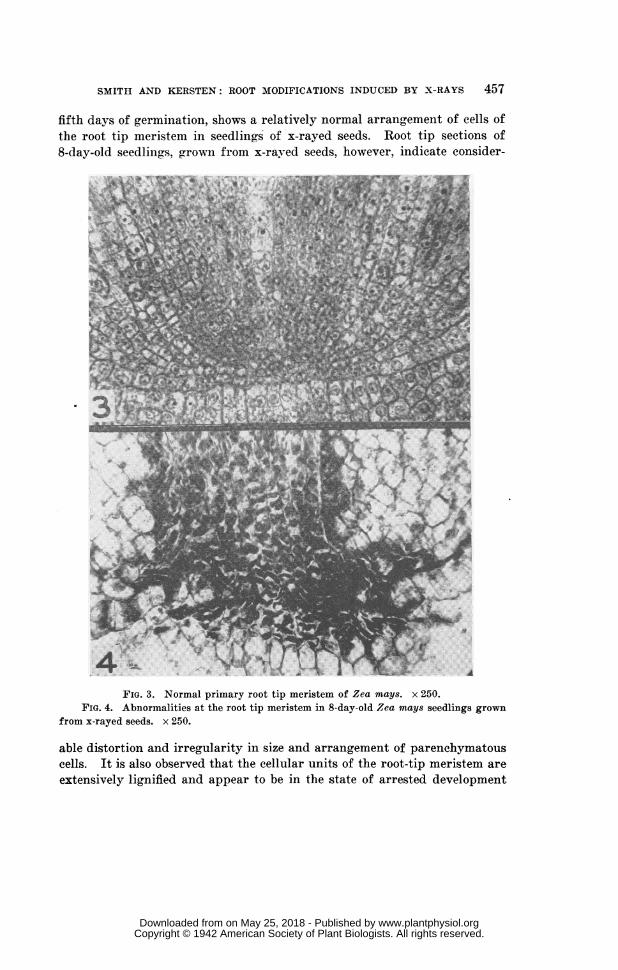
fifth days of germination, shows a relatively normal arrangement of cells ofthe root tip meristem in seedlings of x-rayed seeds. Root tip sections of8-day-old seedlingrs, grown from x-raved seeds, however, indicate consider-
FIG. 3. Normal primary root tip meristem of Zea mays. x 250.FIG. 4. Abnormalities at the root tip meristem in 8-day-old Zea mays seedlings grown
from x-rayed seeds. x 250.
able distortion and irregularity in size and arrangement of parenchymatouscells. It is also observed that the cellular units of the root-tip meristem areextensively lignified and appear to be in the state of arrested development
www.plantphysiol.orgon May 25, 2018 - Published by Downloaded from Copyright © 1942 American Society of Plant Biologists. All rights reserved.
PLANT PHYSIOLOGY
(fig. 4). The arrangement of the root tip meristem in normal Zea maysseedlings is represented in figure 3.
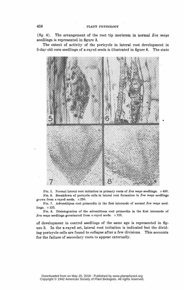
The extent of activity of the pericycle in lateral root development in5-dav-old corn seedlings of x-rayed seeds is illustrated in figure 6. The state
n_~~@~~~~'.~ ~ ~ ~ ~ TV.
X...::. a.7 ^\..t §$sk'; '. ... ..# w ;;>8.48~~~~4:
..7 ~~~~~~~ ~~~~
FIG. 5. Normal lateral root initiation in primary roots of zea mays seedlings. x 450.F9IG. 6. Breakdown of pericycle cells in lateral root formation in zea mays seedlings
grown from x-rayed seeds. x 250.FIG. 7. Adventitious root primordia in the first internode of normal zea mays seed-
lin1gs. X 125.FIG. 8. Disintegration of the adventitious root primordia in the first internode of
zea mays seedlings germinated from x-rayed seeds. x 125.
Of development inl control seedlings of the same age is represented in fig-ure 5. In the x-rayed set, lateral root initiation is indicated1 but the divid-ing pericycle cells are found to collapse after a few divisions. This accountsfor the failure of secondary roots to appear externally.
458
www.plantphysiol.orgon May 25, 2018 - Published by Downloaded from Copyright © 1942 American Society of Plant Biologists. All rights reserved.
SMITI AND KERSTEN: ROOT MODIFICATIONS INDUCED BY X-RAYS 459
The failure of adventitious root formatioln in the internodal reoions ofZea mays seedlings grown from x-rayed seeds is apparently related to thedisintegration of adventitious root primordia. This is shown in figure 8 asit is observed in the first internode of such seedlinogs. The normal forma-tion of these primordia is represented in figure 7.
An interesting series of observations on the root modifications in Zea
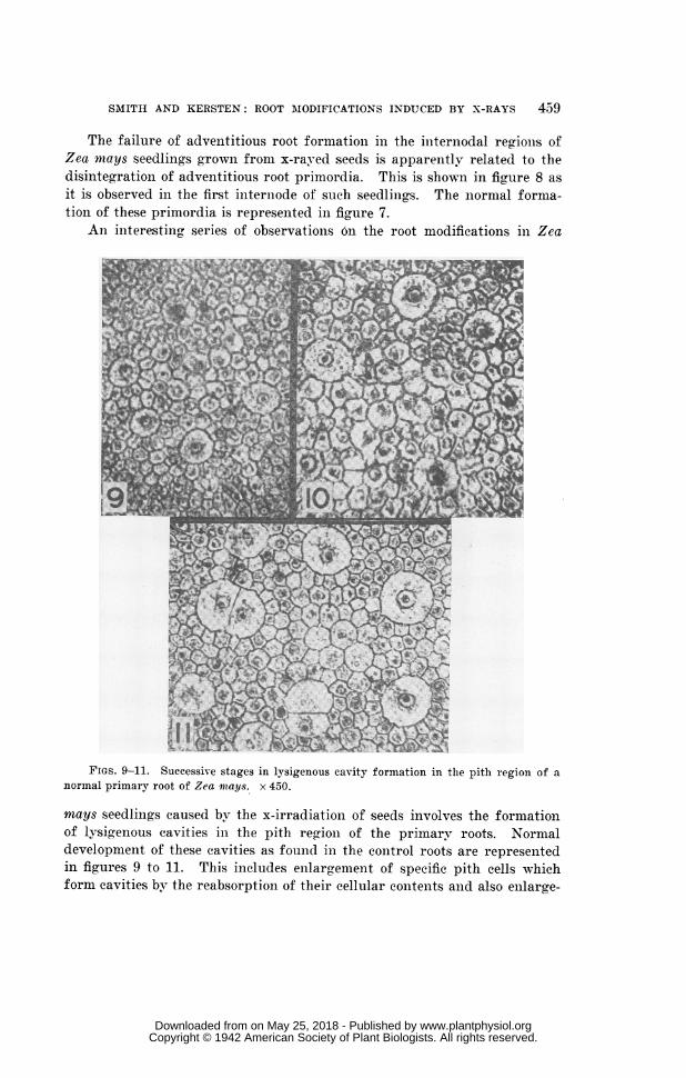
FIGS. 9-11. Successive stages in lysigenous cavity formation in the pith region of anormal primary root of Zea mays. x 450.
mays seedlings caused by the x-irradiationi of seeds involves the formationof lysig,enous cavities in the pith region of the primary roots. Normaldevelopment of these cavities as found in the control roots are representedin figures 9 to 11. This includes enlargement of specific pith cells whichform cavities by the reabsorption of their cellular contents and also enlarge-
www.plantphysiol.orgon May 25, 2018 - Published by Downloaded from Copyright © 1942 American Society of Plant Biologists. All rights reserved.
PLANT PHYSIOLOGY
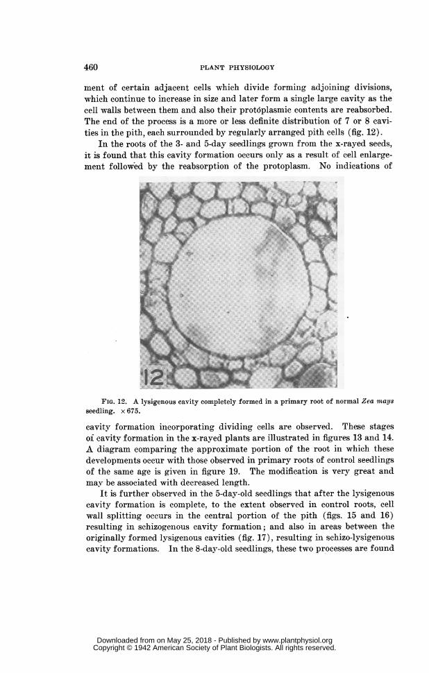
ment of certain adjacent cells which divide forming adjoining divisions,which continue to increase in size and later form a single large cavity as thecell walls between them and also their prot6plasmic contents are reabsorbed.The end of the process is a more or less definite distribution of 7 or 8 cavi-ties in the pith, each surrounded by regularly arranged pith cells (fig. 12).
In the roots of the 3- and 5-day seedlings grown from the x-rayed seeds,it is found that this cavity formation occurs only as a result of cell enlarge-ment followed by the reabsorption of the protoplasm. No indications of
| dP # 2X;; ge f~~~~~S Ri!!
FIG. 12. A lysigenous cavity completely formed in a primary root of normal Zea maysseedling. x 675.
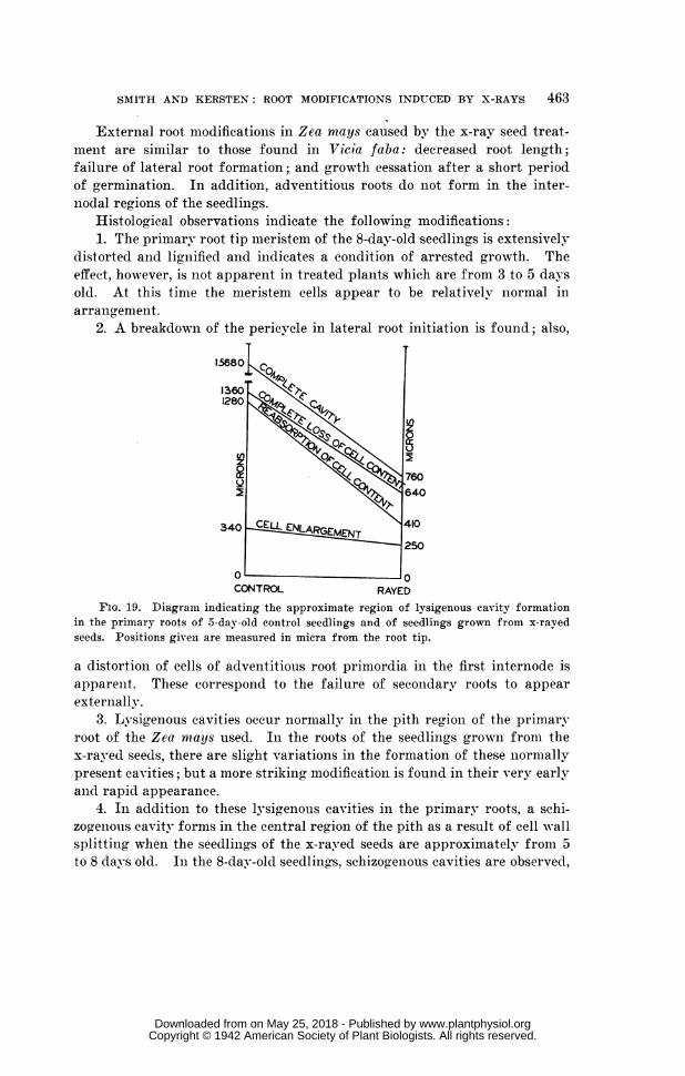
cavity formation incorporating dividing cells are observed. These stagesof cavity formation in the x-rayed plants are 'illustrated in figures 13 and 14.A diagram comparing the approximate portion of the root in which thesedevelopments occur with those observed in primary roots of control seedlingsof the same age is given in figure 19. The modification is very great andmay be associated with decreased length.
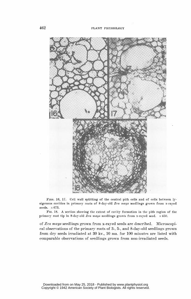
It is further observed in the 5-day-old seedlings that after the lysigenouscavity formation is complete, to the extent observed in control roots, cellwall splitting occurs in the central portion of the pith (figs.~15 and 16)resulting in schizogenous cavity formation; anid also in areasmbetween theoriginally formed lysigenous cavities (fig. 17), resulting in schizo-lysigenouscavity formations. In the 8-day-old seedlings, these two processes are found
460
www.plantphysiol.orgon May 25, 2018 - Published by Downloaded from Copyright © 1942 American Society of Plant Biologists. All rights reserved.

SMITH AND KERSTEN: ROOT MODIFICATIONS INDUCED BY X-RAYS 461
to involve the entire pith portion of the root in its mature region and fol-lowing this to extend downward to include the pith region of the root tip(fig. 18).
These observations are difficult to interpret. Variations in lysiaenous
FIGS. 13-15. Lysigenous cavity formation in primary roots of Zea mays seedlingsgrown from x-rayed seeds. Figure 15 shows the beginning of a schizogenous cavity in thepith. x 450.
cavity formation as compared with that in plants grown from non-irradiatedseeds, and the additional schizogenious and schizo-lysigenous cavities seemto be two separate effects of the x-ray irradiation.
SummaryExternal and histological modifications which occur in the primary roots
www.plantphysiol.orgon May 25, 2018 - Published by Downloaded from Copyright © 1942 American Society of Plant Biologists. All rights reserved.
PLANT PHYSIOLOGY
FIGs. 16, 17. Cell wall splittinig of the cenitral pith cells and of cells between ly-sigenious cavities in primary roots of 8-day-old Zea mitays seedlings growni from x-rayedseeds. x 675.
FIG. 18. A sectioni slhowing the extent of cavity formation in the pitlh region of theprimary root tip in 8-day-old Zea mays seedliings growni from x-rayed seed. x 450.
of Zea nmays seedlinfgs grown from x-rayed seeds are described. Microseopi-cal observations of the primary roots of 3-, 5-, anid 8-day-old seedlin(gs grownfrom dry seeds irradiated at 30 kv., 10 ma. for 100 miinutes are listed withcomparable observations of seedlings grown from noni-irradiated seeds.
462
www.plantphysiol.orgon May 25, 2018 - Published by Downloaded from Copyright © 1942 American Society of Plant Biologists. All rights reserved.
SMITH AND KERSTEN: ROOT MODIFICATIONS INDUCED BY X-RAYS 463
External root modifications in Zea ntays caused by the x-ray seed treat-ment are similar to those found in Vicia faba: decreased root length;failure of lateral root formation; and growth cessation after a short periodof germination. In addition, adventitious roots do not form in the inter-nodal regions of the seedlings.
Histological observations indicate the following modifications:1. The primary root tip meristem of the 8-day-old seedlings is extensively
distorted and lig,nified and indicates a condition of arrested growth. Theeffect, however, is not apparent in treated plants which are from 3 to 5 daysold. At this time the meristem cells appear to be relatively nlormal inarrangement.
2. A breakdown of the pericycle in lateral root initiation is found; also,
15680
13601280
760
640
340 CELL ENLARGEMENT 410
250
0 0CONTROL RAYED
FIG. 19. Diagraimi indicating the approximate region of lysigenous cavity formationin the primary roots of 5-day-old control seedlings and of seedlings grown from x-rayedseeds. Positions given are measured in micra from the root tip.
a distortion of cells of adventitious root primordia in the first internode isapparent. These correspond to the failure of secondary roots to appearexternally.
3. Lysigenous cavities occur normally in the pith region of the primaryroot of the Zea niays used. In the roots of the seedlings grown from thex-rayed seeds, there are slight variations in the formation of these lnormallypresent cavities; but a more striking modification is found in their very earlyand rapid appearance.
4. In addition to these lysigenous cavities in the primary roots, a schi-zogenous eavity forms in the central regioni of the pith as a result of cell wallsplitting when the seedlings of the x-raved seeds are approximatelv from 5to 8 days old. In the 8-day-old seedling-s, schizogeinous cavities are observed,
www.plantphysiol.orgon May 25, 2018 - Published by Downloaded from Copyright © 1942 American Society of Plant Biologists. All rights reserved.
PLANT PHYSIOLOGY
due to continuous cell wall splitting between the previously formed lysig-enous cavities and evidences of this cell wall splitting are found to extenddownward in the pith region to the lowermost part of the root tip.
5. It is not known whether factors involved in lysigenous and schizog-enous cavity formations are comparable. They seem to be separate effectsof the x-ray radiation treatment.
DEPARTMENTS OF BOTANY AND PHYSICSUNIVERSITY OF CINCINNATI
LITERATURE CITED1. DUGGAR, B. M. Biological effects of radiation. Vol. II. McGraw-Hill.
1936.2. HAYWARD, H. E. Structure of economic plants. Macmillan. 1938.3. KERSTEN, H. A gas x-ray tube for irradiation with soft x-rays. Radiol-
ogy 23: 6063. 1934.4. LONG, T. P., and KERSTEN, H. Structural changes produced in leaf tis-
sue of soy bean plants by irradiation of dry seeds with soft x-rays.Plant Physiol. 12: 191-197. 1937.
5. SMITH, G. F., and . Root modifications induced in Viciafaba by irradiating dry seeds with soft x-rays. Plant Physiol. 16:159-170. 1941.
6. SNIDER, G., and . The action of soft x-rays on Cladocera(Daphnia magna). Physiol. Zool. 8: 530-538. 1935.
464
www.plantphysiol.orgon May 25, 2018 - Published by Downloaded from Copyright © 1942 American Society of Plant Biologists. All rights reserved.