Modification of Thrombus Formation on Prosthetic Heart Valves by ...
6
Modification of Thrombus Formation on Prosthetic Heart Valves by the Administration of Low Molecular Weight Dextran LAWRENCE I. BONCHEK, M.D., NINA S. BRAuNwALD, M.D. From the Clinic of Surgery, National Heart Institute, Bethesda, Maryland 20014 THROMBUS formation on prosthetic car- diac valves, with or without emboli, is a major cause of postoperative morbidity and mortality in patients surviving valve re- placement, despite systemic anticoagula- tion. Herr et al.'2 recently reported a series of patients followed for at least 3 months postoperatively in whom the incidence of emboli was 42%o following isolated mitral valve replacement (27 of 65 patients), and 13%b following isolated aortic valve re- placement (12 of 90 patients). Since a sig- nificant number of patients die with throm- bosed prosthetic valves in the early postop- erative period, it is possible that many of the thrombi and emboli occurring after dis- charge from the hospital have their incep- tion in the first few postoperative days before systemic anticoagulation has been instituted or properly regulated. This study was performed in calves to assess the ef- fectiveness of low molecular weight dex- tran (LMWD) in preventing the formation of thrombus on prosthetic tricuspid valves during the immediate postoperative period. During the past decade, investigation of the properties of dextran have extended its use beyond its original application as a plasma expander. Gelin et al.9"10,11 dis- cussed the ability of LMWD to improve capillary flow in shock by diminishsing in- travascular sludging and by decreasing the viscosity of plasma and whole blood. Oth- ers describe the use of LMWD in the treat- Submitted for publication August 1, 1966. ment of frostbite16 and myocardial infarc- tion,13 in prevention of acute renal failure,2 and for pump-oxygenator priming in open- heart surgery.4"5 Numerous reports also describe the ability of dextran to prevent postoperative intravascular thrombosis af- ter arterial reconstructive surgery or ve- nous thrombectomy.5 7 Although these re- ports suggest a trial of LMWD in the pre- vention of prosthetic valve thrombosis, evaluation in a controlled experimental preparation has not been reported. Methods Fifteen healthy Holstein or Angus calves, weighing 61 to 74 Kg., were utilized. Anes- thesia was initiated with open-drop ether, and maintained with endotracheal halo- thane (0.5-1.5%) delivered by a mechani- cal respirator through a vaporizer in a non- rebreathing system. After catheters were inserted in the left femoral artery and vein for pressure measurements, and in the right femoral artery for arterial perfusion, the animal was placed in the left lateral decu- bitus position and the chest opened through the bed of the fifth rib. A disposable bub- ble oxygenator was employed, primed with 1,000 cc. heparinized bovine blood drawn aseptically 24 hours prior to operation, and 1,000 cc. 10% LMWD in saline or dextrose 5% in water (D5W) as discussed below. After the institution of cardiopulmonary bypass, a longitudinal right atriotomy was made and the normal tricuspid valve was 200
Transcript of Modification of Thrombus Formation on Prosthetic Heart Valves by ...
Modification of Thrombus Formation on ProstheticHeart Valves by the Administration of
Low Molecular Weight Dextran
LAWRENCE I. BONCHEK, M.D., NINA S. BRAuNwALD, M.D.
From the Clinic of Surgery, National Heart Institute, Bethesda, Maryland 20014
THROMBUS formation on prosthetic car-
diac valves, with or without emboli, is a
major cause of postoperative morbidity andmortality in patients surviving valve re-
placement, despite systemic anticoagula-tion. Herr et al.'2 recently reported a seriesof patients followed for at least 3 monthspostoperatively in whom the incidence ofemboli was 42%o following isolated mitralvalve replacement (27 of 65 patients), and13%b following isolated aortic valve re-
placement (12 of 90 patients). Since a sig-nificant number of patients die with throm-bosed prosthetic valves in the early postop-erative period, it is possible that many ofthe thrombi and emboli occurring after dis-charge from the hospital have their incep-
tion in the first few postoperative daysbefore systemic anticoagulation has beeninstituted or properly regulated. This studywas performed in calves to assess the ef-fectiveness of low molecular weight dex-tran (LMWD) in preventing the formationof thrombus on prosthetic tricuspid valvesduring the immediate postoperative period.During the past decade, investigation of
the properties of dextran have extended itsuse beyond its original application as a
plasma expander. Gelin et al.9"10,11 dis-cussed the ability of LMWD to improvecapillary flow in shock by diminishsing in-travascular sludging and by decreasing theviscosity of plasma and whole blood. Oth-ers describe the use of LMWD in the treat-
Submitted for publication August 1, 1966.
ment of frostbite16 and myocardial infarc-tion,13 in prevention of acute renal failure,2and for pump-oxygenator priming in open-heart surgery.4"5 Numerous reports alsodescribe the ability of dextran to preventpostoperative intravascular thrombosis af-ter arterial reconstructive surgery or ve-nous thrombectomy.5 7 Although these re-ports suggest a trial of LMWD in the pre-vention of prosthetic valve thrombosis,evaluation in a controlled experimentalpreparation has not been reported.
Methods
Fifteen healthy Holstein or Angus calves,weighing 61 to 74 Kg., were utilized. Anes-thesia was initiated with open-drop ether,and maintained with endotracheal halo-thane (0.5-1.5%) delivered by a mechani-cal respirator through a vaporizer in a non-rebreathing system. After catheters wereinserted in the left femoral artery and veinfor pressure measurements, and in the rightfemoral artery for arterial perfusion, theanimal was placed in the left lateral decu-bitus position and the chest opened throughthe bed of the fifth rib. A disposable bub-ble oxygenator was employed, primed with1,000 cc. heparinized bovine blood drawnaseptically 24 hours prior to operation, and1,000 cc. 10% LMWD in saline or dextrose5% in water (D5W) as discussed below.
After the institution of cardiopulmonarybypass, a longitudinal right atriotomy wasmade and the normal tricuspid valve was
200

THROMBUS FORMATION ON PROSTHETIC HEART VALVES
excised. A standard Melrose valve * wasinserted with 13 to 17 interrupted suturesof 2-0 silk or 2-0 Tevdek, and the atriotomyclosed. Following insertion of a tube forchest drainage and closure of the incisions,a flexible vinyl catheter was placed in theleft external jugular vein for postoperativeadministration of intravenous fluids. Oneanimal died of anesthetic complicationswithin 12 hours of operation and was ex-cluded from the study; 14 surviving calveswere divided into 2 groups.
In Group I, consisting of 7 calves, thepump oxygenator was primed with 1,000cc. of whole blood and 1,000 cc. of 10%oLMWD in saline. Postoperatively, eachcalf in this group received 2 Gm./Kg./day(approximately 1,400 cc.) of 10%o LMWDin saline by continuous intravenous infu-sion. Animal stalls were arranged so thatnormal activity was permitted despite in-travenous tubing.
In Group II, consisting of 7 calves, thepump oxygenator was primed with 1,000cc. of whole blood and 1,000 cc. of D5W.Three calves in this group received nopostoperative intravenous fluids. The other4 received 1,500 cc. a day of dextrose 5%in saline (D5S) by continuous intravenousinfusion.Each animal received 1,200,000 units of
intramuscular procaine penicillin and 1Gm. of streptomycin every 12 hours fromthe night before operation until sacrifice.All animals were given free access to grainand water after operation. In general, theanimals were able to stand and drink wateron the evening of operation and to eat anddrink normal amounts on the followingmorning. All animals that did not die dur-ing the first 3 postoperative days weresacrificed 72 hours postoperatively with anoverdose of intravenous pentobarbital andsuccinylcholine. Immediately prior to sacri-fice, each was given an intravenous injec-
tion of 3 mg./Kg. of heparin to prevent theformation of postmortem thrombus. Thechest was immediately opened, and thepresence or absence of wound hematomasor seromas and the amount of blood orfluid in the chest were recorded. The heartwas then excised, opened, and photo-graphed, and any thrombus present waspreserved in formaldehyde for microscopicexamination. Venous hematocrit was deter-
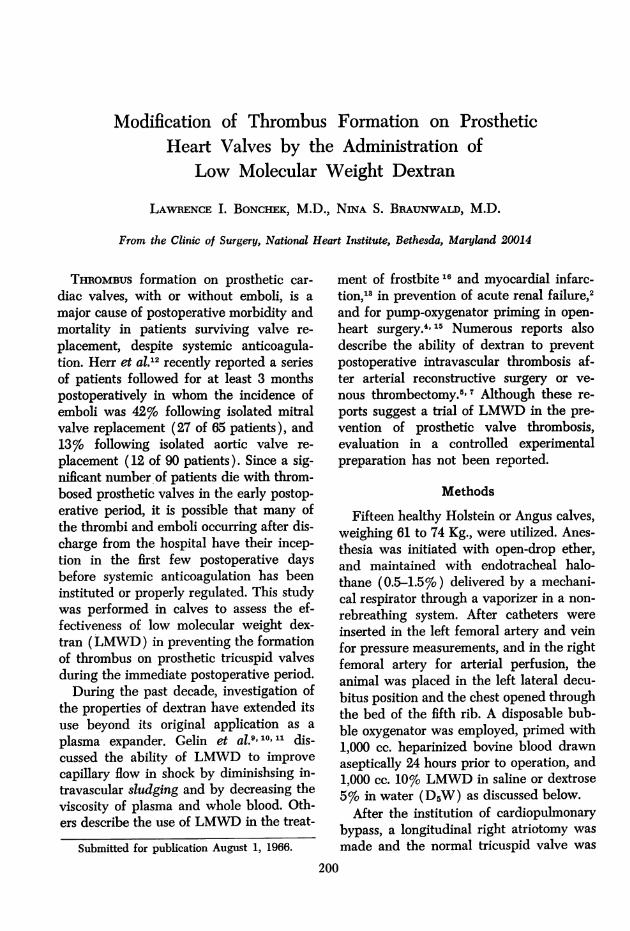
FIG. 1. Atrial view of tricuspid valve prosthesesfrom 2 of the 7 calves that received low molecularweight dextran and were sacrificed 72 hours afteroperation. At the time of sacrifice, 6 valves hadno thrombus, and one had a moderate amountwhich did not interfere with valve function.
Volume 165Number 2 201
* Kindly provided by Mr. Hugh H. Bentall,F.R.C.S.

BONCHEK AND BRAUNWALD Annals of SurgeryFebruary 1967
Six animals in this group had no thrombuson the prosthetic valves at the time (Fig.1). The remaining animal had a moderateamount of white, fibrinous deposit on theatrial side of the valve, sewing ring, andflange that did not interfere with valvefunction.
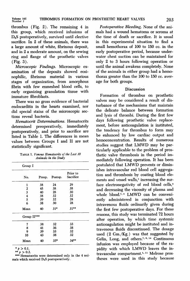
In Group II, the 3 animals that receivedno postoperative infusion died within 48hours of operation with complete occlusionof the prosthetic valves by fresh, red
FIG. 2. Atrial view of tricuspid valve prosthesesfrom 2 of the 3 calves that received no postopera-tive intravenous infusion. All three calves in thisgroup died within 48 hours of operation withvalves occluded by red thrombus.
mined pre- and postoperatively and imme-diately prior to sacrifice in the last 10 ani-mals in the study, by withdrawing bloodfrom the femoral or jugular veins and cen-
trifuging it in heparinized microhematocrittubes.
Results
Thrombus on Valves. All animals inGroup I appeared in good health at thetime of sacrifice, 72 hours postoperatively.
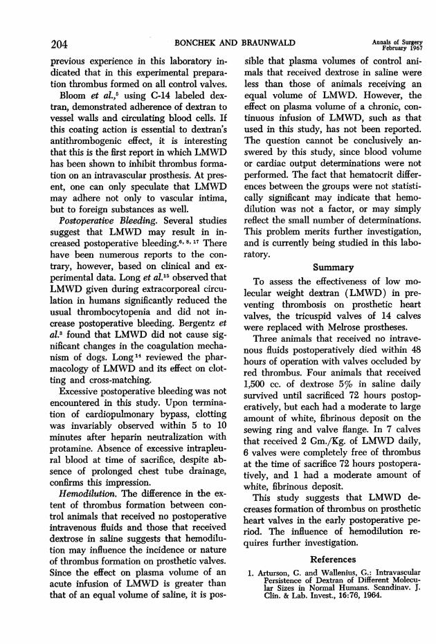
FIG. 3. Atrial view of tricuspid valve prosthesesfrom 2 of the 4 calves that received dextrose insaline and survived until sacrificed 72 hours afteroperation. All of the valves in this group demon-strated thrombus in varying amounts at the timeof sacrifice,
202
THROMBUS FORMATION ON PROSTHETIC HEART VALVES
thrombus (Fig. 2). The remaining 4 inthis group, which received infusions ofD5S postoperatively, survived until electivesacrifice. In 2 of these animals, there was
a large amount of white, fibrinous deposit,and in 2 a moderate amount, on the sewingring and flange of the prosthetic valves(Fig. 3).Microscopic Findings. Microscopic ex-
amination of the deposits showed eosi-nophilic, fibrinous material in variousstages of organization, from amorphousfibrin with few enmeshed blood cells, toearly organizing granulation tissue withimmature fibroblasts.There was no gross evidence of bacterial
endocarditis in the hearts examined, nor
did special stains of the microscopic sec-
tions reveal bacteria.Hematocrit Determinations. Hematocrits
determined preoperatively, immediatelypostoperatively, and prior to sacrifice are
listed in Table 1. The differences in mean
values between Groups I and II are notstatistically significant.
TABLE 1. Venous Hematocrits of the Last 10Animals in the Study
Group I
Prior toNo. Preop. Postop. Sacrifice
1 38 24 292 45 38 383 40 28 304 38 32 325 39 32 286 38 30 28
Mean 40 31* 31**
Group II***
7 38 32 338 41 36 389 39 33 3210 43 38 32
Mean 40 35* 34**
*p > 0.1.**p > 0.2.*** Hematocrits were determined only in the 4 ani-
mals which received DrS postoperatively.
Postoperative Bleeding. None of the ani-mals had a wound hematoma or seroma at
the time of death or sacrifice. It is usualin this experimental situation to find a
small hemothorax of 100 to 150 cc. in theearly postoperative period, because under-water chest suction can be maintained foronly 2 to 3 hours following operation or
until the animal awakens completely. Noneof the animals in either group had a hemo-thorax greater than the 100 to 150 cc. aver-
age for both groups.
DiscussionFormation of thrombus on prosthetic
valves may be considered a result of dis-turbance of the mechanisms that maintainthe delicate balance between formationand lysis of thrombi. During the first fewdays following prosthetic valve replace-ment, before anticoagulation is instituted,the tendency for thrombus to form may
be enhanced by low cardiac output andhemoconcentration. Results of numerous
studies suggest that LMWD may be par-
ticularly applicable to the problem of pros-
thetic valve thrombosis in the period im-mediately following operation. It has beenpostulated that LMWD prevents or dimin-ishes intravascular red blood cell aggrega-
tion and thrombosis by coating blood ele-ments and vessel walls,5 increasing the sur-
face electronegativity of red blood cells,4and decreasing the viscosity of plasma andwhole blood.7 9 LMWD can be conveni-ently administered in conjunction withintravenous fluids ordinarily given duringthe first few postoperative days. For thesereasons, this study was terminated 72 hoursafter operation, by which time systemicanticoagulation might be instituted and in-travenous fluids discontinued. The dosageused (2 Gm./Kg.) was that suggested byGelin, Long, and others.7'9 14 Continuousinfusion was employed because of the ra-
pidity with which LMWD leaves the in-travascular compartment.1," Melrose pros-
theses were used in this study because
Volume 16SNumber 2 203
204 BONCHEK AND BRAUNWALD February196S7previous experience in this laboratory in-dicated that in this experimental prepara-tion thrombus formed on all control valves.Bloom et al.,5 using G-14 labeled dex-
tran, demonstrated adherence of dextran tovessel walls and circulating blood cells. Ifthis coating action is essential to dextran'santithrombogenic effect, it is interestingthat this is the first report in which LMWDhas been shown to inhibit thrombus forma-tion on an intravascular prosthesis. At pres-ent, one can only speculate that LMWDmay adhere not only to vascular intima,but to foreign substances as well.
Postoperative Bleeding. Several studiessuggest that LMWD may result in in-creased postoperative bleeding.8' 8,17 Therehave been numerous reports to the con-trary, however, based on clinical and ex-perimental data. Long et al.'5 observed thatLMWD given during extracorporeal circu-lation in humans significantly reduced theusual thrombocytopenia and did not in-crease postoperative bleeding. Bergentz etal.3 found that LMWD did not cause sig-nificant changes in the coagulation mecha-nism of dogs. Long14 reviewed the phar-macology of LMWD and its effect on clot-ting and cross-matching.
Excessive postoperative bleeding was notencountered in this study. Upon termina-tion of cardiopulmonary bypass, clottingwas invariably observed within 5 to 10minutes after heparin neutralization withprotamine. Absence of excessive intrapleu-ral blood at time of sacrifice, despite ab-sence of prolonged chest tube drainage,confirms this impression.
Hemodilution. The difference in 'the ex-tent of thrombus formation between con-trol animals that received no postoperativeintravenous fluids and those that receiveddextrose in saline suggests that hemodilu-tion may influence the incidence or natureof thrombus formation on prosthetic valves.Since the effect on plasma volume of anacute infusion of LMWD is greater thanthat of an equal volume of saline, it is pos-
sible that plasma volumes of control ani-mals that received dextrose in saline wereless than those of animals receiving anequal volume of LMWD. However, theeffect on plasma volume of a chronic, con-tinuous infusion of LMWD, such as thatused in this study, has not been reported.The question cannot be conclusively an-swered by this study, since blood volumeor cardiac output determinations were notperformed. The fact that hematocrit differ-ences between the groups were not statisti-cally significant may indicate that hemo-dilution was not a factor, or may simplyreflect the small number of determinations.This problem merits further investigation,and is currently being studied in this labo-ratory.
SummaryTo assess the effectiveness of low mo-
lecular weight dextran (LMWD) in pre-venting thrombosis on prosthetic heartvalves, the tricuspid valves of 14 calveswere replaced with Melrose prostheses.Three animals that received no intrave-
nous fluids postoperatively died within 48hours of operation with valves occluded byred thrombus. Four animals that received1,500 cc. of dextrose 5% in saline dailysurvived until sacrificed 72 hours postop-eratively, but each had a moderate to largeamount of white, fibrinous deposit on thesewing ring and valve flange. In 7 calvesthat received 2 Gm./Kg. of LMWD daily,6 valves were completely free of thrombusat the time of sacrifice 72 hours postopera-tively, and 1 had a moderate amount ofwhite, fibrinous deposit.
This study suggests that LMWD de-creases formation of thrombus on prostheticheart valves in the early postoperative pe-riod. The influence of hemodilution re-quires further investigation.
References1. Arturson, G. and Wallenius, G.: Intravascular
Persistence of Dextran of Different Molecu-lar Sizes in Normal Humans. Scandinav. J.Clin. & Lab. Invest., 16:76, 1964.
Volume 165 THROMBUS FORMATION ON PROSTHETIC HEART VALVES 205Number 2202. Atik, M. and Gutierrez-Saenz, R.: Production
of Acute Nephrosis by Incompatible BloodTransfusion and the Protective Effect of LowMolecular Weight Dextran. Surg. Forum,XII:48, 1961.
3. Bergentz, S. E., Eiken, 0. and Nilsson, I. M.:The Effect of Dextran of Various MolecularWeights on Coagulation in Dogs. Thromb.Diath. Haem., 6:15, 1961.
4. Bernstein, E. F., Emmings, F. G., Evans,R. L., Castaneda, A. and Varco, R. L.: Ef-fect of Low Molecular Weight Dextran onRed Blood Cell Charge during Clinical Ex-tracorporeal Circulation. Circulation, 27:816,1963.
5. Bloom, W. L., Harmer, D. S., Bryant, M. F.and Brewer, S. S.: Coating of Vascular Sur-faces and Cells. A New Concept in Preven-tion of Intravascular Thrombosis. Proc. Soc.Exper. Biol. & Med., 115:384, 1964.
6. Breckenridge, J. M. and Walker, W. F.: BloodLoss in Open-Heart Surgery with Low Mo-lecular Weight Dextran. Lancet, 1:1190,1963.
7. Bryant, M. F., Brewer, S. S. and Bloom,W. L.: Use of Dextran in Preventing Throm-bosis of Small Arteries. Clin. Res., 10:49,1962.
8. Garzon, A. A., Fink, L. H., Shaftan, G. W.and Karlson, K. E.: Blood Loss Associatedwith Administration of Low MolecularWeight Dextran. J. Thorac. Cardiov. Surg.,48:873, 1964.
9. Gelin, L. E. and Ingelman, B.: Rheomacrodex-A New Dextran Solution for RheologicalTreatment of Impaired Capillary Flow. ActaChir. Scandinav., 122:294, 1961.
10. Gelin, L. E., Korsan-Bengtsen, K., Ygge, J.and Zederfeldt, B.: Influence of Low ViscousDextran on the Hemostatic Mechanism. Acta.Chir. Scandinav., 122:324, 1961.
11. Gelin, L. E., Solvell, L. and Zederfeldt, B.:The Plasma Volume Expanding Effect ofLow Viscous Dextran and Macrodex. Acta.Chir. Scandinav., 122:309, 1961.
12. Herr, R., Starr, A., McCord, C. W. and Wood,J. A.: Special Problems Following ValveReplacement. Ann. Thor. Surg., 1:403, 1965.
13. Langsjoen, P. H., Falconer, H. S., Sanchez,S. A. and Lynch, D. J.: Observations inTreatment of Acute Myocardial Infarctionwith Low Molecular Dextran. Angiology,14:465, 1963.
14. Long, D. M.: Pharmacology of LMWD andits Effect on Clotting and Crossmatching.In Eiseman, B. and Bosomworth, P. (Ed.):Evaluation of Low Molecular Weight Dex-tran in Shock: Pharmacology and PertinentRheology. Washington, D. C., NationalAcademy of Sciences, 1963.
15. Long, D. M., Sanchez, L., Varco, R. L. andLillehei, C. W.: The Use of Low MolecularWeight Dextran and Serum Albumin asPlasma Expanders in Extracorporeal Circu-lation. Surgery, 50:12, 1961.
16. Mundth, E. D., Long, D. M. and Brown,R. B.: Treatment of Experimental Frostbitewith Low Molecular Weight Dextran. J.Trauma, 4:246, 1964.
17. Smith, B., Omari, A., Melrose, D. G. andBentall, H. H.: Blood Loss after Extra-corporeal Circulation. Surg. Forum, XIV:271, 1963.