Modeling of neuron organization in the CA1 region of rat brain with a
6
Modeling of neuron organization in the CA1 region of rat brain with a helical dendrite structure Sten Andersson Sandforsk, Institute of Sandvik S-38074 Löttorp, Sweden www.sandforsk.se Kåre Larsson KL Chem AB, 23734 Bjärred and Camurus Lipid Research S-22370 Lund, Sweden Marcus Larsson Lund University Children’s Hospital S-22185 Lund, Sweden John C Fiala Department of Biology, Boston University Boston, MA 02215 USA [email protected] Abstract Detailed studies of serial sections of the CA1 region statum radiatum of a 21-day-old rat give two finite structure of neuron packings. One is a double helical arrangement of dendrites which is penetrated by axons, and the other is a distorted bcc arrangement of rods. Introduction In modeling neuron organization we recently used rod packing(ref 1). We now also wish to report an extraordinary case of helical arrangements of neurons as found in two reconstructions of 94 sections from the CA1 region statum radiatum of a 21-day-old rat(ref 2). The D3 volume (45 Mb animated gif) and its double helix During a study of the serial sections from ref 2, the D3 volume, it was clear that two dendrites, D3 and D21, projected after their length moved in a circular orbit. As the two dendrites had opposite positions on the circle, it was equally clear that the motion reflected a double helix structure in 3D. This is shown in section 0 of the volume in Fig 1a, and in section 54 in Fig1b, where the same dendrites are on the way to complete a semicircle.
Transcript of Modeling of neuron organization in the CA1 region of rat brain with a

Modeling of neuron organization in the CA1 region ofrat brain with a helical dendrite structure
Sten AnderssonSandforsk, Institute of SandvikS-38074 Löttorp, Swedenwww.sandforsk.se
Kåre LarssonKL Chem AB, 23734 Bjärred and Camurus Lipid ResearchS-22370 Lund, Sweden
Marcus LarssonLund University Children’s HospitalS-22185 Lund, Sweden
John C FialaDepartment of Biology, Boston UniversityBoston, MA 02215 [email protected]
Abstract
Detailed studies of serial sections of the CA1 region statum radiatum of a21-day-old rat give two finite structure of neuron packings. One is a doublehelical arrangement of dendrites which is penetrated by axons, and the otheris a distorted bcc arrangement of rods.
Introduction
In modeling neuron organization we recently used rod packing(ref 1). Wenow also wish to report an extraordinary case of helical arrangements ofneurons as found in two reconstructions of 94 sections from the CA1 regionstatum radiatum of a 21-day-old rat(ref 2).
The D3 volume (45 Mb animated gif) and its double helix
During a study of the serial sections from ref 2, the D3 volume, it was clearthat two dendrites, D3 and D21, projected after their length moved in acircular orbit. As the two dendrites had opposite positions on the circle, itwas equally clear that the motion reflected a double helix structure in 3D.
This is shown in section 0 of the volume in Fig 1a, and in section 54 inFig1b, where the same dendrites are on the way to complete a semicircle.

Fig 1a Section 0.
Fig 1b Section 54. The dendrites on the way to complete a semicircle
The 3D description of this motion was derived in a mathematical modelusing the exponential scale method which was described in some detail in ref1. The original publication to introduce the exponential scale is given in ref4. Ref 5 also gives a detailed description on the application of the method.
In equation[1] the double helix part comes from the geometry of the DNAmolecule(ref 3). The axons as shown in the Fig 2a were given arbitrarypositions to optimise close packing through the helix. Comparing to theneuron organization in the reconstruction show a remarkable resemblence asshown with the numbering of corresponding elements.

A 3D reconstruction(ref 2) of animation data in the CA1 region statumradiatum of a 21-day-old rat as shown in Fig 2b clearly support the doublehelix model.
†
e-[-xycos1/4pz + 1/2x2sin1/4pz - 1/2y2sin1/4pz + e1/4(x2+ y2 )]
+
e-75((x)2 + (z - 6)2 )2+ e-50((x - y)2 + (z - 4)2 )2
+
e-75((y)2 + (z - 2)2 )2+ e-50((x + y)2 + (z)2 )2
+ e-75((x)2 + (z + 2)2 )2+
e-50((1/2x - y + z - 5)2 + (1/2x + y + z - 9)2 )2+ e-50((-x - 1/2y + z + 2.5)2
+ (x - 1/2y + z - 6.5)2 )2+
e-50((-1/2x + y + z)2+ (-1/2x - y + z - 4)2 )2
+ e-50((x + 1/2y + z + 2.5)2 + (-x + 1/2y + z - 1.5)2 )2+
e-50((-1/2x + y + z - 5)2 + (-1/2x - y + z - 9)2 )2+ e-50((x + 1/2y + z - 2.5)2 + (-x + 1/2y + z - 6.5)2 )2
+
e-50((1/2x - y + z)2+ (1/2x + y + z - 4)2 )2
+ e-50((-x - 1/2y + z + 2.5)2 + (x - 1/2y + z - 1.5)2 )2+
e-100(1/2y + x)2 + (z - 5)2 )2+ e-100(y - 1/2x + 1)2 + (z + 1)2 )2
+ e-100(y - 1/2x + 1)2 + (z - 7)2 )2
-2/5 = 0
Fig 2a. Mathematical model. Fig2b. A 3D reconstruction(ref 2) of
volume data.
In Fig 3 we give another example of a double helix penetrated by rods.

Fig 3 Another view of a double helix.
Rod packing in the D3 volume
Fig 4a below is from the volume D3-D21 at 17 %. Very similar with fig 6ain ref 1, and also fig 3 in ref 1, which however only had axons. There aremore dendrites that belong to this cluster that could have contacts with thisgroup of axons. One thick axon, seen in section 15, that has contact with thedendrite situated up right in picture extends all the way to the D3-D21double helix, and also to a three fold arrangement as visible at 16-17%(shown below), which is close to the D3 – D21 double helix. One of theaxons touches the two helix dendrites, the other two go through the helix andtouches,one by one, one of its dendrites. Easy to see.
The structure is a trigonal distortion of body- centered cubic packing of rods,due to different sizes of rods, axons and dendrites together forming thestructure. The structure in Fig 4b is a part of a model , two meters in heightshown in fig 5.
Fig 4a Three axons and two dendrites form a structure at section 17 b. Rod representation
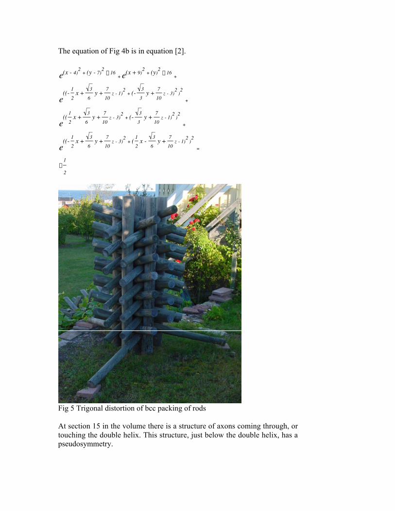
The equation of Fig 4b is in equation [2].
†
e(x - 4)2 + (y - 7)2- 16
+ e(x + 9)2 + (y)2 - 16+
e((- 1
2x + 3
6y + 7
10z - 1)2
+ (- 3
3y + 7
10z - 3)2 )2
+
e(( 1
2x + 3
6y + 7
10z - 3)2
+ (- 3
3y + 7
10z - 1)2 )2
+
e((- 1
2x + 3
6y + 7
10z - 3)2
+ ( 1
2x - 3
6y + 7
10z - 1)2 )2
=
-1
2
Fig 5 Trigonal distortion of bcc packing of rods
At section 15 in the volume there is a structure of axons coming through, ortouching the double helix. This structure, just below the double helix, has apseudosymmetry.

Obviously what is shown is an example how axons end, here via a synaps.Shown in Fig 6
Fig 6. Structure with pseudosymmetry at section 15. Six axons seem to meetdown to right.
References
1 S. Andersson, K. Larsson,M . Larsson and John C Fiala,Modeling neuron organization: a case of rod packing.http://synapses.mcg.edu/anatomy/modeling/modeling.stm
2 John C. Fiala. http://synapses.bu.edu/samples/samples.htm
3 M. Jacob, J.Phys. II France 7 (1997) 1035-1044.
4 S. Andersson, M. Jacob and S. Lidin, Z. Kristallogr. 210, 3 (1995).
5 S. Andersson, K. Larsson, M . Larsson , and M. Jacob,BIOMATHEMATICS, Elsevier, 1999.