Modeling of MR-guided HIFU for Breast and Brain Therapy
42
Utah Center for Advanced Imaging Research Douglas A. Christensen, Allison Payne, Nick Todd, Scott Almquist, Alexis Farrer and Dennis L. Parker University of Utah Salt Lake City, Utah Modeling of MR-guided HIFU for Breast and Brain Therapy
Transcript of Modeling of MR-guided HIFU for Breast and Brain Therapy
Utah Center for Advanced Imaging Research
Douglas A. Christensen, Allison Payne,
Nick Todd, Scott Almquist, Alexis Farrer and Dennis L. Parker
University of Utah
Salt Lake City, Utah
Modeling of MR-guided HIFU for
Breast and Brain Therapy
Utah Center for Advanced Imaging Research
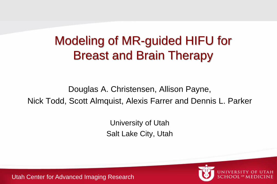
Overview
• High-Intensity Focused Ultrasound (HIFU) Surgery
– Critical needs: locating the beam; full 3D temperature images;
accurate beam modeling
• Beam Modeling with Hybrid Angular Spectrum (HAS)
Method
• Application to Brain and Breast
– Phase aberration correction
– Incorporating absorption and scattering
Utah Center for Advanced Imaging Research
coronal
sagittal
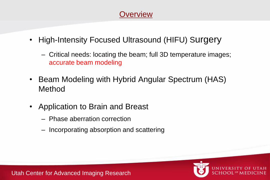
Critical Need 1: Locating Beam
Use Acoustic Radiation Force Imaging (ARFI) with MRI
at geometric focus in phantom:
ARFI ARFI temperature temperature
steered 5,5,5 mm in phantom:
Utah Center for Advanced Imaging Research
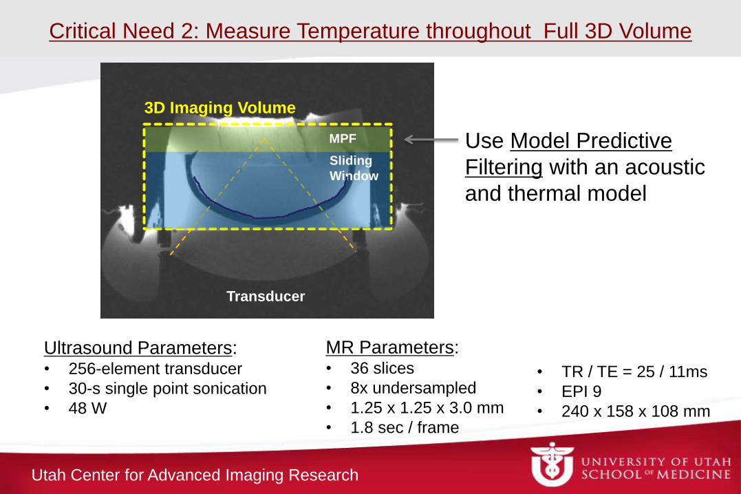
Ultrasound Parameters: • 256-element transducer
• 30-s single point sonication
• 48 W
MR Parameters: • 36 slices
• 8x undersampled
• 1.25 x 1.25 x 3.0 mm
• 1.8 sec / frame
• TR / TE = 25 / 11ms
• EPI 9
• 240 x 158 x 108 mm
Transducer
3D Imaging Volume
MPF
Sliding
Window
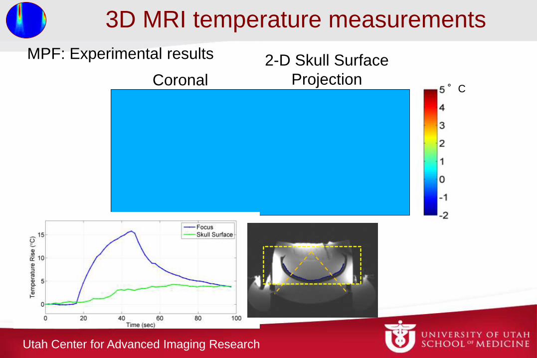
Critical Need 2: Measure Temperature throughout Full 3D Volume
Use Model Predictive
Filtering with an acoustic
and thermal model
Utah Center for Advanced Imaging Research
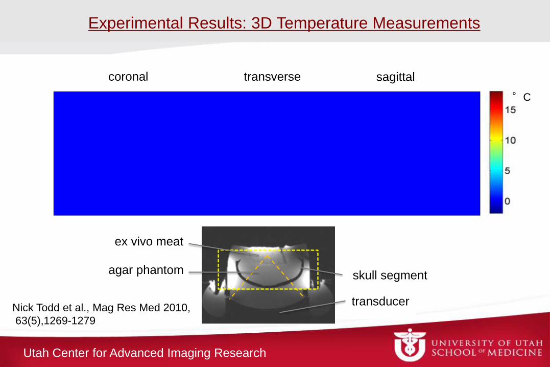
skull segment
coronal transverse sagittal
°C
Experimental Results: 3D Temperature Measurements
agar phantom
ex vivo meat
transducer Nick Todd et al., Mag Res Med 2010,
63(5),1269-1279
Utah Center for Advanced Imaging Research
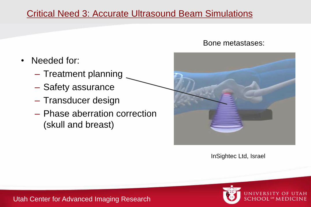
Critical Need 3: Accurate Ultrasound Beam Simulations
• Needed for:
– Treatment planning
– Safety assurance
– Transducer design
– Phase aberration correction
(skull and breast)
6
InSightec Ltd, Israel
Bone metastases:
Utah Center for Advanced Imaging Research
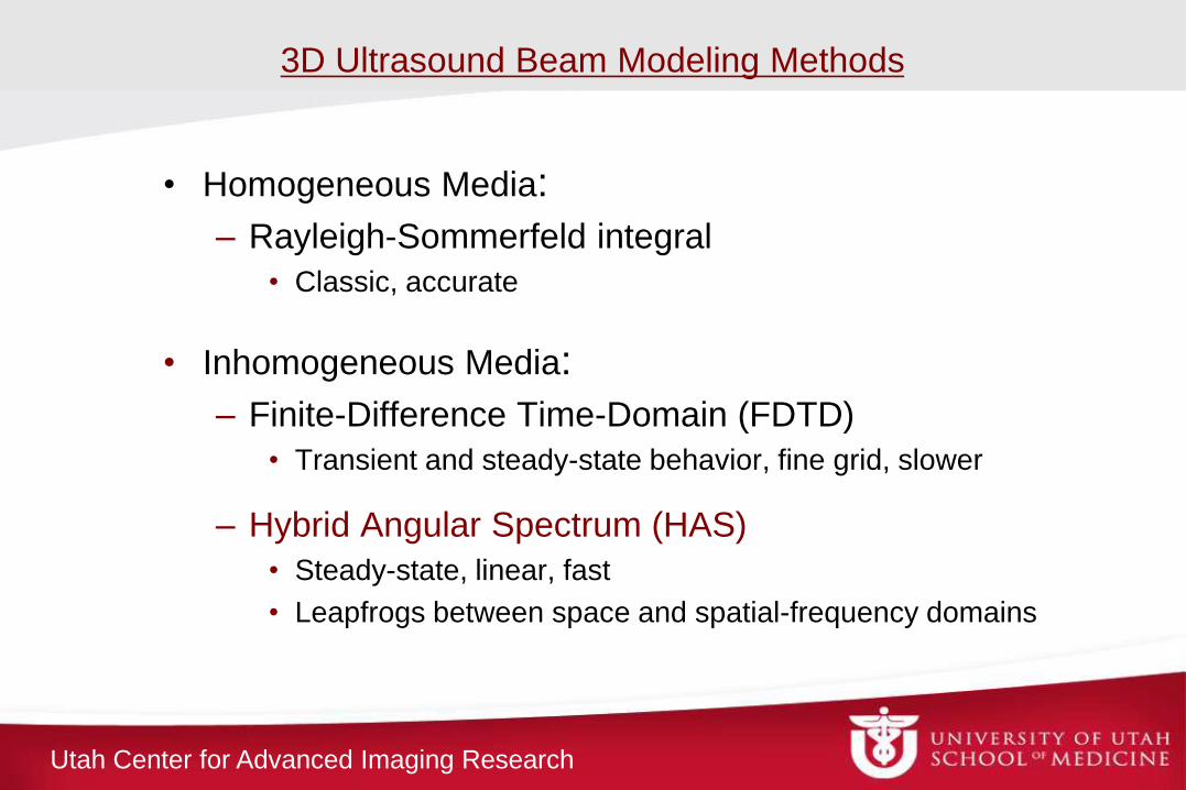
• Homogeneous Media:
– Rayleigh-Sommerfeld integral
• Classic, accurate
• Inhomogeneous Media:
– Finite-Difference Time-Domain (FDTD)
• Transient and steady-state behavior, fine grid, slower
– Hybrid Angular Spectrum (HAS)
• Steady-state, linear, fast
• Leapfrogs between space and spatial-frequency domains
3D Ultrasound Beam Modeling Methods
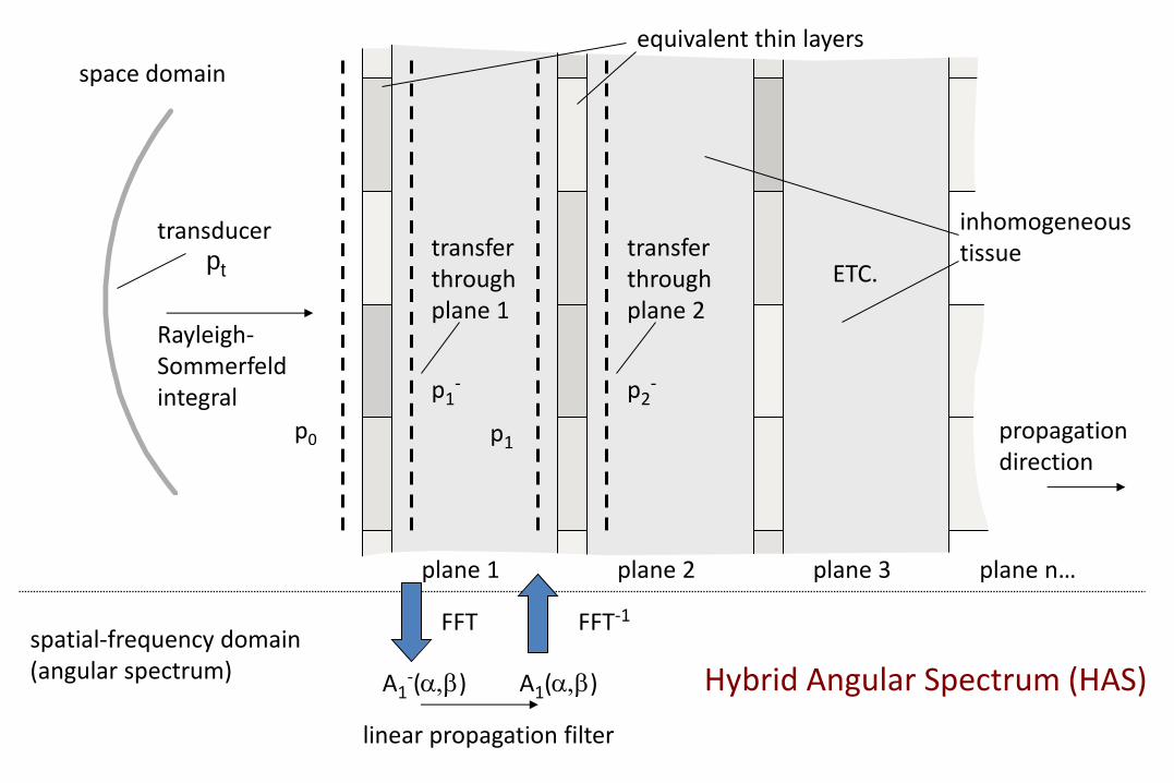
space domain
spatial-frequency domain (angular spectrum)
plane 1 plane 2 plane 3 plane n …
propagation direction
Rayleigh- Sommerfeld integral
p0
plane 1 plane 2 plane 3 plane n…
equivalent thin layers
p1-
transfer through plane 1
FFT
A1-(a,b)
linear propagation filter
A1(a,b)
FFT-1
p1
p2-
transfer through plane 2
ETC.
transducer pt
inhomogeneous tissue
Hybrid Angular Spectrum (HAS)
Utah Center for Advanced Imaging Research
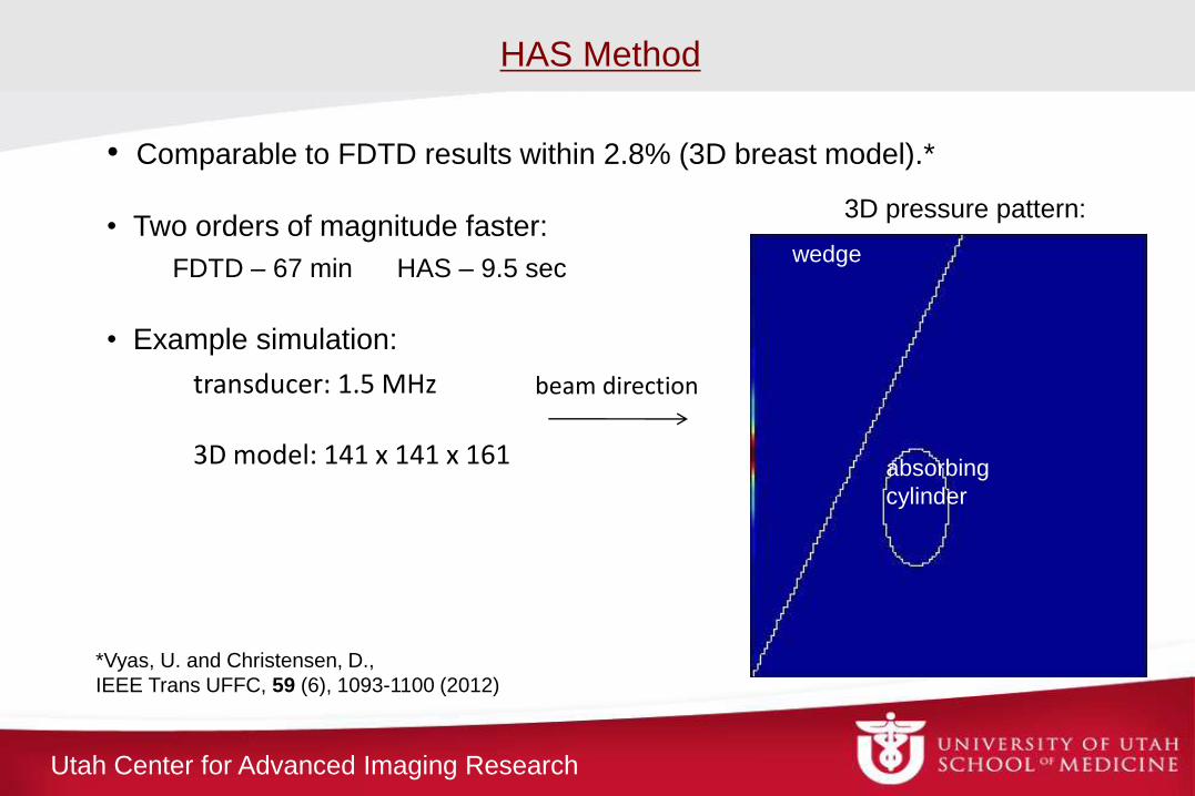
HAS Method
*Vyas, U. and Christensen, D.,
IEEE Trans UFFC, 59 (6), 1093-1100 (2012)
transducer: 1.5 MHz 3D model: 141 x 141 x 161
beam direction
• Comparable to FDTD results within 2.8% (3D breast model).*
• Two orders of magnitude faster:
FDTD – 67 min HAS – 9.5 sec
• Example simulation: wedge
absorbing
cylinder
3D pressure pattern:
wedge
Utah Center for Advanced Imaging Research
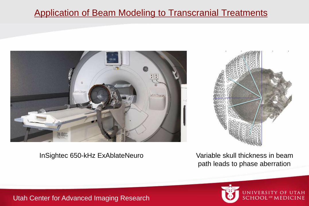
Application of Beam Modeling to Transcranial Treatments
InSightec 650-kHz ExAblateNeuro Variable skull thickness in beam
path leads to phase aberration
Utah Center for Advanced Imaging Research
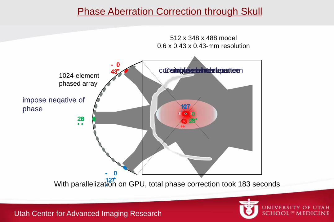
With parallelization on GPU, total phase correction took 183 seconds
512 x 348 x 488 model
0.6 x 0.43 x 0.43-mm resolution
1024-element
phased array
Combine all elements single element pattern
impose neqative of
phase
constructive interference
Phase Aberration Correction through Skull
Utah Center for Advanced Imaging Research
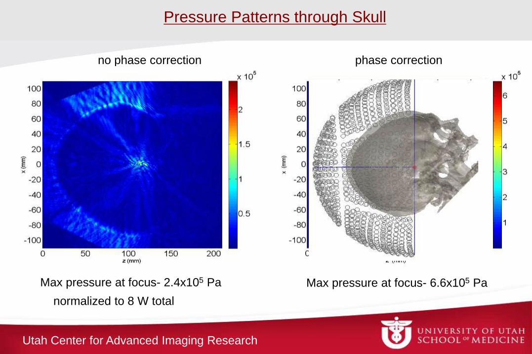
no phase correction phase correction
Max pressure at focus- 2.4x10⁵ Pa
normalized to 8 W total
Max pressure at focus- 6.6x10⁵ Pa
Pressure Patterns through Skull
Utah Center for Advanced Imaging Research
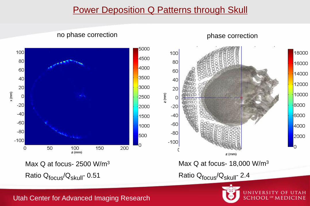
no phase correction phase correction
Ratio Qfocus/Qskull- 0.51
Max Q at focus- 2500 W/m3
Ratio Qfocus/Qskull- 2.4
Max Q at focus- 18,000 W/m3
Power Deposition Q Patterns through Skull
Utah Center for Advanced Imaging Research
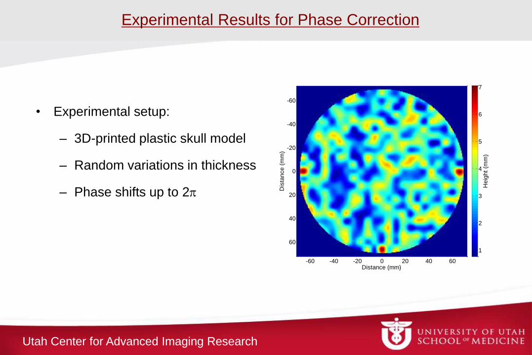
Experimental Results for Phase Correction
• Experimental setup:
– 3D-printed plastic skull model
– Random variations in thickness
– Phase shifts up to 2p
Distance (mm)
Dis
tance (
mm
)
-60 -40 -20 0 20 40 60
-60
-40
-20
0
20
40
60
Heig
ht
(mm
)
1
2
3
4
5
6
7
Utah Center for Advanced Imaging Research
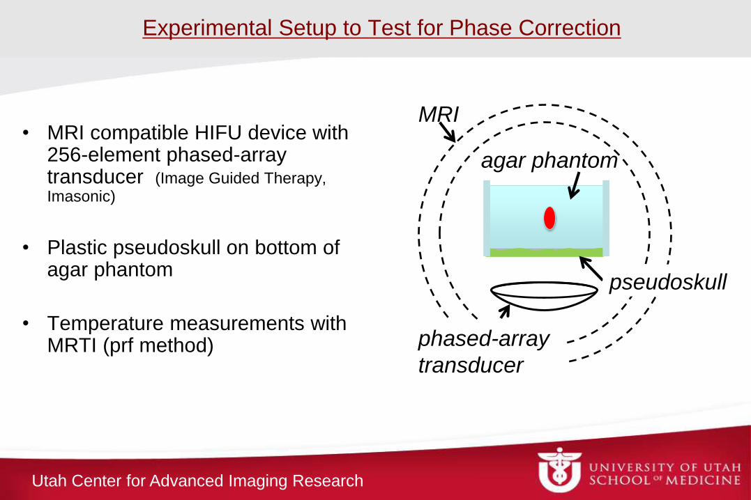
• MRI compatible HIFU device with 256-element phased-array transducer (Image Guided Therapy, Imasonic)
• Plastic pseudoskull on bottom of agar phantom
• Temperature measurements with MRTI (prf method)
agar phantom
MRI
phased-array
transducer
pseudoskull
Experimental Setup to Test for Phase Correction
Utah Center for Advanced Imaging Research Distance (mm)-60 -40 -20 0 20 40 60
0
20
40
Distance (mm)
Dis
tance (
mm
)
-60 -40 -20 0 20 40 600
20
40
-60 -40 -20 0 20 40 60
-60
-40
-20
0
20
40
60
Tem
pera
ture
(C
elc
ius)
0
1
2
3
4
5
6
7
8
9
10
Dis
tance (
mm
)
-60 -40 -20 0 20 40 60
-60
-40
-20
0
20
40
60
-60 -40 -20 0 20 40 60
-60
-40
-20
0
20
40
60
Tem
pera
ture
Ris
e (
Degre
es C
elc
ius)
0
1
2
3
4
5
6
7
8
9
10
Dis
tance (
mm
)
-60 -40 -20 0 20 40 600
20
40
-60 -40 -20 0 20 40 600
20
40
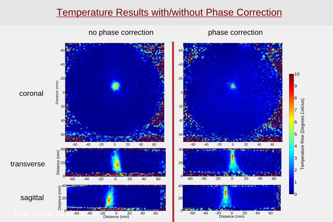
Temperature Results with/without Phase Correction
no phase correction phase correction
transverse
coronal
sagittal
Utah Center for Advanced Imaging Research
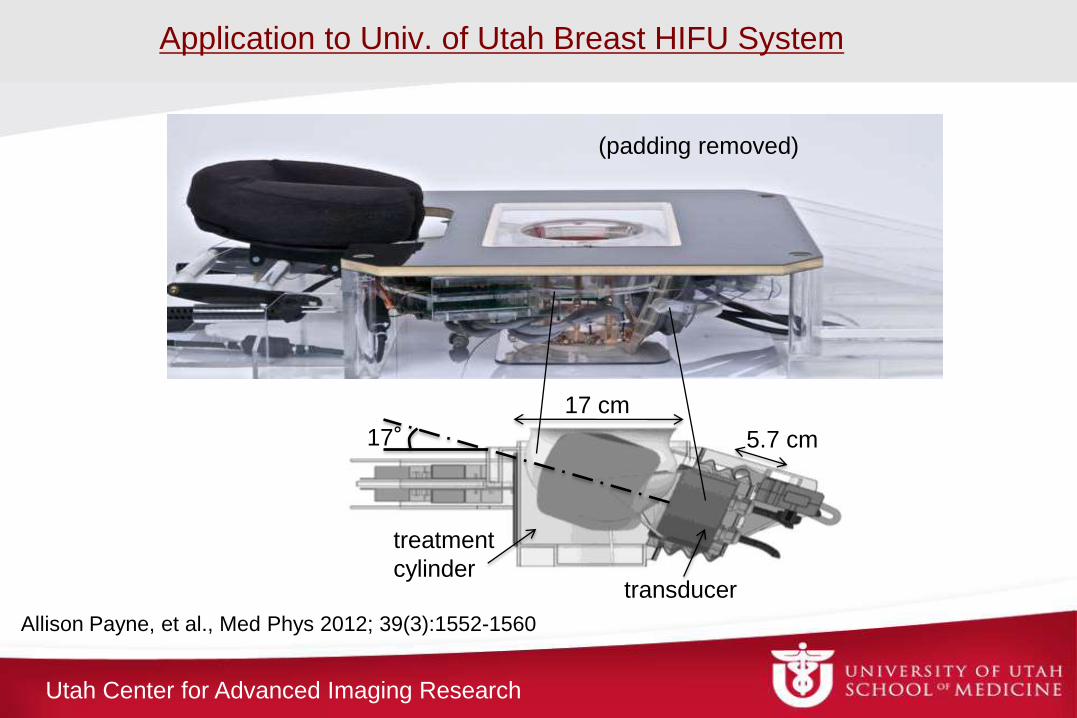
Application to Univ. of Utah Breast HIFU System
5.7 cm
17 cm
transducer
treatment
cylinder
17°
(padding removed)
Allison Payne, et al., Med Phys 2012; 39(3):1552-1560
Utah Center for Advanced Imaging Research
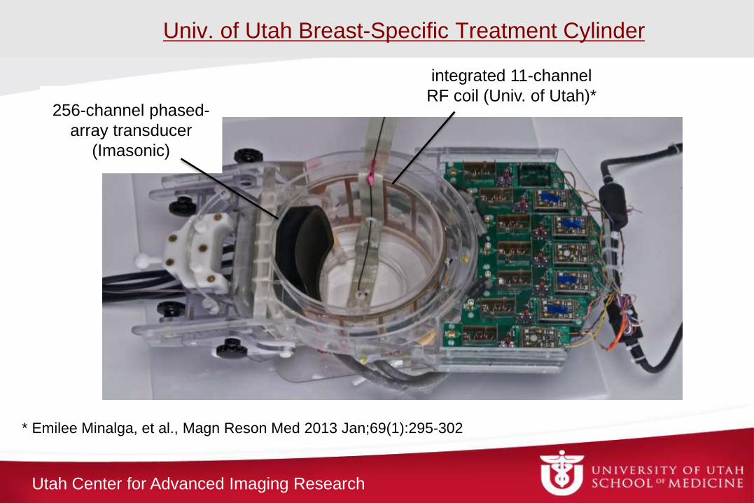
Univ. of Utah Breast-Specific Treatment Cylinder
256-channel phased-
array transducer
(Imasonic)
integrated 11-channel
RF coil (Univ. of Utah)*
* Emilee Minalga, et al., Magn Reson Med 2013 Jan;69(1):295-302
Utah Center for Advanced Imaging Research
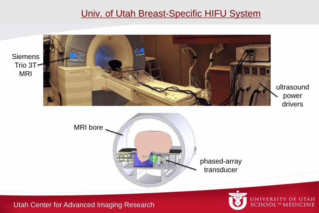
Univ. of Utah Breast-Specific HIFU System
phased-array
transducer
Siemens
Trio 3T
MRI
ultrasound
power
drivers
MRI bore
Utah Center for Advanced Imaging Research
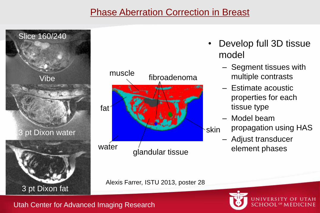
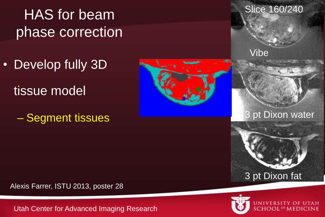
• Develop full 3D tissue
model
– Segment tissues with
multiple contrasts
– Estimate acoustic
properties for each
tissue type
– Model beam
propagation using HAS
– Adjust transducer
element phases
Vibe
3 pt Dixon water
3 pt Dixon fat
Slice 160/240
Alexis Farrer, ISTU 2013, poster 28
Phase Aberration Correction in Breast
glandular tissue
fat
muscle fibroadenoma
water
skin
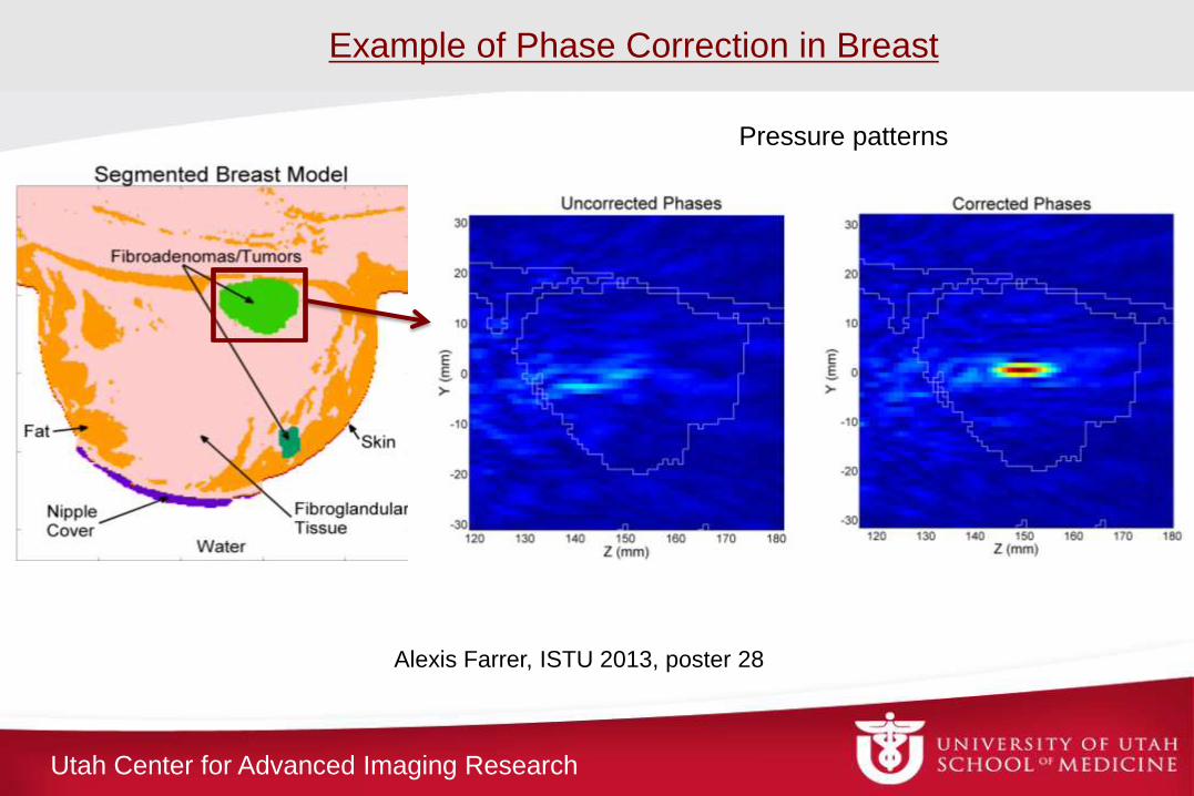
Utah Center for Advanced Imaging Research
Alexis Farrer, ISTU 2013, poster 28
Example of Phase Correction in Breast
Pressure patterns
Utah Center for Advanced Imaging Research
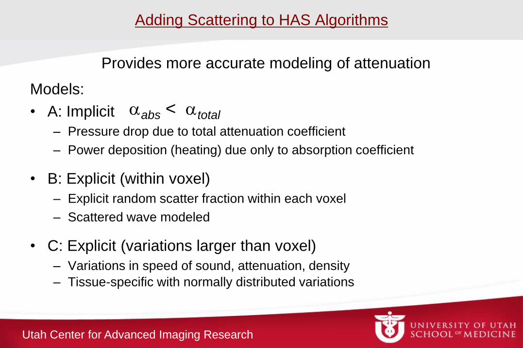
Adding Scattering to HAS Algorithms
Models:
• A: Implicit
– Pressure drop due to total attenuation coefficient
– Power deposition (heating) due only to absorption coefficient
• B: Explicit (within voxel)
– Explicit random scatter fraction within each voxel
– Scattered wave modeled
• C: Explicit (variations larger than voxel)
– Variations in speed of sound, attenuation, density
– Tissue-specific with normally distributed variations
Provides more accurate modeling of attenuation
aabs < atotal
Utah Center for Advanced Imaging Research
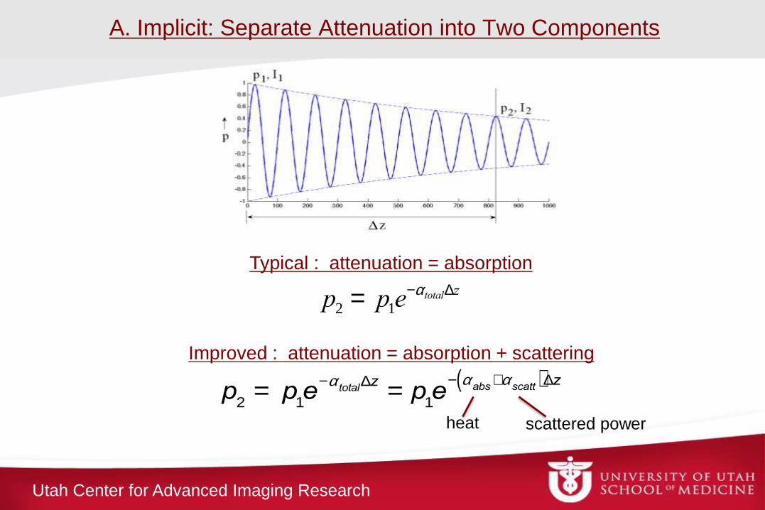
A. Implicit: Separate Attenuation into Two Components
p2 = p1e-atotalDz
Typical : attenuation = absorption
p
2= p
1e
-atotal
Dz= p
1e
- aabs
+ascatt( )Dz
heat scattered power
Improved : attenuation = absorption + scattering
Utah Center for Advanced Imaging Research
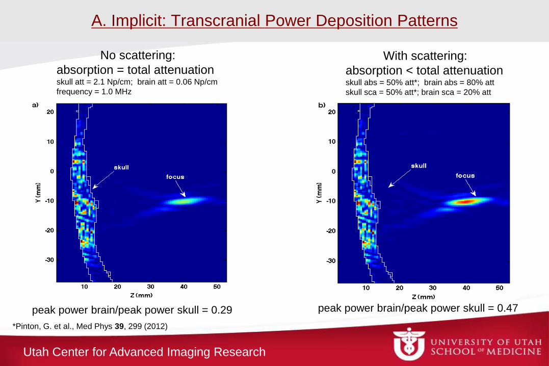
A. Implicit: Transcranial Power Deposition Patterns
No scattering:
absorption = total attenuation skull att = 2.1 Np/cm; brain att = 0.06 Np/cm
frequency = 1.0 MHz
With scattering:
absorption < total attenuation skull abs = 50% att*; brain abs = 80% att
skull sca = 50% att*; brain sca = 20% att
peak power brain/peak power skull = 0.29 peak power brain/peak power skull = 0.47
*Pinton, G. et al., Med Phys 39, 299 (2012)
Utah Center for Advanced Imaging Research
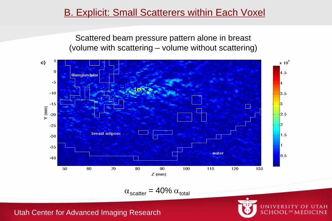
B. Explicit: Small Scatterers within Each Voxel
ascatter = 40% atotal
Scattered beam pressure pattern alone in breast
(volume with scattering – volume without scattering)
Utah Center for Advanced Imaging Research
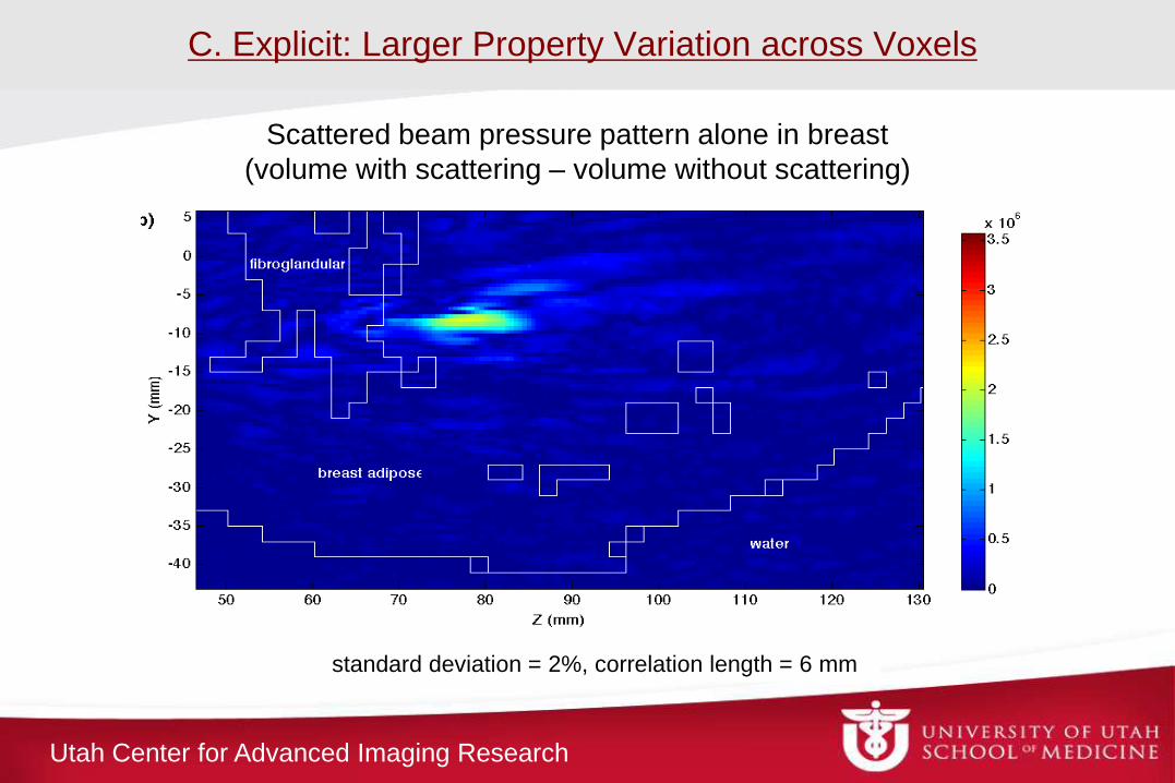
C. Explicit: Larger Property Variation across Voxels
standard deviation = 2%, correlation length = 6 mm
Scattered beam pressure pattern alone in breast
(volume with scattering – volume without scattering)
Utah Center for Advanced Imaging Research
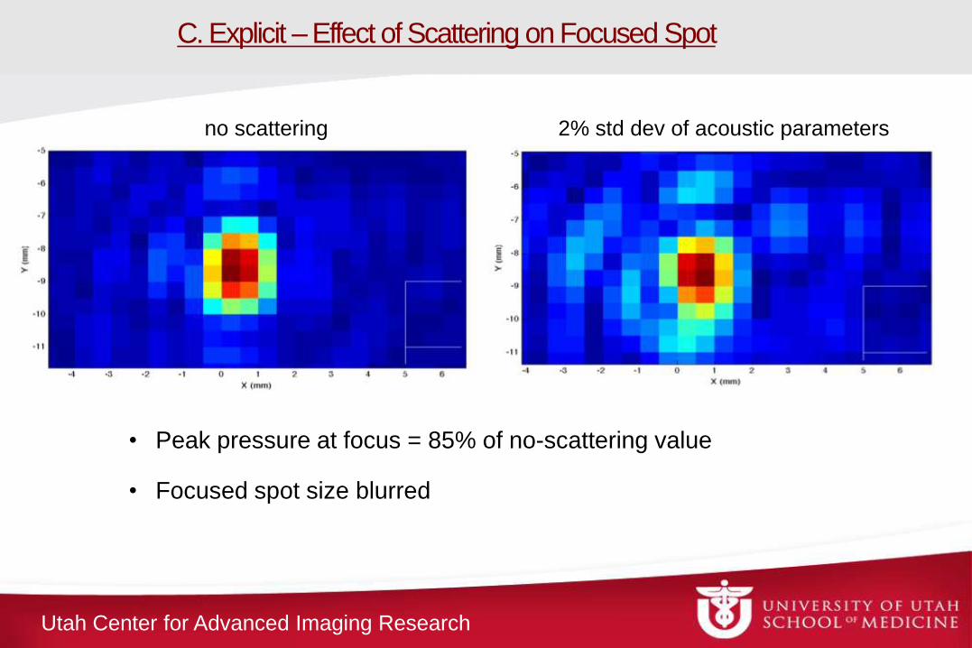
C. Explicit – Effect of Scattering on Focused Spot
• Peak pressure at focus = 85% of no-scattering value
• Focused spot size blurred
no scattering 2% std dev of acoustic parameters
Utah Center for Advanced Imaging Research
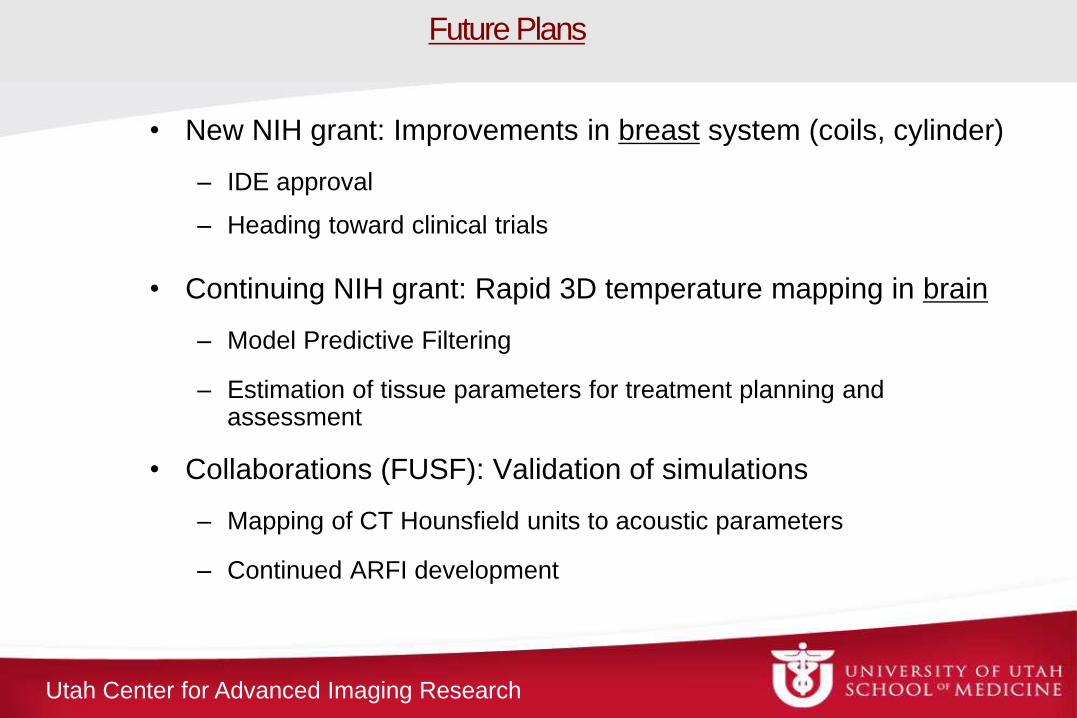
• New NIH grant: Improvements in breast system (coils, cylinder)
– IDE approval
– Heading toward clinical trials
• Continuing NIH grant: Rapid 3D temperature mapping in brain
– Model Predictive Filtering
– Estimation of tissue parameters for treatment planning and assessment
• Collaborations (FUSF): Validation of simulations
– Mapping of CT Hounsfield units to acoustic parameters
– Continued ARFI development
Future Plans

Utah Center for Advanced Imaging Research
Acknowledgments
The UCAIR group
Funding from: The FUS Foundation,
The Margolis Foundation,
Siemens Healthcare AG,
NIH R01s:
CA87785, CA134599, EB013433

Utah Center for Advanced Imaging Research 30
Thank you -
Any questions?
Utah Center for Advanced Imaging Research
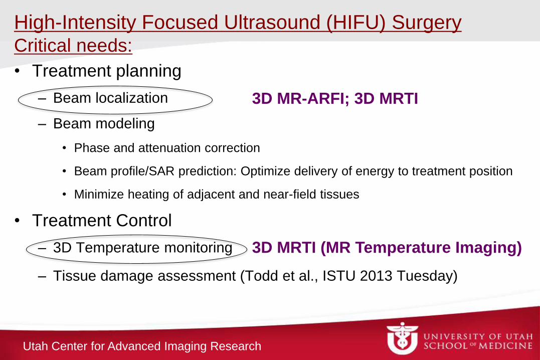
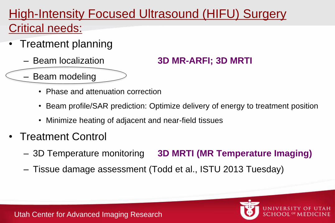
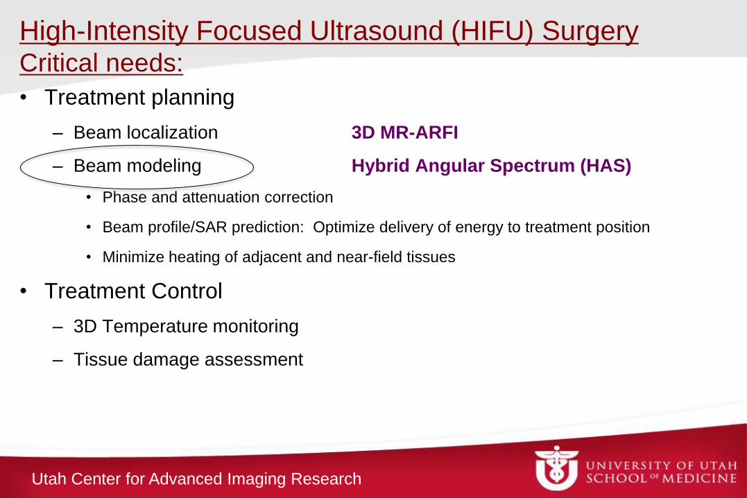
High-Intensity Focused Ultrasound (HIFU) Surgery Critical needs:
• Treatment planning
– Beam localization
– Beam modeling
• Phase and attenuation correction
• Beam profile/SAR prediction: Optimize delivery of energy to treatment position
• Minimize heating of adjacent and near-field tissues
• Treatment Control
– 3D Temperature monitoring
– Tissue damage assessment (Todd et al., ISTU 2013 Tuesday)
3D MR-ARFI; 3D MRTI
3D MRTI (MR Temperature Imaging)
Utah Center for Advanced Imaging Research
Coronal
2-D Skull Surface
Projection °C
3D MRI temperature measurements
MPF: Experimental results
Utah Center for Advanced Imaging Research
High-Intensity Focused Ultrasound (HIFU) Surgery Critical needs:
• Treatment planning
– Beam localization 3D MR-ARFI; 3D MRTI
– Beam modeling
• Phase and attenuation correction
• Beam profile/SAR prediction: Optimize delivery of energy to treatment position
• Minimize heating of adjacent and near-field tissues
• Treatment Control
– 3D Temperature monitoring 3D MRTI (MR Temperature Imaging)
– Tissue damage assessment (Todd et al., ISTU 2013 Tuesday)
Utah Center for Advanced Imaging Research
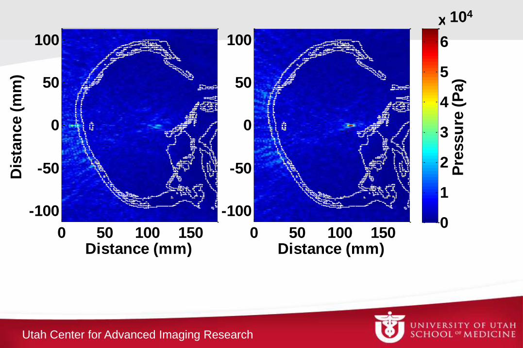
Distance (mm)
0 50 100 150
-100
-50
0
50
100
Pre
ssu
re (
Pa)
0
1
2
3
4
5
6
x 104
Distance (mm)
Dis
tan
ce (
mm
)
0 50 100 150
-100
-50
0
50
100
104
Utah Center for Advanced Imaging Research
High-Intensity Focused Ultrasound (HIFU) Surgery Critical needs:
• Treatment planning
– Beam localization 3D MR-ARFI
– Beam modeling Hybrid Angular Spectrum (HAS)
• Phase and attenuation correction
• Beam profile/SAR prediction: Optimize delivery of energy to treatment position
• Minimize heating of adjacent and near-field tissues
• Treatment Control
– 3D Temperature monitoring
– Tissue damage assessment
Utah Center for Advanced Imaging Research
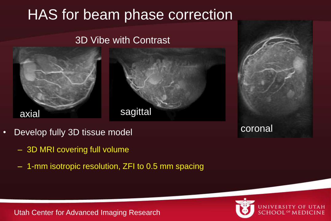
HAS for beam phase correction
• Develop fully 3D tissue model
– 3D MRI covering full volume
– 1-mm isotropic resolution, ZFI to 0.5 mm spacing
3D Vibe with Contrast
axial sagittal
coronal
Utah Center for Advanced Imaging Research
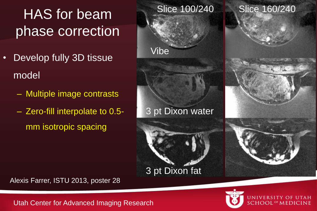
HAS for beam
phase correction
• Develop fully 3D tissue
model
– Multiple image contrasts
– Zero-fill interpolate to 0.5-
mm isotropic spacing
Vibe
3 pt Dixon water
3 pt Dixon fat
Slice 100/240 Slice 160/240
Alexis Farrer, ISTU 2013, poster 28
Utah Center for Advanced Imaging Research
HAS for beam
phase correction
• Develop fully 3D
tissue model
– Segment tissues
Vibe
3 pt Dixon water
3 pt Dixon fat
Slice 160/240
Alexis Farrer, ISTU 2013, poster 28
Utah Center for Advanced Imaging Research
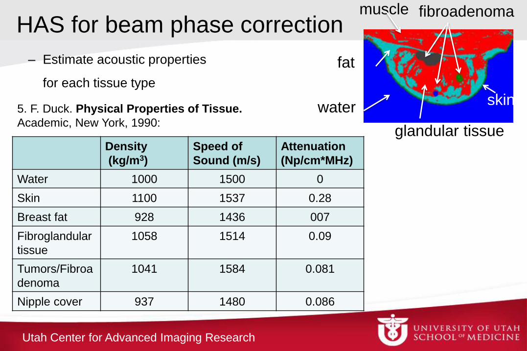
HAS for beam phase correction
– Estimate acoustic properties
for each tissue type
glandular tissue
fat
muscle fibroadenoma
water
Density
(kg/m3)
Speed of
Sound (m/s)
Attenuation
(Np/cm*MHz)
Water 1000 1500 0
Skin 1100 1537 0.28
Breast fat 928 1436 007
Fibroglandular
tissue
1058 1514 0.09
Tumors/Fibroa
denoma
1041 1584 0.081
Nipple cover 937 1480 0.086
5. F. Duck. Physical Properties of Tissue.
Academic, New York, 1990:
skin
Utah Center for Advanced Imaging Research
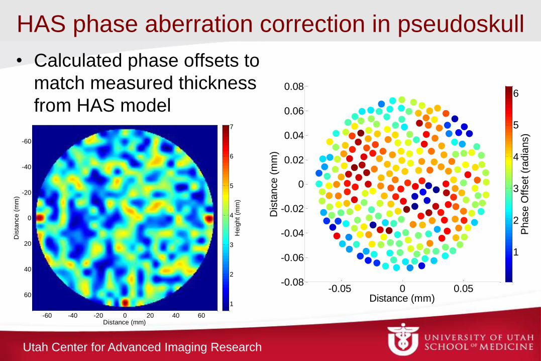
HAS phase aberration correction in pseudoskull
• Calculated phase offsets to
match measured thickness
from HAS model
Figure 4: Diagram of experimental setup. -0.05 0 0.05
-0.08
-0.06
-0.04
-0.02
0
0.02
0.04
0.06
0.08
Distance (mm)
Dis
tance (
mm
)
Phase O
ffset (r
adia
ns)
1
2
3
4
5
6
Distance (mm)
Dis
tance (
mm
)
-60 -40 -20 0 20 40 60
-60
-40
-20
0
20
40
60
Heig
ht
(mm
)
1
2
3
4
5
6
7
Utah Center for Advanced Imaging Research
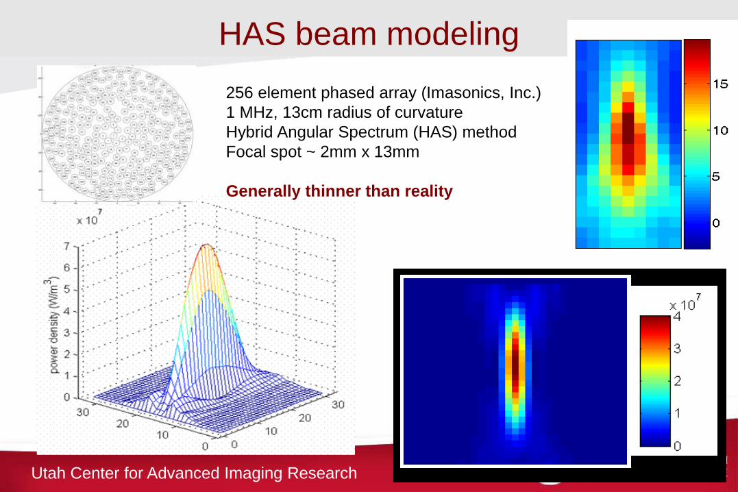
HAS beam modeling
256 element phased array (Imasonics, Inc.)
1 MHz, 13cm radius of curvature
Hybrid Angular Spectrum (HAS) method
Focal spot ~ 2mm x 13mm
Generally thinner than reality
Utah Center for Advanced Imaging Research
Adding scattering to HAS:
• Creates a more realistic picture of transcranial heating
• Provides more accurate model of beam propagation in
scattering media
• Will lead to:
– more accurate understanding of beam focusing for all HIFU
applications
– More accurate SAR prediction


![Original Article High intensity focused ultrasound inhibits breast cancer … · 2020. 4. 27. · HIFU inhibits breast cancer 2206 Int J Clin Exp Med 2020;13(4):2205-2215 [10]. Moreover,](https://static.fdocuments.net/doc/165x107/6002df81a9e7ac55f44947b9/original-article-high-intensity-focused-ultrasound-inhibits-breast-cancer-2020.jpg)


