Modeling auxin transport and plant development Heisler...
29
Modeling auxin transport and plant development Heisler, Marcus; Jönsson, Henrik Published in: Journal of Plant Growth Regulation DOI: 10.1007/s00344-006-0066-x 2006 Link to publication Citation for published version (APA): Heisler, M., & Jönsson, H. (2006). Modeling auxin transport and plant development. Journal of Plant Growth Regulation, 25, 302-312. https://doi.org/10.1007/s00344-006-0066-x General rights Copyright and moral rights for the publications made accessible in the public portal are retained by the authors and/or other copyright owners and it is a condition of accessing publications that users recognise and abide by the legal requirements associated with these rights. • Users may download and print one copy of any publication from the public portal for the purpose of private study or research. • You may not further distribute the material or use it for any profit-making activity or commercial gain • You may freely distribute the URL identifying the publication in the public portal Take down policy If you believe that this document breaches copyright please contact us providing details, and we will remove access to the work immediately and investigate your claim.
Transcript of Modeling auxin transport and plant development Heisler...
LUND UNIVERSITY
PO Box 117221 00 Lund+46 46-222 00 00
Modeling auxin transport and plant development
Heisler, Marcus; Jönsson, Henrik
Published in:Journal of Plant Growth Regulation
DOI:10.1007/s00344-006-0066-x
2006
Link to publication
Citation for published version (APA):Heisler, M., & Jönsson, H. (2006). Modeling auxin transport and plant development. Journal of Plant GrowthRegulation, 25, 302-312. https://doi.org/10.1007/s00344-006-0066-x
General rightsCopyright and moral rights for the publications made accessible in the public portal are retained by the authorsand/or other copyright owners and it is a condition of accessing publications that users recognise and abide by thelegal requirements associated with these rights.
• Users may download and print one copy of any publication from the public portal for the purpose of private studyor research. • You may not further distribute the material or use it for any profit-making activity or commercial gain • You may freely distribute the URL identifying the publication in the public portalTake down policyIf you believe that this document breaches copyright please contact us providing details, and we will removeaccess to the work immediately and investigate your claim.
Department of Theoretical Physics
LUP Lund University Publications
Institutional Repository of Lund University Found at: http://www.lu.se
This is an author produced version of a paper published in Journal of Plant Growth Regulation
This paper has been peer-reviewed but does not include
the final publisher proof-corrections or journal pagination.
Citation for the published paper:
M. Heisler and H. Jönsson Modeling auxin transport and plant development
Journal of Plant Growth Regulation, 2006, 25, 302-312.
http://dx.doi.org/10.1007/s00344-006-0066-x
Access to the published version may require subscription.
Published with permission from: Springer Verlag
Modelling Auxin Transport and Plant development
M. G. Heisler1 and H. Jönsson
2
1Division of Biology, California Institute of Technology, Pasadena, CA, USA,
2Corresponding author, Computational Biology and Biological Physics, Department of
Physics, Lund University, Lund, Sweden, [email protected], Ph.+46 46 222 0667, Fax
+46 46 222 9686
Key words: auxin, phyllotaxis, meristem, Arabidopsis, pattern formation, AUX1,
PINFORMED1, development
Running title: Modelling auxin in plant development
Summary
The plant hormone auxin plays a critical role in plant development. Central to its
function is its distribution in plant tissues, which is in turn, largely shaped by intercellular
polar transport processes. Auxin transport relies on diffusive uptake as well as carrier-
mediated transport via influx and efflux carriers. Mathematical models have been used to
both refine our theoretical understanding of these processes and to test new hypotheses
regarding the localization of efflux carriers in order to understand auxin patterning at the
tissue level. Here we review models for auxin transport and how they have been applied
to patterning processes including the elaboration of plant vasculature and primordium
positioning. Secondly we investigate the possible role of auxin influx carriers such as
AUX1 in pattering auxin in the shoot meristem. We find that AUX1 and its relatives are
likely to play a crucial role in maintaining high auxin levels in the meristem epidermis.
We also show that auxin influx carriers may play an important role in stabilizing auxin
distribution patterns generated by auxin-gradient type models for phyllotaxis.
Introduction
The hormone auxin plays a central role in plant development. It acts both as a
global coordinator, dictating where and when developmental events occur (Benkova and
others 2003), and as a mediator of morphogenesis, both in the context of whole tissues
and individual cells . Critical to its role in plant development is its polar intercellular
transport. Disruption to auxin transport, either by chemical means or through mutation
results in many plant defects including the inhibition of cell elongation, a loss of
asymmetric embryo morphogenesis (Friml and others 2003; Hadfi and others 1998), cell
division defects (Campanoni and Nick 2005), defects in vascular patterning (Sieburth
1999), a loss of tropic responses to light and gravity and the absence of organ initiation
(Okada and others 1991). The importance of auxin transport is further underscored by its
ability to act as a buffer against changes in auxin homeostasis, particularly during embryo
development (Weijers and others 2005). In this review we will briefly outline the current
conception of how auxin transport occurs and how mathematical models and computer
simulations have helped us test this conception against experimental data. Secondly we
will outline how modelling efforts have also enabled us to explore various hypotheses for
how transport routes are specified. Lastly we will present new simulations of auxin
transport that explore the potential role of auxin influx carriers within the shoot apical
meristem.
Our current understanding of auxin transport is based on the chemiosmotic model
proposed by Rubery and Sheldrake (Rubery and Sheldrake 1974) and Raven (Raven
1975). This model is based on the fact that indole-3-acetic acid (IAA), which is the most
predominant naturally occurring auxin, exists in two alternate states depending on pH.
Outside the cell plasma membrane, where the pH is close to 5, a significant proportion of
IAA exists in an uncharged form, which can enter cells passively. However inside cells
the pH is around 7 and the vast majority of IAA present there is negatively charged. Thus
in order to exit a cell, there has to be an efflux carrier present that enables negatively
charged IAA to move through the plasma membrane down the electrochemical gradient
between the inside and outside of the cell. It is the polar subcellular localization of the
efflux carrier that is thought to give auxin transport its overall directionality. In addition
to entering cells passively, auxin is taken up by a saturable influx carrier which symports
two protons with one IAA molecule (Bennett and others 1996; Marchant and others
1999; Rubery and Sheldrake 1974; Yang and others 2006).
Early transport models
Perhaps the earliest attempt to model polar auxin transport is that of Leopold and
Hall in 1966. In this and an accompanying study, a simple polarized transport model
(basal “secretion”) is compared with experiments in which an auxin source is applied to a
file of auxin transporting cells (de la Fuente and Leopold 1966; Leopold and Hall 1966).
These studies reveal that a small basally biased asymmetry in transport polarity at the
cellular level should be sufficient to generate a large asymmetry in auxin transport at the
tissue level. Their model also predicts the observation that the ratio of auxin that
accumulates in basal vs. apical cells grows exponentially with the distance from an apical
source (de la Fuente and Leopold 1966; Leopold and Hall 1966).
Later mathematical models were developed to establish a firm theoretical basis
for the chemiosmotic model. Both Mitchison (1980) and Goldsmith and others (1981)
showed that the model predicted constant velocity for an auxin pulse travelling through
tissue characterized by an asymmetric distribution of anion carrier, as is observed
experimentally (Goldsmith, 1977). Mitchison (1980) also showed that the polarity or
asymmetry in permeability required for polar transport depended on the route of auxin
within cells, i.e. whether transport was mainly restricted to a thin cytoplasmic sheath
surrounding a large vacuole or whether the tonoplast was considered permeable to the
anion. Differences in the mathematical description of the two studies were later
reconciled (Martin and others 1990). Interestingly both Mitchison and Goldsmith and
others noted that to achieve the known auxin transport rate of 1 cm/h in their simulations
their ratio between basipetal and acropetal transport had to be much higher than what had
been suggested by Leopold and Hall. However Mitchison found that this contradiction
could be reconciled assuming that efflux was saturable (Mitchison 1980a). Since these
early theoretical studies considerable experimental progress has been made in identifying
and characterizing the molecular components involved in auxin transport. Members of
the PIN family of membrane proteins (Galweiler and others 1998) have been shown to
mediate auxin efflux (Petrasek and others 2006) and their polar localization has been
shown to be required for polar auxin transport (Wisniewska and others 2006). PIN
protein localization has been documented in many plant tissues and the consequences of
disruption to PIN function have also been characterized (Benkova and others 2003; Friml
and others 2002a; Friml and others 2003; Friml and others 2002b; Galweiler and others
1998; Okada and others 1991; Reinhardt and others 2003). The characterization of auxin
influx carriers has also progressed with the identification of AUX1 and its relatives. AUX1
encodes an amino acid permease like protein that has been shown to mediate auxin influx
(Bennett and others 1996; Yang and others 2006). However so far only AUX1 has been
extensively investigated at the level of function, localization and expression while its
three close relatives remain relatively uncharacterised.
Models for generating auxin transport patterns associated with vascular patterning
One of the most intriguing aspects of plant development linked to auxin transport
is the patterning of plant vasculature. From studying the influence of wounding and
exogenously applied auxin on vasculature patterning, Sachs proposed that plant veins
form along the paths of auxin flow. Sachs also suggested that these preferred flow paths
form because of positive feedback between auxin flux and auxin transport capacity. Cells
with an increased capacity for transport are suggested to act as auxin sinks thereby
depleting auxin from neighbouring cells. A critical aspect of this model not explicitly
defined by Sachs is the way in which a transporting cell becomes a “sink”. Addressing
this issue, Mitchison showed that if the concentration of auxin within a transport route
decreased with increasing flux then auxin would move preferentially into this transport
route simply by diffusion (Mitchison 1980b; Mitchison 1981). Mitchison further showed
that such a scenario could be achieved if transport capacity is increased faster than linear
as a function of flux. Small differences in auxin flux were found in simulations of this
model to be sufficient to destabilize a uniform initial flow pattern into preferred
pathways, as postulated by Sachs. By also changing the location of auxin sources and
sinks Mitchison was able to generate circular transport channels which are suggested to
correspond to the loops of vascular elements induced by experimentally manipulating
exogenous auxin sources (Sachs 1981). One criticism of this model has been that
experimental evidence indicates that auxin levels in developing vascular tissues are
higher than the surrounding cells rather than lower. For instance Sachs and others had
shown that high auxin concentrations are required for vascular cell differentiation and
cell elongation. More recently it has been found that many auxin-regulated genes are
specifically expressed in developing vasculature (Mattsson and others 2003; Scarpella
and others 2004; Scarpella and others 2006). However Mitchison proposed a solution to
this criticism in his 1981 paper in the supplementary discussion section. He suggests that
cells that become incorporated into a preferred transport route need only contain
relatively low auxin concentrations at the beginning of the canalisation process when they
are acting as sinks. During the later differentiation stages, if the auxin anion channel
becomes localized to one end of the cell and depleted from the other sides, auxin can
flow into this cell even if it has a relatively high auxin concentration. This proposal has
now been modelled explicitly and simulations confirm that auxin could specifically
accumulate in transporting cells under these conditions (Feugier and others 2005; Fujita
and Mochizuki 2006).
Recent detailed observations of PIN1 localization in developing veins has
revealed some aspects of localization predicted by the flux model as well as other
patterns that are more puzzling (Scarpella and others 2006). For instance the initiation of
tertiary veins is marked by the expression of PIN1 in a cell adjacent to an existing
secondary vein. Later PIN1 expression then appears in a new cell adjacent to the first cell
but located further away from the pre-existing vein. PIN1 polarity in all the cells is also
directed towards the pre-existing vein. This pattern of localization and expression
resembles efflux carrier distributions in simulations of the flux model in which a new
vein forms from a local pre-existing auxin sink. In such cases a new channel forms from
the sink towards the source. However a critical question here is in what sense do pre-
existing veins represent auxin “sinks”? For instance, do they contain relatively low
concentrations of auxin as Mitchison originally proposed? In fact expression of the DR5
auxin marker in many provascular cells suggests that the majority of these cells contain
high levels of auxin in contradiction to Mitchison’s predictions (Mattsson and others
2003; Scarpella and others 2004; Scarpella and others 2006). However it may be that
cells that contain high levels of auxin can still act as sinks because they express high
levels of auxin influx carriers. In fact specific influx carrier expression has been
suggested to be a precondition for auxin accumulation (Kramer 2004). Another
interesting observation made by Scarpella and others (2006) is that within tertiary vein
loops, there is a cell in which PIN1 is localized to two opposite sides. According to the
flux model, such a bi-polar cell would be expected to act as an auxin source. Intriguingly
a recent modelling study that incorporates local auxin sources together with a simplified
version of the flux model is capable of generating realistic patterns of leaf vein formation
(Runions and others 2005). It will be interesting to examine this model further and test
whether local auxin sources exist in developing leaves at the locations of these bipolar
cells.
Auxin transport and phyllotaxis
Due to the striking geometrical arrangements of plant organs, the study of
phyllotaxis, or arrangement of plant organs, has a long history (Adler and others 1997).
Phyllotactic patterning can be understood as the superimposition of several processes.
Firstly there is a mechanism that restricts primordial emergence to a narrow region called
the peripheral zone, which is located a certain radial distance from the centre of the shoot
apical meristem (Steeves and Sussex 1989). Secondly there is the continuous generation
of new tissue within the meristem that results in the steady displacement of cells away
from the peripheral zone (Reddy and others 2004). Thirdly there is a positioning process
that imposes a regular spacing between the locations of new primordia (Mitchison 1977).
Lastly, for many plant species, the shoot apical meristem can change size gradually
during seedling growth. Most theoretical studies have been concerned with hypotheses
for how spacing mechanisms may work. Generally these can be divided into four
categories. One general type of mechanism involves primordia producing a diffusible
substance that inhibits primordia from initiating nearby (Schoute 1913). A second type of
model involves primordia depleting a positive activator of primordium development with
the activator only reaching the critical initiation concentration threshold a certain distance
away from pre-existing primordia (Reinhardt and others 2003). A recent variant on this
theme, discussed further below, is a polarized transport model based on intercellular
auxin gradients (Jönsson and others 2006; Smith and others 2006). A fourth type of
mechanism involves both positive and negative regulators in a reaction-diffusion network
(Meinhardt and others 1998). Lastly mechanical buckling with positive feedback has also
been proposed (Green and others 1996; Shipman and Newell 2004; Shipman and Newell
2005).
Experimental data support the second two hypotheses listed above in which auxin
acts as a limiting positive regulator. As early as the 1930s, Snow and Snow discovered
that by applying lanolin paste containing auxin to shoot meristems of Lupin, ectopic
growth of leaf or floral tissue could be induced (Snow and Snow 1937). More recently it
was found that plants deficient in auxin transport activity developed pin-like apices
devoid of lateral organs (Okada and others 1991). By applying auxin to such apices organ
growth could be restored at the site of application (Reinhardt and others 2000). These
experiments suggested that auxin acts as a positive regulator of organ initiation and that
its transport-dependant distribution might determine where organs arise. By immuno-
localization of the PIN1 putative auxin efflux carrier, it was found that polar transport
was indeed likely to transport auxin directly to sites of organ initiation since the PIN1
protein was found to be localized on the sides of meristem cells located closest to where a
primordium was predicted to form (Reinhardt and others 2003). PIN1 protein was also
localized basally within primordia and on the apical sides of epidermal cells located
below the meristem. Lastly, the AUX1 protein was found predominantly localized to the
epidermis (Reinhardt and others 2003). These findings suggested a scenario in which
auxin is transported apically to the meristem where it then becomes localized to the
epidermis and sites of primordium emergence. As primordia develop auxin would then
get transported basally down the developing vasculature. These data and previous
findings also prompted Reinhardt and co-workers to propose that it is the transport of
auxin into the vasculature and resulting depletion of auxin in surrounding cells that
inhibits primordial development nearby. The next primordium would therefore be placed
furthest away where auxin levels first reach a certain threshold (Reinhardt and others
2003).
More recently a series of modelling papers have been published in an effort to
bring our theoretical understanding up to date with experimental results and to propose
mechanisms for determining PIN1 polarity patterns. Two of these papers propose positive
feedback between cellular auxin concentrations and auxin transport direction as a
mechanism for positioning primordia (Jönsson and others 2006; Smith and others 2006).
The third paper quantifies PIN1 localization patterns within the meristem to gain a more
accurate picture of probable auxin accumulation patterns (de Reuille and others 2006).
Unlike the flux model, the proposed feedback models suggest cell-cell signalling as a
means of coordinating PIN1 polarity by which cells polarize PIN1 towards neighbouring
cells containing the most auxin. One problem with a model based solely on this
mechanism however is that there is no means by which auxin maxima become fixed to
the underlying cells. Hence in response to the changing geometry of a plant apex and the
formation of new auxin maxima, auxin accumulation patterns are not stable resulting in
variable phyllotactic patterns. This problem is exacerbated by incorporating the
experimental result that in the apical meristem PIN1 expression is auxin induced (Heisler
and others 2005) (as discussed below) since when PIN1 expression intensifies, auxin is
more easily able to move out of cells within the maxima and into neighbouring cells
(unpublished observations). Smith and others incorporate a number of additional
assumptions in their model to try and prevent this phenomenon from occurring. One such
assumption is that auxin synthesis occurs in primordial cells after these cells are first
specified. Another assumption is that PIN1 localization towards the centre of primordia
becomes fixed after they first form (Smith and others 2006).
Here we attempt to explore the possible role of auxin influx carriers in this type of
feedback model. Our aim is to test the previously proposed hypothesis that AUX1 may
help to concentrate auxin in the meristem epidermis (Reinhardt and others 2003) and
secondly, to test whether auxin influx carriers including AUX1 may help stabilize the
concentration feed-back model by maintaining high auxin concentrations in cells in
which auxin first accumulates.
Including influx mediators when modelling auxin patterning in the SAM
Our modelling approach is based on basic biochemical descriptions of molecular
reactions including the chemiosmotic transport theory for auxin where model parameters
are mainly based on experimental estimates. All interactions are described using ordinary
differential equations and we obtain a numerical solution describing molecular
concentrations in a tissue of cell and wall compartments over time. Compared to our
previous study (Jönsson and others 2006), here we add AUX1 as an explicit auxin influx
mediator. We use a symmetric distribution of AUX1 in the membranes surrounding a
cell, which in contrast to the previously used passive influx, allows individual cells to
regulate their auxin influx from surrounding walls. We then discuss the consequences
that the explicit inclusion of an influx mediator has for phyllotaxis models based on auxin
transport since influx mediators have not been included in recent models. More explicitly,
we show that in a model taking into account the whole structure of the SAM, including
epidermal and sub-epidermal cells, and in a model including auxin-induced PIN1, influx
carriers are important to maintain a stable auxin pattern.
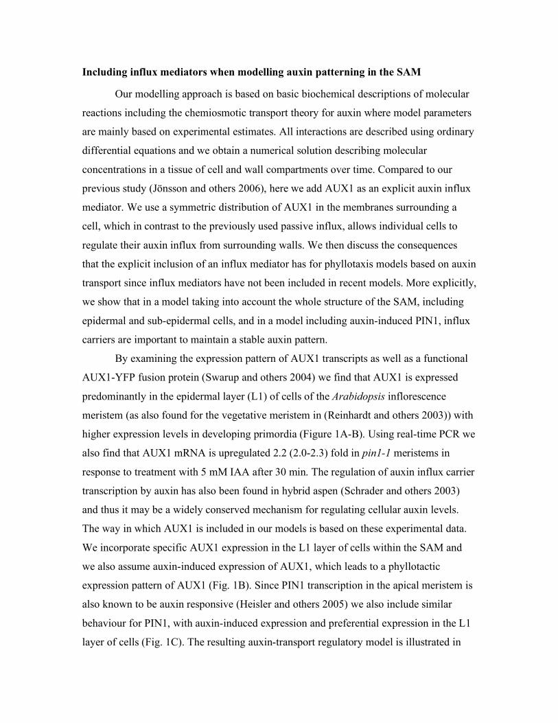
By examining the expression pattern of AUX1 transcripts as well as a functional
AUX1-YFP fusion protein (Swarup and others 2004) we find that AUX1 is expressed
predominantly in the epidermal layer (L1) of cells of the Arabidopsis inflorescence
meristem (as also found for the vegetative meristem in (Reinhardt and others 2003)) with
higher expression levels in developing primordia (Figure 1A-B). Using real-time PCR we
also find that AUX1 mRNA is upregulated 2.2 (2.0-2.3) fold in pin1-1 meristems in
response to treatment with 5 mM IAA after 30 min. The regulation of auxin influx carrier
transcription by auxin has also been found in hybrid aspen (Schrader and others 2003)
and thus it may be a widely conserved mechanism for regulating cellular auxin levels.
The way in which AUX1 is included in our models is based on these experimental data.
We incorporate specific AUX1 expression in the L1 layer of cells within the SAM and
we also assume auxin-induced expression of AUX1, which leads to a phyllotactic
expression pattern of AUX1 (Fig. 1B). Since PIN1 transcription in the apical meristem is
also known to be auxin responsive (Heisler and others 2005) we also include similar
behaviour for PIN1, with auxin-induced expression and preferential expression in the L1
layer of cells (Fig. 1C). The resulting auxin-transport regulatory model is illustrated in
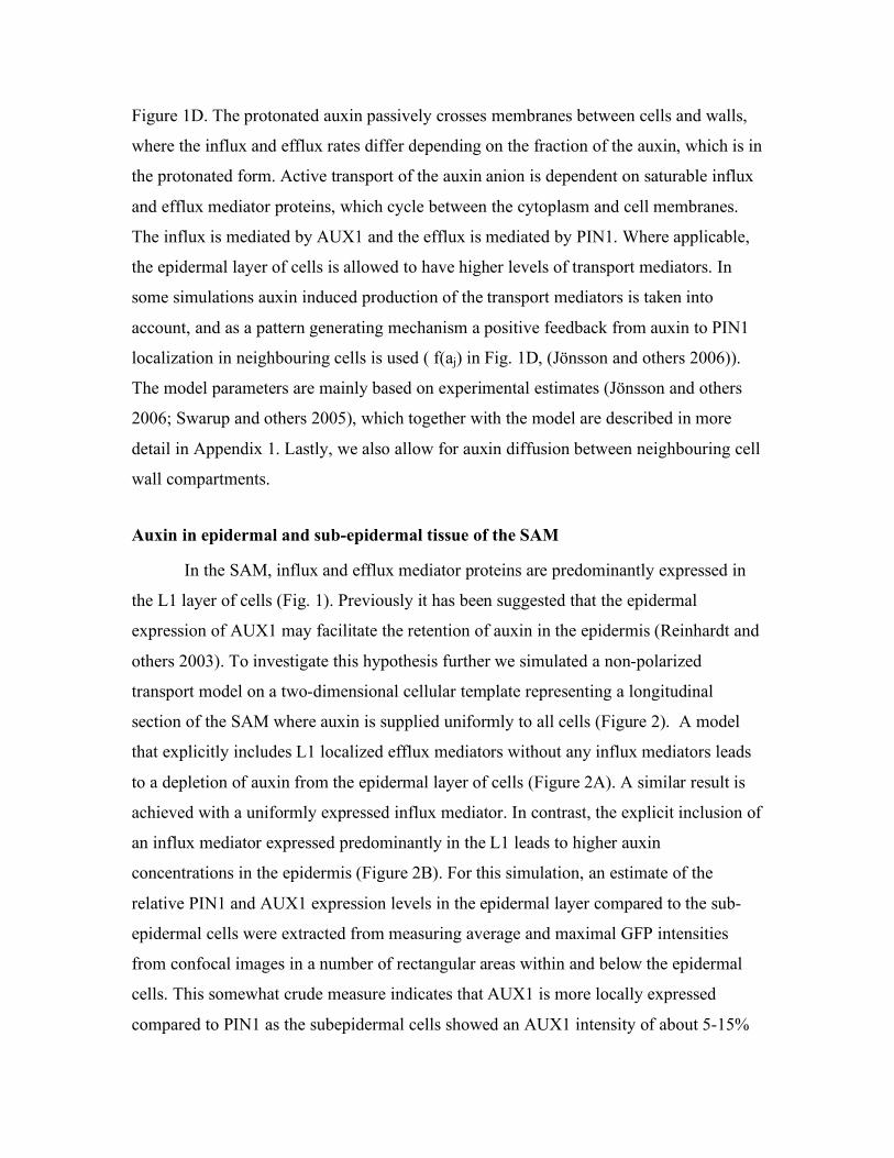
Figure 1D. The protonated auxin passively crosses membranes between cells and walls,
where the influx and efflux rates differ depending on the fraction of the auxin, which is in
the protonated form. Active transport of the auxin anion is dependent on saturable influx
and efflux mediator proteins, which cycle between the cytoplasm and cell membranes.
The influx is mediated by AUX1 and the efflux is mediated by PIN1. Where applicable,
the epidermal layer of cells is allowed to have higher levels of transport mediators. In
some simulations auxin induced production of the transport mediators is taken into
account, and as a pattern generating mechanism a positive feedback from auxin to PIN1
localization in neighbouring cells is used ( f(aj) in Fig. 1D, (Jönsson and others 2006)).
The model parameters are mainly based on experimental estimates (Jönsson and others
2006; Swarup and others 2005), which together with the model are described in more
detail in Appendix 1. Lastly, we also allow for auxin diffusion between neighbouring cell
wall compartments.
Auxin in epidermal and sub-epidermal tissue of the SAM
In the SAM, influx and efflux mediator proteins are predominantly expressed in
the L1 layer of cells (Fig. 1). Previously it has been suggested that the epidermal
expression of AUX1 may facilitate the retention of auxin in the epidermis (Reinhardt and
others 2003). To investigate this hypothesis further we simulated a non-polarized
transport model on a two-dimensional cellular template representing a longitudinal
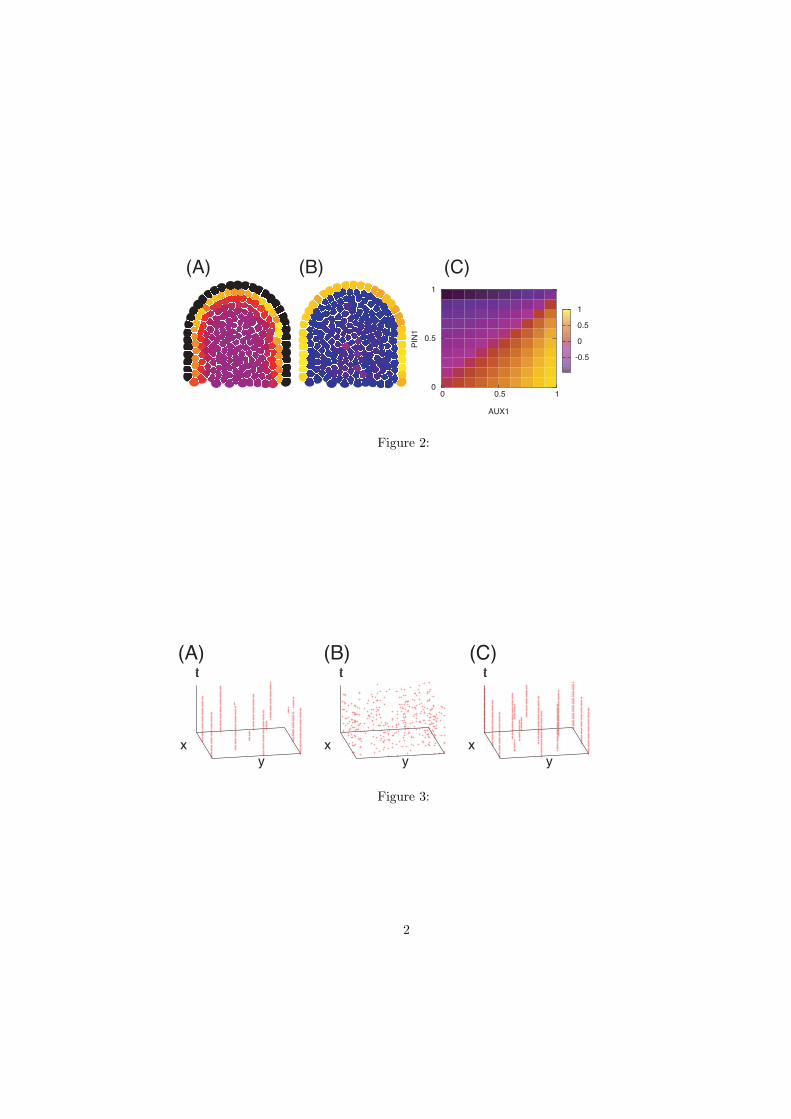
section of the SAM where auxin is supplied uniformly to all cells (Figure 2). A model
that explicitly includes L1 localized efflux mediators without any influx mediators leads
to a depletion of auxin from the epidermal layer of cells (Figure 2A). A similar result is
achieved with a uniformly expressed influx mediator. In contrast, the explicit inclusion of
an influx mediator expressed predominantly in the L1 leads to higher auxin
concentrations in the epidermis (Figure 2B). For this simulation, an estimate of the
relative PIN1 and AUX1 expression levels in the epidermal layer compared to the sub-
epidermal cells were extracted from measuring average and maximal GFP intensities
from confocal images in a number of rectangular areas within and below the epidermal
cells. This somewhat crude measure indicates that AUX1 is more locally expressed
compared to PIN1 as the subepidermal cells showed an AUX1 intensity of about 5-15%
of the intensities measured in the epidermal cells, while the corresponding measure for
PIN1 was 35-45% (compare Fig. 1A and C). To further investigate the expected
sensitivity of the auxin distribution to the expression patterns of transport mediators we
measured the auxin levels in simulations with varying AUX1 and PIN1 expression
patterns. AUX1 and PIN1 were varied from being uniformly expressed (same amount of
protein in the epidermal and sub-epidermal cells) to being completely L1 localized
(protein expressed only in the epidermal layer). The relative average auxin concentration
in the epidermal layer of cells compared to the sub-epidermal cells is presented in Figure
2C. It can be seen that increasing the relative expression of AUX1 in the epidermis leads
to increased auxin in the epidermal layer, while the opposite is true for PIN1. Thus our
results suggest that in order for auxin to accumulate specifically in the epidermis AUX1
needs to also be specifically expressed in the epidermis while PIN1 expression must be
less localized. This prediction is similar to that of a previous study (Kramer 2004), where
it is shown that to maintain high auxin concentrations within a transport channel in which
PIN1 is expressed, it is beneficial to have PIN1 expressed also in surrounding cells. Our
final conclusion is that the observed highly localized L1 expression pattern of influx
carrier is required for retention of auxin in the epidermis, as previously postulated
(Reinhardt and others 2003). However we also predict that such an accumulation of auxin
in the epidermis also requires the expression pattern of PIN1to be relatively less L1
localized, as is observed.
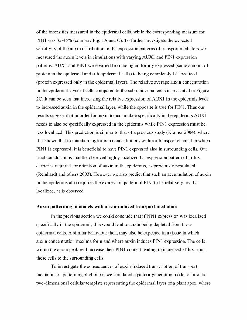
Auxin patterning in models with auxin-induced transport mediators
In the previous section we could conclude that if PIN1 expression was localized
specifically in the epidermis, this would lead to auxin being depleted from these
epidermal cells. A similar behaviour then, may also be expected in a tissue in which
auxin concentration maxima form and where auxin induces PIN1 expression. The cells
within the auxin peak will increase their PIN1 content leading to increased efflux from
these cells to the surrounding cells.
To investigate the consequences of auxin-induced transcription of transport
mediators on patterning phyllotaxis we simulated a pattern-generating model on a static
two-dimensional cellular template representing the epidermal layer of a plant apex, where
we include auxin-induced PIN1 and AUX1 production. Figure 3 shows the time
evolution of peak positions in simulations of different auxin induction scenarios. In a
model without any auxin-induced transport mediator production, peaks form and stay at
constant positions except for some minor slow rearrangements (Fig. 3A), as expected
from our previous work (Jönsson and others 2006). If auxin-induced PIN1 is added to the
model, the stability of the peak positions is lost (Fig. 3B). Here we have used a maximal
PIN1 expression level approximately three times the minimal in accordance with the
variation in cellular PIN1 intensity levels in an epidermal layer template (Fig. 1C in
(Jönsson and others 2006)). Finally, if auxin-induced AUX1 expression is introduced, the
resulting pattern becomes stable (Fig. 3C). These results suggest that the ability of auxin
to modulate PIN1 transcription may be a destabilizing influence on auxin distribution
patterns while auxin-induced influx carrier activity has the converse effect. Although
aux1 single mutants do not exhibit disrupted phyllotaxis, we suggest AUX1 may act
redundantly together with other members of the AUX1 family of proteins to help
stabilize plant organ positioning.
Discussion
Modelling of auxin transport has long been used to complement experimental
results and to formalize and evaluate hypotheses. It is indeed interesting to note that the
chemiosmotic formulation of auxin transport, first introduced over 25 years ago, is still
applicable to new problem settings. However although there is now a consensus on how
auxin transport is modelled in detail, it is important to realise that the data used for
defining parameters in current models come from studies using many different plants and
an assortment of tissue types. Although it should be possible to derive these parameters
through the reverse engineering and optimisation of models, such a strategy requires us to
have a reliable estimate of auxin concentration at subcellular resolution, which, up until
recently, has been beyond our capabilities. However with the identification of ABP1
(Lobler and Klambt 1985) and TIR1 (Dharmasiri and others 2005; Kepinski and Leyser
2005) as two distinct auxin-binding proteins, the development of a fluorescence
resonance energy transfer (FRET) based auxin biosensor such as has become a real
possibility.
Apart from modelling the dynamics of auxin efflux and influx, a future challenge
will be to incorporate other modulators of auxin transport that mediate pattering at the
tissue level. For understanding phyllotaxis for example, a detailed polarized auxin
transport model can only provide part of the explanation. Important issues such as the
demarcation and definition of the peripheral zone, understanding the role of genes such as
PINOID (Benjamins and others 2001; Christensen and others 2000; Friml and others
2004) and MONOPTEROS (Aida and others 2002; Hardtke and Berleth 1998; Przemeck
and others 1996) and incorporating the down stream events leading to primordial growth
are important goals for future experimental and theoretical studies.
ACKNOWLEDGEMENTS
We thank Eric Mjolsness, Elliot M. Meyerowitz, Adrienne Roeder and Bruce
Shapiro for helpful discussions. HJ acknowledge support from the Swedish Research
Council and Human Frontier Science Program. MGH was supported by the National
Science Foundation’s Frontiers in Biological Research (FIBR) program, award number
EF-0330786.
REFERENCES
Adler, I, Barabe, D and Jean, RV. 1997. A history of the study of phyllotaxis. Ann Bot
80: 231-244.
Aida, M, Vernoux, T, Furutani, M, Traas, J and Tasaka, M. 2002. Roles of PIN-
FORMED1 and MONOPTEROS in pattern formation of the apical region of the
Arabidopsis embryo. Development 129: 3965-74.
Benjamins, R, Quint, A, Weijers, D, Hooykaas, P and Offringa, R. 2001. The PINOID
protein kinase regulates organ development in Arabidopsis by enhancing polar
auxin transport. Development 128: 4057-67.
Benkova, E, Michniewicz, M, Sauer, M, Teichmann, T, Seifertova, D, Jurgens, G and
Friml, J. 2003. Local, efflux-dependent auxin gradients as a common module for
plant organ formation. Cell 115: 591-602.
Bennett, MJ, Marchant, A, Green, HG, May, ST, Ward, SP, Millner, PA, Walker, AR,
Schulz, B and Feldmann, KA. 1996. Arabidopsis AUX1 gene: a permease-like
regulator of root gravitropism. Science 273: 948-50.
Campanoni, P and Nick, P. 2005. Auxin-dependent cell division and cell elongation. 1-
Naphthaleneacetic acid and 2,4-dichlorophenoxyacetic acid activate different
pathways. Plant Physiol 137: 939-948.
Christensen, SK, Dagenais, N, Chory, J and Weigel, D. 2000. Regulation of auxin
response by the protein kinase PINOID. Cell 100: 469-78.
de la Fuente, RK and Leopold, AC. 1966. Kinetics of Polar Auxin Transport. Plant
Physiol 41: 1481-&.
de Reuille, PB, Bohn-Courseau, I, Ljung, K, Morin, H, Carraro, N, Godin, C and Traas, J.
2006. Computer simulations reveal properties of the cell-cell signaling network at
the shoot apex in Arabidopsis. Proc Natl Acad Sci U S A 103: 1627-32.
Dharmasiri, N, Dharmasiri, S and Estelle, M. 2005. The F-box protein TIR1 is an auxin
receptor. Nature 435: 441-445.
Feugier, FG, Mochizuki, A and Iwasa, Y. 2005. Self-organization of the vascular system
in plant leaves: inter-dependent dynamics of auxin flux and carrier proteins. J
Theor Biol 236: 366-75.
Friml, J, Benkova, E, Blilou, I, Wisniewska, J, Hamann, T, Ljung, K, Woody, S,
Sandberg, G, Scheres, B, Jurgens, G et al. 2002a. AtPIN4 mediates sink-driven
auxin gradients and root patterning in Arabidopsis. Cell 108: 661-73.
Friml, J, Vieten, A, Sauer, M, Weijers, D, Schwarz, H, Hamann, T, Offringa, R and
Jurgens, G. 2003. Efflux-dependent auxin gradients establish the apical-basal axis
of Arabidopsis. Nature 426: 147-53.
Friml, J, Wisniewska, J, Benkova, E, Mendgen, K and Palme, K. 2002b. Lateral
relocation of auxin efflux regulator PIN3 mediates tropism in Arabidopsis. Nature
415: 806-9.
Friml, J, Yang, X, Michniewicz, M, Weijers, D, Quint, A, Tietz, O, Benjamins, R,
Ouwerkerk, PBF, Ljung, K, Sandberg, G et al. 2004. A PINOID-dependent binary
switch in apical-basal PIN polar targeting directs auxin efflux. Science 306: 862-
865.
Fujita, H and Mochizuki, A. 2006. Pattern formation of leaf veins by the positive
feedback regulation between auxin flow and auxin efflux carrier. J Theor Biol.
Galweiler, L, Guan, C, Muller, A, Wisman, E, Mendgen, K, Yephremov, A and Palme,
K. 1998. Regulation of polar auxin transport by AtPIN1 in Arabidopsis vascular
tissue. Science 282: 2226-30.
Green, PB, Steele, CS and Rennich, SC. 1996. Phyllotactic patterns: A biophysical
mechanism for their origin. Ann Bot 77: 515-527.
Hadfi, K, Speth, V and Neuhaus, G. 1998. Auxin-induced developmental patterns in
Brassica juncea embryos. Development 125: 879-87.
Hardtke, CS and Berleth, T. 1998. The Arabidopsis gene MONOPTEROS encodes a
transcription factor mediating embryo axis formation and vascular development.
Embo J 17: 1405-11.
Heisler, MG, Ohno, C, Das, P, Sieber, P, Reddy, GV, Long, JA and Meyerowitz, EM.
2005. Patterns of auxin transport and gene expression during primordium
development revealed by live imaging of the Arabidopsis inflorescence meristem.
Curr Biol 15: 1899-911.
Jönsson, H, Heisler, MG, Shapiro, BE, Mjolsness, E and Meyerowitz, EM. 2006. An
auxin-driven polarized transport model for phyllotaxis. Proc Natl Acad Sci U S A
103: 1633-8.
Kepinski, S and Leyser, O. 2005. The Arabidopsis F-box protein TIR1 is an auxin
receptor. Nature 435: 446-451.
Kramer, EM. 2004. PIN and AUX/LAX proteins: their role in auxin accumulation.
Trends Plant Sci 9: 578-582.
Leopold, AC and Hall, OF. 1966. Mathematical Model of Polar Auxin Transport. Plant
Physiol 41: 1476-&.
Lobler, M and Klambt, D. 1985. Auxin-Binding Protein from Coleoptile Membranes of
Corn (Zea-Mays-L).2. Localization of a Putative Auxin Receptor. Journal of
Biological Chemistry 260: 9854-9859.
Long, Ja and Barton, MK. 1998. The development of apical embryonic pattern in
Arabidopsis. Development 125: 3027-3035.
Marchant, A, Kargul, J, May, ST, Muller, P, Delbarre, A, Perrot-Rechenmann, C and
Bennett, MJ. 1999. AUX1 regulates root gravitropism in Arabidopsis by
facilitating auxin uptake within root apical tissues. Embo J 18: 2066-73.
Martin, MH, Goldsmith, MHM and Goldsmith, TH. 1990. On Polar Auxin Transport in
Plant-Cells. Journal of Mathematical Biology 28: 197-223.
Mattsson, J, Ckurshumova, W and Berleth, T. 2003. Auxin signaling in Arabidopsis leaf
vascular development. Plant Physiol 131: 1327-39.
Meinhardt, H, Koch, AJ and Bernasconi, G. (1998). Models of pattern formation applied
to plant development. In Symmetry in plants, (ed. D. Barabe and R. V. Jean), pp.
723-758. Singapore: world scientific publishing.
Mitchison, GJ. 1977. Phyllotaxis and Fibonacci Series. Science 196: 270-275.
Mitchison, GJ. 1980a. The Dynamics of Auxin Transport. Proc R Soc Lond Ser B-Biol
Sci 209: 489-511.
Mitchison, GJ. 1980b. Model for Vein Formation in Higher-Plants. Proc R Soc Lond Ser
B-Biol Sci 207: 79-109.
Mitchison, GJ. 1981. The Polar Transport of Auxin and Vein Patterns in Plants. Philos
Trans R Soc Lond Ser B-Biol Sci 295: 461-&.
Okada, K, Ueda, J, Komaki, MK, Bell, CJ and Shimura, Y. 1991. Requirement of the
Auxin Polar Transport System in Early Stages of Arabidopsis Floral Bud
Formation. Plant Cell 3: 677-684.
Petrasek, J, Mravec, J, Bouchard, R, Blakeslee, JJ, Abas, M, Seifertova, D, Wisniewska,
J, Tadele, Z, Kubes, M, Covanova, M et al. 2006. PIN proteins perform a rate-
limiting function in cellular auxin efflux. Science 312: 914-8.
Przemeck, GK, Mattsson, J, Hardtke, CS, Sung, ZR and Berleth, T. 1996. Studies on the
role of the Arabidopsis gene MONOPTEROS in vascular development and plant
cell axialization. Planta 200: 229-37.
Raven, JA. (1975). Transport of indoleacetic-acid in plant-cells in relation to pH and
electrical potential gradients, and its significance for polar IAA transport, (ed.,
pp. 163.
Reddy, GV, Heisler, MG, Ehrhardt, DW and Meyerowitz, EM. 2004. Real-time lineage
analysis reveals oriented cell divisions associated with morphogenesis at the shoot
apex of Arabidopsis thaliana. Development 131: 4225-4237.
Reinhardt, D, Mandel, T and Kuhlemeier, C. 2000. Auxin regulates the initiation and
radial position of plant lateral organs. Plant Cell 12: 507-18.
Reinhardt, D, Pesce, ER, Stieger, P, Mandel, T, Baltensperger, K, Bennett, M, Traas, J,
Friml, J and Kuhlemeier, C. 2003. Regulation of phyllotaxis by polar auxin
transport. Nature 426: 255-60.
Rubery, PH and Sheldrake, AR. 1974. Carrier-Mediated Auxin Transport. Planta 118:
101-121.
Runions, a, Fuhrer, M, Lane, B, Federl, P, Rolland-Lagan, AG and Prusinkiewicz, P.
2005. Modeling and visualization of leaf venation patterns. Acm Transactions on
Graphics 24: 702-711.
Sachs, T. 1981. The Control of the Patterned Differentiation of Vascular Tissues.
Advances in Botanical Research Incorporating Advances in Plant Pathology 9:
151-262.
Scarpella, E, Francis, P and Berleth, T. 2004. Stage-specific markers define early steps of
procambium development in Arabidopsis leaves and correlate termination of vein
formation with mesophyll differentiation. Development 131: 3445-3455.
Scarpella, E, Marcos, D, Friml, J and Berleth, T. 2006. Control of leaf vascular patterning
by polar auxin transport. Genes Dev 20: 1015-27.
Schoute, JC. 1913. Beiträge zur Blattstellungslehre. Récueil des Travaux Botaniques
Néerlandais 10: 153-325.
Schrader, J, Baba, K, May, ST, Palme, K, Bennett, M, Bhalerao, RP and Sandberg, G.
2003. Polar auxin transport in the wood-forming tissues of hybrid aspen is under
simultaneous control of developmental and environmental signals. Proc Natl Acad
Sci U S A 100: 10096-101.
Shipman, PD and Newell, AC. 2004. Phyllotactic patterns on plants. Phys Rev Lett 92: -.
Shipman, PD and Newell, AC. 2005. Polygonal planforms and phyllotaxis on plants. J
Theor Biol 236: 154-197.
Sieburth, LE. 1999. Auxin is required for leaf vein pattern in Arabidopsis. Plant Physiol
121: 1179-90.
Smith, RS, Guyomarc'h, S, Mandel, T, Reinhardt, D, Kuhlemeier, C and Prusinkiewicz,
P. 2006. A plausible model of phyllotaxis. Proc Natl Acad Sci U S A 103: 1301-6.
Snow, M and Snow, R. 1937. Auxin and Leaf Initiation. New Phytol 36: 1-18.
Steeves, TA and Sussex, IM. (1989). Patterns in plant development. Cambridge:
Cambridge University Press.
Swarup, R, Kargul, J, Marchant, A, Zadik, D, Rahman, A, Mills, R, Yemm, A, May, S,
Williams, L, Millner, P et al. 2004. Structure-function analysis of the presumptive
Arabidopsis auxin permease AUX1. Plant Cell 16: 3069-3083.
Swarup, R, Kramer, EM, Perry, P, Knox, K, Leyser, HM, Haseloff, J, Beemster, GT,
Bhalerao, R and Bennett, MJ. 2005. Root gravitropism requires lateral root cap
and epidermal cells for transport and response to a mobile auxin signal. Nat Cell
Biol 7: 1057-65.
Weijers, D, Sauer, M, Meurette, O, Friml, J, Ljung, K, Sandberg, G, Hooykaas, P and
Offringa, R. 2005. Maintenance of embryonic auxin distribution for apical-basal
patterning by PIN-FORMED-dependent auxin transport in Arabidopsis. Plant Cell
17: 2517-26.
Wisniewska, J, Xu, J, Seifertova, D, Brewer, PB, Ruzicka, K, Blilou, I, Rouquie, D,
Benkova, E, Scheres, B and Friml, J. 2006. Polar PIN localization directs auxin
flow in plants. Science 312: 883.
Yang, Y, Hammes, UZ, Taylor, CG, Schachtman, DP and Nielsen, E. 2006. High-
Affinity Auxin Transport by the AUX1 Influx Carrier Protein. Curr Biol 16:
1123-7.

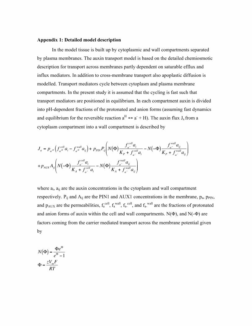
Appendix 1: Detailed model description
In the model tissue is built up by cytoplasmic and wall compartments separated
by plasma membranes. The auxin transport model is based on the detailed chemiosmotic
description for transport across membranes partly dependent on saturable efflux and
influx mediators. In addition to cross-membrane transport also apoplastic diffusion is
modelled. Transport mediators cycle between cytoplasm and plasma membrane
compartments. In the present study it is assumed that the cycling is fast such that
transport mediators are positioned in equilibrium. In each compartment auxin is divided
into pH-dependent fractions of the protonated and anion forms (assuming fast dynamics
and equilibrium for the reversible reaction aH ! a
- + H). The auxin flux Ja from a
cytoplasm compartment into a wall compartment is described by
Ja = paH f
aH
cellai " f
aH
wallaij( ) + pPINPij N #( )
fa"
cellai
KP + fa"
cellai" N "#( )
fa"
wallaij
KP + fa"
wallaij
$
% & &
'
( ) )
+pAUXAij N "#( )fa"
cellai
KA + fa"
cellai" N #( )
fa"
wallaij
KA + fa"
wallaij
$
% & &
'
( ) )
where ai, aij are the auxin concentrations in the cytoplasm and wall compartment
respectively. Pij and Aij are the PIN1 and AUX1 concentrations in the membrane, pa, pPIN,
and pAUX are the permeabilities, facell
, fawall
, fa-cell
, and fa-wall
are the fractions of protonated
and anion forms of auxin within the cell and wall compartments. N("), and N(-") are
factors coming from the carrier mediated transport across the membrane potential given
by
N "( ) ="e
"
e"#1
" =zV
mF
RT
where "=±4.65 has been used assuming a membrane potential Vm=-120 mV (negative
inside). z is the valence, F is the Faraday constant, R is the gas constant, and T is the
absolute temperature.
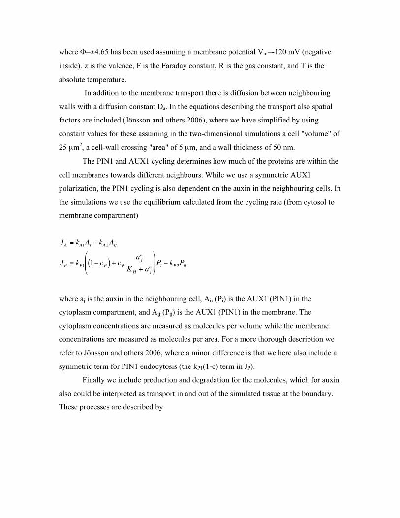
In addition to the membrane transport there is diffusion between neighbouring
walls with a diffusion constant Da. In the equations describing the transport also spatial
factors are included (Jönsson and others 2006), where we have simplified by using
constant values for these assuming in the two-dimensional simulations a cell "volume" of
25 µm2, a cell-wall crossing "area" of 5 µm, and a wall thickness of 50 nm.
The PIN1 and AUX1 cycling determines how much of the proteins are within the
cell membranes towards different neighbours. While we use a symmetric AUX1
polarization, the PIN1 cycling is also dependent on the auxin in the neighbouring cells. In
the simulations we use the equilibrium calculated from the cycling rate (from cytosol to
membrane compartment)
JA = kA1Ai " kA 2Aij
JP = kP1 1" cP( ) + cPa j
n
KH + a j
n
#
$ % %
&
' ( ( Pi " kP 2Pij
where aj is the auxin in the neighbouring cell, Ai, (Pi) is the AUX1 (PIN1) in the
cytoplasm compartment, and Aij (Pij) is the AUX1 (PIN1) in the membrane. The
cytoplasm concentrations are measured as molecules per volume while the membrane
concentrations are measured as molecules per area. For a more thorough description we
refer to Jönsson and others 2006, where a minor difference is that we here also include a
symmetric term for PIN1 endocytosis (the kP1(1-c) term in JP).
Finally we include production and degradation for the molecules, which for auxin
also could be interpreted as transport in and out of the simulated tissue at the boundary.
These processes are described by
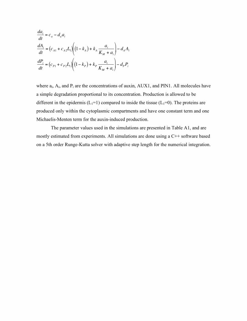
dai
dt= c
a" d
aai
dAi
dt= c
A1+ c
A 2L1( ) 1" kA( ) + k
A
ai
KM
+ ai
#
$ %
&
' ( " dAAi
dPi
dt= c
P1+ c
P 2L1( ) 1" kP( ) + k
P
ai
KM
+ ai
#
$ %
&
' ( " dPPi
where ai, Ai, and Pi are the concentrations of auxin, AUX1, and PIN1. All molecules have
a simple degradation proportional to its concentration. Production is allowed to be
different in the epidermis (L1=1) compared to inside the tissue (L1=0). The proteins are
produced only within the cytoplasmic compartments and have one constant term and one
Michaelis-Menten term for the auxin-induced production.
The parameter values used in the simulations are presented in Table A1, and are
mostly estimated from experiments. All simulations are done using a C++ software based
on a 5th order Runge-Kutta solver with adaptive step length for the numerical integration.
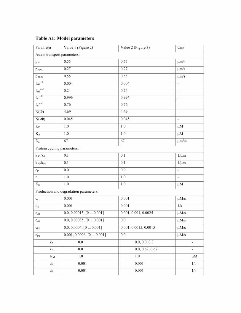
Table A1: Model parameters
Parameter Value 1 (Figure 2) Value 2 (Figure 3) Unit
Auxin transport parameters:
paH 0.55 0.55 µm/s
pPIN_ 0.27 0.27 µm/s
pAUX 0.55 0.55 µm/s
faHcell
0.004 0.004 -
faHwall
0.24 0.24 -
fa-cell
0.996 0.996 -
fa-wall
0.76 0.76 -
N(") 4.69 4.69 -
N(-") 0.045 0.045 -
KP 1.0 1.0 µM
KA 1.0 1.0 µM
Da 67 67 µm2/s
Protein cycling parameters:
kA2/kA1 0.1 0.1 1/µm
kP2/kP1 0.1 0.1 1/µm
cP 0.0 0.9 -
n 1.0 1.0 -
KH 1.0 1.0 µM
Production and degradation parameters:
ca 0.001 0.001 µM/s
da 0.001 0.001 1/s
cA1 0.0, 0.00015, [0 ... 0.001] 0.001, 0.001, 0.0025 µM/s
cA2 0.0, 0.00085, [0 ... 0.001] 0.0 µM/s
cP1 0.0, 0.0004, [0 ... 0.001] 0.001, 0.0015, 0.0015 µM/s
cP2 0.001, 0.0006, [0 ... 0.001] 0.0 µM/s
kA 0.0 0.0, 0.0, 0.8 -
kP 0.0 0.0, 0.67, 0.67 -
KM 1.0 1.0 µM
dA 0.001 0.001 1/s
dP 0.001 0.001 1/s
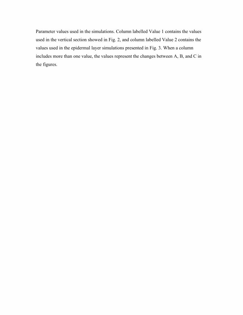
Parameter values used in the simulations. Column labelled Value 1 contains the values
used in the vertical section showed in Fig. 2, and column labelled Value 2 contains the
values used in the epidermal layer simulations presented in Fig. 3. When a column
includes more than one value, the values represent the changes between A, B, and C in
the figures.
Appendix 2: Experimental methods
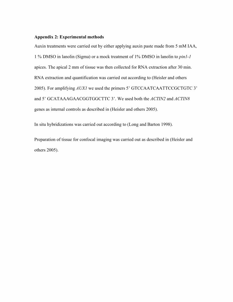
Auxin treatments were carried out by either applying auxin paste made from 5 mM IAA,
1 % DMSO in lanolin (Sigma) or a mock treatment of 1% DMSO in lanolin to pin1-1
apices. The apical 2 mm of tissue was then collected for RNA extraction after 30 min.
RNA extraction and quantification was carried out according to (Heisler and others
2005). For amplifying AUX1 we used the primers 5’ GTCCAATCAATTCCGCTGTC 3’
and 5’ GCATAAAGAACGGTGGCTTC 3’. We used both the ACTIN2 and ACTIN8
genes as internal controls as described in (Heisler and others 2005).
In situ hybridizations was carried out according to (Long and Barton 1998).
Preparation of tissue for confocal imaging was carried out as described in (Heisler and
others 2005).
Figure legends
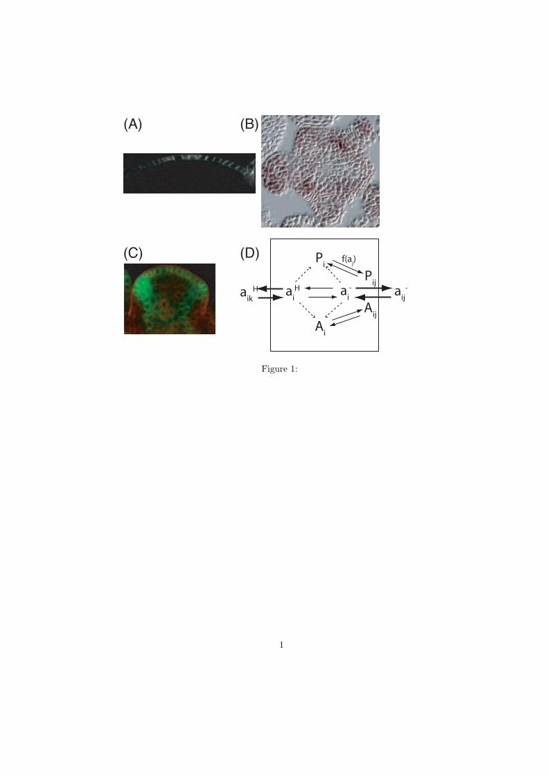
Figure 1. Experimental data and model illustration. A) Longitudinal section showing
AUX1::YFP illustrating the preferential epidermal expression of AUX1. B) Transverse
section of the SAM epidermal layer showing immunostaining of AUX1 mRNA. The
AUX1 is expressed in a phyllotactic pattern similar to assumed auxin localization. C)
Vertical section of PIN1::GFP. D) Illustration of the auxin transport and regulatory
interactions used in the model. Auxin transport between the cell and its surrounding walls
is indicated by solid arrows. The anion transport is mediated by PIN1 and AUX1 in the
membranes. Protein cycling is illustrated by solid arrows where the f(aj) indicates the
dependence on auxin in the neighbouring cell. Dashed arrows indicate the auxin-induced
protein production.
Figure 2. Auxin localization dependence on asymmetrically expressed PIN1 and AUX1
in a longitudinal section simulation. A) and B) show the auxin concentration in the cells
(where the walls in-between have been left out). A) In a model only explicitly including
the efflux mediator PIN1, the equilibrium auxin concentration is high beneath the
epidermal layer when the PIN1 is expressed in the epidermal layer. B) A model that also
includes an asymmetric influx mediator, results in auxin mainly within the epidermal
layer. C) Restrictions on the asymmetry of the influx/efflux mediator expressions apply
to achieve high auxin concentrations in the epidermal cells. The color-coding represents
the measure (aepi - aint)/(aepi + aint) where aepi (aint) is the average auxin concentration in the
epidermal (internal) cells. The axes show the asymmetry where 0 is uniform expression
in all cells and 1 is expression in epidermal cells only.
Figure 3. Auxin peaks in simulations on a static square lattice of cells with walls in-
between. The simulations are started from a close to homogeneous auxin distribution.
Parameter values are described in the text. A) Model without auxin-induced PIN1 and
AUX1. Some minor rearrangements occur, in a stable pattern. B) Model with auxin-
induced PIN1. The pattern is unstable as the peaks move around in the cell tissue. C)
Model with both PIN1 and AUX1 induced by auxin. This results in a stable pattern.
ai-a
iH a
ij-a
ikH
Ai
Pij
Pi
Aij
f(aj)
(A) (B)
(C) (D)
Figure 1:
1
(A) (B) (C)
-0.5
0
0.5
1
PIN
1AUX1
0
0.5
1
0 0.5 1
Figure 2:
(A) (B) (C)t
x
y
t t
x
y
t t
x
y
t
Figure 3:
2