Modeling and Molecular Dynamics of Membrane … and Molecular Dynamics of Membrane Proteins Emad...
83
Modeling and Molecular Dynamics of Membrane Proteins Emad Tajkhorshid Department of Biochemistry, Center for Biophysics and Computational Biology, and Beckman Institute University of Illinois at Urbana-Champagin
-
Upload
vuongkhanh -
Category
Documents
-
view
219 -
download
0
Transcript of Modeling and Molecular Dynamics of Membrane … and Molecular Dynamics of Membrane Proteins Emad...
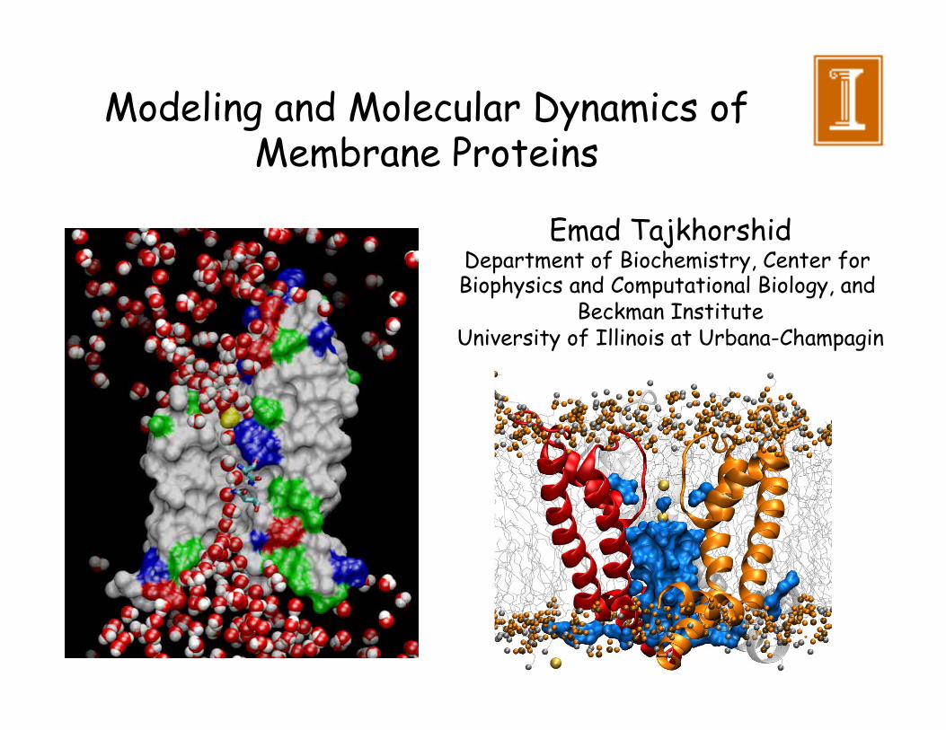
Modeling and Molecular Dynamics of Membrane Proteins
Emad Tajkhorshid Department of Biochemistry, Center for Biophysics and Computational Biology, and
Beckman Institute University of Illinois at Urbana-Champagin

Why Do Living Cells Need Membrane Channels (Proteins)?
Cytoplasm (inside)
Extracellular (outside)
• Living cells also need to exchange materials and information with the outside world
… however, in a highly selective manner.

Phospholipid Bilayers Are Excellent Materials For Cell Membranes
• Hydrophobic interaction is the driving force
• Self-assembly in water • Tendency to close on themselves • Self-sealing (a hole is unfavorable) • Extensive: up to millimeters

Once in several hours! (~ 50 Å in ~ 104 s)
Lipid Diffusion in a Membrane
~9 orders of magnitude slower ensuring bilayer asymmetry
Dlip = 10-8 cm2.s-1 (50 Å in ~ 5 x 10⁻⁶ s)
Dwat = 2.5 x 10⁻⁵ cm2.s-1
Modeling mixed lipid bilayers!

Fluid Mosaic Model of Membrane
Flip-flop Forbidden
Lateral Diffusion Allowed Ensuring the conservation of membrane asymmetric structure
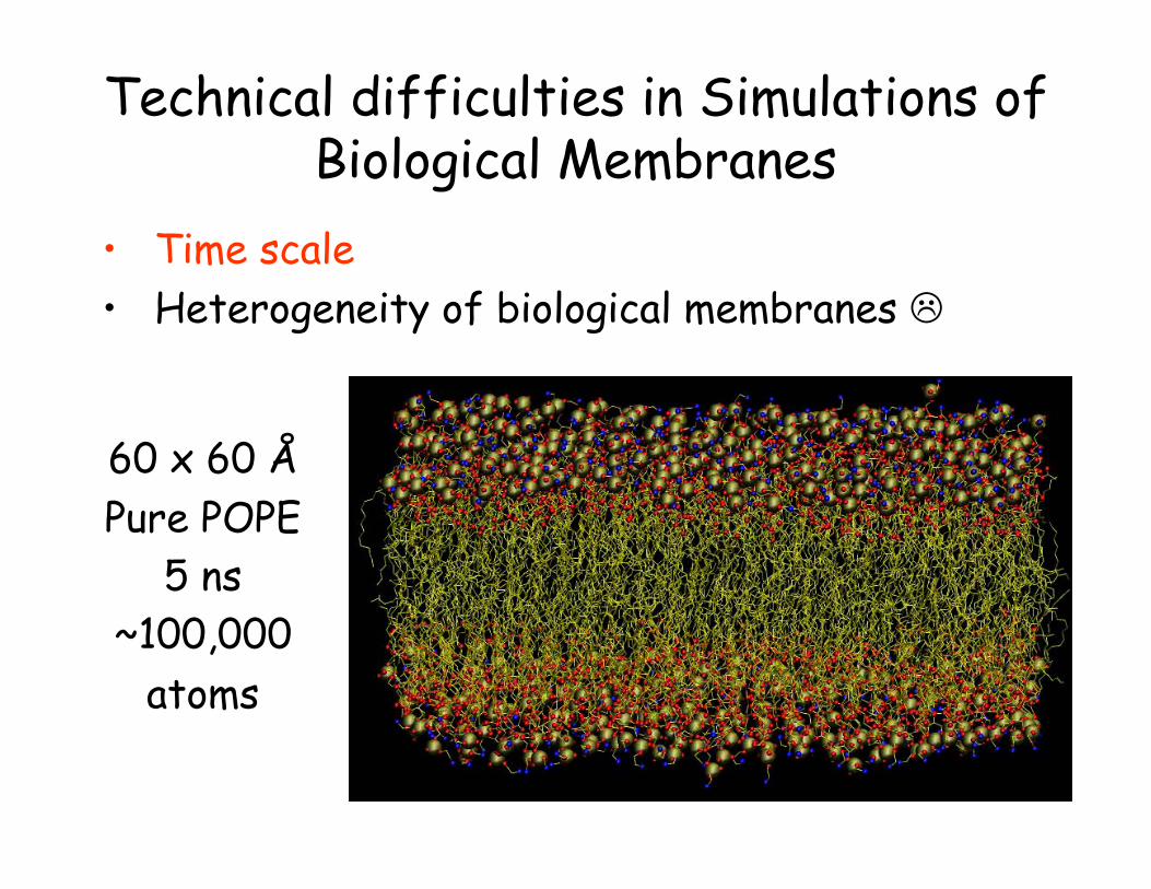
Technical difficulties in Simulations of Biological Membranes
• Time scale • Heterogeneity of biological membranes
60 x 60 Å Pure POPE
5 ns ~100,000
atoms

Coarse-grained modeling of lipids
9 particles!
150 particles
Also, increasing the time step by orders of magnitude.

by: J. Siewert-Jan Marrink and Alan E. Mark, University of Groningen, The Netherlands

• Pure lipid: insulation (neuronal cells) • Other membranes: on average 50% • Energy transduction membranes (75%)
Membranes of mitocondria and chloroplast Purple membrane of halobacteria
• Different functions = different protein composition
Protein/Lipid ratio
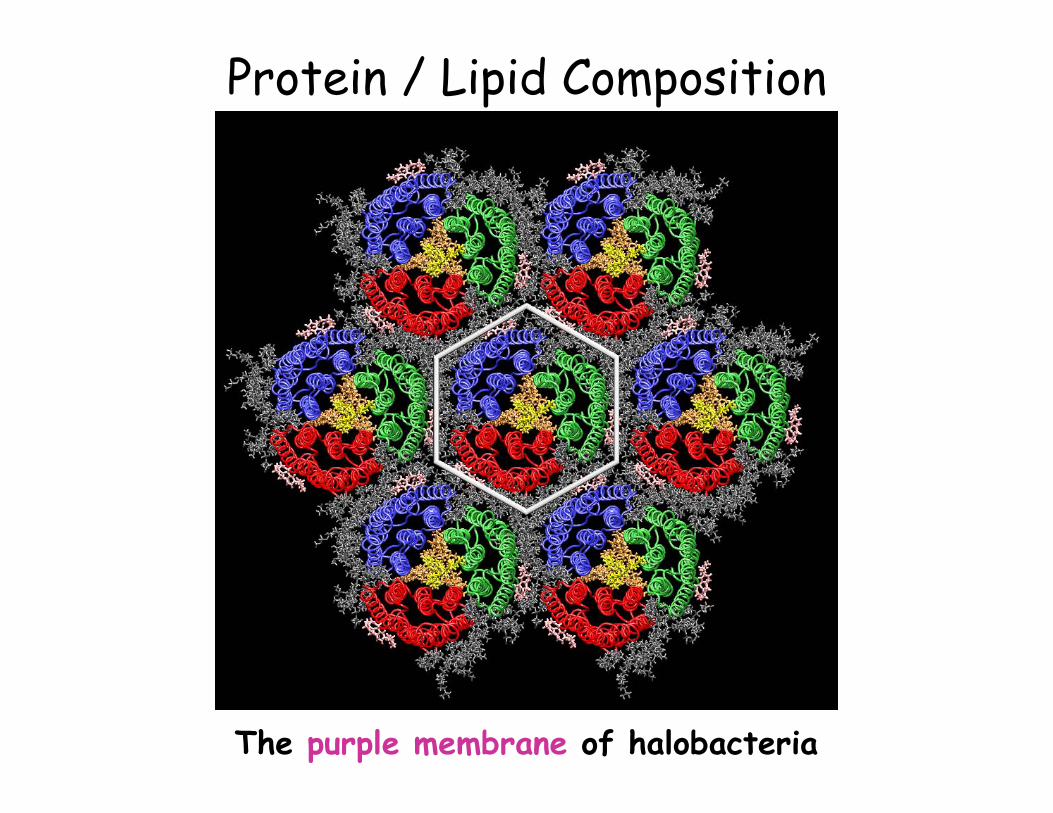
Protein / Lipid Composition
The purple membrane of halobacteria

Gramicidin A Might be very sensitive to the lipid head
group electrostatic and membrane potential

Central cavity

Analysis of Molecular Dynamics Simulations of Biomolecules
• A very complicated arrangement of hundreds of groups interacting with each other
• Where to start to look at?
• What to analyze?
• How much can we learn from simulations?
It is very important to get acquainted with your system
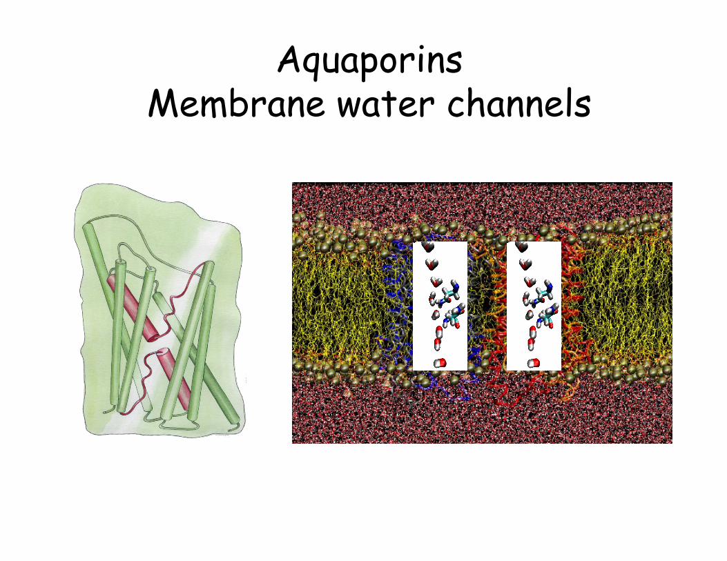
Aquaporins Membrane water channels

Monomeric pores Water, glycerol, …
Tetrameric pore Perhaps ions???

• Tetrameric architecture • Amphipatic channel interior • Water and glycerol transport • Protons, and other ions are
excluded • Conserved asparagine-proline-
alanine residues; NPA motif • Characteristic half-membrane
spanning structure
~100% conserved -NPA- signature sequence NPA NPAR N C E E
Functionally Important Features

A Semi-hydrophobic channel

Molecular Dynamics Simulations Protein: ~ 15,000 atoms Lipids (POPE): ~ 40,000 atoms Water: ~ 51,000 atoms Total: ~ 106,000 atoms
NAMD, CHARMM27, PME
NpT ensemble at 310 K
1ns equilibration, 4ns production 10 days /ns – 32-proc Linux cluster
3.5 days/ns - 128 O2000 CPUs
0.35 days/ns – 512 LeMieux CPUs
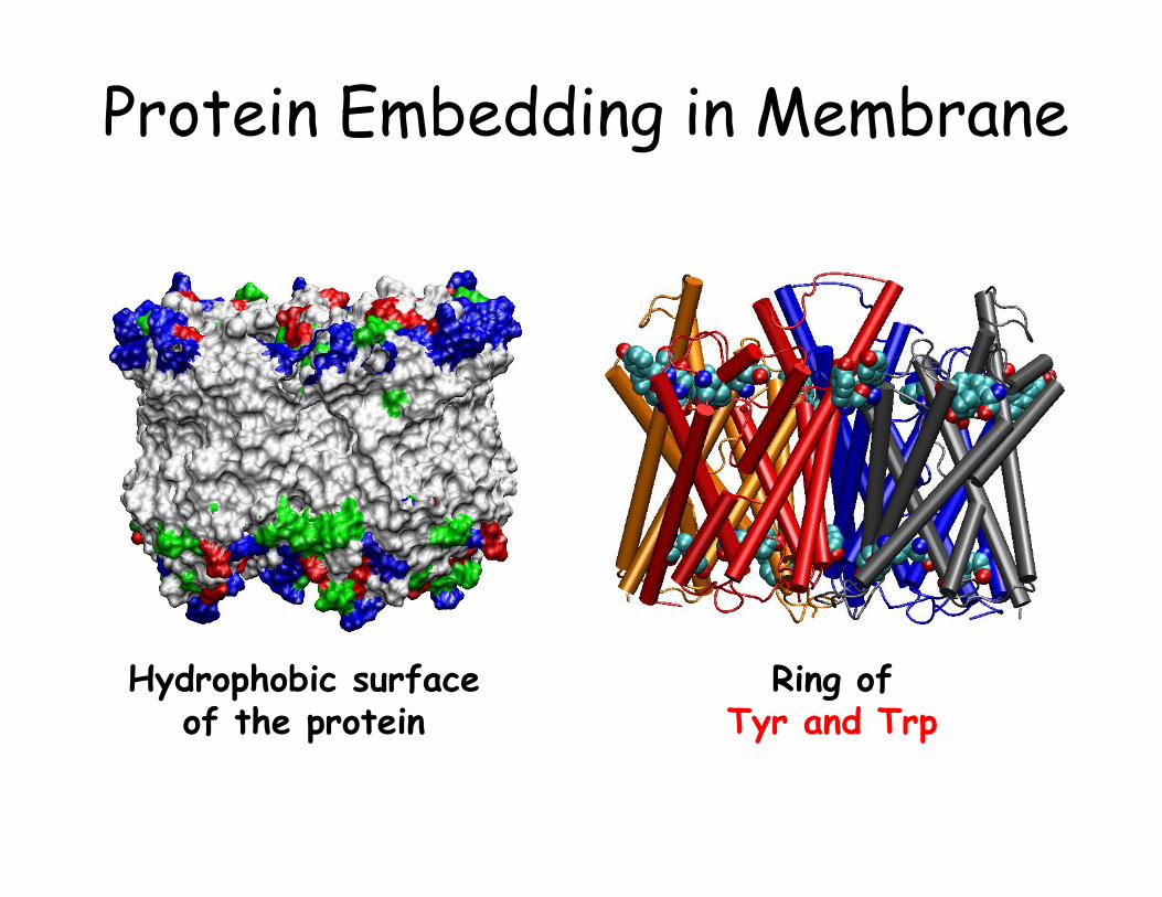
Protein Embedding in Membrane
Ring of Tyr and Trp
Hydrophobic surface of the protein

Embedding GlpF in Membrane
77 A
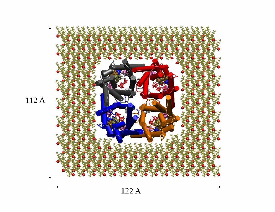
122 A
112 A

A Recipe for Membrane Protein Simulations • Align the protein along the z-axis (membrane normal): OPM, Orient.
• Decide on the lipid type and generate a large enough patch (MEMBRANE plugin in VMD, other sources). Size, area/lipid, shrinking.
• Overlay the protein with a hydrated lipid bilayer. Adjust the depth/height to maximize hydrophobic overlap and matching of aromatic side chains (Trp/Tyr) with the interfacial region
• Remove lipids/water that overlap with the protein. Better to keep as many lipids as you can, so try to remove clashes if they are not too many by playing with the lipids. Add more water and ions to the two sides of the membrane (SOLVATE / AUTOIONIZE in VMD)
• Constrain (not FIX) the protein (we are still modeling, let’s preserve the crystal structure; fix the lipid head groups and water/ion and minimize/simulate the lipid tails using a short simulation.

A Recipe for Membrane Protein Simulations • Continue to constrain the protein (heavy atoms), but release
everything else; minimize/simulate using a short “constant-pressure” MD (NPT) to “pack” lipids and water against the protein and fill the gaps introduced after removal of protein-overlapping lipids.
• Watch water molecules; They normally stay out of the hydrophobic cleft. If necessary apply constraints to prevent them from penetrating into the open cleft between the lipids and the protein.
• Monitor the volume of your simulation box until the steep phase of the volume change is complete (.xst and .xsc files). Do not run the system for too long during this phase (over-shrinking; sometimes difficult to judge).
• Now release the protein, minimize the whole system, and start another short NPT simulation of the whole system.
• Switch to an NPnAT or an NVT simulation, when the system reaches a stable volume. Using the new CHARMM force field, you can stay with NPT.
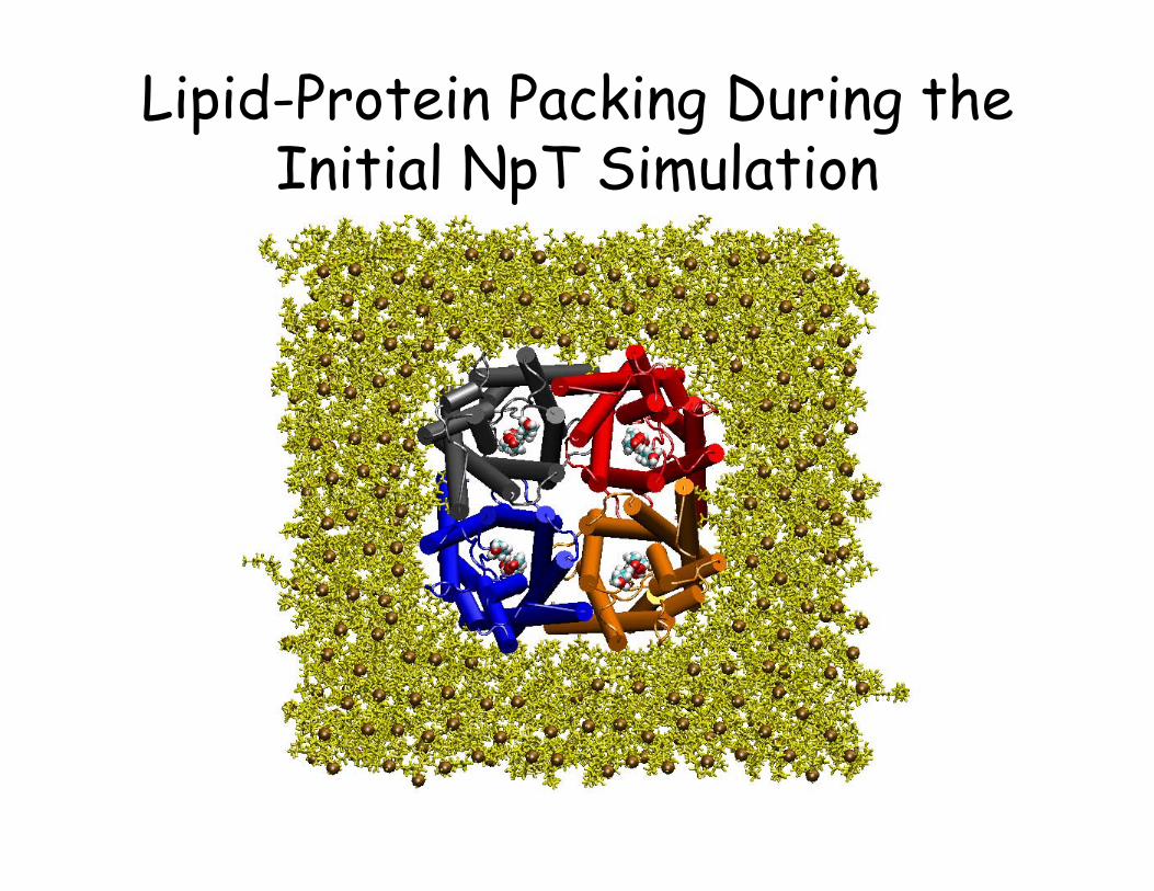
Lipid-Protein Packing During the Initial NpT Simulation
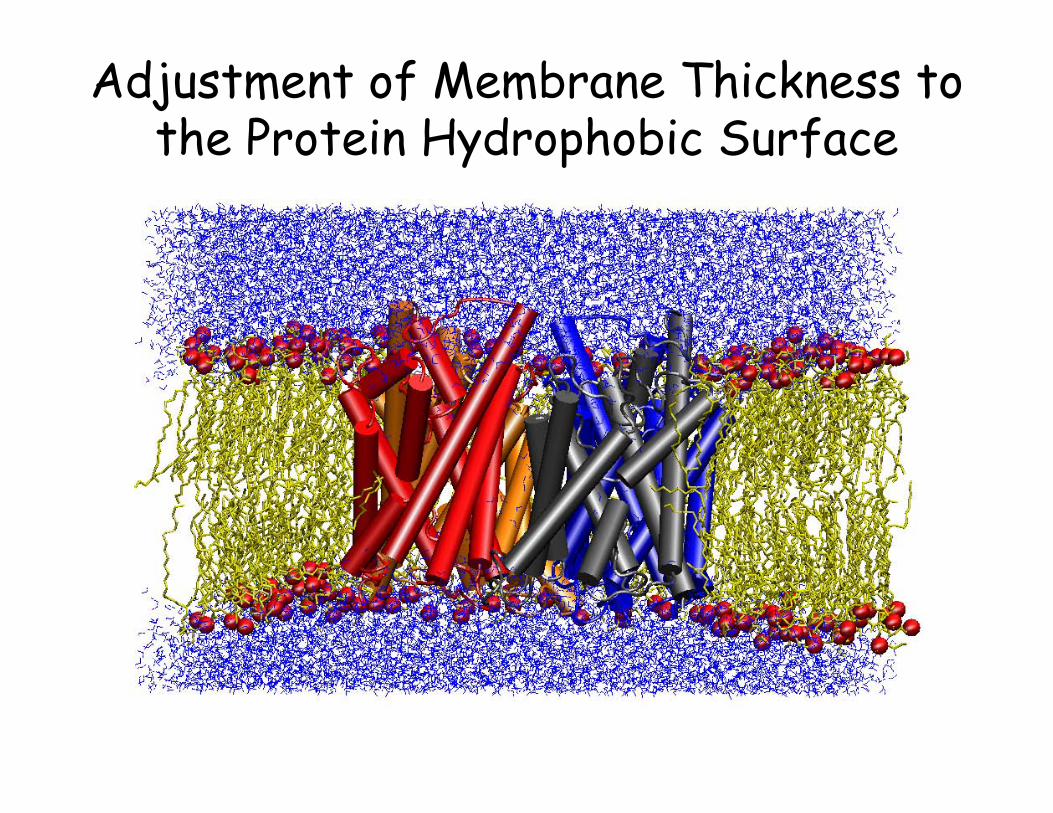
Adjustment of Membrane Thickness to the Protein Hydrophobic Surface

Glycerol-Saturated GlpF


Description of full conduction pathway
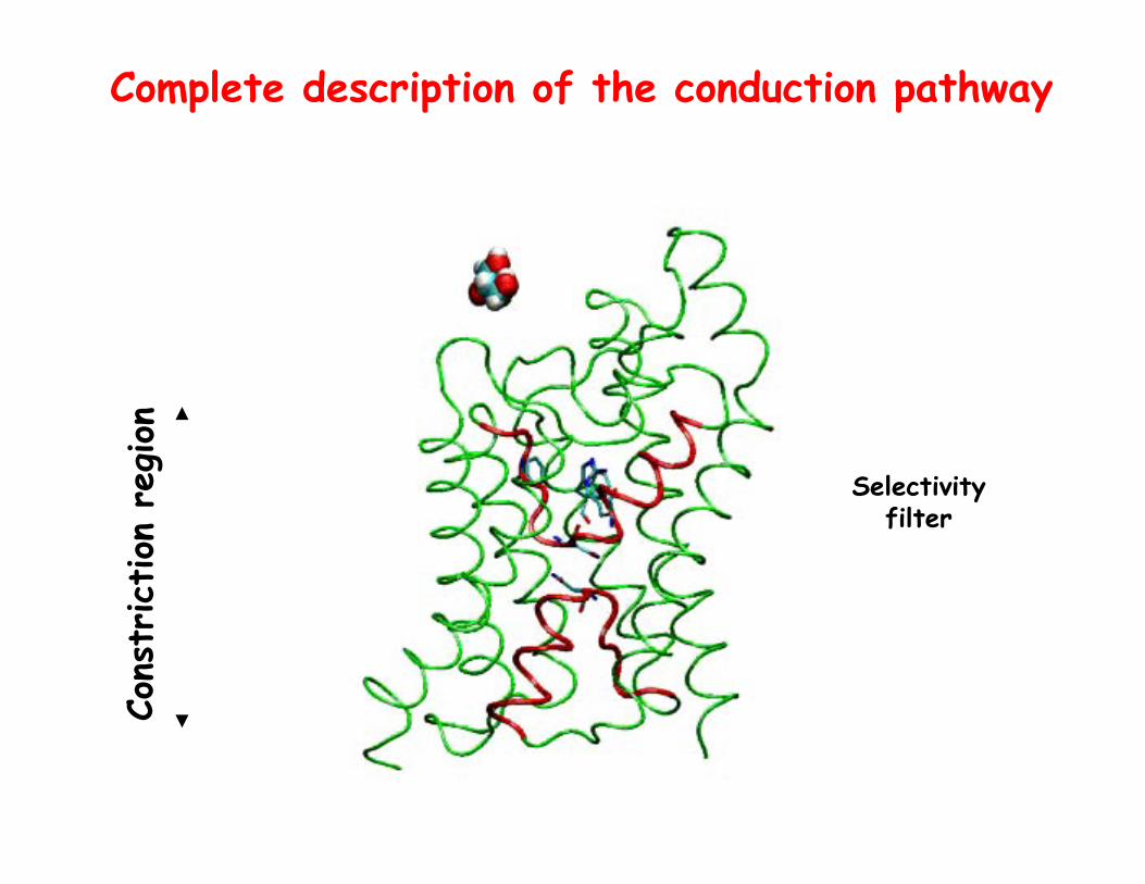
Complete description of the conduction pathway
Selectivity filter
Cons
triction
reg
ion

Channel Hydrogen Bonding Sites
…
{set frame 0}{frame < 100}{incr frame}{
animate goto $frame set donor [atomselect top “name O N and within 2 of (resname GCL and name HO)”] lappend [$donor get index] list1 set acceptor [atomselect top “resname GCL and name O and within 2 of (protein and name HN HO)”] lappend [$acceptor get index] list2
}
…
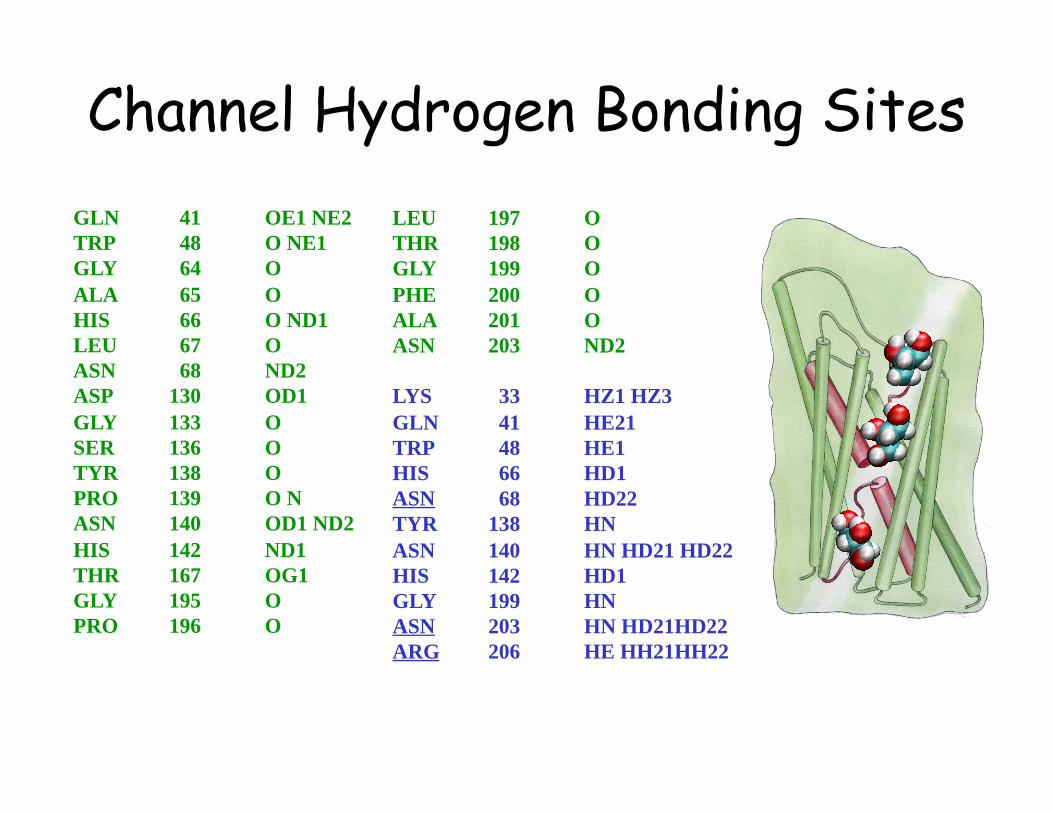
GLN 41 OE1 NE2 TRP 48 O NE1 GLY 64 O ALA 65 O HIS 66 O ND1 LEU 67 O ASN 68 ND2 ASP 130 OD1 GLY 133 O SER 136 O TYR 138 O PRO 139 O N ASN 140 OD1 ND2 HIS 142 ND1 THR 167 OG1 GLY 195 O PRO 196 O
LEU 197 O THR 198 O GLY 199 O PHE 200 O ALA 201 O ASN 203 ND2
LYS 33 HZ1 HZ3 GLN 41 HE21 TRP 48 HE1 HIS 66 HD1 ASN 68 HD22 TYR 138 HN ASN 140 HN HD21 HD22 HIS 142 HD1 GLY 199 HN ASN 203 HN HD21HD22 ARG 206 HE HH21HH22
Channel Hydrogen Bonding Sites

The Substrate Pathway is formed by C=O groups

Non-helical motifs are stabilized by two glutamate residues.
NPA NPAR N C E E
The Substrate Pathway is formed by C=O groups

Conservation of Glutamate Residue in Human Aquaporins

Glycerol – water competition for hydrogen bonding sites
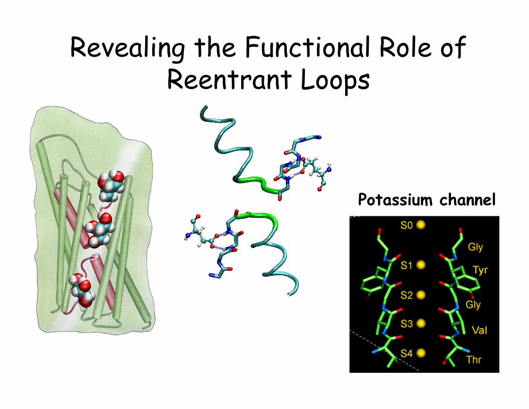
Revealing the Functional Role of Reentrant Loops
Potassium channel

AqpZ vs. GlpF • Both from E. coli • AqpZ is a pure water channel • GlpF is a glycerol channel • We have high resolution structures for both channels

Steered Molecular Dynamics is a non-equilibrium method by nature
• A wide variety of events that are inaccessible to conventional molecular dynamics simulations can be probed.
• The system will be driven, however, away from equilibrium, resulting in problems in describing the energy landscape associated with the event of interest.
W G≥ ΔSecond law of thermodynamics

work W heat Q
λ = λi
λ = λ(t) λ = λf
T T
Transition between two equilibrium states
Jarzynski’s Equality
W G≥ Δ
In principle, it is possible to obtain free energy surfaces from repeated non-equilibrium experiments.
C. Jarzynski, Phys. Rev. Lett., 78, 2690 (1997) C. Jarzynski, Phys. Rev. E, 56, 5018 (1997)
W Ge eβ β− − Δ=1
Bk Tβ =
p(W)
WGΔ
e-βWp(W)
f iG G GΔ = −
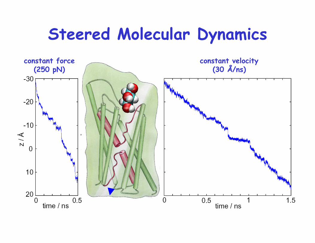
constant velocity (30 Å/ns)
constant force (250 pN)
Steered Molecular Dynamics

Trajectory of glycerol pulled by constant force
SMD Simulation of Glycerol Passage

4 trajectories v = 0.03, 0.015 Å/ps k = 150 pN/Å
Constructing the Potential of Mean Force
])([)( 0 vtztzktf −−−=
∫ ʹ′ʹ′=t
0)()( tvftdtW

• Captures major features of the channel • The largest barrier ≈ 7.3 kcal/mol; exp.: 9.6±1.5 kcal/mol
Features of the Potential of Mean Force
Jensen et al., PNAS, 99:6731-6736, 2002.

Features of the Potential of Mean Force
Asymmetric Profile in the Vestibules
Periplas
m
Cyto
plas
m
Jensen et al., PNAS, 99:6731-6736, 2002.

Artificial induction of glycerol conduction through AqpZ
Y. Wang, K. Schulten, and E. Tajkhorshid Structure 13, 1107 (2005)
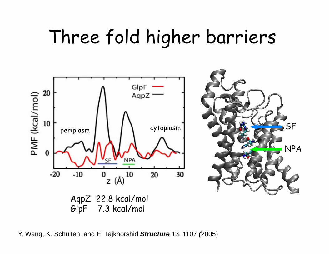
Three fold higher barriers
AqpZ 22.8 kcal/mol GlpF 7.3 kcal/mol
periplasm cytoplasm SF
NPA
Y. Wang, K. Schulten, and E. Tajkhorshid Structure 13, 1107 (2005)

Could it be simply the size?
Y. Wang, K. Schulten, and E. Tajkhorshid Structure 13, 1107 (2005)

It is probably just the size that matters!
Y. Wang, K. Schulten, and E. Tajkhorshid Structure 13, 1107 (2005)
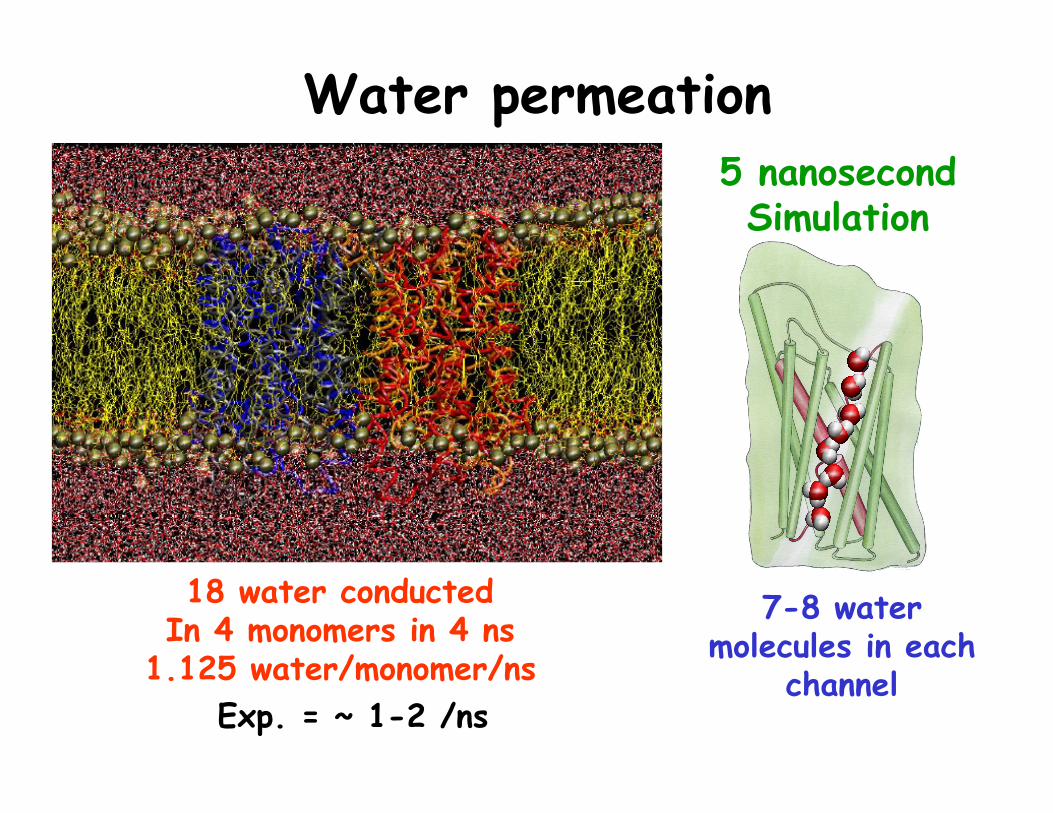
Water permeation
7-8 water molecules in each
channel
5 nanosecond Simulation
18 water conducted In 4 monomers in 4 ns
1.125 water/monomer/ns Exp. = ~ 1-2 /ns


Correlated Motion of Water in the Channel Water pair correlation
The single file of water molecules is maintained.

Experimental value for AQP1: 0.4-0.8 e-5
One dimensional diffusion: 202 ( )tDt z z= −
Diffusion of Water in the channel

Diffusion of Water in the channel 2
02 ( )tDt z z= −
0 1 2 3 4 Time (ns)
Improvement of statistics

Water Bipolar Configuration in Aquaporins

Water Bipolar Configuration in Aquaporins


One of the most useful advantages of simulations over experiments is that you can modify the system as you wish: You can do modifications that are not even possible at all in reality!
This is a powerful technique to test hypotheses developed during your simulations. Use it!
R E M E M B E R:

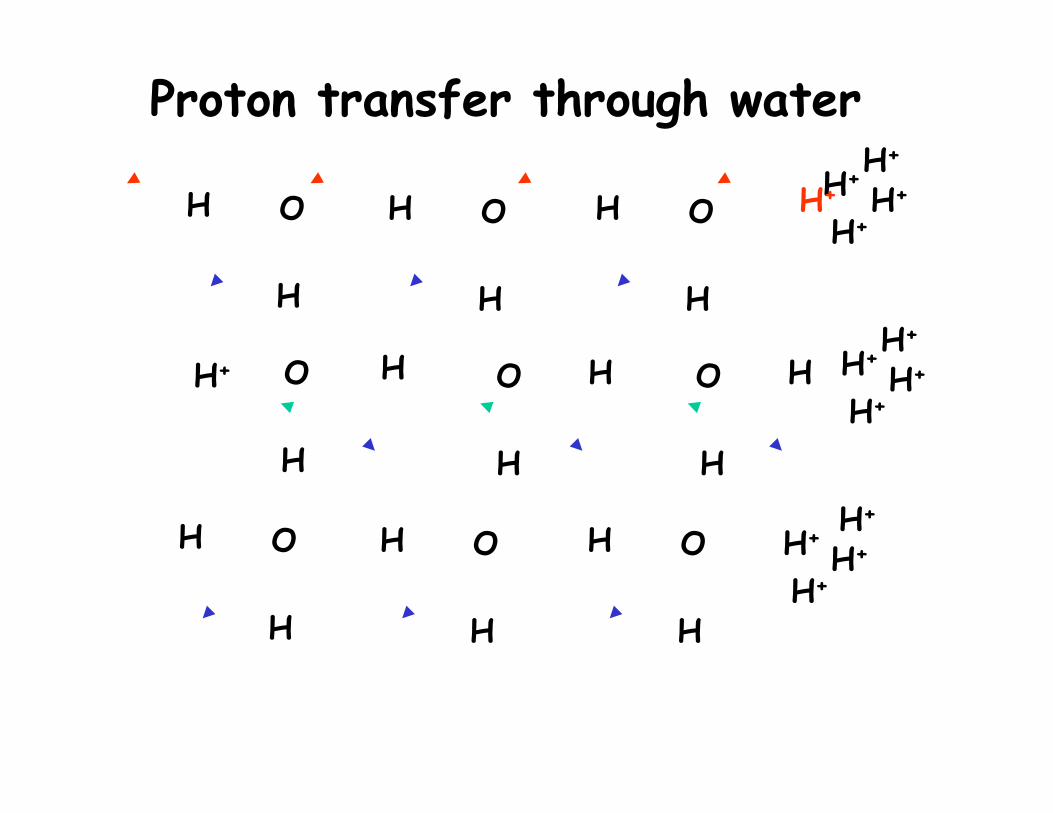
Electrostatic Stabilization of Water Bipolar Arrangement
H
H
O H
H
O H
H
O
H
H
O H
H
O H
H
O H+ H+
H+
H+ H+
H+
H+
H+ H+ H+
H+ H
H
O H
H
O H
H
O H+
H+ H+
Proton transfer through water

K+ channel
Cl- channel
Aquaporins


A Complex Electrostatic Interaction
“Surprising and clearly not a hydrophobic channel”
M. Jensen, E. Tajkhorshid, K. Schulten, Biophys. J. 85, 2884 (2003)
SF
NPA

A Repulsive Electrostatic Force at the Center of the Channel
QM/MM MD of the behavior of an excessive proton
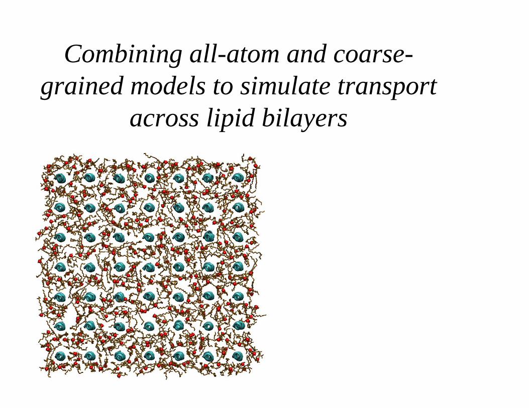
Combining all-atom and coarse-grained models to simulate transport
across lipid bilayers

Peptide aggregation and “Pore” formation in lipid bilayers

Alamethicin
20 residue antimicrobial peptide
• 20-residue peptide
• No charge
• forming pores in the membrane

CG molecular systems allow for time scales of 3-4 orders of magnitude longer,
because: • Significant reduction of the degrees of freedom (or number of interacting particles/beads) • Softer potentials allowing much longer time steps
µm length scale and µs time scale Bilayer, micelle, and vesicle formation Fusion of bilayers and vesicles, …

Alamethicin
20 residue antimicrobial peptide
• 20-residue peptide
• No charge
• forming pores in the membrane

Simulation Setting
• 49 peptides in 288 DMPC
• All-atom model equilibrated 1ns
• Converted to a CG model
• Simulated for 1µs
• 0.5 µs snapshot was reverse-corarse-grained to an all atom model
• All atom model simulated for 20 ns

Coarse-Graining
Employs CHARMM-like force fields parameterized using all-atom simulations
Four-to-one mapping P N C Q
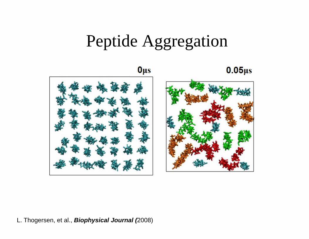
Peptide Aggregation
L. Thogersen, et al., Biophysical Journal (2008)

0
0.02
0.04
0.06
0.08
0 10 20 30 40R / Å
g(R
) for
pep
tides
A g (R )init
t =1_s
0
10
20
30
40
50
0 0.2 0.4 0.6 0.8 1Time / _s
Num
ber o
f clu
ster
s B
0
1
2
3
0 0.2 0.4 0.6 0.8 1Time / _s
Av.
num
ber o
f nei
ghbo
rs C
0
500
1000
0 0.1 0.2 0.3Time / _s
MSD
/ Å
2
lipidpeptide
D

What brings the helices together?

Peptide Insertion
L. Thogersen, et al., Biophysical Journal (2008)
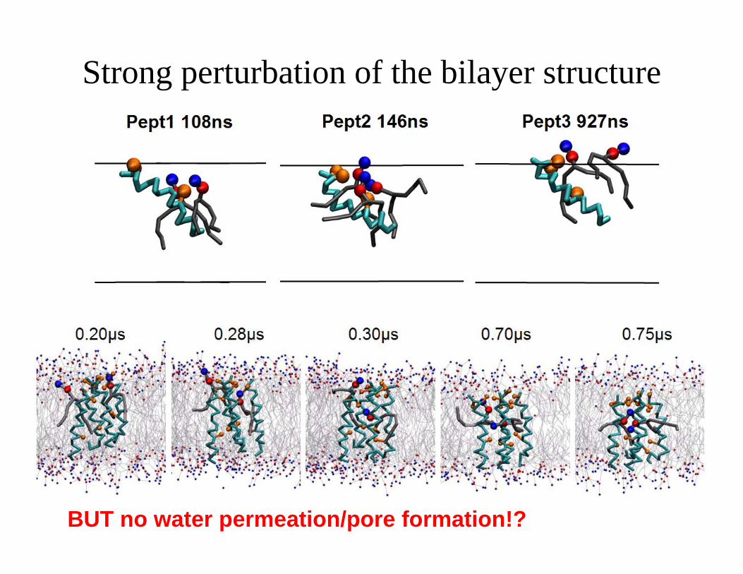
Strong perturbation of the bilayer structure
BUT no water permeation/pore formation!?

Hydration of the head group region in coarse-grained and all-atom models

Reverse Coarse-Graining
X X

Reverse Coarse-Graining • Mapping back CG beads to all-atom clusters • Re-solvating the system • 5000 steps of minimization • Simulated annealing for 20 ps (T changing from
610K to 300K, ΔT = -10K) while constraining atoms to the position of the corresponding CG beads

Hydration of the head group region in coarse-grained and all-atom models

Alemethicin-induced Membrane Poration

Alemethicin-induced Membrane Poration

Alemethicin-induced Membrane Poration