MOBILIZATION - Clinician's View€¦ · · 2015-10-19Joint mobilization is indicated when the...
27
MOBILIZATION FOR THE NEUROLOGICALLY INVOLVED CHILD Assessment and Application Strategies For Pediatric PTs and OTs Sandy Brooks-Scott M.S., PPT, PCS www.clinicians-view.com
Transcript of MOBILIZATION - Clinician's View€¦ · · 2015-10-19Joint mobilization is indicated when the...

MOBILIZATION FOR THE NEUROLOGICALLY INVOLVED CHILD
Assessment and Application Strategies For Pediatric PTs and OTs
Sandy Brooks-Scott M.S., PPT, PCS
www.clinicians-view.com

Chapter I
Pathology and Resultant Immobility
Mobility is a worthy goal. However, before it can be achieved in a child with neurological dysfunction, certain prerequisites must be in place: normal bony alignment; normal muscle
strength, flexibility and endurance; a coordinating nervous system; an efficient cardiorespiratory system; and normal connective tissue flexibility. Immobility after initial neurological
insult affects all these systems, making assessment and treatment of all systems interfering with efficient mobility a paramount goal of the therapist.
Neurological Damage-Original Insult
The motor result of the neurological insult is dependent upon the age, the location, and the extent of the insult (Brann 1988; Costello et al. 1988; Nelson 1988; Pape and Wigglesworth
1979). The most widely discussed reason for brain damage in the pre-, peri- or initial postnatal stages is a change in blood pressure, causing the vessels in the developing organ to hemorrhage or become ischemic (Pape and Wigglesworth 1979). Nelson (1988) notes
that most infants who live through severe asphyxia at birth do not develop cerebral palsy or mental retardation. Brann (1988), in his discussion of the effects of acute total and
prolonged partial asphyxia, clearly demonstrates that the neurological changes and motor outcomes vary depending on the acuteness, duration, and severity of the asphyxia.
Because a child will not have autoregulatory mechanisms to control blood pressure until 3
months of age, excessive heat loss, abnormal partial pressures of oxygen and carbon dioxide, or fluid imbalances can affect a newborn's blood pressure, producing ischemic hypoxia and
neural damage. Hypoxia is a decrease in oxygen to the nerve ceil, which can occur as a result of a hemorrhage or ischemic attack. Hemorrhage occurs when blood leaves the vascular
system, drowning the nerve cells, and ischemia is a decrease in blood flow to the nerves. Either event decreases the oxygen available to the nerve cell, resulting in its death.
User
Rectangle
User
Rectangle
User
Rectangle
User
Text Box
3

Pathology and Resultant Immobility
trauma or meningitis than in hypoxic events. Flaccidity may be a result of damage to the
cerebellum or peripheral nerves and can be maintained or later overshadowed by spasticity; it should not be confused with muscle weakness. Flaccidity is a decrease in stiffness;
conversely, muscle weakness is an inability to create tension sufficient to raise the body part against gravity and age-appropriate resistance. Spasticity is increased stiffness clinically
identified as an increased resistance to high velocity muscle stretch. It is a result of neuro
logical damage. Similar stiffness may be mimicked by muscle spasm, emotional state, and connective tissue tightness.
Taken together, the above factors all affect stiffness. Stiffness is a set of behaviors including muscle activation, response to stretch, kinematic patterns, conscious control, fiber patterns,
and passive elastic properties of muscles and ligaments (Guiliani 1991). Neurological
damage does not cause stiffness, yet stiffness can be affected by it. All factors contributing
to stiffness must be ruled out before spasticity is implicated as the cause of the motor
problem. Consequently, before movement anomalies can be linked to manifestations of spasticity, the role of the other systems' contributions to stiffness, including spasm,
weakness, and connective tissue tightness, must be ruled out. Spasm, weakness, and connective tissue tightness are often sequelae of immobility frequently seen in infants and
children with neurological damage.
Immobility and its Effects
Immobility is frequently more detrimental than the damage that causes the immobility in
the first place, and it affects all tissues of the body (Kessler and Hertling 1983; Kottke 1966;
Salter 1978). Within days of immobility, several physiological changes occur:
> bones lose density
> muscles atrophy and weaken
> synapses deteriorate
> cardiorespiratory inefficiency develops
> connective tissue tightens
Bones Lose Density
Immobility results in calcium loss in bone (Cornwall 1984). Abnormal stresses on weak
bones from incorrect jOint alignment may be responsible for some of the malformations frequently seen in children with cerebral palsy, such as foot deformities and hip dislocations
(Bleck 1987; Cusick 1990). Immobility results in permanent deformity by maintaining infantile bone shape or allowing bones to twist as in some foot deformities or scoliosis
(Badgley 1949; Beals 1969; Raney and Brashear 1971; Salter 1978).
User
Rectangle
User
Rectangle
User
Rectangle
User
Text Box
5

Chapter 2
Principles of Mobilization
In the literature on the management of children with cerebral palsy, the focus has been on
neurological approaches. No texts have discussed application of mobilization to the
management of cerebral palsy. Before mobiliZation can be applied to the management of cerebral palsy, an overview of mobilization is necessary.
This chapter discusses techniques designed to stretch extra-articular structures of synovial jOints. Key concepts to consider in the application of the techniques are joint play, grades of
motion, close-packed or open-packed positions, and the rule of convex and concave.
Joint Play
Joint play is the term for the accessory movements that can be pasSively but not actively
performed at the joint (Kessler and Herding 1983; Maitland 1977) and that enable smooth
gliding of joint surfaces. Joint play is achieved when the extra-articular connective tissue structures are flexible enough to allow normal roll and spin of the articular surfaces, a
prerequisite for normal active range of motion. To achieve normal joint play, grades of
motion are applied in open-pack positions in the direction determined by the rule of
convex and concave.
Grades of Motion
Grades of motion are the passive rhythmic oscillations applied to a joint to increase its
extra-articular connective tissue flexibility (Kessler and Hertling 1983; Maitland 1977). Choice of grade to use depends on the joint's irritability and degree of restriction. Four
"
User
Rectangle
User
Rectangle
User
Rectangle

Mobilization for the Neurologically Involved Child
grades of motion are defined (see Figure 2.1), Grades 1 and 2 decrease irritability in
inflamed or painful joints, and Grades 3 and 4 increase flexibility in joints with extra
articular Joint restrictions. Grades 3 and 4 are frequently used in patients with neurological
impairment to increase the flexibility of jOint structures that have tightened after immobility.
Because these joints are seldom in acute trauma, grades 1 and 2 are used less frequently.
Close-Pack/Open-Pack Positions
In a joint's close-packed position, joint surfaces are maximally congruent; also, capsule and
ligaments become twisted, causing the joint surfaces to approximate and lock so maximum
stability is achieved (Kessler and Herding 1983; Maitland 1977). Any other position is open
pack for that joint.
Table 2.1 lists the major joints and their close-packed positions. Notice that all the close
packed positions are positions in which the joint has a stability function (Le., single limb
stance is close-packed).
------------------ Normal End of Range
Pathological Limit of Range
IVI II III
'----...I.....I..----'-'----.........I..--...r.--Beginning of Range
Figure 2.1. Grades of Motion Reproduced by permission of The Saunders Group, Inc. © 1996
12
User
Rectangle

----------------------------------------------------------
Principles of Mobilization
Table 2.1 Close-Pack Positions for the Joints
Joint Close-Pack Position
Hip Full extension with adduction, and internal rotation.
Knee Full extension with external rotation.
Ankle Full dorsiflexion for talocrural joint; supination for subtalor and midtarsal jOints.
Shoulder Maximal abduction and external rotation.
Elbow Full extension/supination for humeroulnar joint; elbow flexion at 90° and supination at 5° for humeroradial joint.
Wrist Full extension with radial deviation.
Rule of Convex and Concave
The rule of convex and concave determines the direction in which force is applied to
achieve a desired range. Articular surfaces move relative to the shafts depending upon
whether the convex or concave surface is stabilized or frxed. Hence, if the concave surface is stabilized (as the tibia in non-weightbearing dorsiflexion), the convex articular surface of the
talus moves in the opposite direction of the shaft (body) of the talus. Conversely, if the convex talus is frxed (as in standing), the concave surface of the tibia moves in the same
direction as the shaft of the tibia to achieve dorsiflexion. Figure 2.2 illustrates this concept.
Applying this rule, the head of the talus must glide posteriorly relative to the tibia to achieve
ankle dorsiflexion.
Table 2.2 summarizes the major movements used in mobilization for the neurologically impaired client.
13
User
Rectangle
User
Rectangle
User
Rectangle

Mobilization for the Neurologically Involved Child
Table 2.2 Common Mobilization Techniques for the Neurologically Impaired Child
Desired Motion Direction of Force
Hip flexion Femur glides posteriorly and inferiorly.
Hip extension Femur glides anteriorly.
Hip abduction Femur glides medially and inferiorly.
Knee extension If in the last ISo of extension, tibia glides externally; if greater than
the last ISo of extension, tibia glides anteriorly. ~~------~--------
Knee flexion If in the last 15° of extension, tibia glides internally; if greater
than the last 15° on extension, the tibia glides posteriorly.
Ankle dorsiflexion Talus glides posteriorly and the fibula and tibia separate at the mortise.
Shoulder flexion Humerus glides inferiorly, clavicle depresses and rotates at the
sternal articulation.
Shoulder abduction Humerus glides inferiorly, clavicle depresses and rotates at the
sternal articulation.
Supination Head of the radius rotates on the ulna.
Wrist extension Capitate glides palmarly on the scaphoid, the scaphoid glides dorsally
on the lunate, then the scaphoid glides palmarly on the radius.
General Rules of Mobilization
The following are five general rules to observe while using mobilization techniques:
1. The patient must be relaxed.
2. The therapist must be relaxed.
3. One hand stabilizes a body part while the other hand mobilizes its
articulating part.
4. The therapist considers the direction of movement, the velOCity of
movement, and the amplitude of movement.
5. One movement at a time, one joint at a time.
14
User
Rectangle
User
Rectangle

Principles of Mobilization
Indications and Contraindications
Joint mobilization is indicated when the extra-articular connective tissue abnormally restricts
motion of that joint. For example, babies have a 60-degree hip flexion contracture at birth
due to connective tissue tightness (Lee 1977). Many children with cerebral palsy also have a
hip flexion contracture due to connective tissue tightness. Mobilization of the hip joint in
infants is contraindicated because they have normal limitations and the ability to overcome
them with their own oscillatory end-range movements when kicking. Mobilization would be
indicated in the older child with cerebral palsy exhibiting abnormal range limitations
interfering with function caused by extra-articular tissue tightness.
Contraindications fall into four categories:
1. Risk of fracture. The forces in mobilization can cause fractures in weak bones.
Osteogenesis imperfect a or a history of pathological fractures is a contraindication
to mobilization.
2. Joint inflammation. Stretching of articular tissue is contraindicated when the tissue
itself is under stress such as the stretch from inflammation, as in acute juvenile
rheumatoid arthritis.
3. Desired hypomobility. Extra-articular tissue stretching is contraindicated when
fixation is required. Mobilization would be contraindicated at the subtalor joint
following a triple arthrodesis in a child with cerebral palsy but may be indicated at
the talocrural joint subsequent to the immobility from the casting for the surgery
4. Hypermobility. Excessive mobility in a given direction at a specific joint is a contraindi
cation to mobilization, but the techniques may be appropriate in a different direction
at the same joint or in a related joint. For example, it is contraindicated to mobilize
the glenohumeral joint in a child with cerebral palsy with a tendency to sublux the
glenohumeral joint, but mobilization of the sternoclavicular joint may be indicated to
increase shoulder girdle range of motion without stressing the glenohumeral joint.
Figure 2.2. Rule of Convex and Concave
IS
User
Rectangle
User
Rectangle
User
Rectangle

System Assessment
The proposed strength items have not been tested for reliability for muscle testing. They
are frequently used informally in the clinic to document that children are "slightly better" after therapy or surgery because they can now perform these items. Because the items are used informally without a consistent pattern, this attempt to standardize terms and apply
them clinically to children with cerebral palsy is intended to be a starting point for discussion and research. Until reliability and validity can be determined, clinical notation
should define a muscle strength grade in terms of function (Le., fair strength is the ability to move the part full range antigravity with no resistance).
Endurance
Muscular endurance can be a problem for children with neurological impairments. Children
who can perform two to three repetitions of a muscle contraction but then begin to substitute other muscles may be showing poor muscular endurance. Functional mobility often reqUires frequent contractions. For example, if a normal child ambulates 150 steps per
minute, the gastrocnemius unilaterally contracts 75 times in a minute. Now, if the child with cerebral palsy shows appropriate length and strength in the gastrocsoleous but still can only
ambulate a few steps with appropriate push-off, the problem in carryover outside of therapy may be one of muscular endurance. Specific techniques to increase endurance must then
be integrated into the therapeutic program to achieve functional carryover.
Neurological System
Neurological examinations assess the patient's ability to sense and process a stimulus. This information will give clues about the patient's neural damage and the particular stimuli
most likely to elicit responses. A cranial nerve assessment can be critical in determining
treatment options for children with cerebral palsy, because they often have multiple neurological deficits. To perform a cranial nerve assessment, gather together vials of different scents to test the olfactory nerve; a pencil for visual tracking; and a piece of cotton and a
safety pin for the light touch and sharp/dull discrimination. See Table 3.1 for a summary of
cranial nerves, their functions, and common deficits.
25
User
Rectangle
User
Rectangle

Mobilization for the Neurologically Involved Child
Table 3.1 Cranial Nerve Assessment
Nerve Function Deficits
Olfactory Sense of smell Loss of sense of smell (with accompanying decrease in the ability to taste)
Optic Vision Loss of vision
Oculomotor Movement of eyebaJl Ptosis, externaJ stabismus, dilatation of pupil, loss of power of accommodation, and slight prominence of eyebaJI
ltochlear Superior oblique muscle of eyeball
Cannot turn eye downward and outward. Eye twist'S inward, produdng double vision
TrigeminaJ Mastication and sensory nerve of head and face
Face paraJyzed on one or both sides; diminished saJivation and lachrymaJ activity; paralysis of lower jaw
Abducens Focusing element of eye Squinting, pupil contraction
Facial Control of facial expression FaciaJ paraJysis or spasms
Auditory Hearing Unilateral or complete deafness
Glossopharyngeal Gag reflex Increased risk of aspiration
Vagus Control., motor function of Difficulty breathing, coughing, breathing, voice, pharynx, swallOWing, phonating esophagus, stomach, and heart
Accessory Motor control of trapezoids Difficulty with movement in cervicaJ and sternocleidomastoids spine and sternoclavicular joint
Hypoglossal Motor control of tongue Difficulty with speech and swallowing
Other types of nervous system assessments include proprioception ; sensory discrimination
(such as stereognosis and two-point discrimination); motor planning; and cerebellar
functioning and critical thinking. In-depth discussion of neurological assessment can be
found in many texts and will not be discussed here.
26
User
Rectangle

Mobilization for the Neurologically Involved Child
Figure 4.6. Anterior Glide of the Hip Reproduced by permission of The Saunders Group, Inc. © 1996
Figure 4.7. Inferior Glide Reproduced by permission of The Saunders Group, Inc. © 1996
40

Chapter 5
Knee
The knee jOint adjusts leg length during stance phase of gait and controls the excursion of
the center of gravity over the base of support. It also responds to the positions of the hip
and ankle-foot complex. Therefore, hip flexion contractures place excess stress on the
femoral-tibial articulation, excess pressure on the patellar articulating surface, and excess
demands on the strength of the quadriceps. Malalignment of the ankle-foot complex often
stresses the knee more on one side than the other. All of these excess stresses force the
knee to flex or hyperextend to control the center of gravity over the base of support.
Skeletal System
Bones of the knee jOint are the femur, patella, and the tibia (Figure 5.1). The tibiofemoraJ
and patellofemoral joints are maintained within the same capsule, but move very differently.
Common variations in the bones include the tibiofemoral joint angle and tibial torsion.
The articulation of the femur and the tibia forms an angle known as physiologic valgus. The
infant has no angle between these bones. Physiologic valgus at the knee forms as the
femoral angle of inclination decreases, bringing the distal end of the femur in toward the
midline . This change in relationship between the femur and the tibia has the effect of
narrowing the base of support and decreasing the excursion of the center of gravity over
the base of support in gait. Normal angle develops to between 165 and 170 degrees
(increase in the angle is genu varum, decrease is genu valgum).
49
User
Rectangle

Mobilization for the Neurologically Involved Child
physiologic valgus
Figure 5.1. Knee Joint Skeletal System
50
User
Rectangle
Chapter 7
Shoulder
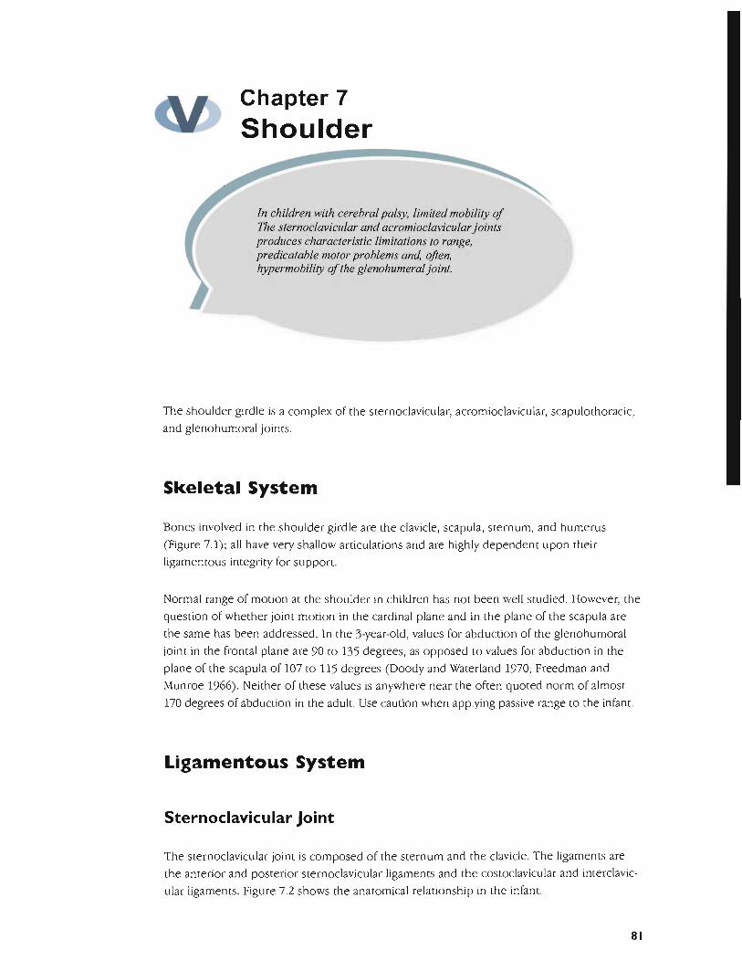
The shoulder girdle is a complex of the sternoclavicular, acromioclavicular, scapulothoracic,
and glenohumoral joints.
Skeletal System
Bones involved in the shoulder girdle are the clavicle, scapula, sternum, and humerus
(Figure 7.1); all have very shallow articulations and are highly dependent upon their
ligamentous integrity for support.
Normal range of motion at the shoulder in children has not been well studied. However, the
question of whether joint motion in the cardinal plane and in the plane of the scapu la are
the same has been addressed. In the 3-year-old, values for abduction of the glenohumoral
joint in the frontal plane are 90 to 135 degrees, as opposed to values for abduction in the
plane of the scapula of 107 to 115 degrees (Doody and Waterland 1970; Freedman and
Munroe 1966). Neither of these values is anywhere near the often quoted norm of almost
170 degrees of abduction in the adult. Use caution when applying passive range to the infant.
Ligamentous System
Sternoclavicular Joint
The sternoclavicular jOint is composed of the sternum and the clavicle. The ligaments are
the anterior and posterior sternoclavicular ligaments and the costoclavicular and interclavic
ular ligaments. Figure 7.2 shows the anatomical relationship in the infant.
81
User
Rectangle

Mobilization fo th N .r e eurologlcally Involved Child
Figure 7.1. Shoulder Girdle
82

Shoulder
anterior sternoclavicular
ligament
sternum
Figure 7.2. Sternoclavicular Ligaments
83
User
Rectangle

Mobilization for the Neurologically Involved Child
The capsular pattern
the distal end of the clavicle relative to the
of is a limitation of elevation and of
end. Even though many children with
cerebral appear to have elevated and rv,")tr'lrtp,; shoulders, the distal end of the
clavicle is not elevated or protracted relative to the end. This apparent paradox is
fJH<>'''-'.U by the alignment of the clavicle relative to the sternum when the is
kyphosed. Children with cerebral appear to have elevated and shoulders when the is kyphosed. Once the spine is the clavicle is neutral relative to
the sternum and the has little or no elevation and both are critical
for reach.
The sternoclavicular joint is the bone connection between the upper extremity and the
trunk. The motions available are clavicular and
rotation. The costoclavicular the axis of motion for elevation and
as well as protraction and retraction. This is located outside of the
and results in a see-saw motion. Rotation occurs along the long axis and
additional movement at the acromioclavicular
Acromioclavicular Joint
The acromioclavicular of the acromion of the scapula and the clavicle.
The are the
is
and inferior acromioclavicular and the coracoclavicular
ligaments.
position of the joint occurs during upward rotation and of the
scapula, In the this comributes to shoulder elevation after 60
degrees and stops comributing motion between 90 and 120 elevation (Kessler
and Because this jOint is a union until almost 2 years of
age (Cailliet 1966), it is unable to contribute to the rhythm in
the child as it does in the adult. This may explain the more limited range of motion norms
identified for children by Freedman and Munroe and Doody and Waterland It
is then that total range of motion in the infant and child is different and
more limited than in the adult. Observation of normal infants does suggest that do not
show 170 of shoulder abduction or flexion.
This maintains the between the clavicle and the scapula in the stages
of elevation and allows additional range of rotation in the latter stages of elevation.
Motions available are scapular and rotation occurs in an
upward/downward direction during upper extremity elevation at the same time that the
over the thorax.
to the thorax.
is not.
occurs when the vertebral border of the
rides is normal of the flat as it
rides forward over the round as occurs in kyphosis. Tipping is frequently seen in
children with cerebral
84
User
Rectangle
User
Rectangle

Mobilization for the Neurologically Involved Child
joint capsule
annular ligament
joint capsule
radial collateral
/////)/;))/ 1 ligamentA////I } II'IIII((I/I;II
Figure 8.2. Elbow Ligaments
96
User
Rectangle

Elbow
Muscular System
The biceps brachii is frequently implicated as the cause of elbow flexion and pronation
contracture in children with cerebral palsy. However, the bicep is a supinator and a
contracture occurs throughout the muscle 's actions , so it cannot be the cause of the
pronation component in the child with cerebral palsy. Lack of joint flexibility must also be
ruled out as a contributing factor in the limited range of motion.
Table 8.2 Elbow Muscle Flexibility Testing for Children with Cerebral Palsy
Muscle Group Testing Technique
Flexors Stabilize the scapula and extend both the shoulder and the elbow.
Extensors Stabilize the scapula and flex both the shoulder and the elbow.
Supinators Stabilize the humerus and pronate the forearm.
Pronators Stabilize the humerus and supinate the forearm.
Figure 8.3. Anterior-Posterior Glide Reproduced by permission of The Saunders Group, Inc. © 1996
97
User
Rectangle

Mobilization for the Neurologically Involved Child
Obtaining the range of motion for extension and supination is once the
connective tissue is stretched. However, strength in the new range is a far more
difficult task, because very few functional activities involve full supination. Functional tasks
tend to occur in or between and The child unable
to once the range has been obtained may need to strengthening
with isometric contractions and work up to eccentric and concentric contractions. Isometric
contractions while objects such as a paper cup and ice in the involved
hand while the child pours water or into the cup, tend to be both functional and hold
the child's interest. The amount the child can hold cup, the cup the cup
ice plus a little is an indication of the isometric strength of the muscle. Drinking the
contents can be an immediate reward and will also decrease the resistance when necessary.
Table 8.l Proposed Strength for Elbow
Strength Criteria Flexors Extensors Pronators
Poor (movement Hand to mouth Extends elbow along with gravity in prone. in prone.
Reaches in prone. Supinate, pronate with elbow flexed.
Reaches in prone while a toy.
resistance) toy in hand.
Toy to mouth.
At 3 months
At months
At 9 months
At 12 months
At 18 months
At 24 months
At months
Lifts age-toys.
to bring hand to mouth
Mouths toys
N.A.
Holds a ball
NA
Reaches in prone while holding age
toy.
Thrusts arm in
Elevates self by arms in prone
Throws a ball
N.A.
NA
resistance.
N.A.
Turns band in front face
N.A.
NA
Pours
NA Not available. Unable to infer from standardized assessments.
98
User
Rectangle

Chapter II
Skill Development
The acquisition of gross and fine motor skills has been relatively well documented in such work as the Denver Developmental Scales, Gesell Scales, and Peabody Gross and Fine Motor Scales (Bailey 1993; Folio and Fewel 1983; Frankel 1973). However, scales such as the Denver were developed to identify early indicators of mental retardation, not motor control problems (Frankel 1973). This is disconcerting given the amount of therapy prescribed based on motor delays identified by these types of tests alone. The tests can document the delay but they cannot identify the cause of the delay. The therapy evaluation must determine if the delay is due to muscular, skeletal, or neurological factors , and if the delay is amenable to therapy.
Although some variability has been recorded in different scales, the general principle is one
of orderly development of neural maturation causing skill acquisition. This principle has
been challenged in several ways. First, the timing of skill appearance has changed in this
culture since Gesell originally developed the scales, hence the need for revision. Second, it
is well established that not all normal children display the same pattern of development. For
example, not all non-neurologically involved children crawl before they walk (Bottos et al.
1989; Robson 1984). Third, different ethnic groups display differing rates of motor
development (Malina 1988). Fourth, different nationalities display differing sequence and
timing of motor milestones (Super 1976; Thornton 1992). An intact neuromusculoskeletal
system and practice on a skill seems to be most important to the acquisition of the skill, not
how early nor in what sequence the skill appears .
Integration for Skill Acquisition
This section focuses on the integration of neurological, orthopedic, and muscular systems
that are prerequisites to motor skills performance.
117
User
Rectangle

Mobilization for the Neurologically Involved Child
The classical theories of motor that the
must be present to express the skill. This is aptly illustrated by motor skill in the child with mental retardation any other neuromotor AJI the systems must be
functioning however, for a child to express motor skills with the
quality of movement characteristic of efficient function. For ~A'UUIJ''"
muscular prone, and a child with get to sit or crawl
Skill and have been related to myelination and maturation.
skill acquisition and performance have not been related to the musculoskeletal
to express the skill. This discussion focuses on the
between musculoskeletal and skill. Infants range and strength on their
own. To range, infants must stretch the connective tissue elements that range.
This is clearly seen in hip connective tissue needed for the hip extension
1977). Infant movements are oscillatory and at the end of their available
range. As the oscillations stretch the children continue oscillatory movements, now
in a new range. The movements at the end of joint range require repetitive
contraction and relu,'Cation of the muscles. Infants continue this pattern of oscillating near
the end range, adding resistance to gain in the new range.
Children new range through movements and add resistance to the oscilla
tions to increase strength. Infants can increase resistance in two ways. an increase in
the lever arm is an increase in resistance. As a child control for reach in prone,
sitting, or standing, the arms are in guard. That they are abducted from the in
the coronal plane, bringing the center of gravity over the base of support. As strength
muscular resistance increases the elbow. 'Pf'r.n,rl
during play with such as An infant will pick up the rattle but not a teddy
up a teddy bear but not a toy. The of the toy is a
measure of the resistance available to the muscle. Standard children's toys can be
and used as measures of strength in the clinical Combining increased
levers and in and
Pictorial Sequence of Development
The sequence of development illustrates these in a child.
are illustrations of one normally developing child drawn from monthly
taken as she matured. A detailed examination of the illustrations
and range over time combined with rol'Ogllcal maturation are all
necessary a skill to appear.
118
User
Rectangle

Skill Development
Head Control
Head control in prone is one of the first skills to emerge. lacey et at. (1985) observed 104
infants, 25 to 33 weeks gestational age. They identified a pattern of head side turning that
decreased spinal extension; however, the central position of the head required more spinal
extension . At 35 to 39 weeks post-conceptual age, the 11 infants with later motor handicap
showed a persistence of decreased spinal extension.
Comparing Figure 11.1 (l-week-old, full-term child) and Figure 11.2 (the same child at 1
month), note the flexed spine with head to side consistent with the description ofLacey et
at. (1985). Note that by 2 months (Figure 11 .3), significant increases in lumbar spine and hip
extension are evident. The hip extension changes are consistent with research on changes
in hip range of motion previously reported. For the head to come up prone and keep the
eyes level, the cervical spine is flexed. To bring the head up, the increased lumbar extension
appears necessary. Figures 11.4 and 11.5 illustrate the continuing increase in lumbar
extension through 11 months. Figure 11.4 shows a marked increase in the thoracolumbar
junction but not a smooth curve throughout the lumbar spine. By 11 months (Figure 11.5),
extension occurs throughout the lumbar spine.
Figure 11.1. One-week-old with lumbar flexion
119
User
Rectangle

Mobilization for the Neurologically Involved Child
Figure 11.2. One-month-old with lumbar extension
Figure 11.3. Two-month-old with lumbar extension
120
User
Rectangle

Skill Development
Figure 11.4. Nine-month-old with lumbar extension
Figure 11.5. Eleven-month-old with lumbar extension
121
User
Rectangle

Mobilization for the Neurologically Involved Child
To achieve head up in prone, it appears that the normal infant must shift his or her head and oscillate weight backwards. This increase the connective tissue
increases range and necessary to
shift may the force necessary to
and hip extension. This in turn
and influence neural stability. The child with does not appear to be able to the range and for normal motor performance et aL among others).
Muscular weakness the head to corne to midline, or oscillate suffiCiently while it
is may lead to failure of the and hips to increase their range. This failure to increase range and strength in the range prevents mobility
for movement of the center of over the base of support in future skills. This failure to increase range to normal values often appears later as a contracture. the
contracture in older children with damage did not but was maintained
from infancy. The contracture "normal" in the infant is "abnormal" in the older child with cerebral
Reach
is a pattern Oscillation of the head and shift toward the throughout normal from head up in prone described above, as the oscillations continue in intact children, or prone occurs. This brings the arms from the surface but them near the body. Yet out
more range and through the shoulder due to the increased length of the lever arm. Thelen et a1. in the infant and concluded that
appears between 12 and 22 weeks, Thelen notes that reaching appears when an infant can intentionally adjust force and of the arm, often using muscle coactiva
tion. These patterns are therefore consequences of the match between system
and the task et a1. 1993). The natural dynamic of the system includes the available range and strength.
At birth, the arms cannot reach this high
must be increased for high
the little had about 60
in pivot prone, nor reach for
and in pivot prone or reaching. In
the of shoulder
backward with the arms on the support. The
As the little girl increased her head up relative to prone, she
child continues this shift across the sternoclavicular Consistent with the function of the sternoclavicular joint range will allow
the infant to raise the arm from its initial 60 abduction and flexion
to approximately 100 necessary for prone on hands and She showed
active shoulder range of less than 90 at 1 month of age to
more than 90 at 2 months 1 6 months, she can reach forward approximately 110 Note the normal side position of the
as she reaches. It is unclear when the full adult range of shoulder elevation is 2
years of age, as some suggest it cannot contribute to "normal" scapulo
humeral it is possible the adult norm of 170 of flexion and
122
User
Rectangle
User
Rectangle
Skill Development
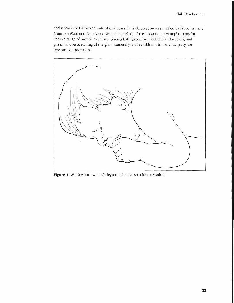
abduction is not achieved until after 2 years. This observation was verified by Freedman and
Munroe (1966) and Doody and WaterJand (1970). If it is accurate, then implications for
passive range of motion exercises, placing baby prone over bolsters and wedges, and
potential overstretching of the glenohumeral joint in children with cerebral palsy are
obvious considerations.
Figure 11.6. Newborn with 60 degrees of active shoulder elevation
123
User
Rectangle