Miyashita, T., V. M. Arbour, L. M. Witmer, and P. J. Currie. 2011. The ...
16
Transcript of Miyashita, T., V. M. Arbour, L. M. Witmer, and P. J. Currie. 2011. The ...


The internal cranial morphology of an armoureddinosaur Euoplocephalus corroborated by X-raycomputed tomographic reconstructionTetsuto Miyashita,1 Victoria M. Arbour,1 Lawrence M. Witmer2 and Philip J. Currie1
1Department of Biological Sciences, University of Alberta, Edmonton, AB, Canada2Department of Biomedical Sciences, Heritage College of Osteopathic Medicine, Ohio University, Athens, OH, USA
Abstract
Internal cranial anatomy is a challenging area to study in fossilized skulls because of small sample sizes and var-
ied post-mortem preservational alterations. This difficulty has led to the lack of correspondence between results
obtained from direct osteological observation and from more indirect reconstruction methods. This paper pre-
sents corroborating evidence from direct osteological observation and from reconstruction based on computed
X-ray tomography (CT) on the internal cranial anatomy of the ankylosaurid dinosaur Euoplocephalus tutus. A
remarkable specimen of Euoplocephalus preserves rarely observed internal cranial structures such as vascular
impressions in the nasal cavity, olfactory turbinates and possible impressions of conchae. Comparison with fos-
sils and CT models of other taxa and other Euoplocephalus specimens adds osteological evidence for the previ-
ously reconstructed nasal cavity in this dinosaur and revises the previously described braincase morphology. A
new interpretation of the ethmoidal homology identifies a mesethmoid, sphenethmoid and ectethmoid. These
ethmoidal ossifications are continuous with the mineralized walls of the nasal cavity. The location of the olfac-
tory fenestra provides further evidence that the olfactory regions of the nasal cavity are pushed to the sides of
the main airway. This implies that the function of the vascular impressions in the nasal cavity and the looping
of the cavity are not related to olfaction. A byproduct of the elongate, looping airway is a dramatic increase in
surface area of the nasal respiratory mucosa, which in extant species has been linked to heat and water bal-
ance. A role in vocalization as a resonating chamber is another possible function of the looping and elongation
of the nasal cavity. Olfaction remains as a possible function for the enlarged olfactory region, suggesting that
multiple functions account for different parts of the ankylosaurid nasal cavity that underwent substantial modi-
fication. Cranial endocasts show negligible variation within Euoplocephalus, which lends some confidence to
interspecific comparisons of endocranial morphology.
Key words: Ankylosauridae; braincase; Dinosaur Park Formation; nasal cavity.
Introduction
Ankylosaurs are a clade of ornithischian dinosaurs com-
monly called ‘armoured dinosaurs’. Due to their highly
modified skulls, detailed description of the cranial anatomy
is particularly important in identification of both basal and
derived conditions within the clade. However, the skulls are
extensively ossified and little is known about the internal
cranial morphology of ankylosaurs. Cranial elements are
rarely preserved individually and it is unusual to find a skull
that shows the internal morphology without the aid of X-ray
computed tomography (CT). Several papers describe ankylo-
saur braincases and cranial endocasts (Maryanska, 1977;
Coombs, 1978a; Kurzanov & Tumanova, 1978; Carpenter
et al. 2001; Averianov, 2002; Vickaryous & Russell, 2003;
Hayakawa et al. 2005; Witmer & Ridgely, 2008; Parsons &
Parsons, 2009). However, these authors provided different
identifications of the foramina perforating the braincases
and this makes comparison difficult. Only a handful of
papers deal with other regions inside ankylosaur skulls,
such as the nasal cavity. Sections of a few skulls (e.g.
Euoplocephalus AMNH 5403) led to reconstruction of the
ankylosaur nasal cavity as a sagittal S-shaped airway
(Maryanska, 1977; Coombs, 1978b; Witmer, 1997). Results
from two-dimensional CT slices supported this view
(Vickaryous & Russell, 2003; Vickaryous, 2006). A three-
dimensional digital reconstruction of a CT scan of the skull
Correspondence
Tetsuto Miyashita, Department of Biological Sciences, University of
Alberta, Edmonton, AB, Canada T6G 2E9. T: + 1 780 2428166;
Accepted for publication 26 August 2011
Article published online 29 September 2011
ªª 2011 The AuthorsJournal of Anatomy ªª 2011 Anatomical Society of Great Britain and Ireland
J. Anat. (2011) 219, pp661–675 doi: 10.1111/j.1469-7580.2011.01427.x
Journal of Anatomy

of the ankylosaurid Euoplocephalus recently overturned
the reconstruction of a simple S-shaped airway in this genus
(Witmer & Ridgely, 2008). According to the new reconstruc-
tion, the nasal cavity of Euoplocephalus follows a complex
path of twists and turns that create a series of loops of the
airway (Fig. 1). The nasal cavity of the nodosaurid ankylo-
saur Panoplosaurus also has the anterior and posterior
loops, although the path of the airway is less complicated
than in Euoplocephalus (Witmer & Ridgely, 2008). The com-
plex pathways of the ankylosaur nasal cavities in Witmer &
Ridgely (2008) also revealed that the internal space within
the skulls previously identified as paranasal sinuses (Witmer,
1997; Vickaryous & Russell, 2003; Vickaryous, 2006) are actu-
ally part of the looping airway, although this was not read-
ily evident in two-dimensional CT slices.
Witmer & Ridgely (2008) radically transformed the previ-
ous view of a straight airway in ankylosaurs because their
reconstruction was possible through sophisticated CT meth-
odology. For instance, Witmer & Ridgely (2008) note that
the nodosaurid ankylosaurs Edmontonia (AMNH 3076) and
Panoplosaurus (ROM 1215) are likely to differ from each
other in the degree of mineralization within the nasal cav-
ity. Whereas their reconstruction showed a looping airway
for Panoplosaurus (Witmer & Ridgely, 2008), a simple,
straight airway was previously reconstructed for Edmonto-
nia due to the lack of apparent subdivision within the nasal
cavity (Vickaryous, 2006). Witmer & Ridgely (2008) observed
thin mineralized laminae as well as heterogeneities in the
matrix within the nasal cavity of the same specimen of
Edmontonia (AMNH 3076), which highlights the sensitivity
of data obtained via CT scanning. For these reasons, corrob-
orative evidence from direct observation of the skull mor-
phology is important. In addition, the ankylosaur literature
dealing with internal cranial anatomy rarely deals with
comparative aspects of the braincase and nasal cavities.
Several specimens of the ankylosaurid Euoplocephalus
from the Campanian (Late Cretaceous) of southern Alberta,
Canada fill this gap. Two of the specimens (AMNH 5238
and UALVP 47977) reveal osteological correlates of the soft
tissues within the skull, whereas the others (AMNH 5405
and UALVP 31) offer new data on braincase anatomy
through three-dimensional reconstructions based on CT
scanning. Cranial endocasts of these specimens establish
correspondence between the cranial nerves and the foram-
ina perforating the braincase wall in UALVP 47977.
Institutional abbreviations: AMNH (American Museum of
Natural History, New York, NY, USA); MPC (Mongolian Pale-
ontological Center, Ulanbaatar, Mongolia; followed by the
collector’s initials and field number); PIN (Paleontological
Institute, Moscow, Russia); ROM (Royal Ontario Museum,
Toronto, Canada); TMP (Royal Tyrrell Museum of Palaeon-
tology, Drumheller, AB, Canada); UALVP (University of
Alberta Laboratory for Vertebrate Paleontology, Edmonton,
AB, Canada); and ZPAL (Institute of Palaeobiology of the
Polish Academy of Sciences, Warsaw, Poland).
Materials and methods
A partial ankylosaurid skull roof (UALVP 47977) was collected
from the Dinosaur Park Formation in Dinosaur Provincial Park in
1971. The precise location and stratigraphic level of the site was
not recorded, but the site is near Happy Jack’s on the north side
of Red Deer River (A.L. Lindoe, personal communication, 2007).
Due to erosion of the skull before field collection, the nasal cav-
ity, the orbital region and the upper half of the braincase are
A B
Fig. 1 Schematic reconstructions of the nasal cavity morphology of two ankylosaur skulls. (A) The ankylosaurid Euoplocephalus tutus (AMNH
5405). The left half of the skull is derived from the reconstruction published by Witmer & Ridgely (2008), whereas the right half represents new
information based on the osteological correlates of the soft tissues within and around the nasal cavity in UALVP 47977. The specimen (UALVP
47977) preserves parts of the main airway (in green), the olfactory region (in blue), and the endocranial cavity (in orange). (B) The nodosaurid
Panoplosaurus mirus (ROM 1215) after Witmer & Ridgely (2008) for comparison. The nasal passage of Euoplocephalus is looped in more complex
ways than that of Panoplosaurus, and the olfactory region of Euoplocephalus is pushed to the side of the main airway. bv, blood vessel trace; co,
groove that possibly housed the concha; dac, dorsal alveolar canal; dpf, descending process fused to the ventral surface of the frontal; olfactory
turbinate; ec, endocranial cavity; ee, ectethmoid; eth, ethmoidal complex; lnv, lateral nasal vessels; lw, lateral wall of the main airway; ma, main
airway; mnv, medial nasal vessels; mnc, medial nasal canal; ns, nasal septum; nv, nasal vestibule; or, olfactory region; os, orbitosphenoid.
ªª 2011 The AuthorsJournal of Anatomy ªª 2011 Anatomical Society of Great Britain and Ireland
Ankylosaurid internal cranial anatomy, T. Miyashita et al.662

exposed ventrally. Two ankylosaurid ankylosaurs are currently
recognized from the Dinosaur Park Formation: Dyoplosaurus,
known from a single specimen recovered from the lower part of
the formation, and Euoplocephalus, known from numerous
specimens throughout the formation and the overlying Horse-
shoe Canyon Formation (Parks, 1924; Arbour et al. 2009). The
holotype specimen of Dyoplosaurus preserves the posterior part
of the skull roof. The skull (along with most of the skeleton) is
affixed to a panel and so the ventral side cannot be observed.
At present, there are no cranial characters that separate Dyoplo-
saurus from Euoplocephalus; the features that distinguish
Dyoplosaurus from Euoplocephalus are restricted to the pelvis
and pes. As such, it is possible that isolated skulls (or skulls
associated with skeletons that do not preserve the pelvis or pes)
currently referred to Euoplocephalus may instead be referable
to Dyoplosaurus. UALVP 47977 preserves distinct flat, polygonal
osteoderms on the dorsal surface of the skull similar to those in
Euoplocephalus. These osteoderms are not preserved in Dyoplo-
saurus, which may reflect either a taphonomic or a true diag-
nostic difference. As such, UALVP 47977 more closely resembles
Euoplocephalus than Dyoplosaurus. For the purposes of this
paper, UALVP 47977 is tentatively referred to Euoplocephalus to
facilitate comparison with other skulls referred to this genus
and pending a revision of the genus by V.M.A.
Computed tomography scans of two additional skulls of Euo-
plocephalus were available for this study. AMNH 5405 and UALVP
31 were both collected from the Steveville locality in Dinosaur
Provincial Park, Alberta, Canada. CT data of AMNH 5405 used by
Witmer & Ridgely (2008) was made publicly available on a website
(http://www.oucom.ohiou.edu/dbms-witmer/3D-Visualization.htm)
and their article may be consulted for technical details of the scan-
ning. UALVP 31 was scanned at the University of Alberta ABACUS
CT scanner, in 1-mm increments. Both skulls were digitally recon-
structed using the thresholding and segmentation tools in the soft-
ware program MIMICS Version 14 (Materialise Inc., Leuven, Belgium).
Digital cranial endocasts were created for each skull and internal
structures were viewed both as two-dimensional slices and as
three-dimensional reconstructions. As an independent check on
Witmer & Ridgely’s (2008) findings on the nasal cavity, V.M.A. per-
formed de novo segmentation of the nasal cavity of AMNH 5405.
Three-dimensional models cropped to resemble the broken sur-
faces of UALVP 47977 were also created to compare internal struc-
tures of the different specimens. A latex cranial endocast was
prepared for UALVP 47977.
Results
This description focuses primarily on UALVP 47977, as it best
presents new information. This paper largely follows Wit-
mer (1995, 1997), Evans (2006) and Witmer & Ridgely (2008)
for homologies in the antorbital region (Figs 1 and 2). The
nasal cavity refers to a respiratory and olfactory passage
that extends between the external naris and the choana,
and is equivalent to the respiratory tract, the respiratory
passage and the airway in other papers.
Skull roof
In UALVP 47977, the dorsal surface of the skull is moder-
ately weathered. The specimen preserves the top and
lateral parts of the nasal cavity, which represent both the
non-olfactory and olfactory regions of the nasal cavity
(Fig. 2; Witmer & Ridgely, 2008). As in most ankylosaurs,
but unlike Cedarpelta (Carpenter et al. 2001) and Pinaco-
saurus (Maryanska, 1977), no sutures can be observed. The
non-olfactory dorsomedial passage of the nasal cavity
A
B
Fig. 2 Illustration (A) and photograph (B) of the ventral view of an
ankylosaurid skull roof (UALVP 47977, Euoplocephalus) from the
Dinosaur Park Formation (Campanian, Upper Cretaceous), southern
Alberta. Hatched lines indicate parts reconstructed with plaster.
Impressions of the soft tissues, including the main airway, nasal
arteries, and possible turbinates, are well defined. The ethmoidal
elements are well ossified and separate regions of the nasal cavity
from each other. No sutures are visible. aw, anterior wall of the cavity
for the olfactory region; cor, cavity for the olfactory region; ls,
laterosphenoid; od, orbital depression; sl, sulcus associated with the
groove in the olfactory region. For other abbreviations, see Fig. 1.
ªª 2011 The AuthorsJournal of Anatomy ªª 2011 Anatomical Society of Great Britain and Ireland
Ankylosaurid internal cranial anatomy, T. Miyashita et al. 663
under the frontals extends posteriorly behind the orbits,
just anterior to the braincase. The olfactory region of the
nasal cavity occupies the large cavity on both sides of the
non-olfactory dorsomedial passage. A well developed bony
wall separates the dorsomedial passage from the olfactory
region for all its preserved length.
The dorsomedial passage of the nasal cavity (main
airway) is divided into right and left passages by the nasal
septum. There is no evidence for a median common cham-
ber as reconstructed for lambeosaurine ornithopods (Evans
et al. 2009). On both the lateral and medial walls of the
nasal cavity proper in UALVP 47977, deep grooves extend
anteroposteriorly and lead to the extensive vascular impres-
sions in the roof of the nasal cavity (Fig. 3). The impressions
branch and extend posteromedially toward the nasal
septum and this pattern is bilaterally consistent. The vascu-
lar impressions are conspicuous in the anterior half of the
preserved length of the main airway but are absent in the
posterior half. Vascular impressions are also preserved in
this region in AMNH 5238. Vascular impressions could not
be reconstructed from the CT scans of AMNH 5405 and
UALVP 31, but this is because of inadequate resolution of
the CT scans.
The cavity that housed the olfactory region of the nasal
cavity occupies a large volume to the side of the dorsomedi-
al part of the main airway. The cavity is surrounded by a
thin sheet of bone (ectethmoid) laterally and by thick bony
walls medially and anteriorly, and is connected posteriorly
with the endocranial cavity through the olfactory fenestra.
The olfactory bulb sat within this fenestra through which
the olfactory nerves [cranial nerve (CN) I], ethmoidal vessels
and their branches passed. This fenestra was previously
identified as an olfactory tract in Talarurus (Carpenter,
2004) but the olfactory tract was located more posteriorly,
well within the endocranial cavity.
In UALVP 47977, a conspicuous descending process fused
to the ventral surface of the frontal develops at the front of
the olfactory fenestra. The descending process accommo-
dates a deep, spacious groove that originates from the
anterior margin of the orbit. Its anterolateral site of origin
is associated with vascular impressions on the medial sur-
face of the lacrimal. The groove extends medially along the
anterior wall of the olfactory region and then posteriorly
along the lateral wall of the main airway, and finally ven-
trally along the descending process. A deep sulcus parallels
the groove medially along the lateral wall of the main air-
way. The soft tissue that filled this groove was extensively
vascularized because of the vascular impressions at the an-
terolateral end of the groove and because of the sulcus
associated with the groove along the lateral wall of the
main airway.
In another skull of Euoplocephalus (AMNH 5405), a tun-
nel extends anterolaterally within the roof of the olfactory
region (Fig. 4) and presumably opens into the descending
process. Witmer & Ridgely (2008) did not reconstruct this
tunnel in AMNH 5405, but it is present on both sides in their
CT data. A re-examination of the CT slices revealed that the
tunnel branches laterally. Although UALVP 47977 does not
A
B
C
Fig. 3 Vascular impressions in the dorsomedial part of the nasal cavity
proper of UALVP 47977. (A) Drawing of skull showing region of
enlargement in diagram (B) and photograph (C). These ethmoidal
vessels are likely to be part of the median nasal canal system. Hatched
area in B represents broken nasal septum (mesethmoid). Anterior is to
the right. For abbreviations, see Fig. 1.
ªª 2011 The AuthorsJournal of Anatomy ªª 2011 Anatomical Society of Great Britain and Ireland
Ankylosaurid internal cranial anatomy, T. Miyashita et al.664

have the tunnel, the vascular impression associated with
the groove in the olfactory region suggests that at least the
vascular component of the tissue filling the groove may cor-
respond to the tissue filling the tunnel in AMNH 5405. The
descending process and the groove could not be recon-
structed from the CT scan of UALVP 31 (Fig. 5). UALVP
47977 has cracks that show the cross-sections of the frontal
and the nasal. None of these cross-sections indicate pneu-
matization within the bones.
Ethmoidal region
UALVP 47977 preserves the mesethmoid, the sphenethmoid
and the ectethmoid, all of which are fully mineralized
(Figs 2 and 7). The mesethmoid is a septum on the midline
that separated the olfactory bulbs and is continuous anteri-
orly with the mineralized nasal septum. The sphenethmoid
is the lateral element of the ethmoidal complex (generally
referred to as a presphenoid in ornithischians: Horner,
1992; Evans, 2005, 2006) that enveloped the olfactory bulb
ventrally and laterally. The sphenethmoid is continuous
with the lateral wall of the main airway. The mineralized
median septum of the olfactory bulbs has been described
for a variety of non-avian theropods (Brochu, 2002; Coria &
Currie, 2002; Sampson & Witmer, 2007; Ali et al. 2008) and
is considered a homologue of the mesethmoid in birds (Ali
et al. 2008). Following this position, the ossified median
septum of the ethmoidal complex in UALVP 47977 is identi-
fied as the mesethmoid. It is not possible to distinguish the
boundary between the mesethmoid and the sphenethmoid
in UALVP 47977 and these two elements probably fused to
each other early in ontogeny.
The ectethmoid forms a thin lateral wall of the olfactory
region, separating it from the orbital depression laterally. It
contacts the orbitosphenoid posteriorly and the lacrimal
anteriorly, although the sutures are not visible at either
end. Because of the skull width and the relatively more
anterior placement of the orbit, the ectethmoid is elongate
A E
B F
C G
D H
Fig. 4 Sagittal sections of a Euoplocephalus
skull (AMNH 5405) from CT data of Witmer
& Ridgely (2008) show a tunnel within the
frontal bone, laterally positioned and passing
medially and slightly posteriorly on both sides.
The most lateral sagittal section for each side
is where the canal disappears into the bone.
Arrowhead indicates the tunnel within the
frontal, and letters A–H indicate the levels of
the CT slices on the skull. Anterior is to the
right in all CT slices and in the 3D model of
the skull. CT data are available from the
website (http://www.oucom.ohiou.edu/
dbms-witmer/3D-Visualization.htm).
ªª 2011 The AuthorsJournal of Anatomy ªª 2011 Anatomical Society of Great Britain and Ireland
Ankylosaurid internal cranial anatomy, T. Miyashita et al. 665

and oriented anterolaterally rather than transversely. The
ectethmoid forms a small, medially overhanging shelf near
the base of the descending process. A small foramen that
pierces the ectethmoid on the left side of the skull from the
orbital depression to the olfactory region may represent
the orbitonasal foramen.
Anteriorly to the ethmoidal complex, the nasal septum
and the lateral walls of the main airway are mineralized
(Figs 2 and 3). There is no suture that distinguishes the min-
eralized septum and walls of the main airway from any of
the cranial elements, including the ethmoidal complex,
nasal, frontal and lacrimal, which they contact. The nasal
septum and the lateral walls converge at the midline ante-
rior to the ethmoidal complex. There is no opening that
connects the dorsomedial passage of the main airway with
the endocranial cavity. The skull roof is damaged and does
not preserve the ventral part of the main airway. On the
right side of the skull, however, the preserved part of the
lateral wall extends ventromedially. This suggests that the
mineralized wall wrapped around the dorsomedial passage
of the main airway ventrally as well as laterally and medi-
ally. The anterior wall of the olfactory region extends lat-
eromedially between the lateral wall of the main airway
and the lacrimal. The anterior wall separates the olfactory
region and the groove filled with the vascularized tissue
from the cavity that housed the posterior loop of the main
airway anteriorly (Witmer & Ridgely, 2008).
Sphenoidal region
The orbitosphenoid contacts the ectethmoid anterolaterally
and the ethmoidal complex (sphenethmoid + mesethmoid)
anteriorly (Figs 2 and 7). The olfactory fenestra opens
between these two contacts. The olfactory nerves (CN I)
would have diffused from this fenestra to both lateral and
medial sides of the descending process. The orbitosphenoid
contacts the laterosphenoid posteriorly and the parasphe-
noid ventrally. The laterosphenoid is short anteroposterior-
ly, but has a long, laterally oriented postorbital process that
is approximately half the width of the transversely
expanded ankylosaurid skull. The element is firmly fused to
the skull roof. Two other sets of foramina pierce the orbit-
osphenoid. The foramen for the optic nerve (CN II) is larger
than all other foramina for the cranial nerves except the
olfactory fenestra and consists of a single exit (Fig. 7). The
shared foramen for the oculomotor (CN III) and trochlear
(CN IV) nerves opens posterior to the optic foramen. The
foramen for the abducens nerve (CN VI) opens directly ven-
tral to the oculomotor ⁄ trochlear foramen, which is consis-
tent with these foramina transmitting motor nerves to the
extraocular muscles.
In the laterosphenoid, the foramen for the trigeminal
nerve (CN V) is posterior to the oculomotor ⁄ trochlear fora-
men. Just dorsal to the trigeminal foramen is an aperture
for the anterior middle cerebral vein. The trigeminal fora-
men is anteroventral with respect to the lateral wing of the
braincase (pila antotica) that contacts the postorbital later-
ally. In addition, the trigeminal nerve is associated with the
prootic in sauropsids. Although no suture can be observed
between the laterosphenoid and prootic, the topographical
relationships of the trigeminal foramen with other brain-
case landmarks suggest that the foramen was mainly within
the laterosphenoid with contribution from the prootic pos-
teriorly. This implies that the prootic extended anteriorly
C
AD
B
Fig. 5 CT-based reconstruction corroborates
direct osteological observation. CT renderings
of the skull roofs of two Euoplocephalus
specimens, AMNH 5405 (A,C) and UALVP 31
(B,D). A and B show in dark grey the portion
of the skull represented in C and D in relation
to the entire skull, in oblique right anterior
view. C and D are sliced to mimic the areas
preserved in UALVP 47977, and show internal
features of the skull in ventral view that
correspond to those of UALVP 47977 (Fig. 2),
with anterior towards the bottom of the
page. oo, ocular osteoderm; pp, paroccipital
process; q, quadrate. For other abbreviations,
see Figs 1 and 2.
ªª 2011 The AuthorsJournal of Anatomy ªª 2011 Anatomical Society of Great Britain and Ireland
Ankylosaurid internal cranial anatomy, T. Miyashita et al.666

below the lateral wing of the laterosphenoid. This inter-
pretation is supported by the location of the foramen for
the facial nerve (CN VII), which is located completely within
the prootic in sauropsids and is just posteroventral to the
trigeminal foramen.
Occipital region
Most of the elements of the occipital region are highly ossi-
fied and fused to each other. The squamosal and parietal
form a roof over a chamber for M. adductor mandibulae
posterior (Holliday & Witmer, 2007). The otic region is ante-
roposteriorly short, and the prootic and the opisthotic are
indistinguishably fused together. The well developed crista
interfenestralis separates the fenestra vestibularis anteriorly
and the jugular foramen posteriorly (Fig. 7). The posterior
foramen for the hypoglossal nerve (CN XII) opens laterally
at the base of the occipital condyle, whereas the anterior
foramen for the hypoglossal nerve is merged to the pos-
teroventral corner of the jugular foramen. In Amtosaurus,
there are three foramina for the hypoglossal nerve (Averia-
nov, 2002). In occipital view, the osteoderms overhang from
the skull roof elements (Fig. 6). The foramen magnum is tal-
ler than it is wide and the margin is inflated into a rim. The
crescentic occipital condyle is oriented posteroventrally.
Cranial endocast
The description of the cranial endocasts focuses on AMNH
5405, which has the best preserved braincase amongst the
specimens used in this study. The newly prepared cranial en-
docasts (AMNH 5405, UALVP 31 and UALVP 47977; Fig. 7)
compare well with the published description of the cranial
endocast of AMNH 5337 (Coombs, 1978a). In all specimens,
the brains were anteroposteriorly short but relatively
straight. The cranial endocast of UALVP 31 has a blockier,
more robust appearance than that of AMNH 5405, because
of the lower resolution of the CT data for UALVP 31. The
endocast is also more strongly bowed dorsoventrally com-
pared to the other specimens, but this is probably a result
of taphonomic distortion of the skull. The anteroposterior
shortening of the olfactory stalk partly accounts for the
short anteroposterior length of the cranial endocasts of
Euoplocephalus. The anteroposterior distance between the
olfactory fenestra and the root of the optic nerve is less
than a quarter the entire anteroposterior length of the
cranial endocast in Euoplocephalus, whereas the distance is
typically more than a third the length of the cranial endo-
cast in other dinosaurs (based on figures in Hopson, 1979;
Brochu, 2002; Sampson & Witmer, 2007; Witmer et al.
2008).
The olfactory bulbs diverge immediately anterior to the
cerebrum at an angle of 80–100º and lead to the olfactory
fenestra opening at the posteromedial end of the olfactory
region. A general condition for ornithischian dinosaurs is
that the olfactory tracts did not diverge as strongly antero-
laterally as in Euoplocephalus (Hopson, 1979; Galton, 1983,
1988, 1989, 1997, 2001; Evans et al. 2009). The cerebral
hemispheres are fairly discrete on the endocast, forming a
rounded swelling immediately posterior to the olfactory
tract. As is often the case in non-coelurosaurian dinosaurs
(Witmer & Ridgely, 2009), however, other major neural
structures such as the optics lobes and cerebellum are lar-
gely obscured by the dural envelope. An important excep-
tion is the flocculus (cerebellar auricle). The flocculus on the
endocast of AMNH 5405 extends posterolaterally as a sub-
stantial, finger-like projection into the region of the inner
ear and breaks the plane of the anterior semicircular canal.
The presence of a flocculus in AMNH 5405 clears up the dis-
crepancy in the reports on AMNH 5337 between Coombs
(1978a: no flocculus) and Hopson (1979: large flocculus) in
favor of the latter interpretation. The structure interpreted
as an epiphysis cerebri (= pineal gland) projecting from the
diencephalon noted in AMNH 5337 by Coombs (1978a) is
also present in the endocasts of UALVP 31 and AMNH 5405,
but is not visible in the endocast of UALVP 47977. Although
being small, this structure is in the position to be the epiph-
ysis. Epiphyses are present in extant birds and have been
reconstructed in some dinosaurs (e.g. some theropods; Wit-
mer & Ridgely, 2009). The lack of the epiphysis in UALVP
47977 is probably due to the presence of plaster infilling,
which was used to strengthen the cracks during preparation
of this specimen.
As already noted in the braincase description, the optic
nerves in the endocast project almost directly laterally, such
that the optic chiasm is oriented transversely rather than
anterolaterally as in other archosaurs. The shared exit for
the oculomotor and trochlear nerves is a large trunk directly
posterior to the optic nerve. Both Coombs (1978a) and Hop-
son (1979) interpreted the smaller twig dorsal to the defini-
tive oculomotor nerve canal as the trochlear nerve. This
interpretation was widely accepted in the subsequent anky-
losaur literature and the corresponding foramen was identi-
fied as that for the trochlear nerve in Amtosaurus
(Averianov, 2002), Saichania (Maryanska, 1977), Sauropelta
Fig. 6 UALVP 47977 (Euoplocephalus) in occipital view. For
abbreviations, see Fig. 5.
ªª 2011 The AuthorsJournal of Anatomy ªª 2011 Anatomical Society of Great Britain and Ireland
Ankylosaurid internal cranial anatomy, T. Miyashita et al. 667
A
B
C
D
F
G
E
H I
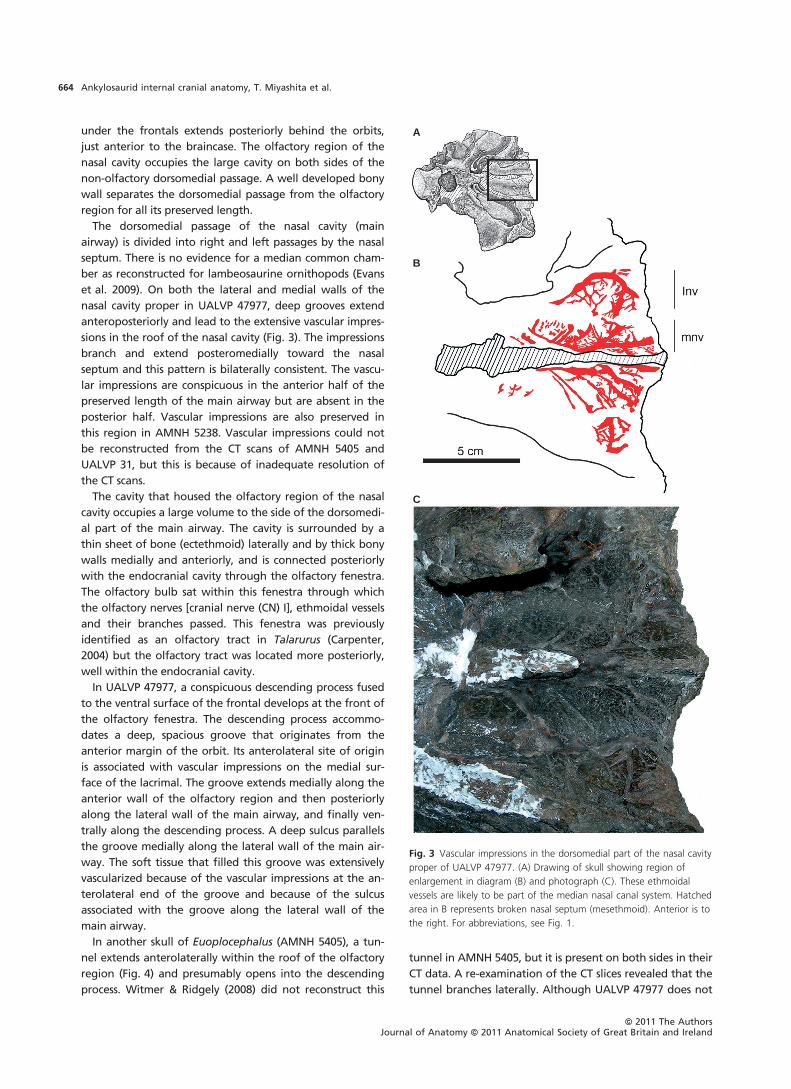
Fig. 7 Comparison of cranial endocasts and that of braincases reveals minor variation amongst specimens referred to as Euoplocephalus. The
braincase of AMNH 5405 in left lateral view (A), the cranial endocast of the same specimen in left lateral (B), dorsal (C) and ventral (D) views, the
braincase of UALVP 47977 in right lateral view (E), the cranial endocast of the same specimen in right lateral (F) and ventral (G) views, and the
cranial endocast of UALVP 31 in right lateral (H) and ventral (I) views. The images E–I were all inverted horizontally to show the right sides in the
same orientation with the left side of AMNH 5405 for the purpose of comparison. In both UALVP 47977 and UALVP 31, the right side is better
preserved. UALVP 47977 is represented by a line drawing of a latex cast, and AMNH 5405 and UALVP 31 are 3D models based on CT data.
Roman numerals refer to either the foramen for, or the trunk of, the cranial nerve. amcv, anterior middle cerebral vein; amp, insertion site for M.
adductor mandibulae posterior; sensu Holliday & Witmer, 2007; fl, flocculus; fv, fenestra vestibularis; ic, internal carotid artery; ocv, orbitocerebral
vein; of, olfactory fenestra; pmcv, posterior middle cerebral vein; sp, sinus of pituitary. For other abbreviations, see Figs 1 and 2.
ªª 2011 The AuthorsJournal of Anatomy ªª 2011 Anatomical Society of Great Britain and Ireland
Ankylosaurid internal cranial anatomy, T. Miyashita et al.668

and Tatankacephalus (Parsons & Parsons, 2009). We instead
regard their putative trochlear nerve as an orbitocerebral
vein. In the endocast of AMNH 5405, the ‘trochlear nerve’
of Coombs (1978a) and Hopson (1979) is comparable to the
orbitocerebral vein canals of sauropods (Sereno et al. 2007;
Witmer et al. 2008), theropods (Sampson & Witmer, 2007;
Witmer & Ridgely, 2009) and other dinosaurs. This feature
in AMNH 5405 emerges from the lateral pole of the
cerebral region and opens into the orbit well dorsal to
the canals for the other nerves supplying the extraocular
muscles.
The trunk of the oculomotor nerve in this study was iden-
tified by Coombs (1978a) as being associated with the pitui-
tary vein. Indeed, this canal shared by the trochlear and
oculomotor nerves seems too large to have transmitted
only these two small nerves. It is likely that veins also tra-
versed this canal. However, the term ‘pituitary vein’ is not
appropriate because the venous drainage of the pituitary
was almost certainly within the pituitary fossa itself and the
cavernous sinus within (see Sampson & Witmer, 2007). The
trunk of the abducens nerve originates from the ventral
side of the brain below the trigeminal nerve and passes
anterolaterally below the oculomotor and trochlear nerves.
The fossa for the pituitary gland projects more or less
straight ventrally in all the specimens as a bulbous structure.
In AMNH 5405, the bulbous structure is twice as wide trans-
versely as long anteroposteriorly. Ventral to the pituitary,
the endocast of the internal carotid artery is oriented ven-
trolaterally, whereas the artery extended anterodorsally in
the cranial endocasts of other dinosaurs (Hopson, 1979;
Witmer et al. 2008). The pituitary in UALVP 31 expands pos-
teriorly, but this is most likely a result of damage to the
ventral portions of the braincase. The pituitary fossa of
AMNH 5405 also preserves large paired apertures dorsal to
the carotid canals, which almost certainly transmitted the
sphenoid branch of the carotid artery into the floor of the
orbit as well as receiving ophthalmic veins.
The single trunk of the trigeminal nerve indicates that
the branches diverged outside the endocranial cavity. The
endocast of this nerve is dorsoventrally taller than antero-
posteriorly long, suggesting that the canal housed the gan-
glion, as in most dinosaurs except for tyrannosaurids and
birds (Witmer et al. 2008). The anterior middle cerebral vein
is preserved above the trigeminal nerve in the cranial endo-
cast of AMNH 5405 (Fig. 7C). The trunk of the facial nerve
originated from the shallow recess shared with that of the
vestibulocochlear nerve (CN VIII). The course of the facial
nerve closely parallels that of the trigeminal nerve anteri-
orly and laterally, and diverges away from that of the ves-
tibulocochlear nerve. The trunk of the vestibulocochlear
nerve has two branches that separate from each other
immediately outside the endocranial cavity. The dorsal
branch is directed laterally toward the vestibule and the
ventral one toward the cochlear ventrally. The trunks of the
glossopharyngeal, vagus and accessory nerves (CNs IX–XI)
exit the endocranial cavity through the jugular foramen.
The jugular foramen is directly anterior to the foramen for
the posterior branch of the hypoglossal nerve. AMNH 5405
has two trunks for the hypoglossal nerve, although there
are possibly three trunks for this nerve in AMNH 5337 (Coo-
mbs, 1978a) and UALVP 47977 (Fig. 7). Even in the case of
three trunks, the proximity and directions of the two smal-
ler anterior trunks suggest that they likely joined to emerge
from a single external foramen. In AMNH 5405, the fora-
men for the anterior trunk of the hypoglossal nerve is
merged to the posteroventral corner of the jugular fora-
men. The foramen for the larger posterior trunk opens
directly posterior to the jugular foramen on the lateral sur-
face of the base of the occipital condyle.
The labyrinth of the inner ear is reasonably well
preserved on the left side of AMNH 5405 and is generally
similar to the one illustrated for AMNH 5337 by Hopson
(1979). The lateral semicircular canal is extremely reduced,
more so than in perhaps any dinosaur described to date.
The anterior canal may seem somewhat elongate but this
may result more from the constraint that the anterior canal
must pass around the flocculus (Witmer et al. 2003). The
cochlea is remarkably elongate in AMNH 5405, as illustrated
also for AMNH 5337 by Hopson (1979). The elongate
cochlea suggests that hearing was an important sense in
Euoplocephalus.
Discussion
A combination of direct visual observation of several partial
specimens (in particular AMNH 5238 and UALVP 47977) and
CT-based digital reconstructions (AMNH 5405, ROM 1215,
UALVP 31) makes the data gained through either technique
interchangeable. The osteological data from UALVP 47977
agree with the reconstruction by Witmer & Ridgely (2008)
and complement it by adding fine-scale details (such as the
vascular impressions in the dorsomedial part of the nasal
cavity) that cannot be imaged using most medical CT scan-
ners. Coupled with the previous observation of large vascu-
lar canals in an ankylosaurid nasal cavity based on CT data
(Witmer & Ridgely, 2008), the vascular impressions (Fig. 3)
provide further evidence of extensive vascularization in the
nasal cavities of ankylosaurids. The medial neurovascular
canal near the nasal septum is consistent with the medial
nasal vessels and nerves, which extend along the nasal sep-
tum under the skull roof in both crocodylians and birds
(Sedlmayr, 2002; Witmer & Ridgely, 2008). Similarly, the lat-
eral canal presumably represents the lateral nasal vessels
and nerves. Tumanova (1987) identified a groove that
extends anteroposteriorly along the dorsolateral part of the
nasal septum in Talarurus as the olfactory nerve impression.
The groove does not represent the olfactory nerve because
the dorsomedial passage of the nasal cavity is not olfactory.
The current evidence suggests that the groove in Talarurus
is a channel of the medial nasal vessels and nerve. The
ªª 2011 The AuthorsJournal of Anatomy ªª 2011 Anatomical Society of Great Britain and Ireland
Ankylosaurid internal cranial anatomy, T. Miyashita et al. 669

presence of the vascular impressions on the dorsal roof of
the nasal cavity proper indicates the mucosa of the cavity
was appressed to the surfaces of the osseous walls. This
makes it more certain that the shape and volume of the
nasal cavity can be estimated from the osseous walls.
Osteological correlates in the olfactory region
Substantial evidence has now accumulated for the presence
of the olfactory region in the cavity lateral to the dorsome-
dial passage of the main airway (Witmer & Ridgely, 2008;
this paper). Based mainly on UALVP 47977, the cavity for
the olfactory region can be divided into three main parts:
(i) the hollow descending process; (ii) the groove along the
lateral wall of the main airway associated with a sulcus
along its medial margin and vascular impressions at the an-
terolateral end; and (iii) the space characterized by the
smooth surface on the ventral side of the frontal and
bound by the groove and the ectethmoid laterally. Miyash-
ita & Arbour (2007) initially hypothesized that the descend-
ing process and the groove were occupied by the
nasolacrimal canal. If the process and the groove repre-
sented an impression of the nasolacrimal canal, the canal
must have extended posteriorly, which would be a novel
pathway amongst vertebrates. Embryologically, the naso-
lacrimal canal passes anteriorly between the frontonasal
process and maxillary eminence along the developing nasal
cavity (Parsons, 1959; Romanoff, 1960; Witmer, 1995). This is
not the case for the tissue filling the descending process
and the groove within the cavity for the olfactory region.
Therefore, it is unlikely that the nasolacrimal canal filled
the process and the groove. Another possible explanation is
that this groove housed a salt gland, such as those found
anterior to the orbits in crocodilians. However, the salt
gland hypothesis is also unlikely because in crocodilians this
structure is typically large, teardrop-shaped and composed
of many smaller lobules (Fernandez & Gasparini, 2008).
Alternatively, the descending process in UALVP 47977
may represent a mineralized posterior wall of the olfactory
turbinate, and the groove associated with the process may
be an impression of the mucosal concha. The position of
the descending process immediately lateral to the olfactory
fenestra supports this hypothesis. Witmer & Ridgely (2008)
also note scroll-like olfactory turbinates in this region in
both Euoplocephalus and Panoplosaurus. The olfactory
nerves would have innervated the concha from both lateral
and medial sides. The turbinate hypothesis is also consistent
with the vascular impressions at the anterolateral end of
the groove. It is uncertain whether tissues other than the
concha (and its turbinate) also participated in filling in this
groove. The posterior conchae of birds are likely a homo-
logue of the conchae of crocodylians, whereas the postcon-
chae of crocodylians are probably a neomorph (Witmer,
1995). Therefore, it is equally plausible that the descending
process housed the posterior concha homologous with
those of birds or the postconcha homologous with those of
crocodylians.
The observed branching strongly suggests that the tun-
nel in AMNH 5405 (Fig. 4) was filled by blood vessels. It
is likely that the same vessels were associated with the
groove in the olfactory region of UALVP 47977, as the
vascular impression at the anterolateral end of the
groove implies. Perhaps the tunnel in AMNH 5405 is the
groove in the olfactory region partly enclosed within the
skull roof. If this were the case, the groove would not be
an impression of the concha. Instead, the most likely can-
didate for filling the groove would be a venous sinus.
However, the sheer size of the groove precludes the pos-
sibility that the groove was entirely an impression of the
venous sinus. Furthermore, the descending process indi-
cates that the tissue filling the groove extended ventrally,
a morphology not seen in the venous system in the olfac-
tory regions of living archosaurs. Although morphological
variation in the olfactory region of Euoplocephalus and
small sample size allow different interpretations, it is pro-
posed here that the groove associated with vascular
impressions in the olfactory region of UALVP 47977 rep-
resents the concha, olfactory turbinate and its associated
blood vessels. There is no impression in the smooth dorsal
surface along the ectethmoid that indicates tissues adja-
cent to the bone.
The area of the dorsal surface of the olfactory cavity in
UALVP 47977 indicates that the olfactory region probably
occupied a volume larger than the endocranial cavity. It is
tempting to link this large volume of the cavity with
increased olfactory acuity. Indeed, the olfactory bulbs of
Euoplocephalus seem somewhat enlarged relative to the
cerebral hemispheres but have not been subjected to the
kind of quantitative analysis that has been done for thero-
pods (Zelenitsky et al. 2009, 2011). The olfactory bulb in
each of the cranial endocasts of Euoplocephalus is medio-
laterally wide and dorsoventrally tall. Taken together, these
findings suggest that olfaction was an important sense for
Euoplocephalus, but we regard this as provisional until we
can put these data in a broader comparative context.
Functional implications of the looping nasal cavity
In addition to the large cavity for the olfactory region, the
looping main airways reconstructed for ankylosaurs (Wit-
mer & Ridgely, 2008) call for functional explanations. The
olfactory nerves do not exit into the dorsomedial passage
of the main airway, but extend into the cavity lateral to the
main airway. Therefore, the olfactory region was outside
the looping pathway of the main airway (Witmer & Ridgely,
2008). This suggests that increased olfactory acuity was not
the primary selective pressure for the unusual looping of
the main airway of Euoplocephalus.
The looping main airway in Euoplocephalus may have
evolved to increase the surface area within the nasal
ªª 2011 The AuthorsJournal of Anatomy ªª 2011 Anatomical Society of Great Britain and Ireland
Ankylosaurid internal cranial anatomy, T. Miyashita et al.670

cavity for other functions, including thermoregulation or
osmoregulation. It has previously been hypothesized that
an antilopine bovid (Saiga tatarica) uses its unusually
large nose as a counter-current heat exchanger (Frey &
Hofmann, 1997). However, Clifford & Witmer (2004)
instead supported an alternate hypothesis that the nose
acts as a filter for particulate matter. This is unlikely to be
the function of the looping nasal passage of ankylosaurids
because the small narial opening in Saiga opens into a
large chamber that slows the velocity of inhaled dust
particles, a morphology not seen in Euoplocephalus. Most
fundamentally, the looping nasal passage of ankylosaurs
results in a dramatic increase in the surface area of the
respiratory mucous membrane (Witmer & Ridgely, 2008).
Extant mammals and birds expand the mucosal surface
area by the development of variously branched and
scrolled conchal structures, which have been shown in
numerous studies to act as intermittent counter-current
heat exchangers, playing a key role in heat and water bal-
ance (e.g. Schmidt-Nielsen et al. 1969, 1970; Ruben, 1996;
Geist, 2000; Van Valkenburgh et al. 2011), although the sit-
uation is clearly complex (Tieleman et al. 1999; Van Valken-
burgh et al. 2004; Nelson et al. 2007). Thus, it is reasonable
to suggest that the increased surface area conferred by the
elongate ankylosaur nasal passage may have been an alter-
nate morphological solution with comparable physiological
functions, which is consistent with the evidence for exten-
sive nasal blood supply. UALVP 47977 shows extensive
vascularization in the nasal cavity, although the vascular
impressions are found in the narrow, posterior part of the
airway medial to the olfactory region. The looping part of
the nasal cavity was also extensively irrigated in other spec-
imens of Euoplocephalus and in Panoplosaurus (Witmer &
Ridgely, 2008). With the evidence of extensive vascularity,
nasal mechanisms for regulating heat and water balance
remain possible selective forces for the looping main air-
way of Euoplocephalus. Indeed, regardless of whether
nasal elongation evolved specifically for these physiological
reasons, it is hard to imagine how such an extensive, moist
surface with air passing over it would not be participating
in these physiological functions.
Witmer & Ridgely (2008) also suggested that the looping
nasal passages may have played a role in vocal resonance.
In addition to using their unusual noses for removing
inhaled dust, rutting males of Saiga tense and elongate the
nasal vestibulum anteriorly to lengthen the vocal tract for
nasal roaring and thereby produce a lower call (Frey et al.
2007). Many birds, such as cranes and swans, have looping
tracheas that achieve the same effect, which exaggerates
the body size of the caller in intraspecific display during
mating (Fitch, 1999). The lengthening and looping of the
nasal passages while retaining relatively small olfactory
areas have been used to support an acoustic function in the
cranial crests of lambeosaurine hadrosaurid dinosaurs
(Weishampel, 1981; Evans, 2006; Evans et al. 2009). Simi-
larly, the complexity of the ankylosaur nasal passage may
have lowered the frequency of nasal roars. Moreover, the
finding here of an elongate cochlea in AMNH 5405 is consis-
tent with this vocalization hypothesis, as argued as well for
lambeosaurines (Evans et al. 2009).
In comparison with other ornithischians, the skull in anky-
losaurids is shorter, but the looping of the nasal cavity more
than compensated for the short skull length (Witmer &
Ridgely, 2008). This strongly suggests that there is a
functional advantage in maintaining or increasing high
volume and surface area of the nasal cavity in ankylosaur-
ids. This inverse correlation between skull length and nasal
cavity complexity may be interpreted partly as a response to
the reduction in the skull length to width ratio in ankylo-
saurids. The net result of the change in ratio is profound in
the morphology of the ankylosaurid skull. The braincase is
reduced in anteroposterior length relative to its width
(Coombs, 1978a; Hopson, 1979); the trunks of the cranial
nerves are oriented predominantly lateroventrally; the max-
illa houses a large cavity (Coombs, 1978b; Maryanska, 1978;
Witmer, 1997) for the loops of the main airway (Witmer &
Ridgely, 2008); the olfactory region sits in a large cavity lat-
eral to the dorsomedial part of the airway (Witmer & Ridg-
ely, 2008); and the orbital depression is anteroposteriorly
elongate, with the orbit in an anterior position in the skull
(relative to positions in other ornithischians), whereas its
medial end shifts posteriorly to align with the exit of the
optic nerve from the anteroposteriorly shortened braincase.
In contrast, lambeosaurine hadrosaurids achieved elonga-
tion of the main airway partly by developing a prominent
crest over the skull roof (Weishampel, 1981; Evans, 2006;
Evans et al. 2009). The development of different arrange-
ments in ankylosaurids and lambeosaurines suggests wide-
spread benefits of looping nasal passages amongst
ornithischian dinosaurs.
Identification of the ethmoidal elements
The ethmoidal elements of ankylosaurids are extensively
ossified. The mesethmoid and the sphenethmoid form the
ethmoidal complex (Fig. 2). The ectethmoid separates the
orbital depression from the olfactory region. There is no
direct evidence that the ethmoidal complex consists of two
mineralized elements rather than a single one. Only the
sphenethmoid (generally referred to as presphenoid in orni-
thischians) is mineralized and forms the lateral and ventral
walls of the olfactory bulbs in hadrosaurids (Evans, 2006),
which lack the ossified median septum between the olfac-
tory bulbs (= mesethmoid). A mineralized sphenethmoid
and a cartilaginous median septum seem to have also been
present in pachycephalosaurids (pachycephalosaurid skull
caps in TMP and UALVP; e.g. TMP 84.5.1; TMP 92.88.1).
Based on these observations, it is highly likely that there are
two distinct centres of mineralization (a mesethmoid and a
sphenethmoid) in the cartilaginous capsule enveloping the
ªª 2011 The AuthorsJournal of Anatomy ªª 2011 Anatomical Society of Great Britain and Ireland
Ankylosaurid internal cranial anatomy, T. Miyashita et al. 671

olfactory bulbs in ornithischians. Therefore, the midline eth-
moidal ossification in UALVP 47977 is treated as a complex
of the mesethmoid and the sphenethmoid.
The ectethmoid is part of the interorbitalis of Vickaryous
& Russell (2003), the sphenethmoid of Vickaryous et al.
(2004) or the anterior orbital wall of Carpenter (2004). A
large part of the interorbitalis of Vickaryous & Russell (2003,
Fig. 5A), however, clearly represents the orbitosphenoid,
which renders the term interorbitalis redundant. In birds,
an ectethmoid divides the antorbital cavity and the orbit,
forming the posterior wall of the olfactory region (Witmer,
1995; Ali et al. 2008). Amongst dinosaurs, pachycephalo-
saurids (observed in UALVP 2, Stegoceras and UALVP casts
of Prenocephale and Homalocephale holotype skulls) have
ossified ectethmoids in the same position as the thin sheet
of bone that forms the anteromedial wall of the orbit in
UALVP 47977. The olfactory nerve passes through neither
the ectethmoid nor the orbitosphenoid (both under
the name ‘interorbitalis’) as Vickaryous & Russell (2003)
suggested; it penetrates the sphenethmoid medial to the
ectethmoid. In pachycephalosaurids (TMP 84.5.1; UALVP 2),
the ectethmoid seems to contact the sphenethmoid posteri-
orly, but not the orbitosphenoid. Sanders & Smith (2005)
used the term ectethmoid to describe an ossified element
enveloping the olfactory tract in the ethmoidal region of
the theropod Ceratosaurus magnicornis but this element is
a sphenethmoid based on its position.
In the dorsomedial part of the anterior part of the main
airway, it has been the general assumption that the ossified
nasal septum in ankylosaurs is an extension of the nasal (Tu-
manova, 1987; Vickaryous & Russell, 2003; Vickaryous, 2006)
with contributions from the premaxilla and vomer (Mar-
yanska, 1977; Hill et al. 2003). In mammals, however, the
nasal septum is largely mineralized anteriorly from the junc-
tion of the septoethmoid and septopresphenoid (Wealthall
& Herring, 2006). Where mineralization occurs in tetrapods,
the nasal septum is always endochondral. In birds, the nasal
septum (confluent with the interorbital septum) develops
from the trabecula communis (Zusi, 1993; Witmer, 1995). In
crocodiles, the cartilaginous nasal septum represents a ven-
tral part of the tectum nasi and an anterior and medial part
of the planum supraseptales, within the homologue of
which the mesethmoid of birds develops (Bellairs & Kamal,
1981; Klembara, 1991; Ali et al. 2008). Comparison with
extant taxa suggests that the mineralized ankylosaurid
nasal septum is largely the endochondral element. How-
ever, the endochondral nature of the entire nasal septum is
incompatible with the observation that at least the premax-
illa (a dermal bone) forms the anterior part of the nasal sep-
tum in ankylosaurids (Maryanska, 1977; Hill et al. 2003).
Comparative morphology of ankylosaurid crania
The ankylosaurids with skulls showing internal structures
include: Euoplocephalus (Vickaryous & Russell, 2003; Wit-
mer et al. 2008) from the Late Cretaceous of western North
America; Pinacosaurus (Maryanska, 1971; Hill et al. 2003),
Saichania (Maryanska, 1977), Talarurus (Tumanova, 1987)
and an unidentified ankylosaurid (MPC PJC 2000.24), all
from the Late Cretaceous of Mongolia; Gobisaurus (Vickary-
ous et al. 2001) from the Early Cretaceous of Asia; and
Cedarpelta (Carpenter et al. 2001) and Takantacephalus
(Parsons & Parsons, 2009) from the Early Cretaceous of
North America. Overall, the skull is internally better ossified
in UALVP 47977 (Euoplocephalus) than in the other ankylo-
saurid skulls. The characters discussed in this section seem to
be independent of body size as some of the skulls com-
pared here (e.g. MPC PJC 2000.14) are larger than UALVP
47977.
Talarurus has a relatively narrower skull than that of UAL-
VP 47977. This is evident from the fact that the cavity for
the olfactory region is more anterior in position than the
orbital depression. In UALVP 47977, the orbital depression
extends anterolaterally and separates the cavity for the
olfactory region medially from the facial elements. The
cavity for the main airway extends posteriorly to the orbit
in UALVP 47977, whereas it is anterior to the orbit in Talaru-
rus. The nasal septum is well developed in all the ankylo-
saurids that have been compared in this study. On the
other hand, the lateral wall of the dorsomedial passage of
the main airway is only defined by a low ridge in Saicha-
nia (Fig. 9 in Maryanska, 1977), Pinacosaurus (Pl. 27 in
Maryanska, 1977) and MPC PJC 2000.14. In contrast, the
thickly ossified walls separate the main airway from the
olfactory region in UALVP 47977 and Talarurus. The fully
mineralized lateral wall of the dorsomedial passage of the
main airway is probably a universal condition in Euoplo-
cephalus because Coombs (1978b) notes this wall and
because CT images show a thick bony structure in each of
the corresponding regions of TMP 1997.32.1 (Vickaryous &
Russell, 2003), AMNH 5405 and UALVP 31. The variable
degrees of development of the septa and walls amongst
these taxa suggest that the septum mineralized indepen-
dently from the lateral and posterior walls and that the
mineralization of the lateral and posterior walls was regu-
lated separately.
The descending process is less robust in an unidentified
ankylosaurid from Mongolia (MPC PJC 2000.14) than in
UALVP 47977. It merely amounts to a fold of a thin sheet of
bone in this ankylosaurid. This is also the case for Saichania
(Fig. 9 in Maryanska, 1977; labeled as ‘ethmoid’). In Talaru-
rus (PIN 3780 ⁄ 1), Tumanova (1987) illustrated and described
a lamina, which extends from the anterior margin of the
olfactory region along the lateral wall of the dorsomedial
passage of the main airway. This lamina was labeled as the
anterior transverse lamina by Tumanova (1987, Fig. 5) and is
also visible in a photograph of the same specimen (Carpen-
ter, 2004; Fig. 3). The location and orientation suggests that
it represents the same groove in the olfactory region as in
UALVP 47977. Pinacosaurus grangeri (ZPAL MgD II ⁄ 1) differs
ªª 2011 The AuthorsJournal of Anatomy ªª 2011 Anatomical Society of Great Britain and Ireland
Ankylosaurid internal cranial anatomy, T. Miyashita et al.672
significantly from UALVP 47977 in this region. It lacks a
descending process but possesses concave ridges that
Maryanska (1971) interpreted as possible turbinates. The
tunnel within the skull roof of Euoplocephalus (AMNH
5405) might have been present in other ankylosaurids that
lack an anterolateral groove on the ventral surface of the
skull roof in the olfactory region, if the groove or the tun-
nel is functionally associated with the descending process.
However, no exits for the tunnel have been described or
can be seen in illustrations of Cedarpelta, Pinacosaurus,
Saichania or MPC PJC 2000.14, which suggests that the tis-
sue filling the groove in UALVP 47977 was separate from
the skull roof in each of these taxa.
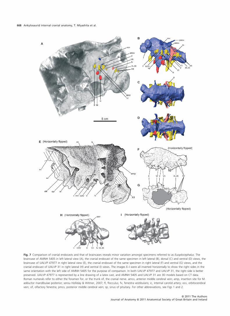
Potential intraspecific variation occurs in the olfactory
region of Euoplocephalus. The tunnel in AMNH 5405
(Fig. 4) cannot be identified in UALVP 47977. This suggests
that a tunnel like that in AMNH 5405 may have formed as a
result of partial enclosure of the groove found in the olfac-
tory region of UALVP 47977. On the other hand, the
descending process is conspicuous in UALVP 47977, whereas
the process is smaller in AMNH 5405. It is uncertain if the
differences were due to individual, ontogenetic, taxonomic
or taphonomic variation. Although UALVP 47977 is
currently best referred to Euoplocephalus, it could also be
Dyoplosaurus, another ankylosaurid from the Dinosaur Park
Formation (Arbour et al. 2009).
Intraspecific variation in cranial endocasts has been doc-
umented in the opossum Monodelphis domestica (Macrini
et al. 2007) and oreodonts (Macrini, 2009). The proportions
of cranial endocasts can vary among individuals and as a
result of ontogeny (Macrini et al. 2007). Witmer et al.
(2008) also showed that the morphology of the dural
expansion varies in Diplodocus. The dural expansion is not
conspicuous in the cranial endocasts of Euoplocephalus
(Fig. 7). The variation in Euoplocephalus cranial endocasts
results primarily from taphonomic distortion and limited
resolution of CT scanning. None of the variations in the
Euoplocephalus cranial endocasts described here are likely
to be taxonomically informative. This does not entirely
reject an influence of ontogeny on morphology of the
endocranial cavity in Euoplocephalus, because the cranial
endocasts compared here do not differ substantially in
size. Nonetheless, the fact that the adult-sized cranial en-
docasts do not substantially vary in morphology implies
that a single cranial endocast of an adult is likely suffi-
ciently to represent a general condition for the taxon, pro-
vided that the endocranial cavity has not been
taphonomically distorted.
Conclusions
A combination of direct osteological observation and CT-
based reconstruction provides corroborating, comple-
mentary evidence for the nasal and endocranial soft tissues
and braincase morphology of the ankylosaurid dinosaur
Euoplocephalus. A partial skull roof (UALVP 47977) reveals
vascular impressions in the nasal cavity, an unusual
descending process (likely representing a turbinate) and
deep groove possibly associated with the concha and the
olfactory fenestra. The fenestra demonstrates that the
cavity beside the dorsomedial passage of the main airway
housed the olfactory region, which is directly anterior to
the endocranial cavity in non-ankylosaur dinosaurs. The
ethmoidal region preserves the ethmoidal complex (mes-
ethmoid + sphenethmoid), the ectethmoid and mineralized
walls of the nasal cavity. The neurovascular foramina of the
braincase were re-interpreted. CT-based reconstructions of
other specimens of Euoplocephalus show that many con-
spicuous osteological correlates are present in these speci-
mens. Manually and digitally prepared cranial endocasts
show minor variation within the taxon. Therefore, a single
cranial endocast is likely to represent a general condition
for a taxon. Two parts of the nasal cavity are unusual in
ankylosaurids: the looping main airway and the large cavity
for the olfactory region. The elongate, looping nasal cavity
in ankylosaurid dinosaurs is not an adaptation for
enhanced olfaction, but likely had thermo- and osmoregu-
latory benefits. An acoustic function is also possible. It is
likely that the improved olfactory acuity is correlated with
the increased volume of the cavity for the olfactory region
in ankylosaurids, which is consistent with the size of the
olfactory bulb, although the olfactory hypothesis requires
corroborative evidence. These hypothesized functions sug-
gest that multiple functional drivers may explain morphol-
ogy in different parts of the ankylosaurid nasal cavity. The
nasal osteological correlates are expressed or preserved dif-
ferently in other ankylosaurid dinosaurs, which invites
extensive interspecific comparison.
Acknowledgements
The authors thank Brandon Strilisky (TMP) for access to the
specimens in his care. Allan Lindoe (University of Alberta) and
David Krause (New York State University at Stony Brook) col-
lected UALVP 47977. Michael James (University of Alberta) pre-
pared and photographed UALVP 47977 (Figs 2 and 6). Ariana
‘Premji’ Paulina Carabajal (University of La Plata) made the latex
endocast of UALVP 47977. CT scanning of UALVP 31 was con-
ducted at the University of Alberta Hospital ABACUS Facility (G.
Schaffler and R. Lambert). T.M. benefited from discussions with
Ariana Paulina Carabajal, David Evans (Royal Ontario Museum),
James Kirkland (Utah Geological Survey), Eric Snively (Ohio Uni-
versity) and Francois Therrien (TMP). Clint Boyd (University of
Texas, Austin) carefully reviewed the earlier versions of the
manuscript. Ryan Ridgely (Ohio University) worked on the 3D
visualization of the endocast of AMNH 5405 and provided assis-
tance in other ways. T.M. appreciates ongoing medical assis-
tance from Kesia Miyashita and family. All authors acknowledge
the logistical support from Eva Koppelhus (UALVP). This
research was supported by NSERC, Alberta Ingenuity Fund,
National Science Foundation, the Dinosaur Research Institute,
and the Department of Biological Sciences at the University of
Alberta.
ªª 2011 The AuthorsJournal of Anatomy ªª 2011 Anatomical Society of Great Britain and Ireland
Ankylosaurid internal cranial anatomy, T. Miyashita et al. 673

Author contributions
T.M., V.M.A. and L.M.W. are responsible for study design, data
acquisition and analysis. All authors contributed to drafting of
the manuscript. T.M. drew Figs 1, 2, 4 and 7E, V.M.A prepared
Figs 3, 5 and 7F–I, and L.M.W. contributed Fig. 7A–D.
References
Ali F, Zelenitsky DK, Therrien F, et al. (2008) Homology of the
‘ethmoidal complex’ of tyrannosaurids and its implications for
the reconstruction of the olfactory apparatus of non-avian
theropods. J Vertbr Paleontol 28, 123–133.
Arbour VM, Burns ME, Sissons RL (2009) A redescription of the
ankylosaurid dinosaur Dyoplosaurus acutosquameus Parks,
1924 (Ornithischia: Ankylosauria) and a revision of the genus.
J Vertbr Paleontol 29, 1117–1135.
Averianov AO (2002) An ankylosaurid (Ornithischia:
Ankylosauria) from the Upper Cretaceous Bissekty Formation
of Uzbekistan. Bull Inst R Sci Natl Belg Sci Terre 72, 97–110.
Bellairs Ad’A, Kamal AM (1981) The chondrocranium and the
development of the skull in recent reptiles. In: Biology of the
Reptilia, Volume 11 (Morphology F) (eds Gans C, Parsons T),
pp. 1–263. New York: Academic Press.
Brochu CA (2002) Osteology of Tyrannosaurus rex: insights from
a nearly complete skeleton and high-resolution computed
tomographic analysis of the skull. Soc Vert Paleontol Memoir
7, 1–138.
Carpenter K (2004) Redescription of Ankylosaurus magniventris
Brown 1908 (Ankylosauridae) from the Upper Cretaceous of
the Western Interior of North America. Can J Earth Sci 41,
961–986.
Carpenter K, Kirkland JI, Burge D, et al. (2001) Disarticulated skull
of a new primitive ankylosaurid from the Lower Cretaceous of
eastern Utah. In: The Armored Dinosaurs (ed. Carpenter K), pp.
211–238. Bloomington: Indiana University Press.
Clifford AB, Witmer LM (2004) Case studies in novel narial
anatomy: 3. Structure and function of the nasal cavity of saiga
(Artiodactyla: Bovidae: Saiga tatarica). J Zool 264, 217–230.
Coombs WP (1978a) An endocranial cast of Euoplocephalus
(Reptilia, Ornithischia). Palaeontogr Abt A 161, 176–182.
Coombs WP (1978b) The families of the ornithischian dinosaur
order Ankylosauria. Palaeontology 21, 143–170.
Coria RA, Currie PJ (2002) The braincase of Giganotosaurus
carolinii (Dinosauria: Theropod) from the Upper Cretaceous of
Argentina. J Vertbr Paleontol 22, 802–811.
Evans DC (2005) New evidence on brain-endocranial cavity
relationships in ornithischian dinosaurs. Acta Palaeontol Pol
50, 617–622.
Evans DC (2006) Nasal cavity homologies and cranial crest
function in lambeosaurine dinosaurs. Paleobiology 32, 109–125.
Evans DC, Ridgely R, Witmer LM (2009) Endocranial anatomy of
lambeosaurine hadrosaurids (Dinosauria: Ornithischia): a
sensorineural perspective on cranial crest function. Anat Rec
292, 1315–1337.
Fernandez M, Gasparini Z (2008) Salt glands in the Jurassic
metriorhynchid Geosaurus: implications for the evolution of
osmoregulation in Mesozoic marine crocodyliforms.
Naturwissenschaften 95, 79–84.
Fitch WT (1999) Acoustic exaggeration of size in birds via
tracheal elongation: comparative and theoretical analyses. J
Zool 248, 31–48.
Frey R, Hofmann RR (1997) Skull, proboscis musculature and
preorbital gland in the saiga antelope and Guenther’s dikdik
(Mammalia, Artiodactyla, Bovidae). Zool Anz 235, 183–199.
Frey R, Volodin I, Volodina E (2007) A nose that roars:
anatomical specializations and behavioural features of rutting
male saiga. J Anat 211, 717–736.
Galton PM (1983) The cranial anatomy of Dryosaurus, a
hypsilophodontid dinosaur from the Upper Jurassic of North
America and East Africa, with a review of the
hypsilophodontids from the Upper Jurassic of North America.
Geol Paleontol 17, 207–243.
Galton PM (1988) Skull bones and endocranial casts of
stegosaurian dinosaur Kentrosaurus Hennig 1915 from Upper
Jurassic of Tanzania, East Africa. Geol Paleontol 22, 123–143.
Galton PM (1989) Crania and endocranial casts from ornithopod
dinosaurs of the families Dryosauridae and Hypsilophodontidae
(Reptilia: Ornithischia). Geol Paleontol 23, 217–239.
Galton PM (1997) Cranial anatomy of the basal
hypsilophodontid dinosaur Thescelosaurus neglectus Gilmore
(Ornithischia: Ornithopoda) from the Upper Cretaceous of
North America. Rev Paleobiol 16, 321–358.
Galton PM (2001) Endocranial casts of the plated dinosaur
Stegosaurus (Upper Jurassic, western USA): a complete
undistorted cast and the original specimens of Othniel Charles
Marsh. In: The Armored Dinosaurs (ed. Carpenter K), pp. 103–
129. Bloomington: Indiana University Press.
Geist NR (2000) Nasal respiratory turbinate function in birds.
Physiol Biochem Zool 73, 581–589.
Hayakawa H, Manabe M, Carpenter K (2005) Nodosaurid
ankylosaur from the Cenomanian of Japan. J Vertbr Paleontol
25, 240–245.
Hill RV, Witmer LW, Norell MA (2003) A new specimen of
Pinacosaurus grangeri (Dinosaur: Ornithischia) from the Late
Cretaceous of Mongolia: ontogeny and phylogeny of
ankylosaurs. Am Mus Novit 3395, 1–29.
Holliday CM, Witmer LM (2007) Archosaur adductor chamber
evolution: integration of musculoskeletal and topological
criteria in jaw muscle homology. J Morphol 268, 457–484.
Hopson JA (1979) Paleoneurology. In: Biology of the Reptilia,
Vol. 9 (eds Gans C, Northcutt RG, Ulinski P), pp. 39–146. New
York: Academic Press.
Horner JR (1992) Cranial morphology of Prosaurolophus
(Ornithischia: Hadrosauridae) with descriptions of two new
hadrosaurid species and an evaluation of hadrosaurid
phylogenetic relationships. Mus Rock Occas Pap 2, 1–119.
Klembara J (1991) The cranial anatomy of early ontogenetic
stages of Alligator mississippiensis (Daudin, 1802) and the
significance of some of its cranial structures for the evolution
of tetrapods. Palaeontogr Abt A 215, 103–171.
Kurzanov SM, Tumanova TA (1978) On the structure of the
endocranium in some ankylosaurs from Mongolia. Paleontol
Zh 1978, 90–96. [In Russian]
Macrini TE (2009) Description of a digital cranial endocast of
Bathygenys reevesi (Merycoidodontidae; Oreodontoidea) and
implications for apomorphy-based diagnosis of isolated,
natural endocasts. J Vertbr Paleontol 29, 1199–1211.
Macrini TE, Rowe T, VandeBerg JL (2007) Cranial endocasts from
a growth series of Monodelphis domestica (Didelphidae,
Marsupialia): a study of individual and ontogenetic variation.
J Morphol 268, 844–865.
Maryanska T (1971) New data on the skull of Pinacosaurus
grangeri (Ankylosauria). Palaeontol Pol 25, 45–53.
ªª 2011 The AuthorsJournal of Anatomy ªª 2011 Anatomical Society of Great Britain and Ireland
Ankylosaurid internal cranial anatomy, T. Miyashita et al.674
Maryanska T (1977) Ankylosauridae (Dinosauria) from
Mongolia. Palaeontol Pol 37, 85–151.
Miyashita T, Arbour VM (2007) New information on the internal
cranial anatomy of Euoplocephalus (Ornithischia,
Ankylosauridae). J Vertbr Paleontol 27 (Suppl), 119A.
Nelson JE, Christian KA, Baudinette RV (2007) Anatomy of the
nasal passages of three species of Australian bats in relation
to water loss. Aust J Zool 55, 57–62.
Parks WA (1924) Dyoplosaurus acutosquameus, a new genus and
species of armored dinosaur; and notes on a skeleton of
Prosaurolophus maximus. Univ Toronto Stud Geol Ser 18, 1–35.
Parsons TS (1959) Studies on the comparative embryology of
the reptilian noses. Bull Mus Comp Zool, 120, 101–277.
Parsons WL, Parsons KM (2009) A new ankylosaur (Dinosauria:
Ankylosauria) from the Lower Cretaceous Cloverly Formation
of central Montana. Can J Earth Sci 46, 721–738.
Romanoff AL (1960) The Avian Embryo: Structural and Functional
Development, 1305 pp. New York: The Macmillan Company.
Ruben JA (1996) Evolution of endothermy in birds, mammals,
and their ancestors. In: Animals and Temperature: Phenotypic
and Evolutionary Adaptation (eds Johnston LA, Bennett AF),
pp. 347–376. Cambridge: Cambridge University Press.
Sampson SD, Witmer LD (2007) Craniofacial anatomy of
Majungasaurus crenatissimus (Theropoda: Abelisauridae) from
the Late Cretaceous of Madagascar. Soc Vert Paleontol
Memoir 8, 32–102.
Sanders RK, Smith DK (2005) The endocranium of the theropod
dinosaur Ceratosaurus studied with computed tomography.
Acta Palaeontol Pol 50, 601–616.
Schmidt-Nielsen K, Kanwisher J, Lasiewski RC, et al. (1969)
Temperature regulation and respiration in the ostrich. Condor
71, 341–352.
Schmidt-Nielsen K, Hainsworth FR, Murrish D (1970)
Countercurrent heat exchange in the respiratory passages:
effect on water and heat balance. Resp Physiol 9, 263–276.
Sedlmayr JC (2002) Anatomy, evolution, and functional
significance of cephalic vasculature in Archosauria, 399 pp.
Unpublished PhD Thesis. Unviersity of Ohio at Athens.
Sereno PC, Wilson JA, Witmer LM, et al. (2007) Structural
extremes in a Cretaceous dinosaur. PLoS ONE 2, 1–9 (e1230).
Tieleman BI, Williams JB, Michaeli G, et al. (1999) The role of
the nasal passages in the water economy of crested larks and
desert larks. Physiol Biochem Zool 72, 219–226.
Tumanova TA (1987) The armoured dinosaurs of Mongolia.
Sovm Sov-Mong Paleontol Eksped Trudy 32, 1–80. [In Russian].
Van Valkenburgh B, Theodor J, Friscia A, et al. (2004)
Respiratory turbinates of canids and felids: a quantitative
comparison. J Zool 264, 281–293.
Van Valkenburgh B, Curtis A, Samuels JX, et al. (2011) Aquatic
adaptations in the nose of carnivorans: evidence from the
turbinates. J Anat 218, 298–310.
Vickaryous MK (2006) New information on the cranial anatomy
of Edmontonia rugosidens Gilmore, a Late Cretaceous
nodosaurid dinosaur from Dinosaur Provincial Park, Alberta. J
Vertbr Paleontol 26, 1011–1013.
Vickaryous MK, Russell AP (2003) A redescription of the skull of
Euoplocephalus tutus (Archosauria: Ornithischia): a
foundation for comparative and systematic studies of
ankylosaurian dinosaurs. Zool J Linn Soc 137, 157–186.
Vickaryous MK, Russell AP, Currie PJ, et al. (2001) A new
ankylosaurid (Dinosauria: Ankylosauria) from the Lower
Cretaceous of China, with comments on ankylosaurian
relationships. Can J Earth Sci, 38, 1767–1780.
Vickaryous MK, Maryanska T, Weishampel DB (2004)
Ankylosauria. In: The Dinosauria, 2nd edn (eds Weishampel
DB, Dodson P, Osmolska H), pp. 363–392. Berkeley: University
of California Press.
Wealthall RJ, Herring SW (2006) Endochondral ossification of
the mouse nasal septum. Anat Rec 288, 1163–1172.
Weishampel DB (1981) The nasal cavity of lambeosaurine
hadrosaurids (Reptilia, Ornithischia): comparative anatomy
and homologies. J Paleontol 55, 1046–1057.
Witmer LM (1995) Homology of facial structures in extant
archosaurs (birds and crocodilians), with special reference to
paranasal pneumaticity and nasal conchae. J Morphol 225,
269–327.
Witmer LM (1997) The evolution of the antorbital cavity of
archosaurs: a study in soft-tissue reconstruction in the fossil
record with an analysis of the function of pneumaticity. Soc
Vert Paleontol Memoir 3, 1–73.
Witmer LM, Ridgely RC (2008) The paranasal air sinuses of
predatory and armored dinosaurs (Archosauria: Theropoda
and Ankylosauria) and their contribution to cephalic
structure. Anat Rec 291, 1362–1388.
Witmer LM, Ridgely RC (2009) New insights into the brain,
braincase, and ear region of tyrannosaurs (Dinosauria,
Theropoda), with implications for sensory organization and
behavior. Anat Rec 292, 1266–1296.
Witmer LM, Chatterjee S, Franzosa J, et al. (2003)
Neuroanatomy of flying reptiles and implications for flight,
posture and behavior. Nature 425, 950–953.
Witmer LM, Ridgely RC, Dufeau DL, et al. (2008) Using CT to
peer into the past: 3D visualization of the brain and ear
regions of birds, crocodiles, and nonavian dinosaurs. In:
Anatomical Imaging: Towards a New Morphology (eds Endo
H, Frey R), pp 67–87. Tokyo: Springer.
Zelenitsky DK, Therrien F, Kobayashi Y (2009) Olfactory acuity
in theropods: palaeobiological and evolutionary implications.
Proc Biol Sci 276, 667–673.
Zelenitsky DK, Therrien F, Ridgely RC, et al. (2011) Evolution of
olfaction in non-avian theropod dinosaurs and birds. Proc Biol
Sci, doi: 10.1098/rspb.2011.0238.
Zusi RL (1993) Patterns of diversity in the avian skull. In: The
Skull: Patterns of Structural and Systematic Diversity, Vol. 2
(eds Hanken J, Hall BK), pp. 391–437. Chicago: University of
Chicago Press.
ªª 2011 The AuthorsJournal of Anatomy ªª 2011 Anatomical Society of Great Britain and Ireland
Ankylosaurid internal cranial anatomy, T. Miyashita et al. 675