Mitosis CELL DIVISION DURING MITOSIS The cell nucleus divides into two identical halves. These...
62
-
Upload
gwenda-stafford -
Category
Documents
-
view
235 -
download
0
Transcript of Mitosis CELL DIVISION DURING MITOSIS The cell nucleus divides into two identical halves. These...

Mitosis
CELL DIVISION
DURING MITOSIS
The cell nucleus divides into two identical halves. These halves contain the same number of chromosomes as the parent cell.
Write This
Chromosome – A double rod shaped structure that contains DNA and protein.
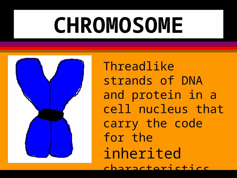
CHROMOSOME
Threadlike strands of DNA and protein in a cell nucleus that carry the
code for the inherited characteristics of an organism.
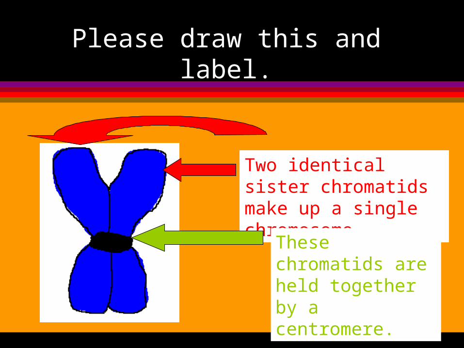
Please draw this and label.
Two identical sister chromatids make up a single chromosome.
These chromatids are held together by a centromere.
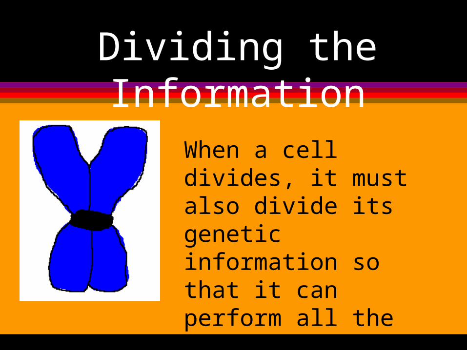
Dividing the Information
When a cell divides, it must also divide its genetic information so that it can perform all the activities of the parent cell.
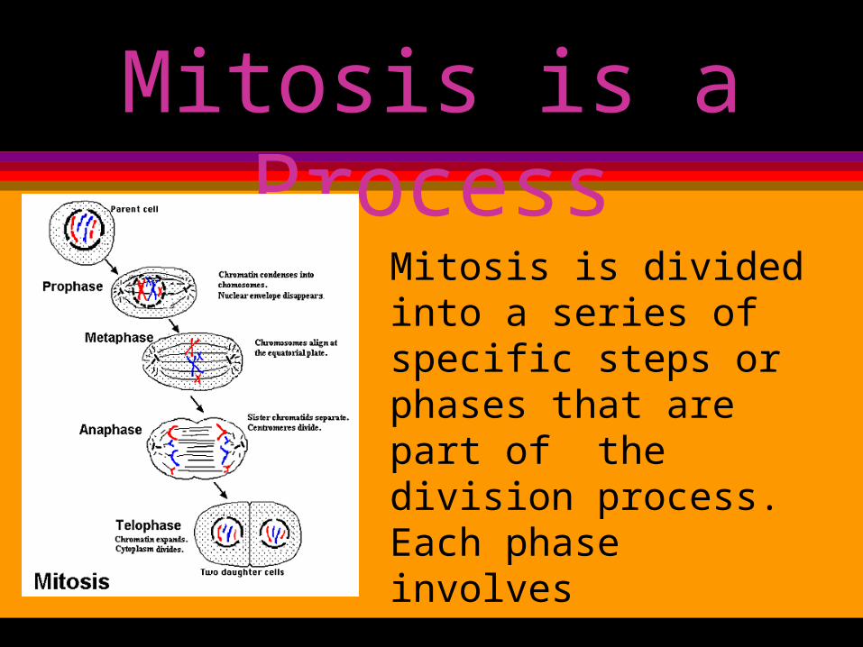
Mitosis is a Process
Mitosis is divided into a series of specific steps or phases that are part of the division process. Each phase involves significant phenomenon with specific events occurring.
INTERPHASE
During interphase the chromosomes are not visible. Amazingly, during this phase the genetic material is making an identical copy of itself. This process is called replication. It is this process of replication that allows chromosomes to remain double stranded, generation after generation.
INTERPHASE
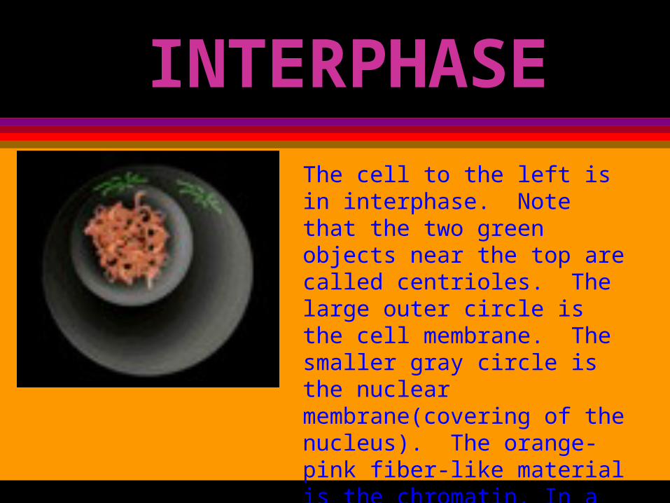
The cell to the left is in interphase. Note that the two green objects near the top are called centrioles. The large outer circle is the cell membrane. The smaller gray circle is the nuclear membrane(covering of the nucleus). The orange-pink fiber-like material is the chromatin. In a later phase, this chromatin will eventually organize into chromosomes.
Write This
Interphase:1. DNA is replicating (making copies
of itself)2. Organelles are replicating.3. Nucleus is visible.4. Genetic information appears as
chromatin.
PROPHASE
During prophase, chromosomes become fully visible. The nucleolus
and the nuclear membrane disappear. The centrioles begin to migrate out
towards opposite ends of the cell. Thin thread-like fibers called spindle fibers
begin to stretch across the cell.
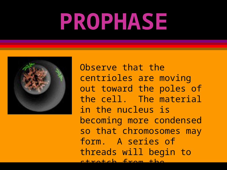
PROPHASE
Observe that the centrioles are moving out toward the poles of the cell. The material in the nucleus is becoming more condensed so that chromosomes may form. A series of threads will begin to stretch from the centrioles toward the chromosomes. These threads make up the spindle apparatus.
Write ThisProphase
1. Chromatin bunches up to form chromosomes.
2. Nuclear envelope disappears.3. Centrioles appear in animal cells
to aid in division. They begin moving to the poles.
METAPHASE
In metaphase, the chromosomes line up along a central line known as the equator or equatorial plate. The spindle apparatus attaches to the centromere of each chromosome. The centrioles have moved to the poles (outer ends) of the cell.
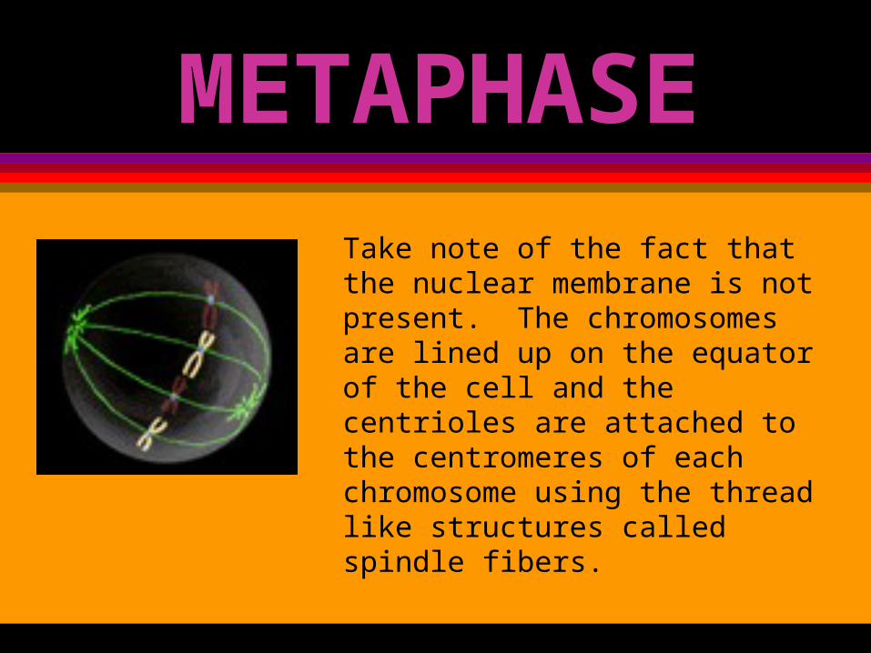
METAPHASE
Take note of the fact that the nuclear membrane is not present. The chromosomes are lined up on the equator of the cell and the centrioles are attached to the centromeres of each chromosome using the thread like structures called spindle fibers.
Write ThisMetaphase
1. Chromosomes line up at the equator.
2. Spindle fibers are attached.3. Centrioles are at the poles.

ANAPHASE
In anaphase, the chromosomes are pulled apart by the spindle fibers. This divides the genetic material(DNA) into two identical halves. One chromatid of each chromosome will end up in each half of the dividing cell.
ANAPHASE
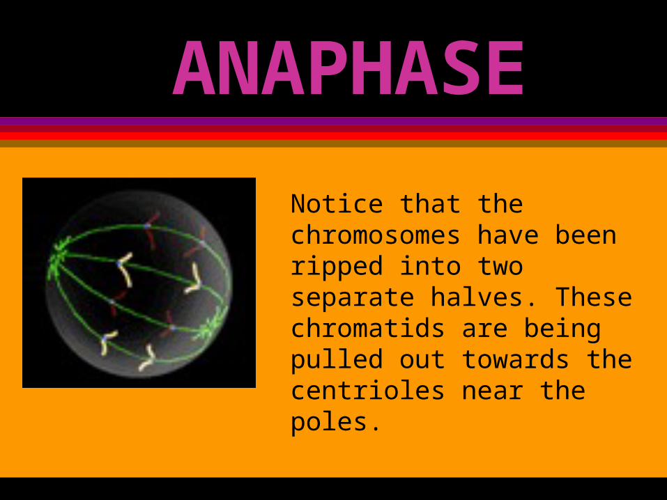
Notice that the chromosomes have been ripped into two separate halves. These chromatids are being pulled out towards the centrioles near the poles.
Write ThisAnaphase
1. Chromosomes get pulled apart so that one chromatid will go toward each pole.
TELOPHASE
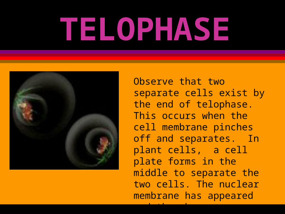
The final step to mitosis is telophase. During this step the centrioles and spindle fibers begin to disappear. The chromosomes begin to spread out and are harder to see. The nuclear membrane and a nucleolus begin to reappear in each newly formed half. The cell membrane pinches off so that each newly formed cell can separate.
TELOPHASE
Observe that two separate cells exist by the end of telophase. This occurs when the cell membrane pinches off and separates. In plant cells, a cell plate forms in the middle to separate the two cells. The nuclear membrane has appeared and the chromosomes are no longer organized. These two new cells are called daughter cells.
Write ThisTelophase
1. Cell membrane pinches inward to divide up the cytoplasm and cell membrane.
2. Nuclear envelope reappears.3. By late telophase the
chromosomes begin to unbunch to appear as chromatin.
REMEMBER
Mitosis is the process of a cell’s nucleus dividing in two. When the process is complete, the two new cells are identical to the parent with the same number of chromosomes as the parent.

What’s it really look like?
The following are pictures of real cells at various
stages of mitosis.
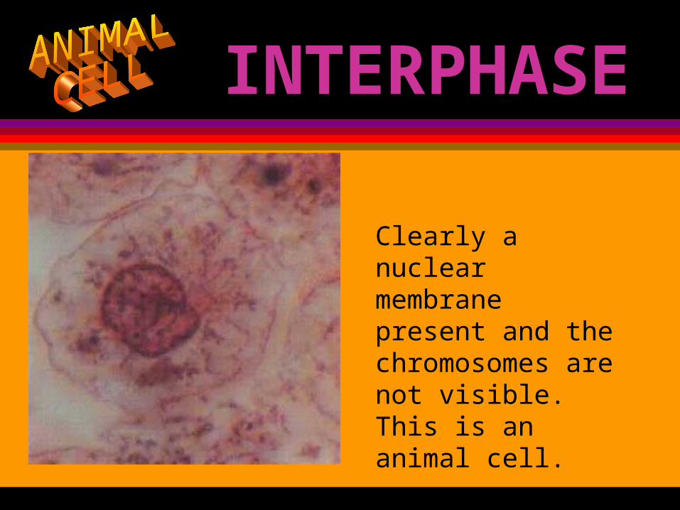
INTERPHASE
Clearly a nuclear membrane present and the chromosomes are not visible. This is an animal cell.
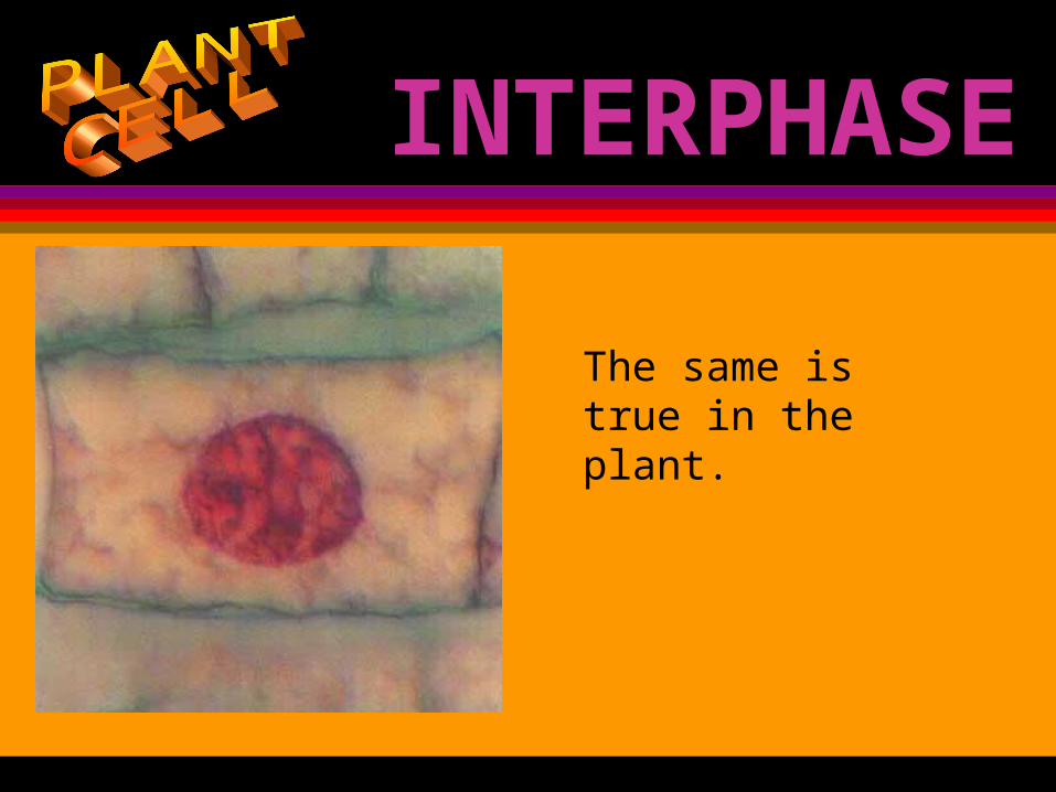
INTERPHASE
The same is true in the plant.
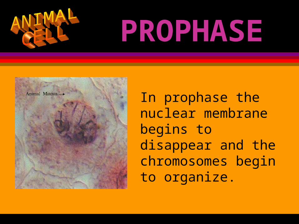
PROPHASE
In prophase the nuclear membrane begins to disappear and the chromosomes begin to organize.
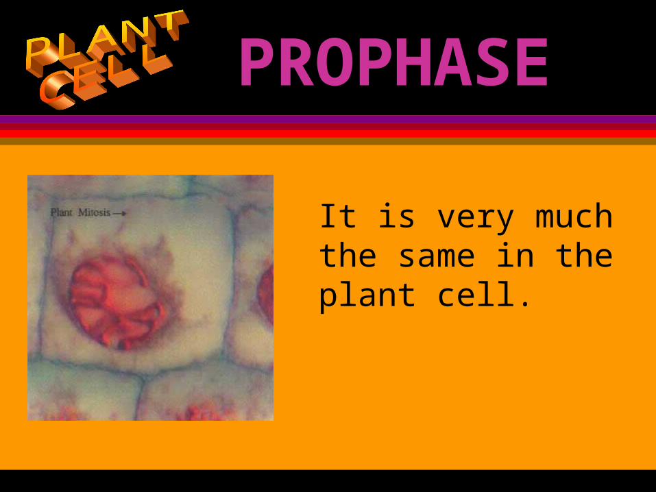
PROPHASE
It is very much the same in the plant cell.
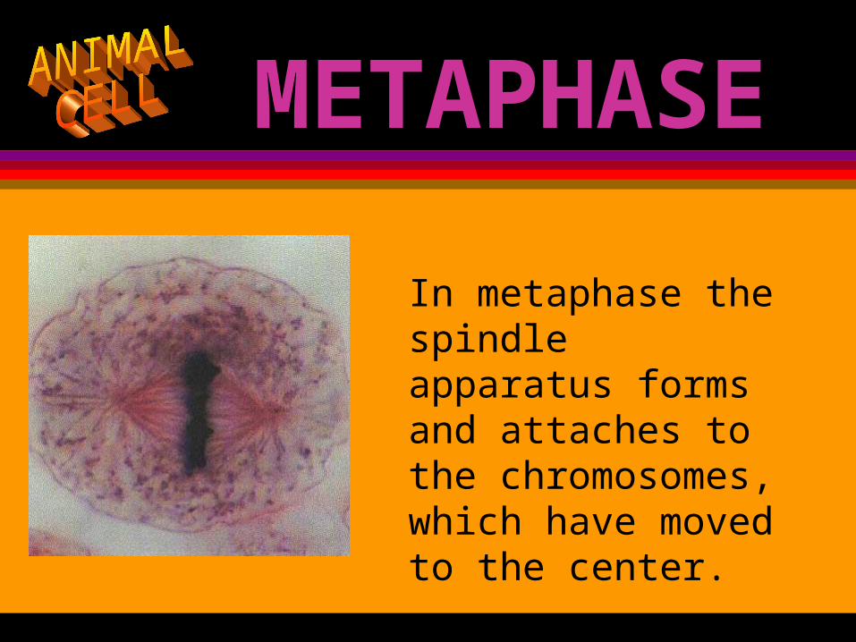
METAPHASE
In metaphase the spindle apparatus forms and attaches to the chromosomes, which have moved to the center.
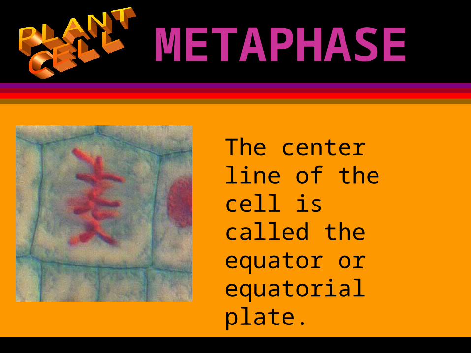
METAPHASE
The center line of the cell is called the equator or equatorial plate.
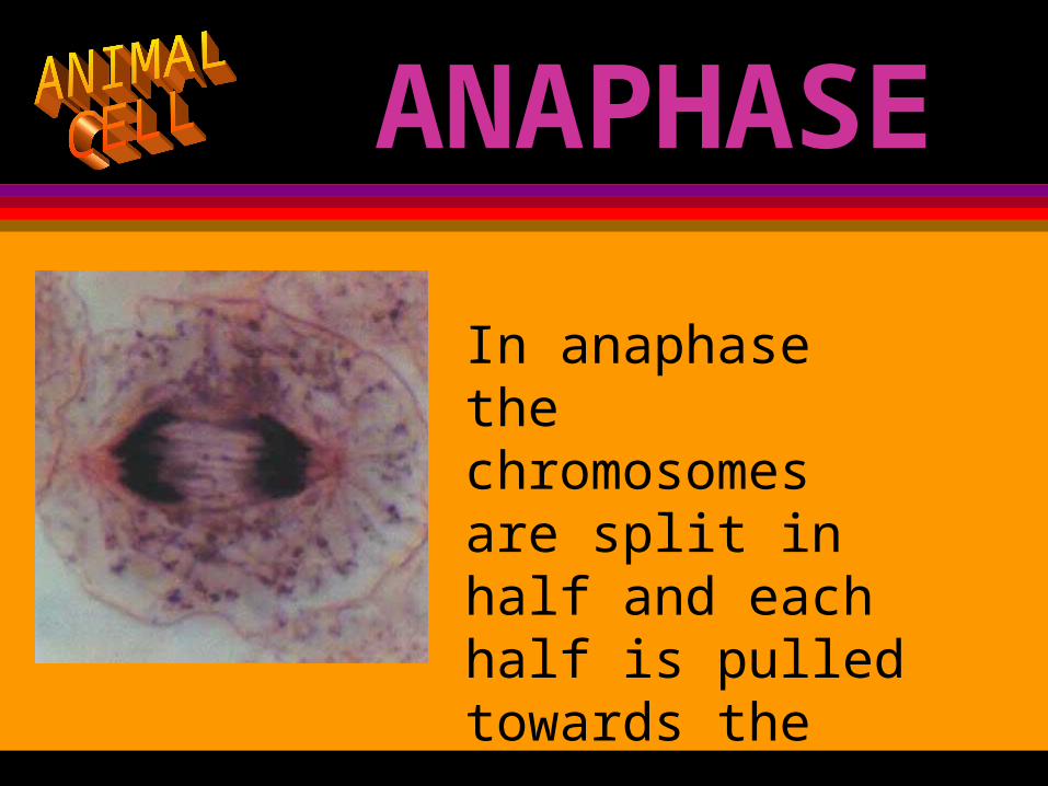
ANAPHASE
In anaphase the chromosomes are split in half and each half is pulled towards the poles.
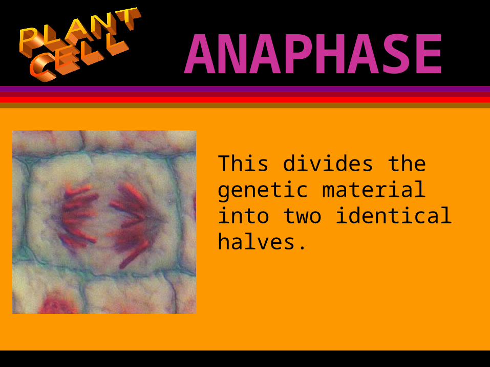
ANAPHASE
This divides the genetic material into two identical halves.
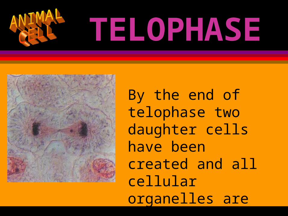
TELOPHASE
By the end of telophase two daughter cells have been created and all cellular organelles are divided.
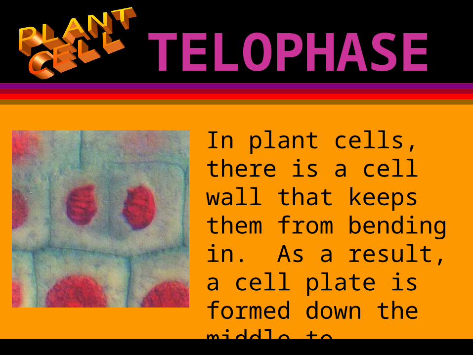
TELOPHASE
In plant cells, there is a cell wall that keeps them from bending in. As a result, a cell plate is formed down the middle to complete the cell’s covering.
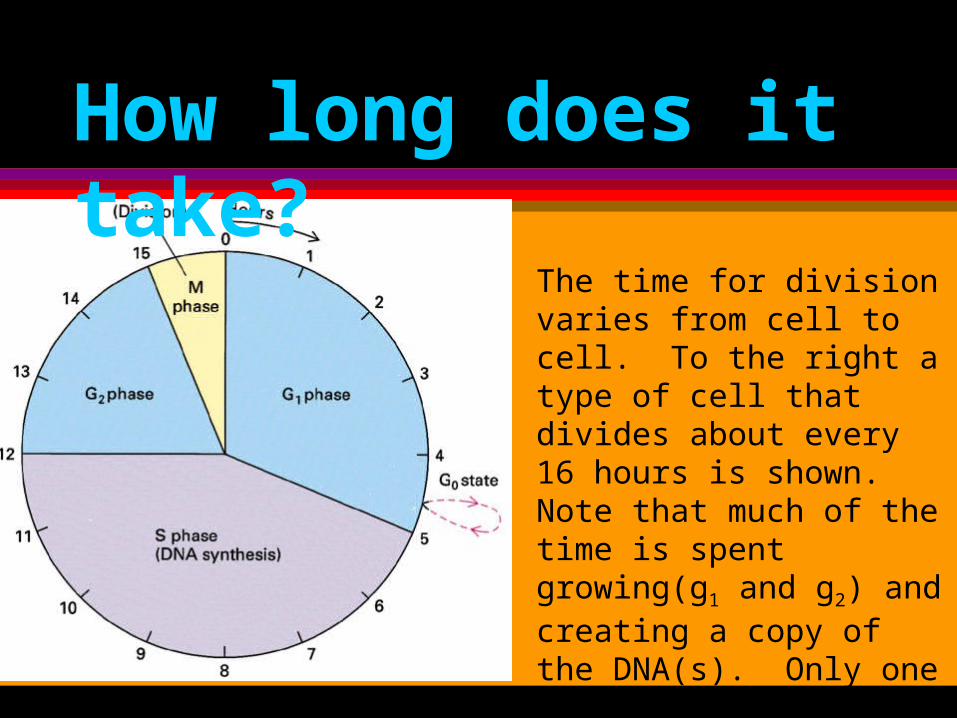
How long does it take?
The time for division varies from cell to cell. To the right a type of cell that divides about every 16 hours is shown. Note that much of the time is spent growing(g1 and g2) and creating a copy of the DNA(s). Only one of the 16 hours is actually spent dividing.
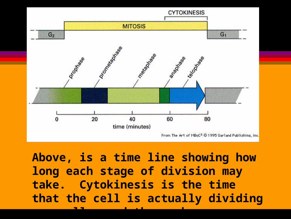
Above, is a time line showing how long each stage of division may take. Cytokinesis is the time that the cell is actually dividing organelles and the membrane.
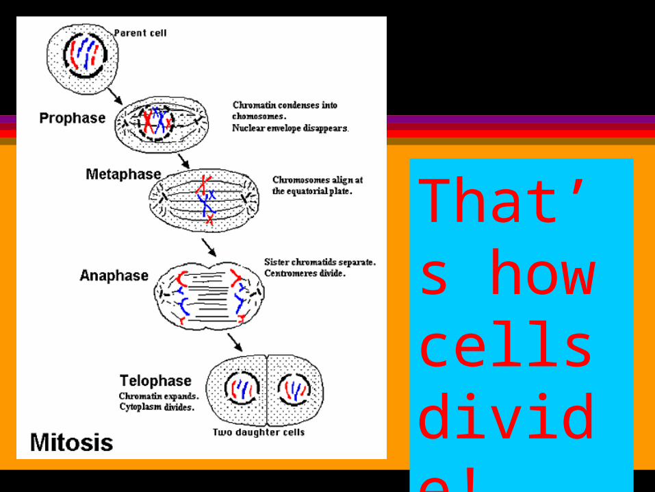
That’s how cells divide!
IT’S TIME TO TAKE SOME NOTES AND
DRAW A DIAGRAM.
Draw the following diagrams into your notes.
Be sure to label the parts.
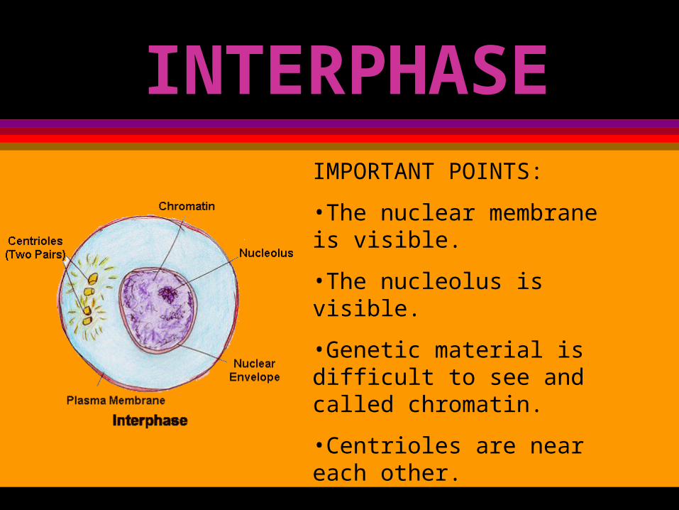
INTERPHASEIMPORTANT POINTS:
•The nuclear membrane is visible.
•The nucleolus is visible.
•Genetic material is difficult to see and called chromatin.
•Centrioles are near each other.
•DNA is replicated(copied).

PROPHASE
IMPORTANT POINTS:
•Chromosome appear
•Centrioles begin to migrate to the poles.
•Chromosomes consist of two sister chromatids.
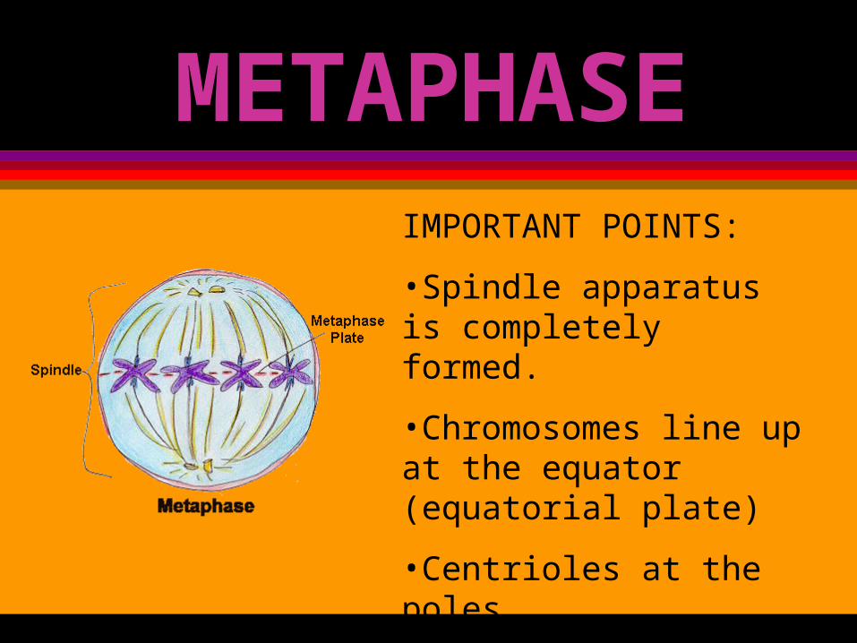
METAPHASEIMPORTANT POINTS:
•Spindle apparatus is completely formed.
•Chromosomes line up at the equator (equatorial plate)
•Centrioles at the poles.
•Nuclear envelope and nucleolus gone
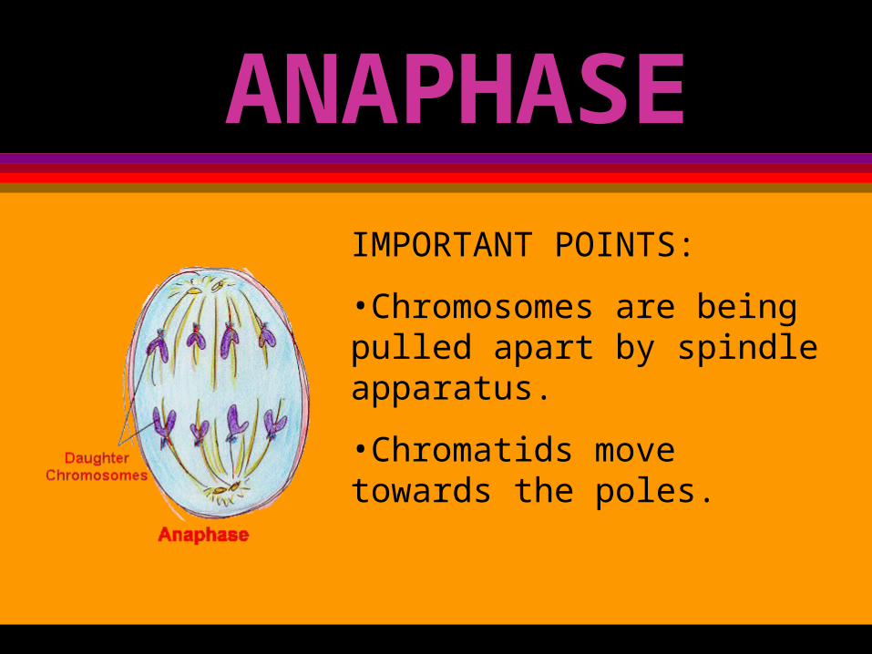
ANAPHASEIMPORTANT POINTS:
•Chromosomes are being pulled apart by spindle apparatus.
•Chromatids move towards the poles.
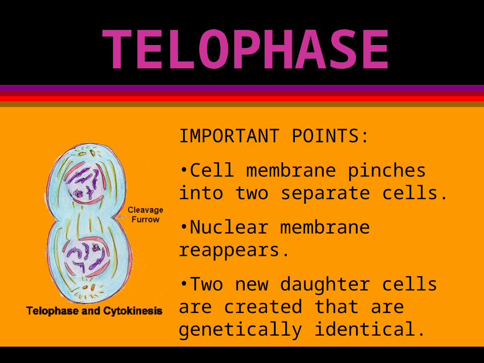
TELOPHASEIMPORTANT POINTS:
•Cell membrane pinches into two separate cells.
•Nuclear membrane reappears.
•Two new daughter cells are created that are genetically identical.

PLEASE LET YOUR TEACHER KNOW YOU ARE DONE!