The Cell Cycle/Division/Mitosis Unit 4. The Cell Cycle Interphase Mitosis Cytokinesis.
1
1
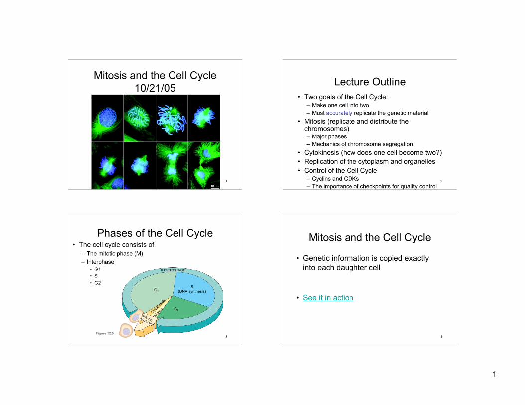
Mitosis and the Cell Cycle10/21/05
2
Lecture Outline• Two goals of the Cell Cycle:
– Make one cell into two– Must accurately replicate the genetic material
• Mitosis (replicate and distribute thechromosomes)– Major phases– Mechanics of chromosome segregation
• Cytokinesis (how does one cell become two?)• Replication of the cytoplasm and organelles• Control of the Cell Cycle
– Cyclins and CDKs– The importance of checkpoints for quality control
3
Phases of the Cell Cycle• The cell cycle consists of
– The mitotic phase (M)– Interphase
• G1• S• G2
INTERPHASE
G1S
(DNA synthesis)
G2Cyto
kines
is
Mitosis
MITOTIC(M) PHASE
Figure 12.54
Mitosis and the Cell Cycle
• Genetic information is copied exactlyinto each daughter cell
• See it in action

2
5
• Each duplicated chromosomeHas two sister chromatids, which separate
during cell division
0.5 µm
Centromere
Sisterchromatids
Centromeres Sister chromatidsFigure 12.4
One chromosome, one DNA molecule
Duplication
One chromosome, two DNA molecules(Two attached chromatids)
Sister chromosomes separateduring mitosis
6
G2 OFINTERPHASE PROPHASE PROMETAPHASE
Centrosomes(with centriole pairs) Chromatin
(duplicated)Early mitoticspindle
AsterCentromere
Fragmentsof nuclearenvelope
Kinetochore
Nucleolus Nuclearenvelope
Plasmamembrane
Chromosome, consistingof two sister chromatids
Kinetochore microtubule Figure 12.6
Nonkinetochoremicrotubules
Overview of Mitosis
DNA replication duringInterphase Prophase:
Chromosomes begin to condense.Spindle starts to form
Prometaphase:Nuclear envelopebreaks down.Chromosomesattach to spindle
7
Centrosome at one spindle pole
Daughter chromosomes
METAPHASE ANAPHASE TELOPHASE AND CYTOKINESIS
Spindle
Metaphaseplate Nucleolus
formingCleavagefurrow
Nuclear envelopeformingFigure 12.6
Overview of Mitosis
Metaphase:Chromosomes align incenter of cell
Anaphase:Sister chromatids separate
Telophase:Completeset ofchromosomes at eachpole
8
Balanced attachment of spindle fibers to both chromatidsaligns chromosomes in metaphase
“tug of war”

3
9
CentrosomeAster
Sisterchromatids
MetaphasePlate
Kinetochores
Overlappingnonkinetochoremicrotubules
Kinetochoresmicrotubules
Centrosome
ChromosomesMicrotubules 0.5 µm
1 µm
Figure 12.7
Nonkinetechoremicrotubules from oppositepoles overlap and pushagainst each other, elongatingthe cell
Kinetochore microtubulesattach to centromeresand direct the polewardmovement ofchromosomes
Both chromatids must be captured byspindle fibers. If any kinetochoresremain unattached, chromosomes willnot separate
10
Mark
Spindle fibers shorten at the kinetochore
11
Kinetochore
Chromosomemovement
Microtubule Motorprotein
Chromosome
Kinetochore
Tubulinsubunits
12
CytokinesisAnimal cells divide by
constriction
Cleavage furrow
Contractile ring ofmicrofilaments Daughter cells
100 µm
(a) Cleavage of an animal cell (SEM)Figure 12.9 A
Daughter cells
1 µmVesiclesforming cell plate
Wall of patent cellCell plateNew cell wall
(b) Cell plate formation in a plant cell (SEM)Figure 12.9 B
Plant cells build apartition (cell plate)
4
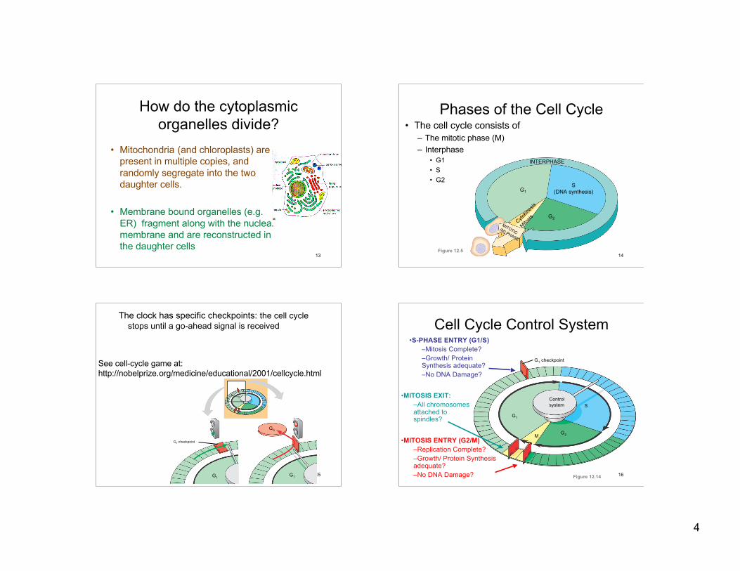
13
How do the cytoplasmicorganelles divide?
• Mitochondria (and chloroplasts) arepresent in multiple copies, andrandomly segregate into the twodaughter cells.
• Membrane bound organelles (e.g.ER) fragment along with the nuclearmembrane and are reconstructed inthe daughter cells
14
Phases of the Cell Cycle• The cell cycle consists of
– The mitotic phase (M)– Interphase
• G1• S• G2
INTERPHASE
G1S
(DNA synthesis)
G2Cyto
kines
is
Mitosis
MITOTIC(M) PHASE
Figure 12.5
15
The clock has specific checkpoints: the cell cyclestops until a go-ahead signal is received
G1 checkpoint
G1G1
G0
(a) If a cell receives a go-ahead signal at the G1 checkpoint, the cell continues on in the cell cycle.
(b) If a cell does not receive a go-ahead signal at the G1checkpoint, the cell exits the cell cycle and goes into G0, a nondividing state.
Figure 12.15 A, B
See cell-cycle game at:http://nobelprize.org/medicine/educational/2001/cellcycle.html
16
Cell Cycle Control System
Figure 12.14
Control system
G1 checkpoint
G1
S
G2M
•MITOSIS EXIT:–All chromosomesattached tospindles?
•S-PHASE ENTRY (G1/S)–Mitosis Complete?–Growth/ ProteinSynthesis adequate?–No DNA Damage?
•MITOSIS ENTRY (G2/M)–Replication Complete?–Growth/ Protein Synthesisadequate?–No DNA Damage?

5
17
The Cell Cycle Clock:Cyclins and Cyclin-dependent kinases
Cyclins– G1 cyclin (cyclin D)– S-phase cyclins (cyclins E and A)– M-phase cyclins (cyclins B and A)
Cyclin-dependent kinases (Cdks)– G1 Cdk (Cdk4)– S-phase Cdk ((Cdk2)– M-phase Cdk (Cdk1)
Cyclin levels in the cell rise and fallwith the stages of the cell cycle.
Cdk levels remain stable, but eachmust bind the appropriate cyclin(whose levels fluctuate) in order to beactivated.
18
Phosphorylation of CDK Targets Changes Their Activity
Now performsa cell cycle function
19
The Human Cell Cycle
~ 10 hours
~ 9 hours
~ 4 hours
~ 1 hour
20
How does the cell cycle cycle?
Focus first on entryand exit frommitosis
6
21
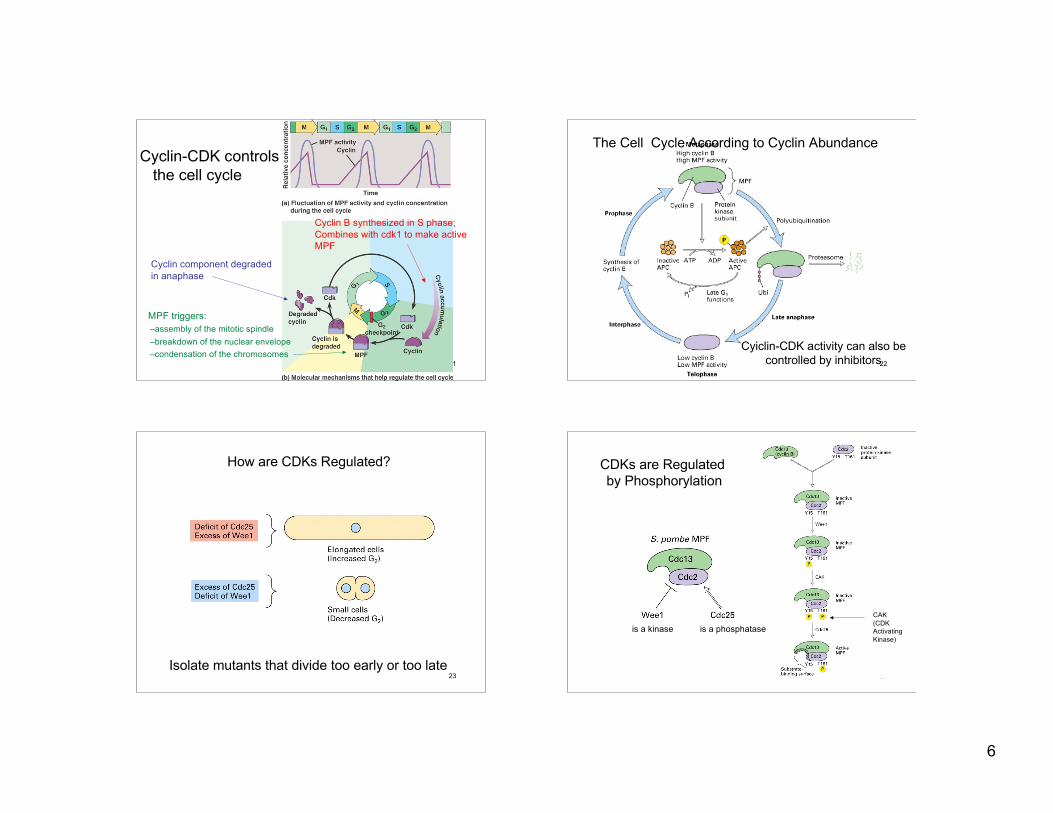
MPF triggers:–assembly of the mitotic spindle –breakdown of the nuclear envelope –condensation of the chromosomes
Cyclin component degradedin anaphase
Cyclin B synthesized in S phase;Combines with cdk1 to make activeMPF
Cyclin-CDK controlsthe cell cycle
22
Cyiclin-CDK activity can also becontrolled by inhibitors
The Cell Cycle According to Cyclin Abundance
23
How are CDKs Regulated?
Isolate mutants that divide too early or too late24
CDKs are Regulated by Phosphorylation
is a kinase is a phosphatase
CAK(CDKActivatingKinase)

7
25
Conformational Changes Associated with CDK Phosphorylation
The T-loop blockssubstrate access
Free CDK CDK + Cyclin T161 phosphorylation
Binding of cyclinmoves the T-loop
Phosporylation movesthe T-loop more 26
Cyclin Dependent Kinase Inhibitors (CKIs)
CyclinCDK
p21
CyclinCDK4
CyclinCDK
CDK4p16
p16
p21
27
Cell Cycle Regulators andCancer
28
G1G2 Metaphase Anaphase
Cyclin-Cdk Cyclin-CdkSecurin
Anaphase promoting complex
APC
Cdc20 E2-Ub Cdh1
Triggers:Chromosome separationBreakdown of cyclin to re-start the cycleBreakdown of geminin(to again allow replication)

8
29MCB Fig. 13-19 30
Prophase Prometaphase
Metaphase Anaphase Telophase