Mitochondrial Cytopathy in Adults 2001 Cohen 625 6
16
C LEVELAN D C LI NIC JO U RN AL O F M EDIC I N E VO LUM E 68 • NU M BER 7 JU LY 2001 625 ITOCHONDRIALCYTOPATHIES —disorders of the e nerg y-produci ng org ane lles of the cells—are an increasingly recognized ca us e o f hum a n i llne s s . A lthoug h this fie ld is still in its infancy, several syndromes have been identified and linked to specific muta- tions in m itocho ndria l DNA . T hes e p rob a bly represent only a few of the mitochondrial function disorders. This p a p er add r esses: • How m itoc ho nd ria l d ise a s e s a ris e • The pres e nta tions a nd d ia g nos is of the various known mitochondri a l di s e as es • Pos s ible trea tm ents (there are no c ure s ). The c h a lle nge f or t h e pr im a r y ca r e p h y s i- cia n is to ide ntify pa ti ents who m ay have a mitochondrial cytopathy and to coordinate m anag em ent. The challeng e for the su bs pe - cialist diagnostician is to provide an accurate diag nosis and a s s is t the prim ary ca re phys ician in caring for the patient. MITOCHONDRIA: POWERHOUSES OF THE CELL Mitochondria, contained in all human cells excep t ma t ure erythrocyt es , perfor m the vital task of generating adenosine triphosphate (ATP) , the m ole cule the ce ll us e s for the b ulk of it s e nergy nee ds ( FIGURE 1 ). M ITOCHOND RIAL DISEASES ARE REM ARKABL Y D IV ERSE A probl e m tha t has vexed the s tudy of m ito- chondri al di s ea s eseve r si nce the fi rst reported cas e (in 1962) 1 is that their manifestations are rem arkabl y di verse . 2 A lthoug h the u nde rlying chara cteris ti c of a ll of them is la ck of ad eq ua te energ y to me et cel lular nee ds , they vary con- BRUCE H. COHEN, M D Chief, Section of Pediatric Neurology, Departments of Neurology , Neurosurgery , and the Tauss ig Cancer Center, Cleveland Clinic DEBORAH R. GOLD, M D Section of Pediatric Neurology, Department of Neurology, Cleveland Cli nic M i to cho nd r i a l cytop a thy i n a d ul ts : W ha t w e k now s o f a r REVIEW M ABSTRACT M itochond rial cytop athies are a divers e group of inh erited and acquired disorders that res ult in inadequ ate energy production.They can be caused by inheritable genetic mut ations , acquired somatic mutations, expos ure to toxins (including some prescription medications), and the aging proces s itself. In addition, a number of well-described diseases can decrease mitochondrial energy production; these include hyperthyroidism, hypothyroidism, and hyperlipidemia. KE Y POIN TS T he m itochond rial cytopathies vary considerably in t heir man ifestat ions, leading to uncertaint y about diagno sis and classification. T here are no absolute diagno stic criteria for mitochondrial cytopathies, and m ost screening tests are neither specific nor sens itive, w hich can lead to false-positive and false- negative diagnoses.
-
Upload
frank-nova -
Category
Documents
-
view
53 -
download
1
Transcript of Mitochondrial Cytopathy in Adults 2001 Cohen 625 6

5/11/2018 Mitochondrial Cytopathy in Adults 2001 Cohen 625 6 - slidepdf.com
http://slidepdf.com/reader/full/mitochondrial-cytopathy-in-adults-2001-cohen-625-6
CLEVELAND CLINIC JOURNAL OF M EDICINE VOLUME 68 • NUM BER 7 JULY 20 01 625
ITOCHONDRIAL CYTOPATHIES—disordersof the energy-producing organelles of
the cells—are an increasingly recognized
cause of human illness. Although this field isstill in its infancy, several syndromes havebeen identified and linked to specific muta-tions in mitochondrial DNA. These probablyrepresent only a few of the mitochondrialfunction disorders.
This paper addresses:• How mitochondrial diseases arise• The presentations and diagnosis of the
various known mitochondrial diseases• Possible treatments (there are no cures).
The challenge for the primary care physi-
cian is to identify patients who may have amitochondrial cytopathy and to coordinatemanagement. The challenge for the subspe-cialist diagnostician is to provide an accuratediagnosis and assist the primary care physicianin caring for the patient.
s MITOCHONDRIA:POWERHOUSES OF THE CELL
Mitochondria, contained in all human cellsexcept mature erythrocytes, perform the vital
task of generating adenosine triphosphate(ATP), the molecule the cell uses for the bulk of its energy needs (FIGURE 1).
s MITOCHONDRIAL DISEASESARE REMARKABLY DIVERSE
A problem that has vexed the study of mito-chondrial diseases ever since the first reportedcase (in 1962)1 is that their manifestations areremarkably diverse.2 Although the underlyingcharacteristic of all of them is lack of adequate
energy to meet cellular needs, they vary con-
BRUCE H. COHEN, MDChief, Section of Pediatric Neurology, Departments
of Neurology, Neurosurgery, and the Taussig
Cancer Center, Cleveland Clini c
DEBORAH R. GOLD, MDSection of Pediatric Neurology, Department of
Neurology, Cleveland Clinic
Mitochondrial cytopathy in adults:What we know so far
REVIEW
Ms ABSTRACT
Mitochondrial cytopathies are a diverse group of inherited
and acquired disorders that result in inadequate energyproduction. They can be caused by inheritable geneticmutations, acquired somatic mutations, exposure to toxins(including some prescription medications), and the agingprocess itself. In addit ion, a number of well-describeddiseases can decrease mitochondrial energy production;these include hyperthyroidism, hypothyroidism, andhyperlipidemia.
s KEY POINTS
The mitochondrial cytopathies vary considerably in theirmanifestations, leading to uncertainty about diagnosis andclassification.
There are no absolute diagnostic criteria for mitochondrialcytopathies, and most screening tests are neither specificnor sensit ive, which can lead to false-positive and false-negative diagnoses.

5/11/2018 Mitochondrial Cytopathy in Adults 2001 Cohen 625 6 - slidepdf.com
http://slidepdf.com/reader/full/mitochondrial-cytopathy-in-adults-2001-cohen-625-6
626 CLEVELAND CLINIC JOURNAL OF M EDICINE VOLUM E 68 • NUM BER 7 JULY 2 00 1
siderably from disease to disease and from case
to case in their effects on different organ sys-tems, age at onset, and rate of progression,even within families whose members haveidentical genetic mutations. No symptom ispathognomonic, and no single organ system isuniversally affected. Although a few syn-dromes are well-described, any combination of organ dysfunctions may occur.3
These diseases most often affect the cen-tral and peripheral nervous systems, but canaffect any organs or tissues that are postmitot-ic at birth (ie, in which the cells have stopped
dividing), including the muscles, liver, kid-
neys, heart, ears, eyes, and endocrine system
(TABLE 1).
Clinical courseSymptoms in adults tend to develop overyears, and therefore it is distinctly uncommonfor these diseases to be diagnosed when symp-toms first begin. The early phase can be mildand may not resemble any known mitochon-drial disease. In addition, symptoms such asfatigue, muscle pain, shortness of breath, andabdominal pain can easily be mistaken for col-lagen vascular disease, chronic fatigue syn-
drome, fibromyalgia, or psychosomatic illness.
MITOCHONDRIAL CYTOPATHY COHEN
HE FIRST DESCRIPTION of a mitochondrialdisease was in 1962, when Luft and col-leagues reported a case of a 35-year-old euthyroidwoman with myopathy, excessive perspiration,heat intolerance, polydipsia with polyuria, and abasal metabolic rate 180% of normal.1 Study of her muscle cells revealed an increase in the num-ber of mitochondria, which were larger than nor-mal and exhibited a wider range of sizes than nor-mal. The ultrastructure of the mitochondriarevealed electron-dense inclusions, subsequentlytermed paracrystalline inclusions, which are coagu-
lated and nonfunctional enzyme complexes.Functional mitochondrial studies in this patient(a technique called polarography) demonstratedthat oxidation and phosphorylation were not cou-pled, meaning that in the absence of ADP andinorganic phosphate, food substrates could be oxi-dized without ATP being produced. Since then,however, only one other patient has been report-ed with a similar presentation, although uncou-pling is occasionally seen in our patients.
It was soon recognized that excessive accumu-lation of abnormal mitochondria were present on
light microscopy. This feature was termed ragged red fibers because of its appearance when muscle tissuewas prepared with a modified Gomori trichromestain. Ragged red fibers were soon associated withthe syndrome of chronic progressive external oph-thalmoplegia (CPEO), a condition affecting adultsthat causes ptosis and paralysis of eye muscles. Theterm CPEO plus was used to describe a syndrome
with additional features including systemic myopa-thy (TABLE 2).In the late 1970s and 1980s, as more cases with
varying features were reported, debate ensued as towhether the new cases represented diseases alreadydefined (a position taken by “lumpers”) or whetherthey were in fact different diseases (a position takenby “splitters”). The term Kearn-Sayre syndrome(KSS), it was agreed, described only the combina-tion of CPEO, cardiac conduction defect, and sen-sorineural hearing loss. Acronyms for other diseasesfollowed, and overlap of clinical features led to the
inclusive but incomplete term mitochondrialmyopathies.
In the 1980s and 1990s the mitochondrialgenome was mapped, and many, but not all, of thedisease acronyms could be linked to specific pointmutations or common deletions in the mtDNA.Common methods of evaluating and classifyingpatients and their diseases included complementarybut distinct methods of molecular genetic analysisand biochemical analysis. This led to increasedconfusion about how best to classify these diseases,which still plagues attempts to develop a rational
classification system for mitochondrial disease,because genetic defects could not be found in manypatients with severe biochemical defects such assevere electron transport defects. As it becameapparent that many organ systems other than mus-cle could be primarily involved, mitochondrialcytopathy became the preferred term for this groupof diseases.
T
Discovery of mitochondrial diseases

5/11/2018 Mitochondrial Cytopathy in Adults 2001 Cohen 625 6 - slidepdf.com
http://slidepdf.com/reader/full/mitochondrial-cytopathy-in-adults-2001-cohen-625-6
CLEVELAND CLINIC JOURNAL OF M EDICINE VOLUM E 68 • NUM BER 7 JULY 20 01 629
s How mitochondria synthesize ATP
FIGURE 1
CCF©2001
Mitochondria synthesize adenosine triphosphate (ATP) fromadenosine diphosphate (ADP) and inorganic phosphate in aprocess called oxidative phosphorylation . Simply put , they burn
food in t he presence of oxygen to produce ATP. The process,greatly simplified, has three main steps.
Fatty acids
Pyruvate
DNA
Betaoxidation
ATP
ADP
Citricacidcycle
I
IIIII
IV
V
Mitochondrion
Cyt COX
Cyt Cred
COQCOQH2
O2NAD+
H+
H+ H+ H+H+
FAD
H2O ATP
III
III IV
V
ADP + Pi
FADH2
NADH
Innermembrane
Intermembranespace
Outermembrane
1 The citric acid cycle breaks down pyruvate (a productof glucose metabo lism) and the beta oxidation spiral breaksdow n f att y acids. Both use the energy released to reduce(ie, add electrons to) the electron carriers nicotinamideadenine dinucleotide (NAD+), yielding NADH, and flavinadenine dinucleotide (FAD), yielding FADH2
2 The electron transport chain (also called therespiratory chain) uses the energy from the electronsto pump hydrogen ions (protons) into theint ermembrane space. The electron transport chaincomprises five complexes designated I through V.
Cell w ithmitochondria
3 ATP synthesis takes place at complex V of the electron transportchain, which uses the energy of prot ons flowing back into the matrixto attach phosphorus atoms to ADP molecules, producing ATP. ATPexits through t he adenosine nucleotide t ranslocase (ANT) channel,where ATP is exchanged f or ADP.
ATP(energy)
ANT

5/11/2018 Mitochondrial Cytopathy in Adults 2001 Cohen 625 6 - slidepdf.com
http://slidepdf.com/reader/full/mitochondrial-cytopathy-in-adults-2001-cohen-625-6
630 CLEVELAND CLINIC JOURNAL OF M EDICINE VOLUME 68 • NUM BER 7 JULY 20 01
No rules accurately predict the course of these diseases: they are usually thought to beprogressive, but some patients’ conditionsremain stable over time, and others evenimprove spontaneously.
s WHY THE DIVERSITY?
Reasons for the diversity in the manifestationsof these diseases may involve the mitochon-dria’s unique genetic makeup. Alone among
the organelles, mitochondria possess theirown DNA, a remnant of their long-ago past asfree-living organisms. Approximately 1.5 bil-lion years ago the aerobic mitochondria took up residence inside the anaerobic ancestor of the modern eukaryotic cell, and althoughmost of the mitochondrial genes migrated tothe nucleus eons ago, 37 of them—some of which encode absolutely vital functions—stillreside within the mitochondria themselves.4
A fertilized ovum contains several hun-dred mitochondria, each of which contains
several copies of the mitochondrial genome
(in double-stranded loops exactly 16,569 basepairs in length). All of the mitochondria andthe mitochondrial DNA (mtDNA) comefrom the ovum itself: the sperm, with its mito-chondria in its tail, contributes none.5
If a percentage of these mtDNA carrydefects, when the ovum divides, one of thedaughter cells may receive more of the defec-tive mtDNA and the other may receive less.With successive cell divisions, the defect maybecome more concentrated in one of the
developing organs or tissues. Since the processin which defective mtDNA becomes concen-trated in an organ is random, this may accountfor the differing manifestations amongpatients with the same genetic defect. Andthe more defective mtDNA becomes concen-trated in any given organ, the worse the dis-ease manifestation.3,6
Mitochondrial diseases may also arisefrom processes other than germline mutationsin mtDNA. A case of a somatic (acquired)mutation causing mitochondrial disease was
recently reported.7 Some mtDNA mutations
MITOCHONDRIAL CYTOPATHY COHEN
Problems associated with mitochondrial cytopathies
ORGAN SYSTEM POSSIBLE PROBLEMS
Muscles Hypotonia, weakness, cramping, muscle pain, ptosis, ophthalmoplegia
Brain Developmental delay, mental retardation, autism, dementia, seizures, neuropsychiatricdisturbances, atypical cerebral palsy, atypical migraines, stroke and stroke-like events
Nerves Neuropathic pain and weakness (which may be intermitt ent), acute and chronicinflammatory demyelinating polyneuropathy, absent deep tendon reflexes, neuropathicgastrointestinal problems (gastroesophageal reflux, constipation, bowel pseudo-obstruction), faint ing, absent or excessive sweating, aberrant temperature regulation
Kidneys Proximal renal tubular dysfunction (Fanconi syndrome), may result in loss of protein(amino acids), magnesium, phosphorous, calcium, and other electrolytes
Heart Cardiac conduction defects (heart blocks), cardiomyopathy
Liver Hypoglycemia, gluconeogenic defects, nonalcoholic liver failure
Eyes Optic neuropathy and retinitis pigmentosa
Ears Sensory-neural hearing loss, aminoglycoside sensitivity
Pancreas Diabetes and exocrine pancreatic failure
Systemic Failure to gain weight, short stature, fatigue, respiratory problemsincluding intermittent air hunger
TABLE 1
mtDNA isinherited solely
from the
mother

5/11/2018 Mitochondrial Cytopathy in Adults 2001 Cohen 625 6 - slidepdf.com
http://slidepdf.com/reader/full/mitochondrial-cytopathy-in-adults-2001-cohen-625-6
may cause disease only when the bearer isexposed to an environmental toxin: amino-glycoside-induced ototoxicity is a case in
point.8 Mutations may accumulate with aging,or with chronic hypoxia, as occurs, for exam-ple, following cardiac ischemia.9
s WHY ARE POSTMITOTIC TISSUESVULNERABLE TO MITOCHONDRIALDISEASE?
Postmitotic t issues such as those in the brain,muscles, nerves, retinas, and kidneys, are vul-nerable for several reasons. They all tend tohave a high demand for energy. Furthermore,
their diseased cells cannot be replaced byhealthier neighbor cells, a process that wouldoccur in tissues with cellular turnover, such asthe skin or mucosa.
In dividing tissues such as mucosal mem-branes, cell populations with healthy mito-chondria would have a selective advantageover those with diseased mitochondria. Overtime, cells with diseased mitochondria woulddisappear from the population, so the tissuetends to remain free of significant mitochon-drial abnormalities.
However, in tissues that are postmitotic atbirth, no selection process weeds out sick cells. In these tissues, mtDNA mutationsaccumulate and result in progressive dysfunc-tion of individual cells and eventually of theorgan itself. These phenomena are clinicallyrelevant because a hallmark of most mito-chondrial diseases is earlier onset of symptomsin persons with a heavier burden of geneticdefects, and worsening disease with age.4,10,11
s WHY DOES MITOCHONDRIAL DNA
MUTATE?
Mitochondrial DNA acquires mutations at sixto seven times the rate of nuclear DNA, pre-sumably because the mitochondria lack pro-tective histones and because the mtDNA is inclose proximity to the electron transportchain, exposing it to high concentrations of free radicals, which can damage thenucleotides. In addition, the mitochondrialack DNA repair mechanisms, which resultsin mutant tRNA, rRNA, and protein tran-
scripts.3
Mostly unknown at this time is howmutations in the nuclear DNA, which con-tains most of the mitochondrion’s genes, may
contribute to mitochondrial diseases, and howthe mitochondria manage to replicate andcarry out their functions with their DNA intwo places.3,6,10
s CLINICAL FEATURES
MusclesWeakness due to myopathy is usually the firstsymptom in people in whom symptoms devel-op in adulthood. The weakness is often mildand can become more severe throughout the
day, a pattern similar to that in myastheniagravis. Involvement of the eyelid and extra-ocular muscles may be severe, which is also acommon feature in myasthenia gravis.However, in myasthenia gravis, electromyo-graphic studies usually show an electrodecre-mental response or the patient has antibodiesto acetylcholine esterase, or both. Neitheroccurs in mitochondrial cytopathies.
Cramping of large and small muscles alsomay occur, which is a nonspecific finding of many muscle diseases.
Despite the subjective weakness, manypatients have minimal objective findings, pos-sibly because fatigability is difficult to quanti-fy in a physician’s office. Only in severe casesor late in the course of the illness are grossmuscle bulk and strength reduced. However, acareful physical examination early on mayreveal doughy muscle consistency, mild atro-phy, and very mild weakness.
Some patients have mildly elevated levelsof the creatine kinase MM fraction, althoughintermittent rhabdomyolysis can occur with
illness or dehydration, causing myoglobulin-uria and creatine kinase MM levels higherthan 10,000 U/L.
Cardiac muscle (see below) and smoothmuscles may also be affected. Poor motility of the esophagus, stomach, and intestines cancause considerable morbidity. Anorexia andweight loss can occur in the MELAS (mito-chondrial encephalomyopathy, lactic acidosisand stroke-like) syndrome12,13 and MNGIE(myoneurogenic gastrointestinal encephalopa-thy) syndrome14,15 (TABLE 2) and may be mis-
taken for anorexia nervosa.
CLEVELAND CLINIC JOURNAL OF M EDICINE VOLUM E 68 • NUM BER 7 JULY 20 01 631
Mitochondrialcytopathies
vary greatly in
presentation

5/11/2018 Mitochondrial Cytopathy in Adults 2001 Cohen 625 6 - slidepdf.com
http://slidepdf.com/reader/full/mitochondrial-cytopathy-in-adults-2001-cohen-625-6
632 CLEVELAND CLINIC JOURNAL OF M EDICINE VOLUME 68 • NUM BER 7 JULY 20 01
The food avoidance that occurs with somemitochondrial illnesses is due to the extremelypoor gastrointestinal motility and subsequentintermittent pseudo-obstruction. Conversely,
the starvation that occurs in anorexia nervosacan eventually cause mitochondrial failure.Ipecac, which is often abused by persons withanorexia nervosa, is a specific mitochondrialpoison.16
BrainMigraine, dementia, seizures, and stroke-like episodes can occur at any stage of thedisease, but like myopathy, brain involve-ment is not required for the diagnosis. Mostadults undergoing initial evaluation for
mitochondrial cytopathy are cognitively
normal. Complex migraine is a commonsymptom. Other symptoms can includetransient hemiparesis, hemisensory loss,aphasia, or altered mentation. Dementia
may or may not occur, but is seen frequent-ly in adult-onset mitochondrial diseasescaused by mtDNA mutations, such asmyoclonic epilepsy and ragged red fibers(MERRF) and MELAS (TABLE 2). Commonly,dementia presents with psychiatric symp-toms, including atypical psychosis.
Strokes and stroke-like episodes are com-mon in some syndromes. The strokes tend notto occur in vascular distributions but mayappear in the occipital lobe and in areas of thebrain that are metabolically active, such as the
basal ganglia, thalamus, and cerebellum. The
MITOCHONDRIAL CYTOPATHY COHEN
Described phenotypes of mit ochondria l diseases
Leber hereditary optic neuropathy (LHON)Key features: Visual loss beginning in young adulthoodOther features: Wolff-Parkinson-White syndrome, mult iple sclerosis-type disease
Mitochondrial encephalomyopathy, lactic acidosis, and stroke-like syndrome (MELAS)Key features: Varying degrees of cognit ive impairment and dementia, lactic acidosis,
strokes, and transient ischemic attacksOther features: Hearing loss, dysmotilit y, weight loss
Myoclonic epilepsy and ragged-red fibers (MERRF)Key features: Progressive myoclonic epilepsy, clumps of diseased mitochondria accumulate
in the subsarcolemmal region of the muscle fiber and appear as “ ragged-red fibers” when muscleis stained with modified Gomori trichrome stain
Other features: Short stature
Leigh syndrome subacute sclerosing encephalopathyKey features: After normal development the disease usually begins late in the first year of life,
but t he onset may occur in adulthood; a rapid decline in function occurs and is marked byseizures, altered states of consciousness, dementia, ventilatory failure
Mutations associated with phenotype: 8993, 8994, pyruvate carboxylase deficiency, pyruvatedehydrogenase deficiency, cytochrome oxidase deficiency, SURF-1 mutation
Neuropathy, ataxia, retinitis pigmentosa, and ptosis (NARP)Key features: Progressive symptoms as described in the acronym, along w ith dementiaMutations associated wi th phenotype: 8993. The same mutation associated wi th the infantile form
of Leigh syndrome, when heteroplasmy is between approximately 70% and 90%, wi ll result in theNARP phenotype
Kearn-Sayre syndrome (KSS)Key features: External ophthalmoplegia, cardiac conduction defects, and sensory-neural hearing loss
Myoneurogenic gastrointestinal encephalopathy (MNGIE)Key features: Gastrointestinal pseudo-obstruction, neuropathyMutations associated with phenotype: Thymidine phosphorylase deficiency
TABLE 2
Poor GImotility can
lead to
food avoidance
resembling
anorexia
nervosa
5/11/2018 Mitochondrial Cytopathy in Adults 2001 Cohen 625 6 - slidepdf.com
http://slidepdf.com/reader/full/mitochondrial-cytopathy-in-adults-2001-cohen-625-6
neurologic deficit may last minutes to monthsand in some cases is irreversible. Complicatedmigraines are often difficult to differentiate
from mild stroke-like events.Neuroimaging is an important compo-
nent of evaluation and can show lesions in thebasal ganglia, unusual leukodystrophies, andareas of infarction. Magnetic resonance spec-troscopy can show regions of elevated lacticacid concentrations.
NervesNerve cells and Schwann cells are extremelyactive metabolically: nerve cells require atremendous amount of energy to maintain the
electrochemical gradient necessary for nervetransmission.
Neuropathy can cause distal weakness,pain, or autonomic features such as tempera-ture instability, inappropriate sweating (orlack of sweating), orthostatic hypotension, orbladder dysfunction. In addition, autonomicneuropathy can contribute to gastrointestinaldysmotility.
Loss of deep tendon reflexes and weaknessare the typical neurologic signs of neuropathy.Disabling neuropathic pain is one of the more
troubling symptoms. In some patients the lack of appropriate sweating can be disabling, ren-dering them susceptible to heat stroke at tem-peratures that should be only mildly uncom-fortable.17,18
HeartThe sinoatrial and atrioventricular nodes arethe most metabolically active tissues in thebody, and the muscular activity of the heartnever ceases. Therefore, cardiac conductiondefects and cardiomyopathy are complications
of mitochondrial dysfunction.In some patients, cardiac disease is the
first sign of mitochondrial cytopathy. Third-degree heart blocks may develop quickly inKearn-Sayre syndrome,19 and Wolff-Parkinson-White syndrome can develop inpatients with Leber's hereditary optic neu-ropathy (LHON).20 For this reason, allpatients with mitochondrial cytopathy shouldundergo electrocardiography regularly. Apacemaker or other intervention should beconsidered before symptoms arise if the elec-
trocardiographic findings worsen.
LiverMaintenance of glucose homeostasis is themost vital moment-to-moment function of
the liver. A number of primary disorders resultin failure of normal gluconeogenesis, due toeither cytoplasmic enzyme dysfunction (eg,glucose-6-phosphatase deficiency) or mito-chondrial enzyme dysfunction (eg, pyruvatecarboxylase deficiency), but these usually pre-sent in childhood. However, secondary gluco-neogenic defects are seen in some patientswith electron transport chain disorders, aswell as disorders of fatty acid oxidation, suchas long-chain acyl-CoA dehydrogenase defi-ciency and carnitine palmitoyltransferase II
deficiency.Prolonged fasting may result in biochemi-
cal disturbances and subsequent mental statuschanges. Abnormal laboratory values caninclude nonketotic hypoglycemia, lactic aci-dosis, and elevated blood ammonia levels.
EyesBoth retinitis pigmentosa21,22 and optic atro-phy may occur in mitochondrial diseases. Notall patients with these conditions have a mito-chondrial disease, but this should be consid-
ered if there is a family history or if there areother features suggestive of multiorganinvolvement. Optic atrophy is a hallmark of LHON—sometimes the only feature.23,24
EarsSensorineural hearing loss occurs in somepatients with mitochondrial diseases. Startingwith high-frequency hearing loss, it canprogress to total deafness. A number of mtDNA point mutations are associated withan extreme otosensitivity to aminoglycoside
antibiotics. However, hearing loss, with orwithout aminoglycoside exposure, is also seenin persons without those identified muta-tions.25,26
KidneysThe proximal renal tubular cells require anabundant and steady energy supply.Mitochondrial cytopathies often cause a lossof amino acids and electrolytes in the urine,especially in affected infants. Aminoaciduria,renal tubular acidosis, and Fanconi syndrome
are often seen in childhood-onset disorders,
CLEVELAND CLINIC JOURNAL OF M EDICINE VOLUM E 68 • NUM BER 7 JULY 20 01 633
No symptomis pathognomic
no single
organ system
is always
affected

5/11/2018 Mitochondrial Cytopathy in Adults 2001 Cohen 625 6 - slidepdf.com
http://slidepdf.com/reader/full/mitochondrial-cytopathy-in-adults-2001-cohen-625-6
634 CLEVELAND CLINIC JOURNAL OF M EDICINE VOLUME 68 • NUM BER 7 JULY 20 01
but are usually not symptomatic in adults.27,28
Pancreas
Diabetes is a common late feature of mito-chondrial diseases.29,30 The common MELASmutation (G to A substitution at position3243 of the mitochondrial DNA) often isassociated with diabetes mellitus, and in somefamilies the phenotype is diabetes mellituswith or without high-frequency hearing loss.Many members of these families do not havethe typical features seen in MELAS, for whichthere is no obvious explanation. As many as1% of patients with adult-onset diabetes mel-litus may have the MELAS A3243G muta-
tion.31
Systemic manifestationsA symptom described by many patients is theintermittent sensation of air hunger, which isnot associated with anoxia, cardiac malfunc-tion, or pulmonary dysfunction, but is proba-bly a physiologic phenomenon resulting fromthe energy failure caused by the relativeinability to reduce molecular oxygen to water(by complex IV of the electron transportchain). Likewise, fatigue following little activ-
ity is a common feature in many patients.Short stature is a key feature in some
genotypic mitochondrial disorders.12
Chronic fatigue syndrome. A few casereports described patients with chronic fatiguesyndrome who ultimately were diagnosed withan energy metabolism disorder that includeselectron transport chain disorders, fatty acidoxidation disorders, and carnitine deficien-cy.32–34
Neither chronic fatigue syndrome normitochondrial cytopathies are caused by single
diseases. The symptoms of both overlap con-siderably, and there is no simple way to screenfor a mitochondrial cytopathy. In all likeli-hood, only a small fraction of those withchronic fatigue syndrome have a mitochon-drial cytopathy. Evaluating every person whohas chronic fatigue syndrome for mitochon-drial cytopathy would not be practical andprobably should be reserved for patients whohave had an exhaustive but uninformativeinvestigation of their illness. Abnormalscreening studies such as elevated creatine
kinase, serum lactate, or reduced serum carni-
tine along with multisystem signs or symptomswould suggest the need for a diagnostic musclebiopsy. However, sometimes only muscle
pathology can yield the diagnostic result.34
s METHODS OF DIAGNOSIS
To recognize mitochondrial illness, one mustbe familiar with the various symptoms, andbecause these symptoms are so diverse, it isoften difficult to comprehend that they couldbe related to the same underlying process.
To make matters more difficult for thephysician, there are no accepted criteria fordiagnosis. The tests used to screen for mito-
chondrial diseases are often complicated tointerpret. Although the gold standard for diag-nosis is a pathologic point mutation that canbe identified in leukocytes, a mutation cannotbe found in many patients, and therefore adiagnosis may require visual and biochemicalexamination of muscle tissue.
A diagnosis of a mitochondrial cytopathycan be established with a combination of mol-ecular genetic, pathologic, or biochemicaldata in a patient who has clinical features con-sistent with the diagnosis. However, there is
no agreed-upon standard method of evalua-tion nor any accepted guidelines to determinewhether the diagnosis is correct.
Rigid criteria for diagnosis require aknown pathologic mutation, severe biochemi-cal deficiency, or well-defined pathologic find-ings in an affected person. However, someexperts feel that looser diagnostic criteria areacceptable. One practical method, referred toas the Thor-Byrne-ier scale (TABLE 3), repre-sents a balanced approach to diagnosis, butstill leaves many patients out of the “definite”
category.35I cannot overemphasize how important it
is to evaluate the patient for other conditionsthat may mimic or secondarily result in mito-chondrial dysfunction. The most common dis-eases that can cause overlapping symptoms arethe endocrinopathies (diabetes and thyroid,parathyroid, and adrenal disorders) and colla-gen vascular diseases. However, hyperthy-roidism, for example, can cause features simi-lar to a mitochondrial cytopathy, and thyroidhormone can uncouple the process of oxida-
tive phosphorylation.
MITOCHONDRIAL CYTOPATHY COHEN
Beforesuspecting
mitochondrial
cytopathy, rule
out other
causes

5/11/2018 Mitochondrial Cytopathy in Adults 2001 Cohen 625 6 - slidepdf.com
http://slidepdf.com/reader/full/mitochondrial-cytopathy-in-adults-2001-cohen-625-6
For example, a patient in our clinic who
had myopathy and biopsy-proven ragged-redfiber disease was found to have intermittentbursts of thyroid hormone from a multinodu-lar goiter. Although this man may have twodistinct diseases, it is more likely that thehyperthyroidism is the primary illness andthat the apparent mitochondrial disorder issecondary.
The overlap of symptoms is further com-plicated by numerous similar clinical features,as in the case of diabetes. Diabetes is a com-mon feature of mitochondrial diseases, but
the ravages of primary diabetes can cause fea-tures seen in mitochondrial diseases, such asneuropathy, retinopathy, and cardiomyopa-thy.
If a patient’s symptoms and signs fit into awell-described clinical phenotype, it is reason-able to proceed directly with mutationalanalysis, which can often be performed onblood lymphocytes. Although any point onthe mtDNA can be tested for a point muta-tion, most laboratories offer routine testing of only a few (usually 3 to 15) specific mutations,
in addition to a Southern blot, which will
detect large deletions or duplications. Some
laboratories offer a panel of the dozen or somost commonly identified mutations. Theentire mtDNA can also be screened, but thisis quite expensive. For example, for an adultwith the clinical syndrome of MELAS, it isreasonable to test for the most common muta-tions associated with MELAS and possiblyeven those associated with NARP (neuropa-thy, ataxia, retinitis pigmentosa, and ptosis) orMERRF, but testing for LHON mutations isgenerally a waste of resources if the patientdoes not have optic atrophy.
It is critical to remember that most of themitochondrial structure is encoded by nDNA,and aside from a few mutations associatedwith mitochondrial disease, the nDNA geneproducts that are relevant to mitochondrialfunction remain unmapped territory.Automated sequencers and DNA chips willmake the analysis of the mtDNA simpler inthe future, but it is likely that many mito-chondrial disorders will be due to nDNAmutations or mutations involving bothnDNA and mtDNA that alone would not be
pathologic.
CLEVELAND CLINIC JOURNAL OF M EDICINE VOLUM E 68 • NUM BER 7 JULY 20 01 635
Thor-Byrne-ier criteriafor diagnosis of mitochondrial cytopathy
Major criteriaA classic mitochondrial clinical phenotype (see TABLE 2), or unexplained death of a newborn or infant> 2% ragged red fibers in a skeletal muscle biopsy< 20% activity of age-adjusted mean on biochemical or polarographic assessment of any electron
transport complex (ETC), or < 30% in cell culture, or 20% to 30% in two different t issuesPathogenic mtDNA abnormality
Minor criteriaIncomplete mitochondrial clinical phenotypeRagged red fibers (but < 2%), or other electron microscopic change20% to 30% residual ETC in t issue as measured by polarography, or 30% to 40% in tissue culture,
or 30-40% in two tissues, or ATP synthesis < 2 SD below the mean,
or galactose-sensit ive cell growthmtDNA abnormality of unproven pathogenicityAbnormal metabolic studies (lactate, 31P magnetic resonance spectroscopy)
ScoringDefinite: 2 major criteria, or 1 major + 2 minor criteriaProbable: 1 major criterion + 1 minor, or 3 minor criteriaPossible: 1 major criterion, or clinical manifestations + 1 minor criterion
ADAPTED FROM WALKER UA, COLLIN S, BYRNE E. RESPIRATORY CHAIN ENCEPHALOMYOPATHIES: A DIAGNOSTIC CLASSIFICATION.
EUR NEUROL 1996; 36:260–267
TABLE 3
Diabetes andthyroid disease
have symptoms
that overlap
those of
mitochondrial
cytopathy
5/11/2018 Mitochondrial Cytopathy in Adults 2001 Cohen 625 6 - slidepdf.com
http://slidepdf.com/reader/full/mitochondrial-cytopathy-in-adults-2001-cohen-625-6
636 CLEVELAND CLINIC JOURNAL OF M EDICINE VOLUME 68 • NUM BER 7 JULY 20 01
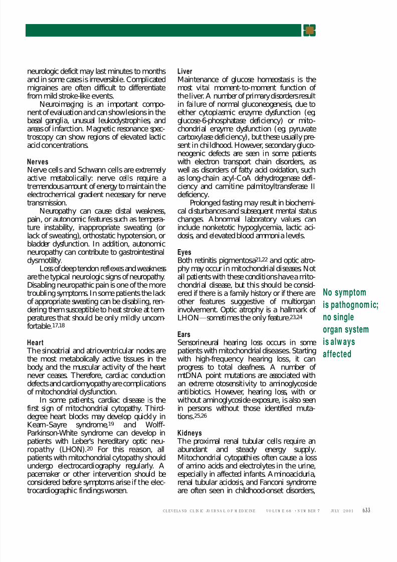
In most instances, a stepwise evaluation ismost sensible (TABLES 4–6). As a general sugges-tion, it is reasonable to evaluate patients withthree or more distinct clinical symptomsinvolving at least two different organ systems.
Before starting an evaluation, especially if the patient has no central or peripheral nervoussystem involvement, he or she should bescreened for common diseases that can producethe symptoms and signs the patient is experi-encing. Thyroid disease, Cushing syndrome,
rheumatic diseases, and inflammatory
myopathies are obvious examples of illnessesthat can cause seemingly unrelated systemicphenomena. Patients should also be advisedthat the evaluation is time-consuming and inva-sive and often will not yield diagnostic results.
Muscle biopsyMuscle tissue can be tested for electron trans-port chain enzyme activity, carnitine disor-ders, fatty acid oxidation activity, and glyco-gen storage disease analysis. In addition, the
mitochondria can be isolated from fresh mus-
MITOCHONDRIAL CYTOPATHY COHEN
Primary evaluation of suspected mitochondrial diseases
TEST COMMENTS
Blood glucose
Hemoglobin A1c
Serum electrolytes Calculate anion gap
Blood counts Anemia, thrombocytopenia, and neutropenia are seen in a varietyof metabolic diseases
Primary and secondary disorders of folate and vitamin B12 metabolismshould be considered
Blood lactate Tourniquet must be released before blood is sampled
Urinalysis High pH may suggest renal tubular acidosis
Plasma ammonia Fasting sample most usefulOrganic acids Measured in urine, cerebrospinal f luid
Samples must be kept refrigerated or frozenUrine collections may be random or t imed, and may be collected after
a fasting period or glucose load, depending on the clinical situationAbnormal amounts of lactate, pyruvate, citric acid cycle intermediates,
or 3-methylglutaconic acid suggest mitochondrial dysfunction3-methylglutaconic acid can be seen in women taking progesterone, or
during extreme stress or glucocorticosteroid use
Ketones Measured in blood or urineSignificant if absent during fasting
Mitochondrial DNA Tested in blood or in muscle biopsy specimens
(mtDNA) point mutations If a patient fi ts into a specific, well-described mitochondrial phenotype,testing for specific, known point mutations may be helpful at this stage
Some centers routinely screen for the 3 to 15 most commonly identifiedmtDNA mutations in all patients
Southern blot If a patient fi ts into a specific, well-described mitochondrial phenotypesuch as CPEO, KSS, or MELAS, Southern blot testing may lead toa rapid diagnosis
Muscle tissue is more sensitive than lymphocytes
Ophthalmology consult Assess for ret init is pigmentosa or optic atrophy
Cardiac evaluation Routine electrocardiogram and echocardiogram
TABLE 4
A stepwiseevaluation of
mitochondrial
cytopathy is
sensible

5/11/2018 Mitochondrial Cytopathy in Adults 2001 Cohen 625 6 - slidepdf.com
http://slidepdf.com/reader/full/mitochondrial-cytopathy-in-adults-2001-cohen-625-6
cle or liver tissue and tested with the previ-ously mentioned studies and oxidative phos-phorylation polarography. The mtDNA canbe extracted from muscle tissue, which can beinformative when the mutation is an acquiredsomatic mutation as opposed to an inheritedgermline mutation that would be also presentin lymphocytes.
Because a muscle biopsy is invasive, the
risks and costs of the procedure must beweighed against the chance the biopsy willyield positive results and the benefits gainedby a diagnosis, such as treatment decisionsand genetic counseling. If a genetic mutationcan be determined by other means, there is noreason to proceed with muscle biopsy or otherdiagnostic tests.
Before a muscle biopsy is performed, a plan
CLEVELAND CLINIC JOURNAL OF M EDICINE VOLUM E 68 • NUM BER 7 JULY 20 01 637
Secondary laborat ory eva luat ionof suspected mitochondrial diseases
TEST COMMENTS
Blood lactate Tourniquet must be released before blood is sampled
Serum pyruvate Proper determination of pyruvate requires the specimen be instantlydeproteinized
Pyruvate not useful i f lactate is normalDisregard results if not properly corrected
Lactate/pyruvate rat io The ratio of lactate to pyruvate can be very helpful in determining if lactateacidosis is due to an oxidative phosphorylation disorder (L/P > 20)
Amino acids Measured in blood, urine, or cerebral spinal fluidUrine collections may be random or timed and may be collected after
a meal or after a fasting period, depending on the clinical situation“ Generalized aminoaciduria” may indicate the presence of proximal renal
tubular dysfunction due to mit ochondrial cytopathy, as well as othermedical conditions
Alanine is the amino acid precursor to pyruvate, and therefore an elevatedalanine can be helpful in diagnosis
Organic acids Measured in urine or cerebral spinal f luidSamples must be kept refrigerated or frozenDifferent techniques, some more sensit ive, are used by certain laboratoriesUrine collections may be random or t imed, and may be collected after
a fasting period or glucose load, depending on the clinical situation
Carnitine analysis Measured in blood, urine, or muscle biopsy specimen
Most laboratories determine the free carnitine and total carnitineFractionation into specific acyl carnitines may be helpful in some situationsUrine collections may be random or t imed, and may be collected after
a fasting period, depending on the clinical situation
Ketones Measured in blood or urineDetermining the ratio of ß-hydroxybutyrate and acetoacetate may
be helpful. This test is most valuable if collected during an acute illnessor after a fast
Urinary acylglycines Useful in disorders of beta oxidation
Skin biopsy Electron microscopy may reveal structural defects in mitochondrial structureA fibroblast culture can be established with the skin obtained from a biopsy
Other testing of skin samples includes testing for electron transport chainactivity, beta-oxidation disorders, and other specific diseases
TABLE 5
Advisepatients that
evaluation is
invasive,
lengthy, and
often
unproductive

5/11/2018 Mitochondrial Cytopathy in Adults 2001 Cohen 625 6 - slidepdf.com
http://slidepdf.com/reader/full/mitochondrial-cytopathy-in-adults-2001-cohen-625-6
638 CLEVELAND CLINIC JOURNAL OF M EDICINE VOLUME 68 • NUM BER 7 JULY 20 01
needs to be arranged for how the sample is tobe distributed. Reference laboratories shouldbe contacted before the biopsy is done so thatthe muscle sample is prepared correctly.
The following tests can be ordered onmuscle samples taken during the biopsy:• Routine light microscopy including
immunohistochemistry. The modified Gomoritrichrome stain is used to demonstrate raggedred fibers, and succinate dehydrogenase stain-ing for ragged-blue fibers, which indicateclumps of diseased mitochondria. Cytochromeoxidase staining can demonstrate fibers absentin this enzyme, and some laboratories have theability to stain for nuclear-encoded and mito-chondrial-encoded subunits of cytochromeoxidase.• Electron microscopy, looking for abnor-mally sized or shaped mitochondria, paracrys-
talline inclusions, and proliferation of mito-chondria, usually beneath the subsarcolemmalmembrane (FIGURE 2).• Electron transport chain activity as deter-mined by spectrophotometric assay. This ispreferably performed on isolated mitochondria(obtained only from fresh muscle), but can beperformed on fresh or flash-frozen musclehomogenate. This study determines the activ-ity of the catalytic components of the variousparts of the electron transport chain.• Oxidative phosphorylation activity
(polarography), which measures rates of oxy-
gen utilization using different concentrationsof ADP, tested by using a variety of substrates.This method can determine the functionalactivity of the five respiratory chain complex-es and the integrity of the inner and outermitochondrial membranes. In addition, thefunctional activity of pyruvate dehydrogenase,
carnitine transport, and fatty acid oxidationcan also be estimated. Polarography requiresfresh mitochondria and therefore can only beperformed within the first few hours after thetissue is removed from the body.• Enzyme activity for beta oxidation disor-ders, including those of the enzymes of the betaoxidation spiral, carnitine transport, and carni-tine palmitoyltransferase I and II activity.• Determination of carnitine, acylcarnitine,and coenzyme Q10 levels.
s TREATMENT
There are no cures for mitochondrial diseases.The focus of treatment should be to maximizenormal organ function and alleviate symp-toms, which includes standard medical thera-pies.
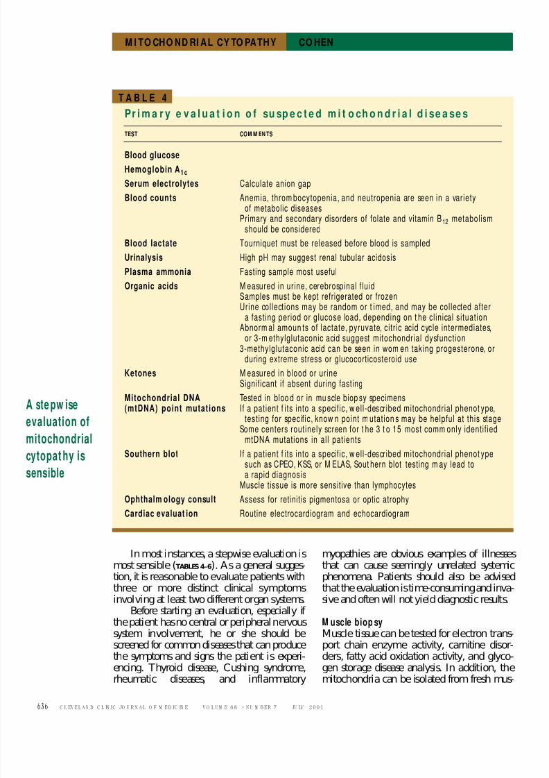
Are vitamin and cofactor supplementsbeneficial?Most patients with mitochondrial cytopathiesask whether supplemental vitamin and cofac-
tor therapies may be helpful (TABLE 7).
Tert iary laboratory evaluationof suspected mitochondrial diseases
TEST COMMENTS
Repeat testing Repeating some of the above-listed tests, sometimes under differentconditions (such as during an il lness), may be helpful
Provocative testing Under monitored condit ions, usually in the hospital, repeating someof the above tests after a fast or after a specific meal or intravenousinfusion may be helpful
mtDNA point mutations If a patient fi ts into a specific, well-described mitochondrial phenotype,testing for specific, known point mutations may be helpful at this stage
mtDNA Southern blot If a patient fi ts into a specific, well-described mitochondrial phenotype,
Southern blot testing may be helpful at this stageCoenzyme Q10 blood test and muscle and mitochondrial levels areprobably more important, but currently not available
TABLE 6
MITOCHONDRIAL CYTOPATHY COHEN
Plan aheadon how to
distribute
muscle biopsy
samples

5/11/2018 Mitochondrial Cytopathy in Adults 2001 Cohen 625 6 - slidepdf.com
http://slidepdf.com/reader/full/mitochondrial-cytopathy-in-adults-2001-cohen-625-6
Aside from a handful of case reports inwhich an enzyme defect was due to cofactor
deficiency or was very cofactor-responsive,there is no overwhelming evidence that theuse of cofactors is helpful in most patients.
However, the value of cofactor therapy isdifficult to measure. These diseases have avaried clinical course, and some patients haveacute exacerbations followed by long periodsof stability or partial recovery. In addition,there are literally hundreds of different defectsthat affect different organ systems in each per-son, making outcome measures almost impos-sible to determine. Even within one family
sharing a common gene defect, the variabilityis so diverse it is not possible to determine aperson’s clinical course on the basis of thecourse of other family members. Furthermore,the treatment duration of many negative stud-ies may not be long enough to determineimprovement. Despite the lack of experi-mental data, most persons with mitochondri-al cytopathies chose to take supplemental vit-amins and cofactors. The cost of these supple-ments can be substantial, and therefore thephysician and patient should use some degree
of restraint deciding about cofactor therapy.
Coenzyme Q10 (CoQ10) is the bestknown cofactor used in treating mitochondri-
al cytopathies. CoQ10 is synthesized in vivoand functions as the mobile electron carrierresiding in the inner mitochondrial mem-brane, transferring electrons from complexes Iand II to complex III. It also can function as apowerful antioxidant. Benefits may includereduction in lactic acid levels,36–39 improve-ment in muscle magnetic resonance spec-troscopy findings,40–42 improved musclestrength,38 and decreased muscle fatigability.36
Central nervous system symptoms generallydo not improve with this therapy. Although
numerous studies found CoQ10 therapy to bebeneficial, others did not.43–45 There are nosignificant side effects.
Levocarnitine (L-carnitine, carnitine), isa cofactor required for the metabolism of fattyacids. Only the levo-isomer is active.Carnitine palmitoyltransferase I (CPT I) cat-alyzes the binding of acyl-CoA with carnitineto form acylcarnitine, which is shuttled acrossthe inner mitochondrial membrane inexchange for free carnitine. Once acylcarni-tine is inside the mitochondrial matrix, CPT
II reverses the reaction, resulting in free carni-
CLEVELAND CLINIC JOURNAL OF M EDICINE VOLUM E 68 • NUM BER 7 JULY 20 01 639
Mi t ochondria l disease i n muscle: Elect ron micrographic appearance
FIGURE 2. Electron photomicrographs of muscle biopsy samples f rom two pat ients with mit ochondrialdisease. Left, large mitochondria with needle-like paracrystalline inclusions (arrow) in a patient withdementia and int ractable epilepsy. Right, pathologic accumulation of mitochondria (arrow ) in thesubsarcolemma of muscle tissue.
Most patientselect to take
vitamins and
cofactors
5/11/2018 Mitochondrial Cytopathy in Adults 2001 Cohen 625 6 - slidepdf.com
http://slidepdf.com/reader/full/mitochondrial-cytopathy-in-adults-2001-cohen-625-6
640 CLEVELAND CLINIC JOURNAL OF M EDICINE VOLUME 68 • NUM BER 7 JULY 20 01
tine and acyl-CoA, which is metabolized viabeta oxidation.
Many disorders of intermediary metabo-lism, including those affecting electron trans-port, can result in carnitine deficiency. Undernormal circumstances, about 25% of the nec-essary carnitine is synthesized in vivo and 75%is consumed in the diet. Carnitine deficiencycan cause clinical myopathy or cardiomyopa-thy and lead to rhabdomyolysis.46
Benefits of levocarnitine therapy areimproved strength (which is sometimes
observed in those who do not have a carnitinedeficiency), reversal of cardiomyopathy, andimproved gastrointestinal motility, which canbe a major benefit to those with poor motilitydue to their disease.47 Supplemental carnitinetherapy is accepted for those with proven car-nitine deficiency, but remains an unprovenbut widely used treatment for those with mito-chondrial disorders.46
Intestinal cramping and pain are themajor side effects, which are alleviated in mostcases by reducing the dose. It is reasonable to
consider a therapeutic trial of levocarnitine in
those with mitochondrial cytopathies.Creatine phosphate is synthesized from
creatine and ATP in a reaction catalyzed bycreatine kinase. Unlike ATP, creatine phos-phate can accumulate in small amounts in thebody, and since creatine phosphate can behydrolyzed to ATP and creatine it thus allowsfor storage of a high-energy phosphate bond.
Creatine is found in muscle, brain, kidney,and other tissues. Muscular creatine may bedepleted in mitochondrial cytopathy, and sup-plemental creatine phosphate has been shown
to be helpful in some patients with weaknessdue to their disease. Because the benefits maybe transient, it is recommended that this ther-apy be reserved for acute crises and discontin-ued as soon as possible.48–50
B vitamins are inexpensive essentialnutrients necessary for the function of a widearray of enzymes associated with energy pro-duction. The need for supplemental B vitamintherapy is not proven, aside from well-docu-mented but rare cases of thiamine (vitaminB1)-responsive pyruvate dehydrogenase defi-
ciency51,52; riboflavin (vitamin B2)-responsive
Vitamins, supplements, and medicat ionsused in mitochondrial diseases
SUPPLEMENT DAILY DOSE COMMENTS
Coenzyme Q10 5–15 mg/kg in divided doses Variable gastrointestinal absorption dependenton formulationMaximal benefit may take months
L-carnitine 30–100 mg/kg in divided doses Prescription brand CarnitorIV and oral preparations available
Thiamine (vitamin B1) 100–800 mg
Riboflavin (vitamin B2) 400 mg
Niacinamide (vitamin B3
) 100–500 mg Avoid niacin form, as it can cause uncomfortable flushing
Folate 1–10 mg
Vitamin E 400–1200 IU in divided doses May interfere with CoQ10 absorption
Selenium 25–50 µg
Lipoic acid 200–600 mg in divided doses
Prednisone 5–60 mg Symptomatic improvement noted on patients,but should be used sparingly as withdrawal of treatmentmay lead to recurrence of symptoms
TABLE 7
MITOCHONDRIAL CYTOPATHY COHEN
Reservecreatine
phosphate
for acute
crises

5/11/2018 Mitochondrial Cytopathy in Adults 2001 Cohen 625 6 - slidepdf.com
http://slidepdf.com/reader/full/mitochondrial-cytopathy-in-adults-2001-cohen-625-6
forms of electron transfer flavoprotein (ETF)and ETF-coenzyme Q10 reductase deficiency(glutaric aciduria type II)53; and biotin-
responsive biotinidase deficiency.54Riboflavin is the best studied of the B vita-mins and has also been proposed to be helpfulin preventing migraine,55–57 and for this rea-son a trial may be reasonable in patients withmitochondrial disease.
Antioxidants. Antioxidant use makes
sense on theoretical grounds.58 Free radicals,which damage lipid membranes such as theinner mitochondrial membrane, are overpro-
duced in disorders of mitochondrial functionand may be scavenged by antioxidants.59,60
These agents have not been systematicallytested in mitochondrial disorders, and anybenefit may not be detectable in a brief trial.Despite the lack of proof, these are routinelyused in patients with these diseases.
CLEVELAND CLINIC JOURNAL OF M EDICINE VOLUM E 68 • NUM BER 7 JULY 20 01 641
s REFERENCES
1. Luft R, Ikkos D, Palmieri G, Ernster L, Afzelius B. A case of severe hyper-
metabolism of nonthyroid origin wi th a defect in t he maintenance of
mitochondrial respiratory control: A correlated clinical, biochemical and
morpholog ical study. J Clin Invest 1962; 41:1776–1804.
2. DiMauro S, Bonilla E. Mitochondrial encephalomyopathies. In: Rosenberg
RN, Prusiner SB, DiMauro S, Barchi RL, editors. The molecular and genetic
basis of neurological disease. Boston: Butt erwort h-Heinemann,
1997:201–235.
3. Shoffner JM, Wallace DC. Oxidat ive phosphorylat ion diseases. In: Scriver
CR, Beaudet AL, Sly WS, et al, editors. The metabolic and molecular bases
of inherited disease. New York: McGraw-Hill, 1995:1535–1610.
4. Wallace DC. Mitochondrial diseases in man and mouse. Science 1999;
238:1482–1488.
5. Giles RE, Blanc H, Cann HM , Wallace DC. Maternal inheritance of human
mitochondrial DNA. Proc Natl Acad Sci USA 1980; 77:6715–6719.
6. Shoffner JM. Mitochondrial myopathy diagnosis. Neurol Clin 2000;
18:105–123.
7. Andreu AL, Hanna MG, Reichmann H, et al. Exercise intolerance due to
mutations in the cytochrome b gene of mitochondrial DNA. N Engl J
Med 1999; 341:1037–1044.
8. Fischel-Ghodsian N, Prezant TR, Chaltraw WE, et al. Mit ochondrial gene
mutation is a significant predisposing factor in aminoglycoside ototoxici-
ty. Am J Otolaryngol 1997; 18:173–178.
9. Corral-Debrinski M, Shoffner JM, Lott MT, Wallace DC. Association of
mitochondrial DNA damage with aging and coronary atherosclerotic
heart disease. Mutat Res 1992; 275:169–180.
10. Wallace DC. Mitochondrial DNA mutations and bioenergetic defects in
aging and degenerat ive diseases. In: Rosenberg RN, Prusiner SB, DiMauro
S, Barchi RL, editors. The molecular and genetic basis of neurologic dis-
ease. Boston, Butterworth-Heinemann, 1997:237–269.
11. Melov S, Shoffner JM, Kaufman A, Wallace DC. Marked increase in t he
number and variety of mitochondrial DNA rearrangements in aging
human skeletal muscle. Nucleic Acids Res 1995; 23:4122–4126.
12. Pavlakis SG, Phillips PC, DiMauro S, DeVivo DC, Rowland LP.
Mitochondr ial myopathy, encephalomyopathy, lactic acidosis and stroke-
like episodes: a distinctive clinical syndrome. Ann Neurol 1984;
16:481–488.
13. Goto Y, Nonaka I, Horai S. A mutation in the tRNALeu(URR) gene associ-
ated with the MELASsubgroup of mitochondrial encephalomyopathies.
Nature 1990; 348:653.
14. Bardosi A, Creutzfeldt W, DiMauro S, et al. Myo-, neuro-, gastrointestinal
encephalomyopathy (MNGIE syndrome) due to partial def iciency of
cytochrome c oxidase: a new mit ochondrial mult isystem disorder. Acta
Neuropathol (Berl) 1987; 74:248–158.
15. Nishino I, Spinazzola A, Hirano M. Thymidine phosphorylase gene muta-
tions in MNGIE, a human mitochondrial disorder. Science 1999;
283:689–692.
16. Lietman PS. Mitochondrial protein synthesis: inhibition by emetine
hydrochloride. Mol Pharmacol 1971; 7:122–128.
17. Huang CC, Chu CC, Pang CY, Wei YH. Tissue mosaicism in the skeletal
muscle and sural nerve biopsies in the MELASsyndrome. Acta NeurolScand 1999; 99:125–129.
18. Peyronnard JM, Charron L, Bellavance A, Marchand L. Neuropathy and
mit ochondrial myopathy. Ann Neurol 1980; 7:262–268.
19. Kenny D, Wetherbee J. Kearns-Sayre syndrome in the elderly: mitochon-
drial myopathy w ith advanced heart block. Am Heart J 1990;
120:440–443.
20. Mashima Y, Kigasawa K, Hasegawa H, Tani M, Oguchi Y. High incidence
of pre-excitation syndrome in Japanese fami lies with Leber’s hereditary
optic neuropathy. Clin Genet 1996; 50:535–537.
21. Kerrison JB, Biousse V, Newman NJ. Retinopathy of NARP syndrome.
Arch Ophthalmol 2000; 118:298–299.
22. Smith PR, Bain SC, Good PA, et al. Pigmentary retinal dystrophy and the
syndrome of maternally inheri ted diabetes and deafness cause by the
mitochondrial DNA 3243 tRNA (Leu) A to G mutation. Ophthalmology
1999; 106:1101–1108.
23. Wallace DC, Singh G, Lott MT, et al. Mitochondrial DNA mutation associ-
ated wi th Leber’s hereditary opt ic neuropathy. Science 1988;
242:1427–1430.
24. Chalmers RM, Schapira AH. Clinical, biochemical and molecular genetic
features of Leber’s hereditary opt ic neuropathy. Biochim Biophys Acta
1999; 1410:147–158.
25. Fischel-Ghodsian N. Mitochondrial genet ics and hearing loss: the missing
link betw een genotype and phenot ype. Proc Soc Exp Biol M ed 1998;
218:1–6.
26. Jacobs HT. Mitochondrial deafness. Ann Med 1997; 29:483–491.
27. Niaudet P. Mitochondrial d isorders and the k idney. Arch Dis Child 1998;
78:387–390.
28. Niaudet P, Rotig A. The kidney in mitochondrial cytopathies. Kidney Int
1997; 51:1000–1007.
29. Odawara M. Involvement of mitochondrial gene abnormaliti es in t he
pathogenesis of diabetes mellitus. Ann NY Acad Sci 1996; 786:72–81.
30. Gerbitz KD, van den Ouw eland JM, Maassen JA, Jaksch M.
Mitochondrial d iabetes mellitus: a review. Biochim Biophys Acta 1995;
1271:253–260.
31. van den Ouweland JM, Lemkes HH, Trembath RC, et al. Maternally
inherit ed diabetes and deafness is a distinct subtype of diabetes and
associates with a single point mutation in the mitochondrial tRNA
(Leu(URR)) gene. Diabetes 1994; 43:746–751.
32. Plioplys AV, Plioplys S. Serum levels of carnitine in chronic fat igue syn-
drome: clinical correlates. Neuropsychobiology 1995; 32:132–138.
33. Kuratsune H, Yamaguti K, Takahashi M, Tagawa S, Kitani T. Acylcarnitine
deficiency in chronic fatigue syndrome. Clin Infect Dis 1994; 18(suppl
1):62–67
34. Griggs RC, Karpati G. Muscle pain, fatigue, and mitochondriopathies. N
Engl J Med 1999; 341:1077–1078.
35. Walker UA, Collins S, Byrne E. Respiratory chain encephalomyopathies: a
diagnostic classification. Eur Neurol 1996; 36:260–267.
36. Goda S, Hamada T, Ishimoto S, Kobayashi T, Goto I, Kuroiw a Y. Clinical
improvement after administrat ion of coenzyme Q10 in a patient w ith
mitochondrial encephalomyopathy. J Neurol 1987; 234:62–63.
37. Ogasahara S, Yorifuji S, Nishikawa Y, et al. Improvement of abnormal
pyruvate metabol ism and cardiac conduction defect wi th coenzyme Q10
in Kearns-Sayre syndrome. Neurology 1985; 35:372–377.
38. Bresolin N, Bet L, Binda A, et al. Clinical and biochemical correlations in
mitochondrial myopathies treated with coenzyme Q10. Neurology 1988;38:892–899.

5/11/2018 Mitochondrial Cytopathy in Adults 2001 Cohen 625 6 - slidepdf.com
http://slidepdf.com/reader/full/mitochondrial-cytopathy-in-adults-2001-cohen-625-6
39. Abe K, Fujimura H, Nishikawa Y, et al. Marked reduction in CSF lactat e
and pyruvate levels after CoQ therapy in a patient with mitochondrial
myopathy, encephalopathy, lactic acidosis and stroke-like episodes
(MELAS). Acta Neurol Scand 1991; 83:356–359.
40. Gold R, Seibel P, Reinelt G, et al. Phosphorous magnet ic resonance spec-
troscopy in the evaluation of mitochondrial myopathies: Results of a 6-month therapy study with coenzyme Q. Eur Neurol 1996; 36:191–196.
41. Barbiroli B, Iotti S, Lodi R. Improved brain and muscle mit ochondrial res-
pirat ion wi th CoQ. An in vivo study by 31P-MR spectroscopy in pat ients
with mitochondrial cytopathies. BioFactors 1999; 9:253–260.
42. Bendahan D, Desnuelle C, Vanuxem D, et al. 31P NMR spectroscopy and
ergometer exercise test as evidence for muscle oxidative performance
improvement with coenzyme Q in mitochondrial myopathies. Neurology
1992; 42:1203–1209.
43. Bresolin N, Doriguzzi C, Ponzetto C, et al. Ubidecarenone in the treat-
ment of mitochondrial myopathies: a mult i-center double-blind t rial. J
Neurol Sci 1990; 100:70–78.
44. Zierz S, von Wersebe O, Bleistein J, Jerusalem F. Exogenous coenzyme Q
(CoQ) fails to increase CoQ in skeletal muscle of tw o pat ients wi th mit o-
chondr ial myopat hies. J Neurol Sci 1990; 95:283–290.
45. Matthews PM, Ford B, Dandurand RJ, et al. Coenzyme Q10 with multi-
ple vitamins is generally ineffective in treatment of mitochondrial dis-ease. Neurology 1993; 43:884–890.
46. Pons R, DeVivo DC. Primary and secondary carnitine deficiency syn-
drome. J Child Neurol 1995; 10(suppl 2):8–24.
47. Campos Y, Huertas R, Lorenzo G, et al. Plasma carnitine insufficiency and
effectiveness of L-carnitine therapy in patients with mitochondrial
myopathy. Muscle Nerve 1993; 16:150–153.
48. Harris RC, Soderlund K, Hultman E. Elevation of creatine in resting and
exercised muscle of normal subjects by creatine supplementation. Clin
Sci 1992; 83:367–374.
49. Tarnopolsky MA, Roy BD, MacDonald JR. A randomized, controlled trial
of creatine monohydrate in patients with mitochondrial cytopathies.
Muscle Nerve 1997; 20:1502–1509.
50. Tarnopolsky MA, Martin J. Creatine monohydrate increases strength in
patients with neuromuscular disease. Neurology 1999; 52:854–857.
51. Naito E, Ito M, Yokota I, et al. Concomitant administration of sodium
dichloroacetate and t hiamine in West syndrome caused by thiamine-
responsive pyruvate dehydrogenase complex deficiency. J Neurol Sci
1999; 171:56–59.
52. DiRocco M, Lamba LD, Minniti G, Caruso U, Naito E. Outcome of thi-
amine treatment in a child with Leigh disease due to thiamine-respon-
sive pyruvate dehydrogenase deficiency. Eur J Paediatr Neurol 2000;
4:115–117.
53. Gregersen N. Ribof lavin-responsive defects of beta-oxidation. J Inherit
Metab Dis 1985; 8(suppl 1):65–69.
54. Wolf B, Heard GS, Jefferson LG, Proud VK, Nance WE, Weissbecker KA.
Clinical findings in four children with biotinidase deficiency detected
through a stat ewide neonat al screening program. N Engl J Med 1985;
313:16–19.
55. Schoenen J, Jacquy J, Lenaerts M. Eff ecti veness of high-dose ribo flavin in
migraine prophylaxis. A randomized controlled trial. Neurology 1998;
50:466–470.
56. Yee AJ. Eff ectiveness of high-dose ribo flavin in migraine prophylaxis.
Neurology 1999; 52:431–432.
57. Mattimoe D, Newton W. High-dose riboflavin for migraine prophylaxis. J
Fam Pract 1998; 47:11.
58. Sastre J, Pallardo FV, Garcia de la Asuncion J, Vina J. Mitochondria,
oxidative stress and aging. Free Radic Res 2000; 32:189–198.
59. Mecocci P, MacGarvey U, Kaufman AE, et al. Oxidative damage to mit o-
chondr ial DNA shows marked age-dependent increases in human brain.
Ann Neurol 1993; 34:609–616.
60. Beal M F. Does impairment of energy metabolism result in excitotoxic
neuronal death in neurodegenerative illness? Ann Neurol 1992;
31:119–130.
ADDRESS: Bruce H. Cohen, M D, Chief , Section of Pediat ric Neurology, S80,
The Cleveland Clinic Foundation, 9500 Euclid Avenue, Cleveland, OH
44195.
642 CLEVELAND CLINIC JOURNAL OF M EDICINE VOLUME 68 • NUM BER 7 JULY 20 01
MITOCHONDRIAL CYTOPATHY COHEN