MISS in Thoracolumbar Fractures -...
34
Guillem Saló Bru, MD, Phd Spine Unit. Orthopaedic Department. Hospital del Mar. Barcelona. Associated Professor. Universitat Autónoma de Barcelona. MISS in Thoracolumbar Fractures
Transcript of MISS in Thoracolumbar Fractures -...

Guillem Saló Bru, MD, PhdSpine Unit. Orthopaedic Department.
Hospital del Mar. Barcelona.
Associated Professor. Universitat Autónoma de Barcelona.
MISS in
Thoracolumbar
Fractures
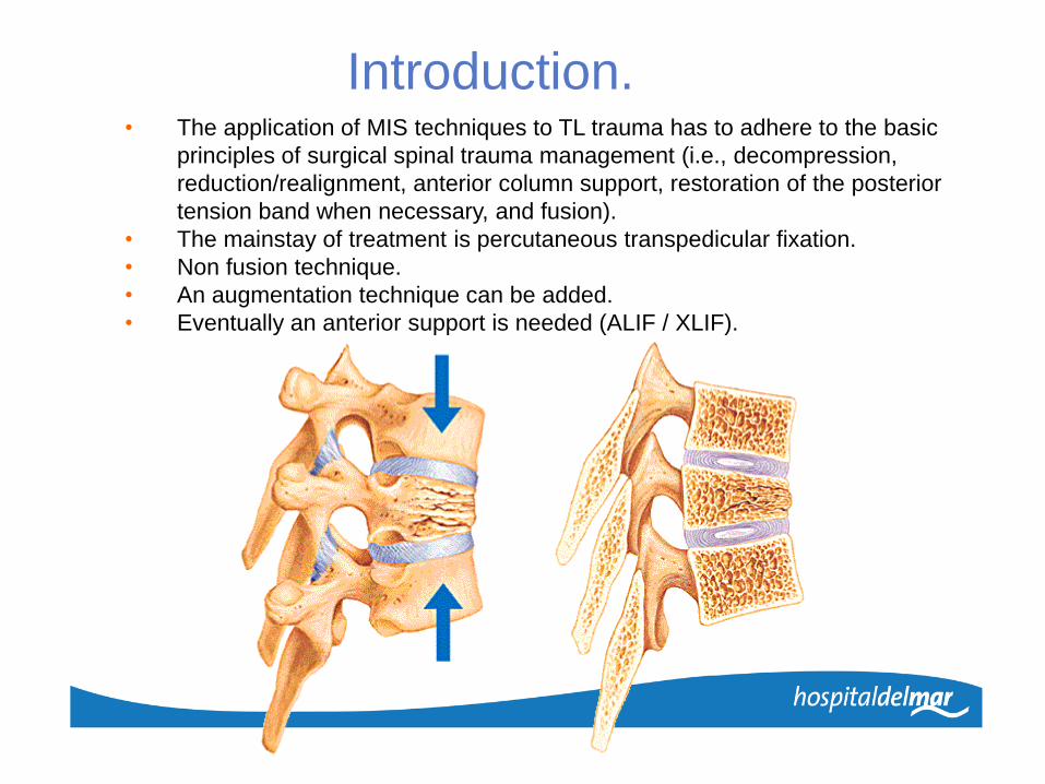
Introduction.• The application of MIS techniques to TL trauma has to adhere to the basic
principles of surgical spinal trauma management (i.e., decompression,
reduction/realignment, anterior column support, restoration of the posterior
tension band when necessary, and fusion).
• The mainstay of treatment is percutaneous transpedicular fixation.
• Non fusion technique.
• An augmentation technique can be added.
• Eventually an anterior support is needed (ALIF / XLIF).
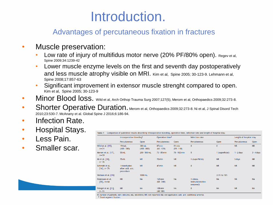
Advantages of percutaneous fixation in fractures
• Muscle preservation: • Low rate of injury of multifidus motor nerve (20% PF/80% open). Regev et al,
Spine 2009;34:1239-42
• Lower muscle enzyme levels on the first and seventh day postoperatively
and less muscle atrophy visible on MRI. Kim et al, Spine 2005; 30-123-9. Lehmann et al,
Spine 2008;17:857-63
• Significant improvement in extensor muscle strenght compared to open. Kim et al, Spine 2005; 30-123-9
• Minor Blood loss. Wild et al, Arch Orthop Trauma Surg 2007;127(5), Merom et al, Orthopaedics 2009;32:273-8.
• Shorter Operative Duration. Merom et al, Orthopaedics 2009;32:273-8. Ni et al, J Spinal Disord Tech
2010;23:530-7. McAnany et al. Global Spine J 2016;6:186-94.
• Infection Rate.
• Hospital Stays.
• Less Pain.
• Smaller scar.
Introduction.

Disadvantages of percutaneous fixation in fractures
• It is a technically demanding surgery:
steep learning curve.
• Requires recognition of anatomy with
fewer landmarks.
• Hand-eye coordination: Lack of tactile
feedback.
• Difficultly in reduction of severe displaced
fractures.
• Difficultly in placement of graft.
• Increase of radiation.
• Increases of Cost of procedures owing to
tecnification.
Introduction.
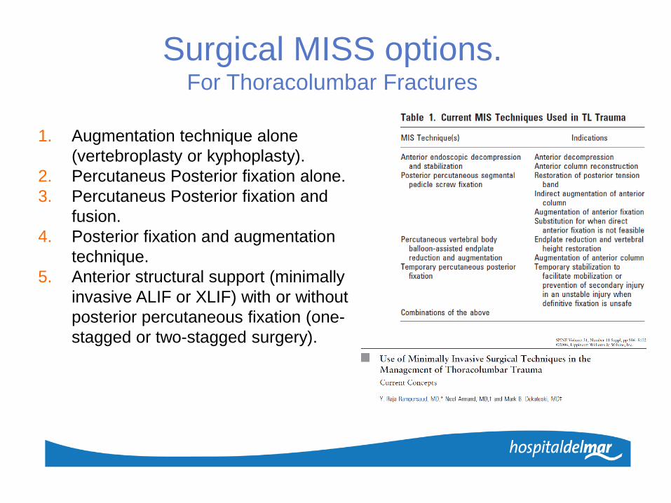
Surgical MISS options.For Thoracolumbar Fractures
1. Augmentation technique alone
(vertebroplasty or kyphoplasty).
2. Percutaneus Posterior fixation alone.
3. Percutaneus Posterior fixation and
fusion.
4. Posterior fixation and augmentation
technique.
5. Anterior structural support (minimally
invasive ALIF or XLIF) with or without
posterior percutaneous fixation (one-
stagged or two-stagged surgery).
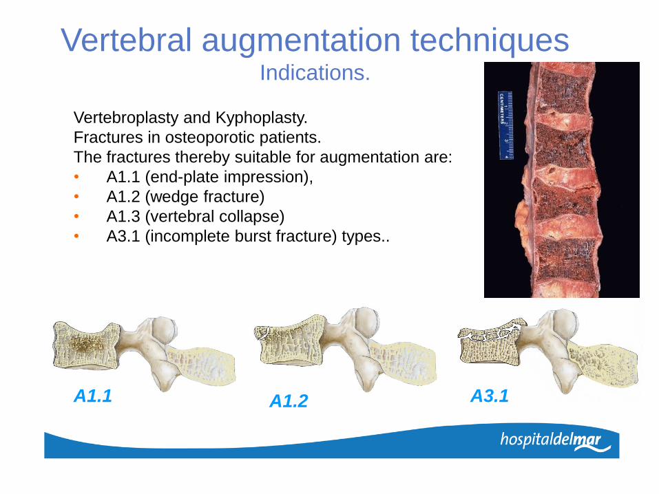
Vertebral augmentation techniquesIndications.
Vertebroplasty and Kyphoplasty.
Fractures in osteoporotic patients.
The fractures thereby suitable for augmentation are:
• A1.1 (end-plate impression),
• A1.2 (wedge fracture)
• A1.3 (vertebral collapse)
• A3.1 (incomplete burst fracture) types..
A1.1 A1.2 A3.1

Vertebral augmentation techniquesTechnique.
A. Placing the biopsy needle at the pedicle entry
site at the angle between the upper articular
process and the transverse process.
B. Kirschner wire fed through the biopsy needle
and acting as a guide.
C. The biopsy needle is removed.
D. Introduction of the cannulated trocar via
guide-wire.
E. Positioning the kyphoplasty balloon in the
drilled channel in the fracture zone. Pressure-
controlled inflation of the kyphoplasty balloon
and the simultaneous gain in height of the
vertebral body.
F. The cavity that remains after the kyphoplasty
balloon has been removed is filled with high-
viscosity augmentation material through the
cannula.
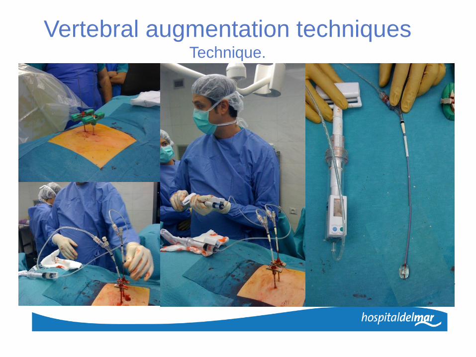
Vertebral augmentation techniquesTechnique.

Vertebral augmentation techniquesTechnique.
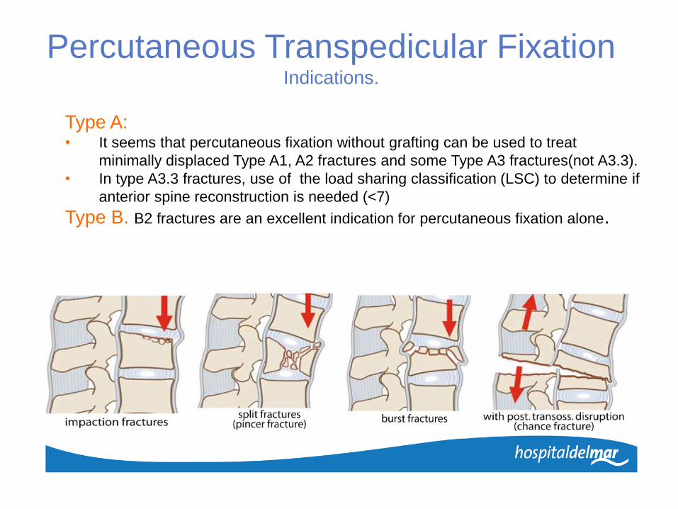
Percutaneous Transpedicular FixationIndications.
Type A: • It seems that percutaneous fixation without grafting can be used to treat
minimally displaced Type A1, A2 fractures and some Type A3 fractures(not A3.3).
• In type A3.3 fractures, use of the load sharing classification (LSC) to determine if
anterior spine reconstruction is needed (<7)
Type B. B2 fractures are an excellent indication for percutaneous fixation alone.
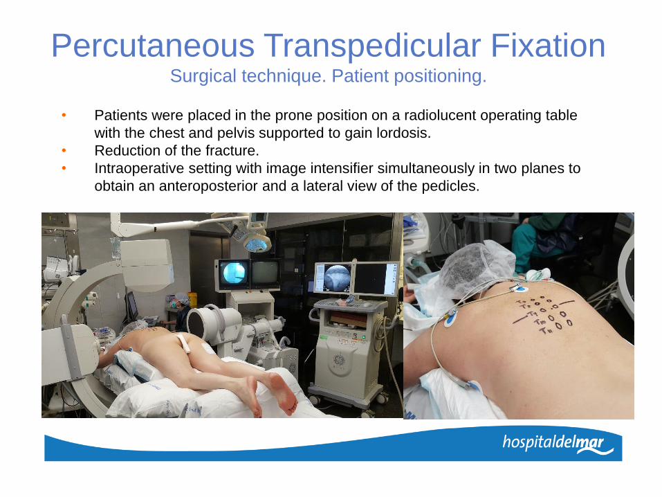
• Patients were placed in the prone position on a radiolucent operating table
with the chest and pelvis supported to gain lordosis.
• Reduction of the fracture.
• Intraoperative setting with image intensifier simultaneously in two planes to
obtain an anteroposterior and a lateral view of the pedicles.
Percutaneous Transpedicular FixationSurgical technique. Patient positioning.

•12
? ?
Percutaneous Transpedicular FixationSurgical technique. Patient positioning.

• The skin incision should be made 1–2 cm laterally so that the Jamshidi
needle can be angled appropriately when inserting it into the pedicle.
• While the Jamshidi needle is advanced into the pedicle, a.p. and lateral
fluoroscopy should be used intermittently as needed to confirm the direction,
making sure that the needle remains lateral to the medial pedicle wall.
• Be carefully when the pedicle is broken.
ABCABC
A
B
C
1-2 cm
Percutaneous Transpedicular FixationSurgical technique.

Percutaneous Transpedicular FixationSurgical technique.

Percutaneous Transpedicular FixationSurgical technique.
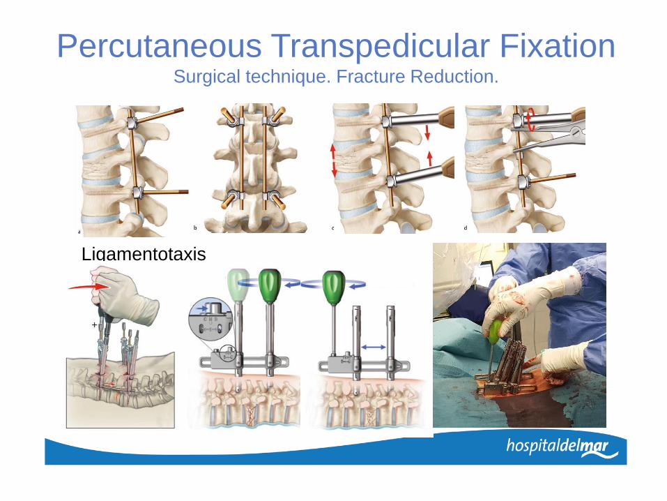
Ligamentotaxis
Percutaneous Transpedicular FixationSurgical technique. Fracture Reduction.

Percutaneous Transpedicular FixationSurgical technique. Fracture Reduction.

Short segment including fracture
• Biomechanically stronger (3-poin fixation)
• Better kyphosis correction (6% loss of
correction)
• Fracture level screws increase stiffness of
the constructs by 30%. (Norton et al, Spine
J 2014).
• Fewer instrument failures.
• Comparable clinical and functional outcome.
• Shields fractured body from anterior loads
Mahar A, Kim C, Wedemeyer M, Mitsunaga L, Odell T, Johnson B, Garfin S. Short- segment
fixation of lumbar burst fractures using pedicle fixation at the level of the fracture. Spine 2007,
32 (14). 1503-7.
Percutaneous Transpedicular FixationUse of screw at the fracture level in the treatment of thoracolumbar fractures.

Indications of long segment fixations:
• Fracture-dislocations (injuries with translation),
PCL injury.
• Severely comminuted vertebral bodies (LSC).
• Osteoporotic spine
• Patient Factors: Past psychological disturbances,
drug abuse, alcoholics or non compliance patients
or co-morbidities.
Advantatges:
• Stronger: lesser chances of implant failure
(multiple fixation points)
• Better alignment of sagittal balance.
Disadvantages: more surgery, more levels fused…
Percutaneous Transpedicular FixationShort versus long constructs.

Percutaneous Transpedicular FixationShort versus long constructs.
Guven O et al, J Spinal Disord Tech 2009; 22:417-21. Compared
short versus long fixations for treatement of thoracolumbar
fractures. 72 patients randomised into 4 groups:1. Posterior instrumentation 2 levels above and below.
2. Posterior instrumentation 2 levels above and below and screws at
fractured level.
3. Posterior instrumentation 1 level above and below
4. Posterior instrumentation 1 level above and below and screws at
fractured level.
• Follow up 26-82 m.
• Intraoperative kyphosis angle correction achieved lowest and
greatest loss of correction on follow up in group 3.
• In all other groups, no differences.
• Comparing with long fixations, a short segment fusion with
screws at fractured level is adequate for intraoperative
correction and for the manintenance of correction on follow up .

Percutaneous Transpedicular FixationAnd Fusion.
• The need for always using a bone graft during surgical treatment of spinal fractures is highly debated and no consensus exists.
• Type B1 fractures with ligament
involvement, are not an indication for
percutaneous fixation alone in our
opinion, since a bone graft must be added
to obtain fusion and make up for the
ligament injuries.
• Minimally invasive procedures for the
fusion have a place here in combination
with percutaneous fixation.

Posterior fixation with recostruction technique.Indications.
• Combination of percutaneous fixation with percutaneous anterior spinal reconstruction
techniques
• A balloon kyphoplasty procedure in combination with posterior short-segment fixation
helps not only to correct angular and vertebral body height losses, but to maintain this
correction over time.
• This combination applies to fractures not needing a graft, but where fixation alone is not
mechanically sufficient (LSC > 6) and requires anterior spinal reconstruction.
• Fractures meeting these criteria included Type B2 bone fractures and Type A3.3
fractures, which have significant vertebral compression leading to loss of vertebral body
height and an anterior bone void.

Posterior fixation with recostruction technique.Technique.
• Percutaneous screw insertion of one
vertebra cranial and one vertebra
caudal to the fracture.
• Fracture reduction.
• Introduction of balloons under the
central depression
• Reduction of the central endplate with
the balloons
• Injection of CPC after removal of the
balloons
• Augmentation or not of the pedicle
screws with PMMA.

McCormack et al. Spine, 1994
Percutaneous fixation with anterior structural support (minimally or open)• Type B1 fractures and even Type C fractures, if the posterior structures are not
dislocated or greatly laterally displaced, which would require an open posterior
reduction.
• In type A3.3 fractures, use of the Load Sharing Classification (LSC) to determine if
anterior spine reconstruction is needed (McCormack 1994)
• These fractures requires an intersomatic graft to avoid angular loss of the disc space.
Anterior Structural Support.Indications.
>6

• Anterior approach.
• Mimimally or open
• XLIF or ALIF
• One or two-staged surgery.
• Special retractors.
• Thoracoscopic or laparoscopic
approach
Anterior Structural Support.Technique.
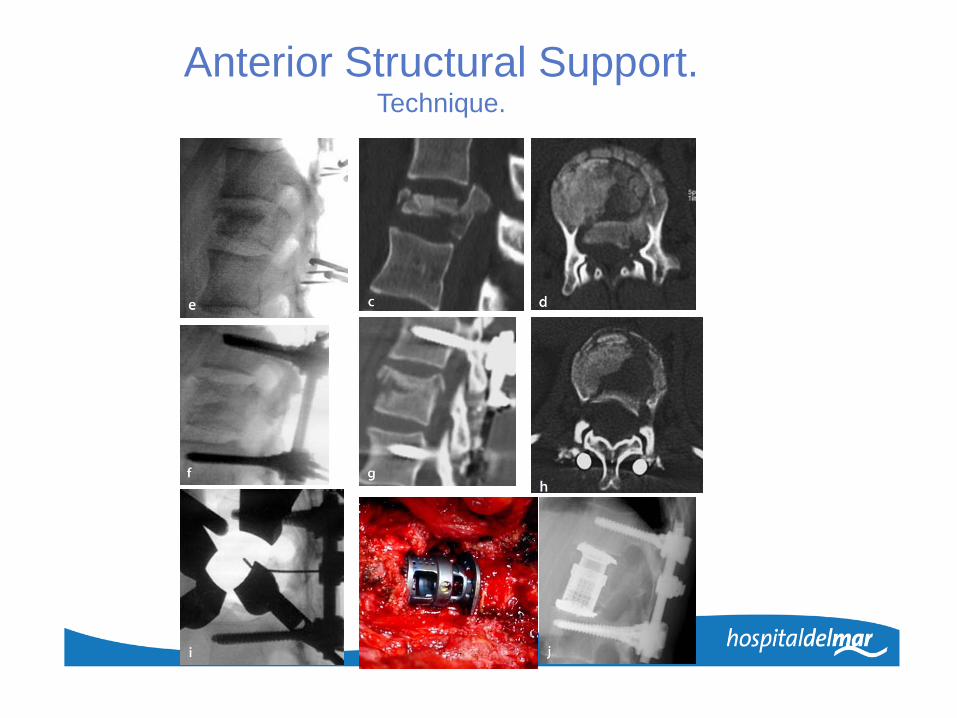
Anterior Structural Support.Technique.

Outcomes of MISS in ThL Fractures.Positioning of pedicle screws placed
percutaneous under fluoroscopy control
• Percutaneous pedicle screw targeting with fluoroscopy guidance, when using proper
technique, leads to fewer pedicle wall violations than when performed open.
Author Year Number of screws % misplacement N revision
Weisner et al. 2000 408 6,6% 2
Ringle et al. 2006 488 3% 9
Pelegriet al. 2008 50 3,8% 1
Ni et al. 2010 104 6,7% 0
Korovessis et al 2008 180 2,7% 0
Heintel et al. 2013 502 2% 1

• 6 studies selected (2016)
• Short segment pedicle screw fixation without fusion.
• MISS superior in therms of less blood loss and shorter
operative duration.
• No difference in terms of vertebral body height, kyphosis
angle and postoperative pain.
• At minimum, percutaneous fixation of thoracolumbar fractures
results in equivalent biomechanics and clinical outcomes
compared to the open group.
Outcomes of MISS in ThL Fractures.

Outcomes of MISS in ThL Fractures.
• Percutaneous MIS can provide a safe and effective treatment for
thoracolumbar junction fractures.
• A significant reduction in blood loss, postoperative pain, surgical
time, and hospital stay are the main advantages associated with
these new minimally invasive techniques.
• These favorable outcomes are particularly important in specific
subgroups of patients, including elderly people and patients with
important comorbidities, and could make the percutaneous
minimally invasive techniques the preferable surgical treatment

Limitations of percutaneous fixation in
thoracolumbar fractures
1. Exposure to X-Rays• Proper percutaneous screw placement requires a precise
• technique and high-quality fluoroscopy
• will be exposed to ionizing radiation.
• Screw placement requires 9.3 seconds of exposure to X-rays
• These data confirm the increased irradiation for the surgical team and the
patient during percutaneous fixation.
• Navigation systems aim to reduce exposure to X-rays while also improving
screw placement.

Limitations of percutaneous fixation in
thoracolumbar fractures
2. Reduction of fracture.• Displaced fractures.
• Patients who needs a great reduction of fracture are in principle a
contraindication to percutaneous fixation, since complete reduction
cannot be performed.
• Specially type C (fractures dislocation) or comminuted burst fracture.

Limitations of percutaneous fixation in
thoracolumbar fractures
3. Neurological decompresion.• Fractures that are complicated by neurological problems are in
principle a contraindication to percutaneous fixation, since
decompression cannot be performed.
• However, percutaneous fixation can be combined with a limited
posterior midline approach to perform the required decompression.

Conclusions.• The role of percutaneous spinal fixation and posterior minimally-
invasive surgery is becoming clearer.
• They do not replace the other open techniques, but add to treatment
options.
• The advantage of these techniques in reducing surgical morbidity,
simplifying the immediate postoperative recovery and improving the
medium-term functional results is well known.
• Percutaneous fixation is not always performed alone; it can be
combined with additional anterior or minimally-invasive posterior
routes.
• Screw at fractured level and indirect reduction by ligamentotaxis.
• Anterior support in comminuted fractures
• Long constructs in type C fractures or >7 points in LSC.