miRCURY LNA™ microRNA ISH Optimization Kit (FFPE) · of additional material recommended for...
32
Instruction manual v1.3 for product # 90000, 90001, 90002, 90003, 90004, 90005, 90006, 90007, 90008 Novmeber 2010 miRCURY LNA™ microRNA ISH Optimization Kit (FFPE)
Transcript of miRCURY LNA™ microRNA ISH Optimization Kit (FFPE) · of additional material recommended for...
Instruction manual v1.3 for product # 90000, 90001, 90002, 90003, 90004, 90005, 90006, 90007, 90008 Novmeber 2010
miRCURY LNA™ microRNA ISH Optimization Kit (FFPE)
2
miR
CU
RY LN
A™ m
icroRN
A ISH O
ptimization K
it (FFPE)
Literature citationsPlease refer to miRCURY LNA™ microRNA Detection probes when describing a procedure for publication using this product or to the following article: Robust One-day in situ hybridization protocol for detection of microRNA in paraffin samples using LNA probes. Jørgensen S, Baker A, Møller S, Nielsen BS. Methods (2010), 52,373-381.
Patents and TrademarksExiqon, LNA™ and miRCURY™ are registered trademarks of Exiqon A/S, Vedbaek, Denmark. Locked Nucleic Acids (LNA™) are covered by patents and patent applications owned by Exiqon A/S. DIG is licensed from Roche Diagnostics GmbH. All other trademarks are the property of their respective owners.
Locked-nucleic Acids (LNA™s) are protected by US Pat No. 6,268,490, US Pat No. 6,770,748, US Pat No. 6,639,059, US Pat No. 6,734,291 and other applications and patents owned or licensed by Exiqon A/S.
DisclaimerProducts are for research use only and not for diagnostic or therapeutic use. The products in their original or any modified form may be used only for the buyer’s internal research purposes and not for commercial, diagnostic, therapeutic, or other use, including contract research. The buyer may not resell products in their original or any modified form. The purchase of products does not include or carry an implied right or license for the buyer to use such products in the provision of services to third parties and a license must be obtained directly from Exiqon A/S for such use.
© Copyright 2010 Exiqon. All rights reserved.
3
miR
CU
RY LN
A™ m
icroRN
A ISH O
ptimization K
it (FFPE)
Table of contents
Abbreviations. FFPE: formalin-fixed and paraffin-embedded. ISH: In situ hybridization. LNA™: locked nucleic acid. Prot-K: Proteinase-K. RT: room temperature. AP: alkaline phosphatase. DIG: digoxigenin. PFA: paraformaldehyde.
Product summary . . . . . . . . . . . . . . . . . . . . . . . . . . . . . . . . . . . . . . . . . . . . . . . . . 4Product summary . . . . . . . . . . . . . . . . . . . . . . . . . . . . . . . . . . . . . . . . . . . . . . 4Kit content. . . . . . . . . . . . . . . . . . . . . . . . . . . . . . . . . . . . . . . . . . . . . . . . . . . . . 5Shipping and storage. . . . . . . . . . . . . . . . . . . . . . . . . . . . . . . . . . . . . . . . . . . . 5Additional required material. . . . . . . . . . . . . . . . . . . . . . . . . . . . . . . . . . . . . . 6Reagents and equipment required, not supplied . . . . . . . . . . . . . . . . . . . . . 7Related products . . . . . . . . . . . . . . . . . . . . . . . . . . . . . . . . . . . . . . . . . . . . . . . 8Product description . . . . . . . . . . . . . . . . . . . . . . . . . . . . . . . . . . . . . . . . . . . 10
Protocol. . . . . . . . . . . . . . . . . . . . . . . . . . . . . . . . . . . . . . . . . . . . . . . . . . . . . . . . . 12Before starting the experiment . . . . . . . . . . . . . . . . . . . . . . . . . . . . . . . . . . 12Workflow overview - One-day microRNA ISH protocol . . . . . . . . . . . . . . . 17One-day microRNA ISH protocol . . . . . . . . . . . . . . . . . . . . . . . . . . . . . . . . . 18
Tips and troubleshooting . . . . . . . . . . . . . . . . . . . . . . . . . . . . . . . . . . . . . . . . . . 23Troubleshooting . . . . . . . . . . . . . . . . . . . . . . . . . . . . . . . . . . . . . . . . . . . . . . . 27Frequently asked questions . . . . . . . . . . . . . . . . . . . . . . . . . . . . . . . . . . . . . 29
References . . . . . . . . . . . . . . . . . . . . . . . . . . . . . . . . . . . . . . . . . . . . . . . . . . . . . . 30
4
miR
CU
RY LN
A™ m
icroRN
A ISH O
ptimization K
it (FFPE)
Product summary
Product summary
The miRCURY LNA™ microRNA ISH Optimization Kit (FFPE) provides the user with reagents and recommendations to ensure the best starting point for successful microRNA in situ hybridizations (ISH) on formalin-fixed paraffin embedded (FFPE) tissue samples. The kit contains 3 digoxigenin (DIG)-labeled probes: one double- (5’ and 3’)-DIG labeled probe for a known cell-specific microRNA, one double-(5’ and 3’)-DIG labeled scrambled probe to use as negative control, and one 5’-DIG labeled probe against U6 snRNA for use in the early-phase assay set-up. In addition, the kit contains a formamide-free hybridization buffer developed specifically for miRCURY LNA™ Detection probe-based ISH. The included Proteinase-K will allow the user to optimize the Proteinase-K treatment for optimal retention of the microRNA target.
The accompanying One-day microRNA ISH protocol minimizes time-consuming optimization steps and enables a fast and optimal microRNA ISH analysis using a colorimetric antibody-based development system with the double-DIG labeled miRCURY LNA™ microRNA Detection probes. In addition, the Instruction Manual carefully covers each step of the FFPE ISH procedure, including tissue sectioning, incubation intervals and temperatures, miRCURY LNA™ microRNA Detection probe concentrations and substrate incubation. The manual further contains a list of additional material recommended for establishing the ISH technology in the lab and making it easier to conduct the experiments.
5
miR
CU
RY LN
A™ m
icroRN
A ISH O
ptimization K
it (FFPE)
Shipping and storage
Upon receiptThe kit is shipped at room temperature. Immediately upon receipt, remove the miRCURY LNA™ microRNA ISH buffer from the package and store at 4°C. Store the DIG-labeled miRCURY LNA™ microRNA Detection probes and Proteinase-K in the package at -20°C or below. Under these conditions the probes are stable for at least 6 months. It is recommended to store the probes in aliquots to avoid freeze-thaw cycles (see recommendations for dilution below). Do not store in frost-free freezer with automatic thaw-freeze.
Before First Use • Proteinase-K stock: reconstitute to 20 mg/mL by adding 600 µL 10mM Tris-HCl,
pH7.5 (RNase-free). Store appropriate aliquots at -20°C. • DIG-labeled LNA™ probes: The following options are possible: 1) The probes may be stored at 4°C if used within 4 weeks. 2) Prepare aliquots to be stored at -20°C or below and avoid multiple freeze-
thaw cycles. Example: divide the 40nM LNA™ Detection probe into 5µL or 10µL aliquots into non-stick RNase-free tubes.
3) Prepare pre-diluted probe aliquots to be stored at -20°C or below in the microRNA ISH buffer. Example: First prepare 20 mL of hybridization buffer by mixing 10 mL of 2x-microRNA-ISH-buffer with 10 mL of RNase-free water. For
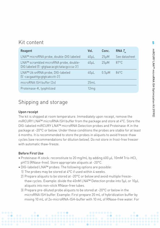
Reagent Vol. Conc. RNA Tm
LNA™ microRNA probe, double-DIG labeled 40µL 25µM See datasheet
LNA™ scrambled microRNA probe, double- DIG labeled (5’-gtgtaacacgtctatacgccca-3’)
40µL 25µM 87°C
LNA™ U6 snRNA probe, DIG-labeled(5’-cacgaatttgcgtgtcatcctt-3’)
40µL 0.5µM 84°C
microRNA ISH buffer (2x) 25mL
Proteinase-K, lyophilized 12mg
Kit content
6
miR
CU
RY LN
A™ m
icroRN
A ISH O
ptimization K
it (FFPE) Additional required material
ISH protocols vary extensively due to different equipment set-up and laboratory routines. The presented One-day miRCURY LNA™ microRNA ISH protocol guides the process of a manual ISH on formalin-fixed and paraffin embedded tissue samples using double-DIG labeled miRCURY LNA™ microRNA Detection probes.
For the ISH steps we recommend a hybridization station that allows precise and fast temperature adjustments, e.g. Dako Hybridizer. The One-day miRCURY LNA™ microRNA ISH protocol is developed using a hybridization station, but if unavailable conventional hybridization ovens may be used (see details in FAQs at page 29).
For the immunohistochemical steps Exiqon has had good experience with both horizontal humidifying chambers and Shandon’s Sequenza Slide Rack systems.
The chromogenic ISH assay is based on the use of DIG-labeled probes and therefore requires proper detection reagents (e.g. alkaline phosphatase-conjugated anti-DIG, DIG wash and blocking reagents, NBT-BCIP substrate).
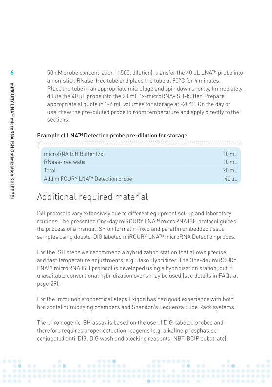
microRNA ISH Buffer (2x) 10 mLRNase-free water 10 mLTotal 20 mLAdd miRCURY LNA™ Detection probe 40 µL
Example of LNA™ Detection probe pre-dilution for storage
50 nM probe concentration (1:500, dilution), transfer the 40 µL LNA™ probe into a non-stick RNase-free tube and place the tube at 90°C for 4 minutes. Place the tube in an appropriate microfuge and spin down shortly. Immediately, dilute the 40 µL probe into the 20 mL 1x-microRNA-ISH-buffer. Prepare appropriate aliquots in 1-2 mL volumes for storage at -20°C. On the day of use, thaw the pre-diluted probe to room temperature and apply directly to the sections.
7
miR
CU
RY LN
A™ m
icroRN
A ISH O
ptimization K
it (FFPE)
Reagents and equipment required, not supplied
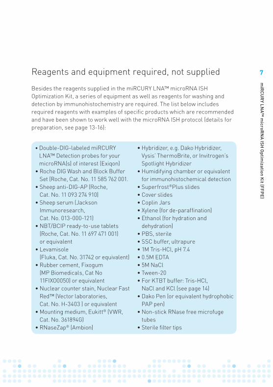
Besides the reagents supplied in the miRCURY LNA™ microRNA ISH Optimization Kit, a series of equipment as well as reagents for washing and detection by immunohistochemistry are required. The list below includes required reagents with examples of specific products which are recommended and have been shown to work well with the microRNA ISH protocol (details for preparation, see page 13-16):
• Double-DIG-labeled miRCURY LNA™ Detection probes for your microRNA(s) of interest (Exiqon)
• Roche DIG Wash and Block Buffer Set (Roche, Cat. No. 11 585 762 001.
• Sheep anti-DIG-AP (Roche, Cat. No. 11 093 274 910)
• Sheep serum (Jackson Immunoresearch, Cat. No. 013-000-121)
• NBT/BCIP ready-to-use tablets (Roche, Cat. No. 11 697 471 001) or equivalent
• Levamisole (Fluka, Cat. No. 31742 or equivalent)
• Rubber cement, Fixogum (MP Biomedicals, Cat No 11FIXO0050) or equivalent
• Nuclear counter stain, Nuclear Fast Red™ (Vector laboratories, Cat. No. H-3403 ) or equivalent
• Mounting medium, Eukitt® (VWR, Cat. No. 361894G)
• RNaseZap® (Ambion)
• Hybridizer, e.g. Dako Hybridizer, Vysis’ ThermoBrite, or Invitrogen’s Spotlight Hybridizer
• Humidifying chamber or equivalent for immunohistochemical detection
• Superfrost®Plus slides• Cover slides • Coplin Jars• Xylene (for de-paraffination)• Ethanol (for hydration and
dehydration)• PBS, sterile• SSC buffer, ultrapure• 1M Tris-HCl, pH 7.4• 0.5M EDTA• 5M NaCl• Tween-20• For KTBT buffer: Tris-HCl,
NaCl and KCl (see page 14)• Dako Pen (or equivalent hydrophobic
PAP pen)• Non-stick RNase free microfuge
tubes• Sterile filter tips
8
miR
CU
RY LN
A™ m
icroRN
A ISH O
ptimization K
it (FFPE)
Functional AnalysisLocalizationExpression
AnalysisIsolation
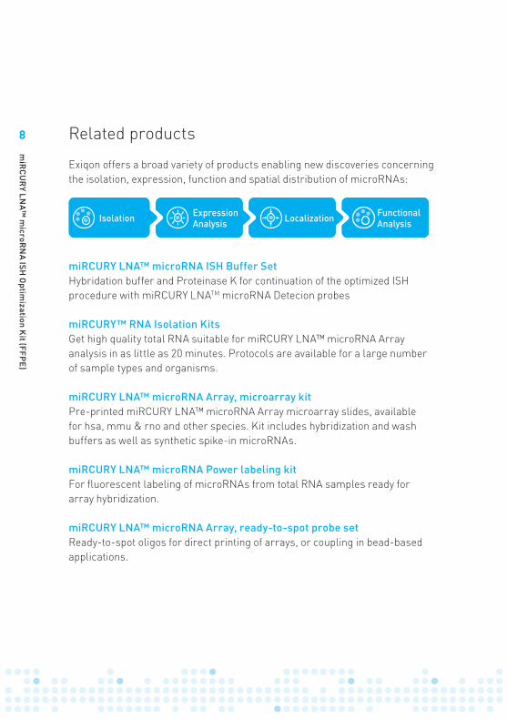
Related products
Exiqon offers a broad variety of products enabling new discoveries concerning the isolation, expression, function and spatial distribution of microRNAs:
miRCURY LNA™ microRNA ISH Buffer SetHybridation buffer and Proteinase K for continuation of the optimized ISH procedure with miRCURY LNATM microRNA Detecion probes
miRCURY™ RNA Isolation KitsGet high quality total RNA suitable for miRCURY LNA™ microRNA Arrayanalysis in as little as 20 minutes. Protocols are available for a large numberof sample types and organisms.
miRCURY LNA™ microRNA Array, microarray kitPre-printed miRCURY LNA™ microRNA Array microarray slides, availablefor hsa, mmu & rno and other species. Kit includes hybridization and washbuffers as well as synthetic spike-in microRNAs.
miRCURY LNA™ microRNA Power labeling kitFor fluorescent labeling of microRNAs from total RNA samples ready forarray hybridization.
miRCURY LNA™ microRNA Array, ready-to-spot probe setReady-to-spot oligos for direct printing of arrays, or coupling in bead-basedapplications.

9
miR
CU
RY LN
A™ m
icroRN
A ISH O
ptimization K
it (FFPE)
miRCURY LNA™ Universal RT microRNA PCRExiqon’s microRNA qPCR system offers the best available combination ofperformance and ease-of-use on the microRNA real-time PCR market. Thecombination of a Universal RT reaction and LNA™-enhanced PCR primersresults in unmatched sensitivity and specifi city. The Ready-to-use microRNAPCR panels enable fast and easy microRNA expression profiling.
miRCURY LNA™ microRNA Detection ProbesFor in situ hybridization and northern blotting of all microRNAs.
miRCURY LNA™ microRNA Inhibitors and Power InhibitorsUnravel the function of microRNAs by microRNA inhibition. SophisticatedLNA™ design ensures potent inhibition of all microRNAs regardless of theirGC content. Chemically modified, highly stable Power Inhibitors for unrivalledpotency.
miRCURY LNA™ microRNA Inhibitor LibraryFor genome-wide high throughput screening of microRNA function
10
miR
CU
RY LN
A™ m
icroRN
A ISH O
ptimization K
it (FFPE)
Product description
In situ hybridization (ISH) is a powerful technique and the most common method for visualizing gene expression and localization in specific tissue and cell types. The technology is far from trivial and is often a very time-consuming and difficult procedure requiring many steps of protocol optimizing to achieve satisfactory ISH results. Detection of microRNA by conventional ISH analysis is no exception.
The miRCURY LNA™ microRNA ISH Optimization kit (FFPE) offers a fast and robust procedure for an easy implementation of microRNA ISH analysis requiring a minimum of optimization. The microRNA ISH buffer is specifically developed for use with the double-DIG labeled miRCURY LNA™ microRNA Detection probes. Used in combination, this provides the best available method for specific and sensitive detection of microRNA expression by ISH in FFPE sections of any tissue specimen.
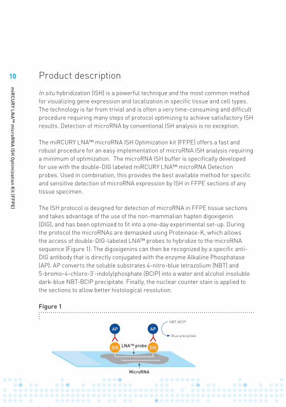
The ISH protocol is designed for detection of microRNA in FFPE tissue sections and takes advantage of the use of the non-mammalian hapten digoxigenin (DIG), and has been optimized to fit into a one-day experimental set-up. During the protocol the microRNAs are demasked using Proteinase-K, which allows the access of double-DIG-labeled LNA™ probes to hybridize to the microRNA sequence (Figure 1). The digoxigenins can then be recognized by a specific anti-DIG antibody that is directly conjugated with the enzyme Alkaline Phosphatase (AP). AP converts the soluble substrates 4-nitro-blue tetrazolium (NBT) and 5-bromo-4-chloro-3’-indolylphosphate (BCIP) into a water and alcohol insoluble dark-blue NBT-BCIP precipitate. Finally, the nuclear counter stain is applied to the sections to allow better histological resolution.
Figure 1
LNA™ probe
MicroRNA
NBT-BCIP
Blue precipitate
DIG
AP
DIG
AP
LNA™ probe
MicroRNA
NBT-BCIP
Blue precipitate
DIG
AP
DIG
AP
11
miR
CU
RY LN
A™ m
icroRN
A ISH O
ptimization K
it (FFPE)
Optimization of the ISH analysis procedure is divided into three steps:1. Optimization of the protocol parameters with the LNA™ U6 snRNA probe by
adjustment of hybridization temperature and Proteinase K treatment.2. Control study at the optimized protocol parameters with the kit-specific double-
DIG miRCURY LNA™ microRNA Detection control probe (a strong specific ISH signal should be obtained).
3. Detection of the microRNA of interest using the appropriate miRCURY LNA™ microRNA Detection probe.
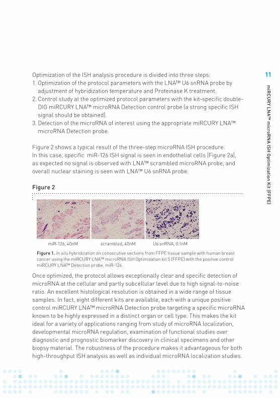
Figure 2 shows a typical result of the three-step microRNA ISH procedure. In this case, specific miR-126 ISH signal is seen in endothelial cells (Figure 2a), as expected no signal is observed with LNA™ scrambled microRNA probe, and overall nuclear staining is seen with LNA™ U6 snRNA probe.
Once optimized, the protocol allows exceptionally clear and specific detection of microRNA at the cellular and partly subcellular level due to high signal-to-noise ratio. An excellent histological resolution is obtained in a wide range of tissue samples. In fact, eight different kits are available, each with a unique positive control miRCURY LNA™ microRNA Detection probe targeting a specific microRNA known to be highly expressed in a distinct organ or cell type. This makes the kit ideal for a variety of applications ranging from study of microRNA localization, developmental microRNA regulation, examination of functional studies over diagnostic and prognostic biomarker discovery in clinical specimens and other biopsy material. The robustness of the procedure makes it advantageous for both high-throughput ISH analysis as well as individual microRNA localization studies.
Figure 2
miR-126, 40nM scrambled, 40nM U6 snRNA, 0.1nM
Figure 1. In situ hybridization on consecutive sections from FFPE tissue sample with human breast cancer using the miRCURY LNA™ microRNA ISH Optimization kit 5 (FFPE) with the positive control miRCURY LNA™ Detection probe, miR-126.
LNA™ probe
MicroRNA
NBT-BCIP
Blue precipitate
DIG
AP
DIG
AP
12
miR
CU
RY LN
A™ m
icroRN
A ISH O
ptimization K
it (FFPE)
Before starting the experiment
In order to ensure that all preparations and reagents for the microRNA ISH analysis are in place once starting the experiment, it is recommended to go through the following sequence of steps:
1) Preparation and storage of kit reagents.2) Establish histology Lab environment for RNA work.3) Fixation of tissue, FFPE preparations.4) Cut FFPE sections using Tissue Sectioning Guidelines.5) Prepare reagents and buffers.6) Become confident with the steps in the One-day miRCURY LNA™ microRNA
ISH protocol.7) Evaluate appropriate Proteinase-K treatment range for each sample type.8) Determine ISH sensitivity level using the control miRCURY LNA™ probe, U6
snRNA. It may be necessary to repeat steps 7 and 8 to gradually improve the performance of the protocol.
9) Run the microRNA ISH protocol with the two supplied control miRCURY LNA™ Detection probes (LNA™ microRNA probe and LNA™ scrambled microRNA probe).
10) Optimize hybridization temperature and probe concentration for each probe.
An overview of the workflow for the One-day miRCURY LNA™ microRNA ISH protocol is shown on page 17.
Protocol
RNA work requires specific handling and precautions to prevent RNase contamination of the reagents and degradation of the RNA sample.
Every step in the microRNA ISH procedure including tissue sectioning, must take place in a clean and nuclease free environment. We recommend that all surfaces are cleaned with RNaseZap®, RNase Away or other RNase removal solution. Wear gloves during the entire process, and only use RNase decontaminated glassware. For preparation of all reagents, use RNase-free water only, e.g. RNase-grade Milli-Q water or DEPC-treated water. All buffers and reagents should be prepared with a minimum of RNase contamination and autoclaved if specified.
Important – cautions for RNA work
13
miR
CU
RY LN
A™ m
icroRN
A ISH O
ptimization K
it (FFPE)
FFPE sample requirements and comments on tissue fixation MicroRNAs are like other RNA fragile molecules sensitive to degradation. Fast and sufficient fixation of tissue specimens is therefore important for successful ISH analyses. For ISH analysis in human specimens, standard overnight fixation in neutral-buffered formalin followed by paraffin embedding often works well. For studies in mouse tissues, perfusion fixation with 4% fresh PFA is recommended. Consult animal care guidelines before setting up this protocol, see e.g. The Laboratory Mouse by Mark A. Suckow, Peggy Danneman, Cory Brayton (CRC Press). Pathology of Genetically Engineered Mice by Jerrold Michael Ward, Joel F. Mahler, Robert R. Maronpot (Iowa State University Press). Optimization of the assay performance should preferably be based on analysis of at least 4 FFPE blocks.
For detailed guidelines to Tissue Sectioning, please see Tip 1 at page 23.
GlasswareIn order to reduce potential RNase contamination of glassware, it is recommended to autoclave all glassware or to heat-treat all glassware for 8 hours at 180°C. Prior to the heat-treatment, it is recommended to wrap all items in aluminum foil, including appropriate stacks of cover slips. Keep apart from untreated glassware.
See tip 1
Preparation of reagents and buffersIn addition to the hybridization buffer and Proteinase-K buffer supplied with the microRNA ISH Optimization Kit a number of other reagents, buffers and equipment needs to be prepared prior to initiating the microRNA ISH experiment. A list of additional required reagents and equipment is provided at page 7. Recipes for preparation of these other required reagents and buffers and the microRNA ISH Optimization Kit buffers are listed in Table 1-3:
14
miR
CU
RY LN
A™ m
icroRN
A ISH O
ptimization K
it (FFPE)
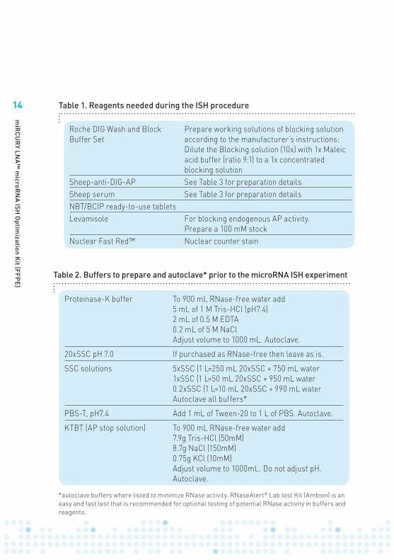
Proteinase-K buffer To 900 mL RNase-free water add5 mL of 1 M Tris-HCl (pH7.4) 2 mL of 0.5 M EDTA0.2 mL of 5 M NaClAdjust volume to 1000 mL. Autoclave.
20xSSC pH 7.0 If purchased as RNase-free then leave as is.
SSC solutions 5xSSC (1 L=250 mL 20xSSC + 750 mL water1xSSC (1 L=50 mL 20xSSC + 950 mL water0.2xSSC (1 L=10 mL 20xSSC + 990 mL water Autoclave all buffers*
PBS-T, pH7.4 Add 1 mL of Tween-20 to 1 L of PBS. Autoclave.
KTBT (AP stop solution) To 900 mL RNase-free water add7.9g Tris-HCl (50mM)8.7g NaCl (150mM)0.75g KCl (10mM)Adjust volume to 1000mL. Do not adjust pH. Autoclave.
*autoclave buffers where listed to minimize RNase activity. RNaseAlert® Lab test Kit (Ambion) is an easy and fast test that is recommended for optional testing of potential RNase activity in buffers and reagents.
Table 2. Buffers to prepare and autoclave* prior to the microRNA ISH experiment
Roche DIG Wash and Block Buffer Set
Prepare working solutions of blocking solution according to the manufacturer’s instructions: Dilute the Blocking solution (10x) with 1x Maleic acid buffer (ratio 9:1) to a 1x concentrated blocking solution
Sheep-anti-DIG-AP See Table 3 for preparation detailsSheep serum See Table 3 for preparation detailsNBT/BCIP ready-to-use tabletsLevamisole For blocking endogenous AP activity.
Prepare a 100 mM stockNuclear Fast Red™ Nuclear counter stain
Table 1. Reagents needed during the ISH procedure
15
miR
CU
RY LN
A™ m
icroRN
A ISH O
ptimization K
it (FFPE)
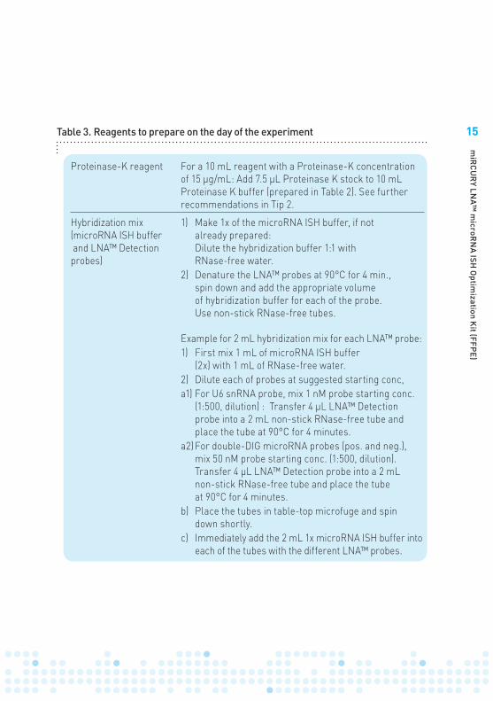
Proteinase-K reagent For a 10 mL reagent with a Proteinase-K concentration of 15 µg/mL: Add 7.5 µL Proteinase K stock to 10 mL Proteinase K buffer (prepared in Table 2). See further recommendations in Tip 2.
Hybridization mix (microRNA ISH buffer and LNA™ Detection probes)
1) Make 1x of the microRNA ISH buffer, if not already prepared: Dilute the hybridization buffer 1:1 with RNase-free water.
2) Denature the LNA™ probes at 90°C for 4 min., spin down and add the appropriate volume of hybridization buffer for each of the probe. Use non-stick RNase-free tubes.
Example for 2 mL hybridization mix for each LNA™ probe: 1) First mix 1 mL of microRNA ISH buffer
(2x) with 1 mL of RNase-free water. 2) Dilute each of probes at suggested starting conc,a1) For U6 snRNA probe, mix 1 nM probe starting conc.
(1:500, dilution) : Transfer 4 µL LNA™ Detection probe into a 2 mL non-stick RNase-free tube and place the tube at 90°C for 4 minutes.
a2) For double-DIG microRNA probes (pos. and neg.), mix 50 nM probe starting conc. (1:500, dilution).Transfer 4 µL LNA™ Detection probe into a 2 mL non-stick RNase-free tube and place the tube at 90°C for 4 minutes.
b) Place the tubes in table-top microfuge and spin down shortly.
c) Immediately add the 2 mL 1x microRNA ISH buffer into each of the tubes with the different LNA™ probes.
Table 3. Reagents to prepare on the day of the experiment
16
miR
CU
RY LN
A™ m
icroRN
A ISH O
ptimization K
it (FFPE)
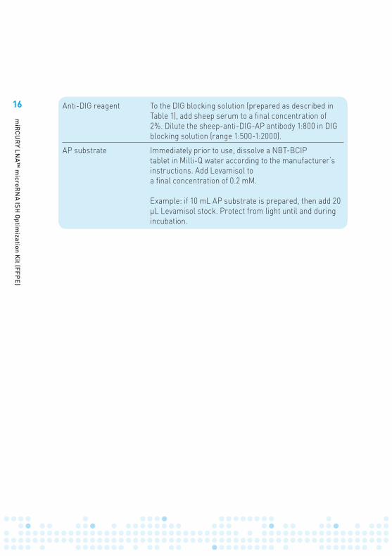
Anti-DIG reagent To the DIG blocking solution (prepared as described in Table 1), add sheep serum to a final concentration of 2%. Dilute the sheep-anti-DIG-AP antibody 1:800 in DIG blocking solution (range 1:500-1:2000).
AP substrate Immediately prior to use, dissolve a NBT-BCIP tablet in Milli-Q water according to the manufacturer’s instructions. Add Levamisol to a final concentration of 0.2 mM.
Example: if 10 mL AP substrate is prepared, then add 20 µL Levamisol stock. Protect from light until and during incubation.
17
miR
CU
RY LN
A™ m
icroRN
A ISH O
ptimization K
it (FFPE)
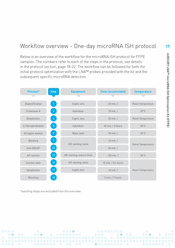
Workflow overview - One-day microRNA ISH protocol
*washing steps are excluded from the overview
Process* Time /accumulatedEquipment
Coplin Jars
Coplin Jars 40 min. / Room Temperature
Room Temperature
Room Temperature
Room Temperature
10 min. / 37°C
55°C
55°C
30°C
20 min. /
30 min. /
15 min. /
60 min. /
120 min. /
10 min. /
5 min. / 7 hours
10 min. / 6½ hours
60 min. / 3 hours
Hybridizer
Hybridizer
Water bath
IHC staining racks
IHC staining racks
Coplin Jars
IHC staining racks in Oven
Deparaffination
Dehydration
Proteinase-K
In Situ hybridization
Stringent washes
Blocking
Anti-DIG/AP
AP reaction
Counter stain
Dehydration
Mounting
TemperatureStep
1
7
15
4
10
18
2
9
17
5
12
Below is an overview of the workflow for the microRNA ISH protocol for FFPE samples. The numbers refer to each of the steps in the protocol, see details in the protocol section, page 18-22. The workflow can be followed for both the initial protocol optimization with the LNA™ probes provided with the kit and the subsequent specific microRNA detection.
18
miR
CU
RY LN
A™ m
icroRN
A ISH O
ptimization K
it (FFPE)
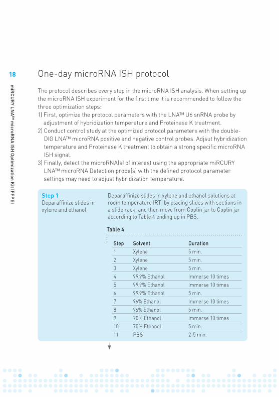
One-day microRNA ISH protocol
Step 1Deparaffinize slides in xylene and ethanol
Deparaffinize slides in xylene and ethanol solutions at room temperature (RT) by placing slides with sections in a slide rack, and then move from Coplin jar to Coplin jar according to Table 4 ending up in PBS.
Step Solvent Duration1 Xylene 5 min.2 Xylene 5 min.3 Xylene 5 min.4 99.9% Ethanol Immerse 10 times5 99.9% Ethanol Immerse 10 times6 99.9% Ethanol 5 min.7 96% Ethanol Immerse 10 times8 96% Ethanol 5 min.9 70% Ethanol Immerse 10 times10 70% Ethanol 5 min.11 PBS 2-5 min.
Table 4
The protocol describes every step in the microRNA ISH analysis. When setting up the microRNA ISH experiment for the first time it is recommended to follow the three optimization steps: 1) First, optimize the protocol parameters with the LNA™ U6 snRNA probe by
adjustment of hybridization temperature and Proteinase K treatment. 2) Conduct control study at the optimized protocol parameters with the double-
DIG LNA™ microRNA positive and negative control probes. Adjsut hybridization temperature and Proteinase K treatment to obtain a strong specific microRNA ISH signal.
3) Finally, detect the microRNA(s) of interest using the appropriate miRCURY LNA™ microRNA Detection probe(s) with the defined protocol parameter settings may need to adjust hybridization temperature.
19
miR
CU
RY LN
A™ m
icroRN
A ISH O
ptimization K
it (FFPE)
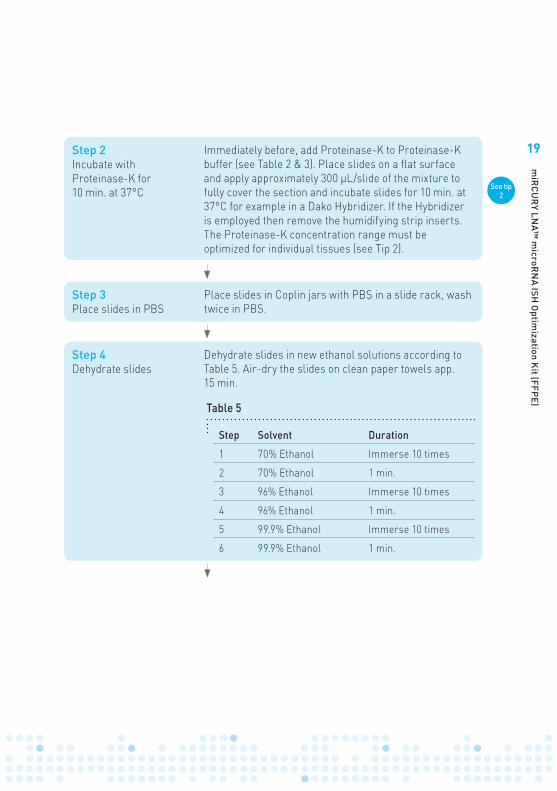
Step 4Dehydrate slides
Dehydrate slides in new ethanol solutions according to Table 5. Air-dry the slides on clean paper towels app. 15 min.
Step Solvent Duration
1 70% Ethanol Immerse 10 times
2 70% Ethanol 1 min.
3 96% Ethanol Immerse 10 times
4 96% Ethanol 1 min.
5 99.9% Ethanol Immerse 10 times
6 99.9% Ethanol 1 min.
Table 5
Step 3Place slides in PBS
Place slides in Coplin jars with PBS in a slide rack, wash twice in PBS.
Step 2Incubate with Proteinase-K for 10 min. at 37°C
Immediately before, add Proteinase-K to Proteinase-K buffer (see Table 2 & 3). Place slides on a flat surface and apply approximately 300 µL/slide of the mixture to fully cover the section and incubate slides for 10 min. at 37°C for example in a Dako Hybridizer. If the Hybridizer is employed then remove the humidifying strip inserts.The Proteinase-K concentration range must be optimized for individual tissues (see Tip 2).
See tip 2
20
miR
CU
RY LN
A™ m
icroRN
A ISH O
ptimization K
it (FFPE)
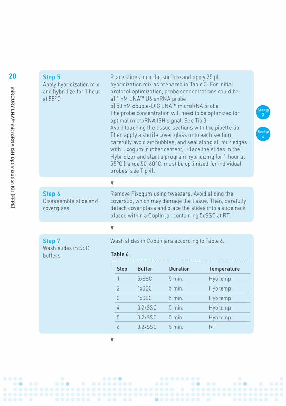
Step 7Wash slides in SSC buffers
Wash slides in Coplin jars according to Table 6.
Step Buffer Duration Temperature
1 5xSSC 5 min. Hyb temp
2 1xSSC 5 min. Hyb temp
3 1xSSC 5 min. Hyb temp
4 0.2xSSC 5 min. Hyb temp
5 0.2xSSC 5 min. Hyb temp
6 0.2xSSC 5 min. RT
Table 6
Step 6Disassemble slide and coverglass
Remove Fixogum using tweezers. Avoid sliding the coverslip, which may damage the tissue. Then, carefully detach cover glass and place the slides into a slide rack placed within a Coplin jar containing 5xSSC at RT.
Step 5Apply hybridization mix and hybridize for 1 hour at 55°C
Place slides on a flat surface and apply 25 µL hybridization mix as prepared in Table 3. For initial protocol optimization, probe concentrations could be:a) 1 nM LNA™ U6 snRNA probeb) 50 nM double-DIG LNA™ microRNA probeThe probe concentration will need to be optimized for optimal microRNA ISH signal. See Tip 3. Avoid touching the tissue sections with the pipette tip. Then apply a sterile cover glass onto each section, carefully avoid air bubbles, and seal along all four edges with Fixogum (rubber cement). Place the slides in the Hybridizer and start a program hybridizing for 1 hour at 55°C (range 50-60°C, must be optimized for individual probes, see Tip 4).
See tip 3
See tip 4
21
miR
CU
RY LN
A™ m
icroRN
A ISH O
ptimization K
it (FFPE)
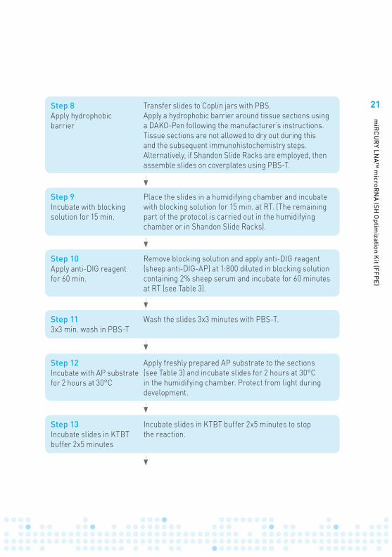
Step 13Incubate slides in KTBT buffer 2x5 minutes
Incubate slides in KTBT buffer 2x5 minutes to stop the reaction.
Step 12Incubate with AP substrate for 2 hours at 30°C
Apply freshly prepared AP substrate to the sections (see Table 3) and incubate slides for 2 hours at 30°C in the humidifying chamber. Protect from light during development.
Step 8Apply hydrophobic barrier
Transfer slides to Coplin jars with PBS. Apply a hydrophobic barrier around tissue sections using a DAKO-Pen following the manufacturer’s instructions. Tissue sections are not allowed to dry out during this and the subsequent immunohistochemistry steps. Alternatively, if Shandon Slide Racks are employed, then assemble slides on coverplates using PBS-T.
Step 9Incubate with blocking solution for 15 min.
Place the slides in a humidifying chamber and incubate with blocking solution for 15 min. at RT. (The remaining part of the protocol is carried out in the humidifying chamber or in Shandon Slide Racks).
Step 10Apply anti-DIG reagent for 60 min.
Remove blocking solution and apply anti-DIG reagent (sheep anti-DIG-AP) at 1:800 diluted in blocking solution containing 2% sheep serum and incubate for 60 minutes at RT (see Table 3).
Step 113x3 min. wash in PBS-T
Wash the slides 3x3 minutes with PBS-T.
22
miR
CU
RY LN
A™ m
icroRN
A ISH O
ptimization K
it (FFPE)
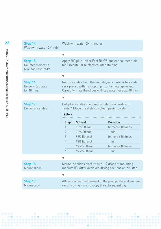
Step 17Dehydrate slides
Dehydrate slides in ethanol solutions according to Table 7. Place the slides on clean paper towels.
Step Solvent Duration1 70% Ethanol Immerse 10 times2 70% Ethanol 1 min.3 96% Ethanol Immerse 10 times4 96% Ethanol 1 min.5 99.9% Ethanol Immerse 10 times6 99.9% Ethanol 1 min.
Table 7
Step 14Wash with water, 2x1 min.
Wash with water, 2x1 minutes.
Step 15Counter stain with Nuclear Fast Red™
Apply 200 µL Nuclear Fast Red™ (nuclear counter stain) for 1 minute for nuclear counter staining.
Step 16Rinse in tap water for 10 min.
Remove slides from the humidifying chamber to a slide rack placed within a Coplin jar containing tap water. Carefully rinse the slides with tap water for app. 10 min.
Step 18Mount slides
Mount the slides directly with 1-2 drops of mounting medium (Eukitt®). Avoid air-drying sections at this step.
Step 19Microscopy
Allow overnight settlement of the precipitate and analyze results by light microscopy the subsequent day.
23
miR
CU
RY LN
A™ m
icroRN
A ISH O
ptimization K
it (FFPE)
Tips and troubleshooting
Tissue Sectioning GuidelinesIt is strongly recommended to wear gloves during paraffin sectioning and in general to maintain an RNase-free environment during all downstream procedures. Use only heat-treated glassware and RNase-free water. Use SuperFrost®Plus slides drawn directly from new packages.
Workstation and MicrotomeBefore starting the tissue sectioning, the whole workstation (bench top, microtome, blade holder, brushes, tweezers, cooling plate, water bath etc.) needs to cleaned with RNase-Zap/RNase Away. Cutting sections1) Prepare a water bath with room temperature RNase-free water and a warm-
water bath with RNase-free water at 40-50°C (depending on the paraffin type).2) Insert a new disposable blade in the knife carrier and place the paraffin block in
the cassette clamp. Trim the block in order to avoid the first couple of sections. It is recommended to cool the FFPE blocks on a cooling plate to app. -15°C before cutting to better control the section thickness.
3) Cut 6 µm-thick paraffin sections and place them in the room temperature RNase-free water, where folding can be reversed. Transfer the sections to the heated water bath, where the tissue section is allowed to stretch shortly. It is recommended to mount sections immediately thereafter on electrostatic treated slides, such as SuperFrost®Plus slides, obtained from a new non-contaminated package.
4) Let the paraffin sections dry for 1-2 hours at room temperature and store at 4°C. Avoid melting the paraffin until the day prior to the in situ hybridization analysis.
5) Melt paraffin in an oven at 60°C for 45 minutes on the day before conducting the ISH experiment. Store slides overnight at 4°C in an RNase-free environment.
Identify appropriate Proteinase-K treatment range The degree of Proteinase-K treatment depends on fixation and tissue of origin. In general terms, the harder fixed the more Prot-K is needed, however there are lower and upper limits. For the Proteinase-K treatment step, it is recommended to vary the concentration or the duration, as indicated in Table 8 (optimal (opt) starting values are shown in parenthesis).
Tip 1
Tip 2
24
miR
CU
RY LN
A™ m
icroRN
A ISH O
ptimization K
it (FFPE)
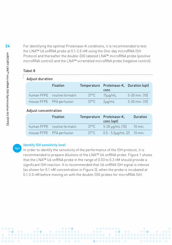
Identify ISH sensitivity level In order to identify the sensitivity of the performance of the ISH protocol, it is recommended to prepare dilutions of the LNA™ U6 snRNA probe. Figure 1 shows that the LNA™ U6 snRNA probe in the range of 0.03 to 0.3 nM should provide a significant ISH reaction. It is recommended that U6 snRNA ISH signal is intense (as shown for 0.1 nM concentration in Figure 3), when the probe is incubated at 0.1-2.0 nM before moving on with the double-DIG probes for microRNA ISH.
Tip 3
Tabel 8
Fixation Temperature Proteinase-K, conc
Duration (opt)
human FFPE routine formalin 37°C 15µg/mL 5-30 min. (10)
mouse FFPE PFA perfusion 37°C 2µg/mL 3-30 min. (10)
Fixation Temperature Proteinase-K, conc (opt)
Duration
human FFPE routine formalin 37°C 5-20 µg/mL (15) 10 min.
mouse FFPE PFA perfusion 37°C 0.5 - 5.0µg/mL (2) 10 min.
Adjust concentration
Adjust duration
For identifying the optimal Proteinase-K conditions, it is recommended to test the LNA™ U6 snRNA probe at 0.1-2.0 nM using the One-day microRNA ISH Protocol and thereafter the double-DIG labeled LNA™ microRNA probe (positive microRNA control) and the LNA™ scrambled microRNA probe (negative control).
25
miR
CU
RY LN
A™ m
icroRN
A ISH O
ptimization K
it (FFPE)
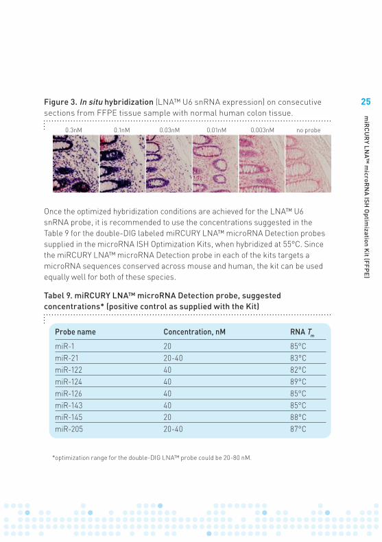
Tabel 9. miRCURY LNA™ microRNA Detection probe, suggested concentrations* (positive control as supplied with the Kit)
Probe name Concentration, nM RNA Tm
miR-1 20 85°CmiR-21 20-40 83°CmiR-122 40 82°CmiR-124 40 89°CmiR-126 40 85°CmiR-143 40 85°CmiR-145 20 88°CmiR-205 20-40 87°C
Once the optimized hybridization conditions are achieved for the LNA™ U6 snRNA probe, it is recommended to use the concentrations suggested in the Table 9 for the double-DIG labeled miRCURY LNA™ microRNA Detection probes supplied in the microRNA ISH Optimization Kits, when hybridized at 55°C. Since the miRCURY LNA™ microRNA Detection probe in each of the kits targets a microRNA sequences conserved across mouse and human, the kit can be used equally well for both of these species.
Figure 3. In situ hybridization (LNA™ U6 snRNA expression) on consecutive sections from FFPE tissue sample with normal human colon tissue.
0.3nM 0.1nM 0.03nM 0.01nM 0.003nM no probe
*optimization range for the double-DIG LNA™ probe could be 20-80 nM.
26
miR
CU
RY LN
A™ m
icroRN
A ISH O
ptimization K
it (FFPE)
Identify optimal hybridization temperature Optimal performance of an ISH probe is related to its signal-to-noise ratio. For the use of oligo probes, there will be a potential risk for the probe to hybridize to highly similar sequences - the lower the hybridization temperature the higher the risk is. The positive control LNA™ probes supplied with the microRNA ISH Optimization kit results in a high signal-to-noise ratio at 55°C using the One-day miRCURY LNA™ microRNA ISH protocol. The LNA™ probes also hybridize at 60°C, but generally provides weaker signal. At 50°C the LNA™ probes give higher signal, but the risk of cross-hybridization to highly similar sequences (in RNA transcripts or genome) will increase at the low hybridization temperatures.
As a rule-of-thumb, hybridization should be performed at 30°C below the given RNA Tm (or 20°C below DNA Tm).
Tip 4
27
miR
CU
RY LN
A™ m
icroRN
A ISH O
ptimization K
it (FFPE)
Troubleshooting
No stain: If no signal is obtained with the LNA™ U6 snRNA probe incubated at 10 nM, it is recommended to test that all reagents are prepared according to the recommendations (pages 14-16). Always test sections from more than one block.
Not sufficient sensitivity level with the LNA™ U6 snRNA probe. It is recommended that the LNA™ U6 snRNA signal is intense when incubated in the range of 0.1-1 nM. If this is not the case ensure that the buffers are prepared correctly and that tissue sections are in the range 5-7 µm. Ensure that the AP-reaction takes place at 30°C. Low sensitivity may also be caused by RNase contamination during sectioning or handling during the in situ hybridization protocol. Ensure to keep an RNase-free environment. Be aware that tissue samples fixed for too short or too long time will result in a low signal. Thus, it is necessary to test several blocks in parallel and avoid concluding on a single sample.
Strong U6 snRNA signal but no microRNA signal. If a strong U6 snRNA signal is obtained when diluted to the range 0.1-0.5 nM but no signal is obtained with the supplied positive control LNA™ microRNA probe, it is most likely due to an unfit Proteinase-K concentration. Hence, the Proteinase-K concentration should be optimized (see Tip 2 for details).
Unspecific staining. It is necessary to clarify whether unspecific staining obtained with the LNA™ scrambled microRNA probe is related to the DIG-labeled probe itself, the detecting antibody or to endogenous enzymatic reactions. This can be done by a systematic approach where the effect of excluding individual reagents is tested: the DIG-labeled probe, the AP-conjugated anti-DIG or both. If staining is obtained in the absence of AP-conjugated anti-DIG then endogenous AP is present. If staining is obtained in the absence of the DIG-labeled probe (and no endogenous AP activity is observed) then staining is related to the detecting antibody. If abundant endogenous enzymatic reactivity (e.g. in some intestinal areas and placenta) cannot be prevented by Levamisol, it may require a change to another detection approach, such as TSA-based fluorescence.
28
miR
CU
RY LN
A™ m
icroRN
A ISH O
ptimization K
it (FFPE)
Some unspecific staining can be caused by improperly maintained SSC buffer washing temperatures. The applied hybridization temperature should be kept during the SSC washes indicated in Table 6.
Unspecific staining of ECM. Unspecific staining of extracellular matrix may occur if the concentration of the detecting antibody is too high.
Sections fall off after de-paraffination. Avoid storage of paraffin sections at -20°C. Small and thick sections more easy fall off than large thin sections. Ensure that the glass slides used are electrostatic treated, such as the SuperFrost®Plus slides.
29
miR
CU
RY LN
A™ m
icroRN
A ISH O
ptimization K
it (FFPE)
Frequently asked questions
Can I use a hybridization oven instead of a Dako Hybridizer? In case you will need to use a hybridization oven during the hybridization step, seal-cover slides with Fixogum as specified in step 6. The slides can be placed as such in the hybridization oven without humidifying conditions. However, humidifying conditions may be tried, e.g. by using 1xSSC. In order to establish a more stable hybridization temperature place a metallic plate, e.g. the inserts from a multiblock heater, in the oven. Place the slides on the plate and hybridize for 1–2 hours. Then go to step 6.
Can I pause the ISH procedure? The individual steps in the One-day protocol have been optimized to accommodate a One-day protocol. PBS steps may be prolonged, but it is not recommended to extend to protocol to more than one day.
What happens if sections dry out? Sections should be maintained in buffered solutions after the hybridization step. Tissue sections that dry out may cause protein denaturation, which may be particularly harmful to the detecting antibody and its conjugated alkaline phosphatase. This may lower the sensitivity of the assay significantly and in addition cause background staining. Drying out of tissue sections may also reduce the quality of the tissue morphology.

30
miR
CU
RY LN
A™ m
icroRN
A ISH O
ptimization K
it (FFPE)
• Robust One-day in situ hybridization protocol for detection of microRNA in paraffin samples using LNA probes. Jørgensen S, Baker A, Møller S, Nielsen BS. Methods (2010), 52, 375-381.
• The Laboratory Mouse. Mark A. Suckow, Peggy Danneman, Cory Brayton. CRC Press, ISBN 0849303222.
• Pathology of Genetically Engineered Mice. Jerrold Michael Ward, Joel F. Mahler, Robert R. Maronpot. Iowa State University Press, ISBN 0813825210.
• www.exiqon.com
References

31
miR
CU
RY LN
A™ m
icroRN
A ISH O
ptimization K
it (FFPE)

Outside North AmericaExiqon A/S · Skelstedet 16DK-2950 Vedbaek · DenmarkPhone +45 45 660 888Fax +45 45 661 888
North AmericaExiqon Inc. · 14 F Gill StreetWoburn, MA 01801 · United StatesPhone (781) 376 4150Fax (781) 376 4152
exiqon.com/contactexiqon.com
13-0
085
- 91
8076
- v
1.3
- 11
/201
0