Minimally Invasive Abdominal and Thoracic Surgery...
9
©Copyright 2012 Vetstreet Inc. This document is for internal purposes only. Reprinting or posting on an external website without written permission from Vetlearn is a violation of copyright laws. Vetlearn.com | May 2012 | Compendium: Continuing Education for Veterinarians ® E1 3 CE Credits Jennifer L. Lansdowne, DVM, MSc, DACVS-SA, DECVS-SA Stephen J. Mehler, DVM, DACVS-SA* Ludovic P. Bouré, DVM, MSc, DACVS, DECVS Abstract: Minimally invasive surgery, including laparoscopy, thoracoscopy, and endoscopic-assisted procedures, is becoming more common in veterinary medicine. The basic principles of laparoscopy and thoracoscopy involve gaining access to a cavity through small incisions, creating a workspace, and maneuvering extracorporeally activated instruments within that workspace. Minimally Invasive Abdominal and Thoracic Surgery: Principles and Instrumentation T he term minimally invasive surgery (MIS) is used for procedures that are performed completely under endoscopic guidance (e.g., laparoscopy, thoracoscopy), negating the need for large inci- sions to enter a cavity. e terms less invasive and endoscopic-assisted imply that procedures are performed with the aid of endoscopic equipment, permitting smaller incisions than traditional procedures. Endoscopic-assisted procedures generally involve using a rigid endo- scope to improve visualization and/or access or to permit completion of the procedure through markedly smaller incisions compared with traditional open techniques. Examples of endoscopic-assisted pro- cedures currently performed in small animals include intestinal biopsy, intestinal foreign-body removal, cystotomy, and gastropexy. Today in human medicine, most abdominal and thoracic surgical procedures can be performed using minimally invasive or less invasive techniques. Although advancement of veterinary medicine in the field of MIS lags behind human medicine, the past 10 years have seen a dramatic increase in techniques described for laparos- copy and thoracoscopy in animals. 1–10 Minimally invasive veteri- nary diagnostic and surgical procedures are currently performed at many academic institutions and private practices around the world. is advancement has largely been driven by a changing client base that recognizes and seeks out less invasive alternatives and by clinicians motivated to learn new procedures and provide cutting-edge techniques to their clientele. ere is little doubt that laparoscopy, thoracoscopy, and endoscopic-assisted procedures have staked a permanent place in the veterinary profession, and their increased use is inevitable. is article summarizes current principles and instrumentation of laparoscopy and thoracoscopy, including indications and contraindications, advantages and dis- advantages, anesthetic implications, and equipment. A companion article summarizes laparoscopic and thoracoscopic techniques that can be performed by clinicians and describes some recent advances and future directions. Basic Principles of Laparoscopy and Thoracoscopy e basic principles of lap- aroscopy and thoracoscopy involve gaining access to a cavity through small inci- sions, creating a workspace, and (using triangulation) maneuvering instruments within that workspace. ese instruments are con- trolled extracorporeally and viewed remotely on a monitor. is is a relatively new concept for veterinary surgeons and involves some specific considerations re- garding patient and sur- geon positioning, effects of creating a workspace, and anesthesia. For more information, please see the companion article, “Minimally Invasive Abdominal and Thoracic Surgery: Techniques.” Key Points • The TV monitor should always be positioned in a straight line with the surgeon’s body, and, ideally, the endoscope should be directed toward the monitor. • Pneumoperitoneum is well tolerated in healthy animals if ventilation can be increased to compensate for the increase in amount of CO 2 requiring elimination. • A controlled pneumothorax is used for many diagnostic procedures in thoracoscopy; however, one-lung ventilation is recommended for more advanced procedures. • Retrieval bags should be used to remove masses from a cavity to help reduce the risk of seeding the portal site. *Dr. Mehler discloses that he has received financial benefits from Infiniti Medical.
Transcript of Minimally Invasive Abdominal and Thoracic Surgery...

©Copyright 2012 Vetstreet Inc. This document is for internal purposes only. Reprinting or posting on an external website without written permission from Vetlearn is a violation of copyright laws.
Vetlearn.com | May 2012 | Compendium: Continuing Education for Veterinarians® E1
3 CE Credits
Jennifer L. Lansdowne, DVM, MSc, DACVS-SA, DECVS-SA Stephen J. Mehler, DVM, DACVS-SA* Ludovic P. Bouré, DVM, MSc, DACVS, DECVS
Abstract: Minimally invasive surgery, including laparoscopy, thoracoscopy, and endoscopic-assisted procedures, is becoming more common in veterinary medicine. The basic principles of laparoscopy and thoracoscopy involve gaining access to a cavity through small incisions, creating a workspace, and maneuvering extracorporeally activated instruments within that workspace.
Minimally Invasive Abdominal and Thoracic Surgery: Principles and Instrumentation
The term minimally invasive surgery (MIS) is used for procedures that are performed completely under endoscopic guidance (e.g., laparoscopy, thoracoscopy), negating the need for large inci-
sions to enter a cavity. The terms less invasive and endoscopic-assisted imply that procedures are performed with the aid of endoscopic equipment, permitting smaller incisions than traditional procedures. Endoscopic-assisted procedures generally involve using a rigid endo-scope to improve visualization and/or access or to permit completion of the procedure through markedly smaller incisions compared with traditional open techniques. Examples of endoscopic-assisted pro-cedures currently performed in small animals include intestinal biopsy, intestinal foreign-body removal, cystotomy, and gastropexy.
Today in human medicine, most abdominal and thoracic surgical procedures can be performed using minimally invasive or less invasive techniques. Although advancement of veterinary medicine in the field of MIS lags behind human medicine, the past 10 years have seen a dramatic increase in techniques described for laparos-copy and thoracoscopy in animals.1–10 Minimally invasive veteri-nary diagnostic and surgical procedures are currently performed at many academic institutions and private practices around the world. This advancement has largely been driven by a changing client base that recognizes and seeks out less invasive alternatives and by clinicians motivated to learn new procedures and provide cutting-edge techniques to their clientele. There is little doubt that laparoscopy, thoracoscopy, and endoscopic-assisted procedures have staked a permanent place in the veterinary profession, and
their increased use is inevitable. This article summarizes current principles and instrumentation of laparoscopy and thoracoscopy, including indications and contraindications, advantages and dis-advantages, anesthetic implications, and equipment. A companion article summarizes laparoscopic and thoracoscopic techniques that can be performed by clinicians and describes some recent advances and future directions.
Basic Principles of Laparoscopy and ThoracoscopyThe basic principles of lap-aroscopy and thoracoscopy involve gaining access to a cavity through small inci-sions, creating a workspace, and (using triangulation) maneuvering instruments within that workspace. These instruments are con-trolled extracorporeally and viewed remotely on a monitor. This is a relatively new concept for veterinary surgeons and involves some specific considerations re-garding patient and sur-geon positioning, effects of creating a workspace, and anesthesia.
For more information, please see the companion article, “Minimally Invasive Abdominal and Thoracic Surgery: Techniques.”
Key Points
• TheTVmonitorshouldalwaysbepositionedinastraightlinewiththesurgeon’sbody,and,ideally,theendoscopeshouldbedirectedtowardthemonitor.
• PneumoperitoneumiswelltoleratedinhealthyanimalsifventilationcanbeincreasedtocompensatefortheincreaseinamountofCO2requiringelimination.
• Acontrolledpneumothoraxisusedformanydiagnosticproceduresinthoracoscopy;however,one-lungventilationisrecommendedformoreadvancedprocedures.
• Retrievalbagsshouldbeusedtoremovemassesfromacavitytohelpreducetheriskofseedingtheportalsite.
*Dr.MehlerdisclosesthathehasreceivedfinancialbenefitsfromInfinitiMedical.

Vetlearn.com | May 2012 | Compendium: Continuing Education for Veterinarians® E2
Minimally Invasive Abdominal and Thoracic Surgery: Principles and Instrumentation
Patient PositioningPatient positioning includes the recumbency of the ani-mal (dorsal, sternal, lateral, or lateral oblique) and whether the head will be tilted down (Trendelenburg position), be tilted up (re-verse Trendelenburg posi-tion), or remain neutral. Thoracoscopy is generally performed with animals in a neutral position, but for laparoscopy, the effect of positioning can greatly im-prove working space and facilitate some procedures. Tilting the head up or down allows gravity to pull the abdominal viscera away
from the area of surgical interest. The head-down or Trendelen-burg position (FIGURE 1) has been reported to be 10° to 30° from horizontal; 10° to 20° is usually adequate for most procedures in small animals and is indicated (for example) for performing laparo-scopic cystotomy or abdominal cryptorchidectomy. Most simple procedures, such as liver biopsies, can be done without tilting the patient.
Equipment PlacementPlacement of the endoscope and instrument ports varies with each procedure. In general, the endoscope is placed in direct line with the organ of interest, and the instrument portals are placed on each side of the endoscope to create a triangle that points away from the organ of interest (FIGURE 2). Placement of instruments too close to the area of interest can severely limit maneuverability within the cavity and prevent completion of the procedure. Portal placement for specific procedures is discussed in the companion article and in depth elsewhere.1,11
The position of the lap-aroscopic/thoracoscopic tower (monitor) is impor-tant in facilitating the pro-cedure. It should always be in a straight line with the surgeon’s body, and, ideally, the endoscope should be directed toward the monitor (FIGURE 3). If the tower is to the side of or behind the surgeon, it makes instru-ment operation difficult, similar to operating in front of a mirror. In advanced procedures in which sur-gery is to be performed on both sides of the patient, an unscrubbed assistant should be available to move the tower and keep it perfectly positioned. The ideal situa-tion is to have two or more monitors available in the operating room.
Positioning of the sur-gical team and the operating instrument table is a skill that can be perfected with experience. Ideal team positioning is different for every procedure. Examples of surgical team position relative to the patient, the tower, and the instrument table can be found elsewhere.1,11
Pneumoperitoneum and Anesthetic Considerations During LaparoscopyPneumoperitoneum must be created to enable visualization of the organs and maneuvering within the abdomen. This is achieved by insufflating the abdomen with carbon dioxide (CO2), known as capnoperitoneum. CO2 is considered to be the safest gas for abdominal insufflation because the risks of air embolization and spark ignition during cauterization are lower than those associated with other gases.12 Pneumoperitoneum lifts the body wall off the organs, creating a workspace (FIGURE 4). Because pneumoperito-neum increases intraabdominal pressure (IAP) and reduces the excursions of the diaphragm, laparoscopy is most commonly performed with the patient under general anesthesia and on assisted or controlled ventilation. Increasing IAP has several effects on the patient’s cardiovascular and respiratory systems.13 The hemodynamic disturbances related to an increase in IAP reported in humans and dogs are decreased cardiac output, elevation of abdominal arterial pressure, and increased systemic and pulmonary vascular resistance.13,14 The decrease in cardiac output is proportional to the increase in IAP.15 However, if IAP is maintained between 10 and 12 mm Hg, cardiac output will remain within physiologically acceptable limits.13 Normal intraperitoneal pressure in dogs ranges
Figure 2. Triangulation of camera and instrument portals. The camera is furthest from the organ of interest, with an instrument portal on each side. The instruments are directed caudally to repair a ruptured bladder in a foal.
Figure 3. Correct tower position. The surgeon is located directly across from the monitor and is performing a liver biopsy through one portal. The camera operator is also positioned directly across from the monitor and acts as the surgeon’s eyes.
Figure 1. Dog in Trendelenburg (head-down) position for caudal abdominal laparoscopy.

Vetlearn.com | May 2012 | Compendium: Continuing Education for Veterinarians® E3
Minimally Invasive Abdominal and Thoracic Surgery: Principles and Instrumentation
from −5 to 7 mm Hg, with an average of 2 mm Hg.16 When IAP is >20 mm Hg, cardiac output may become severely compromised. Pressures >40 mm Hg can create a shock-like state leading to death.16 Decreased cardiac output as a result of high IAP will remain throughout laparoscopic surgery despite surgical stimulus.14 With large increases in IAP (>30 mm Hg), the vena cava is compressed (similar to compartmentalization syndrome in dogs with gastric dilation–volvulus) and venous return (cardiac preload) is decreased.
Abdominal organs are sensitive to increased IAP; in humans, when IAP is increased to 20 mm Hg, blood flow is compromised in all organs except the adrenal glands.17 Renal vascular resistance increases by more than 500%, and renal blood flow and glomerular filtration decrease to less than 25% of normal.14 The actual clinical effects of these physiologic changes on kidney function are not known. However, theoretically, patients with renal or cardiac disease or other underlying conditions may be more susceptible to the adverse consequences of pneumoperitoneum. The visceral capillary system also can partially collapse, leading to hypoxia of the gastro-intestinal (GI) tract. Increases in IAP to between 20 and 40 mm Hg are associated with occlusion of blood flow in peritoneal and splanchnic capillary beds and can lead to severe intestinal ischemia.18 The pressure typically used in laparoscopy should remain as low as possible, between 10 and 12 mm Hg.
Intrathoracic pressure also increases during peritoneal insuf-flation because the diaphragm is pushed cranially. This increases the patient’s work of respiration and decreases tidal volume. Decreased tidal volume adds to the decrease in venous return and increases pulmonary vascular resistance, both of which exacerbate the decreased cardiac output. As this affects the efficiency of sponta-neous breathing in anesthetized patients, positive-pressure ven-tilation is strongly recommended for MIS procedures and is mandatory during long procedures or in compromised patients. It is beneficial to intermittently partially decompress the abdomen manually to about 6 to 10 mm Hg during long procedures, especially when extracorporeal work is being done (e.g., changing instruments, preparing biopsy or culture specimens).
The physiologic changes mentioned above may be exacerbated when the patient is placed in the Trendelenburg position. There
are conflicting reports from human research as to the effects of head-down position on cardiac output. Both an increase in cardiac output due to increased venous return and a decrease in cardiac out-put due to activation of carotid baroreceptors have been reported.13,14 The end result depends on intravascular volume status, presence of associated cardiac disease, choice of anesthetic drugs, and ventilation technique.19 The head-down position does decrease vital capacity, total lung volume, and pulmonary compliance due to increased weight of the abdominal viscera on the diaphragm; it also increases ventilation-perfusion (V/Q) mismatching. Generally, the head-down position is well tolerated in healthy patients. An additional consideration for certain patients is that increases in intracranial and intraocular pressures can worsen acute glaucoma.
Diagnostic laparoscopy can be performed with the patient under heavy sedation using local anesthesia at portal sites.20 Out-patient procedures using local anesthesia are routinely performed in human medicine.21 During these procedures, it is recom-mended that IAP be kept to a minimum and excessive tilting be avoided. In addition, if patients are not intubated, precautions should be taken to decrease the risk of aspiration pneumonia. It is recommended to either let the patient’s head hang over the edge of the table or to keep the Trendelenburg position to let reflux drain from the mouth, as well as to suction out the pharynx before the patient is awake.
Absorption of Exogenous Carbon DioxideCO2 is a product of normal cellular respiration and is highly dif-fusible. It diffuses from cells into the interstitium, where it is absorbed into the blood. From the blood, CO2 enters the lungs, where it is eliminated by respiratory gas exchange. The relatively large volume of CO2 insufflated into the abdomen during laparoscopy is readily absorbed from the peritoneum. If not evacuated manually from the abdomen at the end of the procedure, the remaining CO2 is eliminated from the body as described. This is generally well tolerated in healthy animals if ventilation can be increased to compensate for the higher amount of CO2 requiring elimination. However, elimination is compromised in animals with regional or systemic hypoperfusion. Compromised elimination leads to a buildup of CO2 in the blood (hypercarbia) and causes acid–base disturbances.
Hypercarbia has many effects on the cardiovascular system. The direct effects are myocardial depression, decreased heart contractility and rate, systemic vasodilation, and possibly arrhythmias. By contrast, effects via sympathetic stimulation are increased heart rate (HR), contractility, and vasoconstriction. In healthy patients, an overall increase in HR, blood pressure (BP), and cardiac output and a decrease in systemic vascular resistance are observed in response to hypercarbia. However, patients with cardiopulmonary disease have decreased BP and cardiac output due to an inability to respond to sympathetic stimulation.22 Changes in the arterial concentration of CO2 (Paco2) vary with anesthetic technique, duration of pneumoperitoneum, and patient characteristics. Increasing the ventilation rate can reverse hypercarbia caused by absorption of exogenous CO2. Thus, it is important to monitor
Figure 4. (a) Abdomen before insufflation. (b) Abdomen insufflated to 15 mm Hg. (c) View of the cranial abdomen with 15 mm Hg pneumoperitoneum. The gallbladder (gb) and liver are accessible. d = diaphragm, h = heart (seen through the diaphragm), ll = left lateral liver lobe, lml = left medial liver lobe, si = duodenum.

Vetlearn.com | May 2012 | Compendium: Continuing Education for Veterinarians® E4
Minimally Invasive Abdominal and Thoracic Surgery: Principles and Instrumentation
respiratory rate in animals not on a ventilator. If increasing ven-tilation does not improve acidosis, a release of pneumoperitoneum may be required to improve venous return. Occasionally, Paco2 rises after deflation of the abdomen. This can be explained by the improvement in cardiac output.14
Pneumothorax and Anesthetic Considerations During ThoracoscopyThe biggest challenges in thoracoscopy are working within a con-stantly moving environment and increasing the working space to facilitate surgery. Because the ribs are attached to the sternum and the spine, the thorax does not distend like the abdomen, and with inflation of the lungs, it can be difficult to visualize the relevant structures in the chest. Assisted ventilation is mandatory for thora-coscopic procedures. Thoracoscopy can be performed either with controlled pneumothorax, thereby slightly depressing the lung volume, or by purposely deflating one entire side of the lungs. Reducing the tidal volume and increasing the respiratory rate of patients can create controlled pneumothorax.
Purposely deflating a section of lung is called one-lung ventilation and is one way to provide optimal visualization of the hilus of the lungs during thoracoscopic procedures; this is frequently used during human procedures. Deflation of the surrounding lobes can reduce surgical time and improve the safety of a procedure because it reduces the risk of trauma to the deflated lung lobes, but it is not required in all cases. We induce one-lung ventilation by placing an endobronchial blocker into the appropriate bronchi under bronchoscopic guidance (FIGURE 5). Because the blocker can be displaced when patients are moved from the preparation room into the operating room, it is recommended that one-lung ventilation be induced in the operating room after final positioning of the patient for surgery.9
One-lung ventilation induces a massive right-to-left shunt in the lungs, resulting in a low V/Q mismatch.23 Thus, one-lung ventilation can induce a significant reduction in oxygen saturation. To prevent desaturation, patients should be placed on a peak end-expiratory pressure (PEEP) of 5 cm H2O. PEEP keeps the alveoli
of the ventilator-dependent lung open, which prevents further deterioration of pulmonary gas exchange. It has been shown that one-lung ventilation for open thoracic surgery does not reduce the oxygen delivery index in dogs.23 Application of PEEP does not seem to affect oxygen delivery and cardiac output in dogs during thoracoscopy.24 Monitoring with pulse oximetry and cap-nography is recommended. Using this anesthesia technique, a 2005 study reported no adverse effects in nine of nine dogs, as evidenced by 100% short-term survival.9
InstrumentationThe basic equipment for MIS includes a rigid endoscope, monitor, video camera, high-definition TV monitor, light source, light cable, tower (cart), and high-flow CO2 insufflator, as well as various trocar–cannula units, forceps, and accessory instruments (BOXES 1 and 2). A start-up package may cost US$20,000 to US$45,000. However, because the basic equipment is the same as that used for arthros-copy, it may be possible to start laparoscopy and thoracoscopy
Figure 5. (a) Appearance of endotracheal tube with an endobronchial blocker. (b) Close-up of inflated endobronchial blocker extending beyond regular endotracheal tube end. (c) Bronchoscopic appearance of endobronchial blocker occluding the right bronchus. (d) Intrathoracic view with collapsed lung. Note reddish, atelectic lungs. Courtesy of Dr. E. Monnet
Box 1. Basic Start-up Equipment for Laparoscopy and/or Thoracoscopy
• Tower
• Color,high-qualityTVmonitor
• Halogenorxenonadjustablelightsourcewithcable
• CO2insufflator,pressuremonitor,andadjustablecontrol(forlaparoscopyonly)
• Videoordigitalrecordingsystem
• 5-and10-lbCO2tanks
• Electrosurgeryunita
Endoscopes• 0°5-mmendoscope
• 30°5-mmendoscopea
Instrumentation• Trocars(bluntandsharp;differentsizes)
• Grasping,dissecting,andcuttinginstruments
• Biopsyinstruments
• Suturinginstruments
• Bluntprobes
aOptional.
Box 2. Additional Equipment Specific to Thoracoscopy
• Endotrachealtubewithendobronchialblocker
• Flexibleendoscopeforendobronchialblockerplacement
• Threadedcannulae

Vetlearn.com | May 2012 | Compendium: Continuing Education for Veterinarians® E5
Minimally Invasive Abdominal and Thoracic Surgery: Principles and Instrumentation
without a large initial investment if arthroscopy is already being performed.
The most commonly used endoscope in small-animal laparos-copy and thoracoscopy is a 5-mm, 0° rigid endoscope (FIGURE 6). A 0° endoscope provides the surgeon with a visual field that is in line with the true field. This type of endoscope makes orientation and manipulation of instruments easier. It also maximizes light transmission compared with endoscopes that have an offset viewing angle. The most common angled endoscope used is 30°. This type of endoscope enables visualization over the top of organs and widens the area of visualization. Regardless of type, the endo-scope must be attached to a high-quality light source via a light cable. A high-intensity xenon light source is recommended because it gives the truest organ color.3 At the end of the eyepiece of the endoscope, a video camera unit is connected to a high-definition TV monitor (FIGURE 7). This projects the image at the tip of the endoscope onto a TV video monitor located across from the surgeon. The camera can be sterilized using low-temperature hydrogen peroxide gas plasma technology, or it can be placed into a disposable, sterile camera bag.
A CO2 insufflator is used to create the pneumoperitoneum required to perform laparoscopy. It is not used for thoracoscopy. Most CO2 insufflating systems have a standard means of delivering CO2, an adjustable flow rate, and a constant or intermittent pressure reading. It is recommended to keep IAP between 10 and 12 mm Hg. We insufflate to the lowest pressure that allows the procedure to be performed efficiently and safely.
Trocar–cannula units are used to gain access to the body cavity, provide a means to insufflate with gas, and act as portals for the endoscope and accessory instruments. After access to the cavity has been obtained, the trocar is removed and an endoscope or instrument is placed into the remaining cannula. The cannula
Figure 6. Rigid endoscope, light source, and camera. (a) 0° and 30° ends of a 5-mm scope. (b) Xenon light cable attached to a 10-mm scope. (c) Camera attachment to 10-mm rigid endoscope.
Figure 7. (a) Complete laparoscopic tower with monitor, (b) camera unit, (c) insufflation unit, (d) digital recording, (e) light source, (f) tower with cable attachments.
Figure 8. Various trocar-cannula units: (a) 10-mm threaded cannula with retractable sharp trocar, (b) short 10-mm threaded cannula, (c) 10-mm smooth cannula with blunt trocar, (d) short 5-mm threaded cannula, (e) smooth 5-mm cannula with sharp trocar, (f) 5-mm threaded cannula, (g) 5-mm smooth cannula.
Figure 9. Soft, threaded cannula with blunt trocar.
Vetlearn.com | May 2012 | Compendium: Continuing Education for Veterinarians® E6
Minimally Invasive Abdominal and Thoracic Surgery: Principles and Instrumentation
maintains access into the cavity and prevents unnecessary trauma to the body wall from repeated removal and insertion of instruments. Trocar–cannula units come with sharp or blunt trocars and cor-respond to the size of the instruments to be used (FIGURE 8).
Cannulae come in many types, subdivided into closed and open. Closed cannulae have valves within the tube that prevent gas leakage when instruments are being switched. They are used for laparoscopy and thoracoscopy (during controlled pneumo-thorax). Care must be taken to prevent tension pneumothorax.
Open cannulae can be used for laparoscopy, but leakage of the pneumoperitoneum can prolong procedure time because it will occasionally be necessary to stop and reinsufflate. Open cannulae are most commonly used for thoracoscopy because there is no need for insufflation. Cannulae can also be rigid or soft (FIGURE 9). Rigid cannulae are most commonly used for laparoscopy and protect the endoscope. Soft cannulae are generally limited to thoracoscopy to preserve intercostal neurovascular bundles. Threaded cannulae (FIGURE 9) that screw in are also available and can be helpful during thoracoscopic procedures because they tend to remain reliably locked in place.
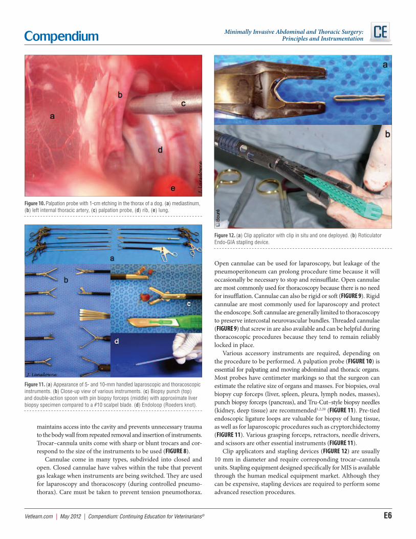
Various accessory instruments are required, depending on the procedure to be performed. A palpation probe (FIGURE 10) is essential for palpating and moving abdominal and thoracic organs. Most probes have centimeter markings so that the surgeon can estimate the relative size of organs and masses. For biopsies, oval biopsy cup forceps (liver, spleen, pleura, lymph nodes, masses), punch biopsy forceps (pancreas), and Tru-Cut–style biopsy needles (kidney, deep tissue) are recommended1,2,20 (FIGURE 11). Pre-tied endoscopic ligature loops are valuable for biopsy of lung tissue, as well as for laparoscopic procedures such as cryptorchidectomy (FIGURE 11). Various grasping forceps, retractors, needle drivers, and scissors are other essential instruments (FIGURE 11).
Clip applicators and stapling devices (FIGURE 12) are usually 10 mm in diameter and require corresponding trocar–cannula units. Stapling equipment designed specifically for MIS is available through the human medical equipment market. Although they can be expensive, stapling devices are required to perform some advanced resection procedures.
Figure 10. Palpation probe with 1-cm etching in the thorax of a dog. (a) mediastinum, (b) left internal thoracic artery, (c) palpation probe, (d) rib, (e) lung.
Figure 11. (a) Appearance of 5- and 10-mm handled laparoscopic and thoracoscopic instruments. (b) Close-up view of various instruments. (c) Biopsy punch (top) and double-action spoon with pin biopsy forceps (middle) with approximate liver biopsy specimen compared to a #10 scalpel blade. (d) Endoloop (Roeders knot).
Figure 12. (a) Clip applicator with clip in situ and one deployed. (b) Roticulator Endo-GIA stapling device.

Vetlearn.com | May 2012 | Compendium: Continuing Education for Veterinarians® E7
Minimally Invasive Abdominal and Thoracic Surgery: Principles and Instrumentation
Endoscopic GI anasto-mosis staplers (such as the Endo-GIA Roticulator 60 3.5 #030458, US Surgical, Norwalk, CT) simultane-ously apply multiple rows of staples and cuts, leaving several rows of staples on the organ and several rows on the section to be re-moved. Most stapling in-struments have an articu-lation that allows for correct placement of the staple line. This is particu-larly useful during lung lo-bectomy because it allows the bronchus, pulmonary artery, and pulmonary vein to be occluded with one cartridge of staples. Many stapling devices come in three different lengths: 30, 45, and 60 mm, with sta-ples 2.0, 2.5, 3.5, or 4.8 mm long. A 2005 study using the Endo-GIA stapling device to perform lung lobectomy in dogs recommended the
60-mm device with 3.5-mm staples.9 Successful lung lobectomy using staples has also been reported in dogs using a stapling de-vice designed for open surgery that applies four rows of staples.
Laparoscopic and thoracoscopic retrieval bags (FIGURE 13) can be useful for removal of large and/or cancerous masses. These bags permit maceration of the tissue to reduce tumor size, thereby allowing removal through a portal site or a minimally extended portal site and preventing seeding of the site with cancerous cells. Although seeding of thoracoscopic incisions has occurred, the risk of cancer recurrence at the cannula site in humans is considered low (three of 934 operations).25 However, as seeding can be a serious complication and is easily prevented, the use of a thoracoscopic retrieval bag is the standard of care in human medicine.26 A human study evaluating the incidence of tumor recurrence at portal sites when a retrieval bag was used in 308 cases reported one case of recurrence after a median follow-up of 25 months.27
Most laparoscopic instruments can perform monopolar electro-cautery at their tip. In addition, the Harmonic Scalpel (Ethicon Endo-Surgery, New Brunswick, NJ) and other coagulation and cutting devices (e.g., Ligasure, Ligasure sealer/divider #LS1500, US Surgical) are available for MIS use.
Advantages and IndicationsLaparoscopy and thoracoscopy have several advantages compared with traditional open techniques. MIS decreases the inherent stresses
on the body during the postoperative period because incisions are significantly smaller, causing less tissue damage and postoperative pain.28–30 Consequently, there is less risk of postoperative seroma, dehiscence, and incisional infection, hospitalization time can be decreased, negative energy balance can be prevented, and return to function can be faster.31 GI function, including myoelectric activity and intestinal motility, has been shown to return to normal more quickly in dogs undergoing laparoscopic versus open pro-cedures.32–34 The incidence of adhesion formation, although not a large factor in dogs and cats, is lower with laparoscopy, presumably because organs are not exposed and permitted to dehydrate.35 In humans undergoing laparoscopic cholecystectomy, several studies have shown that pulmonary complications were minimized compared with open procedures as a result of less pain on deep inspiration, leading to better ventilation, total lung capacity, and saturation,36 with a return to baseline pulmonary function 4 to 10 days sooner.37 Although these parameters have not been studied in dogs and cats, such benefits may play a role in the improved recovery seen clinically.
In addition, as the surgeon’s experience develops, thoracoscopic and laparoscopic procedures can be performed faster than open techniques, decreasing surgical times and the risks of prolonged anesthesia. The use of a camera and light source enables superior visualization and magnification of body cavities and organ systems compared with similar open approaches. This allows the surgeon to precisely localize the area of concern and focus the procedure. Previously inaccessible recesses can now be visualized and ex-plored using angled endoscopes.35 Also, because thoracoscopic and laparoscopic procedures are carried out through a few small portal sites, there can theoretically be a better cosmetic end result when compared with a full-length incision. Although cosmesis is generally not as important in animals as in people, pet owners’ expectations are continually progressing. All these advantages become important when patients have systemic, chronic, or end-stage diseases that may interfere with healing potential and time at home with their family.
Contraindications and DisadvantagesAbsolute contraindications to laparoscopy in animals include septic peritonitis and moderate to severe intraabdominal adhesions or large masses that would preclude visualization and removal. Absolute contraindications to thoracoscopy include large thoracic masses and severe pulmonary disease. Small body size (<2 kg body weight), severe obesity, mild adhesions, ascites, abnormal clotting times, and poor patient condition are relative contrain-dications for MIS. However, endoscopes as small as 2.7 mm have been used successfully in reptiles, fish, and birds.38–40 Excess fat can obscure vision but can be removed or avoided, adhesions can by lysed via laparoscopy, ascites fluid can be drained preoperatively or intraoperatively, and abnormal clotting times from liver disease have not been shown to always correlate with excessive bleeding from liver biopsies.10,20 Laparoscopy and thoracoscopy can be performed in severely ill patients under sedation and local anesthesia if general anesthesia is deemed too risky.20
Figure 13. Endoscopic retrieval bag with large tumor (equine ovarian tumor). Inset shows the opening of the bag after initial deployment.

Vetlearn.com | May 2012 | Compendium: Continuing Education for Veterinarians® E8
Minimally Invasive Abdominal and Thoracic Surgery: Principles and Instrumentation
There are some inherent disadvantages to minimally invasive procedures. Firstly, the equipment required to perform these procedures remains expensive. However, many companies are interested in the veterinary market (e.g., Storz, Stryker, Olympus, Wolf, Dr. Fritz), and refurbished systems are available at reduced cost. Furthermore, as more veterinarians become trained in MIS, the equipment costs become easier to justify and the investment can become profitable. The instrumentation required to perform MIS procedures can be disposable or nondisposable. Disposable instruments have the benefit of always being new but can be ex-pensive. Nondisposable products are less expensive in the long term but can be costly to repair. Similar to companies providing endoscopes and towers, some instrument suppliers are keen to work with veterinarians.
Secondly, MIS techniques are challenging to learn. Surgeons must adapt to the hand–eye coordination requirements and lack of depth perception. As with thoracotomy, the continuous motion of the heart and lungs can pose an additional challenge in thora-coscopy. MIS involves different visual and tactile skills than tra-ditional surgery, and this alone keeps many veterinarians from pursuing further training and advancing their skill. Thirdly, a large room and surgical team are necessary for most advanced procedures. Many procedures require a camera operator and one to two instrument operators working as a team. For thoracoscopy in particular, a knowledgeable anesthetist is also a real asset to the team. Monitoring anesthesia during MIS can be dynamic and challenging considering the effects of pneumoperitoneum, Trendelenburg or reverse Trendelenburg positions, and one-lung ventilation during thoracoscopy.
* * *
Although being confident and competent with MIS requires a large commitment of time and money, it can be rewarding pro-fessionally, financially, and personally.
References1. VanLueSJ,VanLueAP.Equipmentandinstrumentationinveterinaryendoscopy.Vet Clin North Am Small Anim Pract2009;39:817-837.2. FreemanLJ.Gastrointestinallaparoscopyinsmallanimals.Vet Clin North Am Small Anim Pract2009;39:903-924.3. McCarthyTC.Veterinary Endoscopy for the Small Animal Practitioner.St.Louis,MO:ElsevierSaunders;2005.4. CollardF,NadeauM,CarmelE.Laparoscopicsplenectomyfortreatmentofsplenichemangiosarcomainadog.Vet Surg2010;39:870-872.5. MayhewKN,MayhewPD,Sorrell-RaschiL,CiminoBrownD.Thoracoscopicsub-phrenic pericardectomy using double-lumen endobronchial intubation for alternatingone-lungventilation.Vet Surg2009;38:961-966.6. MayhewPD,CiminoBrownD.Prospectiveevaluationoftwointracorporeallysuturedprophylacticlaparoscopicgastropexytechniquescomparedwithlaparoscopic-assistedgastropexyindogs.VetSurg 2009;38(6):738-746.7. MayhewPD,MehlerSJ,RadhakrishnanA.Laparoscopiccholecystectomyformanagementofuncomplicatedgallbladdermucoceleinsixdogs.Vet Surg2008;37:625-630.8. DupréG,FiorbiancoV,SkalickyM,etal.Laparoscopicovariectomyindogs:com-parisonbetweensingleportalandtwo-portalaccess.Vet Surg2009;38:818-824.9. LansdowneJL,MonnetE,TwedtDC,etal.Thoracoscopiclunglobectomyfortreatmentoflungtumorsindogs.Vet Surg2005;34:530-535.10. VasanjeeSC,BubenikLJ,HosgoodG,etal.Evaluationofhemorrhage,samplesize,andcollateraldamageforfivehepaticbiopsymethodsindogs.Vet Surg2006;35:86-93.
11. FreemanL.Veterinary Endosurgery.St.Louis,MO:Mosby;1999.12. MagneML,TamsTR.Laparoscopy:instrumentationandtechnique.In:TamsTR,ed.Small Animal Endoscopy.2nded.St.Louis,MO:CVMosby;1999:397-408.13. DukeT,SteinacherSL,RemediosAM.Cardiopulmonaryeffectsofusingcarbondioxideforlaparoscopicsurgeryindogs.Vet Surg1996;25:77-82.14. JorisJ.Anestheticmanagementoflaparoscopy.In:MillerR,ed.Anesthesia.NewYork,NY:ChurchillLivingstone;1994:2011-2029.15. PawP,Sackier JM.Complicationsof laparoscopy and thoracoscopy.J Intensive Care Med 1994;9:290-304.16. IbertiTJ,KellyKM,GentiliDR,etal.Asimple technique toaccuratelydetermineintra-abdominalpressure.Crit Care Med1987;15:1140-1142.17. CaldwellC,RicottaJ.Changesinvisceralbloodflowwithelevatedintra-abdominalpressure.J Surg Res1987;43:14-20.18. DiebelLN,DulchavskySA,WilsonRF.Effectofincreasedintra-abdominalpressureonmesentericarterialandintestinalmucosalbloodflow.J Trauma1992;33:45-48;discussion48-49.19. HirvonenEA,NuutinenLS,KaukoM.HemodynamicchangesduetoTrendelenburgpositioningandpneumoperitoneumduringlaparoscopichysterectomy.Acta Anaesthesiol Scand1995;39:949-955.20. Monnet E, Twedt DC. Laparoscopy. Vet Clin North Am Small Anim Pract 2003;33:1147.21. ZevallosH,ShahY,MoodyL.Outpatient laparoscopywith localanesthesia. Int J Gynaecol Obstet 1980;17:379-381.22. SafranDB,OrlandoR3rd.Physiologiceffectsofpneumoperitoneum.Am J Surg1994;167:281-286.23. KudnigST,MonnetE,RiquelmeM,etal.Effectofone-lungventilationonoxygendeliveryinanesthetizeddogswithanopenthoraciccavity.Am J Vet Res2003;64:443-448.24. RiquelmeM,MonnetE,KudnigST,etal.Cardiopulmonarychangesinducedduringone-lungventilation inanesthetizeddogswithaclosed thoraciccavity.Am J Vet Res2005;66:973-977.25. McKennaRJJr.Thecurrentstatusofvideo-assisted thoracicsurgery lobectomy.Chest Surg Clin North Am1998;8:775-785,viii;discussion787-788.26. LinJ,IannettoniMD.Theroleofthoracoscopyinthemanagementoflungcancer.Surg Oncol2003;12:195-200.27. ParekhK,RuschV,BainsM.VATSpostsiterecurrence:atechniquedependentproblem.Ann Surg Oncol2001;8:175-178.28. DavidsonEB,MollHD,PaytonME.Comparisonoflaparoscopicovariohysterectomyandovariohysterectomyindogs.Vet Surg2004;33:62-69.29. DevittCM,CoxRE,HaileyJJ.Duration,complications,stress,andpainofopenovariohysterectomyversusasimplemethodoflaparoscopic-assistedovariohysterectomyindogs.J Am Vet Med Assoc2005;227:921-927.30. WalshPJ,RemediosAM,FergusonJF,etal.Thoracoscopicversusopenpartialperi-cardectomyindogs:comparisonofpostoperativepainandmorbidity.Vet Surg1999;28:472-479.31. CulpWT,MayhewPD,BrownDC.Theeffectoflaparoscopicversusopenovariectomyonpostsurgicalactivityinsmalldogs.Vet Surg 2009;38:811-817.32. HotokezakaM,CombsMJ,SchirmerBD.Recoveryofgastrointestinalmotilityfol-lowingopenversuslaparoscopiccolonresectionindogs.Dig Dis Sci1996;41:705-710.33. SchippersE,OttingerAP,AnurovM,etal.[Intestinalmotilityafterlaparoscopicvsconventional cholecystectomy.An animal experiment study and clinical observation].Langenbecks Arch Chir 1992;377:14-18.34. DaviesW,KollmorgenCF,TuQM,etal.Laparoscopiccolectomyshortenspostop-erativeileusinacaninemodel.Surgery1997;121:550-555.35. RemediosAM,FergusonJ.Minimallyinvasivesurgery:laparoscopyandthoracoscopyinsmallanimals.Compend Contin Educ Pract Vet1996;18:1191-1199.36. KarayiannakisAJ,MakriGG,MantziokaA,etal.Postoperativepulmonaryfunctionafterlaparoscopicandopencholecystectomy.Br J Anaesth1996;77:448-452.37. SchauerPR,LunaJ,GhiatasAA,etal.Pulmonaryfunctionafterlaparoscopicchole-cystectomy.Surgery1993;114:389-397;discussion397-399.38. EcholsS.Collectingdiagnosticsamplesinavianpatients.Vet Clin North Am Exot Anim Pract 1999;2:621-649.39. JeklV,KnotekZ.Endoscopicexaminationofsnakesbyaccessthroughanairsac.Vet Rec2006;158:407.40. TaylorM.Endoscopyinbirdsandreptiles.In:TamsTR,ed.Small Animal Endoscopy.2nded.St.Louis:Mosby:1999:433-445.

Vetlearn.com | May 2012 | Compendium: Continuing Education for Veterinarians® E9
Minimally Invasive Abdominal and Thoracic Surgery: Principles and Instrumentation
1. Pneumoperitoneum is ideally created using what gas at what pressure?
a. NO at 4 to 7 mm Hg
b. CO2 at 4 to 7 mm Hg
c. NO at 10 to 12 mm Hg
d. CO2 at 10 to 12 mm Hg
2. Increases in IAP to 20 to 40 mm Hg are associated with
a. no significant abnormalities.
b. myocardial depression and vasoconstriction.
c. occlusion of blood flow in peritoneal and splanchnic capil-lary beds.
d. decreased HR and BP and increased systemic vascular resistance.
3. Patients with one-lung ventilation should be placed on a PEEP of
a. 5 cm H2O to prevent desaturation.
b. 5 cm H2O to promote resaturation.
c. 8 to 10 cm H2O to prevent desaturation.
d. 8 to 10 cm H2O to prevent right-to-left shunting of blood.
4. Absolute contraindications for laparoscopy include
a. small size, abnormal clotting times, and poor patient condition.
b. adhesions, obesity, and sepsis.
c. moderate to severe intraabdominal adhesions, large masses, and septic peritonitis.
d. liver disease and ascites.
5. Advantages of MIS compared with open procedures include
a. smaller incisions.
b. less postoperative pain.
c. faster return to function.
d. all of the above
6. Where should the TV monitor be placed relative to the surgeon?a. to the surgeon’s left
b. to the surgeon’s right
c. directly in front
d. directly behind
7. An angle of 10° to 20° (Trendelenburg position) is best used to facilitate procedures such as
a. pericardial surgery.
b. liver biopsy.
c. abdominal cryptorchidectomy.
d. lung lobectomy.
8. Laparoscopic and thoracoscopic retrieval bags can be useful for
a. obtaining histology samples.
b. preventing seeding of portal sites.
c. removing large masses.
d. all of the above
9. Basic equipment requirements for MIS include
a. a color, high-quality TV monitor.
b. a halogen or xenon light source.
c. a CO2 source.
d. all of the above
10. The ideal MIS team includes at least one
a. camera operator.
b. surgical instrument operator.
c. anesthetist.
d. all of the above
This article qualifies for 3 contact hours of continuing education credit from the Auburn University College of Veterinary Medicine. CE tests must be taken online at Vetlearn.com; test results and CE certificates are available immediately. Those who wish to apply this credit to fulfill state relicensure requirements should consult their respective state authorities regarding the applicability of this program. 3 CE Credits
©Copyright 2012 Vetstreet Inc. This document is for internal purposes only. Reprinting or posting on an external website without written permission from Vetlearn is a violation of copyright laws.

![Minimally invasive non-surgical vs. surgical approach for ...dictable [12]. More recently, minimally invasive surgical therapy (MIST), modified minimally invasive surgical therapy](https://static.fdocuments.net/doc/165x107/5eddda76ad6a402d6669115c/minimally-invasive-non-surgical-vs-surgical-approach-for-dictable-12-more.jpg)