Minimal Clinical Elements for Breast Cancer Detection and ...
13
STATE OF MARYLAND DHMH Maryland Department of Health and Mental Hygiene Larry Hogan, Governor - Boyd K. Rutherford, Lt. Governor - Dennis R. Schrader, Secretary 201 W. Preston Street – Baltimore, Maryland 21201 Toll Free 1-877-4MD-DHMH – TTY/Maryland Relay Service 1-800-735-2258 Web Site: www.dhmh.maryland.gov March 7, 2017 Dear Maryland Breast and Cervical Cancer Provider: Thank you for providing breast cancer screening for uninsured or underinsured women aged 40-64 enrolled in the Maryland Breast and Cervical Cancer Program (BCCP). The Maryland BCCP is a grantee of the National Breast and Cervical Cancer Early Detection Program, funded by the Centers for Disease Control and Prevention. The policies of the national program are based on evidence in scientific literature and recommendations from national organizations such as the American Cancer Society, the United States Preventive Services Task Force, the National Comprehensive Cancer Network and the American College of Radiology. We are pleased to enclose the revised “Minimal Clinical Elements for Breast Cancer Detection and Diagnosis” developed by the Medical Advisory Committee for the BCCP to serve as guidelines for the management of women receiving breast cancer screening and diagnostic services through the BCCP. The revision includes when breast tomosynthesis (3D mammogram) is reimbursable in the Maryland BCC Program. We appreciate your cooperation in using the new guidelines. If you have any questions regarding the new “Minimal Clinical Elements for Breast Cancer Detection and Diagnosis” for the Maryland Breast and Cervical Program, please contact Ken Lin Tai, M.D., M.P.H., Medical Director for the Center for Cancer Prevention and Control (CCPC) at 410-767-2036 or [email protected]. Sincerely, Maryland Breast and Cervical Cancer Program Enclosure Cc Ken Lin Tai, MD, MPH, Medical Director/Acting Director, CCPC Holly Harshbarger, RN, BS, Program Nurse Consultant, BCCP Local BCCP Coordinators Maryland Breast & Cervical Cancer Program Medical Advisory Committee Breast Cancer Subcommittee Stanley P. Watkins, M.D. Chairman Annapolis Medical Specialists Medical Oncology Cecilia Brennecke, M.D. Johns Hopkins Imaging Breast Imaging Specialist Robert Brookland, M.D. Greater Baltimore Medical Center Dept. of Radiation Oncology Regina Hampton, M.D. Doctor’s Community Hospital Breast Surgery Kathy J. Helzlsouer, M.D. Associate Director Epidemiology and Genomics Research Program National Cancer Institute Nagi Khouri, M.D. Johns Hopkins Medical Institutes Diagnostic Radiologist Lorraine Tafra, M.D. Anne Arundel Medical Center Breast Surgery
Transcript of Minimal Clinical Elements for Breast Cancer Detection and ...
STATE OF MARYLAND
DHMH Maryland Department of Health and Mental Hygiene Larry Hogan, Governor - Boyd K. Rutherford, Lt. Governor - Dennis R. Schrader, Secretary
201 W. Preston Street – Baltimore, Maryland 21201
Toll Free 1-877-4MD-DHMH – TTY/Maryland Relay Service 1-800-735-2258
Web Site: www.dhmh.maryland.gov
March 7, 2017
Dear Maryland Breast and Cervical Cancer Provider:
Thank you for providing breast cancer screening for uninsured or underinsured
women aged 40-64 enrolled in the Maryland Breast and Cervical Cancer Program
(BCCP). The Maryland BCCP is a grantee of the National Breast and Cervical
Cancer Early Detection Program, funded by the Centers for Disease Control and
Prevention. The policies of the national program are based on evidence in scientific
literature and recommendations from national organizations such as the American
Cancer Society, the United States Preventive Services Task Force, the National
Comprehensive Cancer Network and the American College of Radiology.
We are pleased to enclose the revised “Minimal Clinical Elements for Breast Cancer
Detection and Diagnosis” developed by the Medical Advisory Committee for the
BCCP to serve as guidelines for the management of women receiving breast cancer
screening and diagnostic services through the BCCP.
The revision includes when breast tomosynthesis (3D mammogram) is reimbursable
in the Maryland BCC Program.
We appreciate your cooperation in using the new guidelines. If you have any
questions regarding the new “Minimal Clinical Elements for Breast Cancer Detection
and Diagnosis” for the Maryland Breast and Cervical Program, please contact Ken
Lin Tai, M.D., M.P.H., Medical Director for the Center for Cancer Prevention and
Control (CCPC) at 410-767-2036 or [email protected].
Sincerely,
Maryland Breast and Cervical Cancer Program
Enclosure
Cc Ken Lin Tai, MD, MPH, Medical Director/Acting Director, CCPC
Holly Harshbarger, RN, BS, Program Nurse Consultant, BCCP
Local BCCP Coordinators
Maryland Breast & Cervical
Cancer Program Medical
Advisory Committee
Breast Cancer Subcommittee
Stanley P. Watkins, M.D.
Chairman
Annapolis Medical Specialists
Medical Oncology
Cecilia Brennecke, M.D.
Johns Hopkins Imaging
Breast Imaging Specialist
Robert Brookland, M.D.
Greater Baltimore Medical Center Dept. of Radiation Oncology
Regina Hampton, M.D.
Doctor’s Community Hospital
Breast Surgery
Kathy J. Helzlsouer, M.D.
Associate Director
Epidemiology and Genomics Research Program
National Cancer Institute
Nagi Khouri, M.D.
Johns Hopkins Medical Institutes
Diagnostic Radiologist
Lorraine Tafra, M.D.
Anne Arundel Medical Center
Breast Surgery

Page left blank intentionally.

Maryland Breast Cancer Minimal Clinical Elements 2017 1
Minimal Clinical Elements for Breast Cancer Detection and Diagnosis Maryland Breast and Cervical Cancer Program
Maryland DHMH, Center for Cancer Prevention and Control
March 2017
Goal:
The goal of the Minimal Clinical Elements for Breast Cancer Detection and Diagnosis is
to provide clients of the Maryland Breast and Cervical Cancer Program (BCCP) with
optimal, up-to-date screening for breast cancer and management of findings
Objective:
To provide clinical guidelines for breast cancer screening and diagnostic testing
including interpretation and management of results of clinical breast examination,
mammography, and diagnostic testing.
To outline appropriate management and approved indications for procedure payment.
Detection and Management of Breast Abnormalities in the Breast and Cervical Cancer
Program—Breast Cancer Minimal Clinical Elements
Section Page
I. Maryland Breast and Cervical Cancer Program (BCCP)—
Eligibility for Screening, Procedures for Screening or Initial Testing, and Eligibility
for Diagnostic Testing when referred by a clinician for follow-up for an abnormal
breast screening exam.
A. BCCP Eligibility and Procedures for Screening or Initial Testing 3
B. Eligibility for Diagnostic Testing in the BCCP 4
II. Findings, Management of Results, Additional Procedures and Program Coverage
A. Results and Reports 5
B. Management of Findings of CBE, Initial Mammogram, and Testing 6
C. Additional Procedures and Program Coverage 6
Attachment A 9
Flow Charts of the Maryland Breast and Cervical Cancer Program:
Management of Clinical Breast Examination and Mammogram Results
I. Management when the Clinical Breast Exam is Normal/Benign 10
II. Management when the Clinical Breast Exam is Abnormal 11

Maryland Breast Cancer Minimal Clinical Elements 2017 2
Members of the Breast Cancer Subcommittee of the
BCCP Medical Advisory Committee:
Stanley Watkins, M.D., Chairman
Hematologist/Oncologist
Annapolis Medical Specialists
Assistant Professor of Oncology, The Johns Hopkins School of Medicine (part time)
Cecilia Brennecke, M.D.
Medical Director, Johns Hopkins Imaging
Diagnostic Radiology
Robert Brookland, M.D.
Chairman, Department of Radiation Oncology
Greater Baltimore Medical Center
Regina Hampton, M.D. FACS
Medical Director, Center for Women’s Wellness
Doctor’s Community Hospital
Kathy J. Helzlsouer, M.D., M.H.S.
Associate Director, Epidemiology and Genomics Research Program National Cancer Institute; National Institutes of Health
Nagi Khouri, M.D.
Associate Professor of Radiology and Radiological Science
Associate Professor of Oncology
Johns Hopkins Medical Institutions
Lorraine Tafra, M.D.
Breast Surgery
Anne Arundel Medical Center
Staff for the Breast Cancer Subcommittee
Center for Cancer Prevention and Control, Maryland Dept. of Health and Mental Hygiene
Ken Lin Tai, M.D., M.P.H., Acting Director, CCPC
Holly Harshbarger, R.N., B.S. Program Nurse Consultant, BCCP

Maryland Breast Cancer Minimal Clinical Elements 2017 3
Detection and Management of Breast Abnormalities in the Breast and
Cervical Cancer Program
Breast Cancer Minimal Clinical Elements (MCE)
I. Maryland Breast and Cervical Cancer Program (BCCP)—Eligibility for Screening,
Procedures for Screening or Initial Testing, and Eligibility for Diagnostic Testing When
Referred by a Clinician for Follow-up for an Abnormal Breast Screening Exam
A. BCCP Eligibility and Procedures for Screening or Initial Testing
1. A woman is eligible for breast cancer screening with clinical breast examination
(CBE) and mammogram1 in the BCCP regardless of symptoms, risk factors, or prior
breast cancer/findings if she:
a. Is 40 – 64 years old or 65+ without Medicare Part B;
b. Meets income eligibility of household income ≤250% of the Federal Poverty
Guideline;
c. Has no health insurance, has health insurance that does not cover breast cancer
screening, or has health insurance but has not met the deductible for the year, and/or
has a patient contribution amount for applicable procedures to include copays and
co-insurance; and
d. Has not had bilateral mastectomies.
2. A woman should have a diagnostic mammogram if a woman has:
a. A CBE with results that include:
i. Nipple discharge that is:
(a) Bloody;
(b) Crystal clear (like water); or
(c) Any other color or clarity (for example, yellow, white, milky, gray, green) if
the discharge is unilateral, single duct, and spontaneous.
ii. Discrete palpable mass—suspicious for cancer;
iii. Nipple/areolar scaliness; or
iv. Skin dimpling/retraction;
b. A recommendation for a diagnostic mammogram from the Medical Case
Manager.
3. A woman should have a screening mammogram as the annual exam if the woman
has:
a. A CBE with Normal findings or a CBE with Benign findings, including:
i. Nipple discharge that does not meet the requirement for diagnostic mammogram
(2., a., i., above);
ii. Breast implant(s);
iii. Fibrocystic changes,
iv. Mastitis;
1 See page 6, section C, #2 for guidance regarding the type of mammogram allowable in the Maryland BCCP.

Maryland Breast Cancer Minimal Clinical Elements 2017 4
v. ―Lumpy‖ breasts;
vi. Family history of breast cancer (premenopausal breast cancer in sister/mother);
or
vii. Prior benign biopsy (within past year) when surgeon or radiologist
recommends screening mammogram.
b. A history of negative screening mammogram(s) (American College of Radiology,
Breast Imaging and Database Reporting System [BI-RADS] category 1, negative,
or BI-RADS 2, benign finding).
4. A woman with a prior history of breast cancer (in situ or invasive, in patient who has
not had bilateral mastectomies) should have a:
a. Diagnostic mammogram for 5 years post diagnosis then;
b. May resume screening mammogram after 5 years at the discretion of the medical
case manager, radiologist and client.
5. CBE should be performed within 90 days prior to the screening mammogram.
a. Each breast should be examined including the retroareolar and peripheral areas
and the upper lateral quadrant into the axilla.
b. The preferred method of CBE is the strip technique using three levels of pressure
in small circular motions with pad of three middle fingers without lubrication
(MammaCare® method).
B. Eligibility for Diagnostic Testing in the BCCP
A woman is eligible for breast cancer diagnostic testing in the BCCP if she:
1. Is 40 – 64 years old, or 65+ without Medicare Part B;
2. Meets income eligibility of ≤250% of the Federal Poverty Guideline;
3. Has no health insurance, has health insurance that does not cover breast cancer
diagnostic testing/visits, or has health insurance but has not met the deductible for the
year, and/or has a patient contribution amount for applicable procedures to include
copays and co-insurance.
4. Has not had bilateral mastectomies; and
5. Provides the BCCP with a recommendation from a clinician for diagnostic workup and
test results of:
a. CBE requiring further diagnosis (see I. A. 2. a.);
b. Mammogram requiring further diagnosis;
c. Ultrasound abnormal finding other than simple cyst(s); or
d. Persistent, unexplained, localized pain in the breast with a negative mammogram.

Maryland Breast Cancer Minimal Clinical Elements 2017 5
II. Findings, Management of Results, Additional Procedures, and Program Coverage
A. Results and Reports
1. CBE findings:
a. Should be reported as:
i. Normal exam
ii. Benign findings
iii. Abnormal findings:
1. Nipple discharge that is bloody, crystal clear (like water) or any other color or
clarity (for example, yellow, white, milky, gray, green) if the discharge is
unilateral, single duct, and spontaneous.
2. Discrete palpable mass—suspicious for cancer
3. Nipple/areolar scaliness
4. Skin dimpling/retraction
b. CBE should report whether there are breast implants; however, this finding would
be categorized as a ―Benign finding‖ if no other abnormalities were found.
c. CBE should report whether the patient has had a lumpectomy or a mastectomy and
which breast was affected; however, this finding would be categorized as a ―Benign
finding‖ if no other abnormalities were found.
2. Mammogram findings should be reported using American College of Radiology
BI-RADS®
(Breast Imaging-Reporting and Database System) Assessment Categories:
a. Assessment is Incomplete
0 Need Additional Imaging Evaluation and/or Prior Mammograms for Comparison
b. Assessment is Complete – Final Categories
1 Negative
2 Benign Finding(s)
3 Probably Benign Finding – Initial Short-Interval Follow-Up Suggested
4 Suspicious Abnormality – Biopsy Should Be Considered
5 Highly Suggestive of Malignancy – Appropriate Action Should Be Taken
6 Known Biopsy-Proven Malignancy – Appropriate Action Should Be Taken
(Category reserved for lesions identified on imaging study with biopsy proof of
malignancy prior to definitive therapy)
Ref. The American College of Radiology BI-RADS® ATLAS and MQSA: Frequently Asked
Questions (Updated: 7/1/09)
c. Breast composition on mammogram should be described for all patients using the
following patterns:
i. The breasts are almost entirely fatty
ii. There are scattered areas of fibroglandular density
iii. The breasts are heterogeneously dense, which may obscure small masses
iv. The breasts are extremely dense, which lowers the sensitivity of mammography
Ref. 2013 ACR® BI-RADS Atlas (5
TH Edition)

Maryland Breast Cancer Minimal Clinical Elements 2017 6
3. Ultrasound definitions: The terms ―simple cyst,‖ ―complicated cyst,‖ and ―complex
cyst‖ are defined by the radiologist and stated in the report of an ultrasound
examination.
4. The radiologist’s diagnostic workup/evaluation report should include the results of the
diagnostic mammogram, ultrasound (when performed), CBE, and the correlation of
each test with each other.
B. Management of Findings of CBE, Initial Mammogram, and Testing
1. See Attachment A. Flow Charts of the Maryland Breast and Cervical Cancer
Program: Management of Clinical Breast Examination and Mammogram Results.
2. A woman with persistent, unexplained, localized pain in the breast should be evaluated
by a breast specialist or surgeon.
3. If a radiologist recommends obtaining results or copies of prior mammograms
following a BI-RADS category 0 result, local programs should assist in obtaining the
results or copies.
4. Image-guided percutaneous needle biopsy is the diagnostic procedure of choice for
image-detected abnormalities, with few exceptions.
5. When a non-palpable or questionably palpable mass that was found on imaging is
excised, the specimen should be verified by using the appropriate imaging modality
while the patient is still in the operating room.
6. At least one breast tissue specimen positive for cancer should be tested for tumor
markers (e.g. estrogen/progesterone receptors, her2neu etc.) to guide clinical
management.
C. Additional Procedures and Program Coverage
1. Providers should consult with the local BCCP for questions about coverage for
payment of procedures.
2. Breast Tomosynthesis (3D mammography)
a. The Maryland BCCP will reimburse for breast tomosynthesis (3D mammogram) if
the medical case manager or radiologist recommends the procedure.
b. The medical provider (medical case manager or radiologist) ordering the
mammogram is responsible for discussing the use of 2D and/or 3D mammography
and its benefits and risks in breast cancer screening with the woman.
3. Magnetic Resonance Imaging (MRI)
a. The Maryland BCCP will reimburse for screening breast MRI performed in
conjunction with a mammogram when a client has the following indications:
Maryland Breast Cancer Minimal Clinical Elements 2017 7
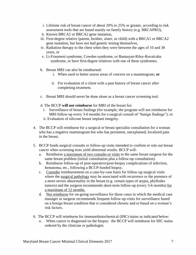
i. Lifetime risk of breast cancer of about 20% to 25% or greater, according to risk
assessment tools that are based mainly on family history (e.g. BRCAPRO),
ii. Known BRCA1 or BRCA2 gene mutation,
iii. First-degree relative (parent, brother, sister, or child) with a BRCA1 or BRCA2
gene mutation, but have not had genetic testing themselves,
iv. Radiation therapy to the chest when they were between the ages of 10 and 30
years, or
v. Li-Fraumeni syndrome, Cowden syndrome, or Bannayan-Riley-Ruvalcaba
syndrome, or have first-degree relatives with one of these syndromes.
b. Breast MRI can also be reimbursed:
i. When used to better assess areas of concern on a mammogram; or
ii. For evaluation of a client with a past history of breast cancer after
completing treatment.
c. Breast MRI should never be done alone as a breast cancer screening tool.
d. The BCCP will not reimburse for MRI of the breast for:
i. Surveillance of breast findings (for example, the program will not reimburse for
MRI follow-up every 3-6 months for a surgical consult of ―benign findings‖); or
ii. Evaluation of silicone breast implant integrity.
4. The BCCP will reimburse for a surgical or breast specialist consultation for a woman
who has a negative mammogram but who has persistent, unexplained, localized pain
in the breast.
5. BCCP funds surgical consults or follow-up visits intended to confirm or rule out breast
cancer when screening tests yield abnormal results. BCCP will:
a. Reimburse a maximum of two consults or visits to the same breast surgeon for the
same breast problem (initial consultation plus a follow-up consultation).
b. Reimburse follow-up of post-operative/post-biopsy complications of infection,
hematoma, etc., following a BCCP-funded biopsy.
c. Consider reimbursement on a case-by-case basis for follow-up surgical visits
where the surgical pathology may be associated with recurrence or the presence of
a more severe abnormality in the breast (e.g. certain types of atypia, phyllodes
tumors) and the surgeon recommends short-term follow-up (every 3-6 months) for
a maximum of 12 months.
d. Not reimburse for on-going surveillance for those cases in which the medical case
manager or surgeon recommends frequent follow-up visits for surveillance based
on a benign breast condition that is considered chronic and or based on a woman’s
risk factors.
6. The BCCP will reimburse for immunohistochemical (IHC) stains as indicated below:
a. When cancer is diagnosed on the biopsy: the BCCP will reimburse for IHC stains
ordered by the clinician or pathologist.

Maryland Breast Cancer Minimal Clinical Elements 2017 8
b. When cancer is suspected or needs to be ruled out: IHC should not be used
reflexively to evaluate every breast biopsy. IHC is best used when ordered by the
pathologist where IHC will clarify an ambiguous pathologic diagnosis. The most
frequent use of this is where the pathologist wants to know whether an adenoma or
papilloma harbors invasive disease, or whether the tumor is ductal or lobular. The
BCCP will reimburse in these cases but may request more information to justify
the use of IHC.

Maryland Breast Cancer Minimal Clinical Elements 2017 9
Attachment A
Flow Charts
of the Maryland Breast and Cervical Cancer Program:
Management of Clinical Breast Examination and
Mammogram Results
I. Management when Clinical Breast Examination is
Normal/Benign Findings
II. Management when Clinical Breast Examination is
Abnormal

Women who: - are asymptomatic; and - have implant(s), fibrocystic changes, mastitis, "lumpy" breasts, family history of breast cancer (premenopausal breast cancer in sister/mother), prior benign biopsy (within past year) when surgeon or radiologist recommends screening mammogram.
Women who have: - a prior history of breast cancer should have a diagnostic mammogram for 5 years post diagnosis then at the discretion of the medical case manager, radiologist and client; or - Medical Case Manager recommendation for Diagnostic Mammogram
Screening mammogram Diagnostic mammogram
Mammogram Results
BI-RADS 0: Needs additional imaging evaluation
BI-RADS 1: Negative BI-RADS 2: Benign Finding
BI-RADS 3: Probably benign--short interval follow-up suggested
BI-RADS 4: Suspicious Abnormality – Biopsy Should Be Considered BI-RADS 5: Highly Suggestive of Malignancy – Appropriate Action Should Be Taken
Follow up per radiologist: - Diagnostic work-up (spot compression, magnification, special views, ultrasound, aspiration…)
Annual follow-up: CBE with screening mammogram (if asymptomatic)
Follow up per radiologist (usually 6 month repeat imaging)
Radiologist communicates findings to the patient, the referring physician
Follow-up based on revised BI-RADS category
See Bi-RADS 1, 2 or See BI-RADS 3 or See Bi-RADS 4 or 5 above.
Refer to surgeon or breast specialist Image-guided biopsy
No cancer on image-guided biopsy; and Surgeon does NOT perform biopsy
Cancer (invasive or DCIS) on prior image-guided biopsy; surgeon performs no additional biopsy
Surgeon performs biopsy
Positive biopsy (invasive cancer or DCIS)
BI-RADS 5: and Negative biopsy OR BI-RADS 4 or 5 with biopsy that is LCIS or atypical ductal hyperplasia
Further follow-up per surgeon*
Management and Treatment of Cancer Surgeon stages and initially manages breast cancer; - Stage 0 must be offered oncologist consult - Stage 1-4 must see oncologist
Positive biopsy (invasive cancer or DCIS+)
BI-RADS 4 or 5 with: Negative biopsy OR biopsy that is LCIS or atypical ductal hyperplasia
Further follow-up per surgeon*
CBE, pathology and imaging results are discordant: - Refer to surgeon or breast specialist.
CBE, pathology and imaging results are concordant: - Radiologist and primary care provider recommend next steps; And - Offer referral to surgeon or breast specialist.
DCIS: Ductal carcinoma in situ LCIS: Lobular carcinoma in situ
*Please refer to The Minimal Clinical Elements for Breast Cancer Detection and Diagnosis, Section II, C, number 4 page 7 for further details and program coverage.
Clinical Breast Examination: Normal or Benign Findings
BI-RADS 4: AND Negative biopsy (that is, biopsy is not cancer, LCIS or atypical ductal hyperplasia)
Maryland Breast Cancer Minimal Clinical Elements 2017 10

Clinical Breast Examination: Abnormal (other than Normal or Benign Finding) **
Diagnostic mammogram (always); with Ultrasound, if recommended
CBE, Mammogram, and Ultrasound Results
BI-RADS 0 BI-RADS 1, 2, or 3 and
- Simple cyst(s), or - Complicated cyst(s)
BI-RADS 1, 2, or 3, and
- Non-cystic palpable mass; - Nipple discharge that is bloody, crystal clear, or any color if unilateral, single duct, and spontaneous; - Nipple/areolar scaliness, or - Skin dimpling/retraction
Any clinical finding and: - Complex cyst(s); - BI-RADS 4: Suspicious Abnormality – Biopsy Should Be Considered; or - BI-RADS 5: Highly Suggestive of Malignancy-Appropriate Action Should Be Taken
BI-RADS 0: Needs additional imaging evaluation
Follow up per radiologist: - Diagnostic work-up (spot compression, magnification, special views, ultrasound, aspiration…)
Follow-up based on revised BI-RADS category
See BI-RADS 1, 2 or 3 or See BI-RADS 4 or 5 above
No aspiration of cyst
Aspirate cyst
Clear fluid Bloody fluid
BI-RADS 1: Negative BI-RADS 2: Benign Finding
BI-RADS 3: Probably benign--short interval follow-up suggested
Annual follow-up: CBE with screening mammogram (if asymptomatic)
Follow up per radiologist (usually 6 month repeat imaging)
Refer to surgeon or breast specialist
Radiologist communicates findings to the patient, the referring physician
Image-guided biopsy
Positive biopsy (invasive cancer or DCIS+)
BI-RADS 5: and Negative biopsy OR BI-RADS 4 or 5 and biopsy that is LCIS or atypical ductal hyperplasia
BI-RADS 4 AND Negative biopsy (that is, biopsy is not cancer, LCIS or atypical ductal hyperplasia)
No cancer on image-guided biopsy; and surgeon does not perform biopsy
Cancer (invasive or DCIS) on prior image-guided biopsy; surgeon does not perform additional biopsy
Surgeon performs biopsy
Further follow-up per surgeon*
Positive biopsy (invasive cancer or DCIS)
Negative biopsy OR Biopsy that is LCIS or atypical ductal hyperplasia
Management and Treatment of Cancer Surgeon stages and initially manages breast cancer; - Stage 0 must be offered oncologist consult - Stage 1-4 must see oncologist
Further follow-up per surgeon*
CBE, pathology and imaging results are discordant: - Refer to surgeon or breast specialist.
CBE, pathology and imaging results are concordant: - Radiologist and primary care provider recommend next steps; And - Offer referral to surgeon or breast specialist.
* Please refer to The Minimal Clinical Elements for Breast Cancer Detection and Diagnosis, Section II, C, number 4 page 7 for further details and program coverage.
** "Abnormal Clinical Breast Exam" includes: -Nipple discharge that is bloody, crystal clear, or of any color if unilateral, single duct, and spontaneous; -Discrete palpable mass—suspicious for cancer -Nipple/areolar scaliness -Skin dimpling/retraction
DCIS: Ductal carcinoma in situ LCIS: Lobular carcinoma in situ Maryland Breast Cancer Minimal Clinical Elements 2017 11