Mini-review Surgical prosthetic treatment Internazionali
7
32 Clinical Cases in Mineral and Bone Metabolism 2010; 7(1): 32-38 Surgical prosthetic treatment Mini-review Christian Carulli Fabrizio Matassi Roberto Civinini Marco Villano Massimo Innocenti 2ª Orthopaedic Clinic (Chief: Prof. Massimo Innocenti) Orthopaedic Traumatologic Center University of Florence Largo Piero Palagi 1, 50139 Florence, Italy Address for correspondence: Christian Carulli, MD 2ª Clinica Ortopedica Centro Traumatologico Ortopedico Largo Piero Palagi 1, 50139 - Firenze, Italy Phone: +39 055 7948287 – Fax: +39 055 432145 E-mail: [email protected] Summary Fragility fractures typically occur in elderly patients related principally to osteoporosis. A significative percentage of these fractures have to be treated surgically but comorbili- ties are often present, and need to be grossly stabilized be- fore surgery. However, there is for these fractures a high rate of morbidity and mortality at short-term. Moreover, patients affected by a fragility fracture are at risk for another fragility fracture later in life. The Authors present an overview of the main patterns of proximal femoral fractures, underlining the peculiar features and choices of surgical treatment, and relating to specific in- dications and results of each treatment. KEY WORDS: Fragility fractures, open reduction and internal fixation, pros- thetic treatment. Introduction Fragility fractures typically occur in elderly patients, with preva- lence on women. Related principally to weakening of bone struc- ture induced by osteoporosis, these fractures are the result of low- energy injuries. Comorbilities are often present and show quick worstening after a femoral fracture: patients’ health status has to be grossly stabilized before surgery to limit peri-operative com- plications. However, there is for these fractures a high rate of mor- bidity and mortality at one year, approximatively 20%-30%, which represents the worst scenario at short follow-up among all frac- tures (1). Moreover, patients affected by a fragility fracture are at risk for another fragility fracture later in life. Even if osteoporosis could virtually concern all bones, some dis- trict is more often involved: fracture of proximal femur is one of the most typical pattern of elderly patients. The incidence of hip fracture doubles for each decade of life after the fifth decade and the number of patients affected is expected to have an increase up to 190% in 2051 respect to present (2, 3). Treatment of fragility fractures of legs is usually a surgical issue, because of the poor quality and weak biomechanical behaviour of the bone. Considered this particular background, surgery is in- tended to avoid additional damage, restore hip articularity, allow quick mobilization and functional recovery. Surgical technique and choice of the implant depend principally on the pattern of the frac- ture. Thus, an exact evaluation of the fracture is of paramount importance to achieve a good clinical result. One of the main problem for the Orthopaedic Surgeon is to con- ciliate the need of a good reduction and stability with surgical ef- ficacy to ensure a fast and longlasting recovery: in fact, a weak bone not only is at risk of fracture, but offers a structure that may be not strong enough to mantain a mechanical device as a nail, plate or implant, in particular after surgery, when mobility is of cap- ital importance to ensure a safe recovery for the patients. On the other side, the huge advancement of the biomedical tech- nology still overcomes the complete knowledge of bone proper- ties and behaviour of this tissue after a fracture, thus at now there is not a single best approach, choice of device or implant and sur- gical technique to obtain a better and longlasting result. Some Authors postulated that osteoporotic bone has no impair- ment of its capacity for fracture healing: tendency to fall is the most important factor in fractures in the elderly, not osteoporosis and impaired function due to poor surgical technique in the elderly is unacceptable. The actual issue is to ensure an adequate fixation or implant stability in a poor bone structure even if technology has reached fine results in the manufacturing devices and prosthe- ses with a favourable mechanical behaviour (4, 5). We agree with the concept that the most of the efforts must be reserved to the acknowledgement of the biologic properties of the bone, while surgical technique and choice of implant have to fol- low and respect these improvements. From an anatomic point of view, fractures of proximal femur may be intracapsular (femoral neck fractures) or extracapsular (trochanteric fractures): the incidence of the two patterns is equal- ly distributed among fragility fractures. The following is an overview of the specific pattern of upper fe- mur fracture and the correlated treatment options. Intracapsular hip fractures Several classifications were proposed to correlate the aspect of femoral neck fractures with better treatment or prognosis. Pauwels grouped these fractures according to the angle of the rim (≤ 30°, 30°-50°, ≥ 70°), hypotesizing a relationship between obliquity of the fracture, stability of the reduction and rate of fail- ure (6). Garden classified femoral neck fractures in four types, accord- ing to displacement of fragments, relating it to a possible vascular damage and, ultimately, to healing of the fracture and survival of the femoral head (7). Recently, AO classification divided femoral neck fractures in type B1, B2 and B3 depending on undisplaced subcapital, displaced transcervical and displaced cervical fractures (8). © CIC Edizioni Internazionali
Transcript of Mini-review Surgical prosthetic treatment Internazionali
32 Clinical Cases in Mineral and Bone Metabolism 2010; 7(1): 32-38
Surgical prosthetic treatmentMini-review
Christian CarulliFabrizio MatassiRoberto CivininiMarco VillanoMassimo Innocenti
2ª Orthopaedic Clinic (Chief: Prof. Massimo Innocenti)Orthopaedic Traumatologic Center University of FlorenceLargo Piero Palagi 1, 50139 Florence, Italy
Address for correspondence: Christian Carulli, MD2ª Clinica OrtopedicaCentro Traumatologico OrtopedicoLargo Piero Palagi 1, 50139 - Firenze, ItalyPhone: +39 055 7948287 – Fax: +39 055 432145E-mail: [email protected]
Summary
Fragility fractures typically occur in elderly patients relatedprincipally to osteoporosis. A significative percentage ofthese fractures have to be treated surgically but comorbili-ties are often present, and need to be grossly stabilized be-fore surgery. However, there is for these fractures a high rateof morbidity and mortality at short-term. Moreover, patients affected by a fragility fracture are at riskfor another fragility fracture later in life.The Authors present an overview of the main patterns ofproximal femoral fractures, underlining the peculiar featuresand choices of surgical treatment, and relating to specific in-dications and results of each treatment.
KEY WORDS: Fragility fractures, open reduction and internal fixation, pros-thetic treatment.
Introduction
Fragility fractures typically occur in elderly patients, with preva-lence on women. Related principally to weakening of bone struc-ture induced by osteoporosis, these fractures are the result of low-energy injuries. Comorbilities are often present and show quickworstening after a femoral fracture: patients’ health status hasto be grossly stabilized before surgery to limit peri-operative com-plications. However, there is for these fractures a high rate of mor-bidity and mortality at one year, approximatively 20%-30%, whichrepresents the worst scenario at short follow-up among all frac-tures (1). Moreover, patients affected by a fragility fracture areat risk for another fragility fracture later in life.Even if osteoporosis could virtually concern all bones, some dis-trict is more often involved: fracture of proximal femur is one of
the most typical pattern of elderly patients. The incidence of hipfracture doubles for each decade of life after the fifth decade andthe number of patients affected is expected to have an increaseup to 190% in 2051 respect to present (2, 3).Treatment of fragility fractures of legs is usually a surgical issue,because of the poor quality and weak biomechanical behaviourof the bone. Considered this particular background, surgery is in-tended to avoid additional damage, restore hip articularity, allowquick mobilization and functional recovery. Surgical technique andchoice of the implant depend principally on the pattern of the frac-ture. Thus, an exact evaluation of the fracture is of paramountimportance to achieve a good clinical result. One of the main problem for the Orthopaedic Surgeon is to con-ciliate the need of a good reduction and stability with surgical ef-ficacy to ensure a fast and longlasting recovery: in fact, a weakbone not only is at risk of fracture, but offers a structure that maybe not strong enough to mantain a mechanical device as a nail,plate or implant, in particular after surgery, when mobility is of cap-ital importance to ensure a safe recovery for the patients. On the other side, the huge advancement of the biomedical tech-nology still overcomes the complete knowledge of bone proper-ties and behaviour of this tissue after a fracture, thus at now thereis not a single best approach, choice of device or implant and sur-gical technique to obtain a better and longlasting result.Some Authors postulated that osteoporotic bone has no impair-ment of its capacity for fracture healing: tendency to fall is the mostimportant factor in fractures in the elderly, not osteoporosis andimpaired function due to poor surgical technique in the elderly isunacceptable. The actual issue is to ensure an adequate fixationor implant stability in a poor bone structure even if technology hasreached fine results in the manufacturing devices and prosthe-ses with a favourable mechanical behaviour (4, 5).We agree with the concept that the most of the efforts must bereserved to the acknowledgement of the biologic properties of thebone, while surgical technique and choice of implant have to fol-low and respect these improvements.From an anatomic point of view, fractures of proximal femur maybe intracapsular (femoral neck fractures) or extracapsular(trochanteric fractures): the incidence of the two patterns is equal-ly distributed among fragility fractures.The following is an overview of the specific pattern of upper fe-mur fracture and the correlated treatment options.
Intracapsular hip fractures
Several classifications were proposed to correlate the aspect offemoral neck fractures with better treatment or prognosis. Pauwels grouped these fractures according to the angle of therim (≤ 30°, 30°-50°, ≥ 70°), hypotesizing a relationship betweenobliquity of the fracture, stability of the reduction and rate of fail-ure (6).Garden classified femoral neck fractures in four types, accord-ing to displacement of fragments, relating it to a possible vasculardamage and, ultimately, to healing of the fracture and survival ofthe femoral head (7). Recently, AO classification divided femoral neck fractures in typeB1, B2 and B3 depending on undisplaced subcapital, displacedtranscervical and displaced cervical fractures (8).
© CIC
Ediz
ioni In
terna
ziona
li
Clinical Cases in Mineral and Bone Metabolism 2010; 7(1): 32-38 33
Surgical prosthetic treatment
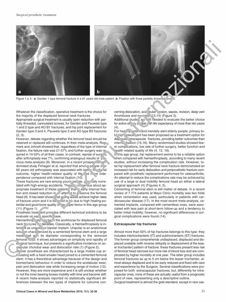
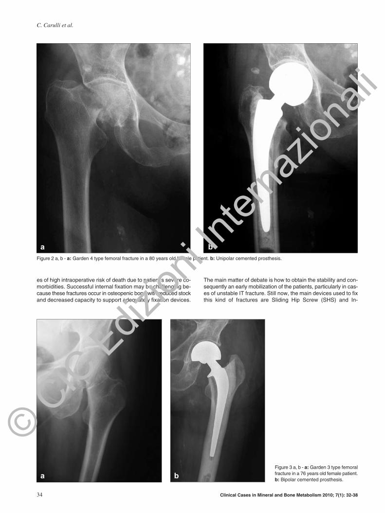
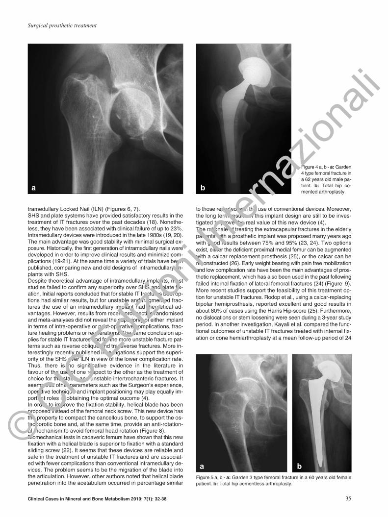
Whatever the classification, operative treatment is the choice forthe majority of the displaced femoral neck fractures. Appropriate surgical treatment is usually open reduction with par-tially threaded, cannulated screws, for Garden and Pauwels type1 and 2 type and AO B1 fractures, and hip joint replacement forGarden type 3 and 4, Pauwels type 3 and AO type B3 fractures(2, 3).However, debate regarding whether the femoral head should beretained or replaced still continues. In their meta-analysis, Rog-mark and Johnell showed that, regardless of the type of internalfixation, the failure rate was 21-57% and further surgery was re-quired in 14-53% of all their cases. In contrast, reprise of surgeryafter arthroplasty was 7%, confirming analogous results of pre-vious meta-analysis (9). Moreover, in a recent prospective ran-domised study Firhagen et al. reported that among people over60 years old arthroplasty was associated with better functionaloutcome, higher health-related quality of life and more inde-pendence compared with internal fixation (10).These fractures are rare among young people, generally corre-lated with high-energy accidents. There is consensus about ap-propriate treatment of these patients, that is early internal fixa-tion and closed reduction in an attempt to preserve the femoralhead (9). It has been shown that young adults achieve higher ratesof fracture union and it is believed to be due to high healing po-tential and good bone quality of the upper femur in this age group(11) (Figure 1).Prosthetic treatment provides different technical solutions to beevaluate on each specific case.Hemiarthroplasty has been the workhorse for displaced femoralneck fractures for decades. Classically, a Hemiarthroplasty is in-tended as unipolar or bipolar implant. Unipolar is an anatomicalsolution characterized by a cemented femoral stem and a largemetal ball with its diameter corresponding to the removedfemoral head. It shows advantages on simplicity and rapidity ofsurgical technique, but presents a significative incidence on ac-etabular chondral wear and dislocation rate (1) (Figure 2).Bipolar prosthesis are characterized by a large mobile cup ar-ticulating with a fixed smaller head joined to a cemented femoralstem: it has a theoretical advantage because of the design andbiomechanic behaviour in order to reduce the acetabular wear,pain and dislocation rate while increasing range of motion (12).However, they are more expensive and it is still unclear whetheror not the inner bearing looses mobility with time and become stiff.A recent meta-analysis reported no statistically significant dif-ferences between the two types of implants for outcome con-
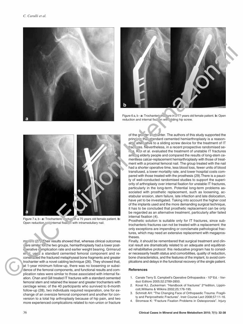
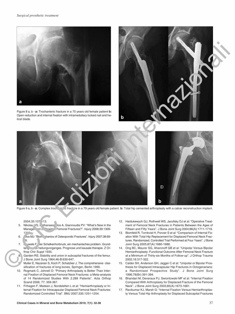
cerning dislocation, acetabular erosion, sepsis, revision, deep veinthrombosis and mortality (13,14) (Figure 3). Additional studies are still needed to evaluate the better choicefor active elderly people with life expectancy of more than ten years(4).For healthy, active and mentally alert elderly people, primary to-tal hip replacement has been proposed as a treatment option fordisplaced intracapsular fractures, providing better outcomes thaninternal fixation (15,16). Many randomised studies showed few-er complications, low rate of further surgery, better function andhealth related quality of life (4, 12, 16).In this age group, hip replacement seems to be a reliable optionwhen compared with hemiarthroplasty, according to many recentstudies, without increasing the complication rate. However, to-tal hip replacement after femoral neck fracture demonstrated anincreased risk for early dislocation and periprosthetic fracture com-pared with prosthetic replacement performed for osteoarthritis.An attempt to reduce the complications rate may be achieved byuse of a large or dual mobility femoral head an either a lateralsurgical approach (4) (Figures 4, 5).Cementing of femoral stem is still matter of debate. In a recentreview of 7.774 patients at Mayo Clinic mortality was two foldswhen cementation was used, particularly in patients with car-diovascular disease (17). In the most recent meta-analysis, ce-mented implants, compared with cementless ones, were asso-ciated with less pain at short-term follow-up and a tendency tobetter initial mobility; however, no significant differences in sur-gical complications were found (14).
Extracapsular hip fractures
Almost more than 50% of hip fractures belongs to this type: theyincludes intertrochanteric (IT) and subtrochanteric (ST) fractures.The former group comprehends undisplaced, displaced and dis-placed unstable (with reverse obliquity or displacement of the less-er trochanter) pattern of fracture: these fractures present less riskof femoral head necrosis but more risk of blood loss and are com-plicated by higher mortality at one year. The latter group includesfemoral fractures as up to 5 cm below the lesser trochanter, al-most always displaced and to be early reduced and fixed with careand experience by the Surgeon. Several classifications were pro-posed for both extracapsular fractures, but, differently for intra-capsular ones, none of these are actually useful from a prognosticpoint of view, representing only a descriptive outline.Surgical treatment is almost the gold standard, except in rare cas-
Figure 1 a, b - a: Garden 1 type femoral fracture in a 61 years old male patient. b: Fixation with three partially threaded screws.
a b
© CIC
Ediz
ioni In
terna
ziona
li
34 Clinical Cases in Mineral and Bone Metabolism 2010; 7(1): 32-38
C. Carulli et al.
es of high intraoperative risk of death due to patient’s severe co-morbidities. Successful internal fixation may be challenging be-cause these fractures occur in osteopenic bone with reduced stockand decreased capacity to support adequately fixation devices.
The main matter of debate is how to obtain the stability and con-sequently an early mobilization of the patients, particularly in cas-es of unstable IT fracture. Still now, the main devices used to fixthis kind of fractures are Sliding Hip Screw (SHS) and In-
Figure 3 a, b - a: Garden 3 type femoralfracture in a 76 years old female patient.b: Bipolar cemented prosthesis.
a b
Figure 2 a, b - a: Garden 4 type femoral fracture in a 80 years old female patient. b: Unipolar cemented prosthesis.
a b
© CIC
Ediz
ioni In
terna
ziona
li
Clinical Cases in Mineral and Bone Metabolism 2010; 7(1): 32-38 35
Surgical prosthetic treatment
tramedullary Locked Nail (ILN) (Figures 6, 7).SHS and plate systems have provided satisfactory results in thetreatment of IT fractures over the past decades (18). Nonethe-less, they have been associated with clinical failure of up to 23%.Intramedullary devices were introduced in the late 1980s (19, 20).The main advantage was good stability with minimal surgical ex-posure. Historically, the first generation of intramedullary nails weredeveloped in order to improve clinical results and minimize com-plications (19-21). At the same time a variety of trials have beenpublished, comparing new and old designs of intramedullary im-plants with SHS.Despite theoretical advantage of intramedullary implants, moststudies failed to confirm any superiority over SHS and plate fix-ation. Initial reports concluded that for stable IT fractures both op-tions had similar results, but for unstable and fragmented frac-tures the use of an intramedullary implant had theoretical ad-vantages. However, results from recent prospective randomisedand meta-analyses did not reveal the superiority of either implantin terms of intra-operative or post-operative complications, frac-ture healing problems or reoperations. The same conclusion ap-plies for stable IT fractures and for the more unstable fracture pat-terns such as reverse oblique and transverse fractures. More in-terestingly recently published investigations support the superi-ority of the SHS over ILN in view of the lower complication rate.Thus, there is no significative evidence in the literature infavour of the use of one respect to the other as the treatment ofchoice for the stable and unstable intertrochanteric fractures. Itseems that other parameters such as the Surgeon’s experience,operative technique and implant positioning may play equally im-portant roles in obtaining the optimal oucome (4).In order to improve the fixation stability, helical blade has beenproposed instead of the femoral neck screw. This new device hasthe property to compact the cancellous bone, to support the os-teoporotic bone and, at the same time, provide an anti-rotation-al mechanism to avoid femoral head rotation (Figure 8).Biomechanical tests in cadaveric femurs have shown that this newfixation with a helical blade is superior to fixation with a standardsliding screw (22). It seems that these devices are reliable andsafe in the treatment of unstable IT fractures and are associat-ed with fewer complications than conventional intramedullary de-vices. The problem seems to be the migration of the blade intothe articulation. However, other authors noted that helical bladepenetration into the acetabulum occurred in percentage similar
to those reported with the use of conventional devices. Moreover,the long term results of this implant design are still to be inves-tigated to prove the real value of this new device (4).The rationale of treating the extracapsular fractures in the elderlypatients with a prosthetic implant was proposed many years agowith good results between 75% and 95% (23, 24). Two optionsexist, either the deficient proximal medial femur can be augmentedwith a calcar replacement prosthesis (25), or the calcar can bereconstructed (26). Early weight bearing with pain free mobilizationand low complication rate have been the main advantages of pros-thetic replacement, which has also been used in the past followingfailed internal fixation of lateral femoral fractures (24) (Figure 9).More recent studies support the feasibility of this treatment op-tion for unstable IT fractures. Rodop et al., using a calcar-replacingbipolar hemiprosthesis, reported excellent and good results inabout 80% of cases using the Harris Hip-score (25). Furthermore,no dislocations or stem loosening were seen during a 3-year studyperiod. In another investigation, Kayali et al. compared the func-tional outcomes of unstable IT fractures treated with internal fix-ation or cone hemiarthroplasty at a mean follow-up period of 24
Figure 4 a, b - a: Garden4 type femoral fracture ina 62 years old male pa-tient. b: Total hip ce-mented arthroplasty.
a b
Figure 5 a, b - a: Garden 3 type femoral fracture in a 60 years old femalepatient. b: Total hip cementless arthroplasty.
a b
© CIC
Ediz
ioni In
terna
ziona
li
36 Clinical Cases in Mineral and Bone Metabolism 2010; 7(1): 32-38
C. Carulli et al.
months (27). Their results showed that, whereas clinical outcomeswere similar for the two groups, hemiarthroplasty had a lower post-operative complication rate and earlier weight bearing. Grimsrudet al. used a standard cemented femoral component and re-constructed the fractured metaphyseal bone fragments and greatertrochanter with a novel cabling technique (26). They showed that,at 1-year minimum follow-up, there was no loosening or subsi-dence of the femoral components, and functional results and com-plication rates were similar to those associated with internal fix-ation. Chan and Gill treated IT fractures with a standard cementedfemoral stem and retained the lesser and greater trochanters withcerclage wires; of the 40 participants who survived to 6-monthfollow-up (28), two individuals required reoperation, one for ex-change of an oversize femoral component and another for con-version to a total hip arthroplasty because of hip pain, and twomore experienced complications related to non-union or fracture
of the greater trochanter. The authors of this study supported theprinciple that standard cemented hemiarthroplasty is a reason-able alternative to a sliding screw device for the treatment of ITfractures. Nevertheless, in a recent prospective randomised se-ries, Kim et al. evaluated the treatment of unstable IT fracturesamong elderly people and compared the results of long-stem ce-mentless calcar-replacement hemiarthroplasty with those of treat-ment with a proximal femoral nail. The group treated with the nailhad a shorter operative time, less blood loss, fewer units of bloodtransfused, a lower mortality rate, and lower hospital costs com-pared with those treated with the prosthesis (29).There is a pauci-ty of well-conducted randomised studies to support the superi-ority of arthroplasty over internal fixation for unstable IT fractures,particularly in the long-term. Potential long-term problems as-sociated with prosthetic replacement, such as loosening, ac-etabular erosion, stem failure, late infection and late dislocation,have yet to be investigated. Taking into account the higher costof the implants used and the more demanding surgical technique,it has to be concluded that prosthetic replacement can be onlybe regarded as an alternative treatment, particularly after failedinternal fixation (4).Prosthetic solution is suitable only for IT fractures, since sub-trochanteric fractures can not be treated with a replacement: theonly exceptions are impending or conclamate pathological frac-tures, which may need an extensive replacement with megapros-theses.Finally, it should be remembered that surgical treatment and clin-ical result are dramatically related to an adequate and equilibrat-ed rehabilitative protocol: this rieducative program has to consid-er necessarily health status and comorbilities, quality of reduction,bone characteristics, and the features of the implant, to avoid com-plications and delays in the functional recovery of the single patient.
References
1. Canale Terry S. Campbell’s Operative Orthopaedics - 10th Ed. - Ver-duci Editore 2005;52:2789-2850.
2. Koval KJ, Zuckerman: “Handbook of fractures” 2ndedition, Lippin-cott,Williams & Wilkins 2002;25;176-195.
3. Schmidt AH: “The Changing Face of Orthopaedic Trauma: Fragili-ty and Periprosthetic Fractures”. Instr Course Lect 2008;57:11-16.
4. Stromsoe K: “Fracture Fixation Problems in Osteoporosis”. Injury
Figure 6 a, b - a: Trochanteric fracture in a 77 years old female patient. b: Openreduction and internal fixation with sliding hip screw.
b
a
Figure 7 a, b - a: Trochanteric fracture in a 75 years old female patient. b:Open reduction and internal fixation with intramedullary nail.
a b
© CIC
Ediz
ioni In
terna
ziona
li
Clinical Cases in Mineral and Bone Metabolism 2010; 7(1): 32-38 37
Surgical prosthetic treatment
2004;35:107-113.5. Nikolau VS, Pathanasopulos A, Giannoudis PV: “What’s New in the
Management of Proximal Femoral Fractures?”. Injury 2008;39:1309-1318.
6. Silva MJ: “Biomechanics of Osteoporotic Fractures”. Injury 2007;38:69-76.
7. Pauwels F. Der Schelkenhorbruck, ein mechanisches problem. Grund-langen des heilungvorganges. Prognose und kausale therapie. Z Or-thop Chir Suppl 1935.
8. Garden RS. Stability and union in subcapital fractures of the femur.J Bone Joint Surg 1964;46-B:630-647.
9. Muller E, Nazarian S, Koch P, Schatzker J. The comprehensive clas-sification of fractures of long bones, Springer, Berlin 1990.
10. Rogmark C, Johnell O: “Primary Arthroplasty is Better Than Inter-nal Fixation of Displaced Femoral Neck Fractures: a Meta-analysisof 14 Randomized Studies With 2.289 Patients”. Acta OrthopScand 2006; 77: 359-367.
11. Frihagen F, Madsen J, Nordsletten L et al: “Hemiarthroplasty or In-ternal Fixation for Intracapsular Displaced Femoral Neck Fractures:Randomized Controlled Trial”. BMJ 2007;335:1251-1254.
12. Haidukewych GJ, Rothwell WS, Jacofsky DJ et al: “Operative Treat-ment of Femoral Neck Fractures in Patients Between the Ages ofFifteen and Fifty Years”. J Bone Joint Surg 2004;86(A):1711-1716.
13. Blomfeld R, Tornkvist H, Ponzer S et al: “Comparison of Internal Fix-ation With Total Hip Replacement for Displaced Femoral Neck Frac-tures. Randomized, Controlled Trial Performed at Four Years”. J BoneJoint Surg 2005;87(A):1680-1688.
14. Ong BC, Maurer SG, Aharonoff GB et al: “Unipolar Versus BipolarHemiarthroplasty: Functional Outcome After Femoral Neck Fractureat a Minimum of Thirty-six Months of Follow-up”. J Orthop Trauma2002;16:317-322.
15. Calder SH, Anderson GH, Jagger C et al: “Unipolar or Bipolar Pros-thesis for Displaced Intracapsular Hip Fractures in Octogenarians:a Randomised Prospective Study”. J Bone Joint Surg1996;78(Br):391-394.
16. Bhandari M, Deveraux PJ, Swiontowski MF et al: “Internal FixationCompared With Arthroplasty for Displaced Fracture of the FemoralNeck”. J Bone Joint Surg 2003;85(A):1673-1681.
17. Ravikumar KJ, Marsh G: “Internal Fixation Versus Hemiarthroplas-ty Versus Total Hip Arthroplasty for Displaced Subcapital Fractures
Figure 9 a, b - a: Complex trochanteric fracture in a 79 years old female patient. b: Total hip cemented arthroplasty with a calcar reconstruction implant.
a b
Figure 8 a, b - a: Trochanteric fracture in a 70 years old female patient b:Open reduction and internal fixation with intramedullary locked nail and he-lical blade.
a
b
© CIC
Ediz
ioni In
terna
ziona
li
of Femur. 13 Year Results of a Prospective Randomised Study”. In-jury 2000;31:793-797.
18. Parvizi J, Ereth MH, Lewallen DJ: “Thirty-day Mortality Following HipArthroplasty for Acute Fracture”. J Bone Joint Surg 2004; 86(A):1983-1988.
19. Bellabarba C, Herscovici D Jr., Ricci WM: “Percutaneous treatmentof pertrochanteric fractures using the gamma nail”. J Orthop trau-ma 2003;17:S38-S50.
20. Bridle SH, Patel AD, Bircher M, Calvert PT: “Fixation of In-tertrochanteric Fractures of the Femur. A Randomised ProspectiveComparison of the Gamma Nail and the Dynamic Hip Screw”. J BoneJoint Surg 1991:73(Br):330-334.
21. Calvert PT: “The Gamma Nail. A significant Advance or a PassingFashion?”. J Bone Joint Surg 1992;74(Br): 329-331.
22. Radford PJ, Needoff M, Webb JK: “A Prospective Randomised Com-parison of the Dynamic Hip Screw and the Gamma Locking Nail”.J Bone Joint Surg 1993;75(Br):789-793.
23. Lenich A, Mayr E, Ruter A et al: “First Results With the TrochantericFixation Nail (TFN): a report on 120 cases. Acta Orthop Trauma Surg2006;126:706-712.
24. Green S, Moore T, Proano F: “Bipolar Prosthetic Replacement For
the Management of Unstable Intertrochanteric Hip Fractures in theElderly”. Clin Orthop 1987;224:169-177.
25. Haentjens P, Casteleyn PP, De Boeck H et al: “Treatment of UnstableIntertrochanteric and Subtrochanteric Fractures in Elderly Patients.Primary Bipolar Arthroplasty Compared With Internal Fixation”. J BoneJoint Surg 1989;71(A):1214-1225.
26. Rodop O, Kiral A, Kaplan H, Akmaz I: “Primary Bipolar Emiprosthe-sis For Unstable Intertrochanteric Fractures. Int Orthop 2002;26:233-237.
27. Grimsud C, Monzon RJ, Richman J, Ries MD: “Cemented Hip Arthro-plasty With a New Cerclage Cable Technique for Unstable In-tertrochanteric Hip Fractures”. J Arthroplasty 2005;20:337-343.
28. Kalyali C, Agus H, Oziuk S, Santi C: “Treatment For Unstable In-tertrochanteric Fractures in Elderly Patients: Internal Fixation Ver-sus Cone Hemiarthroplasty”. J Orthop Surg 2006;14:240-244.
29. Chan C, Gill GS: “Cemented Hemiarthroplasties for Elderly PatientsWith Intertrochanteric Fractures”. Clin Orthop 2000;371:206-215.
30. Kim SY, Kim YG, Hwang JK: “Cementless Calcar-replacement Hemi-arthroplasty Compared With Intramedullary Fixation of Unstable In-tertrochanteric Fractures. A Prospective, Randomized Study”. J BoneJoint Surg 2005;87(A):2186-2192.
38 Clinical Cases in Mineral and Bone Metabolism 2010; 7(1): 32-38
C. Carulli et al.
© CIC
Ediz
ioni In
terna
ziona
li