Mineral Fabrication and Golgi Apparatus Activity in ... · The unicellular organism S. ambiguum...
18
http://www.scirp.org/journal/jbise J. Biomedical Science and Engineering, 2017, Vol. 10, (No. 10), pp: 466-483 https://doi.org/10.4236/jbise.2017.1010036 466 J. Biomedical Science and Engineering Mineral Fabrication and Golgi Apparatus Activity in Spirostomum ambiguum: A Primordial Paradigm of the Stressed Bone Cell? Valerie Fallon, Philippa E. Garner, Jean E. Aaron Institute of Membrane and Systems Biology, Faculty of Biological Sciences, University of Leeds, Leeds, UK Correspondence to: Jean E. Aaron, Keywords: Golgi-Directed Calcification, Mechanosensing Protozoan, Osteocyte Model, Tetracycline Fluorochrome for Bone Mineral, GFP Fluorochrome for Golgi Apparatus Received: September 15, 2017 Accepted: October 24, 2017 Published: October 27, 2017 Copyright © 2017 by authors and Scientific Research Publishing Inc. This work is licensed under the Creative Commons Attribution International License (CC BY 4.0). http://creativecommons.org/licenses/by/4.0/ ABSTRACT The histological basis for acute osteocyte mechanosensitivity remains uncertain. A novel bone cell model of mechanotransduction and inorganic trafficking may be the powerful, silt-burrowing protozoan Spirostomum ambiguum which when being physically challenged fabricates within vesicles populations of bone-like calcium phosphate microspheres, about 1 µm in diameter. These not only attribute considerable compression-resilience but also resemble the Golgi-directed mineral assemblies we recently reported in osteocytes. Advantageously, calcification in the protozoan (confirmed by ultramicroscopy with EDX elemental microanalysis) enabled Golgi comparison under overt, natural phases of both high (i.e. silt-tunnelling) and low (i.e free-swimming) stress. Established hard-tissue microscopy techniques previously positive in bone cells included quantitative fluorescent tetracycline labelling for bone salt together with the same metazoan Golgi body marker (Green Fluorescent Protein-tagged mannosidase II construct). Organellar modulation was monitored by transfection of live organisms in situ (some post-stained with red nuclear fluorochrome TOPRO-3). Results showed that GFP-tagged Golgi fluorescence increased from swimmers (mean 74.5 ± SD 6.7 AU) to burrowers (mean 104.6 ± SD 2.7; p < 0.0001) synchronous with juxtanuclear tetracycline-labelled mineral fluorescence (swimmers, mean 89.7 ± SD 3.3 AU; burrowers, mean 138.0 ± SD 4.0; p < 0.0001). Intracellular dense microspheres, single or bridged, were harvested as pellets rich in Ca, P (Ca:P 0.98) and Si, their polarised alignment moving from trans-axial in swimmers to axial in burrowers. It was concluded that Golgi-directed mineral fabrication in the large, accessi- ble, silt-enclosed ciliate resembles that in the smaller, less-accessible bone cell and may be a conserved early mechanobiological intracellular development predicating force translation Open Access
Transcript of Mineral Fabrication and Golgi Apparatus Activity in ... · The unicellular organism S. ambiguum...
http://www.scirp.org/journal/jbise J. Biomedical Science and Engineering, 2017, Vol. 10, (No. 10), pp: 466-483
https://doi.org/10.4236/jbise.2017.1010036 466 J. Biomedical Science and Engineering
Mineral Fabrication and Golgi Apparatus Activity in Spirostomum ambiguum: A Primordial Paradigm of the Stressed Bone Cell?
Valerie Fallon, Philippa E. Garner, Jean E. Aaron
Institute of Membrane and Systems Biology, Faculty of Biological Sciences, University of Leeds, Leeds, UK
Correspondence to: Jean E. Aaron, Keywords: Golgi-Directed Calcification, Mechanosensing Protozoan, Osteocyte Model, Tetracycline Fluorochrome for Bone Mineral, GFP Fluorochrome for Golgi Apparatus Received: September 15, 2017 Accepted: October 24, 2017 Published: October 27, 2017
Copyright © 2017 by authors and Scientific Research Publishing Inc. This work is licensed under the Creative Commons Attribution International License (CC BY 4.0). http://creativecommons.org/licenses/by/4.0/
ABSTRACT The histological basis for acute osteocyte mechanosensitivity remains uncertain. A novel bone cell model of mechanotransduction and inorganic trafficking may be the powerful, silt-burrowing protozoan Spirostomum ambiguum which when being physically challenged fabricates within vesicles populations of bone-like calcium phosphate microspheres, about 1 µm in diameter. These not only attribute considerable compression-resilience but also resemble the Golgi-directed mineral assemblies we recently reported in osteocytes. Advantageously, calcification in the protozoan (confirmed by ultramicroscopy with EDX elemental microanalysis) enabled Golgi comparison under overt, natural phases of both high (i.e. silt-tunnelling) and low (i.e free-swimming) stress. Established hard-tissue microscopy techniques previously positive in bone cells included quantitative fluorescent tetracycline labelling for bone salt together with the same metazoan Golgi body marker (Green Fluorescent Protein-tagged mannosidase II construct). Organellar modulation was monitored by transfection of live organisms in situ (some post-stained with red nuclear fluorochrome TOPRO-3). Results showed that GFP-tagged Golgi fluorescence increased from swimmers (mean 74.5 ± SD 6.7 AU) to burrowers (mean 104.6 ± SD 2.7; p < 0.0001) synchronous with juxtanuclear tetracycline-labelled mineral fluorescence (swimmers, mean 89.7 ± SD 3.3 AU; burrowers, mean 138.0 ± SD 4.0; p < 0.0001). Intracellular dense microspheres, single or bridged, were harvested as pellets rich in Ca, P (Ca:P 0.98) and Si, their polarised alignment moving from trans-axial in swimmers to axial in burrowers. It was concluded that Golgi-directed mineral fabrication in the large, accessi-ble, silt-enclosed ciliate resembles that in the smaller, less-accessible bone cell and may be a conserved early mechanobiological intracellular development predicating force translation
Open Access

https://doi.org/10.4236/jbise.2017.1010036 467 J. Biomedical Science and Engineering
into compression-resistant mineral fabrication in loaded segments of the osteocyte synci-tium.
1. INTRODUCTION The developmental pathway to metazoan bone has been traced back to the protozoa [1, 2] many ex-
amples of which package mineral as calcium carbonate with varying proportions of calcium phosphate. The common contractile protozoan Spirostomum ambiguum (2 - 3 mm long, visible to the naked eye, ra-pidly motile often in a corkscrew spiralling motion, exceptionally flexible and most typically cigar-shaped) is a successful member of a group of physically versatile, heterotrichous ciliates found in rivers and lakes, subsisting on bacteria and algae. Early attention was drawn to its cyclic, high intracellular apatite content (for example [3, 4]) whereby despite a fragile appearance it can resist substantial compression forces. In consequence its regular calcium phosphate fluctuations and inorganic trafficking may constitute a conve-nient paradigm for stress-related events in bone cells where there remains uncertainty about acute matrix monitoring with various extracellular and intracellular biomechanical theories advanced. In particular, the pervasive osteocyte syncytium [5] is well placed to bridge the gap between mechanical force and cellular signal transduction (see for example [6]). However, its lacunar enclosure within the calcified matrix im-poses limits on direct investigation in vivo, other than by cell culture (for example [7]), where it is ac-knowledged that isolation or manipulation may transform specific threshold characteristics. For this rea-son a readily available, overtly stress-responsive protozoan previously alleged to model certain bone-like properties and that is easily cultured (multiplying rapidly about a week after inoculation) may provide un-usual insight into inherent metazoan bone cell behaviour [8, 9].
Increased mineralization of S. ambiguum relates primarily to its natural channelling predisposition (promoted by supernatant food depletion) when it becomes externally enclosed in silt and internally loaded with spherical, calcified objects including intracellular, calcified filamentous clusters (about 1 mi-cron in diameter) of variable electron density resembling the chemical composition and ultrastructure that is in developing vertebrate bone [10, 11]. Microscopic mineralized objects made by single-celled organisms are generally membrane-bound, leading to the identification of the Golgi apparatus as the source of repeti-tive, organic structural templates manifested in a variety of biomineralization events [12, 13]. Likewise in developing bone there is optical evidence of a direct intracellular association between mineral fabrication and the Golgi apparatus (see for references [7, 8, 14-17]). Diverse primary roles for this organelle (also re-ferred to as the Golgi complex or juxtanuclear body) include a part in muscle differentiation (for example [18]) and in organic macromolecular assembly (for example, acidic polysaccharides [13] and collagen pre-cursors) for which its membranous network is known to host certain reliable resident proteins. The fun-damental expression of these characteristic proteins constitutes a sensitive monitor of Golgi activity, a prop-erty widely adopted as a technological tool. For example, Golgi α-mannosidase II is typically a mammalian transmembrane protein (~125 kDa in size [19]); and since it is a permanent resident of the medial Golgi cisternae its expression may be “tagged” and visualized as a green fluorescent protein (GFP) mannosidase II construct in transfection assays, functioning as a precise temporal marker of Golgi status [18, 20].
The following investigation of the calcifying protozoan is the intended sequel to our recent report on cultured bone cells [7] and extrapolates upon their juxtanuclear calcium phosphate assembly using iden-tical methods. Because of the novelty in applying a mammalian GFP-mannosidase II construct to a proto-zoan such as S. ambiguum an essential pilot objective was to confirm the presence of a conserved proto-zoan Golgi body affinity or non-mammalian equivalent. There followed the main objective in S. ambi-guum of combining this recent GFP-labelled Golgi method with regular techniques of inorganic bone his-tology, including tetracycline labelling (TC) and EDX elemental microanalysis. The aim was to: 1) deter-mine the status of the Golgi apparatus in relation to intracellular mineral load and distribution as the aq-uatic organism became impacted by high-stress tunnelling; 2) examine whether the combined Golgi appa-

https://doi.org/10.4236/jbise.2017.1010036 468 J. Biomedical Science and Engineering
ratus/mineral fabrication process recently identified in bone cells [7] is shared by a ciliate under environ-mental compression; 3) consider the hypothesis that the readily available calcifying protozoan is a unicel-lular paradigm for conserved acute mechanosensitive behaviour in the more advanced but less accessible osteocyte syncytium [5].
2. METHODS The unicellular organism S. ambiguum (Culture Collection of Algae and Protozoa, Cumbria, UK) was
cultured according to the method of Pautard [21]. Calcareous loam (fertile soil) pounded in a mortar was placed to a depth of 1 cm in a 250 ml Schott screw top bottle with distilled water to about 8 cm and was pasteurised in an oven (2 hours at 80˚C) followed by cooling overnight. Ciliates were added with a grain of boiled barley as a food source and the bottle capped loosely for air circulation during incubation at room temperature in dim light. The strain was sub-cultured at four weeks and the free-swimming stock cultures fed monthly to promote cell division. Other cultures were allowed to age without further nutrients to en-courage burrowing and calcification.
2.1. Preparation for Light Microscopy (LM) and Transmission Electron Microscopy (TEM)
Cells drawn from culture by sterile Pasteur pipette were observed directly under the optical micro-scope as wet mounts in plain light. Others were rapidly heat-fixed on glass microscope slides and stained by the von Kossa method for bone mineral (2% silver nitrate under a light source for 30 min, fixed in so-dium thiosulphate [8]) before mounting in Aqua-Mount to confirm general optical calcification features. To establish corresponding ultrastructural calcification features cells extracted from the soil were placed on depression slides for formalin fixation (10%; pH7) and graded dehydration (20%, 40%, 60%, 80% and 100% ethanol) before immersion in a gelatine capsule of LR White resin monomer. After centrifugation (1000 rpm for 5 minutes) and several monomer changes for optimal impregnation polymerisation was heat-induced (60˚C oven for 24 hours). Ultrathin sections (80 nm) cut with a diamond ultramicrotome knife were mounted on formvar-coated copper grids where some were stained for cell detail in uranyl ace-tate (2 hours) and Reynold’s lead citrate (20 minutes), or exposed to osmium vapour alone, while others were left unstained for maximum mineral conservation and minimal artefactual modulation towards crys-tallinity [22].
2.2. Preparation for Scanning Electron Microscopy (SEM) and Mineral Particle Isolation for Energy Dispersive X-Ray Analysis (EDX)
As for TEM, cells from the soil were formalin-fixed/dehydrated on depression slides. They were trans-ferred intact onto carbon-coated aluminium stubs and air-dried for 24 hours before sputter-coating with gold to show cell surface and particulate topography. For particle isolation 50 - 100 ciliates were trans-ferred from soil to a 1.5 ml eppendorf tube and centrifuged (1000 rpm for 10 minutes) followed by resus-pension of the pellet in 10% formalin (pH 7.0), centrifugation and washing in three changes of deionized water. Organic matter was removed from the cell pellet by suspension in hydrazine hydrate (12 hours at 60˚C) and after centrifugation the dense inorganic fraction was re-suspended, washed (three times in deio-nized water) and dehydrated in graded ethanol. The mineral isolate was spread on carbon-coated aluminium stubs, air dried (24 hours) and sputter-coated with gold for SEM and elemental microanalysis (PV9800 EDAX electron microscope) to confirm the nature of the inorganic phase [7, 11].
2.3. Tetracycline (TC) Mineral-Staining Comparison of Tunnelling and Swimming
Six cells from the soil of a burrowing colony and 6 from the supernatant of a swimming colony were gently rinsed on depression slides with water from an undisturbed culture. Each organism was transferred to a standard flat microscope slide and heat-fixed for 30 seconds on a hot plate, before staining with tetra-cycline hydrochloride (10 µg/ml for 30 minutes) to label the calcium phosphate with which it forms a flu-
https://doi.org/10.4236/jbise.2017.1010036 469 J. Biomedical Science and Engineering
orescent complex [15, 23], mounting under glass coverslips in Antifade reagent. Individual cells were ex-amined under a Zeiss confocal microscope to compare fluorescent-mineral complex distribution during swimming and tunnelling, all images digitally captured for quantification of emission intensity and statis-tical analysis (see settings below).
2.4. Controlled Pilot Study of Protozoan Golgi GFP-Mannosidase II Staining Affinity
Random cultured cells on a depression slide were washed in water from an undisturbed culture and transferred to a Petri dish containing 2 ml of similar water (to minimise shock since the disturbed organ-isms can self-destruct). Approximately 20 μl of a pre-prepared GFP-mannosidase II construct (a generous gift from Dr. K.J.M. Zaal supplied by Dr. M. Peckham, University of Leeds) with transfection reagent Fu-GENE 6 ™ (Roche) were added. The live cells were transfected in situ (48 hours at room temperature in dim light), after which they were rinsed in deionized water before transfer to standard flat microscope slides and heat-fixation for 30 seconds. Some preparations were subsequently stained in situ for added de-tail with the nuclear fluorophore TOPRO-3 (to a final concentration of 1 µl/ml for 15 minutes), and all were mounted in Antifade reagent. They were examined under a Zeiss confocal microscope to determine whether the mammalian construct had a specific affinity for the protozoan [8] by reference to unfused GFP-stained controls, whereby to live cells in a Petri dish containing 2 ml of undisturbed culture water was added approximately 20 μl of a pre-prepared mixture of unfused GFP and the transfection reagent as described above (for 48 hours, room temperature, dim light).
2.5. GFP-Mannosidase II Golgi-Staining Comparison of Tunnelling and Swimming
Six rinsed cells from the soil of a burrowing (i.e. well mineralized) culture and 6 from the supernatant of a swimming (i.e. poorly mineralized) colony were put in a Petri dish containing 2 ml of undisturbed culture water to which was added approximately 20 μl of a pre-prepared mixture of a GFP-mannosidase II construct and transfection reagent FuGENE 6 ™ (Roche). After transfection (as above in the pilot study) and heat-fixation before post-staining with TOPRO-3 (1µL/ml) and Antifade mounting, the fluorescent images of individual organisms were digitized under the confocal microscope. As for tetracycline mineral fluorescence the Golgi body status of burrowers and swimmers was compared by quantification (in arbi-trary units, AU) of emission intensity and statistical analysis (see settings below).
Confocal fluorescence microscopy settings for fluorochrome emission measurements and sta-tistical analysis: For TC (excitation laser 360 nm ± 40 i.e. 320 - 400) with emission filters collecting wave-lengths of 528 nm ± 38 i.e. 490 - 566); for GFP (excitation laser 488 nm with emission filters collecting wavelengths of 509 nm); for TOPRO-3 (excitation laser 633 nm with emission filters collecting wave-lengths of 661 nm). The fluorescence intensity distribution of tetracycline and GFP markers (as arbitrary units AU) was quantified (cell by cell) using the Imaris 4.1 (Bitplane) programme. Statistical analysis was by the two-sample t-test, assuming unequal variances.
3. RESULTS 3.1. LM and TEM of S. ambiguum
The elongated form of a large, mature burrowing and small, younger ciliate is shown in Figure 1(a), Figure 1(b) with the typical transparent, water-expelling contractile vacuole at the rear and surrounded by cytoplasmic granularity consistent with particulate bone mineral accumulation. Histochemically this was supported by the von Kossa stain as an intracellular dissemination of calcium phosphate/carbonate par-ticles, about 1 µm in diameter, which tended to be especially aggregated in the juxtanuclear region. It was not uncommon during observation for the live animals to expel spontaneously copious vesicles and mi-crospheres and this may emulate a sudden natural response to secrete surplus particles in the wild. The ultrastructural complexity of the “simple” organism means interpretation of the static image is not always easy. In stained sections, 80 nm thick, the exceptionally long meganucleus (appearing in artefactual planar

https://doi.org/10.4236/jbise.2017.1010036 470 J. Biomedical Science and Engineering
Figure 1. Typical LM and TEM features. (a) Intact, extended, mature Spirostomum in dense tunnelling mode, and (b) young, less dense swimmer both with a rear transparent secretory vacuole (arrowed); unstained, scale bar 1 mm. (c) Ultrathin section with many mito-chondria (M), cilia (Ci top left), vesicles and vacuoles (V) containing calcified microspheres with less dense centres (horizontal arrow) and others uncontained (vertical arrows). (d) In detail, microsphere with less dense centre (bottom arrow) is attached to two others (top ar-rows) by a bifurcated bridge (apposing arrowheads), while (e-f) illu-strate the microsphere beaded substructure in radiating calcified fi-laments (encircled and white arrowheads) with a fine attachment (black arrowhead) extending to the surrounding vacuole wall; (c, d) uranyl acetate, lead citrate stain; (e, f) unstained.
segments) was surrounded by a vacuolated cytoplasm, numerous vesicles and many mitochondria with occasional elongated juxtanuclear Golgi membrane stacks. Calcified microspheres (about 1 µm diameter) were contained singly in vesicles (Figure 1(c)) and also in bridged groups (Figure 1(d)) within large va-cuoles often distributed circumferential to the inner perimeter wall, while smaller microspheres (<0.5 µm diameter) with a more uniform electron density were sometimes free cytoplasmic entities. In unstained preparations the microsphere substructure regularly consisted of mineralised beaded filaments (5 nm di-
(a)
(f)(e)
(d)(c)
(b)

https://doi.org/10.4236/jbise.2017.1010036 471 J. Biomedical Science and Engineering
ameter; as in vertebrate bone) randomly clustered around a less dense centre. Others had a finely granular centre surrounded by a specific pattern of radiating calcified filaments of variable density and were often finely attached to their enclosure wall (Figure 1(e), Figure 1(f)), all observations supporting previous de-scriptions [21, 24].
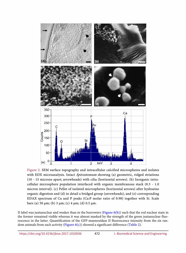
3.2. SEM, EDX and Isolated Mineral Particles
SEM of S. ambiguum confirmed brush borders of cilia upon a geometric pattern of surface ridges (Figure 2(a)) beneath which the location of contractile myonemes has been reported by others (see Dis-cussion). Clusters of contiguous microspheres, each about 1 µm in diameter, were observed in close en-doplasmic association with the ridges (Figure 2(b)) and also near membranous stacks. Cell digestion with hydrazine hydrate released numerous mineralized microspheres, up to 1 µm in diameter (Figure 2(c)), fractionated as a dense pellet. Calcified bridge-like interconnections were found between some of the mi-crospheres to form chains and convoluted assemblies (Figure 2(d)), and their elemental analysis indicated calcium and phosphorus with lesser amounts of silicon (Figure 2(e)); the calcium/phosphate (Ca:P) molar ratio was 0.98 i.e. typical of an amorphous or non-crystalline phase of bone mineral (as is also the presence of Si).
3.3. TC-Mineral Marker Comparison of Tunnelling and Swimming
Staining for calcification of both swimming and burrowing S. ambiguum with TC confirmed two distinct mineral loading states, one high and the other relatively low (Figure 3; Figure 4). In the free-swimming animals (Figure 3(a)) the fluorescent microspheres outlined the unstained shadow of the convoluted me-ganucleus (c.f. the specific meganuclear TOPRO-3 stained configuration in Figure 5(a)). At the same time, a proportion were disseminated throughout the cytoplasm as discrete, micron-sized fluorescent granules and looping or chain-like assemblies, sometimes extended into elongated arrays that appeared to be pre-dominantly transaxial (Figure 4(top)). The fluorophore signal from these swimming ciliates was weak in comparison to those tunnelling (Figure 3(b)) where the domain of juxtanuclear fluorescence was almost masked by the emission strength from the expansive spread of particle populations in a pattern that was predominantly axial (Figure 4(bottom)). Quantification of the TC fluorescence intensity of the six ran-dom animals from each activity (Figure 3(c)) showed a significant difference (Table 1). In summary, the weaker fluorescence observed visually and arithmetically in the swimmers indicated not only fewer miner-al microspheres in comparison to burrowers, but also a pattern that related, on the one hand, to the “string of pearls” contours of a juxtanuclear apparatus least obscured in the swimmers and, on the other, to the geometric arrays most evident during tunnelling, supporting Pautard [1] that mineralization in S. ambi-guum is directly proportional to the amount of burrowing.
3.4. GFP-Mannosidase II-Golgi Marker and Unfused GFP Control
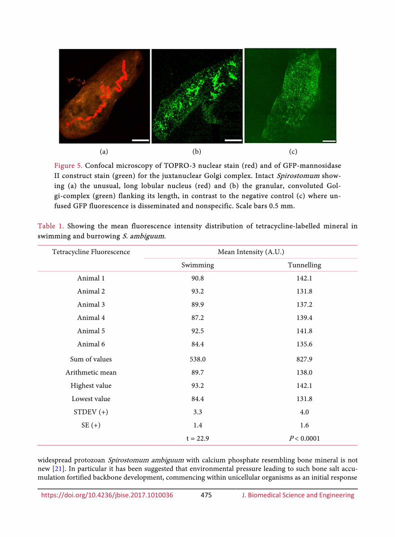
Preliminary treatment of S. ambiguum with the nuclear stain TOPRO-3 confirmed a long lobular macronucleus extending the length of the animal (Figure 5(a)). The novel transfection of S. ambiguum with the mammalian GFP/mannosidase II construct (Figure 5(b)), showed a convoluted pattern of fluores-cence loci flanking the macronuclear area in Figure 5(a) (similar in disposition to mineral fluorescence in Figure 3(a)). Application of unfused GFP control solution to S. ambiguum demonstrated its weak, general random dissemination throughout the organism (Figure 5(c)), supporting the intracellular non-specificity of unfused GFP and confirming the discrete specificity of the mammalian GFP-mannosidase II construct for the Golgi apparatus in its non-mammalian counterpart.
3.5. GFP-Golgi-Marker Comparison of Tunnelling and Swimming
Transfection of both swimming and burrowing organisms with the GFP-mannosidase II construct produced differences in Golgi fluorophore intensity. In the swimmers (Figure 6(a)) the GFP-mannosidase
https://doi.org/10.4236/jbise.2017.1010036 472 J. Biomedical Science and Engineering
Figure 2. SEM surface topography and intracellular calcified microspheres and isolates with EDX microanalysis. Intact Spirostomum showing (a) geometric, ridged striations (10 - 15 microns apart; arrowheads) with cilia (horizontal arrows). (b) Inorganic intra-cellular microsphere population interfaced with organic membranous stack (0.5 - 1.0 micron interval). (c) Pellet of isolated microspheres (horizontal arrows) after hydrazine organic digestion and (d) in detail a bridged group (arrowheads), and (e) corresponding EDAX spectrum of Ca and P peaks (Ca:P molar ratio of 0.98) together with Si. Scale bars (a) 50 µm; (b) 3 µm; (c) 4 µm; (d) 0.5 µm.
II label was juxtanuclear and weaker than in the burrowers (Figure 6(b)) such that the red nuclear stain in the former remained visible whereas it was almost masked by the strength of the green juxtanuclear fluo-rescence in the latter. Quantification of the GFP-mannosidase II fluorescence intensity from the six ran-dom animals from each activity (Figure 6(c)) showed a significant difference (Table 2).

https://doi.org/10.4236/jbise.2017.1010036 473 J. Biomedical Science and Engineering
(a) (b)
(c)
Figure 3. Confocal microscopy of tetracycline stain (yellow) for calcium phosphate/carbonate. Intact Spirostomum showing a convoluted juxtanuc-lear pattern of particulate fluorescence (c.f. with Figure 5(a), Figure 5(b)) in (a) swimmer and (b) burrower; scale bar, 0.5 mm. Corresponding histo-gram (c) with quantified fluorochrome intensity (A.U. arbitrary units) low in swimming, high in tunnelling (whiskers indicate highest and lowest, n = 6; see Table 1).
4. DISCUSSION Natural history records how simple organisms can provide unsuspected insight into conserved an-
cient phenomena such as might be exemplified by an evolved mechanobiology at the soft-to-hard material interface common to both a bone cell at the calcification front and to a calcifying protozoan under extra-neous compression. The inorganic chemistry of bone mineral (crystalline and amorphous) as particulate calcium phosphate (see [25] for references) with carbonate, silicon and various modulating trace elements is fundamental to biological beginnings (see [4, 17, 26] for references). In this regard the association of a

https://doi.org/10.4236/jbise.2017.1010036 474 J. Biomedical Science and Engineering
Figure 4. Stylised central drawing of Spirostomum culture, separating images of tetracycline-stained mineral particle density and alignment (red arrows) in (top quadrant) swimmers with weak transaxial pattern, (bottom quadrant) burrow-ers with strong axial pattern. (A, C) Confocal fluorescence modes with (B) cor-responding plain light, relative to (D) summarising diagram with main stress axis indicated (black arrows). Fluorescent particle dimension approximately 1 micron diameter.
A B
DC
A B
DC
A B
DC
https://doi.org/10.4236/jbise.2017.1010036 475 J. Biomedical Science and Engineering
(a) (b) (c)
Figure 5. Confocal microscopy of TOPRO-3 nuclear stain (red) and of GFP-mannosidase II construct stain (green) for the juxtanuclear Golgi complex. Intact Spirostomum show-ing (a) the unusual, long lobular nucleus (red) and (b) the granular, convoluted Gol-gi-complex (green) flanking its length, in contrast to the negative control (c) where un-fused GFP fluorescence is disseminated and nonspecific. Scale bars 0.5 mm.
Table 1. Showing the mean fluorescence intensity distribution of tetracycline-labelled mineral in swimming and burrowing S. ambiguum.
Tetracycline Fluorescence Mean Intensity (A.U.)
Swimming Tunnelling
Animal 1 90.8 142.1
Animal 2 93.2 131.8
Animal 3 89.9 137.2
Animal 4 87.2 139.4
Animal 5 92.5 141.8
Animal 6 84.4 135.6 Sum of values 538.0 827.9
Arithmetic mean 89.7 138.0
Highest value 93.2 142.1
Lowest value 84.4 131.8
STDEV (+) 3.3 4.0
SE (+) 1.4 1.6
t = 22.9 P < 0.0001
widespread protozoan Spirostomum ambiguum with calcium phosphate resembling bone mineral is not new [21]. In particular it has been suggested that environmental pressure leading to such bone salt accu-mulation fortified backbone development, commencing within unicellular organisms as an initial response

https://doi.org/10.4236/jbise.2017.1010036 476 J. Biomedical Science and Engineering
(a) (b)
(c)
Figure 6. Confocal microscopy of GFP-mannosidase II construct stain (green) for the Golgi complex combined with TOPRO-3 nuclear stain (red). Intact Spi-rostomum (a) swimming when the green juxtanuclear Golgi fluorescence is coin-cidental with, though weaker than, the red lobular nuclear fluorescence; in (b) tun-nelling the green Golgi fluorescence is stronger, masking and expanding beyond the red nucleus. Scale bar 0.5 mm. Corresponding histogram (c) with quantified fluorochrome intensity (A.U. arbitrary units) low in swimming, high in tunnel-ling (whiskers indicate highest and lowest, n = 6; see Table 2).
to store-depleting levels of vital phosphate as competition intensified [24] and supported by the apparent paradox of an inverse relationship between environmental and intracellular P (see [27] for references). Bone salt characteristics have been established by the X-ray diffraction pattern [3] and isotope tracing with 32P and 45Ca [28, 29]. To these previous inorganic observations is now added Si, known to occur in essen-tial traces in bone development (for example [30-32]), the three elements co-located within myriads of

https://doi.org/10.4236/jbise.2017.1010036 477 J. Biomedical Science and Engineering
Table 2. Showing the mean fluorescence intensity distribution of GFP/mannosidase II-labelled Golgi apparatus in swimming and burrowing S. ambiguum.
GFP/Manosidase II Fluorescence Mean Intensity (A.U.)
Swimming Tunnelling
Animal 1 68.5 107.9
Animal 2 77.8 106.2
Animal 3 85.9 103.6
Animal 4 70.1 100.1
Animal 5 75.2 104.7
Animal 6 69.3 105.3
Sum of values 446.8 627.8
Arithmetic mean 74.5 104.6
Highest value 85.9 107.9
Lowest value 68.5 100.1
STDEV (±) 6.7 2.7
SE (±) 2.7 1.1
t = 10.27 P < 0.0001
microspheres and precursor nano-sized objects. Their cytoplasmic numbers reflect the dynamic diversity of the ciliate life cycle and they can be readily isolated from the cells by density fractionation for future analysis of their complex inorganic/organic macromolecular interrelationship and indigenous significance relative to bone (see also [33]).
A prominent mitochondrial presence is consistent with high motility, some authors attributing mi-tochondrial granules to the calcification process [34], and their frequent proximity to the prolific cytop-lasmic vacuoles with cargoes of inorganic objects perhaps facilitating phosphate interchange. The chemical similarity to bone mineral matches the morphological one of populous, enclosed calcified filamentous clusters ([11, 35]), accompanied by smaller uncontained and less complex mineral particles (also found in bone as nanospheres [22, 32]) suggesting a developmental sequence in which the laws of inorganic chemi-stry are modulated by the energetic forces of biology [1]. The calcified particles transported by cytoplasmic ebb and flow may be either independent or interlinked in polarised chains and looped assemblies to influ-ence fluid properties and prevent destruction of the otherwise fragile cell, a remarkable capacity that with-stands the substantial rigour of high pressure chambers [36]. Showers of such particles may be jettisoned by the emerging ciliate. However, in extremis the organisms apparently become so packed with mineral particles that axial flexion is compromised (a phenomenon likened to osteoarthritic stiffening [21]). In marked contrast, after a period of multiplication there follows an immobile (third) phase where the cells hang vertically in fragile, stationary curtains, supported entirely by water and deficient in mineral.
The inception of initially immature, uncalcified objects, which grow attached to membranous enclo-sures and their liberation into the surrounding cytoplasm as mature mineralised entities is reminiscent of algal calcified coccolith formation and orientation. The global unicellular coccolithophorids (essential carbon dioxide-fixing marine algae) have the advantage of particularly distinctive, dumbbell-contoured Golgi mem-branous templates matching their uniquely-shaped calcium carbonate coccoliths and they are of sufficient
https://doi.org/10.4236/jbise.2017.1010036 478 J. Biomedical Science and Engineering
fundamental interest to have given rise to an extensive literature ([12, 13, 37-39] citing P. van der Wal). Sim-ilar random examples include calcified objects in the fresh-water ciliate Euplotes eurystomus [2] and in the hepatopancreas of the snail Helix pomatia [40]. Within such a diverse invertebrate context (mainly cal-cium carbonate) neither the tetracycline-stained calcium phosphate defining the juxtanuclear Golgi of S. ambiguum above nor that similarly demonstrated in the Golgi body of the descendent vertebrate bone cell seem at all extraordinary [7, 15, 41-44].
First characterised in 1898 by its argentophilic property the chameleon-like Golgi apparatus is proba-bly the least understood organelle. However, skeletally there is evidence that its fabrication of calcified mi-crosphere populations may fundamentally influence vertebrate bone microskeletal “quality” with unsuspected clinical implications. By forming calcified objects that are apparently too small in osteoporosis and too large in osteoarthritis [32] it potentially contributes to age-related morbidity and the differential orthopaedic risk of inorganic particle slip, on the one hand, and fatigue microfracture, on the other [45]. The specificity of the fused GFP-mannosidase II construct for the protozoan Golgi apparatus was validated by the con-trasting diffuse and featureless distribution of the unfused GFP-negative control vector. The GFP-mannosidase II construct is typically mammalian and consequently it was predicted to fail in its application to a proto-zoan where there is no literature on resident Golgi proteins. The consistently positive results throughout a series of random individuals support the view that the mannosidase II enzyme or some non-mammalian equivalent is conserved and expressed in a unicellular organism, with attendant evolutionary implications. The unexpected and consistent success of the specific transfection of S. ambiguum demonstrated that sub-jection to burrowing stress raised fluorescence intensity in the Golgi region and confirmed that mineral fabrication events (i.e. as registered by TC-labelled fluorescence) took place in the Golgi complex (i.e. as monitored by GFP-labelled fluorescence). A spatial and temporal synchronicity between the mapped bio-logical markers supported a direct cause and effect association (summarising diagram, Figure 7). Moreo-ver, this biomechanically significant process was a recapitulation of intrinsic events reported earlier in calci-fying colonies of cultured bone cells using exactly the same method with the added benefit of specific mo-lecular Golgi “switches” [7]. Thus Brefeldin A “switched on” bone cell Golgi activity with an immediate resultant rise in mineralogenesis; forscolin “switched off” Golgi activity and mineralogenesis promptly fell in synchrony. These specific observations were enabled by recent advances in cellular technology applied to bone cells for the first time (Figure 8); the implication of these results for skeletal biology concerns the pivotal initiation site of calcification and the transference of its location from the predominantly held do-main of extracellular chemistry to that of a complex intracellular major organelle located within intermit-tently energetic “young” osteocytes.
It has been remarked by others that in this proposed protozoan paradigm for bone cells there will be in a typical, random sample of each, individuals immediately metabolising their calcium phosphate pack-ets while others amass them for storage or strength [10, 26, 46]. In the ciliate, added resilience may be de-rived from rudimentary “musculoskeletal” convergence between mineralized particles and muscle “myo-nemes” widespread below the surface grooves and ridges as “cobweb-like systems of long, narrow myofi-brils” [47-49]. Such mineral conjunction separates the unconstrained and functionally robust S. ambi-guum from the limited activities of other more delicate, contractile ciliates such as the perpetually uncalci-fied Stentor polymorphus that eschews sediment encapsulation, being confined to gentle swimming and interludes of sedentary anchorage for its retractile trumpet-like extension [47]. The difference illustrates the compelling stiffening impact of mineralogenesis within the extraneously pressured silt-dweller, a dis-tinction that perhaps resonates with the transformative dynamics of osteocyte encapsulation at the calcifi-cation front of vertebrate bone [17]. Here initiation of a Golgi-activated mineral loading and unloading cycle has been described in the literature within coherent groups of transitorily “switched on” osteocytes in developing locations, each cycle lasting about 15 min (in situ observations in immature murine calvarium [50]), with intermittent bouts of re-activation in mature bone perhaps mediated by the various other acute stress vehicles reported (e.g. Sharpey’s fibres [51]; integrins, gap junctions, primary cilia, matrix strain, fluid flow [52]; cytoskeletal nanotubules [53].
Despite the mutual relevance of one for the other suggested by the evidence for interchange above
https://doi.org/10.4236/jbise.2017.1010036 479 J. Biomedical Science and Engineering
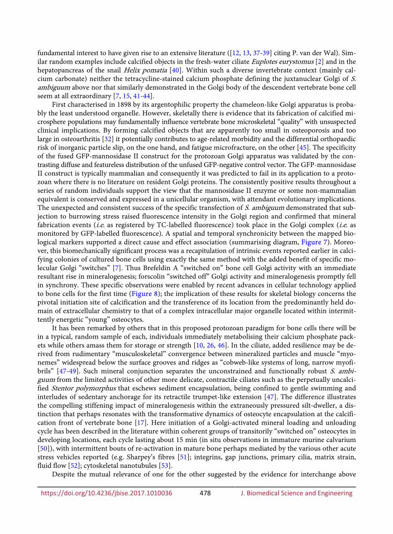
Figure 7. Drawing summarising key features of Golgi-directed, mechano-related mineralogenesis in Spirostomum. (a) In whole organism, typical spiral motion and rear landmark secretory vacuole (sv) and (b) convoluted intact meganuc-leus flanked by juxtanuclear populations of calcified microspheres (about 1 micron diameter). (c) Thin section showing eight nuclear segments with adja-cent Golgi stack and juxtanuclear complexity of vesicles and vacuoles contain-ing calcified objects. (d) Myoneme-associated striated topography with pola-rised chains of calcified microspheres and (e) in detail, magnified microsphere substructure of dense, peripheral radiating filaments, granular centre, with ex-ternal surface bridging and perhaps proliferation by “budding” (Based upon original photomicrographs by undergraduate student Jacqueline Allen).
and consistent with our previous bone cell report, the interpretation of the protozoan perspective on me-tazoan osteocytes may, nevertheless, seem questionable in view of the wide evolutionary separation across a biomineralization field where vertebrate calcification has been traditionally regarded as primarily extra-cellular. On the other hand, the four, central, interrelated factors found using the same techniques (Figure 8; MC3T3-E1 osteocytes) have to be otherwise explained: first is the similar mineral pattern, second is the same Golgi association, third is expression of identical cisternal resident proteins, fourth is intermittent biomechanical initiation. There follows the implication that all mammalian cells in possession of a Golgi
(a) (b)
(c)
(d) (e)
https://doi.org/10.4236/jbise.2017.1010036 480 J. Biomedical Science and Engineering
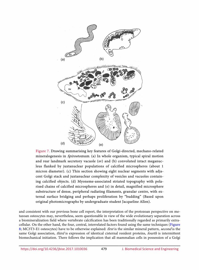
Figure 8. Bone cells in vitro using the same Golgi/mineral staining methods [7] as applied to S. ambiguum. Fluorescent markers of the Golgi body (ar-rowed) showing (left) the distribution of juxtanuclear GFP/mannosidase II construct and (right) the tetracycline mineral stain; demonstrating the si-milarity in fluorescent patterns prevalent in both cases (c.f. Figure 3(a) and Figure 5(b)). Osteocytes a generous gift from Dr. P. Genever, University of York; image courtesy of J. Biomed. Sci. & Eng. [7].
body are thereby equipped with an ancient force-resisting calcification capacity expressed to a lesser or greater degree according to cell type (for which there is substantiating evidence in soft as well as hard tis-sues; see [17] for references), and exhibiting the greatest manifestation in the osteocyte lineage. By virtue of a shared inorganic history at the invertebrate/vertebrate mineralization interface there seems to be as much in common as that which divides and implicit, the widening of an apparently limited academic S. ambiguum horizon to encompass a universal “calcification gene”.
5. CONCLUSION In conclusion, the calcification with phosphate of a stressed protozoan is placed within the context of
an invertebrate mechanobiology conserved in vertebrate bone cells. Previous descriptions of S. ambiguum as a putative “protozoan paradigm for vertebrate calcification” [46] are supported by evidence of a mutual process of Golgi-directed fabrication of compression-resistant, mineralized microsphere populations pola-rised along changing axes of stress. An ancient unicellular mechanism that commenced to protect the in-vertebrates may be conserved beneath the vertebrate bone surface and recapitulated within variably loaded segments of osteocyte syncytium to modulate orthopaedic performance and skeletal restitution. Resultant assemblies of biodynamic objects may be harvested from membranous Golgi containers for insight into a self-organising inorganic/organic primordial partnership [39] that may have engineered the “origin of back-bones” and which may constitute a future portal to innovative, mechanobiological surgical fillers and ar-tificial analogues. Fundamental questions remain about the special nature of the Golgi apparatus in relation to its physicochemical modulation and the identity of factors governing its optimal skeletal performance and failure with age or disease. The incidental encounter noted between contractile myonemes (of unde-fined immunohistochemistry and tensile adhesion) and compression-resistant mineralized microspheres provides a putative bedrock for primitive musculoskeletal integration, further reiterating how the proto-zoan past may enlighten the metazoan present. On the basis of their unique dual geological and biological nature [26] there arises the possibility that the ubiquitous, bacterium-like calcified inclusions [17] that ef-fectively equip a fragile, giant cell to survive adversity may fulfil criteria less accommodating of an inert inorganic precipitate or obscure ultrastructural dense body, and more descriptive of a dynamic early or-ganelle that encloses the unexplored labyrinths envisaged by F.G.E. Pautard.

https://doi.org/10.4236/jbise.2017.1010036 481 J. Biomedical Science and Engineering
ACKNOWLEDGEMENTS V.F. was supported by a Postgraduate Anatomy Scholarship, University of Leeds, P.E.G. by an EPSRC
Postgraduate Studentship and J.E.A. by an EPSRC Challenging Engineering Programme. We are grateful to Dr. Gareth Howell, Microscopy Suite, Leeds University, for confocal microscopy expertise and a supply of unfused Green Fluorescent Protein. The contribution of a number of undergraduate students including Michael Fudge and Sarah Bush is recognised and in particular is Jacqueline Allen whose exceptional light and transmission electron microscopy images were the basis for Figure 7. Dedicated to the pioneering of Dr F.G.E. Pautard, Universities of Leeds and Austin, founding editor of Calcified Tissue Research.
REFERENCES 1. Pautard, F.G.E. (1970) Calcification in Unicellular Organisms. In: Schraer, H., Ed., Biological Calcification: Cel-
lular and Molecular Aspects, Appleton-Century-Crofts, New York, 105-201. https://doi.org/10.1007/978-1-4684-8485-4_4
2. Ruffolo Jr., J.J. (1978) Intracellular Calculi of the Ciliate Protozoon Euplotes eurystomus. Transactions Ameri-can Microscopical Society, 97, 381-386. https://doi.org/10.2307/3225990
3. Pautard, F.G.E. (1958) Bone Salts in Unicellular Organisms. Acta Biochimica Biophysica, 28, 514-520. https://doi.org/10.1016/0006-3002(58)90513-4
4. Pautard, F.G.E. (1962) The Molecular-Biologic Background to the Evolution of Bone. Clinical Orthopaedics, 24, 230-244.
5. Garner, P.E., Wilcox, R., Hordon, L.D. and Aaron, J.E. (2013) Quantification of Cell Networks: Computer-Assisted Method for 3D Mapping of Osteocyte Populations in the Ageing Human Femur. IBMS BoneKEy, 10, Article 333.
6. Vatsa, A., Breuls, R.G., Semeins, C.M., Salmon, P.L., Smit, T.H. and Klein-Nulend, J. (2008) Osteocyte Mor-phology in Fibula and Calvaria—Is There a Role for Mechanosensing? Bone, 43, 452-458. https://doi.org/10.1016/j.bone.2008.01.030
7. Fallon, V., Carter, D.H. and Aaron, J.E. (2014) Mineral Fabrication and Golgi Apparatus Activity in the Mouse Calvarium. Journal of Biomedical Science and Engineering, 7, 769-779. https://doi.org/10.4236/jbise.2014.79075
8. Fallon, V. (2006) The Fabrication of Mineral Particles by Bone Cells and Unicellular Organisms. Ph.D. Disserta-tion, University of Leeds, Leeds.
9. Garner, P.E., Fallon, V. and Aaron, J.E. (2011) Spirostomum ambiguum: A Protozoan Model for Primordial Musculoskeletal Exchange. Bone, 48, S140. https://doi.org/10.1016/j.bone.2011.03.290
10. Pautard, F.G.E. (1981) Calcium Phosphate Microspheres in Biology. Progress in Crystal Growth Characteristics, 4, 89-98. https://doi.org/10.1016/0146-3535(81)90049-6
11. Aaron, J.E., Oliver, B., Clarke, N. and Carter, D.H. (1999) Calcified Microspheres as Biological Entities and Their Isolation from Bone. Histochemical Journal, 31, 455-470. https://doi.org/10.1023/A:1003707909842
12. Manton, I. and Leedale, G.F. (1963) Observations on the Microanatomy of Crystallolithus hyalinus Gaarder and Markali. Archives Mikrobiologica, 47, 115-136. https://doi.org/10.1007/BF00422518
13. Marsh, M.E. (1994) Polyanion-Mediated Mineralization-Assembly and Reorganisation of Acidic Polysaccha-rides in the Golgi System of a Coccolithophorid Alga during Mineral Deposition. Protoplasma, 177, 108-122. https://doi.org/10.1007/BF01378985
14. Kashiwa, H.K. (1970) Calcium Phosphate in Osteogenic Cells. A Critique of the Glyoxal bis (2-hydroxyanil) and the Dilute Silver Acetate Methods. Clinical Orthopaedics, 70, 200-211.
15. Aaron, J.E. and Pautard, F.G.E. (1973) A Cell Cycle in Bone Mineralization. In: Balls, M. and Billett, F.S., Eds.,
https://doi.org/10.4236/jbise.2017.1010036 482 J. Biomedical Science and Engineering
The Cell Cycle in Development and Differentiation, Cambridge University Press, Cambridge, 325-330.
16. Aaron, J.E. (1974) The Development of the Bone Cell and its Role in Mineralization and Resorption. PhD Dis-sertation, University of Leeds, Leeds.
17. Aaron, J.E. (2016) Cellular Ubiquity of Calcified Microspheres: A Matter of Degree, Ancient History and the Golgi Body? Journal of Biomedical Sciences, 5, 1-5. https://doi.org/10.4172/2254-609X.100037
18. Lu, Z., Joseph, D., Bugnard, E., Zaal, K.J.M. and Ralston, E. (2001) Golgi Complex Reorganization during Mus-cle Differentiation: Visualization in Living Cells and Mechanism. Molecular Biology of the Cell, 12, 795-808. https://doi.org/10.1091/mbc.12.4.795
19. Moremen, K.W. and Touster, O. (1986) Topology of Mannosidase II in Rat Liver Golgi Membranes and Release of the Catalytic Domain by Selective Proteolysis. Journal of Biology and Chemistry, 261, 10945-10951.
20. Zaal, K.J.M., Smith, C.L., Polishchuck, R.S., Altan, N., Cole, N.B., Ellenberg, J., Hirschberg, K., Presley, J.F., Ro-berts, T.H., Siggia, E., Phair, R.D., Lippincott and Schwartz, J. (1999) Golgi Membranes Are Absorbed into and Re-Emerge from the ER during Mitosis. Cell, 99, 589-601.
21. Pautard, F.G.E. (1959) Hydroxyapatite as Developmental Feature of Spirostomum ambiguum. Acta Biochimica and Biophysica, 35, 33-46.
22. Carter, D.H., Hatton, P.V. and Aaron, J.E. (1997) The Ultrastructure of Slam-Frozen Bone Mineral. Histochem-ical Journal, 29, 783-793. https://doi.org/10.1023/A:1026425404169
23. Aaron, J.E., Makins, N.B., Francis, R.M. and Peacock, M. (1984) Staining of the Calcification Front in Human Bone using Contrasting Fluorochromes in Vitro. Journal of Histochemstry and Cytochemistry, 32, 1251-1261. https://doi.org/10.1177/32.12.6209330
24. Pautard, F.G.E. (1961) Calcium Phosphate and the Origin of Backbones. New Scientist, 12, 364-366.
25. Dorozhukin, S.V. (2015) Calcium Orthophosphates: Applications in Nature, Biology and Medicine. http://www.amazon.com/Calcium-Orthophosphates-Applications-Biology-Medicine/dp/9814316628
26. Pautard, F.G.E. (1978) Phosphorus and Bone. In: Williams, R.J.P. and Da Silva, J.R.R.F., Eds., New Trends in Bio-Inorganic Chemistry, Academic Press, London, New York, San Francisco, 261-354.
27. Anning, T., Nimer, N., Merrett, M.J. and Brownlee, C. (1996) Costs and Benefits of Calcification in Coccolitho-phorids. Journal of Marine Systems, 9, 45-56.
28. Jones, A.R. (1966) Uptake of 45-Calcium by Spirostomum ambiguum. Journal of Protozoology, 13, 422-428. https://doi.org/10.1111/j.1550-7408.1966.tb01933.x
29. Jones, A.R. (1967) Calcium and Phosphate Accumulation in Spirostomum ambiguum. Journal of Protozoology, 14, 220-225. https://doi.org/10.1111/j.1550-7408.1967.tb01987.x
30. Carlisle, E.M. (1981) Silicon: A Requirement in Bone Formation Independent of Vitamin D. Calcified Tissue International, 33, 27-34. https://doi.org/10.1007/BF02409409
31. Linton, K.M., Tapping, C.R., Adams, D.G., Carter, D.H., Shore, R.C. and Aaron, J.E. (2013) A Silicon Cell Cycle in a Bacterial Model of Calcium Phosphate Mineralogenesis. Micron, 44, 419-432.
32. Linton, K.M., Hordon, L.D., Shore, R.C. and Aaron, J.E. (2014) Bone Mineral “Quality”: Differing Characteris-tics of Calcified Microsphere Populations at the Osteoporotic and Osteoarthritic Femoral Articulation Front. Journal of Biomedical Science and Engineering, 7, 739-755. https://doi.org/10.4236/jbise.2014.79073
33. Hirschman, P.N. and Nichols, G. (1972) The Isolation and Partial Characterization of a Calcium-Rich Particu-late Fraction from Bone Cells. Calcified Tissue Research, 9, 67-79. https://doi.org/10.1007/BF02061946
34. Matthews, J.L., Martin, J.H., Sampson, H.W., Kunin, A.S. and Roan, J.H. (1970) Mitochondrial Granules in the Normal and Rachitic Rat Epiphysis. Calcified Tissue Research, 5, 91-99. https://doi.org/10.1007/BF02017539

https://doi.org/10.4236/jbise.2017.1010036 483 J. Biomedical Science and Engineering
35. Aaron, J.E. and Pautard, F.G.E. (1972) Ultrastructural Features of Phosphate in Developing Bone Cells. Israel Journal of Medical Science, 8, 625-629.
36. Bien, S.M. (1967) High Hydrostatic Pressure Effects on Spirostomum ambiguum. Calcified Tissue Research, 1, 170-172. https://doi.org/10.1007/BF02008087
37. Outka, D.E. and Williams, D.C. (1971) Sequential Coccolith Morphogenesis in Hymenomonas carterae. Journal of Protozoology, 18, 285-297. https://doi.org/10.1111/j.1550-7408.1971.tb03319.x
38. Pienaar, R.N. (1994) Ultrastructure and Calcification of Coccolithophores. In: Winter, A. and Siesser, W.G., Eds., Coccolithophores, Cambridge University Press, Cambridge, 13-38.
39. Young, J.R., Davis, S.A., Bown, P.R. and Mann, S. (1999) Coccolith Ultrastructure and Biomineralization. Jour-nal of Structural Biology, 126, 195-215. https://doi.org/10.1006/jsbi.1999.4132
40. Abolins-Krogis, A. (1970) Electron Microscope Studies of the Intracellular Origin and Formation of Calcifying Granules and Calcium Spherites in the Hepatopancreas of the Snail Helix pomatia. L.Z.Zellforsch, 108, 501-515. https://doi.org/10.1007/BF00339656
41. Aaron, J.E. (1973) Osteocyte Types in the Developing Mouse Calvarium. Calcified Tissue Research, 12, 259-279. https://doi.org/10.1007/BF02013740
42. Aaron, J.E. (1978) Histological Aspects of the Relationship between Vitamin D and Bone. In: Lawson, D.E.M., Ed., Vitamin D, Academic Press, London, 201-265.
43. Park, H.Z. and Kashiwa, H.K. (1975) Calcium Localized in Juxtanuclear Granules of Epiphyseal Chondrocytes with a Dilute Glyoxal bis(2-hydroxyanil) Solution. Calcified Tissue Research, 19, 189-197. https://doi.org/10.1007/BF02564003
44. Chang, Y.L., Stanford, C.M. and Keller, J.C. (2000) Calcium and Phosphate Supplementation Promotes Bone Cell Mineralization: Implications for Hydroxyapatite (HA)-Enhanced Bone Formation. In: Lawson, D.E.M., Ed., Journal of Biomedical Material Research, 52, 270-278. https://doi.org/10.1002/1097-4636(200011)52:2<270::AID-JBM5>3.0.CO;2-1
45. Aaron, J.E. (2003) Bone Turnover and Microdamage. Advances in Fracture Management, 2, 102-110.
46. Pautard, F.G.E. and WILLIAMS, R.J.P. (1982) Biological Minerals. Chemistry in Britain, 18, 14-16.
47. Randall, J.T. and Fitton-Jackson, S. (1958) Fine Structure and Function in Stentor polymorphus. Journal of Bi-ophysics, Biochemistry and Cytology, 4, 807-832. https://doi.org/10.1083/jcb.4.6.807
48. Yagiu, R. and Shigenaka, Y. (1963) Electron Microscopy of the Longitudinal Fibrillar Bundle and the Contractile Fibrillar System in Spirostoum ambiguum. Journal of Protozoology, 10, 364-369. https://doi.org/10.1111/j.1550-7408.1963.tb01689.x
49. Lehman, W.J. and Rebhun, L.I. (1971) The Structural Elements Responsible for Contraction in the Ciliate Spi-rostomum. Protoplasma, 72, 153-178. https://doi.org/10.1007/BF01279048
50. Aaron, J.E. and Pautard, F.G.E. (1973) Dynamic Studies of Bone in Vivo Incident Light Interference Contrast Microscopy. In: Czitober, H. and Eschberger, J., Eds., Calcified Tissues, Facta Publications, Vienna, 197-201.
51. Aaron, J.E. (2012) Periosteal Sharpey’s Fibers: A Novel Bone Matrix Regulatory System? Frontiers in Endocri-nology, 3, 1-10.
52. Vaughan, T.J., Verbruggen, S.W. and McNamara, L.M. (2013) Are All Osteocytes Equal? Multiscale Modelling of Cortical Bone to Characterise the Mechanical Stimulation of Osteocytes. International Journal of Numerical Methods in Biomedical Engineering, 29, 1361-1372. https://doi.org/10.1002/cnm.2578
53. Ingber, D.E. (2003) Tensegrity I. Cell Structure and Hierarchical Systems Biology. Journal of Cell Science, 116, 1157-1173. https://doi.org/10.1242/jcs.00359