MIG-10 and CSN-5 interactions during nervous system ...€¦ · Ena/VASP proteins are actin...
35
Submitted to: Professor Elizabeth Ryder, Advisor MIG-10 and CSN-5 interactions during nervous system development in C. elegans Major Qualifying Project Report submitted to the Faculty of Worcester Polytechnic Institute in partial fulfillment of requirements for the Degree of Bachelor of Science Submitted by: Roxanne Tymon 4/25/2013 This report represents work of a WPI undergraduate student submitted to the faculty as evidence of a degree requirement. WPI routinely publishes these reports on its web site without editorial or peer review. For more information about the projects program at WPI, see http://www.wpi.edu/Academics/Projects.
Transcript of MIG-10 and CSN-5 interactions during nervous system ...€¦ · Ena/VASP proteins are actin...

Submitted to:
Professor Elizabeth Ryder, Advisor
MIG-10 and CSN-5 interactions during nervous system
development in C. elegans Major Qualifying Project Report submitted to the Faculty of Worcester Polytechnic Institute in
partial fulfillment of requirements for the Degree of Bachelor of Science Submitted by:
Roxanne Tymon
4/25/2013
This report represents work of a WPI undergraduate student submitted to the faculty as evidence
of a degree requirement. WPI routinely publishes these reports on its web site without editorial
or peer review. For more information about the projects program at WPI, see
http://www.wpi.edu/Academics/Projects.
2
Table of Contents Table of Figures ............................................................................................................................................. 3
Abstract ......................................................................................................................................................... 4
Acknowledgements ....................................................................................................................................... 5
Introduction .................................................................................................................................................. 6
Cell Migration, Axon Guidance, and Actin Polymerization ....................................................................... 6
C. elegans as a Model Organism ............................................................................................................... 8
MIG-10 and its Role in Axon Guidance and Cell Migration....................................................................... 9
ABI-1 and its Role in Actin Polymerization .............................................................................................. 11
Interactions Between ABI-1 and MIG-10 ................................................................................................ 13
CSN-5 ....................................................................................................................................................... 14
Project Goals ........................................................................................................................................... 15
Methods ...................................................................................................................................................... 16
Molecular Biology ................................................................................................................................... 16
PCR ...................................................................................................................................................... 16
BP/LR Reactions and Product Analysis ................................................................................................ 17
Growing CSN-5::GFP, MIG-10A::V5, and MIG-10 C-terminus::V5 Expression Clone Cultures ............ 17
Biochemistry ........................................................................................................................................... 18
Cell Maintenance ................................................................................................................................ 18
Transfections ....................................................................................................................................... 18
Co-immunoprecipitation ..................................................................................................................... 18
Western Blotting ................................................................................................................................. 19
Results ......................................................................................................................................................... 21
MIG-10 and CSN-5 Interactions .............................................................................................................. 21
Possible MIG-10 Binding Domains .......................................................................................................... 23
Discussion.................................................................................................................................................... 27
References .................................................................................................................................................. 29
Appendix ..................................................................................................................................................... 31
Appendix A: Buffer Solutions .................................................................................................................. 31
Appendix B: Expression Clone Maps with Restriction Enzyme Cut Sites ................................................ 32
Appendix C: Additional Western Blot of MIG-10 and CSN-5 Interactions .............................................. 35

3
Table of Figures
Figure 1: Axon Growth Cone (Sanes et al., 2005). ........................................................................................ 8
Figure 2: MIG-10 Isoforms (McShea, 2011). ............................................................................................... 10
Figure 3: Schematic of MIG-10 Pathway (McShea, 2011). ......................................................................... 11
Figure 4: ABI-1 Domains (McShea, 2011). .................................................................................................. 12
Figure 5: Actin Polymerization (Disanza et al., 2005). ................................................................................ 13
Figure 6: CSN-5 domains (Shackleford and Claret, 2010) ........................................................................... 14
Figure 7: Model of ubiquitin ligases activity (Cope and Deshaies, 2003). .................................................. 15
Figure 8: Co-IP and Western Blot Schematic of MIG-10 and CSN-5 Interactions (Adapted from Sullender,
2012) ........................................................................................................................................................... 22
Figure 9: MIG-10 and CSN-5 Interaction. ................................................................................................... 23
Figure 10: Gateway Cloning System (Sullender, 2012). .............................................................................. 24
Figure 11: No expression of MIG-10 A and B N-terminus in whole cell lysates. ........................................ 25
Figure 12: MIG-10 C-terminus possibly not interacting with CSN-5.. ......................................................... 26
4
Abstract
Mutations in genes involved in nervous system development can result in disorders such as
Down’s syndrome and autism. MIG-10 is a cytoplasmic adaptor protein in the model system C.
elegans that functions in neuronal migration and axon guidance during development. To study
possible interactions between MIG-10 and candidate protein CSN-5, co-immunoprecipitation
and western blotting were performed. An interaction between MIG-10 and CSN-5 was
confirmed, suggesting that CSN-5 is needed to stabilize MIG-10.

5
Acknowledgements
I would like to thank Charu Jain for her assistance with maintaining cells and also
additional help with the Co-IP and Westerns throughout my MQP. Also, I would like to thank
the Duffy Lab for their cooperation with equipment and supplies.
Thank you, Erin Flaherty for her support while in lab and continued interest in my
project. It made lab more enjoyable having a friendly face around.
I would especially like to thank Meagan Sullender and Erin Maloney for giving me a
foundation in lab by training me so that I could do it on my own throughout my MQP. I’m
especially thankful for the many answered questions and support they provided me throughout
the year. And a special thank you to Meagan for the constructs.
A huge thank you goes to Liz Ryder for being such a dedicated advisor. I’m grateful for
the time she spent answering my questions, helping me troubleshoot, and preparing me for my
future career. She taught me how to be extremely efficient in lab which is an important trait I
definitely will need throughout my career. It truly amazes me how she manages to do so much
and keep everything straight (of course with some friendly reminders).

6
Introduction
The nervous system is a complex series of circuits that come together to allow organisms
to feel sensations, think, and generate behaviors. A major component of the nervous system is
the brain which through its billions of cells allows organisms to function. Within the brain lie
neurons that send messages throughout the body. In order to understand how the nervous system
functions, it is important to comprehend how it develops. The complicated interconnections of
the nervous system cannot function properly if they do not form appropriately during the
beginning of development (Sanes et al., 2005). Defects in nerve connections can lead to
neurological disorders such as Down’s syndrome and autism. Further research in developmental
neuroscience could lead to treatment of neurodegenerative diseases or injury caused by damaged
neuronal connections (Quinn and Wadsworth, 2008). Additionally, inappropriate cell migration
can lead to mental disorders, cancer, vascular diseases, or dysfunctional homeostatic processes.
The study of molecular biology and biotechnology is critical to understanding these problems
and developing an approach to solve them (Horwitz and Webb, 2003). Specifically, this project
studied the MIG-10 protein, which functions in cell migration, and a potential interactor, CSN-5,
which functions in protein stability and degradation among additional areas that continue to be
researched.
Cell Migration, Axon Guidance, and Actin Polymerization Developmental neurobiology is a large area of study. Two specific areas of interest are
cell migration and axon guidance. Within the brain, histogenesis is of great importance for a
properly functioning nervous system. Neurons and glial cells are formed during embryogenesis
in one location and must be moved to their final location within the complex circuitry of the
nervous system. Cell migration is a key element in this process (Sanes et al., 2005).
Embryogenesis begins the process of forming the cells, and then the cells move to their target
location where differentiation occurs into a particular cell type. The process of cell migration
occurs as a result of signals which then cause polarization and protrusion of the cell.
Phosphatidylinositol (3,4,5) trisphosphate (PIP3) is an initial component of this pathway. The
polarization and accumulation of PIP3 leads to the activation of Rho GTPases. Rac, a member of
the Rho family GTPases, is a regulator of actin polymerization which is important in the

7
protrusion of the cell during migration. In neurons, an important element in this process is the
growth cone at the tip of the axon (Horwitz and Webb, 2003).
The circuitry of the nervous system is very complex, and it is important during the
development of the neurons to make the right connections. Axon growth begins with growth
cones which are located at the tip of the axon. Guidance cues are needed to activate the growth
cone in order to move towards or away from a target location. Examples of guidance cues are
netrins, slits, bone morphogenic proteins (BMPs), wnts, ephrins, and semaphorins. Asymmetric
axon outgrowth is guided by actin and microtubules as a response to these guidance cues (Quinn
and Wadsworth, 2008). Actin polymerization is guided by the extracellular cues functioning as
attractants or repellants. This therefore directs the orientation of axon growth due to whether the
growth cone is promoted or inhibited by the guidance cues (Quinn et al., 2006). The structure of
the growth cone is shown in Figure 1. Lamellipodia are considered to be at the edges with
filopodial projections extending outward. These components contain actin which is important in
cell movement and guidance. Ena/VASP proteins are actin regulators which function in response
to the attractive and repulsive guidance cues (Sanes et al., 2005). These proteins are important in
actin polymerization and can be found at the leading edge of filopodia and lamellipodia of the
growth cone (Quinn et al., 2006). The extension of actin filaments is promoted by Ena/VASP
proteins which prevent capping proteins from terminating growth along the barbed ends of the
actin (Chang et al., 2006).

8
Figure 1: Axon Growth Cone (Sanes et al., 2005). The leading edge of the axon is composed of filopodia and lamellipodia which are structurally supported by actin.
C. elegans as a Model Organism Model organisms are crucial to scientific research. Caenorhabditis elegans is a model
organism often used in neurobiology. This nematode worm is easy to work with, reproduces
rapidly, and has a small genome (Hart and Bernstein, 2000). C. elegans is especially useful
within the study of neuroscience because of the animals’ simple nervous system composed of
only 302 neurons and 56 glial cells (Sanes et al., 2005). These features allow for an easier study
during scientific research. C. elegans and humans share similar genes providing a better
understanding of humans without the process being time-consuming or unethical.
9
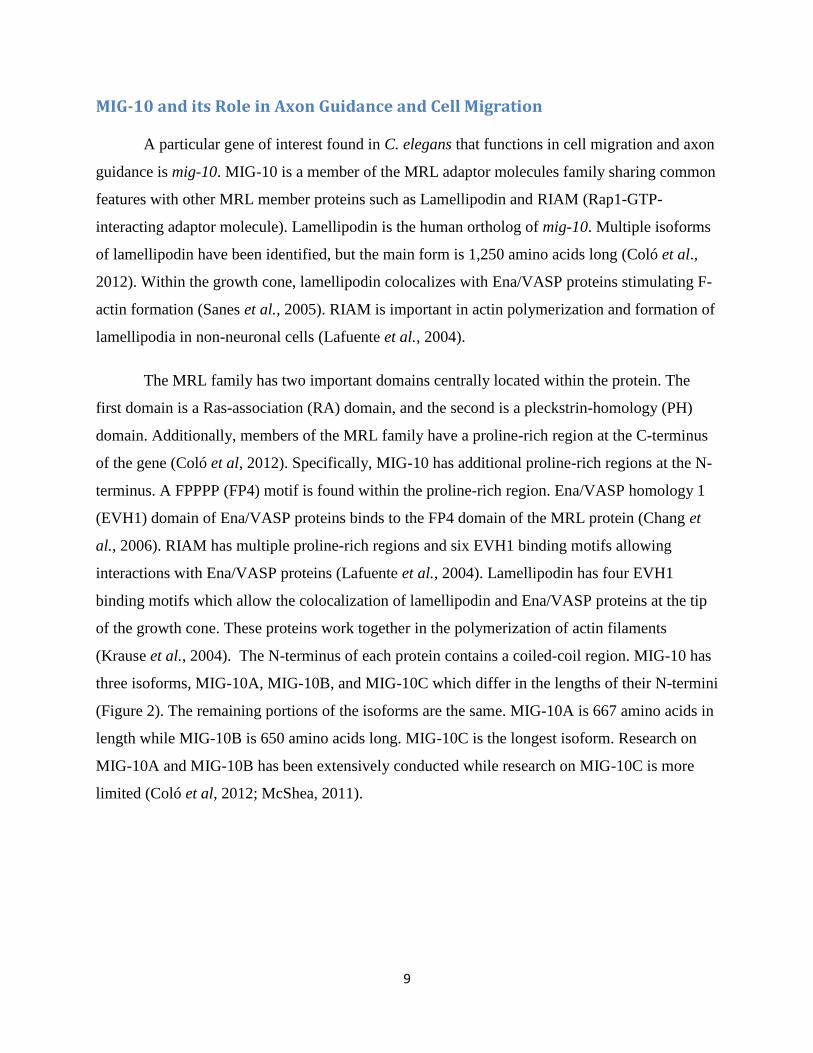
MIG-10 and its Role in Axon Guidance and Cell Migration
A particular gene of interest found in C. elegans that functions in cell migration and axon
guidance is mig-10. MIG-10 is a member of the MRL adaptor molecules family sharing common
features with other MRL member proteins such as Lamellipodin and RIAM (Rap1-GTP-
interacting adaptor molecule). Lamellipodin is the human ortholog of mig-10. Multiple isoforms
of lamellipodin have been identified, but the main form is 1,250 amino acids long (Coló et al.,
2012). Within the growth cone, lamellipodin colocalizes with Ena/VASP proteins stimulating F-
actin formation (Sanes et al., 2005). RIAM is important in actin polymerization and formation of
lamellipodia in non-neuronal cells (Lafuente et al., 2004).
The MRL family has two important domains centrally located within the protein. The
first domain is a Ras-association (RA) domain, and the second is a pleckstrin-homology (PH)
domain. Additionally, members of the MRL family have a proline-rich region at the C-terminus
of the gene (Coló et al, 2012). Specifically, MIG-10 has additional proline-rich regions at the N-
terminus. A FPPPP (FP4) motif is found within the proline-rich region. Ena/VASP homology 1
(EVH1) domain of Ena/VASP proteins binds to the FP4 domain of the MRL protein (Chang et
al., 2006). RIAM has multiple proline-rich regions and six EVH1 binding motifs allowing
interactions with Ena/VASP proteins (Lafuente et al., 2004). Lamellipodin has four EVH1
binding motifs which allow the colocalization of lamellipodin and Ena/VASP proteins at the tip
of the growth cone. These proteins work together in the polymerization of actin filaments
(Krause et al., 2004). The N-terminus of each protein contains a coiled-coil region. MIG-10 has
three isoforms, MIG-10A, MIG-10B, and MIG-10C which differ in the lengths of their N-termini
(Figure 2). The remaining portions of the isoforms are the same. MIG-10A is 667 amino acids in
length while MIG-10B is 650 amino acids long. MIG-10C is the longest isoform. Research on
MIG-10A and MIG-10B has been extensively conducted while research on MIG-10C is more
limited (Coló et al, 2012; McShea, 2011).

10
Figure 2: MIG-10 Isoforms (McShea, 2011). The three isoforms differ only in the N-terminus. The RA and PH domains can be seen in the center with a FP4 domain on each end of the gene.
Research has shown that in C. elegans, mig-10 is important during embryogenesis and
the cell migration of embryonic neurons. Two mutations of mig-10, ct41 and e2527, have been
identified which result in the truncation of the excretory canal of the excretory cell within
developing C. elegans. Shortened migration of CAN, ALM, and HSN embryonic neurons was
also an effect in these two mutants (Manser et al., 1997). In additional experiments, over-
expression of MIG-10 in C. elegans AVM neuron, without guidance cues, has shown misguided
axon outgrowth. Further experiments with the addition of guidance cues such as netrin and slit
showed enhanced axon guidance (Quinn and Wadsworth, 2008).
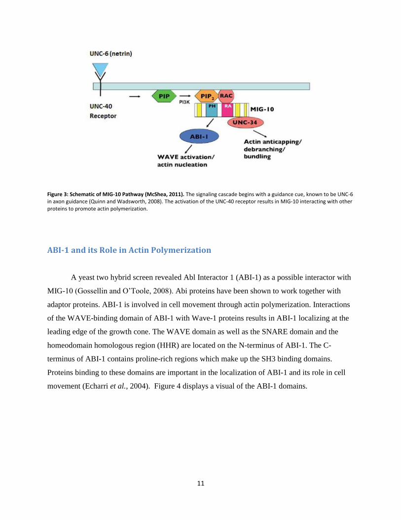
Figure 3 provides a model of the role MIG-10 plays in cell migration and actin
polymerization. In this example, netrin, a guidance cue, is recognized by its receptor UNC-40 at
the membrane of the leading edge of the growth cone. This binding triggers the phosphorylation
of PIP to PIP2 , and the activation of RAC (GTPase), which then recruit MIG-10 to the
membrane. Once bound, MIG-10 can bind to other proteins which are involved in actin
polymerization, axon guidance, or cell migration. Here, ABI-1 is displayed as an interactor of
MIG-10 (Sullender, 2012). UNC-34 is the sole C. elegans homolog of the Ena/VASP proteins
(Quinn et al., 2006). In this signaling cascade, MIG-10 represents an effector protein which
binds to RAC. The asymmetric activation of RAC and phosphorylation of PIP is what brings
MIG-10 to the membrane and allows binding through the RAPH domain. As a result,
lamellipodia and filopodia are formed asymmetrically during actin polymerization, allowing
outgrowth in a particular direction (Quinn and Wadsworth, 2008).
11
Figure 3: Schematic of MIG-10 Pathway (McShea, 2011). The signaling cascade begins with a guidance cue, known to be UNC-6 in axon guidance (Quinn and Wadsworth, 2008). The activation of the UNC-40 receptor results in MIG-10 interacting with other proteins to promote actin polymerization.
ABI-1 and its Role in Actin Polymerization
A yeast two hybrid screen revealed Abl Interactor 1 (ABI-1) as a possible interactor with
MIG-10 (Gossellin and O’Toole, 2008). Abi proteins have been shown to work together with
adaptor proteins. ABI-1 is involved in cell movement through actin polymerization. Interactions
of the WAVE-binding domain of ABI-1 with Wave-1 proteins results in ABI-1 localizing at the
leading edge of the growth cone. The WAVE domain as well as the SNARE domain and the
homeodomain homologous region (HHR) are located on the N-terminus of ABI-1. The C-
terminus of ABI-1 contains proline-rich regions which make up the SH3 binding domains.
Proteins binding to these domains are important in the localization of ABI-1 and its role in cell
movement (Echarri et al., 2004). Figure 4 displays a visual of the ABI-1 domains.

12
Figure 4: ABI-1 Domains (McShea, 2011). The SNARE and HHR domains can be seen on the N-terminus of ABI-1. The SH3 binding domain can be seen on the C-terminus. The middle portion of ABI-1 contains serine and proline-rich regions.
Actin polymerization transforms the globular actin protein into a helical structure as
shown in Figure 5 (Disanza et al., 2005). The helical filaments have a fast and a slow growing
end which are affected by polarization. G-actin begins the process at the growing end (positive
end). Then, ATP hydrolysis plays a key role as ATP-actin is converted to ADP-actin. This slow
process ends with the depolymerization of the actin filaments. The Arp2/3 complex and the
WAVE complex interact and localize at the plasma membrane to begin branching of actin
filaments. Capping proteins attach to portions of the actin filaments after branching has begun to
prevent further elongation (Disanza et al., 2005). ABI-1 binds to Wave-1 through its wave-
binding domain at the amino terminus. Once bound, the complex can localize at the
lamellipodium. Mutations in the wave-binding domain within ABI-1 prevent the localization of
Wave-1 at the leading edge of the growth cone. Wave-1 is needed to activate Arp2/3 which
initiates actin filament branching. If Wave-1 does not localize at the lamellipodium, actin
polymerization will be disrupted (Echarri et al., 2004).

13
Figure 5: Actin Polymerization (Disanza et al., 2005). Model of actin formation and the components involved.
Interactions Between ABI-1 and MIG-10 Past research has been conducted displaying that MIG-10 binds ABI-1. Co-
immunoprecipitation experiments have isolated specific regions of each protein to target the
particular binding domains of the proteins. The SH3 domain of the C-terminus of ABI-1 has
been shown to be important in binding MIG-10. As a comparison, the middle portion of ABI-1
lacking the SH3 domain was analyzed and did not bind MIG-10 (McShea et al., 2013).
Preliminary co-immunoprecipitation experiments have been conducted to examine the possible
interactions between the C-terminus or the middle portion (RAPH domain) of MIG-10 and ABI-
1. These preliminary results have shown that the C-terminus as well as the RAPH domain of
MIG-10 binds ABI-1. The N-terminus of MIG-10 has not been studied to examine its individual
interactions with ABI-1 within axon guidance.

14
CSN-5 The COP9 signalosome (CSN) is a complex of eight subunits that is involved in protein
stability, gene transcription, modulating signal transduction, cell-cycle control, and DNA-
damage response (Shackleford and Claret, 2010). One particular subunit of interest is CSN-5
which is highly conserved. CSN-5 is 334 amino acids long and contains a MPN domain (Figure
6) unlike a majority of the subunits which have a PCI domain (Wei and Deng, 2003). The MPN
domain contains a JAMM motif (metalloenzyme) which together are important for protein
interactions and enzymatic activity such as degradation (Shackleford and Claret, 2010). The
primary function of CSN-5 is the degradation of proteins involved in the ubiquitin-mediated
proteolysis pathway (Zhou and Wee, 2003). The MPN domain of CSN-5 is important in the
removal of Nedd8 from cullin-based E3 ubiquitin ligases (Figure 7). Additionally, CSN-5 acts as
a transcriptional co-activator by playing a role in protein stability (Chamovitz, 2009). CSN-5 has
been shown to stabilize c-Jun (member of activating protein-1 complex) and hypoxia inducible
factor α (HIF-1α) (Shackleford and Claret, 2010). Research in muscle cells of C. elegans has
shown that CSN-5 both stabilizes and degrades M-line proteins, such as UNC-96 and UNC-98
respectively (Miller et al, 2009).
Figure 6: CSN-5 domains (Shackleford and Claret, 2010). The largest and most important domain of CSN-5 is the MPN domain which contains a JAMM motif. This domain plays a role in protein interactions and degradation.

15
Figure 7: Model of ubiquitin ligases activity (Cope and Deshaies, 2003). CSN-5 is needed to remove Nedd8 from the ubiquitin ligases so CAND1 can be recruited. This cycle is needed for protein degradation.
Project Goals The first goal of this project was to study a possible interaction between MIG-10 and
CSN-5. A yeast-two hybrid screen revealed CSN-5 as a possible interactor of MIG-10 (Gossellin
and O’Toole, 2008). Further research was conducted in vitro through western blotting and co-
immunoprecipitation to provide more evidence of this protein interaction. A lack of time and
repetition of experiments resulted in inconclusive results (Sullender, 2012). In the current
project, research was continued to further examine the possible interaction between MIG-10 and
CSN-5, and how they play a role in axon guidance or cell migration. Experiments were
conducted using constructs from previous research through co-immunoprecipitation experiments
and western blotting.
The second goal of this project was to determine which specific domains of MIG-10 are
involved in binding CSN-5. The N-termini of MIG-10 A and B were cloned into insect vectors
with a V5 tag. Regions shown to be involved in the binding ability of MIG-10 such as proline-
rich regions are lacking in the N-terminus of MIG-10B. Additionally, a previously cloned
construct of the C-terminus of MIG-10 was investigated. Experiments were conducted to
determine whether interactions occur between each MIG-10 domain (N-terminus and C-
terminus) and CSN-5.
16
Methods
Molecular Biology
PCR
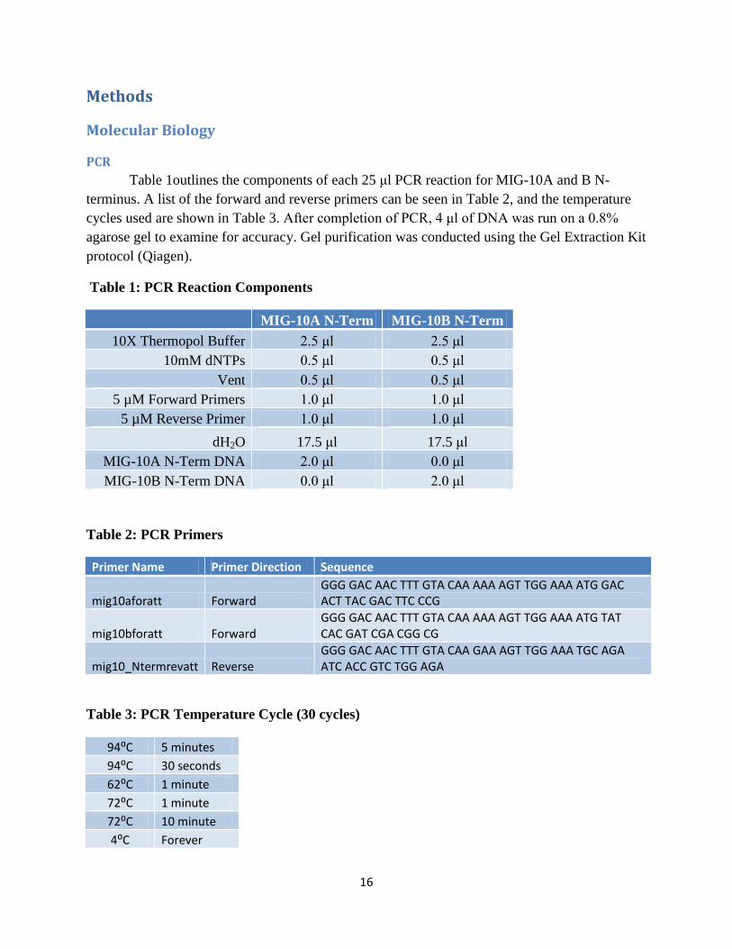
Table 1outlines the components of each 25 μl PCR reaction for MIG-10A and B N-
terminus. A list of the forward and reverse primers can be seen in Table 2, and the temperature
cycles used are shown in Table 3. After completion of PCR, 4 μl of DNA was run on a 0.8%
agarose gel to examine for accuracy. Gel purification was conducted using the Gel Extraction Kit
protocol (Qiagen).
Table 1: PCR Reaction Components
MIG-10A N-Term MIG-10B N-Term
10X Thermopol Buffer 2.5 μl 2.5 μl
10mM dNTPs 0.5 μl 0.5 μl
Vent 0.5 μl 0.5 μl
5 µM Forward Primers 1.0 μl 1.0 μl
5 µM Reverse Primer 1.0 μl 1.0 μl
dH2O 17.5 μl 17.5 μl
MIG-10A N-Term DNA 2.0 μl 0.0 μl
MIG-10B N-Term DNA 0.0 μl 2.0 μl
Table 2: PCR Primers
Primer Name Primer Direction Sequence
mig10aforatt Forward GGG GAC AAC TTT GTA CAA AAA AGT TGG AAA ATG GAC ACT TAC GAC TTC CCG
mig10bforatt Forward GGG GAC AAC TTT GTA CAA AAA AGT TGG AAA ATG TAT CAC GAT CGA CGG CG
mig10_Ntermrevatt Reverse GGG GAC AAC TTT GTA CAA GAA AGT TGG AAA TGC AGA ATC ACC GTC TGG AGA
Table 3: PCR Temperature Cycle (30 cycles)
94⁰C 5 minutes
94⁰C 30 seconds
62⁰C 1 minute
72⁰C 1 minute
72⁰C 10 minute
4⁰C Forever

17
BP/LR Reactions and Product Analysis
Gel purified PCR products were used in the Gateway Cloning System to produce
constructs for co-immunoprecipitation and western blotting. An outline of this cloning system
can be seen in Figure 10 in the Results. BP and LR reactions are performed similarly, except for
a few minor differences which are highlighted below. A mixture with a total reaction volume of
8 µl containing 15-150 ng of PCR product (BP) or 50-150 ng of Entry clone (LR), 1 μl of Donor
vector (BP: 150 ng/μl) or Destination vector (LR: 150 ng/μl), 1-3 μl of TE buffer (pH 8), and 2
μl of BP Clonase II or LR Clonase II enzyme (Invitrogen) was briefly vortexed,
microcentrifuged, and incubated at 25⁰C. After 1 hour, 1 μl of Proteinase K solution (Invitrogen)
was added and placed at 37⁰C for 10 minutes. Then, the DNA was transformed by adding 5 μl of
the BP or LR Reaction to 50 μl of maximum efficiency bacterial cells (New England Biolabs)
and incubated on ice for 30 minutes. The cells were then heat shocked for 30 seconds in a 42⁰C
water bath followed by the addition of 400 μl of SOC. After 3 hours gently rotating on a nutator
at 37⁰C, 50 μl and 150 μl samples were each spread on a 50 µg/ml Kan (BP) or 50 µg/mL Amp
(LR) plate and allowed to incubate at 37⁰C overnight. The next day, 5 individual colonies were
picked from each plate using a pipette tip which was ejected into a conical tube of 5 mL of liquid
LB media + 50 µg/mL Kan (BP) or 50 µg/mL Amp (LR). After rotating overnight, DNA was
isolated by using a miniprep kit (Qiagen). To verify that the samples were correct, a digest was
performed for each reaction for 2 hours at 37⁰C. The BP entry clone digestions combined 2 μl of
isolated DNA with 1 μl of BanII, 15 μl of dH2O, and 2 μl of 10X Buffer 4. LR expression clone
digestions contained 2 μl of isolated DNA, 1 μl of BamHI, 13 μl of dH2O, 2 μl of Buffer 3, and
2 μl of BSA. After digestion, ~18 μl of DNA was run on a 0.8% agarose gel and observed for
accuracy. Additionally, a sample was sent out for sequencing using GENEWIZ protocol to
analyze the accuracy of the sequence of interest.
Growing CSN-5::GFP, MIG-10A::V5, and MIG-10 C-terminus::V5 Expression Clone Cultures
A frozen glycerol stock of CSN-5::GFP, MIG-10A::V5, and MIG-10 C-terminus::V5,
each in an insect expression vector from a previous MQP (Sullender, 2012) were individually
streaked on a 50 µg/mL Amp plate and left overnight at 37⁰C. Colonies were picked and a
miniprep was performed as stated for the BP/LR Reactions. A restriction digest was performed
by mixing 2 μl of isolated DNA, 1 μl of HindIII, 15 μl of dH2O, and 2 μl of Buffer 2 for each
expression clone. After 2 hours of digestion at 37⁰C, ~18 μl of DNA was run on a 0.8% agarose

18
gel and observed for accuracy. Additionally, a sample of each clone was sent out for sequencing
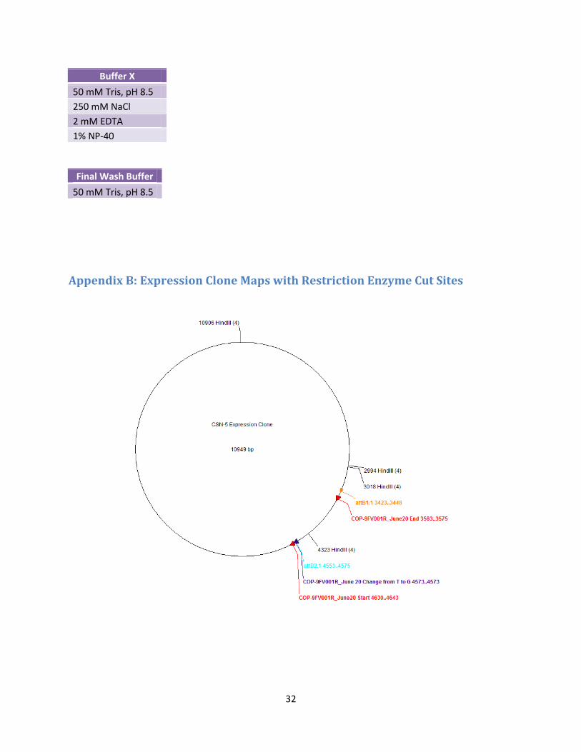
using GENEWIZ protocol to analyze the accuracy of the sequence of interest. Maps of the
expression clones can be seen in Appendix B.
Biochemistry
Cell Maintenance
Two flasks of an S3 cell line of D. melanogaster cells were maintained throughout the
project. The cells were fed with Schneider’s media + 12.5% FBS and kept at 25⁰C. Every 3-4
days cells were split using a 1:5 dilution. All cell culture work was conducted in a tissue culture
hood using sterile technique.
Transfections
In preparation for western blotting, 2 mL of a 1:5 dilution of the parent flask was plated
into each well of a six-well plate. After 3 days at 25⁰C, the plate was transfected with the
preferred DNA samples using the Effectene reagent kit (Qiagen). A dilution of the DNA samples
with TE buffer was prepared before transfection to reach an ideal final concentration of 66.6
ng/μl. Then, 2 μl of each desired expression clone DNA was mixed with 4 μl of Arm-GAL4
DNA (66.6 ng/ μl) and the appropriate amounts of reagents from the transfection kit following
the Qiagen protocol. The actual concentrations of DNA were below the desired amount of 66.6
ng/μl for many experiments. Lastly, 600 μl of Schneider’s media + 12.5% FBS was combined
with the mixture and added drop-wise to the desired well. The plate was left at 25⁰C for 4 days at
which point it was harvested for a Co-IP and/or western blot.
Co-immunoprecipitation
After 4 days at 25⁰C, the six-well plate was observed under a fluorescent microscope to
examine the transfection efficiency. Cells were re-suspended in media, and a 200 μl sample of
cells was reserved from each well of the plate, centrifuged at 8,000 rpm for 5 minutes, and re-
suspended with 50 μl of 1X Sample Buffer (Appendix A). These samples were used as whole

19
cell lysates (WCL). WCL samples were frozen at -20⁰C overnight or used the same day. After
reserving 200 μl from each sample in the six-well plate for WCL, the remaining sample was
transferred into a 15 mL conical tube and centrifuged at 2,000 rpm for two minutes in a bench-
top centrifuge. The supernatant was removed, and the pellet was re-suspended in 1 mL of Lysis
Buffer (Appendix A). The tubes were incubated on ice for 15 minutes, transferred into 1.5 mL
Eppendorf tubes, and centrifuged in a microfuge at 14,000 rpm for 10 minutes at 4⁰C. The
supernatants were transferred into new 1.5 mL Eppendorf tubes, combined with 2 μl of anti‐GFP
(Rabbit polyclonal from Clontech), and incubated at 4⁰C for 30-45 minutes on the rotating
nutator. Next, 100 μl of Protein A Magnetic Beads (Miltenyi) was added, and incubated at 4⁰C
for 30-45 minutes on the nutator.
Columns were placed on a magnetic board with a rack of tubes below to collect the
liquids that were run through the columns. First, 200 μl of Lysis Buffer was run through the
columns. Once the buffer was completely through the column, the lysate samples were applied to
the columns. After the samples had passed entirely through the column, 200 μl of Lysis Buffer
was run through the columns four times. Once clear, 100 μl of Buffer X (Appendix A) was run
through the columns followed by 100 μl of Final Wash Buffer (Appendix A). Next, 20 μl of
preheated 2X Sample Buffer (Appendix A) was added to the columns and allowed to incubate
for 5 minutes. Then, an additional 50 μl of 2X Sample Buffer was added to the columns. The
collection tubes were frozen overnight or used the same day, and then boiled for 5 minutes
before loading them onto the western gels.
Western Blotting
The same day or the following day, 2D polyacrylamide gels were prepared. The gels
were placed in an electrophoresis apparatus which was then filled with protein electrophoresis
buffer. Each well was rinsed by pipetting up and down followed by loading 10 μl of each WCL
or Co-IP sample (pre-boiled for approximately 5 minutes) plus one lane with 10 μl of ladder
(Novex®
Sharp Pre‐Stained Protein Standards). Once loaded, the gels were run at 20mA for
roughly 50 minutes.
Roughly 15-20 minutes before the run was complete, components needed to transfer the
gel onto a nitrocellulose/ECL membrane were soaked in transfer buffer. Once the gels ran

20
completely, a ‘sandwich’ was created to surround the gel and membrane during transfer. Each
‘sandwich’ cassette was prepared with a fiber pad on the bottom, a piece of Whatman paper, the
gel, the membrane (notched top right corner), another piece of Whatman paper, and another fiber
pad. The cassettes were placed in a transfer apparatus with an ice pack and filled with transfer
buffer. Transfer of the gels was run at 100V for 1 hour. After transfer, the membranes were
removed, flipped, and placed in separate containers depending upon the experiment. Each
membrane soaked and rotated for 1 hour in blocking solution made of a mixture of 1.25 g of
non‐fat dry milk (NFDM) and 25 mL of TBST (10X TBS + 0.1% Tween). Next, the membranes
incubated in primary antibody overnight at 4⁰C. Gels testing for V5 tags were incubated in a
mixture of 4 μl of anti-V5 antibody (mouse monoclonal, Invitrogen) in 1% NFDM and 10 mL
TBST. Experiments testing for GFP tags incubated in a mixture of 10 μl of anti-GFP antibody
(mouse monoclonal JL‐8, Clontech) in 0.5 % NFDM and 10 mL TBST.
The following day, the membrane containers were washed with TBST five times for five
minutes each (enough TBST to cover/move the membranes). Then, the membranes incubated in
0.5 μl of secondary antibody (HRP conjugated goat anti‐mouse, Jackson ImmunoResearch) in
5% NFDM and 10 mL of TBST for 1 hour. Following the secondary antibody, the membranes
were washed again with TBST five times for five minutes each. Once the washes were complete,
the membranes were removed and placed on plastic wrap after dabbing off excess liquid. A 1:1
mixture of peroxide:luminol solution was used to cover the membranes and soak for 10 minutes.
Finally, the membranes were developed onto film where they could be analyzed for protein
expression.
21
Results
Through this project, interactions of MIG-10 were investigated through co-
immunoprecipitation and western blotting. The first part of the project looked at the possible
interaction between MIG-10 and a candidate protein, CSN-5. Secondly, the N-terminus and C-
terminus of MIG-10 were examined as possible binding domains.
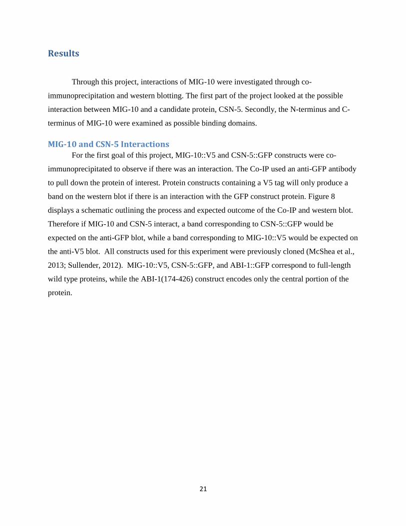
MIG-10 and CSN-5 Interactions For the first goal of this project, MIG-10::V5 and CSN-5::GFP constructs were co-
immunoprecipitated to observe if there was an interaction. The Co-IP used an anti-GFP antibody
to pull down the protein of interest. Protein constructs containing a V5 tag will only produce a
band on the western blot if there is an interaction with the GFP construct protein. Figure 8
displays a schematic outlining the process and expected outcome of the Co-IP and western blot.
Therefore if MIG-10 and CSN-5 interact, a band corresponding to CSN-5::GFP would be
expected on the anti-GFP blot, while a band corresponding to MIG-10::V5 would be expected on
the anti-V5 blot. All constructs used for this experiment were previously cloned (McShea et al.,
2013; Sullender, 2012). MIG-10::V5, CSN-5::GFP, and ABI-1::GFP correspond to full-length
wild type proteins, while the ABI-1(174-426) construct encodes only the central portion of the
protein.
22
Figure 8: Co-IP and Western Blot Schematic of MIG-10 and CSN-5 Interactions (Adapted from Sullender, 2012) Constructs were co-transfected into Drosophila S3 cells, co-immunoprecipitated, probed with either an anti-GFP or anti-V5 antibody, and observed for precipitation on a western blot. A precipitate is seen on the anti-V5 gel where CSN-5::GFP pulled down MIG-10::V5.
All samples were co-transfected, harvested as either whole cell lysates or Co-IPs, run on
a gel, and blotted. As seen highlighted in the yellow box on the V5 gel of Figure 9, a precipitate
was observed indicating the interaction between MIG-10 and CSN-5. ABI-1::GFP and ABI-
1(174-426)::GFP were each co-transfected with MIG-10 as a positive and negative control
respectively (Lanes 6,7). Research has shown that MIG-10 binds to ABI-1 but not ABI-1(174-
426) because the mutant lacks the SH3 domain which is important in binding (McShea et al.,
2013). MIG-10 and CSN-5 were individually transfected as additional negative controls. WCL
samples were used as additional controls for expression. Overall, the anti-GFP blot serves as a
control for the Co-IP. Two additional western blots confirming the interaction between MIG-10
and CSN-5 can be seen in Appendix C which used the same DNA concentrations as Figure 9.

23
Figure 9: MIG-10 and CSN-5 Interaction. The table above the blots gives a breakdown of what DNA was transfected in each lane. Lanes 1-4 represent WCL samples, while lanes 6-10 show Co-IP samples, with a ladder in lane 5. The yellow box highlights where MIG-10 (66.6 ng/μl) was pulled down by CSN-5 (40.4 ng/μl) on the anti-V5 gel. The green arrow points to the expression of the CSN-5 WCL on the anti-GFP gel, and MIG-10 WCL expression is emphasized by the blue arrow on the anti-V5 gel. Precipitate was observed for the co-transfection of MIG-10 and ABI-1 (66.6 ng/μl), but not for the co-transfection of MIG-10 and ABI-1(174-426) (66.6 ng/μl) on the anti-V5 gel which is expected. The red arrow at ~50kDa shows a background band observed from the Co-IP samples not to be confused with the proteins of interest. Arm-GAL4 used during the transfection had a concentration of 17.55 ng/μl.
Possible MIG-10 Binding Domains In order to investigate the possible binding domains of MIG-10, constructs of MIG-10A
and B N-terminus were cloned to insert a 6XHis tag. This was achieved through the Gateway
Cloning System which is outlined in Figure 10.

24
Figure 10: Gateway Cloning System (Sullender, 2012). Constructs were cloned to contain a 6XHis tag through BP and LR Reactions.
The N-termini of MIG-10 A and B were cloned with a V5 tag using the Gateway Cloning
System. Whole cell lysate samples of these two constructs were prepared through transfection
and applied to a western blot, but no expression was observed after three experiments (Figure
11). Thus, it cannot be concluded whether MIG-10 N-terminus is a binding domain. The Co-
immunoprecipitation assay was not conducted because expression of the whole cell lysates could
not be seen. The N-terminus of MIG-10 is only ~14 kDa, and therefore it was possible the
samples ran off the gel in the initial experiment. A shorter run time for the gel was conducted,
but still no expression was observed. Expression of ABI-1 and MIG-10A (whole) control whole
cell lysates was observed which showed that the transfection was efficient and the western was
conducted properly. Additionally, sequence analyses of the N-terminus constructs showed no
mistakes from PCR or cloning.

25
Figure 11: No expression of MIG-10 A and B N-terminus in whole cell lysates. The table above the gels gives a breakdown of what DNA was transfected in each lane. The anti-GFP gel displays the expression of the ABI-1 (66.6 ng/μl) control which is GFP tagged. Expression of MIG-10A (whole: 66.6 ng/μl) can be seen on the anti-V5 gel. No expression was seen in the MIG-10A (66.6 ng/μl) and B N-terminus (66.6 ng/μl) lanes.
Further experiments explored the C-terminus of MIG-10 as a possible binding domain. A
MIG-10 C-terminus::V5 construct was previously cloned using the Gateway Cloning System
(Sullender, 2012). Preliminary results showed no precipitation where MIG-10 C-terminus was
co-transfected with CSN-5 (Figure 12). This experiment was only performed once, and can
therefore not be confirmed. The co-transfection of MIG-10 and CSN-5 was used as a positive
control where interaction was confirmed. Single transfections of MIG-10 C-terminus, MIG-10,
and CSN-5 were used as negative controls. Whole cell lysate samples were used as controls for
expression. Overall, the anti-GFP gel serves as a control for the Co-IP.

26
Figure 12: MIG-10 C-terminus possibly not interacting with CSN-5. The table above the gels gives a breakdown of what DNA was transfected in each lane. Lanes 1-4 represent WCL samples while lanes 6-10 show Co-IP samples, with a ladder in lane 5. No precipitate is seen in lane 7 on the anti-V5 gel indicating no interaction between MIG-10 C-terminus (55 ng/μl) and CSN-5 (88.8 ng/μl). As a control, interaction between MIG-10 (88.8 ng/μl) and CSN-5 is seen in lane 6. The green arrow points to the expression of the CSN-5 WCL on the anti-GFP gel, and MIG-10 C-terminus WCL expression is emphasized by the blue arrow on the anti-V5 gel. The red arrow at ~50kDa shows a background band observed from the Co-IP samples not to be confused with the proteins of interest. Arm-GAL4 used during the transfection had a concentration of 66.6 ng/μl.

27
Discussion
The overall goal of this project was to research protein interactions with MIG-10 during
nervous system development. First, interactions between MIG-10 and a candidate protein, CSN-
5, were investigated through co-immunoprecipitation and western blotting. CSN-5 was identified
as a possible interactor of MIG-10 in previous yeast-two hybrid experiments. Secondly, specific
binding domains of MIG-10 were investigated.
Through this project, the interaction between MIG-10 and CSN-5 was confirmed. Four
repetitions of the co-transfection of MIG-10 and CSN-5 experiment displayed the co-
immunoprecipitation of MIG-10 by CSN-5, confirming interaction. Since CSN-5 is involved in
both protein stability and degradation (Miller et al, 2009), it is possible MIG-10 is regulated by
CSN-5. One possible model could be that CSN-5 is needed to degrade MIG-10 once the neuronal
migration and axon guidance is complete. Preliminary csn-5(RNAi) experiments within C.
elegans appear to disprove this theory (McMasters, 2013). These preliminary results show the
truncation of ALML neuron migration, suggesting CSN-5 may be needed to stabilize rather than
degrade MIG-10 during neuronal migration. Although these results are surprising, they are not
unprecedented, since CSN-5 has been shown to stabilize other proteins (Miller et al., 2009).
Additionally, CSN-5 has been shown to regulate transcription by stabilizing transcription factors,
which could indirectly affect MIG-10 protein levels (Chamovitz, 2009). Further future research
should be conducted to observe interactions between MIG-10 and CSN-5 in vivo. csn-5 null
mutants could be crossed into marker strains to observe the effects on ALM migration. If CSN-5
acts to stabilize MIG-10, as predicted by csn-5(RNAi) experiments, then csn-5 mutants would be
expected to result in the truncation of the neuronal migration.
Additionally through this project, the C-terminus and N-terminus of MIG-10 were
investigated as possible binding domains. Preliminary experiments involving the co-transfection
of MIG-10 C-terminus and CSN-5 did not display interaction. These preliminary results suggest
that the C-terminus of MIG-10 is not a binding domain. The N-terminus of MIG-10 did not show
any expression from the whole cell lysate samples on the western blot. It is possible that since
the N-terminus segment of MIG-10 protein is so small that it is not stable for such experiments.
Therefore, no conclusion can be made about the N-terminus of MIG-10 being a binding domain.

28
Throughout the project, a couple of technical issues were encountered. For example, the
best results were seen when the Co-IP and western blot were performed in one day (“marathon”).
When the procedures were broken up into two days, the WCL and Co-IP samples were either
placed at 4°C or frozen at -20°C overnight. This appeared to have a negative effect on the Co-IP
samples, especially when left at 4°C. Therefore, future experiments should be performed as a
“marathon” to achieve ideal results. Additionally throughout the project, the Arm-GAL4
concentration (17.55 ng/µl) was much lower than the ideal concentration of 66.6 ng/µl. As a
result bands appeared fainter and needed a longer exposure time to be seen. For the last
experiment (Figure 12), Arm-GAL4 with the ideal concentration was used, and gave a much
stronger signal. Based on these findings, it is important to have DNA concentrations of 66.6
ng/µl for the best results.
Future research investigating the different domains of MIG-10 would be beneficial to
understand what part of the protein is binding. Continued co-immunoprecipitation experiments
with MIG-10 C-terminus and CSN-5 could confirm that the C-terminus is not a binding domain.
Further co-immunoprecipitation experiments with the RAPH domain of MIG-10 and CSN-5
would confirm if this region of MIG-10 is a binding domain. Additionally, the N-terminus could
further be examined as a possible binding domain by creating expression clones that increase the
length of the N-terminus. This might increase the stability of the protein which could be used in
Co-IP experiments with CSN-5.
Based on the results of this project, the regulation of MIG-10 can be more thoroughly
understood. MIG-10 which is found in C. elegans serves as a model for the human ortholog,
lamellipodin. Research of MIG-10 can be used to get a better grasp of the effects of mutations
within the development of the nervous system. Overall, such research aids in better
understanding neurological disorders such as Down’s syndrome and autism.

29
References Chamovitz DA (2009). Revisiting the COP9 signalosome as a transcriptional regulator. EMBO
Rep 10(4):352-8.
Chang C, Adler CE, Krause M, Clark SG, Gertler FB, Tessier-Lavigne M, Bargmann CI (2006).
MIG-10/Lamellipodin and AGE-1/PI3K promote axon guidance and outgrowth in
response to slit and netrin. Current Biology 16(9):854-862.
Coló GP, Lafuente EM, Teixidó J (2012). The MRL proteins: Adapting cell adhesion, migration
and growth. European Journal of Cell Biology 91(11-12):861-868.
Cope GA and Deshaies RJ (2003). COP9 Signalosome: A Multifunctional Regulator of SCF
and Other Cullin-Based Ubiquitin Ligases. Cell 114:663-671
Disanza A, Steffen A, Hertzog M, Frittoli E, Rottner K, Scita G (2005). Actin polymerization
machinery: The finish line of signaling networks, the starting point of cellular movement.
Cellular and Molecular Life Sciences 62(9):955-970.
Echarri A, Lai MJ, Robinson MR, Pendergast AM (2004). Abl interactor 1 (abi-1) wave-binding
and SNARE domains regulate its nucleocytoplasmic shuttling, lamellipodium
localization, and wave-1 levels. Molecular and Cellular Biology 24(11):4979-4993.
Gosselin J, O'Toole SM (2008). MIG-10, an adapter protein, interacts with ABI-1, a component
of actin polymerization machinery. WPI MQP: 1-37
Hart AH, Reventar R, Bernstein A (2000). Genetic analysis of ETS genes in C. elegans.
Oncogene 19(55):6400-6408.
Horwitz R, Webb D (2003). Cell migration. Current Biology 13(19):R756-R759.
Krause M, Way M, Yaffe MB, Boussiotis VA, Gertler FB, Leslie JD, Stewart M, Lafuente EM,
Valderrama F, Jagannathan R, Strasser GA, Rubinson DA, Liu H (2004). Lamellipodin,
an Ena/VASP ligand, is implicated in the regulation of lamellipodial dynamics.
Developmental Cell 7(4):571-83.
Lafuente EM, Boussiotis VA, van Puijenbroek,André A F L., Krause M, Carman CV, Freeman
GJ, Berezovskaya A, Constantine E, Springer TA, Gertler FB (2004). RIAM, an
Ena/VASP and profilin ligand, interacts with Rap1-GTP and mediates Rap1-induced
adhesion. Developmental Cell 7(4):585-595.
Manser J, Roonprapunt C, Margolis B (1997). C. elegans Cell Migration Gene mig-10 Shares
Similarities with a Family of SH2 Domain Proteins and Acts Cell Nonautonomously in
Excretory Canal Development. Developmental Biology 184(1): 150-164.
McMasters J (2013). The role of CSN-5 in neuron migration in C. elegans. WPI MQP

30
McShea M (2011). Evidence of an interaction between the actin cytoskeletal regulators MIG-10
and ABI-1. WPI Thesis: 1-78
McShea M, Schmidt KL, Dubuke ML, Baldiga CE, Sullender ME, Reis AL, Zhang S, O’Toole
S, Jeffers MC, Warden RM, Kenney AH, Gosselin J, Kuhlwein M, Hashmi SK,
Stringham EG, Ryder EF (2013). Abelson interactor-1 (ABI-1) interacts with MRL
adaptor protein MIG-10 and is required in guided cell migrations and process outgrowth
in C.elegans
Miller RK, Qadota H, Stark TJ, Mercer KB, Wortham TS, Anyanful A, Benian GM (2009).
CSN-5, a component of the COP9 signalosome complex, regulates the levels of UNC-96
and UNC-98, two components of M-lines in caenorhabditis elegans muscle. Molecular
Biology of the Cell; 20(15):3608-16.
Quinn CC, Pfeil DS, Chen E, Stovall EL, Harden MV, Gavin MK, Forrester WC, Ryder EF,
Soto MC, Wadsworth WG (2006). UNC-6/Netrin and SLT-1/Slit guidance cues orient
axon outgrowth mediated by MIG-10/RIAM/Lamellipodin. Current Biology 16(9):845-
853.
Quinn CC, Wadsworth WG (2008). Axon guidance: Asymmetric signaling orients polarized
outgrowth. Trends Cell Biol 18(12):597-603
Sanes DH, Reh TA, Harris WA (2005). Development of the nervous system. London Academic:
1-388
Shackleford TJ, Claret FX (2010). JAB1/CSN5: A new player in cell cycle control and cancer.
Cell Division 5(1):26.
Sullender M (2012). Do ARX-3 and CSN-5 Protein Subunits Interact with MIG-10 to
Promote Axon Outgrowth and Neuronal Migration in C. elegans? WPI MQP: 1-53
Wei N, Deng XW (2003). The COP9 signalosome. Annu Rev Cell Dev Biol 19(1):261-86.
Zhou C, Wee S (2003). The COP9 signalosome: An assembly and maintenance platform for
cullin ubiquitin ligases? Nat Cell Biol 5(12):1029-33.

31
Appendix
Appendix A: Buffer Solutions
Calculations: 1x Sample Buffer (100µl 1x Sample Buffer needed per WCL sample)-
1.
= Volume 5x Sample Buffer (µl)
2. Add BME (5% of the volume of 5x Sample Buffer used (µl)).
3. Add TBS to reach total 1x Sample Buffer volume.
Lysis Buffer (2mL Lysis Buffer needed per Co-IP sample)- 1. 2x Protease Inhibitors:
= Volume Stock Protease Inhibitor (µl)
2. 5mM Phosphatase Inhibitors:
= Volume Stock Phosphatase Inhibitor (µl)
3. 1mM Sodium Orthovanadate (Na3VO4):
= Volume Stock Sodium Orthovanadate (µl)
4. Add a volume of EBC Buffer to reach the final volume of Lysis Buffer needed for the Co-IP samples.
2x Sample Buffer (90µl per Co-IP sample)-
1.
= Volume 5x Sample Buffer (µl)
2. Add BME (5% of the volume of 5x Sample Buffer used (µl)). 3. Add TBS to reach total 2x Sample Buffer volume. 4. Heat to ~ 95OC using boiling water before using in columns.
32
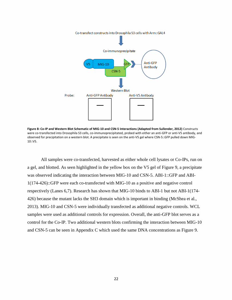
Buffer X
50 mM Tris, pH 8.5
250 mM NaCl
2 mM EDTA
1% NP-40
Final Wash Buffer
50 mM Tris, pH 8.5
Appendix B: Expression Clone Maps with Restriction Enzyme Cut Sites

33

34

35
Appendix C: Additional Western Blot of MIG-10 and CSN-5 Interactions