Mid Face Fractures 1
12
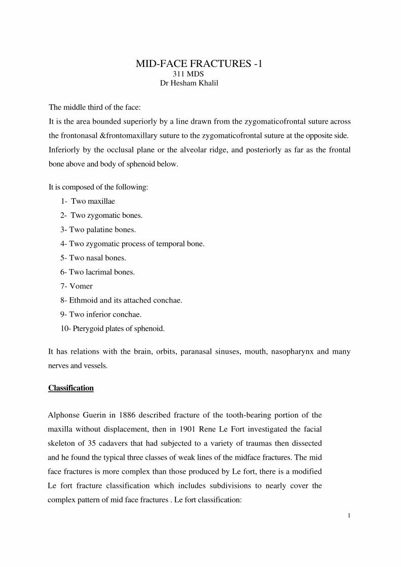
1 MID-FACE FRACTURES -1 311 MDS Dr Hesham Khalil The middle third of the face: It is the area bounded superiorly by a line drawn from the zygomaticofrontal suture across the frontonasal &frontomaxillary suture to the zygomaticofrontal suture at the opposite side. Inferiorly by the occlusal plane or the alveolar ridge, and posteriorly as far as the frontal bone above and body of sphenoid below. It is composed of the following: 1- Two maxillae 2- Two zygomatic bones. 3- Two palatine bones. 4- Two zygomatic process of temporal bone. 5- Two nasal bones. 6- Two lacrimal bones. 7- Vomer 8- Ethmoid and its attached conchae. 9- Two inferior conchae. 10- Pterygoid plates of sphenoid. It has relations with the brain, orbits, paranasal sinuses, mouth, nasopharynx and many nerves and vessels. Classification Alphonse Guerin in 1886 described fracture of the tooth-bearing portion of the maxilla without displacement, then in 1901 Rene Le Fort investigated the facial skeleton of 35 cadavers that had subjected to a variety of traumas then dissected and he found the typical three classes of weak lines of the midface fractures. The mid face fractures is more complex than those produced by Le fort, there is a modified Le fort fracture classification which includes subdivisions to nearly cover the complex pattern of mid face fractures . Le fort classification:
description
Fracture
Transcript of Mid Face Fractures 1
1
MID-FACE FRACTURES -1 311 MDS
Dr Hesham Khalil
The middle third of the face:
It is the area bounded superiorly by a line drawn from the zygomaticofrontal suture across
the frontonasal &frontomaxillary suture to the zygomaticofrontal suture at the opposite side.
Inferiorly by the occlusal plane or the alveolar ridge, and posteriorly as far as the frontal
bone above and body of sphenoid below.
It is composed of the following:
1- Two maxillae
2- Two zygomatic bones.
3- Two palatine bones.
4- Two zygomatic process of temporal bone.
5- Two nasal bones.
6- Two lacrimal bones.
7- Vomer
8- Ethmoid and its attached conchae.
9- Two inferior conchae.
10- Pterygoid plates of sphenoid.
It has relations with the brain, orbits, paranasal sinuses, mouth, nasopharynx and many
nerves and vessels.
Classification
Alphonse Guerin in 1886 described fracture of the tooth-bearing portion of the
maxilla without displacement, then in 1901 Rene Le Fort investigated the facial
skeleton of 35 cadavers that had subjected to a variety of traumas then dissected
and he found the typical three classes of weak lines of the midface fractures. The mid
face fractures is more complex than those produced by Le fort, there is a modified
Le fort fracture classification which includes subdivisions to nearly cover the
complex pattern of mid face fractures . Le fort classification:

2
1-Le Fort I (Low-level fracture):
This is a relatively horizontal fracture in the body of the maxilla that results in a
maxillary tooth bearing fragment being detached from the middle face. The
fracture line passes above the teeth, below the zygomatic process, through the
maxillary sinuses, the tuberosities to the inferior portion of the pterygoid plate; it
can be unilateral or bilateral.
2-Le Fort II (Pyramidal fracture):
In this fracture the maxilla will be separated from the base of the skull by
fractures of the nasal bone and the frontal processes of maxillae. The fracture
extends laterally through the lacrimal bones, floors of the Orbits, and
inferiorly through the zygomaticomaxillary sutures, then passes posteriorly
along the lateral wall of the maxilla, across the ptery-gomaxillary fossa and
through the pterygoid plates.
3-Le Fort III (Craniofacial dysjunction):
The fracture line passes the nasal bones and frontal processes of maxillae or nasofrontal and
frontomaxilary sutures, across the floors of the orbit through the ethmoid and sphenoid
sinuses and the zygomaticofrontal suture. It passes across both pterygoid plates where they
arise from the sphenoid bone, so it separates the middle third from the cranium.
3
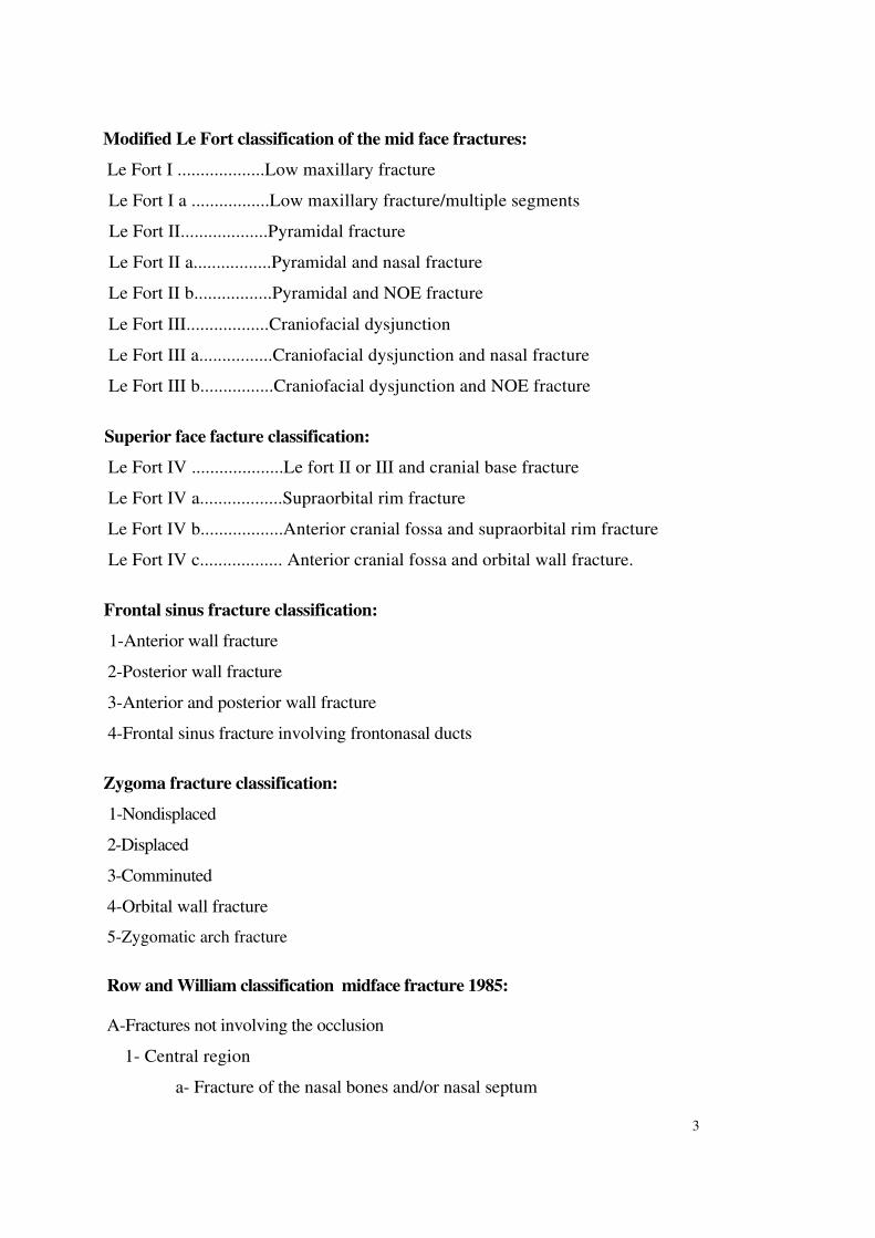
Modified Le Fort classification of the mid face fractures:
Le Fort I ...................Low maxillary fracture
Le Fort I a .................Low maxillary fracture/multiple segments
Le Fort II...................Pyramidal fracture
Le Fort II a.................Pyramidal and nasal fracture
Le Fort II b.................Pyramidal and NOE fracture
Le Fort III..................Craniofacial dysjunction
Le Fort III a................Craniofacial dysjunction and nasal fracture
Le Fort III b................Craniofacial dysjunction and NOE fracture
Superior face facture classification:
Le Fort IV ....................Le fort II or III and cranial base fracture
Le Fort IV a..................Supraorbital rim fracture
Le Fort IV b..................Anterior cranial fossa and supraorbital rim fracture
Le Fort IV c.................. Anterior cranial fossa and orbital wall fracture.
Frontal sinus fracture classification:
1-Anterior wall fracture
2-Posterior wall fracture
3-Anterior and posterior wall fracture
4-Frontal sinus fracture involving frontonasal ducts
Zygoma fracture classification:
1-Nondisplaced
2-Displaced
3-Comminuted
4-Orbital wall fracture
5-Zygomatic arch fracture
Row and William classification midface fracture 1985:
A-Fractures not involving the occlusion
1- Central region
a- Fracture of the nasal bones and/or nasal septum
4
-Lateral nasal injuries
-Anterior nasal injuries
b- Fractures of the frontal process of the maxilla
c- Fractures of type (a) and (b) which extend into the frontal bone
2- Lateral region
Fractures involving the zygomatic bone, arch and ,maxilla (zygomatic Complex)
excluding the dento-alveolar component. B-Fractures involving the occlusion
1- Dento-alveolar
2- Subzygomatic
a- Le fort I
b- Le fort II
3- Suprazygomatic (Le fort III)
Henderson's classification of malar bone fractures:
I- Undisplaced fracture, any site
II Zygomatic arch fracture only
III Tripod fracture with undistracted frontozygomatic suture
IV Tripod fracture with distracted frontozygomatic suture
V Pure blow out fracture of the orbit
VI Fracture of the orbital rim only
VII Comminuted fracture or other than above
Aetiology:
Aetiology is best considered under six broad headings:
1- Assaults
2- Falls
3- Road traffic accidents
4- Industrial injuries
5- Sports injuries
5
6- War injuries
Signs and symptoms:
Le Fort Fractures
1- Pain
2- Swelling and edema
3- Step deformity
4- Mobility
5- Anaesthesia or parasthesia
6- Diplopia
7- Enophthalmus
8- Epistaxis
9- CSF rhinorrhoea.
10- Subconjunctival haemorrhage
11-Dish face deformity
12- Limitation of ocular movement
13- Difficulty of mouth opening
14-Disturbed occlusion
15- Cracked-pot sound on percussion
16- Occasional haematoma at the palate
17- Circumorbital ecchymosis
18-Lengthening of the face
19-Battle's sign
20- Orbital emphysema
21-Paralysis of facial muscles
Zygomatic complex fracture:
Articulation of the zygomatic bone with the other facial bones:
1-The zygomatico frontal suture
2-The infraorbital rim
6
3-The zygomatico maxillary buttress
4-The zygomatic arch
5-The zygomatico sphenoid suture
The zygoma forms the lateral wall and the floor of the orbital cavity and the roof and
lateral wall of the maxillary sinus.
It provides attachment for the following muscles:
1-The zygomaticus major
2-The zygomaticus minor
3-The levator labii superioris
4-Levator anguli
5-masseter
The temporalis muscle passes underneath the zygomatic arch.
Nerves passing through or near the zygomatic bone:
1-Zygomatico temporal
2-Zygomatico facial
3-Infraorbital (about 10 mm from the inferior orbital rim)
At the Whitnall's tubercle the zygomatic bone provides attachment to:
1- Lateral canthus ligament
2- Lockwood's ligament
40% of the zygomatic bone fractures associated with ocular injuries.
Signs and symptoms ocular injury:
1- pain
2- Swelling
3- Asymmetry
4- Periorbital haematoma
5- Subconjunctival haemorrhage
6- Limitation of ocular movement
7- Ecchymosis and tenderness over the area
8- Diplopia
9- Enophthalmus
7
10-Dystopia
11-Epis taxis
12- Step deformity
13- Limitation of mandibular movement
14- Anasthesia
15- Gagging of occlusion
16- Flattening of the malar prominence
17- Changes in eyelid position e.g. Antimogoloid slant of the palpebral fissure
Treatment of zygomatic fracture:
1-Closed reduction and fixation
2-Open reduction and fixation
Approaches to the zygomatic bone:
1-Gilles temporal approach 1927 e.g. using Rowe elevator
2-Keen intra oral approach 1909
3-Dingman supra orbital approach 1964
4-Transcutaneous Cheek approach then using bone hook
5-Intra oral approach though the sinus Caldwell-Luc
6-Combination of upperlid blepharoplasty, midtarsal incision and intraoral incision
7-Combination of Coronal flap, midtarsal incision and intra oral incision.
The more recent advances:
1-Endoscopic assisted procedures
2-Intra-operative CT evaluation
Radiology:
The Midface fractures generally were used to be treated by closed reduction. As a
result, the preoperative imaging needs were only those that can identify the
presence of the fracture. Surgeons today are concerned with the comprehensive,
8
three-dimensional nature of the midface fracture so that restoration of the
preinjury position can be accomplished. Imaging of the middle third can include
the following
1- Occipitomental (standard ,10°, 15° and 30°)
2- True lateral
3- Soft tissue lateral
4- Occlusal
5- Intra orals
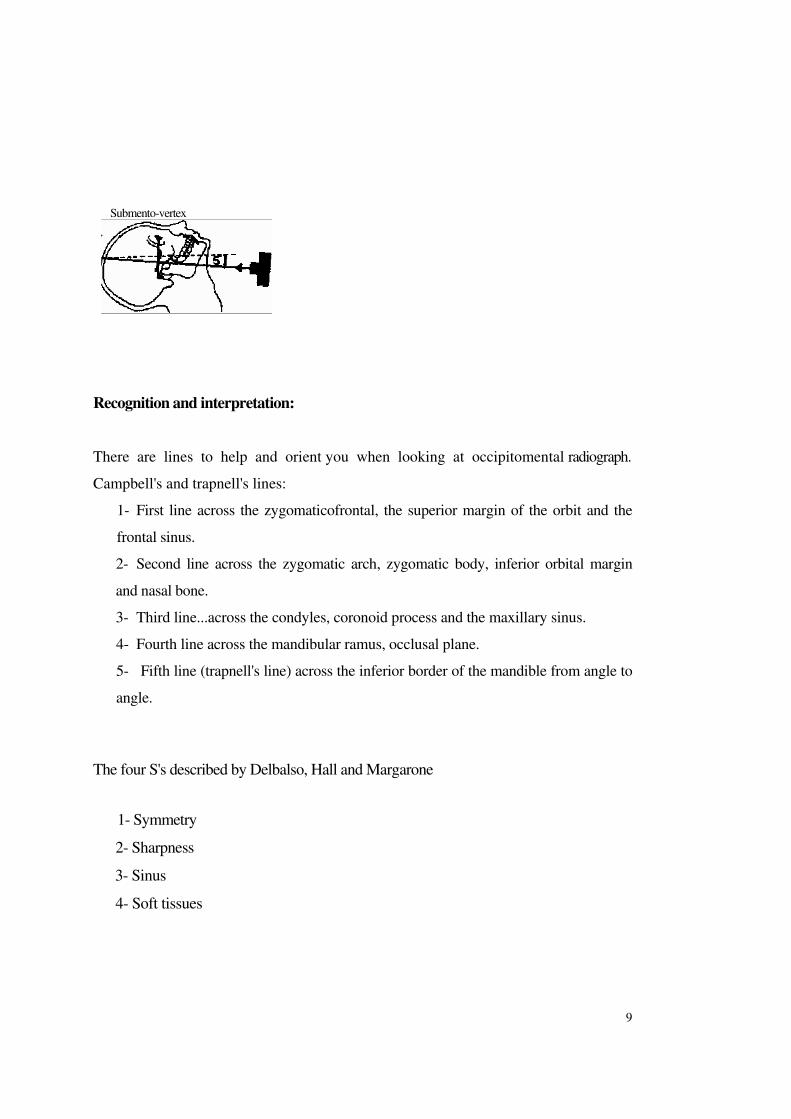
6- Submento-vertex
7- C.T Scan
8- 3D C.T Scan
9- MRI (to detect CSF leaks and fistula)
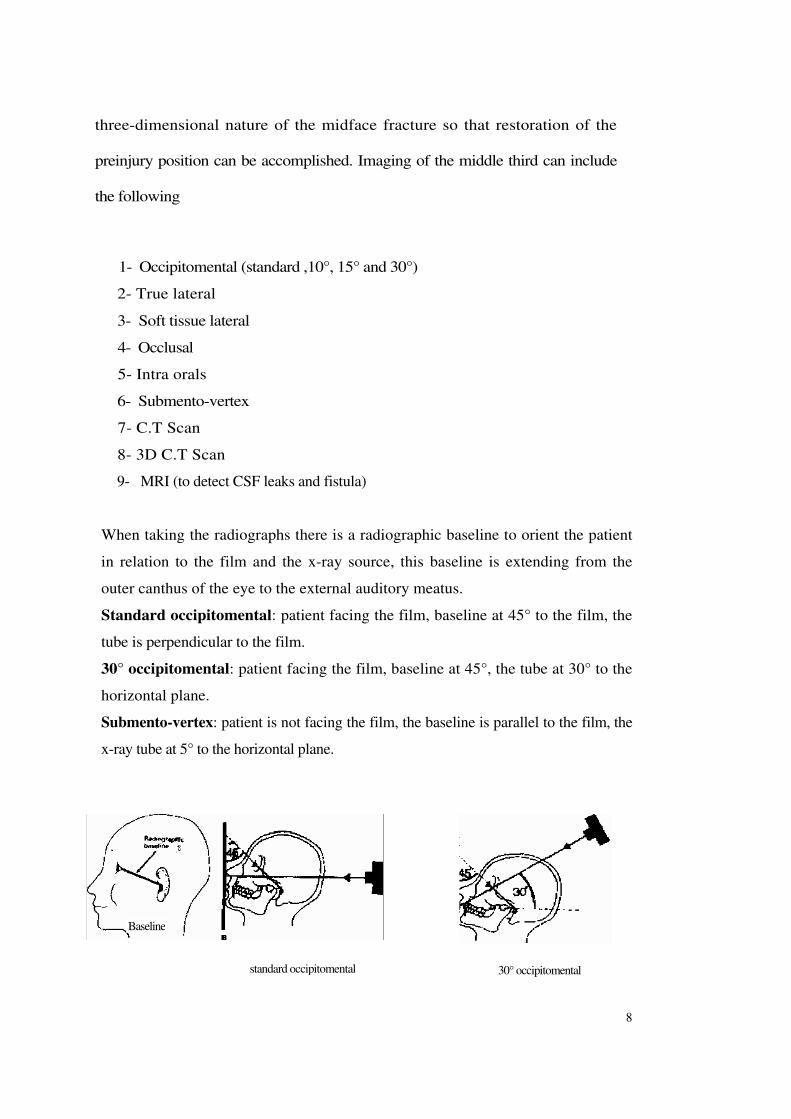
When taking the radiographs there is a radiographic baseline to orient the patient
in relation to the film and the x-ray source, this baseline is extending from the
outer canthus of the eye to the external auditory meatus.
Standard occipitomental: patient facing the film, baseline at 45° to the film, the
tube is perpendicular to the film.
30° occipitomental: patient facing the film, baseline at 45°, the tube at 30° to the
horizontal plane.
Submento-vertex: patient is not facing the film, the baseline is parallel to the film, the
x-ray tube at 5° to the horizontal plane.
30° occipitomental
Baseline
standard occipitomental
9
Submento-vertex
Recognition and interpretation:
There are lines to help and orient you when looking at occipitomental radiograph.
Campbell's and trapnell's lines:
1- First line across the zygomaticofrontal, the superior margin of the orbit and the
frontal sinus.
2- Second line across the zygomatic arch, zygomatic body, inferior orbital margin
and nasal bone.
3- Third line...across the condyles, coronoid process and the maxillary sinus.
4- Fourth line across the mandibular ramus, occlusal plane.
5- Fifth line (trapnell's line) across the inferior border of the mandible from angle to
angle.
The four S's described by Delbalso, Hall and Margarone
1- Symmetry
2- Sharpness
3- Sinus
4- Soft tissues

10
Management of midface fracture:
1- Emergency treatment and stabilization of the patient.
2- Definitive treatment with reduction and fixation
Methods of fixation:
1- Wiring
2- Plates and screws
3- IMF
4- Internal suspension: e.g. circumzygomatic, infraorbital
5- Craniofacial Suspension: e.g. supraorbital pins, box frame, Halo frame
Timing of surgery:
Although most maxillofacial can wait, late repair after healing is extremely difficult,
early treatment within 1-10 days gives the best results, but immediate surgery can
be carried out for life threatening injuries or if the patient is going for the theatre for
other reasons.
Surgical exposure of the midface:
Intra oral:
-The typical incision line is within the unattached mucosa 4-5 mm from the attached
gingiva
-Marginal rim incision
- Crestal incision
11
Extra oral:
- subciliary incision
- transconjunctival
- blepharoplasty
- brow incision
- bicoronal flap
- midface degloving
-Weber Ferguson
-Gillies temporal approach
Diplopia:
-incidence between 15 to 56%
- the most common cause is blow out fractures
Cerebrospinal fluid leakage:
- CSF was described by willis in 1976
- It is usually due to dural tear
- Antibiotic cover?
- Risk of meningitis?

12