Microstructural Changes in Wheat Starch Dispersions During ...
12
Food Structure Food Structure Volume 8 Number 1 Article 6 1989 Microstructural Changes in Wheat Starch Dispersions During Microstructural Changes in Wheat Starch Dispersions During Heating and Cooling Heating and Cooling M. Langton A. M. Hermansson Follow this and additional works at: https://digitalcommons.usu.edu/foodmicrostructure Part of the Food Science Commons Recommended Citation Recommended Citation Langton, M. and Hermansson, A. M. (1989) "Microstructural Changes in Wheat Starch Dispersions During Heating and Cooling," Food Structure: Vol. 8 : No. 1 , Article 6. Available at: https://digitalcommons.usu.edu/foodmicrostructure/vol8/iss1/6 This Article is brought to you for free and open access by the Western Dairy Center at DigitalCommons@USU. It has been accepted for inclusion in Food Structure by an authorized administrator of DigitalCommons@USU. For more information, please contact [email protected].
Transcript of Microstructural Changes in Wheat Starch Dispersions During ...

Food Structure Food Structure
Volume 8 Number 1 Article 6
1989
Microstructural Changes in Wheat Starch Dispersions During Microstructural Changes in Wheat Starch Dispersions During
Heating and Cooling Heating and Cooling
M. Langton
A. M. Hermansson
Follow this and additional works at: https://digitalcommons.usu.edu/foodmicrostructure
Part of the Food Science Commons
Recommended Citation Recommended Citation Langton, M. and Hermansson, A. M. (1989) "Microstructural Changes in Wheat Starch Dispersions During Heating and Cooling," Food Structure: Vol. 8 : No. 1 , Article 6. Available at: https://digitalcommons.usu.edu/foodmicrostructure/vol8/iss1/6
This Article is brought to you for free and open access by the Western Dairy Center at DigitalCommons@USU. It has been accepted for inclusion in Food Structure by an authorized administrator of DigitalCommons@USU. For more information, please contact [email protected].
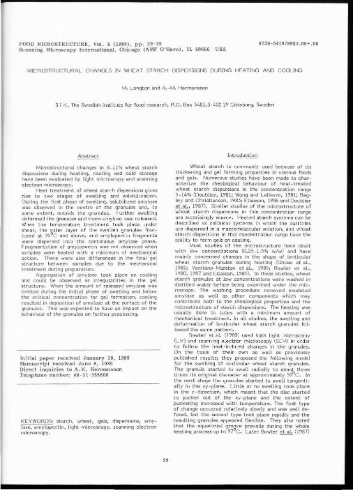
FOOD MICROSTRUCTURE, Vol. 8 (1989), pp. 29 - 39 0730- 5419/89$3 . 00+ .00 Scanning Microscopy International , Chicago (AMF O'Hare), IL 60666 USA
MICROSTRUCTURAL CHANGES IN WHEAT STARCH DISPERSIONS DURING HEATING AND COOLING
M. Lang ton and A . ~M. Hermansson
S I K, The Swed ish institute for food research, P .O . Box 5401,5-402 29 Gi:iteborg, Sweden
Microstructural changes in 8-11% wheat starch dispersions during heating, cooling and cold storage have been evaluated by light microscopy and scanning electron microscopy.
Heat treatment of wheat starch dispersions gives rise to two stages of swelling and solubilization. During the first phase of swelling, solubilized amylose was observed ln the centre of the granules and, to some extent, outside the granules. Further swelling deformed the granules and more amylose was released. When the temperature treatment took place under shear, the outer layer of the swo llen granules fra ctured at 94°C and above, and amylopectin fragments were dispersed into the cont inuous amylose phase. Fragmentation of amylopectin was not observed when samples were heated with a minimum of mechanical action. There were also differences in the final gel structure between samples due to the mechanical treatment during preparation.
Aggregation of amylose took place on cooling and could be observed as irregularities in the gel structure. When the amount of released amylose was limited during the initial phase of swelling and below the critical concentration for gel formation, cooling resulted in deposition of amylose at the surface of the granules. This was expected to have an impact on the behaviour of the granules on further processing.
Initial paper r eceived January 30, 1989 Manuscr ipt received June 8, 1989 Direct inquiries to A.M. Hermansson Telephone number: 46-31-355600
KEYWORDS: starch, wheat, gels, dispersions, amylose, amylopectin, ligh t microscopy, scanning electron microscopy.
29
Introduction
Wheat starch is commonly used because of its thickening and gel forming properties in viscous foods and gels. Numerous studies have been made to characterize the rheological behaviour of heat-treated wheat starch dispersions in the concentration range 5-14% (Doublier, 1981; Wong and Lelievre, 1981; Bagley and Christianson, 1983; Eliasson, 1986 and Doublier eta!., 1987). Similar studies of the microstructure of wheat s tarch dispersions in thi s concentration range are surprisingly scarce. Heated starch systems can be described as colloidal systems in which the particles BI'B dispersed in a macromolecular so lution, and wheat s tarch dispersions in this concentration range have the ab ility to form gels on cooling.
Most studies of the microstructure have dealt with low concentrations (0.25-l.S% w/w) and have mainly concerned changes in the shape of lenticular wheat starch granules during heating (Ghiasi et a l. , 1982; Varriano-Mars ton et al., 1985; Bowler et al., 1980, 1987 and Eliassen, 1985). In these studies, wheat starch granules at low concentrations were washed in distilled water before being examined under the microscope. The washing procedure removed exudated amylose as well as other components which may contribute both to the rheological properties and the microstructure of starch dispersions. The heating was usually done in tubes with a minimum amount of mechanical treatment. In all studies, the swelling and deformation of lenticular wheat starch granules fol lowed the same pattern.
Bow ler et al. (1980) used both light microscopy (LM) end scanning electron microscopy (SEM) In order to follow the heat-induced changes in the granules. On the basis of their own as well as previously pub! ished results; they proposed the following model for the swelling of lenticular wheat starch granu les. The granule star ted to swell radially to about three times its original diameter at approximately S0°C. In the next s tage the granules started to swell tangentially in the xy-plane. Little or no swelling took place in the z-direction, which meant that the disc started to pucker out of the xy-plane and the extent of puckering increased with temperature. The first type of change occurred relatively slowly and was well defined, but the second t ype took place rapid ly and the resul ting granules appeared fl ex ible. They also noted that the equatorial groove prevails during the whole heating process up to 97°C. Later Bowler ~ (1987)

M. Langton and A.-M. Hermansson
investigated the effect of different preparation techniques for SEM, such as dehydration and critical point drying, freeze drying and cryo stage techniques. Even if differences were observed, the preparation techniques did not affect the above described swelling patterns of the granules. The effects of surfactants on the swelling of wheat starch granules have been investigated by Ghiasi et at. (1982) and by Eliasson (1985). They found that surfactants restricted the first but not the second stage of the swelling of wheat starch granules.
The amount of amylose released from wheat starch granules during heating has been investigated by means of iodine-binding at low concentrations in the range 0.25-2.5% (Doublier, 1981; Ghiasi et al., 1982; Eliasson, 1986). Here again two stages wefe observed. In the lower temperature range of about 55-80°C the release of amylose was slow, but it increased considerably at higher temperatures.
The aim of this study was to evaluate structural changes in wheat starch dispersions mainly by light microscopy and, to some extent, by SEM o during heating to 75-120°C, taking the effect of
mechanical t reatment during preparation into account during cooling and cold storage It is well known that, when evaluating complex
food structures it is wise to use more than one technique for preparing the sample for electron microscopy (Hermansson and Buchheim, 1981; Bowler eta!., 1987). The same holds true for light microscopy, and this study demonstrates the advantages of using three techniques in parallel.
e xperimental
Materials --~-n-this study commerc ial wheat starch from CPC-Cerestar was used. Concentrations in the range of 2 to 11% w/v starch were investigated, but in the main study 8% w/v was used if not otherwise stated.
In a preliminary study different ba tches from three producers were tested to get some idea of the effects of the wheat quality, and of environmental and processing conditions. Samples were purchased from Raision Tehtaat OY, KrOner and CPC. AU wheat starch samples showed the same type of general behaviour even if there were some discrepancies in the temperatures at which different phenomena occurred. Sample preparation
Samples were prepared by mixing 36 g wheat starch with 414 ml distilled water.
The starch dispersion was heated at the rate of 1.5°C/min to a required temperature, held at that temperature for 30 minuteS and cooled to 25°C at a rate of 1.5°C/min in a Brabender Amylograph. The temperature dependency was studied by performing experiments at the following max. temperatures 60, 70, 75, 80, 85, 90, 94, 95, 97 and 98°C. This temperature treatment was combined with mechanical treatment cons isting of continuous stirring at 75 rpm. Mechanical treatment was kept to a minimum for two samples by preheating them in the Brabender, as described above. One sample wascrreheated to 85°C and then heated for 30 min at 95 C in a water bath without any stirring. The other sample was preheated to 85°C, whereafter it was canned and heated at 120°C for 30 min in a retort autoclave. After cooling, samples were stored at +4°C.
30
Light microscopy Samples were taken immediately on reaching the
required temperature, but also after being incubated for 30 minutes and after cooling to room temperature, as well as after storage in a refrigerator. Three techniques were used to prepare the samples for light microscopy: smearing, cryosectioning and embedding followed by thin sectioning. All three techniques were used on all samples, and the few micrographs presented here have been chosen out of several hundred.
AU samples were stained with iodine in a diluted 1:1 Lugol's solution for one minute, whereafter the sample was covered and sealed. Preliminary studies were also made with ather stains, such as safranin, Congo red, gent ian violet and aniline blue with Orange G, in combination with different illumination techniques. The prepared samples were examined with a Nikon macrophot Fx microscope.
Smears. The starch dispersion was quickly and gently smeared out onto an object glass and stained directly. The advantage of this technique is that the hydrated sample can be observed without freezing or dehydration. It is also possible to observe intact swelled granules and obtain a three-dimensional impression by focusing through the depth of the sample. The disadvantage is that a sample with swelled granules gets rather thick, which decreases the resolution and results in a loss of detail.
C ryosections. Samples were frozen in liquid nitrogen and sectioned frozen. A Leitz cryostat was used and 7-8 1um thick sections were cut. The advantage of cr'yosections is that very quick changes in the microstructure are detectable as the structure is "frozen". A better resolution is obtained with this technique than with the smearing technique. The disadvantage is that the freezing can induce ice crystals that can damage the microstructure. This damage is more severe in the gel than in the fluid, viscous starch systems, where the dissolved macromolecules act as cryoprotectants.
Embedded sections. Samples were chemically fixed in 1% glutaraldehyde with 0.1% ruthenium red, dehydrated and embedded in historesin, LKB, as recommended by the manufacturer. A Re ichert-Jung Ultracut E, with glass knives, was used to section the samples. Thin sections, 1 ~um, were cut to obtain the
~eesr~ p~~~ib~~ raecs~il~~~o~e~tedr t~~cn~~~sstcs\iZi~~,n~:4 ~~~~ technique gives the best resolution for light microscopy, and no freeze damage or mechanical redistribution is induced in the sample. The samples were stained after sectioning, which means that microstructural changes due to the presence of iodine in the staining solution can be ruled out.
The disadvantage is that it may be difficult to interpret such thin sections with regard to dispersions with small and large irregular particles. The chemical fixation and dehydration used in this technique may also affect the macromolecular phase, but such effects cannot be seen from the resolution given by light microscopy. Scanning electron microscopy
Samples of heat-treated starch dispersion were taken after they had reached room temperature as well as after storage. Two techniques were used: critical point drying and a cryo-stage technique.
The samples were examined in a Cambridge Stereoscan 200 equipped with a Hexland cold stage using accelerating voltage between 2 kV-10 kV .

Microstructural changes in wheat starch dispersions during heating and cooling
jFi gures 1 and 2 are color micrographs at page 33.
!J9.:..2· Scanning electron micrograph of a critical point dried 8% starch dispersion heated to 90°C and cooled to room temperature.
Critica l point drying. Sma ll samples were chemica lly fixed in 1% glutar
aldehyde with 0.1% ruthenium red, dehydrated, through graded alcohol, and critical point dried. The dried samples were fractured and mounted on SEM with "Leit-C", carbon glow. The samples were sputter coated, Au/Pd in a Polaron E5000 before examination in the SEM. 3
Cryo-stage. Very smaiJ samples about l mm were placed in copper sandwich holders and extra rapidly fast frozen in a propane jet freezer, Balzer. Frozen samples were transferred to the cold stage in the microscope, fractured and etched at -90- -l00°C. Samp les were examined at -150- -176°C, both uncoated and gold coa ted at low accelerating voltage 2-J kV.
Resu lts and Discussion
Heat- induced c hanges in starch dispersions Microstructural changes in wheat starch have
been studied B!l a function of temperature combined with mechanical treatment. Samples were taken at various stages of heat treatment from 60 to 120°C for microstructural evaluation. Results from some heating temperatures were chosen to illustrate the most significant heat-induced changes. Wheat starch granules heated to 75°C are representative of the first stage of swelling, where the release of amylose is limited and the concentration of amylose is not h~h enough forge! formation . Heating temperatures of 90 C and above were chosen to illustrate phenomena occur~ ring during the second s tage of swelling.
The effect of heating an 8% wheat starch dispersion to 75°C is illustrated by the iodine-stained smear shown in Figure 1. There is a variation in the degree of swelling of the starch granules, and it is important
31
~· Scanning electron micrograph of a 4% starch dispersion heated to 90°C and cooled to room temperature, frozen rapidly and examined frozen on a cryo stage .
lo realize that starch consists of a population of granules which vary with regard to factors such as shape, deforma tion, release of amylose during heating, etc. From Figure 1 it can be seen that the majority of the granules heated to 75°C have retained their original shape and that they are stained dark blue which means that they are amylose-rich. These granules are in the first stage of swelling according to the swelling pattern described by Bowler et al . (1980). There is an exudated blue phase surrounding these granules, which means that the restricted leakage of amylose out of granules in the first stage of swelling can be detec ted by light microscopy. A number of granules in the second stage of swelling can also be seen from Figure 1. These are more brown and less blue in colour end have a more irregular shape. Iodine stains pure amylose blue and amylopectin beige-brown with vary~ ing intensity.
Figure 2 shows the structure of the 8% starch dispersion at 90°C. More amylose leaks out at this temperature, and the continuous pilose surrounding the swelled granules is stained blue. There are also some blue-stained granules in the first stage of swelling at this temperature, but the majority of the granu les are in the second stage of swelling. They are ma inly beige in colour, and especially the outer layers are very weakly stained, indicating that amylose has leaked out. From Figures 1 and 2 it is also evident that the starch dispersion contains small granules of 8-starch as well as lenticular shaped A-starch granules.
From the smears it is not possible to characterize the exact shapes of the lenticular A-starch granules at different stages of swelling. Corroborati ve SEM studies of heated dispersion show that the changes found in the shape of the granules in 4-8% dispersions were similar to those found in studies of

M. Langton and A.·M. Hermansson
7
OOQ ~· Schematic illustration of fragment ation of the outer layer of swollen granules.
dilute suspensions (Bowler et al., 1980; Ghiasi et a l. , 1982; Eliassen, 1985; Varriano-Marston eta!., 1985). Figure 3 shows a SEM micrograph of a granule in an 8% dispersion after cooling from 90°C. The granule has a shape typical of the second stage of swelling and is surrounded by a network structure of amylose. This sample was prepared by chemical fixation, dehydration and critical point drying. Figure 4 shows a fractu&ed granule in a 4% dispersion after cooling from 90 C, preparation by rapid freezing and examination on a cryostage. The use of low-temperature techniques for isolated granules has been discussed by Bowler et al. (1987). The advantage of this technique is that the samp le can be investigated fully hydrated without additions of chem icals, provided that the free z ing rote is high enough to prevent ice crystal formation, which can damage tho structure. Some difficulties attached to working with highly aqueous samples of dispersions and gels are worth pointing out. Figure 4 shows a samp le which should have been deep!~ etched under the etch ing conditions of 15 min at -90 C. However, the water is firmly held by the swollen granu le and by the biopolymer-rich continuous phase. Therefore, it is very difficult to sublimate water from the surface, which makes the contrast low and makes it difficult to reveal fine details of the structure. F ragmentation of amylopectin
Brabe~d~~n t~he95bt~~c~h:i:~~s;o~y:~s o~e:~:d s;ol~:~ granules fractured and fragments were released into the blue stained amylose solution. These fragments are weakly stained and believed to be composed mainly of hydrated amylopectin and not any amylose with an affinity for iodine. Figures 5 and 6 show 8% wheat starch dispersion heated to 95°C. Amylopectin fragments of varying size, are dispersed in the exudated blue amylose phase as seen in Figure 5. The micrograph at a higher magnification shown in Figure 6 illustrates a granule where the outer layer has been partly sheared off . In three dimensions the outer layer that frac tures would correspond to the outer part of the puckered granule as described by Bowler et a l. (1980). The mechanism of fragmentation is schematically illustrated in Figure 7, showing the swelling of the granule, the formation of an outer layer and the fracture of the outer layer into fragments.
Fragmentation started at 94-95°C for samp les prepared in the Brabender Amylograph at 75 rpm, and the amount of dispersed fragments increased with e levated temperatures and holding times at 94°C or above. This phenomenon has not previously been demonstrated by microscopy. The smear technique was chosen to introduce heat - induced changes and the presence of small fragments in the continuous amy lose
32
~· Smear of an 8% wheal starch dispersion heated to 75 C.
Fig. ~ Smear of an 8% wheat starch dispersion heated to 90 C.
Fig. ~· Smear of an 8% wheat starch dispe rsion heated to 95 C.
~ Smear of an B% wheat starch dispersion heated to 95 C .
~· Cryosection of an 11% wheat starch dispersion heated at 94°C for 30 min and subjected to mechanical treatment.
~· Cryosection of an 11% wheat sta rch dispersion heated at 94°C for 30 min and not subjected to mechanical treatment.
Fig. 10. Embedded section of an 8% gel, formed after heating at 95°C, cooling and storage at room temperature for 5 h.
Fig. 11. Embedded section of an 8% gel formed after heat treatment at 120°C for 30 min.
phase. With any of the sectioning techniques, a small particle as seen under the microscope may have been part of a bigger partic le that has been sectioned c lose to its outer surface. Fragmentation in dispersions and gels
The amylopectin fragments seemed to be fractured from the outer layer of the swolhm granu les, and it was considered of interest to study the e ffect of the mechanical treatment of the dispersion during preparation in the Brabender with regard to amylopectin fragmentation.
Figure B shows a cryosy,ction of an 11% wheat starch dispersion heated to 94 C and held at 30 min in the Brabender. During continuous stirring a large number of small fragments can be seen as particles dispersed in the blue stained amy lose phase. Figure 9 shows a cryosection of an 11% wheat starch dispersion where the sample has been heated to 85°C in the Brabender and then held at 94°C for 30 min in a water bath without any mechanical treatment. In this area hardly any fragments are seen. The difference with regard to the degree of fragmentation between the two samp les is striking.
The higher concentration used in th is experiment means a higher friction during preparation and a higher degree of fragmentation. Fragmentat ion of amylopectin will have a bearing on rheological properties (Svegmark and Herrnansson, to be published).
The mechanism of amylopectin fragmentation explains some observations from solubility studies made by Doublier (1981). By stirr ing at a rotation speed of 750 rpm and rapid heatin~ they found that all amylose was so lubilized at 92-93 C and thereafter a large fraction of the amylopectin was "apparently solubilized" and recovered in the supernatant. When the rotation speed was lowered to 200 rpm, total solubilization was achieved at 94°C but no amylopectin was found in the supernatant. In a third experiment at 750 rpm and a heating rate of 1°C/min, which is closer to the heating rate used in this study, they found that the solubilization of amy lose was not

33

34

Microstructural changes in wheat starch dispersions during heating and cooling
Fig. 14. Cryoscc tion of an 8% wheat starch dispersion heated at 95°C for 30 min. -Fig. 15. Cryosection of an 8% wheat starch dispersion heated at 95°C for 30 min and cooled to room temperature.
Fig. 16. Smear o f an 8% wheat starch dispersion heated at 75°C for 30 min.
Fig. 17. Smear of an B% wheat starch dispersion heated at 75°C for 30 min and cooled to room temperature .
Fig. 18. Smear o f an 8% wheat starch dispersion heated at 75°C for 30 min, cooled to room temperature and stored 15 days in a refrigerator.
Fig. 19. Embedded thin section of an 8% wheat starch dispersion heated at 75°C for 30 min cooled to room temperature and stored 4 days in a refrigerator.
Fig. 20. Embedded thin section of an 8% wheat starch dispersion heated at 75°C for 30 min, cooled to room temperature and stored for 24 h.
total and only a sma ll part of the amylopectin was found in the so luble fraction. These are interesting observations, but direct comparisons are difficult to make, since the rotation speeds were considerably higher, the equ ipment different and the concentrations of wheat starch used in the so lubility experiments lower than in thi s study.
Figuren 8 and 9 show cryosec tions that are 7-B
(,um thick, which means that swollen lenticular graules are sectioned and only parts of the granules can
be seen. Taking this into accoun t , there is a good agreement between smears and cryosections. The cryosections are not thin enough to provide fine details of the granules. The appearance depends on how the granules have been sectioned and how structural components are super imposed. By embedding and sectioning 1 1um sections, details of the interior of the swollen gran'ules can be seen. The thin sections in Figures 10 and 11 show clear ly that there is a blue stained amylose phase inside the swollen granules.
Figure 10 shows an embedded thin section of an 8% gel formed after cooling a dispersion heated to 95°C by continuously stirring in the Brabender . The amylopectin rich fragments in the thin sections stained very poor·ly and appear a lmost co lourless in Figures 10 and 11. A large number of amy lopectin fragments are dispe rsed in the amylose phase of the gel shown in Figure 10, and it is reasonable to assume that the degree of fragmentation increases due to the shearing during cooling. The large number of amylopectin fragments In the amylose phase will probably interfere with the gelation of amylose. Shearing will also disrupt the amylose network, and it can be noted that it look five h for this sample to form a gel after it had been cooled to room temperature. There is an unstained amylopectin-rich outer layer surrounding the granules in Figure 10.
Additional studies showed that there was a tendency towards phase separation of the small amy lo
~:~t~~o~~adgr;t:!t~:om the amylose phase when the gel
Figure 11 shows a thin section of an 8% gel formed after cooling fro m 120°C without mechanical
35
~· Scanning e lectron mi§:rograph of a gel formed aTterheat treatment at 120 C for 30 min and prepared by the rapid fr eez ing technique.
Fig. 13. Illustration of the mixed ge l structure formed after heat treatment at 120°C and not subjected to mechanica l treatment showing amylose in the centre of the granu le a, outer layer containing mainly amylopectin corresponding to the flak y structure in SEM, Figure 12 and the very weak ly stained areas in LM, Figure 11 ap, and the aggregated released continuous amylose network corresponding to the aggregated structure in Figure 12 and to the blue continuous aggregated structure in Figure 11 aa.
treatment. Even after heat treatment at l20°C for 30 min, swo llen granules still exist and their inner parts are larger than those shown in Figure 10. As in the dispersion shown in Figure 9, there are hardly any

M. Langton and A.-M. Hermansson
visible amy lopectin fragments in the gel formed without mechanica l treatment. Instead the outer amylopectin-rich layer of the granules occupies a much larger volume than was the case in the gel shown in Figure 10, where these layers have been partly sheared off. The ge l formed from 120°C without mechanical treatment seems to have two continuous structures separated from each other; one consisting of a network o f released amylose and one consisting of granules with a highly swollen amylopectin- rich layer connec ting them together. The network of raleased amylose formed from heat treatment at 120 C differed from that formed at 95°C, but evaluation of amylose network structures requires the resolution given by transmission electron microscopy (TEM) and will be the subject of a separate study.
The amy lopectin-rich areas of the thin sections stained very poorly, and it can be questioned whether these areas really contain highly swollen amylopectin and are not just water-filled voids caused by phase separation. Corroborative studies of these regions were made by differential interference contrast of cryosections (LM) and cryo -SEM. The results showed that these regions had a structure which cannot have been caused by low molecular components in a water solution . Figure 12 is a SEM micrograph obtained by the low temperature technique, showing the smooth flaky structure of the amylopectin-rich outer layer
~~dh::~ t~~:t'~=~~~~g~~~b~.f released amylose formed
rnent :~6t25~6~c~~~e n~~ !~~ji~~e!0~:"~de:~a~~c~tl ~~=:~: ment is summarized by the schematic drawing in Figure l3. The swo llen granules with amylose in the centre and the outer aqueous amylopectin-rich region form a continuum, and the network of aggregated released amylose forms another continuous phase. This gel was considered f irmer than that formed by heat treatment at 95°C and mechanical treatment, and no storage at room temperature was necessary for a firm gel to form.
Apart from providing fine details of the structure, the embedding and thin-sectioning technique was more suitable for gels than the cryotechnique. The amylose network was sensi t ive to freezing, and freeze artifacts were often observed after cryosectioning of gels. The structure often fractured at the interface between the amylose and the amylopectin-rich regions. Before gel formation amylose acted as a cryoprotectant, and cryosectioning gave the best results for hot and cooled dispersions. Aggregation of amy lose during coo ling from the second stage of swe lling and solubilization
Flgure 14 shows a c ryosect ion of an 0% dispers ion heated and treated mechanically at 95°C for 30 min. This dispersion was prepared in the same way as the 11% dispersion shown in Figure 8. The staining of amylopectin fragments was better at the higher concentration and the high water content may be one factor contributing to the lack of staining intensity. The degree of fragmentation was somewhat highe r at the higher concentration due to the difference in shear force. Otherwise the two structures are similar in character.
When the dispersion was cooled to 25°C no gel was formed directly but amylose started to aggregate. Aggregates can be seen in Figure 15 as dark blue areas . The presence of amylose aggregation can a lso be seen from the thin sect ion of the gel in Figure 10 even if the differences in colour intensity are not os
36
striking in the 1 1um section as in the 8 1um c r yosection shown in Ffgure 15. The same type df amylose aggregation cannot be observed in Figure 11, and one possibility is that amylose is deposited on amy lopec t in fragments . From the cryosections it is not possib le to say whether the amylose aggregates are so lid or whether they cover an amylopectin fragment. The s ize of a fragment is about 10 1um in diameter which corresponds approximately to the thickness of the section. Despite the Jack of resolution, cryosectioning is the best technique for dispersion due to the poss ibility of freezing the structu1·e at any given state of aggregation. Aggregation of amylose during the fir st s tage of swelling and solubilization
Interesting observations were made from dispersions heated to 75°C. At this temperature the amount of amylose released from the granules is limited, gels do not form, and there is no amylopectin fragmentation. Fi~re 16 shows a smear of a dispersion kept at 75 C for JO min. More amylose has leaked out due to the holding time than observed in Figure 1 when the dispersion had just reached 75°C and the majority of the granules are in the first stage of swelling. When this dispersion was coo led to room tempera ture, shown in Figure 17, the interesting observation was made that therg was less amy lose outside the granules than at 75 C. Thus, the released amylose seemed to have been readsorbed by the swollen granu les. After 15 days o f s torage at +4°C this effect was striking and no amylose was visib le outs ide the granules, which can be seen from Figure 18. So me of the granules in this sample had a hollow appea r ance.
Preparation by embedding and thin-sectioning gave information about the structural states of granules after heat treatment at 75°C, cooling and storage at +4°C. Figure 19 shows granules stored for 24 h and Figure 20 granules stored for 4 days. The granules already have an amylose-rich zone in the centre a fter heating to 75°C. There seems to be a passage through the equatorial groove facilitating transport of amylose from the interior zone out of the granules and vice versa . It may then be possible that released amylose can be readsorbed to the central zone on cooling from 75°C.
It can also be seen from Figures 19 and 20 that a thin layer of amylose has been deposited on the surface of the granules. It is possible that such a layer of amylose can form a film around the granules, especia ll y if the dispersion is dried. This amylose film may have a similar effect as hardening of the granules and results in a delay of the swell ing of the granu les on rehydration and reheating.
Iodine has been added at different stages of preparation, so possible effects of iodine on the state of aggregation of amylose as seen by the light microscope can be excluded. In the thin sections iodine was added after the structure was complete ly fixed by chemical fixation, dehydration and embedding and no rearrangements were possible. New aspects of the release of amylose
As described above, amylose is already released from the structure and concentrated in the centre of the granules at 75°C when the solubil ization of amylose determined by analysis o f the supernatant is still limited (Doublier, 1981; Gh iasi et al., 1982; Eliassen, 1986). The reason for this can be the adsorption of water and the concentration of solubi lized amylose in the amorphous central part of the

Microstructural changes in wheat starch dispersions during heating and cooling
grano.Jles. It is postulated that there are openings in the equatorial groove allowing for transport of amylose out of and into the central zone without diffusion through the granular structurej as depicted by the sche'Tiatic drawing in F igure 21. Amylose is probably squeezed out of the central zone during mechanical shear or centrifugation during preparation of samples. A comparison of Figures 10 and 11 shows that the sheared granules are more deformed and elongated and have o sma ller amy lose zone in the centre than the unsheared sample . The effect of processing conditions on the amount of amy lose in the centre may be a reason for the differences in the amount of amylose in the sediment as described by Doublier (1981) even if it is very difficu lt to compare experimental conditions.
Amylose a lso diffuses through the granular structure and very little amy lose is left in the outer layer of the granules in the second stage of swelling and solubilization as shown by Figures 6, 10, 11, 14 and IS.
There seems to be a difference in the state of aggregation between amylose in the centre and amylose outside the granules, as well as differences in the network due to processing conditions, but transmission eleclron microscopy is needed to reveal differences at this dimensional level. It is impossible to say whether a difference between the amylose structure inside the granules and that outs ide the granules is due to concentration, fractionation of amylose molecules o f different molecular weights and degree of branching or if such a difference is due to environmental fa c tor·s during processing.
Concluoiom;
By a combination of microscopy techniques the following observations have been made on heating, cooling and cold storage of 8-11% starch dispersions. • The presence of an amylose-rich phase in the
centre of the g ranules already in the first s tage of swelling when the amount of amylose released from the granules is limited. The fragmentation of the outer amylopectin layer induced by shear during the heating process. The deformation of swollen granules and re lease of amylose from the centra l zone on mechanical treatment. Aggregation of amylose on cooling. Amy lose was deposited on the surface of the granules and was expected to affect the ir behaviour when they were reheated.
Acknowledgement
The authors would like to thank Siw Kidman for her expert technica l work and Karin Svegmark for collaboration and va luable discussions. The financial support from STU - The Swedish Board for Technical Development is gra tefull y acknowledged.
Bagley EB, Chr istianson DO. (1983). Yield stresses in cooked wheat starch dispersions. Starch 35, 81-86.
Bowler P, Williams MR, Ango l RE. l1980). A hypothesis for the morphological changes which occur on heating lenticular wheat starch in water. Starch 32, 186-189. - Bow ler P, Evers AD, Sargent J. (1987). Dehydra-
37
21
F ig. 21. Schematic drawing of a swollen gl'anule with an enriched amylose centre with a passage through the granule at the equatorial groove.
tion artefacts in gelatinized starches. Starch 39,46-49. Boyle A, Wood C. (1969). PreparationOf animal
tissues for su rface - scanning electron microscopy. J. Microscopy 90, 221-249.
Cohen AL. (1977). A critical look at critical point drying - theory, practice and artefacts. Scanning E lectron Microscopy 1977; 1: 525-536.
Doublier JL. (1981). Rheologica l studies on starch- flow behaviour of wheat starch pastes. Starch 33, 415-420. -
Doublie r JL, Llamas G, Le Meur M. (1987). A rheological investigation of cereal starch pastes and gels. E ffect of pasting procedures. Carbohydrate Polymers 7, 251-275.
EliaSsen AC. (1985). Starch gelatinization in the presence of emulsifiers. Starch 37, 411-415.
Eliassen AC. (1986). Viscoe18stic behaviour during gelatinization of starch. J . Texture Studies 27, 253--265. -
Gh iasi K, Hoseney RC, Varriano-Marston E. (1982). Gelatinization of wheat starch. I. Excess-water sys tems. Cereal Chern. 59, 81-85.
Hermansson AM, B"'Uchheim W. (1981). Characterization of protein gels by scanning and transmission electron microscopy. Co li. Int. Sci. 81, 519-530.
Hermansson AM. (1988). Gel strUcture of food biopolymers In: Food Structure: Its creating and eva luation. (Eds.) Blanshard, JMV, Mitchell JR, Butterworth (25-40).
Hermansson AM, Langton M. (1968). Fi lamentous structures of bovine myosin in diluted suspensions and ge ls. J . Sci . o f Fd and Agr. 42 335-369.
Hermansson AM. {1989). Rheo logica l and microstructure evidence for transient sta tes during ge lation of kappa-carrageenan in the presence of potassium. Carbohydrate Polymers 10, 103-181.
Varriano -Marston E-:-zeleznak K. and Nowotina A. (1985). Structural character istics of gelatinized starch. Starch 37 326-329.
Wong KBK'; Lelievre J. (1981). Viscoelastic Be-haviour of Wheat Starch Pastes. Rhea!. Acta 20, 299-307. -
Discussion with Reviewers
E.A. Davis: When smears were made on slides of hydrated material did thi s result in dehydration by air drying? What care was taken to ensure that the sample was hydrated when viewed? Authors: Samples were applied on the objec t glass and stained immediately. Excess of stain so lution was gen tl y removed when the cover glass was adap ted. The

M. Langton and A.-M. Hermansson
cover glass was sealed with entellan to prevent evaporation and the sample was examined in the hydrated state. This is a very important point since only fully hydrated smears provide evidence for the fragmentation of granules and the distribution of fragments in the continuous amy lose phase. With any of the sectioning techniques, a small particle as seen under the microscope may have been part of a bigger particle as stated in the text. Techniques were combined made and compared in order to make sure that the preparation technique did not induce any structural changes that cou ld be misleading. The drawback of smears is the lack of reso lution due to the sample thickness.
E.A. Davis: When c ryosect ions were made, were they viewed on a cryostage for a light microscope or were they freeze-dried? J. Grider: To fully understand the findings from the light microscopy cryosections, it is necessary to know whether the cryosections were examined at low temperatures or air dried and viewed at room temperature. When was the staining procedure done in the cryosection method? Authors: Cryosectioning was used as a routine method for screening a large number of wheat starch samples. The cryosections were picked up on slides at room temperature and kept in a desiccator before staining. Thereafter the staining solution was added and the sections were rehydrated before examination under the light microscope. In preliminary experi1nents staining was done direct ly, before drying. No structund differences could be observed in the wheot starch sa mples between sections stained before and after drying.
E.M. Varriano-Marston: Liquid nitrogen is not neces sarily quick- freezing because the boiling vapor causes insulation and reduced cooling. It wou ld have been better to freeze rapidly in isopentane cooled in liquid nitrogen. Authors: We are aware o f this and small samples were rapidly frozen in a propane jet freezer for cryo-SEM. However, in our experience, rapid freezing often results in cracking of the larger samples (8 x 8 mm) prepared for light microscopy. Furthermore, rapid freezing is only possible over a very small distance and not re levan t for larger samples. ln order to prevent the sample from cracking, it was repeatedly dipped in liquid nitrogen.
As stated in the text, freeze artifacts were observed for wheat starch gels and no cryosections of gels are presented in the paper. Freeze art ifacts were not observed in hot or cooled dispersions. This indicates that solubilized starch has a cryoprotective effect and that this effect is lost when the gel network is formed.
E.M. Varriano-Marston: Figure 4 is more like the real struc ture than Figure 3, where the polymers have been obviously diso rgani zed by the fixation and c rit ical point drying techniques. For Figure 3, you cannot tell if the particles surrounding the starch are amylose when using the SEM technique. Chemical fixation and CPO techniques have been shown to alter the polymer structure. Carbohydrate chemists have long known that polymer morphology and functionality are dramatically affected by drying techniques. These problems should be mentioned with the CPO technique. Authors: There is a lot of documentation about drawbacks of chemica l fixation, dehydration and critical
38
point drying of biological specimens (see e .g. Boyle and Wood, 1969 or Cohen, 1977). Bearing this in mind, the cryotechnique for SEM is a very attractive approach. However, we have encountered practical problems with highly aqueous samples and we thought it would be of general interest to men tion some of the difficulties. The crucial question is whether it is possib le to contro l the sublimation of amorphous ice during preparation of highly aqueous biopolymer network st ructures. Despi te the documented disadvantages of the CPO technique, we have obtained good results with this technique for biopolymer network st ructures and we have also compared this technique with other EM techniques (see e.g. Hermansson and Buchheim, 1981; Hermansson and Langton, 1988; Hermansson, 1988). In this paper we mainly wanted to illustrate the shape of the swollen granules by SEM. For detailed studies of amy lose aggregates and network structures, it would be better to prepare amylose samples for TEM, for example, by the sandwich mica technique (1--iermansson, 1989).
D.O. Christianson: Is the retograded amylose in beads? lt looks like this from the structures at the surface of the granules . ls there a temperature dependence with regard to the structure of amylose aggregates? Authors: A separate study is planned to evaluate the fine structure of the amylose phase. As indicated above, we will use TEM rather than SEM for this study.
D.O. Christianson: You make an very intriguing point about phase separation of small amy lopect in fragments from amylose. The kinetics should be s tudied of separation of both small and large fragments. J. Grider: You make reference to additional studies showing the tendency towards phase separation of small amylopectin fragments fro~ the amylose phase when the gel was stored at -+4 0 C . What evidence from these studies support this conclusion? Authors: The number of colour micrographs presented had to be limited and phase separation of amylose on cooling is illustrated by a cryosection of a coo led dispersion. The effect of cooling and co ld storage of gels was also studied by embedding and thin sectioning. From these micrographs we observed a tendency towards phase separation with storage . Amy lopectin fragments clustered together and the amylose phase became more even. The kinetics of this process needs to be studied further.
E.A. Dav is: Do you think that as amylopectin is so lubilized it no longer looks "brownish" in the presence of iodine or is it mainly a concentration effect? Authors: In this case we think that the concentration is an important factor. However, other factors may also contribute to the ability of amylopectin to stain. We have observed differences in staining intensity between wheat starch from different types of wheat and differences between pota to starch subjected to different degrees of chemical modification and na tive potato starch.
D.O. Christianson: Is readsorption o f amylose evidence for the annealing process in starch? What correlations can be expected with DSC measurements? Authors: The annealing process of starch is not fully understood. Genera ll y, annealing refers to rearrangements of molecules inside the starch granule to a more

Microstructural changes in wheal starch dispersions during heating and cooling
ordered structure. An alternative is reabsorption of amylose and the formation of an amylose film on the surface of the granule. This may have a similar effect on physical properties in heating as changes inside the granules, and it is possible that both phenomena are involved in the, so called annea ling process.
K. Ghiasi: The authors suggest the possibility of amylose coming out of the granules and going back in . Th is is a very interesting hypothesis. Do the authors have any other data on thi s? Authors: This is a hypothesis based on observations of di fferences between the size and shape of the amylose zone in the centre of the granules. The thin sections ob t ained by the plastic embedding technique have made it possible to observe the inner zone of the granules, wh ich is not the case for smears or cryosections. The size and shape of the central amylose zone of mechanically treated granu les are smaller and narrower than for granu les heated under st atic conditions, indicating that amylose has been squeezed out of the granules. The thin sections show that amylose is adsorbed on the granule surface on cooling. The possibility of amy lose going back into the granule is a speculation and further studies are needed in order to elucidate the transport mechan isms of a mylose.
39