
Microspheres & Particles Handling Guide - Polysciences Inc
16
Microspheres & Particles Handling Guide
Transcript of Microspheres & Particles Handling Guide - Polysciences Inc
Microspheres & Particles Handling Guide

For technical questions, [email protected] or (800) 523-25752
Microspheres & Particles Handling Guide
For technical questions, [email protected] or (800) 523-2575
General InformationMicrospheres offer a highly convenient and flexible system for developing reagents for assays and bioseparations and for use as instru-
ment standards. As there are many varieties of microspheres available, it is important to think about the demands the application will
place on them when making a base bead selection. Physical and optical properties should be considered in the context of handling and
detection and thought should also be given to requirements for diameter and size distribution, composition, surface chemistry and any
other needed properties.
Particle SizeMicrosphere size may be critical to the proper function of an assay, or it may be secondary to other characteristics. Considering
traditional diagnostic methods, the test or assay format commonly dictates particle size, such as the use of very small spheres (~0.1-
0.4µm) to ensure satisfactory wicking in lateral flow tests, or the use of larger, cell-sized spheres (~4-10µm) for bead-based flow
cytometric assays.
In magnetic separations, particularly those involving capture and elution of target, the exact size of the magnetic particle may be
unimportant provided that the particles are in some general size range and offer desired separation characteristics.
Diameter also determines surface area. Small-diameter spheres present more surface area per unit weight, while larger spheres present
more surface area per bead. Size also effects ease of handling, process considerations (such as the method used for separations
[centrifugation, dialysis, filtration]) and the amount of reagent needed for coating.
All sizes listed in this catalog are nominal sizes. For most products, the mean diameter of your particles will be printed on the label with
the standard deviation.
Microspheres and Particles Handling GuidePolysciences, Inc. offers a wide selection of microspheres and particles. The information contained in this section covers many of the frequently asked questions about these products. Please contact us with any further questions you might have.
Property Considerations
Size
Composition
Surface chemistry
Special properties
Diameter
Uniformity / distribution
Density
Refractive index
Hydrophobicity / -philicity
Nonspecific binding
Autofluorescence
Reactive groups
Level of functionalization
Charge
Visible dye / fluorophore
Superparamagnetic

For more information please call (800) 523-2575 or visit: www.polysciences.com3
Microspheres & Particles Handling Guide
Particle CompositionCommon microsphere compositions include polystyrene (PS), poly(methyl methacrylate) (PMMA) and silica. These materials possess
different physical and optical properties, which may present advantages or limitations for different applications.
Polymer beads are generally hydrophobic and, as such, have high protein binding abilities. However, they often require the use of
some surfactant (e.g. 0.01-0.1% Tween® 20 or SDS) in the storage buffer to ensure ease of handling. During synthesis, functional
monomers may be co-polymerized with styrene or methyl methacrylate to develop beads with reactive surface groups. Functional
groups may be used in covalent binding reactions and also aid in stabilizing the suspension.
Silica microspheres are inherently hydrophilic and negatively charged. Consequently, aqueous silica suspensions rarely require the
use of surfactants or other stabilizers. Carboxyl- and amine-functionalized silica spheres are available for use in common covalent
coating protocols and plain silica microspheres may be modified using a variety of silanes to generate functional groups or alter
surface properties.
Particle SuspensionThe number of particles per ml will vary with the specified weight to volume (w/v) concentration, diameter of the particle and density
of particle composition. The number of particles per milliliter can be calculated using the following equation:
x = particle weight (g) per ml
y = density (g/ml)
z = diameter (µm)
For more information please call (800) 523-2575 or visit: www.polysciences.com3
6x • 1012
y • π • z3
Composition Refractive Index Density Glass Transition (589nm) (g/cm3) Temperature (˚C)
PS
PMMA
Silica
1.59
1.49
1.43-1.46*
95
105
>>1000
1.05
1.19
2.0*
*Determined using representative samples. Other values are as reported in the literature for bulk polymer or silica.

For technical questions, [email protected] or (800) 523-25754
Microspheres & Particles Handling Guide
The following grid gives the estimated particles per ml for polystyrene (y = 1.05g/ml) suspended at 2.5% solids (w/v) and silica
(y = 2.0g/ml) suspended at 10% solids, at common diameters:
Surface to Volume RatiosUse the following formulas as a rough guide to estimate the surface area or the volume of a sphere.
Surface Area = 4 π r2
Volume = 4 π r3/3
Determination of the surface area of polystyrene spheres is complicated by the unique form of the polymer. These beads are made by the
formation of many single chain polymers which may be likened to a ball of wool. Thus, the surface area will be much greater than that
predicted by the simple formula. This is particularly important for protein binding applications and charge calculations.
CentrifugationWashing particles may be done via centrifugation. This procedure must be performed carefully as excess centrifugation may result in
resuspension difficulties. For the purposes of pelleting, it is important to understand the settling velocities of particles. Centrifuging mag-
netic particles is not recommended. For spherical particles, settling velocity can be calculated using Stokes’ Law:
V = Velocity (cm/sec)
G = G force (cm/sec2)
ρ1 = density of particle (g/cm3)
ρ2 = density of suspending media (g/cm3)
n = coefficient of viscosity (poise; g/cm-sec)
a = radius of spherical particle in cm
For technical questions, [email protected] or (800) 523-25754
Polystyrene SilicaDiameter 2.5% Solids 10% Solids
(µm) (particles/ml) (particles/ml)
0.05 3.64 x 10¹⁴ N/A
0.10 4.55 x 10¹³ 9.74 x 10¹³
0.20 5.68 x 10¹² 1.22 x 10¹³
0.35 1.06 x 10¹² 2.27 x 10¹²
0.50 3.64 x 10¹¹ 7.8 x 10¹¹
0.75 1.08 x 10¹¹ 2.31 x 10¹¹
1.00 4.55 x 10¹⁰ 9.74 x 10¹⁰
1.50 1.35 x 10¹⁰ 2.89 x 10¹⁰
2.00 5.68 x 10⁹ 1.22 x 10¹⁰
3.00 1.68 x 10⁹ 3.61 x 10⁹
Polystyrene SilicaDiameter 2.5% Solids 10% Solids
(µm) (particles/ml) (particles/ml)
4.50 4.99 x 10⁸ 1.07 x 10⁹
6.00 2.10 x 10⁸ 4.5 x 10⁸
10.0 4.55 x 10⁷ N/A
15.0 1.35 x 10⁷ N/A
20.0 5.68 x 10⁶ N/A
25.0 2.91 x 10⁶ N/A
45.0 4.99 x 10⁵ N/A
75.0 1.08 x 10⁵ N/A
90.0 6.24 x 10⁴ N/A
2Ga2 (ρ1 - ρ2)
9n
r
V=

For more information please call (800) 523-2575 or visit: www.polysciences.com5
Microspheres & Particles Handling Guide
For more information please call (800) 523-2575 or visit: www.polysciences.com5
For calculating the settling velocity of polystyrene spheres at 1 G in 20°C water, Stokes Law can be expressed in the following
formula where d = diameter (µm) (ρ1 = 1.05 g/cm3, ρ2 = 1.00g/cm3 and n = 1.002 cp); V= 2.77 x 10⁻⁶ d2. To estimate appropriate
times for centrifugation, settling velocity is multiplied by the G forces generated by the centrifuge. The resultant velocity is then
compared to the height of the centrifuge tube.
For example:
A 1.0µm polystyrene particle placed in a microcentrifuge generating 10,000 G will settle at a velocity of 2.77 x 10⁻² cm/sec.
Pelleting the particle in a 4cm high tube would require a 144 second (minimum) centrifuge run. The actual time required to form
an acceptable pellet could possibly be 50% longer. These calculations are intended to be used as guidelines to assist in determining
centrifugation time. Different size particles yield dramatically different settling velocities. A 10.0µm particle could settle in 2 seconds
under the aforementioned conditions, whereas a 0.01µm particle could take at least 4 hours to settle. Brownian motion and
particle concentration also affect settling rate.
Handling and Storage
Our microspheres are synthesized in water and should be stored in water. Deionized water is the best suspending medium since
high concentrations of ions may result in irreversible aggregation. Storage of particles over long periods of time should be at 4-8°C
to deter the growth of microbes and the particle suspensions must not be allowed to freeze. Dyed and fluorescent particles should
be protected from light. Biocides may be added for extended storage.
Washing
Microspheres sold as instrument standards can often be used as-is, or simply diluted in an appropriate buffer or aqueous solution.
Conversely, microspheres that will be coated or otherwise modified should be washed to remove additives and residuals that could
interfere with the binding reactions or other processes.
Common washing and separation methods for non-magnetic beads include centrifugation, filtration and dialysis. Selection of the
“best” method will depend on scale, required throughput and microsphere characteristics. Centrifugation is often used for small-
scale separations of ≥0.5µm, dialysis for spheres <0.5µm and filtration for small spheres <0.5µm, or to achieve higher throughput.
Superparamagnetic microparticles are separated using rare earth or electro-magnets.
Aggregation
Our microspheres are available in a variety of compositions, including polystyrene, poly(methyl methacrylate), and silica. Though
polymer microspheres are more susceptible to hydrophobic-mediated aggregation, there are several factors that may influence the
dispersity of the suspension. For example, low surface charge, small diameter (high surface area : volume ratio), high microsphere
concentration and suboptimal buffer composition or pH may promote aggregation. Strategies that are effective in addressing
aggregation thus counter these conditions, i.e. use of surfactant to reduce hydrophobicity (e.g. 0.01-0.1% Tween® 20 or SDS),
sonication to disrupt aggregates and adjusting microsphere concentration or buffer pH to deter contact between individual spheres.

For technical questions, [email protected] or (800) 523-25756
Microspheres & Particles Handling Guide
For technical questions, [email protected] or (800) 523-25756
Polystyrene Microspheres Polystyrene Microsphere General Characteristics
Polystyrene Microsphere StabilityPolysciences offers a one year shelf life. Unless noted, biocides are not added and the particles are shipped in DI water with residual sur-
factant. All polystyrene products should be stored at 4°C to prevent microbial growth. Microsphere suspensions must be protected from
freezing to safeguard against irreversible aggregation. If long-term storage is required, the addition of biocide is recommended.
Polystyrene Microsphere MonodispersityThe following chart lists our specifications for the uniformity of our particles, expressed as the coefficient of variance (CV).
The actual diameter (D) and the standard deviation (SD) for each lot is printed on the label. The % CV is expressed as the SD/D x 100.
Polystyrene Microsphere Sterility and Shelf LifeOur polystyrene microspheres are packaged as non-sterile suspensions. We have made the decision to give the customer the option of
adding biocides or preservatives into the product upon receipt. The particles will be stable for up to one year after the date of sale. Degra-
dation of the particles, their functional groups, or the incorporated dyes is not expected under normal conditions and our primary concern
is the quality of the DI water. We make every effort to insure that our water source and packaging procedures will allow us to meet our one
year shelf life. If a sterile product is necessary, then the particles can be gamma irradiated, which results in some darkening of the products.
Additions of biocides, such as thimerosol or sodium azide, are common. For research applications involving in vivo studies or live cells, the
particles can be suspended in alcohols prior to use.
Diameter (µm) CV Maximum (%) 0.05 <_15 0.10 <_25 0.20 <_20 0.35 <_5 0.50 <_11 0.75 <_3 1.00 <_6
Diameter (µm) CV Maximum (%) 1.50 <_5 2.00 <_5 3.00 <_5 4.50 <_7 6.00 <_10 10.00 <_10 15.00 - 19.00 <_10
Parameter Description
Size
Monodispersity
Concentration
Suspending Medium
Color
Functionality
Stability
Protein Affinity
Glass Transition
Bead Density
Refractive Index (589nm)
Biocides
0.05 - 150µm for a wide range of applications.
Coefficient of variance <_10% for size range 0.5 - 90µm.
2.5%
DI water with residual surfactant, to ensure stable dispersions.
Undyed, red, yellow, black, blue, violet, orange, green and several fluorescent colors.
Plain, COOH, -NH2, -OH and CH2Cl acrylated.
Inert, safe for handling and ideal for biological studies.
Covalent coupling for functionalized spheres or passive adsorption possible.
~94˚C, stable to moderate heating temperature; some diameters feature a low level of DVB crosslinking.
~1.05 g/cm3, similar to cell densities.
~1.59-1.60, ideally suited for instrumentation applications.
None (except where noted), particles are compatible with azide, thimerosal and other treatments.

For more information please call (800) 523-2575 or visit: www.polysciences.com7
Microspheres & Particles Handling Guide
Embedding Tissues Containing Polystyrene MicrospheresPolystyrene microspheres have been visualized by light microscopy in unembedded coverslip monolayers, in fixed or unfixed frozen
sections, in paraffin sections and in glycol methacrylate kits. For paraffin sections, n-butyl alcohol must be used for clearing and
deparaffination since the typical organic solvents, such as toluene, THF, or ethyl acetate, will destroy the beads. The beads cannot
be embedded in methyl or butyl methacrylate media. TEM embedments in Epon and Spurrs have been successful.
Polybead® and Fluoresbrite® Dyed Particles
Types of Dyes UsedFor our Fluoresbrite®, Polybead® and some of our Flow Check® products, water insoluble dyes* are used. This minimizes the incidence
of dye leaching from the particles into aqueous buffers. Our visibly dyed microspheres are available as black, blue, red, violet, orange,
green and yellow. Other colors and intensities are available at the customer’s request. Our Flow Check® Alignment and Compensation
particles are “surface dyed” using the same fluorochromes used with conjugated antibodies. Polysciences can custom manufacture
Fluoresbrite® particles with a customer’s dye of choice. Polysciences’ most popular fluorescent dyes match the following filter settings:
Dyed Microspheres and MicroscopyA 6µm visibly dyed (non-fluorescent) particle is the smallest colored particle that can reasonably be observed under light microscopy
conditions (400x). Infinite magnification of a dyed particle will result in an undyed appearance. Fluorescently-labeled Fluoresbrite®
microspheres are recommended for microscopic viewing of particles smaller than 6µm. Fluoresbrite® 0.05µm particles have been
identified using a fluorescent microscope set at 100x objective and 10x ocular magnification.
For more information please call (800) 523-2575 or visit: www.polysciences.com7
Polysciences’Fluorescent Particles Filter Setting
BB (Bright Blue) Coumarin
YG (Yellow Green) Fluorescein
YO (Yellow Orange)* Rhodamine
PC (Polychromatic Red) Phycoerythrin
Dyed Excitation EmissionParticle Max. (nm) Max. (nm)
BB 360 407
YG 441 486
YO 529 546
PC 491; 512 554
Ruby Red 475; 600 663
FITC 492 512
PE 492 512
Rhodamine 556 573
Sulforhodamine 540 645
APC 583 654
* YO has limited water solubility, some leaching may occur with aggressive washing.

For technical questions, [email protected] or (800) 523-25758
Microspheres & Particles Handling Guide
For technical questions, [email protected] or (800) 523-25758
Fluoresbrite® for Phagocytosis or Retrograde TransportThese consistently sized polystyrene particles are ideal for cell interaction. Identification is made
easy by the intense fluorescence and polystyrene has long been recognized as a biologically
active surface for cell attachment. Using two different fluorescent particles allows researchers
to track two populations in one study.
Fluoresbrite® to Calibrate Flow CytometersOur Fluoresbrite® particles have had a long history of use as flow
cytometry standards. As a manufacturer of the base particle, as well as
an innovator in the incorporation of fluorescent dyes, we have advised
even instrument manufacturers on the proper types of beads to use for
aligning flow cytometers.
BioMag®, BioMag®Plus & BioMag® Maxi
BioMag® Physical CharacteristicsOur conventional BioMag® are irregularly shaped iron oxide particles that vary in size between
1-2µm, with most particles measuring ~1.5µm. BioMag®Plus are similar, but have undergone
additional processing for the reduction of outliers. BioMag® Maxi are ~6µm. The particles are
covered with an aminosilane coating, which provides functional groups for the attachment of
proteins or antibodies. The irregular shape of the BioMag®, BioMag®Plus and BioMag® Maxi
particles provide increased surface area and therefore increased binding capacity.
BioMag® particle surface area: >100m2/g
BioMag® particle density: 2.5g/cc
BioMag® settling rate: 4% in 30 minutes
BioMag® StabilitySince the BioMag® base particle is composed of coated iron oxide, the particle itself is very stable. However, any proteins or
antibodies attached to the BioMag® particle are susceptible to degradation over time. BioMag® should not be frozen or exposed
to extreme heat.
Data for this chart was obtained on a EPICS V instrument with fluorescent polystyrene beads, 2µm in diameter. Data courtesy of Kristi Harkins, University of Nebraska / Lincoln.

For more information please call (800) 523-2575 or visit: www.polysciences.com9
Microspheres & Particles Handling Guide
For more information please call (800) 523-2575 or visit: www.polysciences.com9
BioMag® Stability in SolventsBioMag® particles have been used in various coupling buffers at pH ranging from 5.5 to 8.0. It is best to test the suitability of
BioMag® usage in organic solvents or extreme pH conditions.
BioMag® Magnetic ResponsivenessBioMag® particles are superparamagnetic meaning they have no magnetic memory and will readily resuspend if the magnetic force
is removed. The particles are greater than 90% magnetite content and have a magnetization of 25-35 emu/g (Electromagnetic Units).
Positive and Negative Selection with BioMag®
BioMag® particles can be used for both positive and negative selections. In negative selection, the unwanted components are pulled
out of solution by the BioMag® particles. After magnetic separation, the resulting supernatant is enriched for the target cells or mol-
ecules. In positive selection, the BioMag® particles are used to pull out of solution only the target cells or molecules of interest. All
unwanted cell populations, cells or molecules will be left in the supernatant and can be discarded, resulting in a purified suspension
of the target components.
Magnetic Separator for BioMag®
For maximum performance, BioMag® particles should be used with one of the Polysciences
magnets. Since BioMag® particles are so small, they require a strong magnetic field in order to
separate them in a timely fashion. Polysciences’ magnets are rare earth (Neodymium-Iron-Boron)
magnets embedded in plastic housings, with magnetic strengths ranging from 27 megagauss
Oersteds to 35 megagauss Oersteds, depending on the magnet.
Coupling Efficiency DeterminationCoupling efficiency can be determined by measuring the change in absorbance
of the supernatant before and after coupling.
1. Set spectrophotometer wavelength to 280nm. Blank with the Coupling Buffer.
2. Measure the absorbance of the Pre-Coupling Solution. A further dilution may be necessary to read an absorbance depending
upon the amount of protein added (D = dilution factor).
3. Measure the absorbance of the Post-Coupling Solution. A dilution may be necessary to read the absorbance (D = dilution factor).
4. Calculate the coupling efficiency, expressed as the % Protein Uptake, as follows. Typical values of Protein Uptake are >60%.
[(A280 Pre-Coupling Solution1 x D) – (A280 Pre-Coupling Solution2 x D)] x 100
(A280 Pre-Coupling Solution1 x D)
For complete technical information for each BioMag® product, refer to the Technical Information page of our website.
For technical questions, [email protected] or (800) 523-257510
Microspheres & Particles Handling Guide
For technical questions, [email protected] or (800) 523-257510
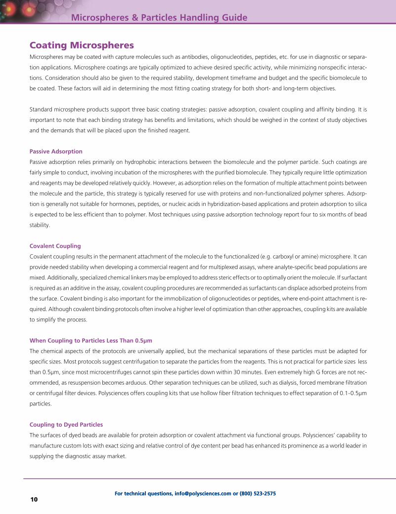
Coating MicrospheresMicrospheres may be coated with capture molecules such as antibodies, oligonucleotides, peptides, etc. for use in diagnostic or separa-
tion applications. Microsphere coatings are typically optimized to achieve desired specific activity, while minimizing nonspecific interac-
tions. Consideration should also be given to the required stability, development timeframe and budget and the specific biomolecule to
be coated. These factors will aid in determining the most fitting coating strategy for both short- and long-term objectives.
Standard microsphere products support three basic coating strategies: passive adsorption, covalent coupling and affinity binding. It is
important to note that each binding strategy has benefits and limitations, which should be weighed in the context of study objectives
and the demands that will be placed upon the finished reagent.
Passive Adsorption
Passive adsorption relies primarily on hydrophobic interactions between the biomolecule and the polymer particle. Such coatings are
fairly simple to conduct, involving incubation of the microspheres with the purified biomolecule. They typically require little optimization
and reagents may be developed relatively quickly. However, as adsorption relies on the formation of multiple attachment points between
the molecule and the particle, this strategy is typically reserved for use with proteins and non-functionalized polymer spheres. Adsorp-
tion is generally not suitable for hormones, peptides, or nucleic acids in hybridization-based applications and protein adsorption to silica
is expected to be less efficient than to polymer. Most techniques using passive adsorption technology report four to six months of bead
stability.
Covalent Coupling
Covalent coupling results in the permanent attachment of the molecule to the functionalized (e.g. carboxyl or amine) microsphere. It can
provide needed stability when developing a commercial reagent and for multiplexed assays, where analyte-specific bead populations are
mixed. Additionally, specialized chemical linkers may be employed to address steric effects or to optimally orient the molecule. If surfactant
is required as an additive in the assay, covalent coupling procedures are recommended as surfactants can displace adsorbed proteins from
the surface. Covalent binding is also important for the immobilization of oligonucleotides or peptides, where end-point attachment is re-
quired. Although covalent binding protocols often involve a higher level of optimization than other approaches, coupling kits are available
to simplify the process.
When Coupling to Particles Less Than 0.5µm
The chemical aspects of the protocols are universally applied, but the mechanical separations of these particles must be adapted for
specific sizes. Most protocols suggest centrifugation to separate the particles from the reagents. This is not practical for particle sizes less
than 0.5µm, since most microcentrifuges cannot spin these particles down within 30 minutes. Even extremely high G forces are not rec-
ommended, as resuspension becomes arduous. Other separation techniques can be utilized, such as dialysis, forced membrane filtration
or centrifugal filter devices. Polysciences offers coupling kits that use hollow fiber filtration techniques to effect separation of 0.1-0.5µm
particles.
Coupling to Dyed Particles
The surfaces of dyed beads are available for protein adsorption or covalent attachment via functional groups. Polysciences’ capability to
manufacture custom lots with exact sizing and relative control of dye content per bead has enhanced its prominence as a world leader in
supplying the diagnostic assay market.

For more information please call (800) 523-2575 or visit: www.polysciences.com11
Microspheres & Particles Handling Guide
Affinity BindingAffinity binding is a straightforward method for immobilizing primary antibodies or biotinylated molecules. Protein A and G and
Fc-specific antibody coatings permit the directed immobilization of primary antibodies and streptavidin is used extensively for the
binding of biotinylated molecules, such as antibodies, peptides and oligonucleotides.
Alternative for BSA as a Blocking AgentAny innocuous protein may be used to block the effects of non-specific adsorption. In selecting an alternative to BSA, it is suggested that
the size of the active protein and the size of the blocking protein be compared. BSA is highly recommended for IgG coupling. However,
the large size of BSA will obscure the activity of smaller, active proteins. Glycine or small polypeptides may be used as alternatives.
For more information please call (800) 523-2575 or visit: www.polysciences.com11
Well dispersed 10µm Polybead® Microspheres
End-point attachment to preserve the activity of the peptides.
End-point attachment to permit hybridization of probe sequence with target
sequence.
Common proteins are generally large enough that multi-point attachment and
nonspecific orientation do not compromise their activity. However, linkers or
spacers (covalent or SA/B) may be employed to address steric effects or
sub-optimal orientation.
Peptides
Nucleic acids
Proteins (e.g.
antibodies)
Covalent
Streptavidin / biotin
Covalent
Streptavidin / biotin
Covalent
Adsorption
Biomolecule Coating Strategy Notes

For technical questions, [email protected] or (800) 523-257512
Microspheres & Particles Handling Guide
For technical questions, [email protected] or (800) 523-257512
Protein Binding ProtocolsThe following sequences serve as guidelines for protein binding. Technical Data Sheets (TDS) with detailed step-by-step protocols can be downloaded from our website.
Adsorbing Protein on ParticlesTDS #238E
Plain Particles
• Initial Buffer: 0.1M Borate Buffer, pH 8.5
• Suspend in buffer, spin down and resuspend 2 or 3 times.
• Suspend in borate buffer.
• Add protein and mix end-to-end overnight.
• Spin and save supernatant for protein determination.
• Resuspend in BSA in appropriate buffer and spin down twice.
• Resuspend in PBS, pH 7.4, containing BSA and glycerol (storage buffer).
• Protein bound directly on surface.
Coupling by Carbodiimide TDS #238C & #644
Carboxylate Functional Particles
• Initial Buffer: 0.1M Carbonate Buffer
• Suspend in buffer, spin down and resuspend 2 or 3 times.
• Suspend in phosphate buffer.
• Add fresh carbodiimide solution dropwise and incubate for 15-30 minutes.
• Wash to remove excess carbodiimide, then resuspend in borate buffer.
• Add protein and mix end-to-end for 2-4 hours.
• Add ethanolamine, mix for 30 minutes.
• Spin and save supernatant for protein determination.
• Resuspend in BSA-containing buffer and spin down twice.
• Resuspend in PBS, pH 7.4, containing BSA and glycerol (storage buffer).
• Protein bound 2-3 carbon atoms from surface.
Coupling by Glutaraldehyde TDS #238D & #238G
Amino or Blue Dyed Particles
• Initial Buffer: 0.02M PBS, pH 7.4
• Suspend in buffer, spin down and resuspend 2 or 3 times.
• Suspend in PBS.
• Suspend in 8% glutaraldehyde in PBS, pH 7.4 and mix end-to-end for 4-6 hours.
• Wash to remove excess glutaraldehyde and resuspend in PBS buffer.
• Add protein and mix end-to-end overnight.
• Add ethanolamine, mix for 30 minutes.
• Spin and save supernatant for protein determination.
• Resuspend in BSA-containing buffer and spin down twice.
• Resuspend in PBS, pH 7.4, containing BSA and glycerol (storage buffer).
• Protein bound 5 carbon atoms from surface of blue dyed beads and 11-12 carbon atoms from surface of amino beads.
PolyPointerEasily find all of our Technical Data Sheets by visiting www.
polysciences.com

For more information please call (800) 523-2575 or visit: www.polysciences.com13
Microspheres & Particles Handling Guide
Protein Coupling TroubleshootingBelow are some solutions for protein coupling troubleshooting. If you do not see a solution to the problem you are experiencing, please email us at: [email protected]
Special PropertiesMany applications in the life sciences demand added properties, such as fluorescence or a visible color, or iron oxide inclusions for
magnetic separations. Polymer spheres (and some polymer-based magnetic spheres) are often internally dyed via organic solvent swelling
and many standard products are available. Dye concentrations can be adjusted to produce beads with different intensities to meet
special needs, such as QuantumPlex™ for multiplexed flow cytometric assays or our Dragon Green or Flash Red Intensity standards, which
support imaging applications and associated instrument QC. Many surface- or internally-labeled fluorescent beads are also available as
specialized flow cytometry standards.
Various types of superparamagnetic microparticles are available as well – with different matrices, magnetite content, surface groups,
etc. For new assays or applications, magnetic beads should be evaluated with application demands in mind.
The following tables provide product suggestions for common microsphere applications. These are offered as general guidelines only.
Further literature research and screening experiments may be appropriate.
For more information please call (800) 523-2575 or visit: www.polysciences.com13
Problem Solution
Clumping prior to use
Clumping after procedure
• Carbodiimide addition causes clumping
• Glutaraldehyde addition causes clumping
• Protein addition causes clumping
• Washing causes clumping
Low binding
Variable coating
Coating, but no reaction
Centrifuge not practical
Nonspecific adsorption
Small proteins bound, but not reactive
Long-term storage leaches protein
Careful sonication
Isolate which step causes clumping
Add slowly, agitate beads, decrease bead concentration
Add slowly, agitate beads, decrease bead concentration; add an excess of glu-
taraldehyde to avoid chemical crosslinking of particles; clumping will typically
resolve by the conclusion of the protein coupling
Increase protein concentration
Add surfactant or reduce washing steps
Move pH of binding closer to protein isoelectric point
Use pure water - no contaminants; use fresh reagents
Optimize pH away from isoelectric point
Use membrane filtration, dialysis, or spin fibers for small particles
Use an alternative for BSA (glycine, casein)
Use a crosslinking agent to extend coupling away from
the surface of the bead
Try covalent attachment or lyophilize final product

For technical questions, [email protected] or (800) 523-257514
Microspheres & Particles Handling Guide
For technical questions, [email protected] or (800) 523-257514
Microsphere Selection for Common Test and Assay Formats:
Suggested Products for Magnetic Assays:
Suggested Products for Magnetic Separations:
Assays Suggested Products
Immunoassays
Hybridization-based assays
ProMag™ or BioMag®
ProMag™
Test / Assay Format Bead Size Bead Type Coating Strategy Detection Strategy
Flow cytometric
(suspension array)
Lateral flow
Lateral flow -
Boulders in the Stream
Dipstick
Latex Agglutination Tests
(LATs)
Light Scattering
(automated LAT -
Nephelometric or
Turbidimetric)
2-15µm
0.1-0.4µm
0.1-0.4µm
~2-3µm
0.1-0.4µm
0.2-1.0µm
0.36-0.76µm
(diameter equal
to wavelength
of light being
scattered)
QuantumPlex™
QuantumPlex™M (encoded
populations for multiplexing)
Non-fluorescent
Dyed (visible or
fluorescent)
Dyed (visible mobile phase)
Undyed (capture phase)
Dyed (visible)
Undyed
Visibly dyed
Undyed
Flow cytometer
Visual or automated reader
(absorbance, fluorescence)
Visual
Visual
Visual (may be microscope-
assisted)
Nephelometer
Turbidimeter
Covalent
Streptavidin / biotin
Covalent
Adsorption
Covalent
Adsorption
Covalent
Adsorption
Covalent
Adsorption
Covalent
Magnetic Separations Suggested Products
Cells
Subcellular organelles
Immunoprecipitates
mRNA
Biotinylated oligonucleotide capture or binding
Biopanning
BioMag® anti-CD marker or secondary antibody
BioMag®
BioMag®
BioMag® Oligo dT (20) or mRNA Purification System
ProMag™ or BioMag®
ProMag™ or BioMag®

For more information please call (800) 523-2575 or visit: www.polysciences.com15
Microspheres & Particles Handling Guide
For more information please call (800) 523-2575 or visit: www.polysciences.com15
Instrument Quality Control For confidence in test or assay results, a facility must employ a comprehensive quality assurance program that encompasses employee
training and routine instrument maintenance and quality control. Facilities should also ensure that studies, particularly those involving
quantitative fluorescence measurements, are conducted with appropriate standardization.
Microsphere standards aid in defining the instrument’s capabilities and limitations in terms of sensitivity, precision and accuracy and
provide a means for ensuring that the instrument is stable and suitable for use. They are also helpful in understanding the effects of
extraneous factors, such as temperature, humidity and electronic noise. The comparison of daily and historical QC data aids in the
identification of random errors (due to electronic noise, air bubbles, etc.) and systematic errors (due to bias, shifts and trends caused by
temperature variation, laser deterioration, misalignment, etc.) so that suitable corrective action may be taken.
Daily Controls
Flow cytometers are highly configurable and results can vary dramatically with different instrument settings. Establishing a common “Win-
dow of Analysis” for each detector, with the upper and lower fluorescence limits defined, allows reference populations to be positioned in
approximately the same place on the same scale. This type of standardized instrument set-up ensures consistency of results from specific
instruments and enables meaningful comparison between instruments. Therefore, the general status and stability of the cytometer must
be checked daily. In addition, by plotting values over time, random and systemic errors may be identified and corrected. Manually aligned
instruments must be aligned daily. If the data from multiple instruments and/or sites are to be compared, all instruments must be standard-
ized daily, using the same standard bead. If multi-color analysis and/or quantitation is being performed, then the appropriate standards
must be run on that same day.
Weekly Controls
If only qualitative analyses are being run, weekly checks of the detection threshold, resolution and linearity are sufficient to establish sensi-
tivity. Some cytometers are fixed alignment instruments. For these, alignment may be verified weekly rather than established daily.
In addition to a regular QA/QC program, specific analyses may require other controls, such as count standards, size standards or
reference standards. Experiments and analyses must be critically evaluated for inclusion of the appropriate controls prior to
instrumental analysis.
U.S. Corporate Headquarters | 400 Valley Road, Warrington, PA 18976 | 1(800) 523-2575 / (215) 523-2575 | Fax 1(800) 343-3291 | [email protected]
Polysciences Europe GmbH | Handelsstrasse 3 D-69214 Eppelheim, Germany | +(49) 6221-765767 | Fax +(49) 6221-764620 | [email protected]
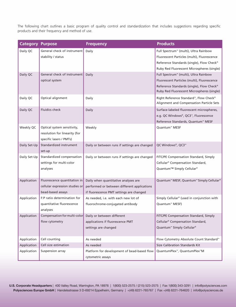
Category Purpose Frequency Products
Daily QC
Daily QC
Daily QC
Daily QC
Weekly QC
Daily Set-Up
Daily Set-Up
Application
Application
Application
Application
Application
Application
Daily
Daily
Daily
Daily
Weekly
Daily or between runs if settings are changed
Daily or between runs if settings are changed
Daily when quantitative analyses are
performed or between different applications
if fluorescence PMT settings are changed
As needed, i.e. with each new lot of
fluorochrome-conjugated antibody
Daily or between different
applications if fluorescence PMT
settings are changed
As needed
As needed
Platform for development of bead-based flow
cytometric assays
General check of instrument
stability / status
General check of instrument
optical system
Optical alignment
Fluidics check
Optical system sensitivity,
resolution for linearity (for
specific lasers / PMTs)
Standardized instrument
set-up
Standardized compensation
settings for multi-color
analyses
Fluorescence quantitation in
cellular expression studies or
bead-based assays
F:P ratio determination for
quantitative fluorescence
analyses
Compensation for multi-color
flow cytometry
Cell counting
Cell size estimation
Suspension array
Full Spectrum™ (multi), Ultra Rainbow
Fluorescent Particles (multi), Fluorescence
Reference Standards (single), Flow Check®
Ruby Red Fluorescent Microspheres (single)
Full Spectrum™ (multi), Ultra Rainbow
Fluorescent Particles (multi), Fluorescence
Reference Standards (single), Flow Check®
Ruby Red Fluorescent Microspheres (single)
Right Reference Standard™, Flow Check®
Alignment and Compensation Particle Sets
Surface-labeled fluorescent microspheres,
e.g. QC Windows®, QC3™, Fluorescence
Reference Standards, Quantum™ MESF
Quantum™ MESF
QC Windows®, QC3™
FITC/PE Compensation Standard, Simply
Cellular® Compensation Standard,
Quantum™ Simply Cellular®
Quantum™ MESF, Quantum™ Simply Cellular®
Simply Cellular® (used in conjunction with
Quantum™ MESF)
FITC/PE Compensation Standard, Simply
Cellular® Compensation Standard,
Quantum™ Simply Cellular®
Flow Cytometry Absolute Count Standard™
Size Calibration Standards Kit
QuantumPlex™, QuantumPlex™M
The following chart outlines a basic program of quality control and standardization that includes suggestions regarding specific products and their frequency and method of use.


