Microsensors allow continuous monitoring of myocardial ...
44
1 Microsensors allow continuous monitoring of myocardial ischemia in cardiac surgery Stefan Hyler The Intervention Centre, Oslo University Hospital, Oslo, Norway Institute for Clinical Medicine, Faculty of Medicine, University of Oslo, Norway 2015
Transcript of Microsensors allow continuous monitoring of myocardial ...

1
Microsensors allow continuous monitoring of myocardial ischemia in cardiac surgery
Stefan Hyler
The Intervention Centre, Oslo University Hospital, Oslo, Norway
Institute for Clinical Medicine, Faculty of Medicine, University of Oslo, Norway
2015

© Stefan Hyler, 2016 Series of dissertations submitted to the Faculty of Medicine, University of Oslo ISBN 978-82-8333-174-5 ISSN 1501-8962 All rights reserved. No part of this publication may be reproduced or transmitted, in any form or by any means, without permission. Cover: Hanne Baadsgaard Utigard Printed in Norway: 07 Media AS – www.07.no

3
TABLE OF CONTENTS
Acknowledgements ............................................................................................................ 4
Abbreviations ..................................................................................................................... 5
List of papers ...................................................................................................................... 6
Introduction ........................................................................................................................ 7
Myocardial ischemia in cardiac surgery .................................................................................. 7
Sensors for monitoring cardiac muscle metabolism and function ....................................... 11
Aim of the study ............................................................................................................... 14
Materials and methods ..................................................................................................... 15
Experimental model .............................................................................................................. 15
Sensor Systems ..................................................................................................................... 17
Hemodynamic measurements .............................................................................................. 20
Reference methods ............................................................................................................... 21
Statistics ................................................................................................................................ 23
Summary of results ........................................................................................................... 24
Study 1 ‐ Epicardial ultrasound sensor ................................................................................. 24
Study 2 ‐ Tissue PtCO2 Sensor ............................................................................................... 26
Study 3 ‐ Accelerometer sensor ............................................................................................ 28
Discussion ......................................................................................................................... 29
Microsensors for myocardial ischemia detection ................................................................. 29
The relation between coronary flow, myocardial ischemia and function ............................ 32
Clinical use of microsensors .................................................................................................. 33
Limitations ............................................................................................................................. 34
Future perspectives .............................................................................................................. 35
Conclusions ....................................................................................................................... 36
References ........................................................................................................................ 37

4
ACKNOWLEDGEMENTS
The work has been carried out from 2009 to 2015 at Intervention Centre, Oslo University Hospital Rikshospitalet and University of Oslo, Norway. The research project has been founded by The Regional Health Authority of South‐Eastern Norway (Helse Sør‐Øst).
I would like to express my deep gratitude to my three supervisors who have supported me throughout these years: Steinar Halvorsen; The Intervention Centre, Professor Erik Fosse; The Intervention Centre, and Helge Skulstad; Department of Cardiology. Their outstanding knowledge and broad experience within experimental cardiovascular research has been of important value for this work.
My colleague Søren Pischke, he has been a key person and contributed essentially to the studies. He shared his office with me and we spent a lot of time at the operation theatre during the experiments. I would in particular like to thank him for the excellent collaboration and many discussions, on topic or not.
The microsensors used in this project has been developed and refined by The Vestfold University College and the group around IscAlert. Without these people the project wouldn’t have been possible, I am grateful for the good collaboration and technical support.
My sincere thanks are given to the staff at The Intervention Centre for their excellent skills and teamwork, to Marianne Berg for her support, encouragement and unselfishly facilitating the PhD student’s everyday life. I feel very privileged to have been a part of this inspiring and motivated multidisciplinary team.
I am deeply grateful to my family. My three daughters Linnea, Camilla and Elizabeth – thank you for reminding me every day what is important in life. And finally, I want to thank my dear Liv for her patience and great support during all these years.
Kristiansand, June 2015
Stefan Hyler

5
ABBREVIATIONS
LV Left ventricle LAD Left anterior descending coronary artery Cx Circumflex coronary artery LIMA Left internal mammary artery CABG Coronary artery bypass grafting ECG Electrocardiogram NSTEMI non‐ST elevation myocardial infarction HR Heart rate MAP Mean arterial pressure dP/dtmax Time derivative of left ventricular pressure LVEDP Left ventricular end‐diastolic pressure LVSW Left ventricular stroke work Vsys Peak systolic velocity Vpost Peak postsystolic velocity PtCO2 Partial pressure of carbon dioxide in the tissue PtO2 Partial pressure of oxygen in the tissue OR Operation room CT Computer tomography IFI Fluorescence imaging 2D Two dimensional ROC Receiver operating characteristic

6
LIST OF PAPERS
Paper I
Hyler S, Pischke S, Halvorsen PS, Espinoza A, Bergsland J, Tønnessen TI, Fosse E, Skulstad H. Continuous monitoring of regional function by a miniaturized ultrasound transducer allows early quantification of low grade myocardial ischemia. J Am Soc Echocardiograph 2015; 28: 486–494
Paper II
Pischke SE, Hyler S, Tronstad C, Bergsland J, Fosse E, Halvorsen PS, Skulstad H, Tønnessen TI. Myocardial tissue CO2 tension detects coronary blood flow reduction after coronary artery bypass in real‐time. Br J Anaesth 2015; 114: 414–422
Paper III
Hyler S, Espinoza A, Skulstad H, Fosse E, Halvorsen PS. Left ventricular function can be continuously monitored with an epicardially attached accelerometer sensor. Eur J Cardiothorac Surg 2014; 46: 313–320

7
INTRODUCTION
Following the construction of the first functional bubble oxygenator by Richard de Wall in the 1955, and the first successful surgeries by C Walton Lillehei, surgery rapidly became the main treatment option for a number of cardiac disorders. Soon after the first successful m surgeries for congenital disease, coronary revascularization, and surgery for valvular disease became some of the most common operations in major hospitals. Eventually cardiac transplant became an established treatment for myocardial infarction. Along with improved systems for extracorporeal circulation, anaesthesia and monitoring, cardiac surgery could be offered to patients with severe disease and with a number or concomitant diseases. In 1979 the German cardiologist Andreas Grüntzig presented coronary angioplasty in patients with coronary stenosis [1]. Since the 1980ies, an increasing number of patients with simple coronary disease were treated with interventional procedures, and since the late 1990ies congenital disorders like atrial septal defects and in some patients ventricular septal defects can also be treated by radiology‐guided intervention. April 16 2002 Prof Alain Cribier at Hôpital Charles Nicolle, at the University of Rouen performed the first catheter‐based aortic valve replacement [2]. Since then a number of aortic valves for catheter based treatment of valvular disease has been developed. Thus the profile of the patients scheduled to cardiac surgery has changed dramatically during the last decades [3–6]. There is a trend towards older patients, a population that is expected to increase in the near future. Among elderly people, extensive coronary artery disease and impaired left ventricular function is more common. Further comorbidities will be present to a higher extent; they are more likely to have a history of coronary artery graft surgery and a higher risk for urgent surgical procedures. This selection of high risk patients is thus more prone to unfavourable outcome. In order to assure a beneficial outcome in these high risk patients, it is an increasing need for more accurate and advanced systems for perioperative monitoring.
MYOCARDIAL ISCHEMIA IN CARDIAC SURGERY
Cardiac ischemia implies insufficient blood supply to the heart by either full or partial blocking of a coronary artery. The majority of patients undergoing cardiac surgery experiences one or several episodes of myocardial ischemia. The reported incidence in the course of coronary artery bypass graft surgery is varying between 36% and 58%. The great variance is due to different diagnostic criteria and inhomogeneity of the selected patient populations [7–9]. Two pathomechanisms are mainly involved in the development of perioperative ischemia:

8
(i) Graft failure: Early graft occlusion is a well known and serious complication in coronary bypass surgery. As much as 8% of grafts were found to be occluded by intraoperative coronary angiography, requiring revision [10]. Major causes are due to failure in surgical technique, such as misplaced suture leading to stenosis at the toe or heel of the anastomosis, dissection or graft kinking [11]. Endothelial trauma of the target vessel may lead to thrombosis or dissection. Other reasons may be vasospasm [12,13], edema, rupture of a vulnerable plaque resulting in acute coronary thrombosis, or peripheral arterial disease causing reduced run off resulting in thrombosis of the graft.
(ii) General postoperative causes leading to oxygen supply/demand imbalance: Clinically, stenosis of a coronary artery graft is more frequent than total occlusion and leads to tissue hypoxia rather than tissue anoxia. The main postoperative causes of reduced oxygen supply include hypotension, hypovolemia, anemia and arrhythmia. Especially tachycardia may increase the oxygen demand of the patient and particularly of the myocardium. Stress induced by pain, hypertension and hyperthermia, may also increase the oxygen demand [14].
Regardless of cause, ischemia will result from an imbalance between myocardial oxygen supply and demand in the patient with extensive coronary disease. In high risk patients, most of the ischemic episodes happen within the first days after surgery [15]. Throughout the entire perioperative period, ischemia occurs considerably more frequent postoperatively [16], and the majority of ischemic episodes are asymptomatic (silent) [15,17].
For patients undergoing peripheral vascular surgery the risk for myocardial infarction is substantially increased with the length of ischemic attacks and as well with their cumulative duration due to repetitive ischemia [14]. During coronary bypass grafting Ischemia has been identified to be an important and independent predictor for perioperative myocardial infarction[8] and is associated with a 3‐fold increased risk [9]. Acute myocardial ischemia is followed by immediate reduction in myocardial contraction [18,19]. Heart failure is an independent predictor for unfavorable outcome after coronary bypass surgery (CABG) and ischemia is a leading cause in the impairment of left ventricular (LV) function [7,20,21]. A main goal of coronary bypass grafting, besides relief of symptoms and prolonged survival, is the preservation of myocardial function. Therefore monitoring for early detection of ischemia during and after cardiac surgery is essential for better survival without adverse cardiac events.
Recurrent coronary surgery is associated with increased risk for perioperative myocardial infarction and development of low cardiac output syndrome, indicating an increased need for accurate monitoring [22].

9
CURRENT METHODS FOR DETECTION OF ISCHEMIA IN CARDIAC SURGERY
VISUAL ASSESSMENT During surgery, when the heart is beating, ischemia is often detected by visual assessment of the heart wall motions in the surgical field. Visual assessment is subjective and subtle changes in heart wall motion may not be detected. Visual assessment also does not provide quantitative data.
CONTINUOUS ECG Electrocardiography (ECG) is mandatory for perioperative monitoring of all patients undergoing cardiac surgery. It is inexpensive, easily disposable and requires little training. By computerized ST‐segment analysis ECG provide continuous information about the electrical impulses of the heart. Although these impulses may be affected by myocardial ischemia, ECG does not give direct information of the blood supply to the heart. Thus, following cardiac surgery, interpretation may be challenging because of pacemaker dependency and intermittent branch blocks or arrhythmias, due to edema or handling of the heart that may cause disturbances. Proper placement of the electrodes and selection of the leads are essential for the sensitivity of the method and can further distinctly be increased when several precordial leads were selected [23]. However ECG performs with lower sensitivity and specificity in ischemia detection compared to echocardiography [7,24].
HAEMODYNAMIC PARAMETERS Arterial and central venous pressure are measured in all patients undergoing cardiac surgery, however these pressures are unspecific, as they are a product of the degree of left ventricular filling, the myocardial function and of peripheral resistance. Measurements from pulmonary artery catheter inserted directly in the pulmonary artery may give additional information. A pulmonary artery pressure measurement has a limited role in ischemia detection but provides valuable information supplementary to arterial pressure, central venous pressure and ECG. A Swan Ganz catheter allows both measurements of central venous pressure, pulmonary artery pressure and pulmonary capillary wedge pressure, as well as intermittent measurements of cardiac output. With all these invasive parameters a number of calculations may be performed, such as left ventricular stroke work, right ventricular stroke work and peripheral vascular resistance. Myocardial ischemia may thus be revealed by hemodynamic abnormalities due to changes in systolic and diastolic function. Ischemia induced reduction of contractile function leads to enlargement of the left ventricle with increase of end‐systolic and end‐diastolic volume, resulting in decreased cardiac output together with decreased LV pressure and increased left ventricular end‐diastolic pressure. Finally mitral regurgitation may be detected with the pulmonary balloon catheter in wedge position by sudden appearance of an enlarged V‐wave, resembling a pulmonary artery trace.
The changes detected by invasive hemodynamic monitoring are due to deterioration of global ventricular function, thus not able to discover minor changes in graft function or early stages of ischemia.

10
ECHOCARDIOGRAPHY Impairment in regional myocardial function often precedes the changes in global hemodynamic parameters. It is desirable to detect regional changes before it reaches global or systemic consequences. Echocardiography has shown to be more sensitive than hemodynamic measurements in detecting perioperative ischemia [7,25]. Unfortunately, routine echocardiography necessitates intermittent monitoring by skilled examinators. In addition, sensitive echocardiographic methods, such as tissue Doppler imaging and speckle tracking strain, are time consuming and impractical on a routine basis. A main advance is, however, that the result of the surgical procedure as for instance valve replacement can be evaluated instantly, thus perioperative echo has became a standard procedure in cardiac surgery.
CORONARY ANGIOGRAPHY Conventional angiography still accounts as the gold standard method to assess coronary flow and graft patency. However, most of the operating theaters are lacking advanced equipment that is available in modern angiographic facilities. Other major limitations are the requirement of trained personnel with catheter skills, increasing operation time and the potential risk to harm the patient by potentially nephrotoxic agents or a thromboembolic event.
GRAFT FLOW MEASUREMENT Several ultrasound based method were used to asses proper graft function. Intraoperative graft flow measurement with a handheld Doppler probe is simple to use however velocity is dependent on the insonation angle. In a study comparing the transit time flow measurements with recordings of mean flow and pulsatility index (the difference between maximum forward flow and maximum negative flow divided by mean flow demonstrated god ability to evaluate graft patency after coronary artery bypass surgery [11]. However when on table angiography was used as reference method, Hol and coworkers demonstrated that transit time flow had poor sensitivity and specificity [26]. This confirmed the findings by Elbeery et al from 1998 that transit time flow measurements do not predict graft patency when comparing with angiography [27].
Hol et al also investigated the ability of epicardial Color Doppler Ultrasonography with a high frequency probe to detect ischemia, also with this method they found only poor correlation between angiographic and ultrasound findings [10].
Both these methods are designed for intraoperative control and not for continuous monitoring.

11
OTHER IMAGING MODALITIES Fluorescence imaging (IFI) is a non‐invasive method based on indocyanin green dye which is systemically administered and bound to plasma proteins. Native coronary arteries and grafts are visualized when illuminated with infrared light. As limiting factors it has to be mentioned that the method only allows semiquantitative assessment of the graft patency and second precise imaging of anastomosis is not possible because of limited tissue penetration of the fluorescent light.
Newly, thermal coronary angiography was presented, in which differences in epicardial surface temperature of are visualized with an infrared camera. However this method has not become a standard in the clinical routine, limited data are available and it fails to detect flow from intramyocardial arteries [28].
Dual source CT scanning is a promising tool for non‐invasive diagnostics of the coronary arteries. Studies have demonstrated high sensitivity and specificity for the detection of impaired coronary flow [29]. However so far this technology has not reached the ORs and it still is an intermittent examination. Although radiation dose has been drastically reduced in modern CTs this still has to be taken into account, when repeated examinations are considered.
SENSORS FOR MONITORING CARDIAC MUSCLE METABOLISM AND FUNCTION
ULTRASOUND TRANSDUCERS
Ultrasonic methods to measure changes in cardiac dimensions were first introduced into cardiac research in 1956 by Rushmer and Franklin’s prototype with two piezoelectrical crystals, working independently as transmitter and receiver. [30] Those “microcardiometers” were positioned on opposite sides of the left ventricle and pulsed waves were sent across the cardiac chamber. The signals, picked up by the receiver were transformed into a voltage, proportional to the transit time, allowing to estimate changes in ventricle dimensions. Ultrasonic crystals have been used in several experimental studies on myocardial function [31,32]. A new Doppler based method with a single crystal, intermittently acting as transmitter and receiver was introduced in 1983 and gave the opportunity to measure wall thickening without penetrating the myocardium with transducers [30]. Changes in wall thickening were estimated as the velocity of myocardial layers, gained from a volume sample at a selected depth, was integrated by time. Recently, an ultrasonic transducer was developed and validated at the Intervention Centre, which can be attached to the epicardium [34]. The method enables continuous monitoring of heart wall contractions by use of an M‐mode image and real‐time calculation of myocardial velocities. This transducer has shown to be superior in detecting myocardial

12
ischemia compared to ECG and invasive hemodynamic monitoring during off‐pump coronary artery bypass surgery [35].
METABOLIC SENSORS
Decrease in sufficient oxygen supply to myocardium results in alteration of the energy metabolism which is normally finely regulated to the demand of cardiac myocytes. The majority of energy originates from oxidation of fatty acids and carbohydrates in the mitochondria. However during ischemia aerobic metabolism is increasingly inhibited and glycolysis becomes the main source of energy synthesis. Downregulation in mitochondrial oxidation and switch to anaerobic metabolism impedes removal of intermediate products from glycolysis leading to accumulation of lactate and H+‐ions. Depending on the severity of the ischemia, a substantial fall in pH and increase of tissue PCO2 (PtCO2) by buffering with bicarbonate will be expected. Several methods have been used to monitor changes in myocardial energy metabolism during ischemia. Microdialysis catheters where used to collect aliquots with metabolic intermediates from the myocardium and helped to understand local metabolic changes in the ischemic heart [36–38]. The method is based on the principle that solutes diffuse along a concentration gradient and finally pass across a selective barrier. Methods better suited to immediately detect ongoing myocardial ischemia were based on changes in H+‐ion concentrations measured by electrodes and fibre‐optic probes [36,37] or tissue PCO2 (PtCO2) by mass spectrometry and fibre‐optic probes [37,38]. A newly invented conductometric PtCO2 sensor (IscAlertTM) demonstrated reliable and immediate detection of tissue anoxia within three minutes of complete occlusion of the left anterior descending coronary artery (LAD) [42]. The mass spectrometry for ischemia detection in the heart was a purely experimental method used in animal models. Fiberoptic probes are commercially not available, which restricts their application. Conductometric PtCO2 sensors (IscAlertTM) are approved for use in human beings.
ACCELEROMETER
Accelerometers are electromechanical devices that measure acceleration forces. They were primarily developed for military purpose and became essential in navigation systems for missiles, space crafts and airplanes. The technology has early been commercialized and the sensors have radically been miniaturized during the last decades. Today accelerometers have become inexpensive and are found in many consumer electronic devices such as cell phones and gaming consoles. The new generation of accelerometers consist of piezoelectric crystals, a material that changes its surface charge when mechanical stress is applied to it and therefore allow estimation of movements by changes in voltage. Accelerometers are widely used in medicine to analyze body posture and movements and have found many indications in neurology and rehabilitation medicine [43,44] (e.g. optimizing physical training and rehabilitation, fall prevention in elderly, diagnosis of tremor or abnormal movements,

13
pedometers). While in cardiology accelerometers have been used in many years to steer rate adaptive pacemakers by measuring the extent of physical activity [45]. When attached to the heart, movement or acceleration of the cardiac surface will generate changes in voltage. These changes can mathematically be transformed to estimate heart motion expressed by acceleration, velocity or displacement [46]. Heart motion within different phases during one cardiac cycle can be identified by combining electrocardiograms (ECGs) and accelerometer signals. This technology is a promising method for continuous monitoring of LV performance, which allows beat‐to‐beat analysis of heart motion in real‐time [47]. Further, minimal size and low energy consumption make it possible to assemble the sensor with a pacemaker lead, which make the device even more applicable for preoperative monitoring. In previous experimental and clinical studies, we have demonstrated that epicardially attached accelerometers were able to provide information similar to that from echocardiography during regional LV dysfunction induced by temporary occlusion of the left descending artery [48,49]. We hypothesized that:
1. Microsensors perform better in detecting myocardial ischemia than existing methods.
2. There is a correlation between coronary flow reduction, myocardial ischemia and
function.

14
AIM OF THE STUDY
Main aims:
1. To evaluate the strengths and limitations in the three sensors ability to detect myocardial ischemia.
2. Demonstrate the association between myocardial contraction, metabolism and cardiac function as detected by microsensors.
Specific aims:
1. Metabolic ‐ Demonstrate that PCO2 monitoring by myocardial microsensors allows detection and quantification of ischemia that correlates with contractile function.
2. Ultrasound ‐ Demonstrate the ability of miniaturized epicardial ultrasound transducer
to detect and quantify low‐grade regional myocardial ischemia.
3. Accelerometer ‐ Demonstrate that epicardial accelerometers allow detection of ischemia that correlates with contractile function.

15
MATERIALS AND METHODS The same open chest large animal model was used in all three studies. All studies were approved by the National Animal Research Authority (NARA). In our large animal model we chose to perform the studies on the pig because the pig heart has anatomical and structural similarities to humans. Furthermore, the pigs heart has limited collateral coronary artery perfusion [50], which makes it easier to consistently regulate coronary flow and induce myocardial ischemia.
EXPERIMENTAL MODEL
All experiments were conducted in Noroc pigs of either sex with a weight between 50 and 60 kg. General anesthesia was used in all animals. Barbiturates and morphine were used for induction of anesthesia and a continuous infusion morphine and inhaled isoflurane were used for maintains of anesthesia. In all studies the surgical preparation included a tracheotomy and sternotomy and placement of intravascular and ventricular pressure catheters and placement of sensors on the left ventricle. A detailed description of the surgical preparation and placement of sensors are described in study I‐III.
REGIONAL MYOCARDIAL ISCHEMIA
LAD is the most important coronary artery to the left ventricle and significant stenosis of the proximal LAD is among the major indications for CABG [51]. Revascularization by grafting the left internal mammary artery (LIMA) has shown to have the most favorable long‐term outcome [52]. In our experimental model LAD flow impediment was therefore the most clinical relevant situation to test the different sensors ability to detect regional myocardial ischemia. In the graded ischemia experimental set up (study I and II) LIMA was grafted to the LAD and the LAD occluded proximal to the anastomosis. Then a vascular occluder and a 4 mm flow probe were placed around LIMA for precise adjustment of flow impediment to the left ventricular apical region (figure 1).
In study I and II coronary artery flow reductions were induced by LIMA occlusion. LIMA flow was reduced to 25%, 50% and 75% for 18 min at each flow reduction. The flow reductions were not randomized because of the risk of ventricular fibrillation and loss of the animal during severe ischemia. Complete reperfusion of the LIMA was performed between each flow reduction and baseline measurements undertaken. In study III a snare was placed around LAD and a 3 minutes period of no flow introduced by tightening the snare similar to what is done during LAD grafting in off pump coronary artery surgery. The reference method to detect ischemia in study I and II was serum lactate and in study III myocardial strain by echocardiography.

16
In order to test the different sensors ability to measure regional changes two sets of sensors were placed in two different LV areas, in the intervention region supplied by the LAD and in a control region supplied by the circumflex coronary artery.
INTERVENTIONS ON GLOBAL LEFT VENTRICULAR FUNCTION
To test the ability of the accelerometer to distinguish regional dysfunction from changes in global left ventricular function preload, afterload and contractility were altered by an infusion of fluid and injections of esmolol and epinephrine respectively (study III).
Figure 1: The experimental set up in study I and II. The left internal mammary artery (LIMA) was grafted to the left anterior descending artery (LAD), which was occluded proximal to the anastomosis by a silicon snare. A vascular occluder on LIMA was used to impede LIMA flow stepwise, based on flow measurements by a flow probe distally to the occluder. Placements of the different sensors on the left ventricle are indicated together with a pressure catheter in the left ventricle.

17
SENSOR SYSTEMS
EPICARDIAL ULTRASONIC SYSTEM
In study I myocardial tissue velocity was obtained by epicardial ultrasonic sensor system. The system included a miniaturized 10‐MHz ultrasonic transducer (Imasonic SA, Besancon, France) with dimensions of 5mm in diameter and 4 mm in height. The transducer acted as both an ultrasonic transmitter and a receiver. Placed on the left ventricle the sensor provided a real‐time M‐mode image of the heart wall where systolic thickening and diastolic thinning could be visualized. From this M‐mode picture the corresponding wall thickening and thinning myocardial velocities were displayed and measured [34,35]. In order to measure myocardial velocities within the cardiac cycle the ECG and left ventricular pressure were displayed together with the M‐mode picture and the velocity curve obtained from the ultrasonic transducer. Systole was defined as the period from onset of the R‐wave on ECG to the left ventricular dP/dtmin. This made it possible to assess both peak systolic and post systolic velocities which have been extensively validated as markers of regional left ventricular dysfunction and myocardial ischemia by tissue Doppler echocardiography [53,54]. Peak systolic myocardial velocity obtained from the ultrasonic transducers is
Figure 2: Recordings from the ultrasonic system during incremental flow reduction are shown as M‐mode images (upper panel). Velocity traces obtained from the subendocardium (yellow lines) are synchronized with aortic and LV pressure, LV dP/dt and ECG (lower panel). During ischemia the velocity traces show the characteristic pattern with decrease in systolic (Vsys) and increase in postsytolic (Vpost) velocities.

18
unaffected by moderate changes in preload but has the sensitivity to detect changes in contractility [34,55,56]. The time‐velocity trace was obtained with dedicated software from a range‐gated volume sample at a fixed depth in the subendocardium (figure 2) as this layer of the ventricular wall is most vulnerable to coronary artery flow reduction and myocardial
ischemia [57]. Intermittent recordings were obtained and stored for post hoc analysis.
The epicardial positioning of the transducers, using three eyes for suture fixation to the epicardium, ensured a perpendicular insonation angle on the myocardium, enabling the assessment of regional myocardial function unaffected by global heart movements (figure 3).
PTCO2 SENSORS
Two different PtCO2 sensors were used:
1) A fiberoptical multiparameter sensor that in addition to real‐time assessment of PtCO2 also measured PtO2, pH and temperature (Neurotrend®, Codman, MA, USA).
2) A conductometric sensor, which also measured PtCO2 and temperature in real‐time (IscAlertTM , Sensocure AS, Horten, Norway)
Insertion of both PtCO2 sensors into the myocardium of the left ventricle was performed by use of a splittable introducer with diameter of 1 mm.
THE FIBEROPTICAL MULTIPARAMETER SENSOR The fiberoptic multiparameter sensor was used in study II and was placed in the LIMA graft dependent myocardium (figure 1). The sensor is a 0.5 mm thick single‐use device, which has been FDA approved for use in human brain in neurosurgery. The sensor has also been used in experimental studies to detect ischemia in the heart [40,42], liver [58,59] and intestine [60]. At the tip of the sensor three optical sensors and one thermocouple are included in a 4 cm long gas‐permeable poly‐ethylene membrane. The oxygen sensor consists of a fiber containing ruthenium based dye. The fiber is illuminated with blue light (wave length 460 nm), which excite ruthenium complexes. Excess energy from this process is emitted as fluorescent light (wavelength of 620 nm) and is measured by the monitor (Codman, MA, USA). In the presence of oxygen, however, parts of the energy are absorbed and the intensity of the emitted fluorescent light is reduced, a phenomenon called oxygen
Figure 3: Epicardial ultrasonic sensor attached to the heart.
Figure 3: Epicardial ultrasonic sensor attached to the heart.
19
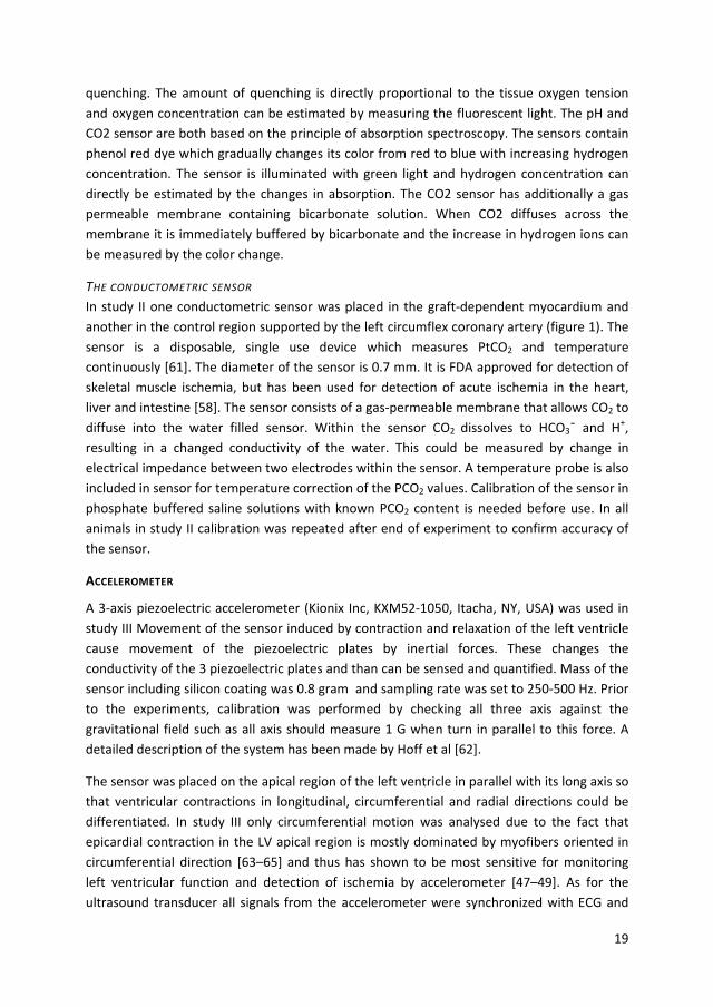
quenching. The amount of quenching is directly proportional to the tissue oxygen tension and oxygen concentration can be estimated by measuring the fluorescent light. The pH and CO2 sensor are both based on the principle of absorption spectroscopy. The sensors contain phenol red dye which gradually changes its color from red to blue with increasing hydrogen concentration. The sensor is illuminated with green light and hydrogen concentration can directly be estimated by the changes in absorption. The CO2 sensor has additionally a gas permeable membrane containing bicarbonate solution. When CO2 diffuses across the membrane it is immediately buffered by bicarbonate and the increase in hydrogen ions can be measured by the color change.
THE CONDUCTOMETRIC SENSOR In study II one conductometric sensor was placed in the graft‐dependent myocardium and another in the control region supported by the left circumflex coronary artery (figure 1). The sensor is a disposable, single use device which measures PtCO2 and temperature continuously [61]. The diameter of the sensor is 0.7 mm. It is FDA approved for detection of skeletal muscle ischemia, but has been used for detection of acute ischemia in the heart, liver and intestine [58]. The sensor consists of a gas‐permeable membrane that allows CO2 to diffuse into the water filled sensor. Within the sensor CO2 dissolves to HCO3ˉ and H
+, resulting in a changed conductivity of the water. This could be measured by change in electrical impedance between two electrodes within the sensor. A temperature probe is also included in sensor for temperature correction of the PCO2 values. Calibration of the sensor in phosphate buffered saline solutions with known PCO2 content is needed before use. In all animals in study II calibration was repeated after end of experiment to confirm accuracy of the sensor.
ACCELEROMETER
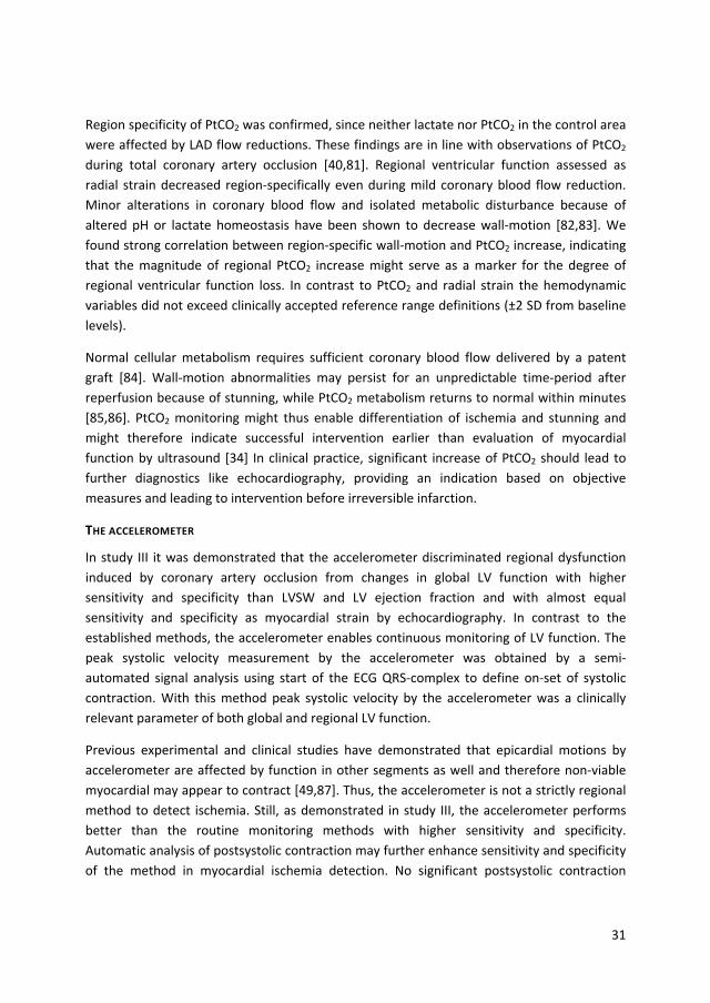
A 3‐axis piezoelectric accelerometer (Kionix Inc, KXM52‐1050, Itacha, NY, USA) was used in study III Movement of the sensor induced by contraction and relaxation of the left ventricle cause movement of the piezoelectric plates by inertial forces. These changes the conductivity of the 3 piezoelectric plates and than can be sensed and quantified. Mass of the sensor including silicon coating was 0.8 gram and sampling rate was set to 250‐500 Hz. Prior to the experiments, calibration was performed by checking all three axis against the gravitational field such as all axis should measure 1 G when turn in parallel to this force. A detailed description of the system has been made by Hoff et al [62].
The sensor was placed on the apical region of the left ventricle in parallel with its long axis so that ventricular contractions in longitudinal, circumferential and radial directions could be differentiated. In study III only circumferential motion was analysed due to the fact that epicardial contraction in the LV apical region is mostly dominated by myofibers oriented in circumferential direction [63–65] and thus has shown to be most sensitive for monitoring left ventricular function and detection of ischemia by accelerometer [47–49]. As for the ultrasound transducer all signals from the accelerometer were synchronized with ECG and

20
pressures in order to determine contractions within the heart cycle (Figure 4). The acceleration raw signal was numerically integrated to obtain an epicardial velocity‐time signal which was continuously and in real‐time displayed on a monitor throughout the whole experiment. Records were saved for post hoc analyses. Early peak systolic velocities were determined at the maximum value within the initial 150 ms in systole (Figure 4) and used as our accelerometer variable for quantifying left ventricular function [47,66].
HEMODYNAMIC MEASUREMENTS
PRESSURES AND ECG In all three studies pressures in the left ventricle, aorta and vena cava superior were continuously sampled by micromanometer tipped catheters (MPC 500, Millar Instruments, Houston, TX, USA) with a sample rate of 500 Hz. From the pressure catheter in the left ventricular cavity peak systolic and end‐diastolic pressure were measured and the positive (dP/dtmax) and negative (dP/dtmin) pressure time derivate were calculated. The left ventricular pressure was also used in study I to generate pressure/displacement curves as a measure of regional work and to calculate the left ventricular relaxation constant (tau) during isovolumic relaxation. Together with ECG the pressure curves were also used to define systole and diastole within the cardiac cycle which was continuously obtained from a
Figure 4: Circumferential peak systolic velocity by the accelerometer (tissue velocity) synchronized with left ventricular and aortic pressures and ECG at baseline and interventions on global and regional left ventricular function. The dark thick line indicates the 150 ms systolic accelerometer measurement interval which starts at Q on the ECG.

21
patient hemodynamic monitor (SC 900XL, Siemens AG, Erlangen, Germany). The analog signals were synchronized by a synchronizing impulse with pressure data and recordings from the ultrasonic sensor and accelerometer.
CARDIAC OUTPUT In study I and II cardiac output was determined continuously by arterial pulse‐contour analysis using a PiCCO® monitor (Pulsion, Munich, Germany). The PiCCO® catheter was placed in a femoral artery and the arterial pulse‐contour cardiac output calibrated by the average of 3 transthoracic measurements (10 ml ice‐cold glucose 5%) at all baselines. In study III cardiac output was determined by measurement of the ascending aortic flow with a transit time flow probe (Medistim, Oslo, Norway) with a diameter of 16 mm. Flow was averaged in a 7 second interval. This enabled an almost real‐time and continuous measure of cardiac output.
REFERENCE METHODS
MICRODIALYSIS
In study I and II myocardial lactate by microdialysis was used as the reference method for detecting anaerobic metabolism and regional myocardial ischemia. One microdialysis catheters (CMA 71, M Dialysis, Solna, Sweden) was placed in the LIMA graft‐dependent myocardium and another in the control region perfused by the left circumflex coronary artery (figure 1). The catheter, being 0.6 mm in diameter, has a 2 cm long semi‐permeable membrane at the tip to allow small metabolic substances to pass along their concentration gradient through 100 kDa pores. Within the sensor the metabolic substances are mixed with a microdilysate fluid, which was pumped through the sensor at a fixed rate of 1 μl/s. The dialysate was collected in small microvials and analyzed bed side in a microdialysis analyzer (Iscus, M Dialysis, Solna, Sweden). In addition to lactate microdialysis catheters were also used for measuring myocardial pyruvate, glucose and glycerol.
The method is widely used in experimental animal research as it is a reliable method for measuring organ ischemia [67]. Microdialysis has also being used clinically to detect ischemia, most often to monitor patients after brain injury [68], but also for monitoring liver transplants [69] and viability of muscle flaps after reconstructive surgery [70] and for evaluating coronary perfusion after coronary artery bypass surgery [38]. Furthermore, peritoneal microdialysis of lactate has also been applied in gastrointestinal surgery to monitor anastomotic leaks and infections [71,72]. Insertion of the microdialysis catheters into the myocardium of the left ventricle was performed similarly to the PtCO2 sensors.

22
LEFT VENTRICULAR STROKE WORK
Left ventricular stroke work was used as a clinical reference method in study III for the evaluation of global left ventricular function. Stroke work was calculated on the basis of aortic flow (CO), heart rate (HR) and left ventricular end‐diastolic (LVEDP) and mean arterial pressures (MAP) using the following equation:
LVSWMAP LVEDP CO
HR 0.0136
ECHOCARDIOGRAPHY
Together with left ventricular stroke work echocardiography is considered the clinical gold standard method for assessment of left ventricular function. Echocardiography was used in study III as the clinical reference method for detection of myocardial ischemia and in study II to investigate the relationship between anaerobic metabolism and regional contractile left ventricular dysfunction. The echocardiographic images were obtained by a Vivid 7 scanner (GE Vingmed AS, Horten, Norway) by use of a hand held probe placed on the left ventricle with abundant of gel beneath in order to achieve high quality images and not apply excessive pressure to the left ventricle. Two‐dimensional grey‐scale recordings were obtained from the apex in two‐ and four‐chamber long‐axis views in which both the LAD/LIMA artery supply region affected by ischemia and the control region supplied by the circumflex coronary artery were included. In the short‐axis views images were taken from apical and basal regions. The images were analyzed post hoc by EchoPAC software (GE Vingmed Ultrasound AS, Horten, Norway). In all echocardiographic analyses the mean of three consecutive heart cycles was used.
Myocardial strain by echocardiography was used in study II‐III to estimate left ventricular function. Two‐dimensional strain by echocardiography is a dimensionless measure to describe myocardial deformation. It is described as the proportional change of an initial length (L0) within a time interval and is expressed by the equation: Strain L ‐ L0 / L0 Δ L / L0
From the gray scale images peak systolic strain was obtained by speckle tracking [64,73]. This method is based on tracking of the movement of characteristic speckle patterns that are generated when the ultrasound beam interferes with myocardial fibers. Speckle tracking has the advantage to be independent from insonation angle, compared to Doppler based methods. During left ventricular contraction in systole, the myocardium shortens in

23
circumferential and longitudinal direction, thus by definition, strain becomes negative whereas wall thickening in radial direction results in positive strain. In study III myocardial peak systolic circumferential strain was used to detect and quantify regional myocardial ischemia.
Ejection fraction in study III was obtained by the Simpson’s method of biplane disc summation was used to calculate left ventricular ejection fraction. The endocardial borders were determined by manual tracing in the end of diastole and systole. The biplanes were focused on the region of maximal dysfunction during coronary occlusion. The orientation of each plane was chosen such as the area with maximal dysfunction was in focus.
STATISTICS
Data are presented as mean mean ± standard deviation (paper I and II) and median with 25th and 75th percentiles (interquartile range) in study III. A P‐value of ≤ 0.05 was considered statistically significant. Analyses were performed by SPSS version 18 (SPSS, Inc, Chicago, IL) and GraphPad Prism 5 (GraphPad Software, La Jolla, CA, USA). Comparison of data from repeated measurements in study I and II were analyzed by a linear mixed model. This model takes into account the covariations of each individual, created by longitudinally collected data. A random intercept model with individuals as random effect was chosen to control these dependencies. For the main effects, post hoc all pair wise comparisons were performed. For study III a non‐parametric approach was chosen. Friedman’s two‐way analysis of variance was chosen for multiple comparisons and, if appropriate, pair wise comparisons were performed with the Wilcoxon signed‐rank test. Significances were Bonferroni corrected for multiple comparisons. The association between two continuous variables was quantified by Pearson correlation coefficients in study I and II, while a non‐parametric method with Spearman’s correlation coefficient was used in study III. Receiver‐operating characteristic (ROC) curves were generated in study II and III to determine cut‐off values for ischemia. Optimal cut‐off values were found by the shortest distance to 100% sensitivity and specificity.

24
SUMMARY OF RESULTS
STUDY 1 ‐ EPICARDIAL ULTRASOUND SENSOR
In this study we evaluated whether miniaturized epicardial ultrasound probes are able to detect and quantify gradual changes in regional blood supply to the heart. In 10 pigs we demonstrated that graded occlusion of the coronary vessel immediately led to a decrease in peak systolic velocity (Vsys). Correspondingly, postsystolic contraction in the early diastole became apparent and measured as positive tissue velocities (figure 2 and 5). These typical velocity changes were established as early as 5 min after coronary flow reduction was initiated. Thereafter, during maintained degree of flow reduction, the velocity pattern remained unchanged indicating that the affected myocardium had adapted to the reduced oxygen availability. When increasing the degree of flow reduction, both systolic and postsystolic velocities demonstrated to be flow‐dependant with more pronounced changes with increasing degree of flow reduction. Velocities in the remote control area were not affected by any of the interventions.
Figure 5. Regional systolic and postsystolic velocities during three levels of flow reduction.
Upper panel: Tissue velocity in the LAD region during mild, moderate, and severe flow reduction. Data are presented as mean and standard deviation from baseline, 5, 10, and 15 min of reduced flow. The changes in systolic and postsystolic velocities appeared during the first 5 min of flow reduction. Prolonged, continuous impairment of flow for additional 10 min did not lead to further aggravation of myocardial function. Lower panel: Flow reduction was kept constant for 15 min at each level. Thick line represents mean flow and gray zone the standard deviation. *P < 0.01 and #P < 0.05 versus before occlusion.
Vsys = peak systolic velocity (red), Vpost = peak postsystolic velocity (blue).

25
We estimated regional myocardial work by left ventricular pressure ‐ displacement loops. Flow dependant decrease of the loop areas (p <0.01) indicates gradual loss of regional myocardial function due to increasing ischemia. However the loop areas remained mainly positive, which indicates that the affected myocardial segment still contracts actively. Regional work was strongly correlated with peak systolic velocity (r = 0.90, p < 0.01) The degree of ischemia was confirmed by the increasing levels of tissue lactate and we found good correlation between tissue velocity and lactate (r = 0.74, p < 0.01) (figure 6). An important finding in this study was that mild coronary stenosis did not lead to changes in hemodynamic parameters, despite considerable alterations in tissue velocities. Left ventricular pressure and dP/dtmax decreased after 50% reduction in coronary flow, while a 75% reduction was necessary for changes in LVEDP.
Figure 6: Close relation was found between ischemia (lactate) and parameters of systolic function. Data are presented as mean and SD at baseline (open circles) and during mild (triangles), moderate (squares) and severe (diamonds) flow reduction.

26
STUDY 2 ‐ TISSUE PTCO2 SENSOR
In this we evaluated whether tissue myocardial tissue CO2 tension (PtCO2) was able to detect different levels of impeded coronary flow and to correlate it corresponding changes in contractile function. In 10 pigs we demonstrated that PtCO2 significantly and flow dependently increased during each intervention with flow reduction in the LIMA graft dependant area and returned to baseline values after reperfusion (figure 7). Concomitant
Figure 7. Myocardial levels of PtCO2, PH, PtO2 during different levels of flow reduction. During reliable blood flow in the LIMA graft to 75, 50 and 25% of baseline levels (panel at the bottom), the myocardial PtCO2 increased significantly (*). Maximum PtCO2 during 25% blood flow was significantly different from the two minor flow reductions (#). Myocardial pH was significantly reduced during every blood flow reduction event (‡) and was significantly more reduced during 25% blood flow when compared with 50 and 75% blood flow (#). Myocardial PtO2 decreased significantly during every blood flow reduction (†) and reached zero during 50 and 25% blood flow. All values expressed as median (pink lines) and quartile range (blue area). P ≤ 0.05, exact values presented in study 2.

27
decrease in myocardial PtO2 was seen. During all intervention critical hypoxia (< 1 mmHg) was met and anoxia achieved when flow was reduced by 50 and 75%. As expected, myocardial pH decreased in a flow dependent manner, similar to PtCO2. Myocardial tissue concentration for lactate and glycerol gradually increased with higher degree of stenosis, while glucose decreased, demonstrating the impact of coronary flow reduction on myocardial metabolism and damage of cellular structures as a consequence of ischemia. Radial strain by speckle tracking from 2D echocardiography was used as a reference method for regional myocardial function. Increasing regional dysfunction according to the degree of flow reduction was seen in the LIMA dependent area, while strain remained unaffected in the control area. Strong correlation was seen between PtCO2 and pH (r = ‐0.905, p < 0.001), lactate (r = 0.863, p < 0.001) and myocardial strain (r = ‐0.699, p < 0.001). For PtCO2 values above 10.6 kPa, critical hypoxia could be predicted with high accuracy as shown by ROC analysis. Hemodynamic parameters were unchanged during the interventions except a decrease in mean arterial pressure and stroke volume during severely reduced coronary flow.

28
STUDY 3 ‐ ACCELEROMETER SENSOR
In this study the experiments were performed in 11 pigs. We tested how the accelerometer sensor performed during changes in global and regional left ventricular function. Peak systolic velocity from the accelerometer increased during epinephrine and fluid loading from 14.1 [10.2; 17.3] to 25.4 [16.7; 28.5] (p < 0.05) and 14.8 [12.5; 18.5] cm/s (p < 0.05), respectively (figure 4). Esmolol infusion significantly decreased accelerometer peak systolic velocity to 9.4 [7.3; 10.7] cm/s (p < 0.05). Minor changes were seen in the echocardiographic measurements, with significant changes only observed in myocardial strain during the interventions with esmolol and epinephrine. Regional LV dysfunction was clearly detected by the accelerometer during LAD occlusion (figure 4), and peak systolic velocity was reduced from 14.1 [10.2; 17.3] to 5.7 [5.0; 6.8] cm/s (p < 0.05). The accelerometer demonstrated higher sensitivity and specificity for the detection of myocardial ischaemia than LVSW and ejection fraction (figure 8). For all interventions, accelerometer peak systolic velocity correlated strongly with LVSW (r = 0.81, p < 0.01) and myocardial strain (r = 0.80, P < 0.01). The decrease in peak systolic velocity by the accelerometer during LAD occlusion differed significantly from the value observed during the intervention with esmolol, whereas the values for left ventricular stroke volume and ejection fraction did not. The accelerometer peak systolic velocity demonstrated higher sensitivity/specificity for the detection of regional myocardial ischemia than left ventricular stroke work index and echocardiography ejection fraction, with sensitivity of 100% and specificity of 91%.
Figure 8: The ROC curves represent the accuracy of the methods to discriminate between LV regional dysfunction from ischemia and changes in global LV function. Myocardial strain was used as reference method for ischemia detection.

29
DISCUSSION
MICROSENSORS FOR MYOCARDIAL ISCHEMIA DETECTION
THE MINIATURIZED ULTRASOUND TRANSDUCER
The miniaturized epicardial ultrasound transducer demonstrated excellent capability in monitoring myocardial ischemia. The method was more sensitive than invasive hemodynamic monitoring to detect different levels of flow reduction and ischemia as the degree of myocardial dysfunction was correlated with the extent of coronary flow reductions and increase in S‐lactate. Both peak systolic and post systolic velocities by the ultrasound transducer are sensitive to detect even low‐grade ischemia. The technique has therefore the potential to improve the monitoring of cardiac surgery patients. Especially in the clinical setting of CABG surgery, this method may be able to detect graft stenosis or insufficiency before a fulminant occlusion is established. This allows that adequate remedial action can be performed to avoid myocardial damage.
In cardiac surgery reduced myocardial contractility may occur for other reasons than regional or global ischemia, for instance during general anesthesia, perioperative treatment with beta blocking agents, hypothermia or sepsis. For ischemia detection by the ultrasound transducer a combination of peak systolic and postsystolic velocities may enhance the accuracy of the method in distinguishing such situations from reduced systolic contraction due to ischemia. It has been demonstrated that coronary stenosis or occlusion reduces systolic and increases postsystolic velocities [35]. These findings are extended in this thesis. In study I it was demonstrated that the increase in postsystolic velocity was dependent on the magnitude of ischemia. Postsystolic contraction is caused by both passive and active processes. Although contractile function is attenuated during ischemia the affected myocardium may still have the ability to contract as soon as LV pressure is lowering during isovolumic relaxation in end systole and wall tension is decreased. On the other side following myocardial infarction the affected segment will not contract actively anymore. During systole the segment is even lengthened as a consequence of the systolic increase in LV pressure. In early diastole, however, the passive segment recoils back. These events can be precisely displayed by using the M‐mode picture obtained by the ultrasound transducer (figure 2). In the M‐mode picture an infarcted segment may be differentiated from an ischemic segment, as the infarcted segment has neither wall thickening during systole nor wall thinning during diastole. Together with reduced peak systolic contraction the postsystolic contraction can be quantified by automated signal analysis with the ultrasound transducer [35]. The ultrasound transducer thereby enables an operator independent and precise detection of myocardial ischemia.

30
No significant changes were observed in the control region by the ultrasound transducer during the graded flow reductions. These results confirm previous experimental and clinical studies by use of the technique [34]. Compared to previous studies, much longer periods of reduced coronary flow was used at different levels of reduced flow in study I. The results from study I thus further extend the knowledge on the clinical utility of the method in regional ischemia detection. Despite 18 min of 75% coronary flow reduction, there were no significant changes in peak systolic and post systolic velocities in the control region. This demonstrates that ultrasound transducer must be considered as an excellent method for regional ischemia detection.
All methods that measure myocardial contraction, including tissue velocities and strain, are load dependent [74,75]. In a clinical setting, preload and afterload will vary, and peak myocardial velocities may change concomitantly. Thus, changes in parameters of myocardial function may result from alterations in loading conditions. Ideally, myocardial wall stress or regional work should be measured to evaluate myocardial function. The regional work from the LV pressure–displacement loops obtained by the ultrasound transducer also reflected the degree of ischemia (figure 6). In a recent clinical study, noninvasive regional work by echocardiography detected ischemia with higher accuracy than regional strain among patients with NSTEMI [76]. However in our study regional work was not found to perform better than myocardial velocities in the detection of different levels of ischemia. This further enhance clinical utility of tissue velocity measurements by the ultrasound transducer as in the clinical setting LV pressure is seldom measured. The global measures of myocardial dysfunction, such as LV dP/dt and LV systolic pressure, were not changed during low‐grade ischemia and the LV end‐diastolic pressure not increased until flow was reduced by 75%. Furthermore, cardiac index the perhaps most used method for pre‐ and postoperative monitoring, did not uncover any level of coronary stenosis and is in accordance with previous findings [77,78], emphasizes the need for more sensitive methods to detect ischemia in the course of cardiac surgery.
THE CO2‐SENSOR
The results from study II confirm the close interaction of variables of myocardial metabolism and LV function [79]. Stepwise decreases in coronary blood flow caused concurrent increases lactate, tissue PtCO2, glycerol and depletion of oxygen and glucose (study II) as well as reduced myocardial strain. Myocardial metabolism and ventricular function correlated strongly with PtCO2 and a PtCO2 increase >10.6 kPa predicted severe myocardial hypoxia with PtO2 <1 kPa with good sensitivity and specificity. Another finding in study II was that even small alterations of coronary blood flow may lead to myocardial cell damage as measured by an increase in glycerol by microdialysis. The occurrence of extracellular glycerol is due to cell membrane damage and lipolysis of membrane phospholipids [80]. This underlines the significance of PtCO2 monitoring as a marker of both myocardial ischemia and cell damage.
31
Region specificity of PtCO2 was confirmed, since neither lactate nor PtCO2 in the control area were affected by LAD flow reductions. These findings are in line with observations of PtCO2 during total coronary artery occlusion [40,81]. Regional ventricular function assessed as radial strain decreased region‐specifically even during mild coronary blood flow reduction. Minor alterations in coronary blood flow and isolated metabolic disturbance because of altered pH or lactate homeostasis have been shown to decrease wall‐motion [82,83]. We found strong correlation between region‐specific wall‐motion and PtCO2 increase, indicating that the magnitude of regional PtCO2 increase might serve as a marker for the degree of regional ventricular function loss. In contrast to PtCO2 and radial strain the hemodynamic variables did not exceed clinically accepted reference range definitions (±2 SD from baseline levels).
Normal cellular metabolism requires sufficient coronary blood flow delivered by a patent graft [84]. Wall‐motion abnormalities may persist for an unpredictable time‐period after reperfusion because of stunning, while PtCO2 metabolism returns to normal within minutes [85,86]. PtCO2 monitoring might thus enable differentiation of ischemia and stunning and might therefore indicate successful intervention earlier than evaluation of myocardial function by ultrasound [34] In clinical practice, significant increase of PtCO2 should lead to further diagnostics like echocardiography, providing an indication based on objective measures and leading to intervention before irreversible infarction.
THE ACCELEROMETER
In study III it was demonstrated that the accelerometer discriminated regional dysfunction induced by coronary artery occlusion from changes in global LV function with higher sensitivity and specificity than LVSW and LV ejection fraction and with almost equal sensitivity and specificity as myocardial strain by echocardiography. In contrast to the established methods, the accelerometer enables continuous monitoring of LV function. The peak systolic velocity measurement by the accelerometer was obtained by a semi‐automated signal analysis using start of the ECG QRS‐complex to define on‐set of systolic contraction. With this method peak systolic velocity by the accelerometer was a clinically relevant parameter of both global and regional LV function.
Previous experimental and clinical studies have demonstrated that epicardial motions by accelerometer are affected by function in other segments as well and therefore non‐viable myocardial may appear to contract [49,87]. Thus, the accelerometer is not a strictly regional method to detect ischemia. Still, as demonstrated in study III, the accelerometer performs better than the routine monitoring methods with higher sensitivity and specificity. Automatic analysis of postsystolic contraction may further enhance sensitivity and specificity of the method in myocardial ischemia detection. No significant postsystolic contraction

32
occurred during interventions on global LV function, including infusion with beta blocker which substantially decreased LV contractility (figure 4).
The results from the interventions on global LV function in study III indicate that the accelerometer can be used to guide effects of treatment and for hemodynamic stabilization of the patient. The accelerometer had the ability to measure global LV function and peak systolic velocity by the accelerometer can be considered a measure of LV contractility and pumping capacity. This is important as changes in LV function frequently occur in cardiac surgery due to alterations in depth of anesthesia, volume status and vascular resistance in addition to myocardial ischemia. In contrast to the accelerometer, the ultrasonic transducer is less capable in monitoring changes in LV load and pumping capacity [34], and the PtCO2 must be considered a method solely for ischemia detection.
THE RELATION BETWEEN CORONARY FLOW, MYOCARDIAL ISCHEMIA AND FUNCTION
A main finding in this thesis was that it is a close relation in the triad of regional coronary flow, level of ischemia, and reduced myocardial function. There were significant correlations between increasing ischemia due to flow reduction and myocardial function, assessed by myocardial velocities and regional work (study I) and myocardial strain (study II). It has been assumed that the reduction in myocardial function during reduced coronary flow is caused by ischemia and increase in myocardial lactate due to anaerobic metabolism [36]. There were linear correlations between increases in lactate and decreases in myocardial performance during the stepwise reductions in coronary blood flow (figure 6), indicating that the magnitude of ischemia and coronary artery flow reduction is possible to assess accurately in real‐time by both methods. Thus, minor reductions in graft patency and coronary perfusion can be detected as subtle changes in myocardial function or by an increase in myocardial PtCO2. Assessed by the ultrasound transducer the mechanical dysfunction induced by decreased coronary flow appeared immediately, and interestingly remained stable at different levels during the whole period of reduced flow (figure 5). Immediate changes in PtCO2 also occurred at any flow reduction. These results indicate that there is an almost instant reduction in mechanical function in response to reduced coronary artery flow. These results are of great clinical value as real‐time monitoring of both low graded and severe myocardial ischemia can be detected accurately by these sensors. In contrast to the microsensors, the global hemodynamic and echocardiographic methods were not able to detect a mild degree of ischemia. Cardiac index was not reduced at any level of flow reduction, where as significant changes in ejection fraction and mean arterial blood pressure (MAP) only were observed when coronary flow was reduced by 75%. This confirms that regional loss of ventricular function is to a large extent compensated by other non‐ischemic regions [35,88]. Hemodynamic variables thus have less sensitivity in detecting perioperative myocardial ischemia in contrast to direct monitoring of myocardial function

33
[35] and metabolism [38]. The results in study I and II therefore clearly demonstrate a limited value of routine hemodynamic monitoring in detecting myocardial ischemia and graft failure.
CLINICAL USE OF MICROSENSORS
Clinical utility of microsensors for myocardial ischemia detection in cardiac surgery does not solely depend on the sensors sensitivity and specificity, but also easiness of use, the possibilities to miniaturize the sensor and to develop real‐time automated signal analysis which can be easily interpreted bedside by both physicians and nurses.
The results from the studies included in this thesis demonstrate that continuous monitoring and early detection of ischemia by the microsensors enable the possibility for reinterventions which can preserve the myocardium. Both the miniaturized ultrasonic transducers and a PtCO2 sensor must be considered strictly regional methods for ischemia detection. In order to monitor ischemia in both LV and RV, this imply that at least one sensor has to be placed in all three coronary artery supply regions. Further studies are needed to determine the minimal number of ultrasonic transducers and PtCO2 sensors that are needed to discriminate between regional and global dysfunction.
The sensitivity of the accelerometer to detect low graded ischemia was not tested in this thesis. The possibility for the accelerometer to detect graft failure therefore remains to be investigated. However, in study III and in previous experimental and clinical studies examining sensitivity of the accelerometer in ischemia detection, temporary short occlusions of LAD induced specific and great changes in the measured epicardial motions by the accelerometer. LV dysfunction in response to this acute flow reduction was detected immediately with the accelerometer, similar to the other microsensors investigated in this thesis.
A characteristic with the accelerometer is that the measurements are affected by a considerable tethering effect. In a recent closed chest study, it was demonstrated that temporary LAD occlusion even reduced epicardial motions on the right ventricle [87]. This makes the method less capable to determine which region is ischemic. Yet, due to the effects of tethering, it implies that one accelerometer can be used to monitor multiple heart regions.
This is an advantage of the accelerometer compared to the two other techniques. Furthermore, sensing and pacing functionality have recently been included in a further miniaturized accelerometer sensor system. This has facilitated insertion and postoperative removal of the accelerometer, which now can be performed in the same way as in temporary pacemaker lead placement. Together with an algorithm for automated signal

34
analysis [47] this new generation of accelerometer sensor systems open a new dimension for real‐time ischemia monitoring and enhances therefore clinical utility of the sensor in cardiac surgery.
The small size of a PtCO2 sensor allows clinical testing in cardiac surgery patients. The fibreoptic PtCO2 sensor (Neurotrend) is no longer available, but the IscAlert PtCO2 sensor is under development and a system for a semiautomatic calibration has recently been made. This together with newly developed software for signal analysis, have enhanced the potential for clinical use of the sensor. As demonstrated in study I, the wall motion abnormalities induced by ischemia develops immediately, but stabilize at a certain level depending of the magnitude of flow reduction. Wall motion analysis by motion sensors therefore may provide limited information about the length of the ischemic period. Such information was obtainable by the PtCO2 sensors. Furthermore, after cardiac surgery the heart may have prolonged reduced pumping capacity and contractility due to myocardial stunning. The PtCO2 sensor may have an advantage compared to motion sensors to distinguish these situations, as the stunned myocardium is viable and PtCO2 therefore suspected to be normal. Nevertheless, measurements of PtCO2 do not give any information on pumping capacity and contractility, which is of vital importance in cardiac surgery. Such added information is obtainable by the ultrasonic transducer and the accelerometer. The optimal sensor would therefore be a combined PtCO2 and motion sensor. This would allow accurate ischemia detection in addition to assessment of LV performance. Which of the two motion sensors that will be preferred in future, depends on further improvement and miniaturization of the technology.
LIMITATIONS
The pig has less coronary collaterals, making them more prone to myocardial ischemia, ventricular arrhythmias and infarction than patients [50]. Thus, our results have to be confirmed in clinical studies. Such studies should also aim to determine cut‐off values for the occurrence of ischemia by the different methods.
Extensive instrumentation was performed in this animal model, with transducers attached to the epicardium, catheters in the cavities, and microcatheters in the myocardium. By the choice of an off‐pump CABG surgery model, additional alteration of the myocardium by surgical manipulation was minimized. However, the manipulation of the coronary arteries may have led to minor disturbances in myocardial function. We attempted to compensate for these possible effects by the design of the study, with baseline registration between each intervention.
All data were obtained in an open chest model. Our results may be different in a closed chest model, as no outer borders limit cardiac motion in an open chest model. Even though
35
preliminary data using a further miniaturized sensor indicate that the technique works also in a closed chest situation, this remains to be established.
In the myocardium there is a tight balance between oxygen consumption and delivery during changes in contractility, load and heart rate. Such alterations were not performed during the different levels of flow reductions and myocardial ischemia in our studies. It is possible that an increase in oxygen consumption would further reduce peak systolic velocity and increase PtCO2. This emphasizes the importance of relating the findings from the microsensors to the hemodynamic situation in the patient and also points out the clear advantage of having a sensor capable of monitoring both regional myocardial ischemia and global ventricular function.
Preconditioning and stunning because of multiple ischemic events in study I‐II cannot be ruled out and would most probably lead to reduction of metabolic changes and peak systolic velocity. Still, myocardial tissue monitoring reliably detected ischemia despite possible preconditioning effects. A small but significant reduction in peak systolic velocity was seen in the baseline values before 75% flow reduction (study I, Table 1). This represents most likely a result of stunning, as lactate levels had returned to baseline levels, excluding ongoing ischemia. This reduction in baseline peak systolic velocity may have had some influence on the measurements during severe flow reduction. It did not, however, have an impact on the major findings, demonstrating the method’s sensitivity to detect low‐grade ischemia.
FUTURE PERSPECTIVES
Continuous information on LV function and contractility is preferable in cardiac surgery in order to detect adverse clinical outcomes earlier and to guide effects of treatment more precisely. The results from study I‐III therefore also apply to other cardiac surgery procedures than CABG, especially in high risk patients and procedures that would benefit from improved monitoring. This includes heart transplantation, combined procedures and implantation of ventricular assist devices. Long‐term use of microsensors can also be a future option for candidates receiving implantable mechanical circulatory support. Maybe these high‐risk patients receiving high‐cost therapy are the likely entry point for this kind of technology, for which cost‐benefit considerations may favor its use.

36
CONCLUSIONS
1. Regional myocardial ischemia was detected with high sensitivity and specificity by miniaturized ultrasonic transducer, PtCO2 ‐sensor and accelerometer.
2. The microsensors performed better in regional myocardial ischemia detection than routine hemodynamic monitoring.
3. There were close associations between coronary artery flow reduction, PtCO2 increase and regional myocardial dysfunction assessed by the miniaturized ultrasonic transducer. These results indicate that microsensors can be used to detect graft failure after CABG much earlier than routine hemodynamic monitoring, before a fulminant occlusion is established. This allows that adequate remedial action can be performed to avoid myocardial damage.
4. The optimal microsensor should preferably have the possibility to detect both regional myocardial ischemia and global ventricular function in order to distinguish viable from non‐viable myocardium. Such a sensor would therefore be a combined PtCO2 and motion sensor.
5. The choice of technique for continuous ischemia detection by microsensors depends on the possibilities for further miniaturization of the sensor and development of robust signal analysis that allows high reliability to detect ischemia. The information from microsensor must be easily to interpret in the clinical setting by both physicians and nurse dealing with the patient during the whole hospital stay.

37
REFERENCES [1] Grüntzig AR, Senning A, Siegenthaler WE. Nonoperative dilatation of coronary‐artery
stenosis: percutaneous transluminal coronary angioplasty. N. Engl. J. Med. 1979;301:61–68.
[2] Cribier A, Eltchaninoff H, Bash A, Borenstein N, Tron C, Bauer F, et al. Percutaneous transcatheter implantation of an aortic valve prosthesis for calcific aortic stenosis: first human case description. Circulation 2002;106:3006–3008.
[3] Drury NE, Nashef SAM. Outcomes of cardiac surgery in the elderly. Expert Rev. Cardiovasc. Ther. 2006;4:535–542.
[4] Alexander KP, Anstrom KJ, Muhlbaier LH, Grosswald RD, Smith PK, Jones RH, et al. Outcomes of cardiac surgery in patients age ≥80 years: results from the National Cardiovascular Network. J. Am. Coll. Cardiol. 2000;35:731–738.
[5] Warner CD, Weintraub WS, Craver JM, Jones EL, Gott JP, Guyton RA. Effect of Cardiac Surgery Patient Characteristics on Patient Outcomes From 1981 Through 1995. Circulation 1997;96:1575–1579.
[6] Friedrich I, Simm A, Kötting J, Thölen F, Fischer B, Silber R‐E. Cardiac surgery in the elderly patient. Dtsch. Ärztebl. Int. 2009;106:416–422.
[7] Leung JM, O’Kelly B, Browner WS, Tubau J, Hollenberg M, Mangano DT. Prognostic importance of postbypass regional wall‐motion abnormalities in patients undergoing coronary artery bypass graft surgery. SPI Research Group. Anesthesiology 1989;71:16–25.
[8] Jain U, Laflamme CJ, Aggarwal A, Ramsay JG, Comunale ME, Ghoshal S, et al. Electrocardiographic and hemodynamic changes and their association with myocardial infarction during coronary artery bypass surgery. A multicenter study. Multicenter Study of Perioperative Ischemia (McSPI) Research Group. Anesthesiology 1997;86:576–591.
[9] Slogoff S, Keats AS. Does perioperative myocardial ischemia lead to postoperative myocardial infarction? Anesthesiology 1985;62:107–114.
[10] Hol PK, Andersen K, Skulstad H, Halvorsen PS, Lingaas PS, Andersen R, et al. Epicardial ultrasonography: a potential method for intraoperative quality assessment of coronary bypass anastomoses? Ann. Thorac. Surg. 2007;84:801–807.
[11] D’Ancona G, Karamanoukian HL, Ricci M, Schmid S, Bergsland J, Salerno TA. Graft Revision After Transit Time Flow Measurement in Off‐Pump Coronary Artery Bypass Grafting. Eur. J. Cardiothorac. Surg. 2000;17:287–293.
[12] Buxton AE, Hirshfeld JW Jr, Untereker WJ, Goldberg S, Harken AH, Stephenson LW, et al. Perioperative coronary arterial spasm: long‐term follow‐up. Am. J. Cardiol. 1982;50:444–451.

38
[13] Hol PK, Fosse E, Lundblad R, Nitter‐Hauge S, Due‐Tønnessen P, Vatne K, et al. The importance of intraoperative angiographic findings for predicting long‐term patency in coronary artery bypass operations. Ann. Thorac. Surg. 2002;73:813–818.
[14] Landesberg G, Mosseri M, Zahger D, Wolf Y, Perouansky M, Anner H, et al. Myocardial infarction after vascular surgery: the role of prolonged, stress‐induced, ST depression‐type ischemia. J. Am. Coll. Cardiol. 2001;37:1839–1845.
[15] Mangano DT, Wong MG, London MJ, Tubau JF, Rapp JA. Perioperative myocardial ischemia in patients undergoing noncardiac surgery‐‐II: Incidence and severity during the 1st week after surgery. The Study of Perioperative Ischemia (SPI) Research Group. J. Am. Coll. Cardiol. 1991;17:851–857.
[16] Mangano DT, Hollenberg M, Fegert G, Meyer ML, London MJ, Tubau JF, et al. Perioperative myocardial ischemia in patients undergoing noncardiac surgery‐‐I: Incidence and severity during the 4 day perioperative period. The Study of Perioperative Ischemia (SPI) Research Group. J. Am. Coll. Cardiol. 1991;17:843–850.
[17] Mangano DT, Browner WS, Hollenberg M, London MJ, Tubau JF, Tateo IM. Association of perioperative myocardial ischemia with cardiac morbidity and mortality in men undergoing noncardiac surgery. The Study of Perioperative Ischemia Research Group. N. Engl. J. Med. 1990;323:1781–1788.
[18] Tennant R, Wiggers CJ. THE EFFECT OF CORONARY OCCLUSION ON MYOCARDIAL CONTRACTION. Am. J. Physiol. ‐‐ Leg. Content 1935;112:351 –361.
[19] Theroux P, Franklin D, Ross J, Kemper WS. Regional myocardial function during acute coronary artery occlusion and its modification by pharmacologic agents in the dog. Circ. Res. 1974;35:896–908.
[20] Rao V, Ivanov J, Weisel RD, Ikonomidis JS, Christakis GT, David TE. Predictors of low cardiac output syndrome after coronary artery bypass. J. Thorac. Cardiovasc. Surg. 1996;112:38–51.
[21] Algarni KD, Maganti M, Yau TM. Predictors of low cardiac output syndrome after isolated coronary artery bypass surgery: trends over 20 years. Ann. Thorac. Surg. 2011;92:1678–1684.
[22] Al‐Jughiman M, Algarni K, Yau T. Outcomes of Isolated Reoperative Coronary Artery Bypass Grafting in Elderly Patients. J. Card. Surg. 2015;30:41–46.
[23] London MJ, Hollenberg M, Wong MG, Levenson L, Tubau JF, Browner W, et al. Intraoperative myocardial ischemia: localization by continuous 12‐lead electrocardiography. Anesthesiology 1988;69:232–241.
[24] Comunale ME, Body SC, Ley C, Koch C, Roach G, Mathew JP, et al. The concordance of intraoperative left ventricular wall‐motion abnormalities and electrocardiographic S‐T segment changes: association with outcome after coronary revascularization.

39
Multicenter Study of Perioperative Ischemia (McSPI) Research Group. Anesthesiology 1998;88:945–954.
[25] Skulstad H, Andersen K, Edvardsen T, Rein KA, Tønnessen TI, Hol PK, et al. Detection of ischemia and new insight into left ventricular physiology by strain Doppler and tissue velocity imaging: assessment during coronary bypass operation of the beating heart. J. Am. Soc. Echocardiogr. Off. Publ. Am. Soc. Echocardiogr. 2004;17:1225–1233.
[26] Hol PK, Fosse E, Mork BE, Lundblad R, Rein KA, Lingaas PS, et al. Graft control by transit time flow measurement and intraoperative angiography in coronary artery bypass surgery. Heart Surg. Forum 2001;4:254–257; discussion 257–258.
[27] Elbeery JR, Brown PM, Chitwood WR. Intraoperative MIDCABG arteriography via the left radial artery: a comparison with Doppler ultrasound for assessment of graft patency. Ann. Thorac. Surg. 1998;66:51–55.
[28] Iwahashi H, Tashiro T, Morishige N, Hayashida Y, Takeuchi K, Ito N, et al. New method of thermal coronary angiography for intraoperative patency control in off‐pump and on‐pump coronary artery bypass grafting. Ann. Thorac. Surg. 2007;84:1504–1507.
[29] Alkadhi H, Stolzmann P, Desbiolles L, Baumueller S, Goetti R, Plass A, et al. Low‐dose, 128‐slice, dual‐source CT coronary angiography: accuracy and radiation dose of the high‐pitch and the step‐and‐shoot mode. Heart Br. Card. Soc. 2010;96:933–938.
[30] Ellis RM, Franklin DL, Rushmer RF. Left ventricular dimensions recorded by sonocardiometry. Circ. Res. 1956;4:684–688.
[31] Bugge‐Asperheim B, Leraand S, Kiil F. Local dimensional changes of the myocardium measured by ultrasonic technique. Scand. J. Clin. Lab. Invest. 1969;24:361–371.
[32] Skulstad H, Edvardsen T, Urheim S, Rabben SI, Stugaard M, Lyseggen E, et al. Postsystolic Shortening in Ischemic Myocardium. Circulation 2002;106:718 –724.
[33] Hartley CJ, Latson LA, Michael LH, Seidel CL, Lewis RM, Entman ML. Doppler measurement of myocardial thickening with a single epicardial transducer. Am. J. Physiol. 1983;245:H1066–1072.
[34] Espinoza A, Halvorsen PS, Hoff L, Skulstad H, Fosse E, Ihlen H, et al. Detecting myocardial ischaemia using miniature ultrasonic transducers‐‐a feasibility study in a porcine model. Eur. J. Cardio‐Thorac. Surg. Off. J. Eur. Assoc. Cardio‐Thorac. Surg. 2010;37:119–126.
[35] Espinoza A, Halvorsen PS, Skulstad H, Lundblad R, Bugge JF, Hoff L, et al. Automated detection of myocardial ischaemia by epicardial miniature ultrasound transducers‐‐a novel tool for patient monitoring during cardiac surgery. Eur. J. Cardio‐Thorac. Surg. Off. J. Eur. Assoc. Cardio‐Thorac. Surg. 2011;39:53–59.

40
[36] Guth B, Wisneski J, Neese R, White F, Heusch G, Mazer C, et al. Myocardial lactate release during ischemia in swine. Relation to regional blood flow. Circulation 1990;81:1948 –1958.
[37] Schaefer S, Schwartz GG, Gober JR, Wong AK, Camacho SA, Massie B, et al. Relationship between myocardial metabolites and contractile abnormalities during graded regional ischemia. Phosphorus‐31 nuclear magnetic resonance studies of porcine myocardium in vivo. J. Clin. Invest. 1990;85:706–713.
[38] Mantovani V, Kennergren C, Goiny M, Ungerstedt U, Lönnroth P, Sala A, et al. Microdialysis for myocardial metabolic surveillance: developing a clinical technique. Clin. Physiol. Funct. Imaging 2006;26:224–231.
[39] Kumbhani DJ, Healey NA, Biswas KS, Birjiniuk V, Crittenden MD, Treanor PR, et al. Adverse 30‐day outcomes after cardiac surgery: predictive role of intraoperative myocardial acidosis. Ann. Thorac. Surg. 2005;80:1751–1757.
[40] Soller BR, Hsi C, Favreau J, Cingo N, Lancey RA, Okike ON, et al. Multiparameter fiber optic sensor for the assessment of intramyocardial perfusion. J. Card. Surg. 2004;19:167–174.
[41] Khuri SF, Kloner RA, Hillis LD, Tow DE, Barsamian EM, Maroko PR, et al. Intramural PCO2: a reliable index of the severity of myocardial ischemic injury. Am. J. Physiol. 1979;237:H253–259.
[42] Pischke SE, Tronstad C, Holhjem L, Halvorsen PS, Tønnessen TI. Perioperative detection of myocardial ischaemia/reperfusion with a novel tissue CO2 monitoring technology. Eur. J. Cardio‐Thorac. Surg. Off. J. Eur. Assoc. Cardio‐Thorac. Surg. 2012;42:157–163.
[43] Choquette S, Hamel M, Boissy P. Accelerometer‐based wireless body area network to estimate intensity of therapy in post‐acute rehabilitation. J. Neuroengineering Rehabil. 2008;5:20.
[44] Raso I, Hervas R, Bravo J. m‐Physio: Personalized Accelerometer‐based Physical Rehabilitation Platform. In; 2010:416–421.
[45] Cao Y, Zhang Y, Su Y, Bai J, Wang W, Ge J. Assessment of adaptive rate response provided by accelerometer, minute ventilation and dual sensor compared with normal sinus rhythm during exercise: a self‐controlled study in chronotropically competent subjects. Chin. Med. J. (Engl.) 2015;128:25–31.
[46] Elle OJ, Halvorsen S, Gulbrandsen MG, Aurdal L, Bakken A, Samset E, et al. Early recognition of regional cardiac ischemia using a 3‐axis accelerometer sensor. Physiol. Meas. 2005;26:429–440.
[47] Halvorsen PS, Remme EW, Espinoza A, Skulstad H, Lundblad R, Bergsland J, et al. Automatic real‐time detection of myocardial ischemia by epicardial accelerometer. J. Thorac. Cardiovasc. Surg. 2010;139:1026–1032.

41
[48] Halvorsen PS, Fleischer LA, Espinoza A, Elle OJ, Hoff L, Skulstad H, et al. Detection of myocardial ischaemia by epicardial accelerometers in the pig. Br. J. Anaesth. 2009;102:29–37.
[49] Halvorsen PS, Espinoza A, Fleischer LA, Elle OJ, Hoff L, Lundblad R, et al. Feasibility of a three‐axis epicardial accelerometer in detecting myocardial ischemia in cardiac surgical patients. J. Thorac. Cardiovasc. Surg. 2008;136:1496–1502.
[50] Hedström E, Engblom H, Frogner F, Aström‐Olsson K, Ohlin H, Jovinge S, et al. Infarct evolution in man studied in patients with first‐time coronary occlusion in comparison to different species ‐ implications for assessment of myocardial salvage. J. Cardiovasc. Magn. Reson. Off. J. Soc. Cardiovasc. Magn. Reson. 2009;11:38.
[51] Hillis LD, Smith PK, Anderson JL, Bittl JA, Bridges CR, Byrne JG, et al. 2011 ACCF/AHA Guideline for Coronary Artery Bypass Graft Surgery A Report of the American College of Cardiology Foundation/American Heart Association Task Force on Practice Guidelines. Circulation 2011;124:e652–e735.
[52] Tatoulis J, Buxton BF, Fuller JA. Patencies of 2127 arterial to coronary conduits over 15 years. Ann. Thorac. Surg. 2004;77:93–101.
[53] Edvardsen T, Aakhus S, Endresen K, Bjomerheim R, Smiseth OA, Ihlen H. Acute regional myocardial ischemia identified by 2‐dimensional multiregion Doppler imaging tissue technique. J. Am. Soc. Echocardiogr. 2000;13:986–994.
[54] Derumeaux G, Ovize M, Loufoua J, André‐Fouet X, Minaire Y, Cribier A, et al. Doppler tissue imaging quantitates regional wall motion during myocardial ischemia and reperfusion. Circulation 1998;97:1970–1977.
[55] Hestenes SM, Halvorsen PS, Skulstad H, Remme EW, Espinoza A, Hyler S, et al. Advantages of strain echocardiography in assessment of myocardial function in severe sepsis: an experimental study. Crit. Care Med. 2014;42:e432–440.
[56] Espinoza A, Kerans V, Opdahl A, Skulstad H, Halvorsen PS, Bugge JF, et al. Effects of therapeutic hypothermia on left ventricular function assessed by ultrasound imaging. J. Am. Soc. Echocardiogr. Off. Publ. Am. Soc. Echocardiogr. 2013;26:1353–1363.
[57] Gallagher KP, Matsuzaki M, Koziol JA, Kemper WS, Ross J. Regional myocardial perfusion and wall thickening during ischemia in conscious dogs. Am. J. Physiol. ‐ Heart Circ. Physiol. 1984;247:H727–H738.
[58] Pischke SE, Tronstad C, Holhjem L, Line PD, Haugaa H, Tønnessen TI. Hepatic and abdominal carbon dioxide measurements detect and distinguish hepatic artery occlusion and portal vein occlusion in pigs. Liver Transplant. Off. Publ. Am. Assoc. Study Liver Dis. Int. Liver Transplant. Soc. 2012;18:1485–1494.
[59] Soller BR, Heard SO, Cingo NA, Hsi C, Favreau J, Khan T, et al. Application of fiberoptic sensors for the study of hepatic dysoxia in swine hemorrhagic shock. Crit. Care Med. 2001;29:1438–1444.

42
[60] Waelgaard L, Dahl BM, Kvarstein G, Tønnessen TI. Tissue gas tensions and tissue metabolites for detection of organ hypoperfusion and ischemia. Acta Anaesthesiol. Scand. 2012;56:200–209.
[61] Tronstad C, Pischke SE, Holhjem L, Tønnessen TI, Martinsen OG, Grimnes S. Early detection of cardiac ischemia using a conductometric pCO(2) sensor: real‐time drift correction and parameterization. Physiol. Meas. 2010;31:1241–1255.
[62] Hoff L, Elle OJ, Grimnes MJ, Halvorsen S, Alker HJ, Fosse E. Measurements of heart motion using accelerometers. In 26th Annual International Conference of the IEEE Engineering in Medicine and Biology Society, 2004. IEMBS ’04.Vol 1.; 2004:2049 –2051.
[63] Sengupta PP, Krishnamoorthy VK, Korinek J, Narula J, Vannan MA, Lester SJ, et al. Left Ventricular Form and Function Revisited: Applied Translational Science to Cardiovascular Ultrasound Imaging. J. Am. Soc. Echocardiogr. Off. Publ. Am. Soc. Echocardiogr. 2007;20:539–551.
[64] Helle‐Valle T, Crosby J, Edvardsen T, Lyseggen E, Amundsen BH, Smith H‐J, et al. New noninvasive method for assessment of left ventricular rotation: speckle tracking echocardiography. Circulation 2005;112:3149–3156.
[65] Rademakers FE, Rogers WJ, Guier WH, Hutchins GM, Siu CO, Weisfeldt ML, et al. Relation of regional cross‐fiber shortening to wall thickening in the intact heart. Three‐dimensional strain analysis by NMR tagging. Circulation 1994;89:1174–1182.
[66] Grymyr O‐JHN, Remme EW, Espinoza A, Skulstad H, Elle OJ, Fosse E, et al. Assessment of 3D motion increases the applicability of accelerometers for monitoring left ventricular function. Interact. Cardiovasc. Thorac. Surg. 2015;20:329–337.
[67] Ungerstedt U. Microdialysis‐‐a new technique for monitoring local tissue events in the clinic. Acta Anaesthesiol. Scand. Suppl. 1997;110:123.
[68] Hillered L, Vespa PM, Hovda DA. Translational neurochemical research in acute human brain injury: the current status and potential future for cerebral microdialysis. J. Neurotrauma 2005;22:3–41.
[69] Haugaa H, Almaas R, Thorgersen EB, Foss A, Line PD, Sanengen T, et al. Clinical experience with microdialysis catheters in pediatric liver transplants. Liver Transplant. Off. Publ. Am. Assoc. Study Liver Dis. Int. Liver Transplant. Soc. 2013;19:305–314.
[70] Edsander‐Nord A, Röjdmark J, Wickman M. Metabolism in pedicled and free TRAM flaps: a comparison using the microdialysis technique. Plast. Reconstr. Surg. 2002;109:664–673.
[71] Pedersen ME, Dahl M, Qvist N. Intraperitoneal microdialysis in the postoperative surveillance after surgery for necrotizing enterocolitis: a preliminary report. J. Pediatr. Surg. 2011;46:352–356.

43
[72] Ellebaek Pedersen M, Qvist N, Bisgaard C, Kelly U, Bernhard A, Møller Pedersen S. Peritoneal microdialysis. Early diagnosis of anastomotic leakage after low anterior resection for rectosigmoid cancer. Scand. J. Surg. SJS Off. Organ Finn. Surg. Soc. Scand. Surg. Soc. 2009;98:148–154.
[73] Lyseggen E, Skulstad H, Helle‐Valle T, Vartdal T, Urheim S, Rabben SI, et al. Myocardial strain analysis in acute coronary occlusion: a tool to assess myocardial viability and reperfusion. Circulation 2005;112:3901–3910.
[74] Burns AT, Gerche AL, D’hooge J, MacIsaac AI, Prior DL. Left ventricular strain and strain rate: characterization of the effect of load in human subjects. Eur. J. Echocardiogr. 2010;11:283–289.
[75] Weidemann F, Jamal F, Sutherland GR, Claus P, Kowalski M, Hatle L, et al. Myocardial function defined by strain rate and strain during alterations in inotropic states and heart rate. Am. J. Physiol. ‐ Heart Circ. Physiol. 2002;283:H792–H799.
[76] Boe E, Russell K, Eek C, Eriksen M, Remme EW, Smiseth OA, et al. Non‐invasive myocardial work index identifies acute coronary occlusion in patients with non‐ST‐segment elevation‐acute coronary syndrome. Eur. Heart J. Cardiovasc. Imaging 2015.
[77] Skulstad H, Urheim S, Edvardsen T, Andersen K, Lyseggen E, Vartdal T, et al. Grading of myocardial dysfunction by tissue Doppler echocardiography: a comparison between velocity, displacement, and strain imaging in acute ischemia. J. Am. Coll. Cardiol. 2006;47:1672–1682.
[78] Nierich, AP, Diephuis, J, Jansen, EW, Borst, C, Knape, JT. Heart displacement during off‐pump CABG: how well is it tolerated? Ann. Thorac. Surg. 2000;70:466–472.
[79] Vaughan‐Jones RD, Spitzer KW, Swietach P. Intracellular pH regulation in heart. J. Mol. Cell. Cardiol. 2009;46:318–331.
[80] Metzsch C, Liao Q, Steen S, Algotsson L. Myocardial glycerol release, arrhythmias and hemodynamic instability during regional ischemia‐reperfusion in an open chest pig model. Acta Anaesthesiol. Scand. 2006;50:99–107.
[81] Pischke SE, Tronstad C, Holhjem L, Halvorsen PS, Tønnessen TI. Perioperative Detection of Myocardial Ischaemia/Reperfusion with a Novel Tissue CO2 Monitoring Technology. Eur. J. Cardiothorac. Surg. 2012.
[82] Samaja M, Allibardi S, Milano G, Neri G, Grassi B, Gladden LB, et al. Differential depression of myocardial function and metabolism by lactate and H+. Am. J. Physiol. ‐ Heart Circ. Physiol. 1999;276:H3–H8.
[83] Ferrari R, Cargnoni A, Bernocchi P, Pasini E, Curello S, Ceconi C, et al. Metabolic Adaptation During a Sequence of No‐Flow and Low‐Flow Ischemia A Possible Trigger for Hibernation. Circulation 1996;94:2587–2596.

44
[84] Doenst T, Bugger H, Schwarzer M, Faerber G, Borger MA, Mohr FW. Three good reasons for heart surgeons to understand cardiac metabolism. Eur. J. Cardiothorac. Surg. 2008;33:862–871.
[85] Ceconi C, La Canna G, Alfieri O, Cargnoni A, Coletti G, Curello S, et al. Revascularization of hibernating myocardium: rate of metabolic and functional recovery and occurrence of oxidative stress. Eur. Heart J. 2002;23:1877–1885.
[86] Alamanni F, Parolari A, Repossini A, Doria E, Bortone F, Campolo J, et al. Coronary blood flow, metabolism, and function in dysfunctional viable myocardium before and early after surgical revascularisation. Heart Br. Card. Soc. 2004;90:1291–1298.
[87] Grymyr O‐JHN, Nguyen A‐TT, Tjulkins F, Espinoza A, Remme EW, Skulstad H, et al. Continuous monitoring of cardiac function by 3D accelerometers in a closed‐chest pig model. Interact. Cardiovasc. Thorac. Surg. 2015.
[88] Jain U, Laflamme CJ, Aggarwal A, Ramsay JG, Comunale ME, Ghoshal S, et al. Electrocardiographic and hemodynamic changes and their association with myocardial infarction during coronary artery bypass surgery. A multicenter study. Multicenter Study of Perioperative Ischemia (McSPI) Research Group. Anesthesiology 1997;86:576–591.