NATSYCO. microscopy Optical microscopy Electron microscopy Scanning probe microscope.
Upload
sara-broomCategory
view
226download
2
Microscopy Lecture I
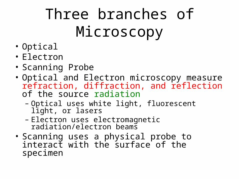
Three branches of Microscopy
• Optical• Electron• Scanning Probe• Optical and Electron microscopy measure
refraction, diffraction, and reflection of the source radiation– Optical uses white light, fluorescent light, or lasers– Electron uses electromagnetic radiation/electron beams
• Scanning uses a physical probe to interact with the surface of the specimen
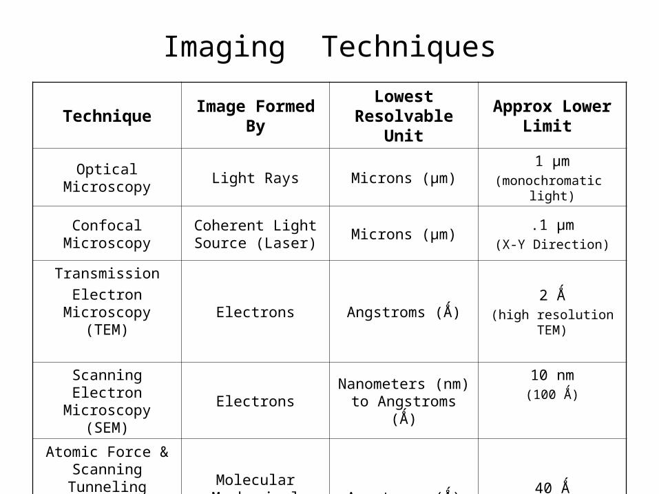
Imaging Techniques
Technique Image Formed ByLowest Resolvable
UnitApprox Lower
Limit
Optical Microscopy Light Rays Microns (μm)1 μm
(monochromatic light)
Confocal MicroscopyCoherent Light Source
(Laser)Microns (μm)
.1 μm(X-Y Direction)
Transmission
Electron Microscopy (TEM)
Electrons Angstroms (Ǻ)2 Ǻ
(high resolution TEM)
Scanning Electron Microscopy (SEM)
ElectronsNanometers (nm) to
Angstroms (Ǻ)
10 nm(100 Ǻ)
Atomic Force & Scanning Tunneling
Microscopies (AFM/STM)
Molecular Mechanical Probes
Angstroms (Ǻ)40 Ǻ
(theoretical)
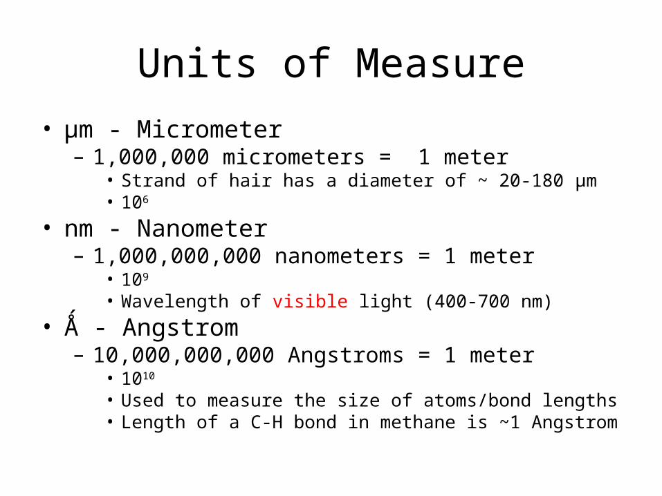
Units of Measure
• μm - Micrometer – 1,000,000 micrometers = 1 meter
• Strand of hair has a diameter of ~ 20-180 μm• 106
• nm - Nanometer– 1,000,000,000 nanometers = 1 meter
• 109
• Wavelength of visible light (400-700 nm)
• Ǻ - Angstrom– 10,000,000,000 Angstroms = 1 meter
• 1010
• Used to measure the size of atoms/bond lengths• Length of a C-H bond in methane is ~1 Angstrom
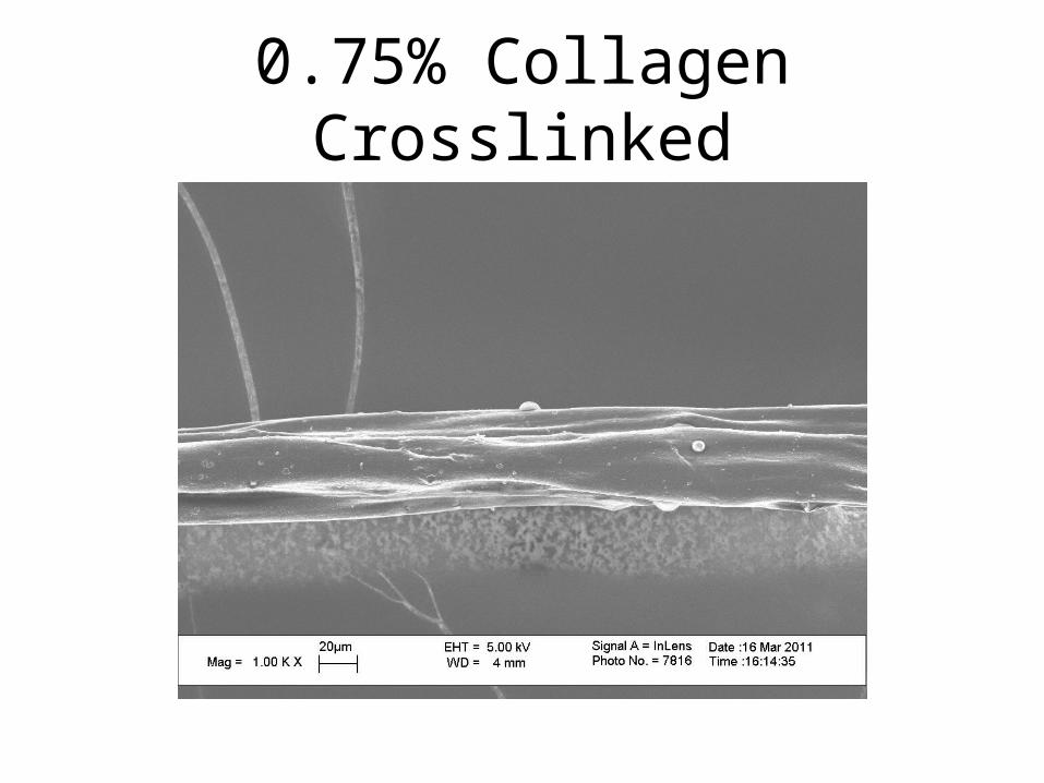
0.75% Collagen Crosslinked
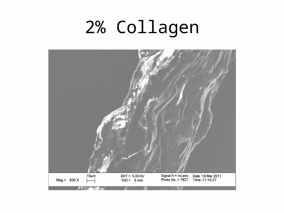
2% Collagen
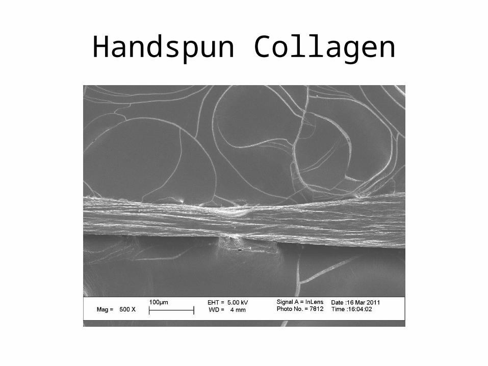
Handspun Collagen
Optical Microscopy
Properties of light
• Reflection
• Refraction
• Numerical Aperture
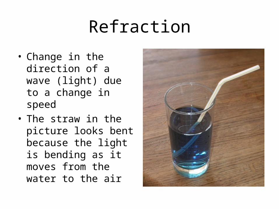
Refraction
• Change in the direction of a wave (light) due to a change in speed
• The straw in the picture looks bent because the light is bending as it moves from the water to the air
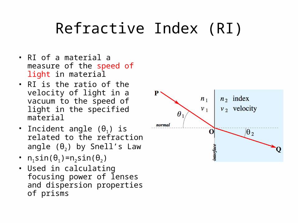
Refractive Index (RI)
• RI of a material a measure of the speed of light in material
• RI is the ratio of the velocity of light in a vacuum to the speed of light in the specified material
• Incident angle (θ1) is related to the refraction angle (θ2) by Snell’s Law
• n1sin(θ1)=n2sin(θ2)• Used in calculating focusing
power of lenses and dispersion properties of prisms
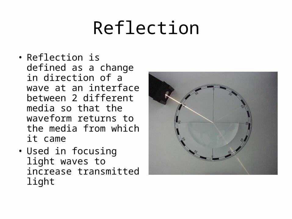
Reflection
• Reflection is defined as a change in direction of a wave at an interface between 2 different media so that the waveform returns to the media from which it came
• Used in focusing light waves to increase transmitted light
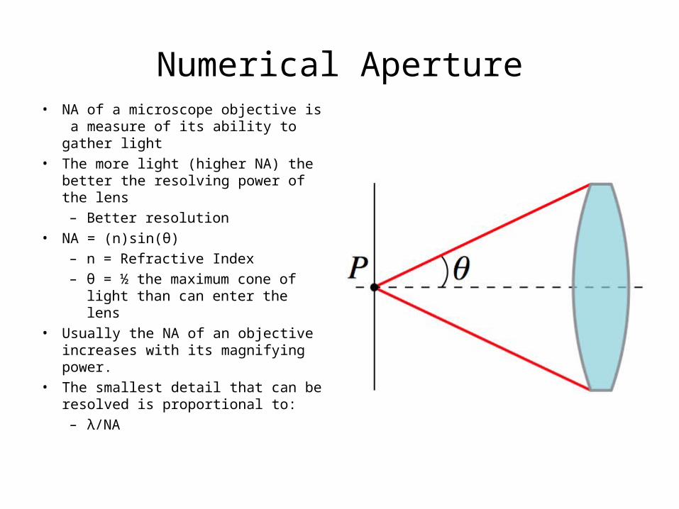
Numerical Aperture• NA of a microscope objective is a
measure of its ability to gather light
• The more light (higher NA) the better the resolving power of the lens
– Better resolution
• NA = (n)sin(θ)
– n = Refractive Index
– θ = ½ the maximum cone of light than can enter the lens
• Usually the NA of an objective increases with its magnifying power.
• The smallest detail that can be resolved is proportional to:
– λ/NA
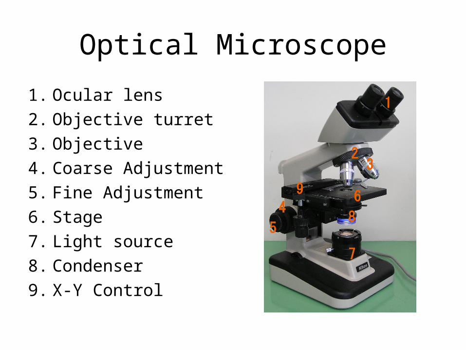
Optical Microscope
1. Ocular lens
2. Objective turret
3. Objective
4. Coarse Adjustment
5. Fine Adjustment
6. Stage
7. Light source
8. Condenser
9. X-Y Control
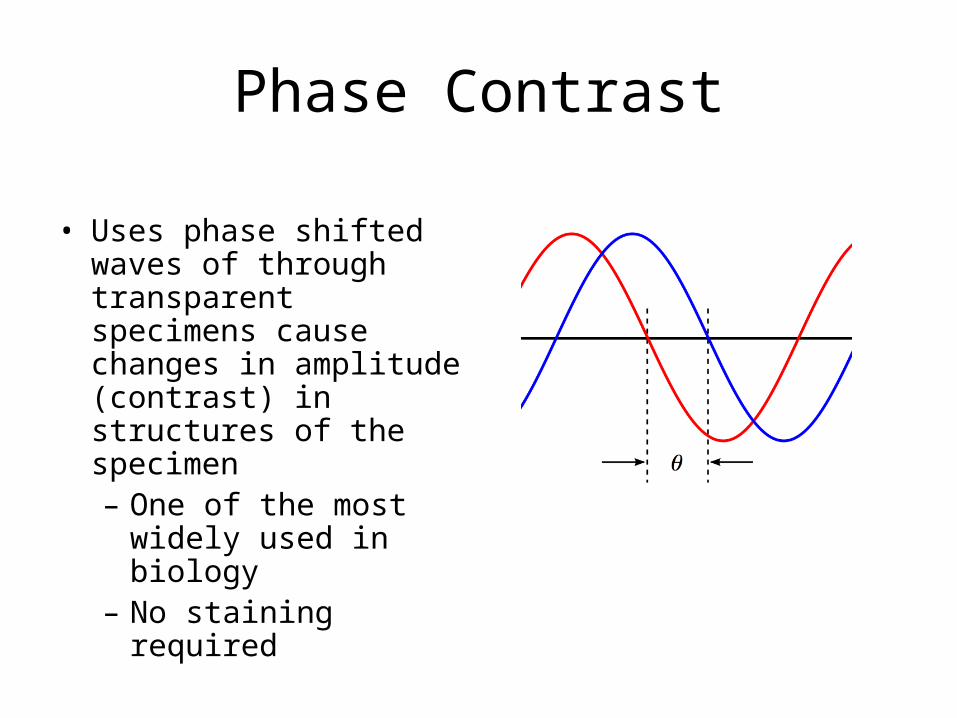
Phase Contrast
• Uses phase shifted waves of through transparent specimens cause changes in amplitude (contrast) in structures of the specimen– One of the most widely
used in biology– No staining required
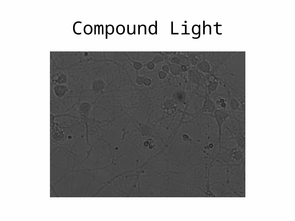
Compound Light
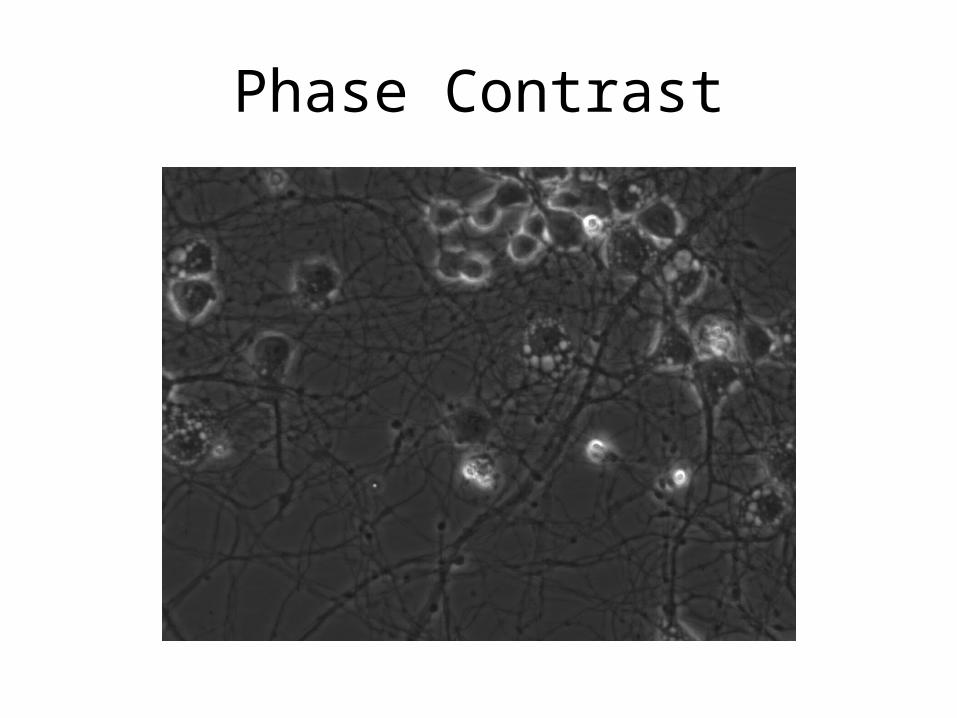
Phase Contrast
Fluorescence
• Fluorescence utilizes fluorescent dyes/stains that fluoresce when radiated with specific wavelengths of light– Typically use mercury or xenon lamps
• Fluorescent dyes are extremely useful in identifying/highlighting specific parts of cells that can otherwise go undetected using simple phase contrast
• http://www.invitrogen.com/site/us/en/home/support/Tutorials.html
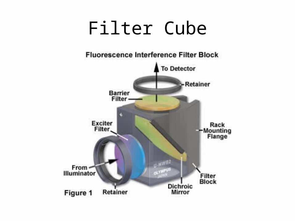
Filter Cube
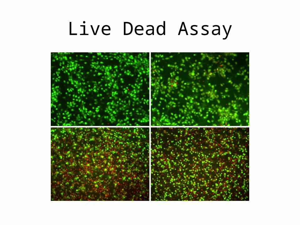
Live Dead Assay
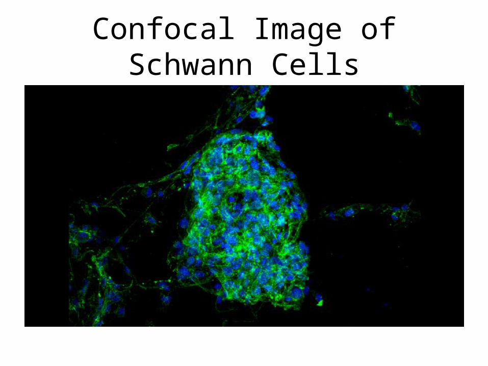
Confocal Image of Schwann Cells
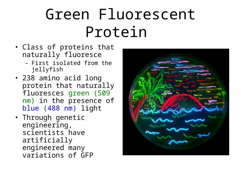
Green Fluorescent Protein
• Class of proteins that naturally fluoresce– First isolated from the
jellyfish
• 238 amino acid long protein that naturally fluoresces green (509 nm) in the presence of blue (488 nm) light
• Through genetic engineering, scientists have artificially engineered many variations of GFP
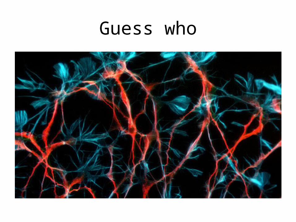
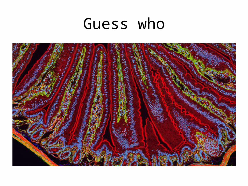
Guess who
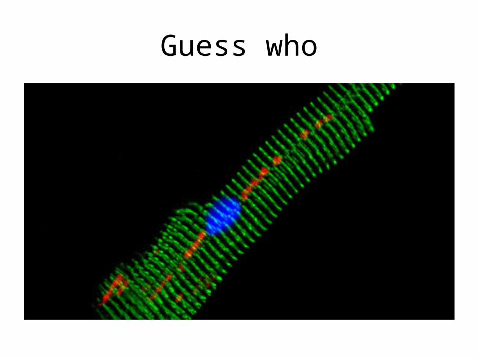
Guess who
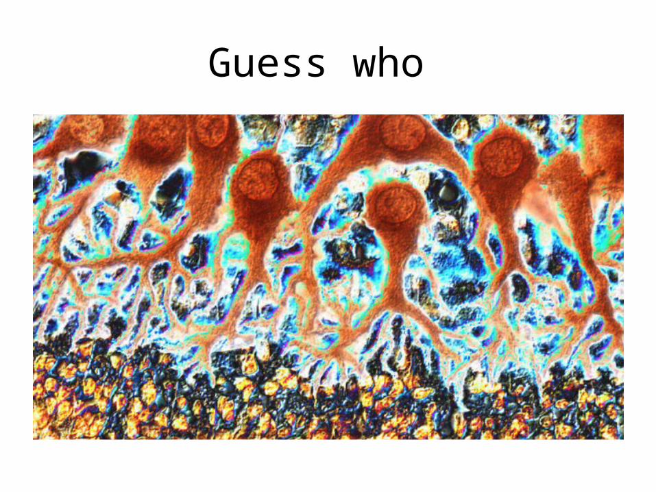
Guess who
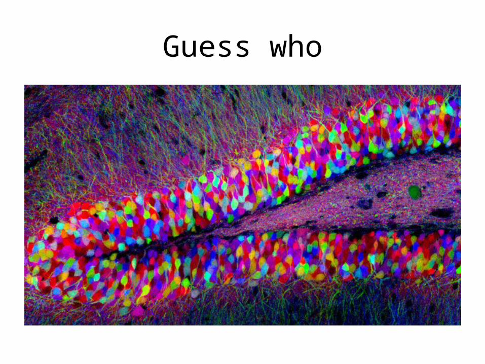
Guess who
Guess who


![Near-Field Optical Microscopy - Indico [Home]indico.ictp.it/event/a04179/session/16/contribution/11/material/0/0.pdf · Optical microscopy Electron microscopy' Near-field optical](https://static.fdocuments.net/doc/165x107/5ed73d31d37f9f58ca6a86bf/near-field-optical-microscopy-indico-home-optical-microscopy-electron-microscopy.jpg)