Microscopes "The evolution of a science often parallels the invention of instruments that extend...
10
Microscopes "The evolution of a science often parallels the invention of instruments that extend human senses to new limits." (Campbell 2002)
-
Upload
linda-gregory -
Category
Documents
-
view
213 -
download
0
Transcript of Microscopes "The evolution of a science often parallels the invention of instruments that extend...

Microscopes
"The evolution of a science often parallels the invention of instruments that extend
human senses to newlimits." (Campbell 2002)

• Compound Microscope• Fluorescence microscope• Phase-contrast microscope• Transmission electron microscope (TEM)• Scanning Electron Microscope (SEM)• Scanning Probe Microscope (SPM)

Optical…compound…light…?
• Confused? Don’t be!
• Any “optical” microscope is a microscope that uses visible “light” and lenses to magnify images of small samples.
• Compound microscope uses series of lenses.• Simple/Single lens microscope only uses one
lens.

Compound Microscopeuses multiple lenses to collect light from the sample and then an another set of lenses to focus the light into the eye
Invented: 1609: Galileo Galilee

Light Microscope: passes visible light through a specimen & series glass lenses that magnify the specimen
1665: Robert Hook: First cell discovered (simple optical microscope)
Light microscopes are effective in magnifying images up to1000x.
Limitations:The smallest wavelengths of visible light are too large to resolve images smaller than0.25 micrometers (um).

Fluorescence microscope
• Another form of “optical/light” microscope • Specimen/sample is illuminated with a specific
light of a wavelength which “excites” the florescence within the specimen/sample.
• In other words, the specimen receives some light, absorbs it, then makes its own light (and that’s what we see & measure)
• Invented in 1911
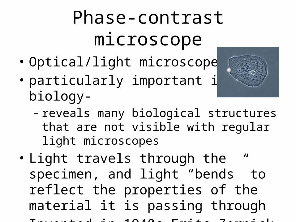
Phase-contrast microscope
• Optical/light microscope• particularly important in biology-– reveals many biological structures that are not
visible with regular light microscopes• Light travels through the specimen, and light
“bends” to reflect the properties of the material it is passing through
• Invented in 1940s-Frits Zernick

Transmission electron microscope (TEM)
• 150,000 x• 1931-Knoll & Ruska• Electrons are used to produce magnified images• Electrons pass through specimen and produce
flat image

Scanning electron microscope (SEM)
• 150,000 x • 1926- Hans Busch• Electrons are used to produce magnified
images• Electrons bounce off the surface of specimen
and produce 3D image

Scanning Probe Microscope (SPM)
• Uses physical probe that scans the specimen• The probe scans the specimen line by line
Invented in 1982 by Binning and Rohrer
It really doesn’t “magnify” things. It can detect things as small as 1 billionth of a mm (pico-meter)