Microscopes/. It is estimated that the human race grows daily by about 214,000 people. It takes only...
46
Microscopes/
-
Upload
shawn-norton -
Category
Documents
-
view
213 -
download
0
Transcript of Microscopes/. It is estimated that the human race grows daily by about 214,000 people. It takes only...
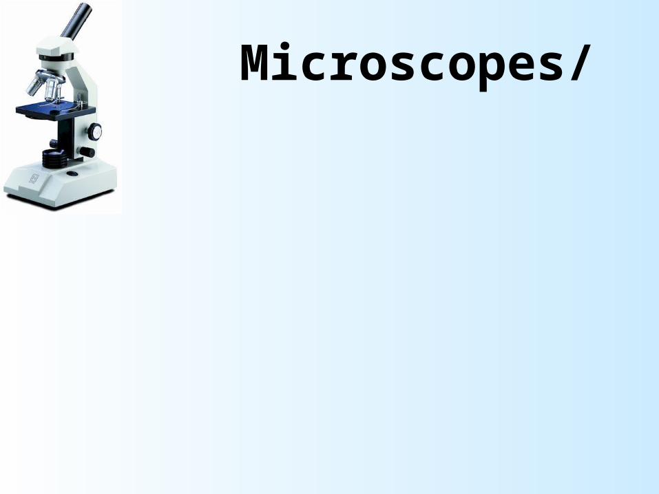
Microscopes/

It is estimated that the human race grows daily by about 214,000 people.
It takes only 15 watts of electricity going through a human body to stop the heart. Common lightbulbs run on about 25 to 75 watts of electricity.
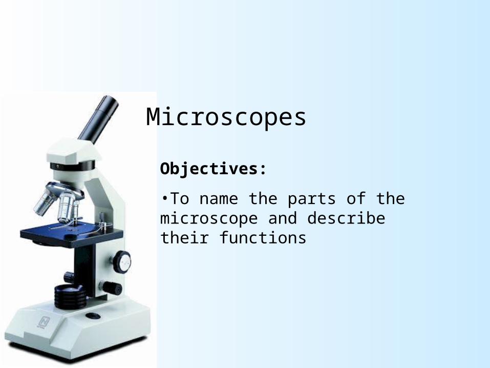
Microscopes
Objectives:
•To name the parts of the microscope and describe their functions
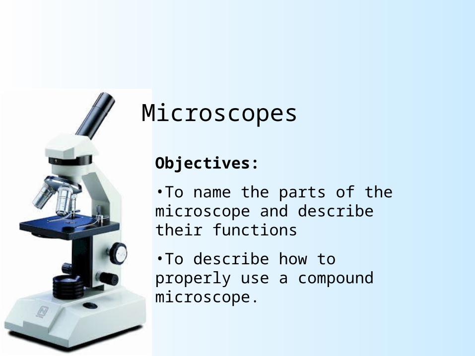
Microscopes
Objectives:
•To name the parts of the microscope and describe their functions
•To describe how to properly use a compound microscope.
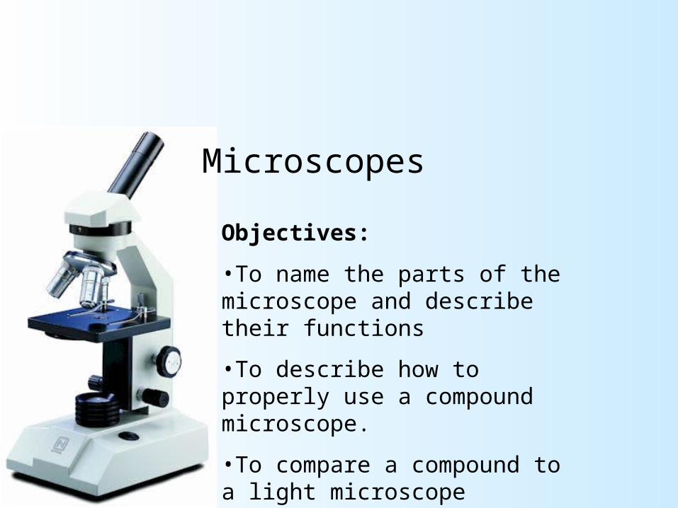
Microscopes
Objectives:
•To name the parts of the microscope and describe their functions
•To describe how to properly use a compound microscope.
•To compare a compound to a light microscope

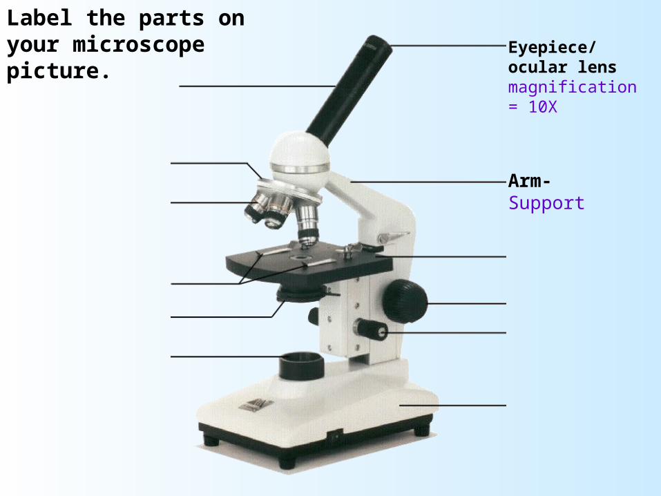
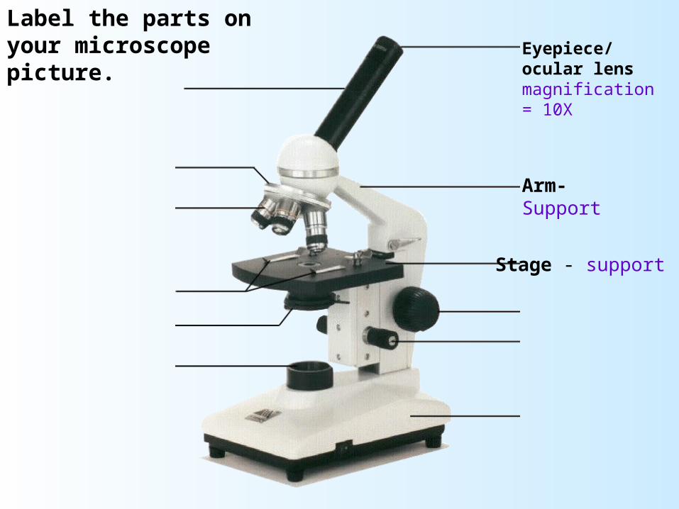
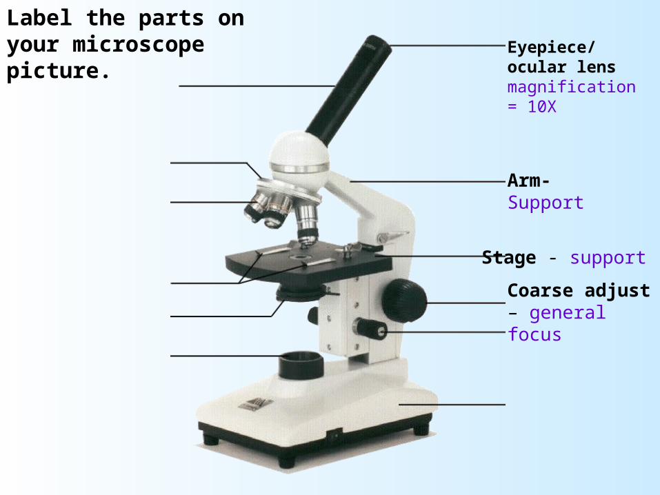
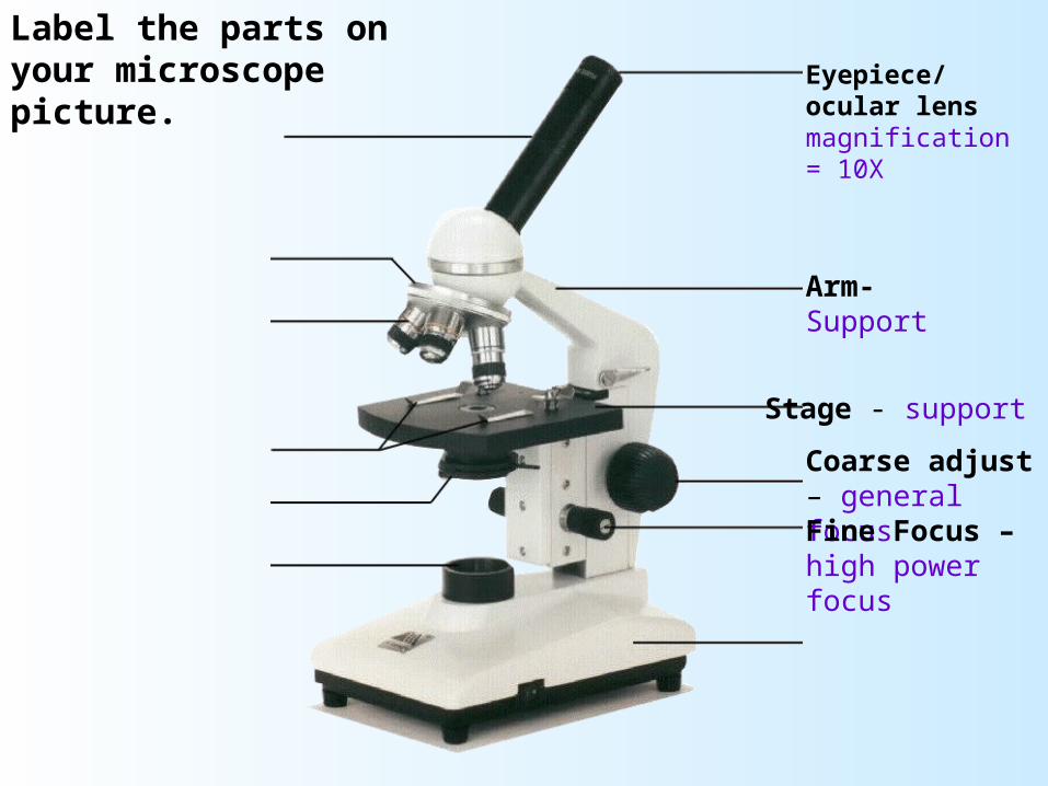
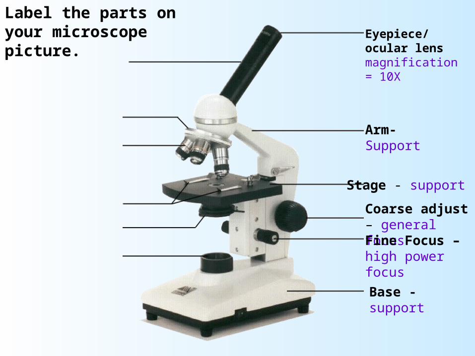
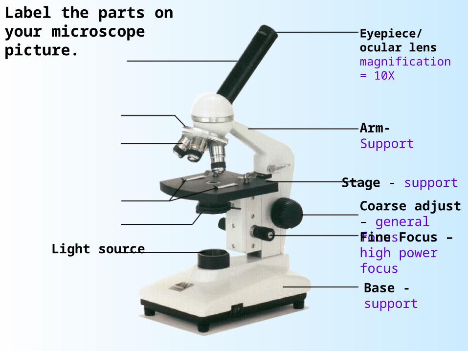
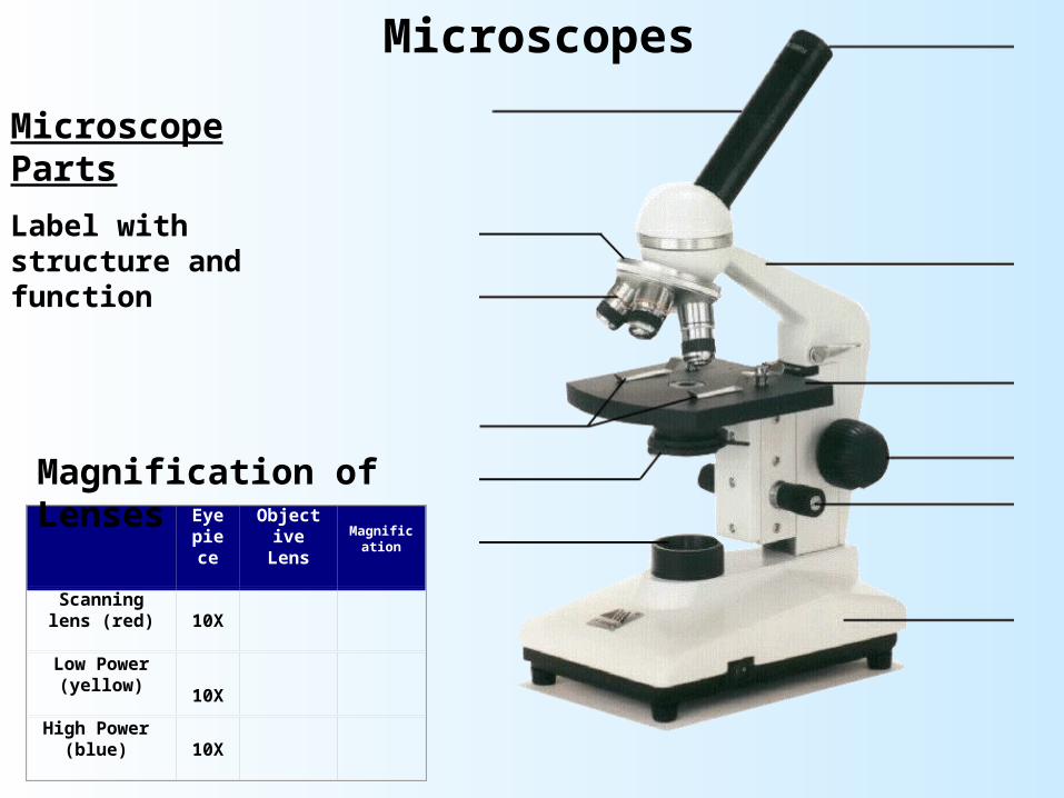
Label the parts on your microscope picture. Eyepiece/ocular lens
magnification = 10X
Label the parts on your microscope picture. Eyepiece/ocular lens
magnification = 10X
Arm-Support
Label the parts on your microscope picture. Eyepiece/ocular lens
magnification = 10X
Arm-Support
Stage - support
Label the parts on your microscope picture. Eyepiece/ocular lens
magnification = 10X
Arm-Support
Stage - support
Coarse adjust – general focus
Label the parts on your microscope picture. Eyepiece/ocular lens
magnification = 10X
Arm-Support
Stage - support
Coarse adjust – general focusFine Focus – high power focus
Label the parts on your microscope picture. Eyepiece/ocular lens
magnification = 10X
Arm-Support
Stage - support
Coarse adjust – general focusFine Focus – high power focus
Base - support
Label the parts on your microscope picture. Eyepiece/ocular lens
magnification = 10X
Arm-Support
Stage - support
Coarse adjust – general focusFine Focus – high power focus
Base - support
Light source

Label the parts on your microscope picture. Eyepiece/ocular lens
magnification = 10X
Arm-Support
Stage - support
Coarse adjust – general focusFine Focus – high power focus
Base - support
Light source
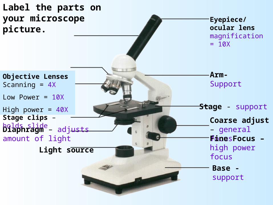
Diaphragm – adjusts amount of light

Label the parts on your microscope picture. Eyepiece/ocular lens
magnification = 10X
Arm-Support
Stage - support
Coarse adjust – general focusFine Focus – high power focus
Base - support
Light source
Diaphragm – adjusts amount of light
Stage clips – holds slide
Label the parts on your microscope picture. Eyepiece/ocular lens
magnification = 10X
Arm-Support
Stage - support
Coarse adjust – general focusFine Focus – high power focus
Base - support
Light source
Diaphragm – adjusts amount of light
Stage clips – holds slide
Objective Lenses Scanning = 4X
Low Power = 10X
High power = 40X
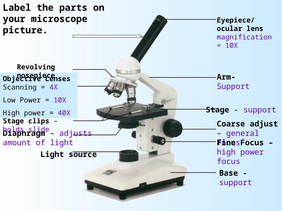
Revolving nosepiece
Label the parts on your microscope picture. Eyepiece/ocular lens
magnification = 10X
Arm-Support
Stage - support
Coarse adjust – general focusFine Focus – high power focus
Base - support
Light source
Diaphragm – adjusts amount of light
Stage clips – holds slide
Objective Lenses Scanning = 4X
Low Power = 10X
High power = 40X
Revolving nosepiece
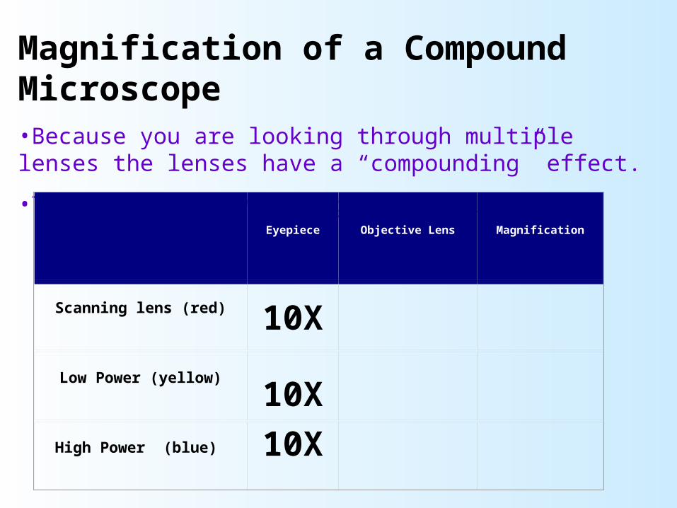
Magnification of a Compound Microscope•Because you are looking through multiple lenses the lenses have a “compounding” effect.
•The eyepiece always magnifies 10X
Eyepiece Objective Lens Magnification
Scanning lens (red)
10X
Low Power (yellow)
10X
High Power (blue)
10X
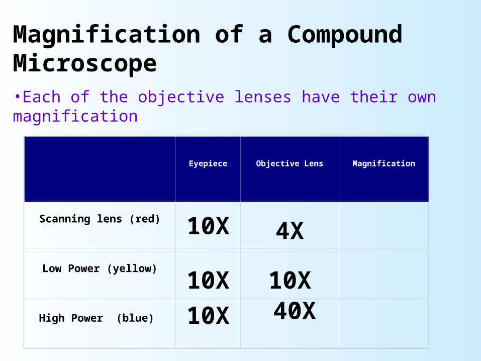
Magnification of a Compound Microscope•Each of the objective lenses have their own magnification
Eyepiece Objective Lens Magnification
Scanning lens (red)
10X
4X
Low Power (yellow)
10X
10X
High Power (blue)
10X 40X
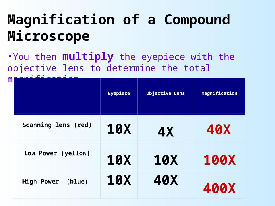
Magnification of a Compound Microscope•You then multiply the eyepiece with the objective lens to determine the total magnification
Eyepiece Objective Lens Magnification
Scanning lens (red)
10X
4X
Low Power (yellow)
10X
10X
100XHigh Power (blue)
10X
40X
400X
40X
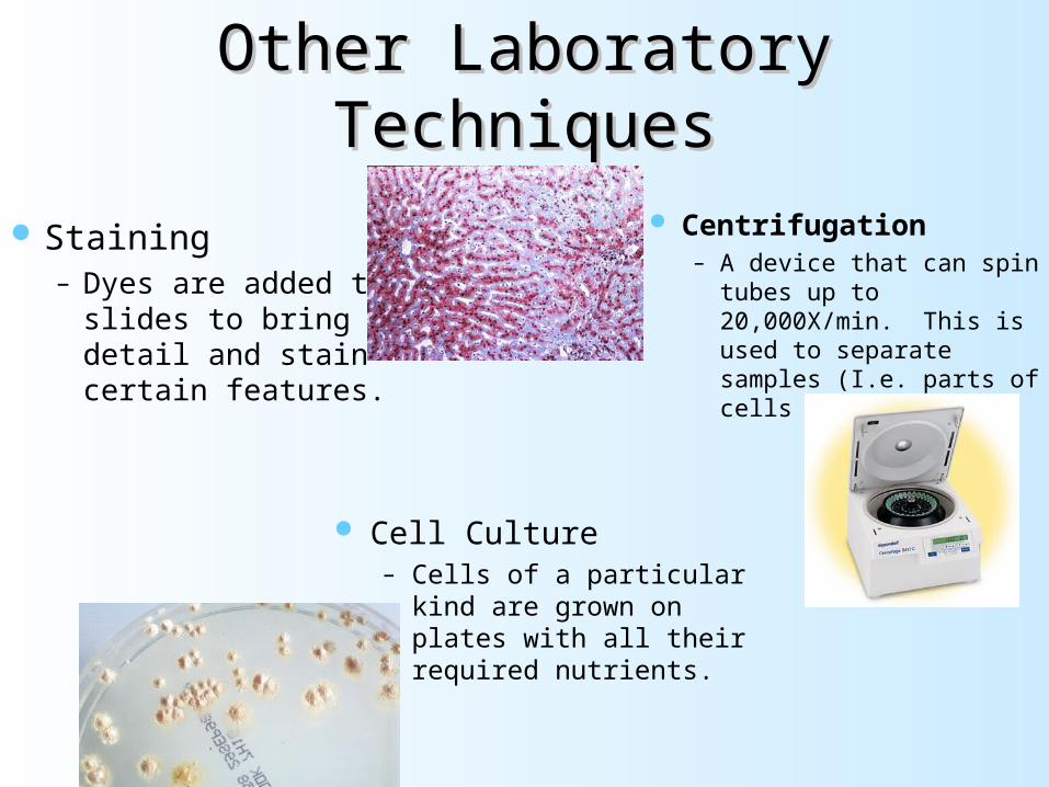
Other Laboratory TechniquesOther Laboratory Techniques
Staining– Dyes are added to
slides to bring out detail and stain certain features.
Centrifugation– A device that can spin tubes
up to 20,000X/min. This is used to separate samples (I.e. parts of cells
Cell Culture– Cells of a particular kind
are grown on plates with all their required nutrients.
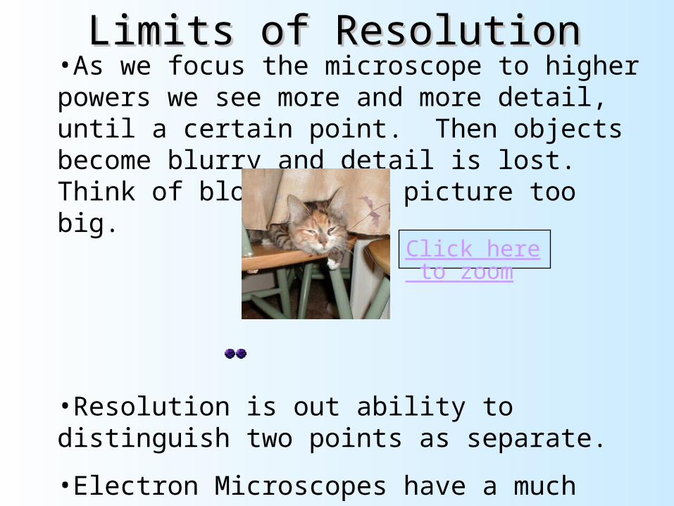
Limits of ResolutionLimits of Resolution•As we focus the microscope to higher powers we see more and more detail, until a certain point. Then objects become blurry and detail is lost. Think of blowing up a picture too big.
•
•Resolution is out ability to distinguish two points as separate.
•Electron Microscopes have a much higher limit of resolution.
Click here to zoom

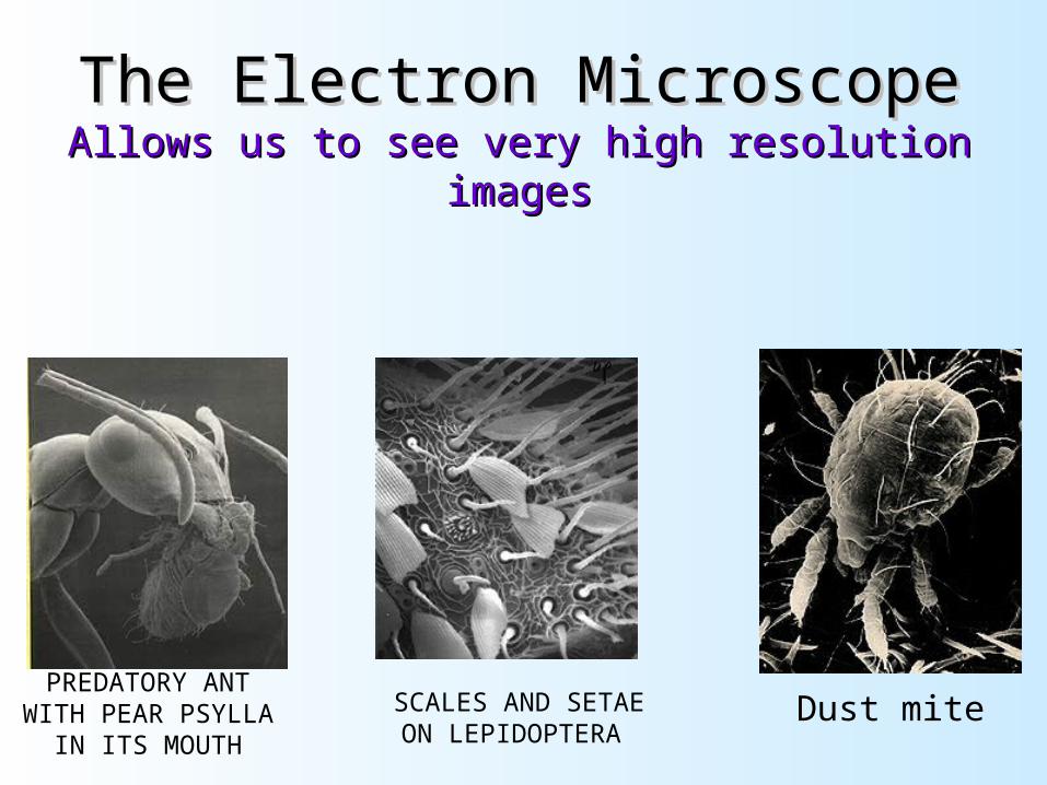
The Electron MicroscopeThe Electron MicroscopeAllows us to see very high resolution imagesAllows us to see very high resolution images
PREDATORY ANT WITH PEAR PSYLLA
IN ITS MOUTH
SCALES AND SETAE ON LEPIDOPTERA Dust mite
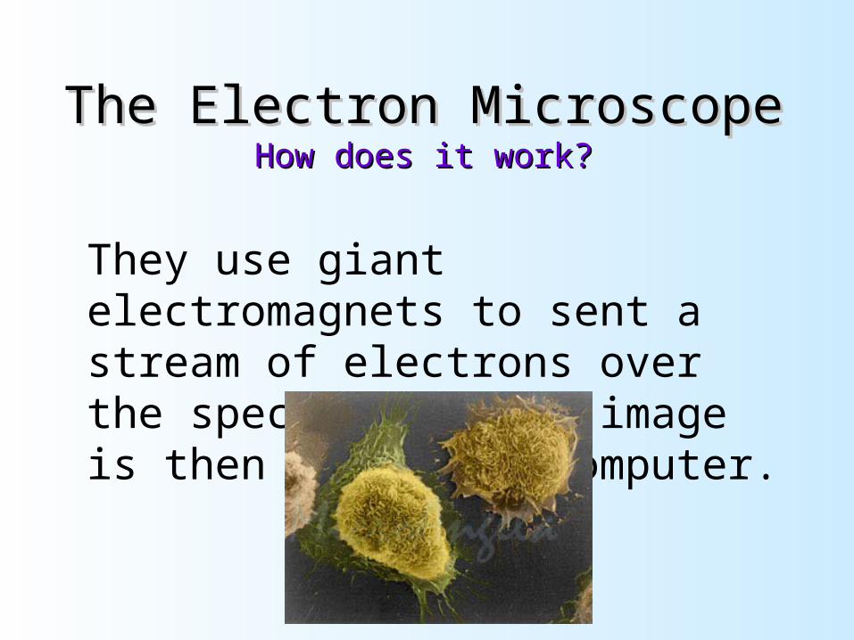
The Electron MicroscopeThe Electron MicroscopeHow does it work?How does it work?
They use giant electromagnets to sent a stream of electrons over the specimen. This image is then read by a computer.
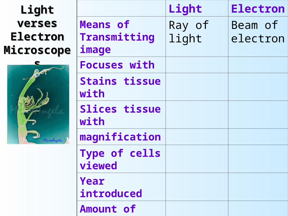
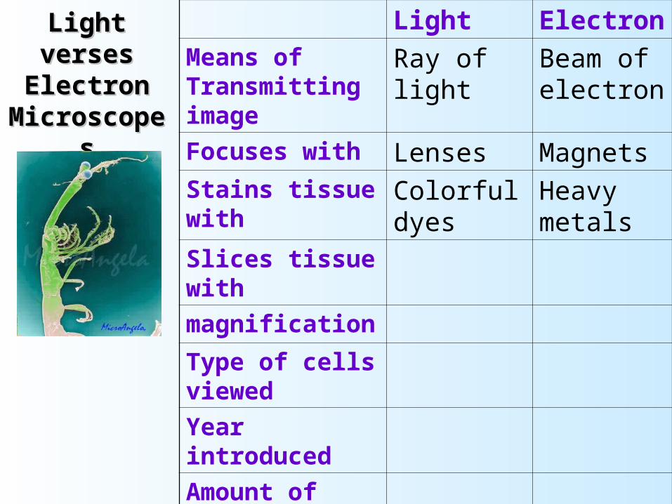
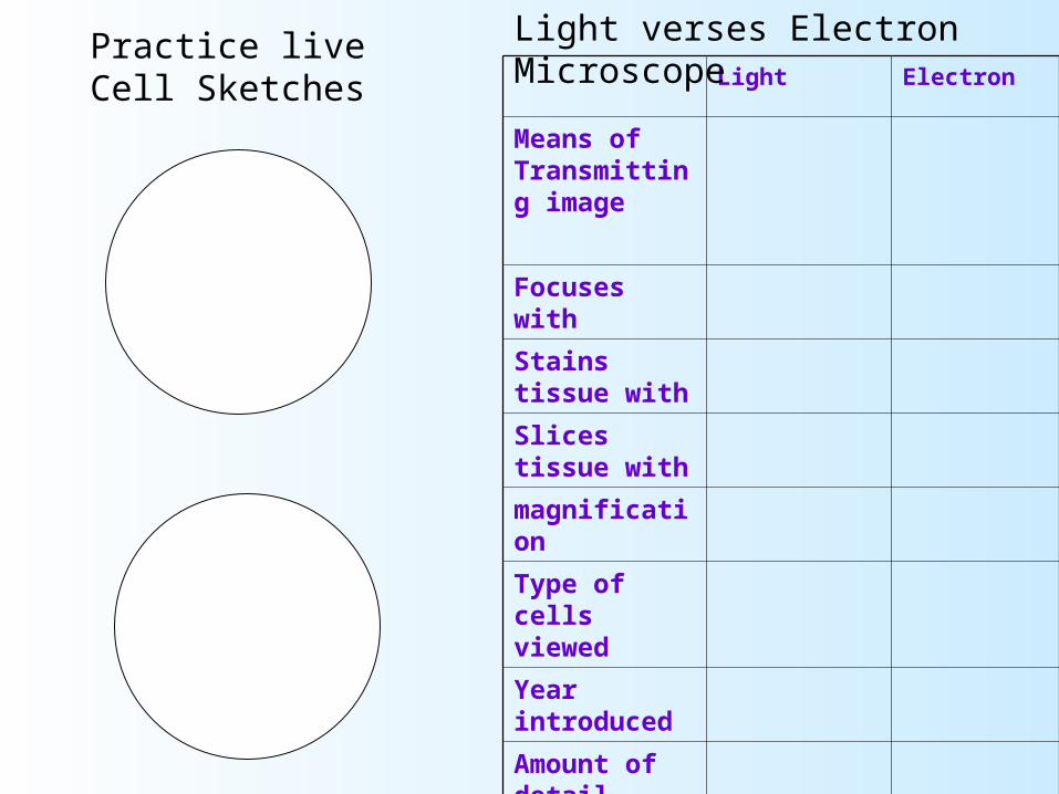
Light verses Light verses Electron Electron
MicroscopesMicroscopes
Light Electron
Means of Transmitting image
Ray of light Beam of electron
Focuses with
Stains tissue with
Slices tissue with
magnification
Type of cells viewed
Year introduced
Amount of detail
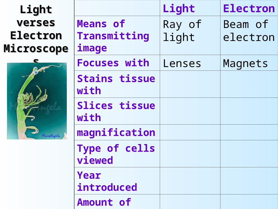
Light verses Light verses Electron Electron
MicroscopesMicroscopes
Light Electron
Means of Transmitting image
Ray of light Beam of electron
Focuses with Lenses Magnets
Stains tissue with
Slices tissue with
magnification
Type of cells viewed
Year introduced
Amount of detail
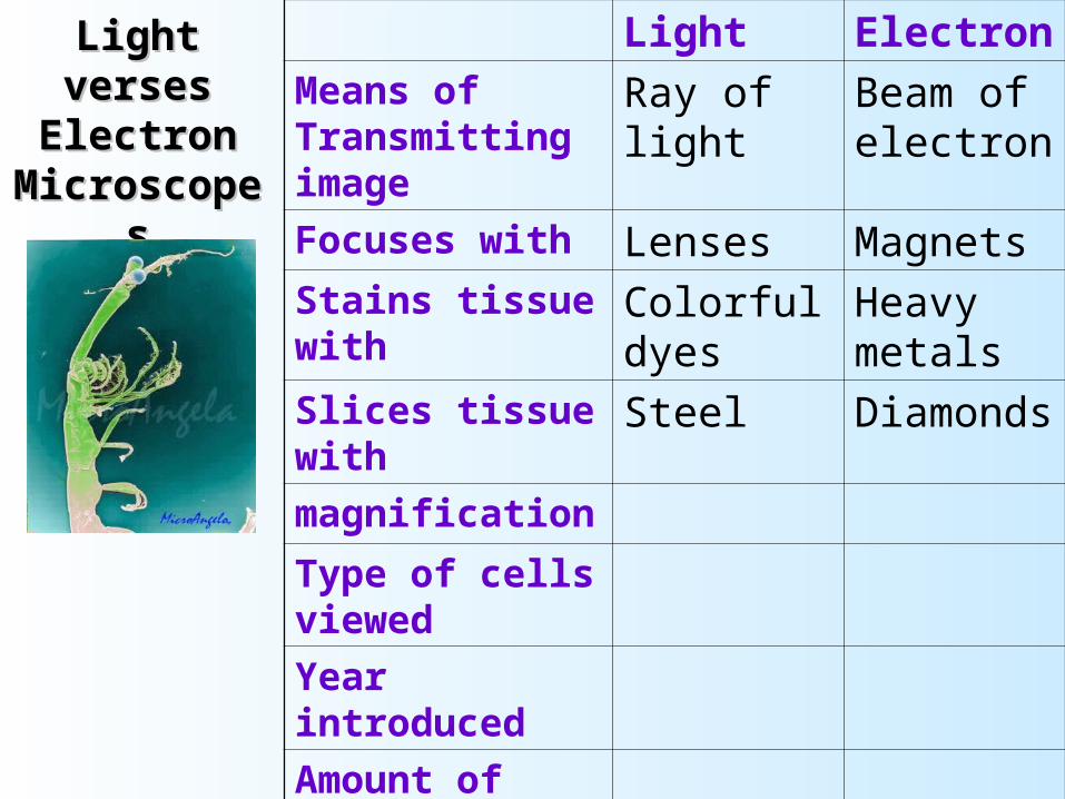
Light verses Light verses Electron Electron
MicroscopesMicroscopes
Light Electron
Means of Transmitting image
Ray of light Beam of electron
Focuses with Lenses Magnets
Stains tissue with Colorful dyes
Heavy metals
Slices tissue with
magnification
Type of cells viewed
Year introduced
Amount of detail
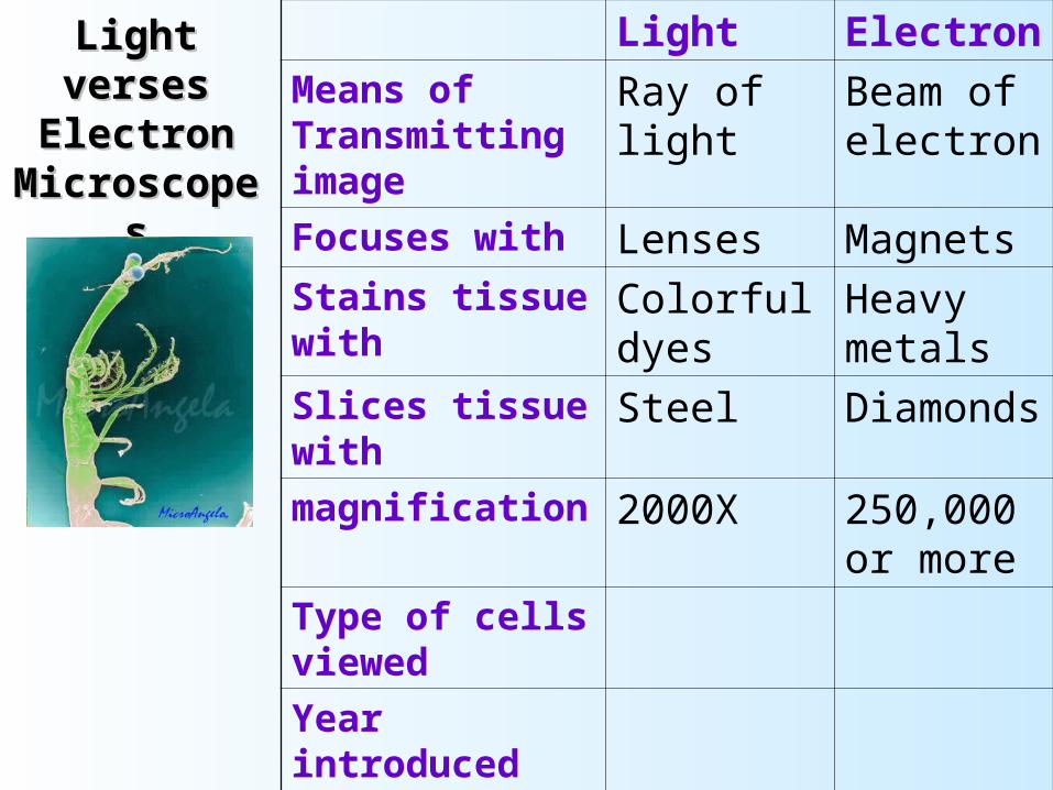
Light verses Light verses Electron Electron
MicroscopesMicroscopes
Light Electron
Means of Transmitting image
Ray of light Beam of electron
Focuses with Lenses Magnets
Stains tissue with Colorful dyes
Heavy metals
Slices tissue with Steel Diamonds
magnification
Type of cells viewed
Year introduced
Amount of detail
Light verses Light verses Electron Electron
MicroscopesMicroscopes
Light Electron
Means of Transmitting image
Ray of light Beam of electron
Focuses with Lenses Magnets
Stains tissue with Colorful dyes
Heavy metals
Slices tissue with Steel Diamonds
magnification 2000X 250,000 or more
Type of cells viewed
Year introduced
Amount of detail
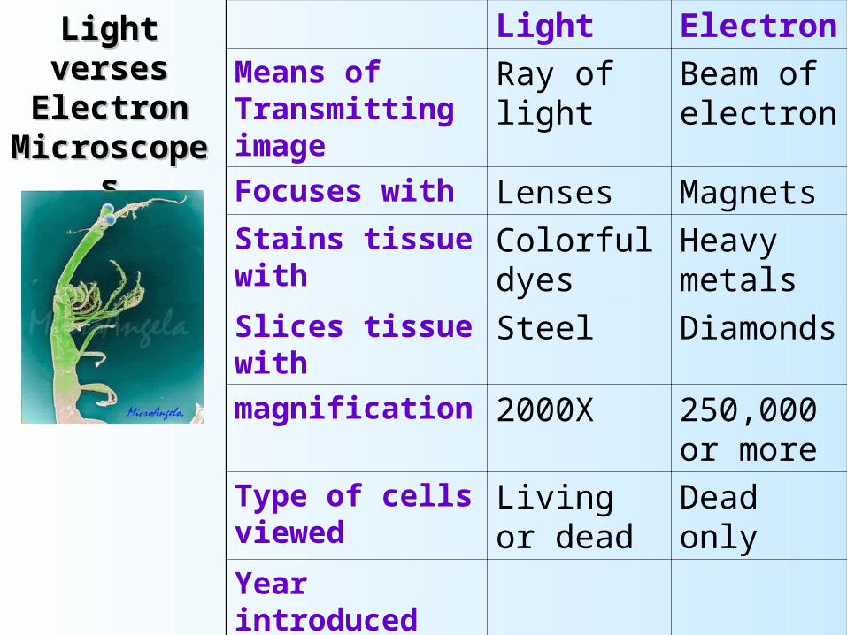
Light verses Light verses Electron Electron
MicroscopesMicroscopes
Light Electron
Means of Transmitting image
Ray of light Beam of electron
Focuses with Lenses Magnets
Stains tissue with Colorful dyes
Heavy metals
Slices tissue with Steel Diamonds
magnification 2000X 250,000 or more
Type of cells viewed
Living or dead
Dead only
Year introduced
Amount of detail
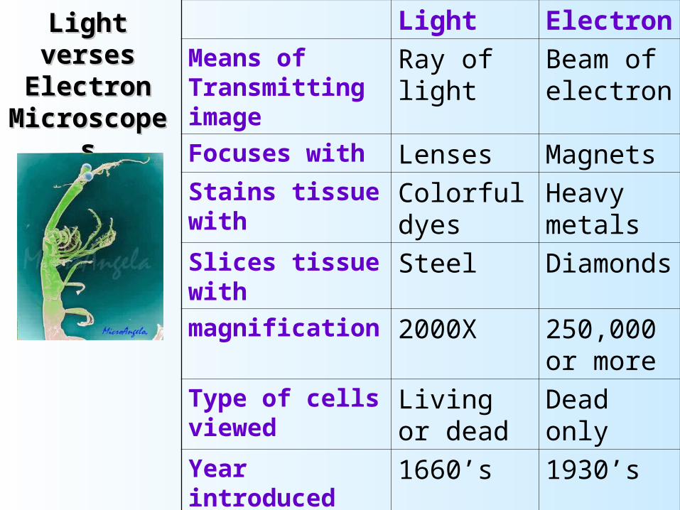
Light verses Light verses Electron Electron
MicroscopesMicroscopes
Light Electron
Means of Transmitting image
Ray of light Beam of electron
Focuses with Lenses Magnets
Stains tissue with Colorful dyes
Heavy metals
Slices tissue with Steel Diamonds
magnification 2000X 250,000 or more
Type of cells viewed
Living or dead
Dead only
Year introduced 1660’s 1930’s
Amount of detail Flat image 3-D image
General Procedures
Make sure all backpacks are out of the aisles before you get a microscope! Always carry the microscope with one hand on the Arm and one hand on the Base.
Wear your glasses, the microscope will focus to your eyesight!
Keep both eyes open, your brain will learn to ignore the other eye.
Focusing a Specimen
Always start on low power or scanning.
Focusing a Specimen
Always start on low power or scanning.
Focus the specimen using the course adjust.

Focusing a Specimen
Always start on low power or scanning.
Focus the specimen using the course adjust.
Re-center. If high power is needed, turn only after re-centering!
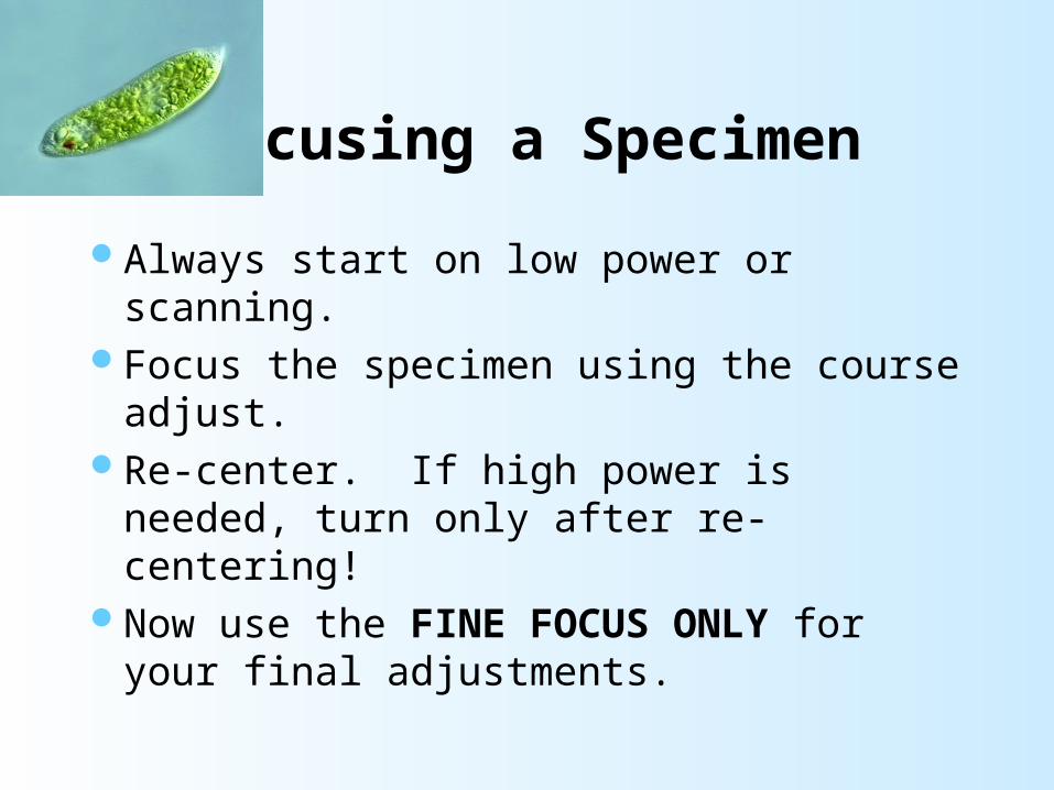
Focusing a Specimen
Always start on low power or scanning.Focus the specimen using the course adjust.Re-center. If high power is needed, turn
only after re-centering!Now use the FINE FOCUS ONLY for
your final adjustments.
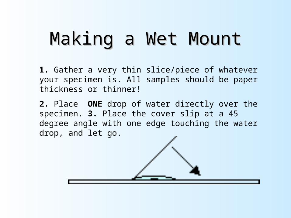
Making a Wet MountMaking a Wet Mount
1. Gather a very thin slice/piece of whatever your specimen is. All samples should be paper thickness or thinner!
2. Place ONE drop of water directly over the specimen. 3. Place the cover slip at a 45 degree angle with one edge touching the water drop, and let go.
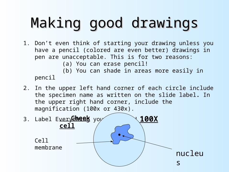
Making good drawingsMaking good drawings1. Don’t even think of starting your drawing unless you have a
pencil (colored are even better) drawings in pen are unacceptable. This is for two reasons: (a) You can erase pencil! (b) You can shade in areas more easily in pencil
2. In the upper left hand corner of each circle include the specimen name as written on the slide label. In the upper right hand corner, include the magnification (100x or 430x).
3. Label Everything you identify!
100X Cheek cell
nucleus
Cell membrane
Student handoutsStudent handouts
Microscope notes page
Microscope Parts
Label with structure and function
Eyepiec
e
Objective
Lens
Magnification
Scanning lens (red)
10X
Low Power (yellow)
10X
High Power (blue)
10X
Magnification of Lenses
Microscopes
Light Electron
Means of Transmitting image
Focuses with
Stains tissue with
Slices tissue with
magnification
Type of cells viewed
Year introduced
Amount of detail
Light verses Electron MicroscopePractice live Cell Sketches
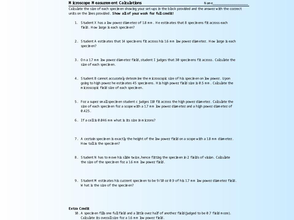
Microscope Measurement Calculations Name_________________
Calculate the size of each specimen showing your set-ups in the block provided and the answer with the correct units on the lines provided. Show all of your work for full credit!
1. Student X has a low power diameter of 1.8 mm. He estimates that 8 specimens fi t across each
field. How large is each specimen?
2. Student A estimates that 14 specimens fi t across his 1.6 mm low power diameter. How large is each specimen?
3. On a 1.7 mm low power diameter field, student I judges that 30 specimens fi t across. Calculate the size of each specimen.
4. Student B cannot accurately determine the microscopic size of his specimen on low power. Upon going to high power he estimates 45 specimens. His high power field size is 0.5 mm. Calculate the microscopic field size of each specimen.
5. For a super small specimen student c judges 110 fi t across the high power diameter. Calculate the size of each specimen for a scope with a 1.7 mm low power diameter and a high power diameter of 0.425.
6. I f a cell is 0.046 mm what is its size in microns?
7. A certain specimen is exactly the height of the low power field on a scope with a 1.8 mm diameter.
How tall is the specimen?
8. Student N has to move his slide twice, hence fi tting the specimen in 2 fields of vision. Calculate
the size of the specimen for a 1.6 mm low power field.
9. Student M estimates his current specimen to be 9/ 10 or 0.9 of his 1.7 mm low power diameter field.
What is the size of the specimen? Extra Credit
10. A specimen fi lls one full field and a little over half of another field(judged to be 0.7 field more). Calculate its overall size for a 1.6 mm low power field.
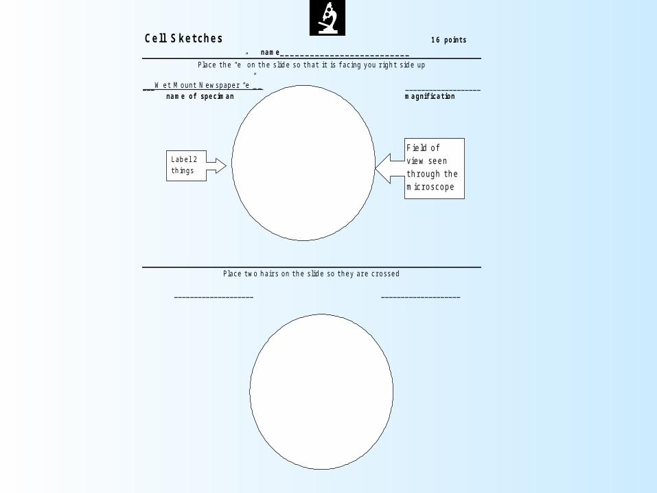
Cell S ket ches 16 point s
name_ _ _ _ _ _ _ _ _ _ _ _ _ _ _ _ _ _ _ _ _ _ _ _ _ _
Plac e t h e “e” on t h e s lid e s o t h at i t is f ac ing you r igh t s id e up
_ _ _ W et M ount N ews paper “e”_ _ _ _ _ _ _ _ _ _ _ _ _ _ _ _ _ _ _ _ _ name of speciman magnifi cat ion
Plac e t wo h air s on t h e s lid e s o t h ey ar e c r os s ed _ _ _ _ _ _ _ _ _ _ _ _ _ _ _ _ _ _ _ _ _ _ _ _ _ _ _ _ _ _ _ _ _ _ _ _ _ _ _ _
F ie ld of view seen t h r ough t h e mic r osc ope
L ab e l 2 t h ings
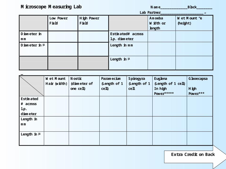
Microscope Measuring Lab Name____________Block_______
Lab Partner___________________
Low Power Field
High Power Field
Amoeba Width or length
Wet Mount “e” (height)
Diameter in mm
Estimated# across l.p. diameter
Diameter in Length in mm
Length in
_
Wet Mount Hair (width)
Nostic (diameter of one cell)
Paramecium (Length of 1 cell)
Spirogyra (Length of 1 cell
Euglena (Length of 1 cell) I n high Power*****
Glaoecapsa High Power***
Estimated # across l.p. diameter
Length in mm
Length in
Extra Credit on Back