MicroRNA Alterations of Pancreatic Intraepithelial...
13
Human Cancer Biology MicroRNA Alterations of Pancreatic Intraepithelial Neoplasias Jun Yu 1 , Ang Li 1 , Seung-Mo Hong 1 , Ralph H. Hruban 1,2 , and Michael Goggins 1,2,3 Abstract Purpose: MicroRNA (miRNA) alterations are likely to contribute to the development of pancreatic cancer and may serve as markers for the early detection of pancreatic neoplasia. Experimental Design: To identify the miRNA alterations that arise during the development of pancreatic cancer, we determined the levels of 735 miRNAs in 34 pancreatic intraepithelial neoplasias (PanIN) and 15 normal pancreatic duct samples isolated by laser capture microdissection using TaqMan miRNA micro- arrays. Differential expression of selected miRNAs was confirmed by FISH analysis and by quantitative real- time reverse transcription PCR (qRT-PCR) analysis of selected candidate miRNAs in an independent set of PanIN and normal duct samples. Results: We identified 107 aberrantly expressed miRNAs in different PanIN grades compared with normal pancreatic duct samples and 35 aberrantly expressed miRNAs in PanIN-3 lesions compared with normal pancreatic duct samples. These differentially expressed miRNAs included those that have been previously identified as differentially expressed in pancreatic ductal adenocarcinomas (PDAC; including miR-21, miR-200a/b/c, miR-216a/b, miR-217, miR-146a, miR-155, miR-182, miR-196b, miR-203, miR- 222, miR-338-3p, miR-486-3p, etc.) as well as miRNAs not previously described as differentially expressed in these lesions (miR-125b, miR-296-5p, miR-183 , miR-603, miR-625/ , miR-708, etc.). miR-196b was the most selectively differentially expressed miRNA in PanIN-3 lesions. Conclusions: Many miRNAs undergo aberrant expression in PanIN lesions and are likely to be important in the development of PDAC. The miRNAs, such as miR-196b, whose expression is limited to PanIN-3 lesions or pancreatic cancers could be useful as diagnostic markers. Clin Cancer Res; 18(4); 981–92. Ó2011 AACR. Introduction Pancreatic cancer is the fourth leading cause of cancer- related deaths in the United States (1). In the United States in 2010, 43,140 new patients were diagnosed, and approximately 36,800 Americans died from pancreatic cancer (1). The poor prognosis and high mortality rate result, at least in part, from the generally late presentation of the disease and the lack of effective therapies (2). Although early detection is considered the best way to cure pancreatic cancer, most early-stage pancreatic can- cers do not cause symptoms. As a result, there is consid- erable interest in pancreatic screening for individuals considered to be at significantly increased risk of devel- oping pancreatic cancer, such as those with an inherited predisposition. Because even early-stage invasive pancre- atic cancer is usually incurable, the primary goal of pancreatic screening programs has been to prevent the development of invasive pancreatic cancer by detecting and treating pancreatic precursor lesions. The most com- mon of these precursor lesions are pancreatic intraepithe- lial neoplasias (PanIN; ref. 3). Because PanINs are too small to be reliably detected by pancreatic imaging tests (4, 5), there is considerable interest in identifying markers of advanced PanINs (6, 7) to improve our ability to detect advanced PanINs during pancreatic screening. Although numerous studies have described the molecular altera- tions of pancreatic ductal adenocarcinomas (PDAC; refs. 8–11), fewer studies have investigated the timing of such alterations in PanIN lesions. Understanding the molecular alterations of PanINs may not only identify markers for pancreatic screening but also to identify important biologic pathways. Changes in the expression of microRNAs (miRNA) are important to the development of cancer. miRNAs are small endogenous noncoding RNAs of 14 to 24 nucleotides that negatively regulate protein expression at the posttranscrip- tional level by inhibiting translation and/or by targeting Authors' Affiliations: Departments of 1 Pathology, 2 Oncology, and 3 Med- icine, The Sol Goldman Pancreatic Cancer Research Center, Johns Hop- kins Medical Institutions, Baltimore, Maryland Note: Supplementary data for this article are available at Clinical Cancer Research Online (http://clincancerres.aacrjournals.org/). Corresponding Author: Michael Goggins, The Sol Goldman Pancreatic Cancer Research Center, The Johns Hopkins Medical Institutions, CRBII Room 342, 1550 Orleans Street, Baltimore, MD 21231. Phone: 410-955- 3511; Fax: 410-614-0671; E-mail: [email protected] doi: 10.1158/1078-0432.CCR-11-2347 Ó2011 American Association for Cancer Research. Clinical Cancer Research www.aacrjournals.org 981 on October 3, 2018. © 2012 American Association for Cancer Research. clincancerres.aacrjournals.org Downloaded from Published OnlineFirst November 23, 2011; DOI: 10.1158/1078-0432.CCR-11-2347
Transcript of MicroRNA Alterations of Pancreatic Intraepithelial...
Human Cancer Biology
MicroRNA Alterations of Pancreatic IntraepithelialNeoplasias
Jun Yu1, Ang Li1, Seung-Mo Hong1, Ralph H. Hruban1,2, and Michael Goggins1,2,3
AbstractPurpose:MicroRNA (miRNA) alterations are likely to contribute to the development of pancreatic cancer
and may serve as markers for the early detection of pancreatic neoplasia.
ExperimentalDesign: To identify themiRNA alterations that arise during the development of pancreatic
cancer, we determined the levels of 735miRNAs in 34 pancreatic intraepithelial neoplasias (PanIN) and 15
normal pancreatic duct samples isolated by laser capture microdissection using TaqMan miRNA micro-
arrays. Differential expression of selectedmiRNAs was confirmed by FISH analysis and by quantitative real-
time reverse transcription PCR (qRT-PCR) analysis of selected candidate miRNAs in an independent set of
PanIN and normal duct samples.
Results: We identified 107 aberrantly expressed miRNAs in different PanIN grades compared with
normal pancreatic duct samples and 35 aberrantly expressed miRNAs in PanIN-3 lesions compared with
normal pancreatic duct samples. These differentially expressed miRNAs included those that have been
previously identified as differentially expressed in pancreatic ductal adenocarcinomas (PDAC; including
miR-21, miR-200a/b/c, miR-216a/b, miR-217, miR-146a, miR-155, miR-182, miR-196b, miR-203, miR-
222, miR-338-3p, miR-486-3p, etc.) as well as miRNAs not previously described as differentially expressed
in these lesions (miR-125b,miR-296-5p,miR-183�,miR-603,miR-625/�,miR-708, etc.).miR-196bwas the
most selectively differentially expressed miRNA in PanIN-3 lesions.
Conclusions:ManymiRNAsundergo aberrant expression in PanIN lesions and are likely to be important
in thedevelopmentofPDAC.ThemiRNAs, suchasmiR-196b,whoseexpression is limited toPanIN-3 lesions
or pancreatic cancers could be useful as diagnostic markers. Clin Cancer Res; 18(4); 981–92.�2011 AACR.
Introduction
Pancreatic cancer is the fourth leading cause of cancer-related deaths in the United States (1). In the UnitedStates in 2010, 43,140 new patients were diagnosed, andapproximately 36,800 Americans died from pancreaticcancer (1). The poor prognosis and high mortality rateresult, at least in part, from the generally late presentationof the disease and the lack of effective therapies (2).Although early detection is considered the best way tocure pancreatic cancer, most early-stage pancreatic can-cers do not cause symptoms. As a result, there is consid-erable interest in pancreatic screening for individuals
considered to be at significantly increased risk of devel-oping pancreatic cancer, such as those with an inheritedpredisposition. Because even early-stage invasive pancre-atic cancer is usually incurable, the primary goal ofpancreatic screening programs has been to prevent thedevelopment of invasive pancreatic cancer by detectingand treating pancreatic precursor lesions. The most com-mon of these precursor lesions are pancreatic intraepithe-lial neoplasias (PanIN; ref. 3). Because PanINs are toosmall to be reliably detected by pancreatic imaging tests(4, 5), there is considerable interest in identifying markersof advanced PanINs (6, 7) to improve our ability to detectadvanced PanINs during pancreatic screening. Althoughnumerous studies have described the molecular altera-tions of pancreatic ductal adenocarcinomas (PDAC;refs. 8–11), fewer studies have investigated the timing ofsuch alterations in PanIN lesions. Understanding themolecular alterations of PanINs may not only identifymarkers for pancreatic screening but also to identifyimportant biologic pathways.
Changes in the expression of microRNAs (miRNA) areimportant to the development of cancer. miRNAs are smallendogenous noncoding RNAs of 14 to 24 nucleotides thatnegatively regulate protein expression at the posttranscrip-tional level by inhibiting translation and/or by targeting
Authors' Affiliations: Departments of 1Pathology, 2Oncology, and 3Med-icine, The Sol Goldman Pancreatic Cancer Research Center, Johns Hop-kins Medical Institutions, Baltimore, Maryland
Note: Supplementary data for this article are available at Clinical CancerResearch Online (http://clincancerres.aacrjournals.org/).
Corresponding Author: Michael Goggins, The Sol Goldman PancreaticCancer Research Center, The Johns Hopkins Medical Institutions, CRBIIRoom 342, 1550 Orleans Street, Baltimore, MD 21231. Phone: 410-955-3511; Fax: 410-614-0671; E-mail: [email protected]
doi: 10.1158/1078-0432.CCR-11-2347
�2011 American Association for Cancer Research.
ClinicalCancer
Research
www.aacrjournals.org 981
on October 3, 2018. © 2012 American Association for Cancer Research. clincancerres.aacrjournals.org Downloaded from
Published OnlineFirst November 23, 2011; DOI: 10.1158/1078-0432.CCR-11-2347
mRNAs for degradation (12). Furthermore, becausemiRNAs are stable and detectable in human plasma, theyare being investigated for their use as diagnostic serummarkers (13).
Alterations in the expression of miRNAs are suspected tocontribute to the development and progression of pancre-atic and other cancers (14–16). PDACs overexpress severalmiRNAs including miR-21, miR-34, miR-146a, miR-155,miR-196a-2, and miR-200a/b (13, 17–24).
Although several studies have reported miRNA altera-tions of pancreatic cancer (4, 17, 20, 22, 24), the role ofthese alterations during early pancreatic neoplastic devel-opment is not well understood. Kent and colleaguesrevealed that the repression of the miR-143/145 cluster byoncogenic Ras promotes pancreatic cancer development(25). But to date, few miRNAs have been examined foralterations in mouse or human PanIN lesions (26, 27).
In this study, we used the TaqMan array human miRNAcards (Sanger miRbase v16) to comprehensively profilePanINmiRNA expression relative to normal pancreatic ductcells.
Materials and Methods
PanIN specimensFresh pancreatic tissues were snap frozen in liquid nitro-
gen, embedded in Tissue-Tek optimum cutting temperature(OCT) compound medium (Sakura FineTek) and stored at�80�C. The samples were subsequently sectioned onto UV-treated PALM membrane slides (Carl Zeiss MicroImaging,Inc.) for laser capture microdissection (LCM) and stored at�80�C (5). In each case, the PanIN lesions were examinedhistologically and the diagnosis was confirmed by two ofthe authors (R.H. Hruban and S.-M. Hong), who are expert
pancreatic pathologists. PanINs were graded as PanIN-1(low grade), PanIN-2 (intermediate grade), and PanIN-3(high grade) as previously described (28). All specimenswere collected and analyzed with the approval of the JohnsHopkins Committee for Clinical Investigation (29).
Tissue microarray constructionTissue microarrays (TMA) were constructed from the
archival formalin-fixed, paraffin-embedded tissue blocksof surgically resected primary PDAC using a manual TissuePuncher/Arrayer (Beecher Instruments) as previouslydescribed (30). A total of ninety-four 1.4-mm cores (42PDAC, 44 PanINs, and 8 normal pancreatic ducts) werearrayed on the recipient blocks. The PanIN lesions com-prised 13 PanIN-1, 15 PanIN-2, and 16 PanIN-3 lesions.
Cells and culture conditionsThe human pancreatic ductal epithelial cell line (HPDE;
provided by Dr. Ming-sound Tsao at the University ofToronto, Toronto, ON, Canada) was cultured in serum-freekeratinocyte media supplemented with supplied growthfactors according to the manufacturer’s instructions.Twenty-nine pancreatic cancer cell lines, including A38-41, A38-44, AsPC-1, BxPC-3, CAPAN-1, CAPAN-2,CFPAC-1, HPAFII, Hs766T, Mia PACA-2, Pa01C, Pa02C,Pa03C, Pa07C, Pa08C, Pa09C, Pa14C, Pa16C, Pa18C,Pa20C, Pa21C, Pa28C, PANC-1, Panc486, PK8, PK9, PL11,Su86.86, and SW1990were used in this study. All cancer celllines were maintained in Dulbecco’s Modified Eagle’sMedium (4.5 mg/mL glucose; Invitrogen) supplementedwith 10% fetal bovine serum and antibiotics (100 mg/mLstreptomycin and 100 units/mL penicillin).
LCMAs a guide, one frozen section slide was stained with
hematoxylin and eosin. Thirty-four PanIN lesions (12PanIN-1, 11 PanIN-2, and 11 PanIN-3 lesions) and 15samples of normal pancreatic ductal epithelial cells adja-cent to PanIN lesions from patients with PDAC (n ¼ 6) orother diagnoses [intraductal papillary mucinous neo-plasms (IPMN) or serous cystadenoma, n ¼ 9] were selec-tively isolated with the PALM laser microdissection plat-form (PALM, Carl Zeiss MicroImaging, Inc.) according tothe manufacturer’s protocols (ref. 31; Supplementary Fig.S1A–S1D). A separate set of PanIN lesions (6 PanIN-1lesions, 3 PanIN-2 lesions, and 2 PanIN-3 lesions) and 9samples of normal pancreatic ductal epithelium [frompatients with either PDAC (n ¼ 1) or benign neoplasms(serous cystadenoma, IPMN, n ¼ 8)] were laser capturemicrodissected in the same fashion to validate the differ-ential expression of candidate miRNAs. In addition, tomeasure the expression of candidatemiRNAs in pancreaticcancer, neoplastic cells from 14 primary PDACs were alsolaser capturemicrodissected.Wemicrodissected amean of20,000 PanIN and normal ductal epithelial cells to helpensure the detection of abundance for the microarrayanalysis and a mean of 4,000 normal, PanINs and PDACcells for the validation analysis.
Translational Relevance
Pancreatic cancer is the fourth leading cause of cancerdeath in the United States and is characterized byadvanced disease at the time of diagnosis and resistancetomost therapeutic treatments. Investigating the precur-sor neoplasms of pancreatic cancer can help elucidatethe molecular mechanisms responsible for the develop-ment of pancreatic cancer and can also identify markersthat could potentially be used tohelp identify high-gradeprecursor neoplasms among patients undergoing pan-creatic screening. In this study, we conducted compre-hensive quantitative analysis of more than 700 micro-RNAs (miRNA) in pancreatic intraepithelial neoplasias(PanIN) and normal pancreatic duct samples to identifydifferentially expressedmiRNAs at eachPanINgrade.Wefound numerous differentially expressed miRNAs. Themost specifically overexpressed miRNA in PanIN-3lesions (carcinoma in situ) was miRNA-196. MiRNA-196 has potential use as a marker of PanIN-3 lesions.
Yu et al.
Clin Cancer Res; 18(4) February 15, 2012 Clinical Cancer Research982
on October 3, 2018. © 2012 American Association for Cancer Research. clincancerres.aacrjournals.org Downloaded from
Published OnlineFirst November 23, 2011; DOI: 10.1158/1078-0432.CCR-11-2347
RNA isolationTotal RNA was extracted using mirVana miRNA isolation
kit (Ambion1560) for cultured cells andRNAqueous-Microkit (Ambion 1931) for microdissected cells, following themanufacturer’s protocols (total RNA isolation procedureand RNAqueous-Micro procedure for LCM, respectively).The extracted RNA was quantified by the absorbance at 260nm, and the purity of the extracted RNA was evaluated bythe absorbance ratio at 260 or 280 nm with a NanoDropND-1000 spectrophotometer (NanoDrop Technologies).
miRNA expression profilingComprehensive miRNA expression profiling was carried
out with the TaqMan Array Human miRNA Cards (Cards Aand B, v2.1 and v3.0, respectively; Applied Biosystems)using the 7900HT thermocycler (Applied Biosystems).These 2 cards are designedwith 750unique assays of humanmiRNAs from Sanger miRbase v14, of which we identified735 human miRNAs from Sanger miRbase v16 (Supple-mentary Table S1). miRNAs were amplified after specificreverse transcription and preamplification using MegaplexAssay Performance (Megaplex RT Primer Pools and Mega-plex PreAmp pools, both from Applied Biosystems) accord-ing to manufacturer’s instructions (Applied Biosystems)and normalized against RNU6B (U6 snRNA, an endoge-nous control assay designed in both cards). Relative expres-sion was determined using the DDCt method and a �32 Ct
value was interpreted as amplification too low to quantify.
Individual miRNA expression detectionmiRNAs that were candidates for being differentially
expressed were analyzed using the TaqMan Small RNAAssay (Applied Biosystems), a 2-step quantitative PCR(qPCR). The 7900HT Thermocycler was used to measurethe abundance of individual candidate differentiallyexpressedmiRNAs. CandidatemiRNAswere amplified afterspecific reverse transcription and preamplification usingMegaplex Assay Performance for LCM samples or afterspecific reverse transcription using TaqMan MicroRNAReverse Transcription Kit (Applied Biosystems) for culturedcell samples, according to manufacturer’s instructions(Applied Biosystems) and normalized against RNU6B. Allcandidate miRNAs’ and RNU6B’s primers were providedfromApplied Biosystems. Each sample was run in triplicate.Relative expression was determined using the DDCt methodand a�32Ct value was interpreted as amplification too lowto quantify.
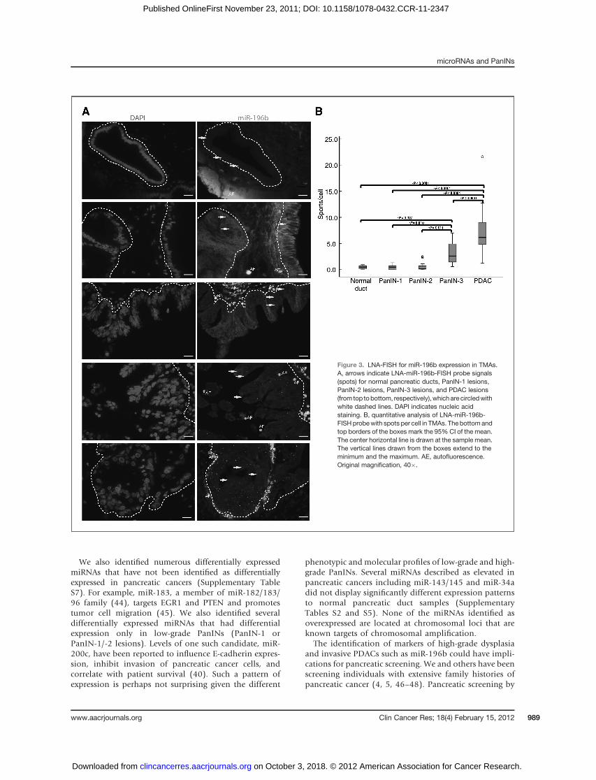
Locked nucleic acid-FISHLocked nucleic acid-FISH (LNA-FISH) was carried out
on TMA slides using LNA oligonucleotide probes againstmiR-196b and U6, both labeled with fluorescein at the50-end (Exiqon), according to the protocol described in thework of de Planell-Saguer and colleagues (32). LNA-U6wasused as a positive measure of probe specificity. Briefly, afterdeparaffinization, slides were prehybridized for 30minutesat 50�C (30�C below the RNA Tm of LNA-miR-196b probewhich is 80�C) in a humid chamber with prehybridization
buffer, then incubatedwith hybridization buffer containingLNA-miR-196b probe (1:1,000) for 1 hour at 50�C in ahybridization oven. After several washes, the slides wereincubated in 2-nitro-5-thiobenzoate (TNB) blocking buffer(PerkinElmer) containing goat anti-fluorescein antibodyperoxidase conjugate [fluorescein isothiocyanate (FITC)/horseradish peroxidase (HRP); Rockland; 1:1,000) for 30minutes in a humid chamber at room temperature]. Thenthe signals were amplified using tyramide signal amplifica-tion (TSA; PerkinElmer) for 10minutes in ahumid chamberat room temperature. After incubation in 40,6-diamidino-2-phenylindole (DAPI) staining solution for 5 minutes atroom temperature, slides weremounted with Prolong Goldanti-fade reagent (Invitrogen) and incubated overnight at4�C and signals were visualized using a fluorescent micro-scope. All steps beginning with hybridization were carriedout in the dark. The LNA-miR-196b-FISH results werequantified at a single-cell level by counting expression spotsper cell as previously described (33).
Statistical analysisPrincipal component analysis (PCA) mapping of com-
prehensive miRNA expression profiling was carried out byusing Partek Genomics Suite software (Partek Incorporat-ed). Differences in median expression were determinedusing the Mann–Whitney U test, differences in proportionsof expressing sampleswere determinedwith Pearson c2 test,and differences in LNA-FISH abnormalities between cate-gorical variables were determined with the Student t test.Statistical significance was defined as a value of P < 0.05. Toadjust for multiple comparisons, we calculated the falsediscovery rate (FDR) from the subset of significantly deregu-latedmiRNAswithP<0.05 (34). All statistical analyseswereconducted using the SPSS Statistics 19.0 software.
Results
miRNA expression differs in PanIN lesions and normalpancreatic ducts
The miRNA profiles of 34 laser capture–microdissectedPanIN lesions were compared with 15 LCM samples ofnormal pancreatic duct using TaqMan Array HumanMicroRNACards containing735humanmiRNAassays. ThemiRNA concentrations were normalized against RNU6B.Wefirst used PCA to compare the globalmiRNAs expressionprofile of PanIN lesions with that of samples of normalpancreatic ductal epithelial cells (Supplementary Fig. S1E)using thedata fromTaqManArrayHumanMicroRNACards.This analysis revealed that miRNAs effectively separatedPanIN lesions (all PanINs or each group of PanINs,PanIN-1, PanIN-2, and PanIN-3) and normal pancreaticducts, whereas PanIN lesions of different grades overlappedin their miRNA profiles (Supplementary Fig. S1E).
Numerous miRNAs are aberrantly expressed in PanINlesions
To identify differentially expressed miRNAs amongPanINs overall relative to normal pancreatic duct samples,
microRNAs and PanINs
www.aacrjournals.org Clin Cancer Res; 18(4) February 15, 2012 983
on October 3, 2018. © 2012 American Association for Cancer Research. clincancerres.aacrjournals.org Downloaded from
Published OnlineFirst November 23, 2011; DOI: 10.1158/1078-0432.CCR-11-2347
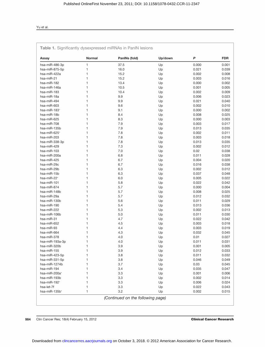
Table 1. Significantly dysexpressed miRNAs in PanIN lesions
Assay Normal PanINs (fold) Up/down P FDR
hsa-miR-486-3p 1 37.5 Up 0.000 0.001hsa-miR-875-5p 1 16.0 Up 0.021 0.039hsa-miR-422a 1 15.2 Up 0.002 0.008hsa-miR-21 1 15.2 Up 0.003 0.016hsa-miR-182 1 13.4 Up 0.000 0.002hsa-miR-146a 1 10.5 Up 0.001 0.005hsa-miR-183 1 10.4 Up 0.002 0.009hsa-miR-18a 1 9.9 Up 0.006 0.023hsa-miR-494 1 9.9 Up 0.021 0.040hsa-miR-603 1 9.6 Up 0.002 0.010hsa-miR-183� 1 9.1 Up 0.000 0.002hsa-miR-18b 1 8.4 Up 0.008 0.025hsa-miR-625 1 8.3 Up 0.000 0.003hsa-miR-708 1 7.9 Up 0.003 0.017hsa-miR-135b 1 7.9 Up 0.013 0.035hsa-miR-625� 1 7.8 Up 0.002 0.011hsa-miR-203 1 7.8 Up 0.003 0.018hsa-miR-338-3p 1 7.8 Up 0.013 0.035hsa-miR-429 1 7.3 Up 0.002 0.012hsa-miR-103 1 7.0 Up 0.02 0.038hsa-miR-200a 1 6.8 Up 0.011 0.028hsa-miR-425 1 6.7 Up 0.004 0.020hsa-miR-29c 1 6.7 Up 0.016 0.038hsa-miR-29b 1 6.3 Up 0.002 0.012hsa-miR-15b 1 6.3 Up 0.037 0.048hsa-miR-22� 1 6.0 Up 0.005 0.022hsa-miR-101 1 5.8 Up 0.022 0.042hsa-miR-874 1 5.7 Up 0.000 0.004hsa-miR-148b 1 5.7 Up 0.008 0.025hsa-miR-29a 1 5.7 Up 0.012 0.032hsa-miR-130b 1 5.6 Up 0.011 0.029hsa-miR-190 1 5.4 Up 0.013 0.036hsa-miR-222 1 5.3 Up 0.002 0.013hsa-miR-106b 1 5.0 Up 0.011 0.030hsa-miR-31 1 4.7 Up 0.022 0.042hsa-miR-652 1 4.5 Up 0.003 0.018hsa-miR-93 1 4.4 Up 0.003 0.019hsa-miR-664 1 4.3 Up 0.032 0.045hsa-miR-378 1 4.0 Up 0.01 0.027hsa-miR-193a-3p 1 4.0 Up 0.011 0.031hsa-miR-320b 1 3.9 Up 0.001 0.005hsa-miR-155 1 3.9 Up 0.012 0.033hsa-miR-423-5p 1 3.8 Up 0.011 0.032hsa-miR-331-5p 1 3.8 Up 0.046 0.049hsa-miR-1274b 1 3.7 Up 0.03 0.045hsa-miR-194 1 3.4 Up 0.035 0.047hsa-miR-200a� 1 3.3 Up 0.001 0.006hsa-miR-193b 1 3.3 Up 0.002 0.014hsa-miR-192� 1 3.3 Up 0.006 0.024hsa-let-7f 1 3.3 Up 0.022 0.043hsa-miR-135b� 1 3.2 Up 0.002 0.015
(Continued on the following page)
Yu et al.
Clin Cancer Res; 18(4) February 15, 2012 Clinical Cancer Research984
on October 3, 2018. © 2012 American Association for Cancer Research. clincancerres.aacrjournals.org Downloaded from
Published OnlineFirst November 23, 2011; DOI: 10.1158/1078-0432.CCR-11-2347
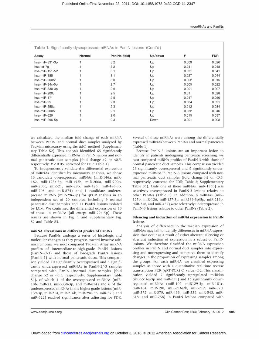
we calculated the median fold change of each miRNAbetween PanIN and normal duct samples analyzed byTaqMan microarray using the DDCt method (Supplemen-tary Table S2). This analysis identified 65 significantlydifferentially expressed miRNAs in PanIN lesions and nor-mal pancreatic duct samples (fold change >2 or <0.5,respectively; P < 0.05, corrected for FDR; Table 1).To independently validate the differential expression
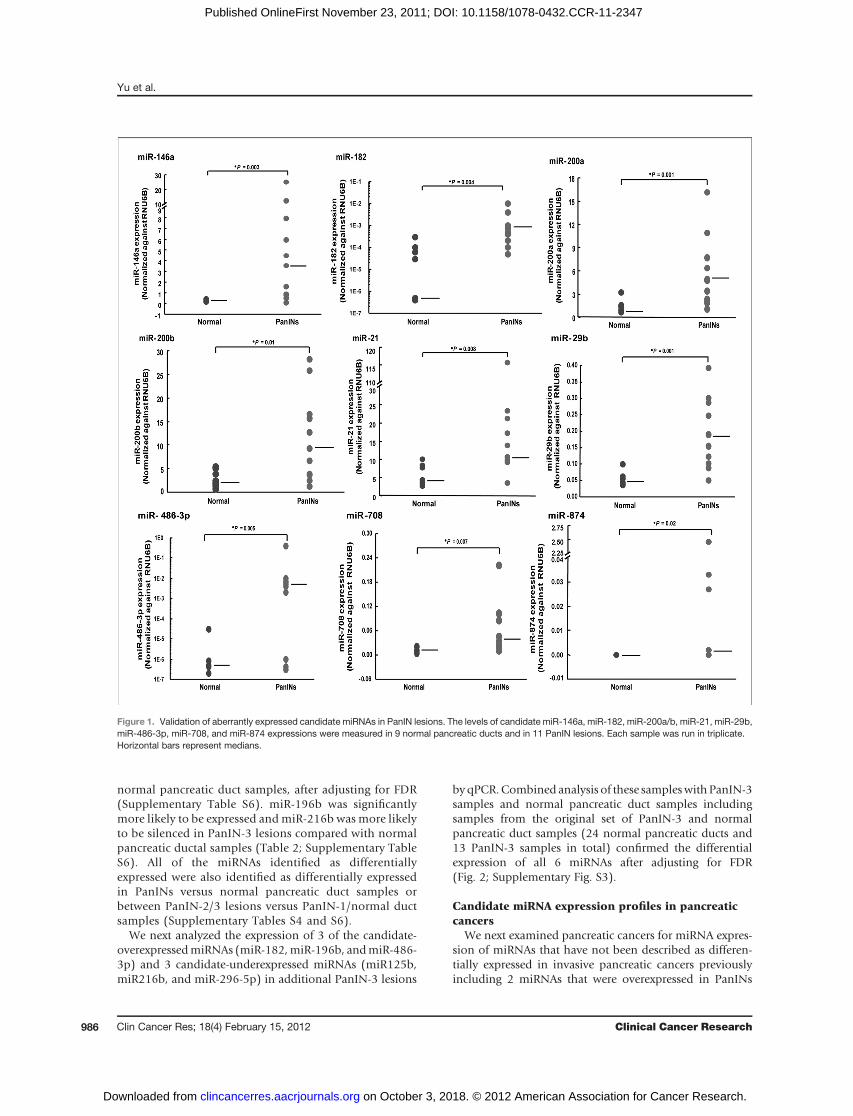
of miRNAs identified by microarray analysis, we chose13 candidate overexpressed miRNAs (miR-146a, miR-182, miR-193a-3p, miR-193b, miR-200a, miR-200b,miR-200c, miR-21, miR-29b, miR-425, miR-486-3p,miR-708, and miR-874) and 1 candidate underex-pressed miRNA (miR-296-5p) for qPCR analysis in anindependent set of 20 samples, including 9 normalpancreatic duct samples and 11 PanIN lesions isolatedby LCM. We confirmed the differential expression of 13of these 14 miRNAs (all except miR-296-5p). Theseresults are shown in Fig. 1 and Supplementary Fig.S2 and Table S3.
miRNA alterations in different grades of PanINsBecause PanINs undergo a series of histologic and
molecular changes as they progress toward invasive ade-nocarcinoma, we next compared TaqMan Array miRNAprofiles of intermediate-to-high-grade PanIN lesions(PanIN-2/-3) and those of low-grade PanIN lesions(PanIN-1) with normal pancreatic ducts. This compari-son yielded 10 significantly overexpressed and 8 signifi-cantly underexpressed miRNAs in PanIN-2/-3 samplescompared with PanIN-1/normal duct samples (foldchange >2 or <0.5, respectively; Supplementary TableS4), of which 4 of the overexpressed miRNAs (miR-18b, miR-21, miR-338-3p, and miR-874) and 6 of theunderexpressed miRNAs in the higher grade lesions (miR-139-3p, miR-214, miR-216b, miR-296-5p, miR-370, andmiR-622) reached significance after adjusting for FDR.
Several of these miRNAs were among the differentiallyexpressed miRNAs between PanINs and normal pancreata(Table 1).
Because PanIN-3 lesions are an important lesion toidentify in patients undergoing pancreatic screening, wenext compared miRNA profiles of PanIN-3 with those ofnormal pancreatic duct samples. This comparison yielded26 significantly overexpressed and 9 significantly under-expressed miRNAs in PanIN-3 lesions compared with nor-mal pancreatic duct samples (fold change >2 or <0.5,respectively; corrected for FDR; Table 2; SupplementaryTable S5). Only one of these miRNAs (miR-196b) wasselectively overexpressed in PanIN-3 lesions relative toother PanINs (Table 1). In addition, 8 miRNAs (miR-125b, miR-126, miR-127-3p, miR139-3p/5p, miR-216b,miR-218, and miR-452) were selectively underexpressed inPanIN-3 lesions relative to other PanINs (Table 2).
Silencing and induction of miRNA expression in PanINlesions
Analysis of differences in the median expression ofmiRNAs may fail to identify differences in miRNA expres-sion that occur as a result of either aberrant silencing oraberrant induction of expression in a subset of PanINlesions. We therefore classified the miRNA expressionprofiles in PanIN and normal duct samples into expres-sing and nonexpressing and compared them to identifychanges in the proportion of expressing samples amongthe groups. For each miRNA, we classified expressingsamples as those with a quantitative real-time reversetranscription PCR (qRT-PCR) Ct value <32. This classifi-cation yielded 2 significantly upregulated miRNAs(miR-516a-3p and miR-659) and 16 significantly down-regulated miRNAs (miR-107, miR129-3p, miR-181c,miR-184, miR-198, miR-216a/b, miR-217, miR-370,miR-372, miR-379, miR-433, miR-539, miR-543, miR-618, and miR-758) in PanIN lesions compared with
Table 1. Significantly dysexpressed miRNAs in PanIN lesions (Cont'd )
Assay Normal PanINs (fold) Up/down P FDR
hsa-miR-331-3p 1 3.2 Up 0.009 0.026hsa-let-7g 1 3.2 Up 0.041 0.048hsa-miR-151-5P 1 3.1 Up 0.021 0.041hsa-miR-185 1 3.1 Up 0.027 0.044hsa-miR-200b� 1 3.0 Up 0.002 0.015hsa-miR-34c-5p 1 2.7 Up 0.005 0.022hsa-miR-330-3p 1 2.6 Up 0.001 0.007hsa-miR-200c 1 2.5 Up 0.01 0.028hsa-miR-17� 1 2.5 Up 0.047 0.050hsa-miR-95 1 2.3 Up 0.004 0.021hsa-miR-550a 1 2.3 Up 0.012 0.034hsa-miR-200b 1 2.1 Up 0.032 0.046hsa-miR-629 1 2.0 Up 0.015 0.037hsa-miR-296-5p 1 0.3 Down 0.001 0.008
microRNAs and PanINs
www.aacrjournals.org Clin Cancer Res; 18(4) February 15, 2012 985
on October 3, 2018. © 2012 American Association for Cancer Research. clincancerres.aacrjournals.org Downloaded from
Published OnlineFirst November 23, 2011; DOI: 10.1158/1078-0432.CCR-11-2347
normal pancreatic duct samples, after adjusting for FDR(Supplementary Table S6). miR-196b was significantlymore likely to be expressed andmiR-216b was more likelyto be silenced in PanIN-3 lesions compared with normalpancreatic ductal samples (Table 2; Supplementary TableS6). All of the miRNAs identified as differentiallyexpressed were also identified as differentially expressedin PanINs versus normal pancreatic duct samples orbetween PanIN-2/3 lesions versus PanIN-1/normal ductsamples (Supplementary Tables S4 and S6).
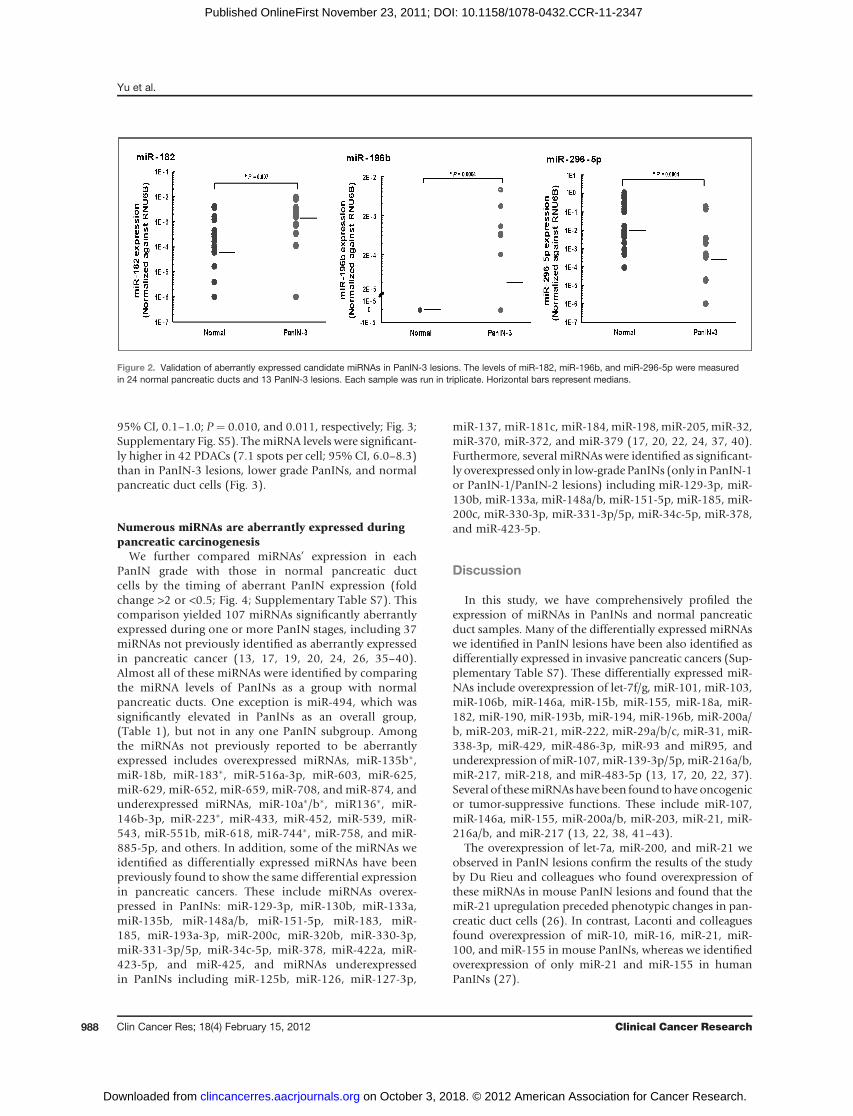
We next analyzed the expression of 3 of the candidate-overexpressedmiRNAs (miR-182,miR-196b, andmiR-486-3p) and 3 candidate-underexpressed miRNAs (miR125b,miR216b, and miR-296-5p) in additional PanIN-3 lesions
byqPCR.Combined analysis of these sampleswithPanIN-3samples and normal pancreatic duct samples includingsamples from the original set of PanIN-3 and normalpancreatic duct samples (24 normal pancreatic ducts and13 PanIN-3 samples in total) confirmed the differentialexpression of all 6 miRNAs after adjusting for FDR(Fig. 2; Supplementary Fig. S3).
Candidate miRNA expression profiles in pancreaticcancers
We next examined pancreatic cancers for miRNA expres-sion of miRNAs that have not been described as differen-tially expressed in invasive pancreatic cancers previouslyincluding 2 miRNAs that were overexpressed in PanINs
Figure 1. Validation of aberrantly expressed candidate miRNAs in PanIN lesions. The levels of candidate miR-146a, miR-182, miR-200a/b, miR-21, miR-29b,miR-486-3p, miR-708, and miR-874 expressions were measured in 9 normal pancreatic ducts and in 11 PanIN lesions. Each sample was run in triplicate.Horizontal bars represent medians.
Yu et al.
Clin Cancer Res; 18(4) February 15, 2012 Clinical Cancer Research986
on October 3, 2018. © 2012 American Association for Cancer Research. clincancerres.aacrjournals.org Downloaded from
Published OnlineFirst November 23, 2011; DOI: 10.1158/1078-0432.CCR-11-2347

(miR-182 andmiR-196b) and one that was underexpressed(miR-296-5p). Expression of these miRNAs was examinedin 29 pancreatic cancer cell lines and in the nonneoplasticpancreatic epithelial line, HPDE. As shown in Supplemen-tary Fig. S4, only miR-296-5p showed differential expres-sion in pancreatic cancer cell lines relative to HPDE.To confirm that miRNA expression patterns of pancreaticcancer cell lines were the same in primary pancreatic cancercells, we examined the expression of these 3 candidatemiRNAs in 14 samples of laser capture–microdissectedPDAC cells compared with 8 samples of microdissectednormal pancreatic ductal cells. As shown in SupplementaryFig. S4, the expression of these 3 miRNAs was significantlydifferent in primary PDAC samples relative to normalpancreatic ducts for all 3 miRNAs, including miR-182
(P < 0.001), miR-196b (P < 0.001), and miR-296-5p(P ¼ 0.002), respectively.
Expression of miRNA-196b in PanIN-3 lesions byLNA-FISH
BecausemiRNA-196b is the most differentially expressedmiRNA in PanIN-3 lesions by miRNA microarray analysis,we further examined its expression in PanIN lesions usingLNA-FISH.We found that themean expression ofmiR-196bin 16 PanIN-3 lesions was 3.1 spots per cell [95% confi-dence interval (CI), 2.0–4.3] versus 0.5 spots per cell (95%CI, 0.2–0.7) in 8 normal pancreatic duct cells (P ¼ 0.027).MiRNA-196b levels were also significantly higher in thePanIN-3 lesions than in 13 PanIN-1 (0.4 spots per cell;95%CI, 0.2–0.7) and 15 PanIN-2 lesions (0.6 spots per cell;
Table 2. Aberrantly expressed miRNAs in PanIN-3 lesions
Assay Normal PanIN-3 (fold) Up/down PanINsa P FDR
hsa-miR-196b 1 64.9 Up — 0.002 0.010hsa-miR-486-3p 1 49.8 Up Up 0.005 0.013hsa-miR-21 1 23.2 Up Up 0.017 0.024hsa-miR-338-3p 1 14.9 Up Up 0.024 0.033hsa-miR-18a 1 14.7 Up Up 0.039 0.041hsa-miR-183� 1 14.5 Up Up 0.001 0.003hsa-miR-182 1 12.8 Up Up 0.002 0.009hsa-miR-18b 1 11.9 Up Up 0.014 0.020hsa-miR-183 1 10.6 Up Up 0.017 0.026hsa-miR-422a 1 10.2 Up Up 0.039 0.043hsa-miR-603 1 9.6 Up Up 0.036 0.040hsa-miR-190 1 7.1 Up Up 0.031 0.039hsa-miR-29b 1 6.8 Up Up 0.039 0.044hsa-miR-93 1 6.7 Up Up 0.024 0.034hsa-miR-425 1 6.7 Up Up 0.017 0.027hsa-miR-146a 1 6.6 Up Up 0.011 0.016hsa-miR-874 1 5.6 Up Up 0.017 0.029hsa-miR-101 1 5.3 Up Up 0.039 0.046hsa-miR-652 1 5.1 Up Up 0.012 0.017hsa-miR-193a-3p 1 4.8 Up Up 0.039 0.047hsa-miR-625 1 4.5 Up Up 0.014 0.021hsa-miR-135b 1 3.4 Up Up 0.045 0.050hsa-miR-320b 1 3.1 Up Up 0.024 0.036hsa-miR-135b� 1 2.8 Up Up 0.021 0.031hsa-miR-222 1 2.6 Up Up 0.039 0.049hsa-miR-106b 1 2.4 Up Up 0.014 0.023hsa-miR-452 1 0.4 Down — 0.002 0.011hsa-miR-126 1 0.3 Down — 0.018 0.030hsa-miR-218 1 0.3 Down — 0.006 0.014hsa-miR-125b 1 0.3 Down — 0.012 0.019hsa-miR-127-3p 1 0.2 Down — 0.024 0.037hsa-miR-139-3p 1 0.2 Down — 0.001 0.007hsa-miR-139-5p 1 0.2 Down — 0.001 0.004hsa-miR-216b 1 0.1 Down — 0.000 0.001hsa-miR-296-5p 1 0.1 Down Down 0.001 0.006
aAberrantly expressed miRNAs in PanIN lesions in Table 1.
microRNAs and PanINs
www.aacrjournals.org Clin Cancer Res; 18(4) February 15, 2012 987
on October 3, 2018. © 2012 American Association for Cancer Research. clincancerres.aacrjournals.org Downloaded from
Published OnlineFirst November 23, 2011; DOI: 10.1158/1078-0432.CCR-11-2347
95% CI, 0.1–1.0; P ¼ 0.010, and 0.011, respectively; Fig. 3;Supplementary Fig. S5). The miRNA levels were significant-ly higher in 42 PDACs (7.1 spots per cell; 95% CI, 6.0–8.3)than in PanIN-3 lesions, lower grade PanINs, and normalpancreatic duct cells (Fig. 3).
Numerous miRNAs are aberrantly expressed duringpancreatic carcinogenesis
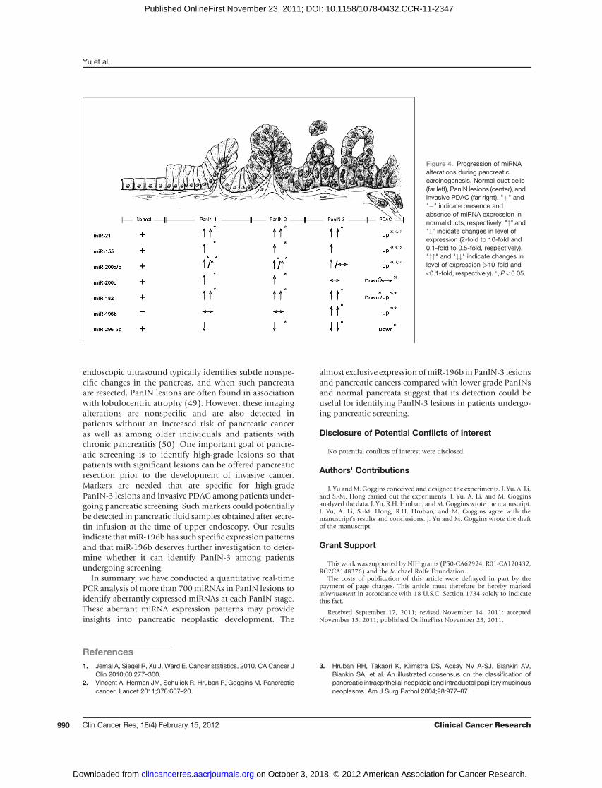
We further compared miRNAs’ expression in eachPanIN grade with those in normal pancreatic ductcells by the timing of aberrant PanIN expression (foldchange >2 or <0.5; Fig. 4; Supplementary Table S7). Thiscomparison yielded 107 miRNAs significantly aberrantlyexpressed during one or more PanIN stages, including 37miRNAs not previously identified as aberrantly expressedin pancreatic cancer (13, 17, 19, 20, 24, 26, 35–40).Almost all of these miRNAs were identified by comparingthe miRNA levels of PanINs as a group with normalpancreatic ducts. One exception is miR-494, which wassignificantly elevated in PanINs as an overall group,(Table 1), but not in any one PanIN subgroup. Amongthe miRNAs not previously reported to be aberrantlyexpressed includes overexpressed miRNAs, miR-135b�,miR-18b, miR-183�, miR-516a-3p, miR-603, miR-625,miR-629, miR-652, miR-659, miR-708, and miR-874, andunderexpressed miRNAs, miR-10a�/b�, miR136�, miR-146b-3p, miR-223�, miR-433, miR-452, miR-539, miR-543, miR-551b, miR-618, miR-744�, miR-758, and miR-885-5p, and others. In addition, some of the miRNAs weidentified as differentially expressed miRNAs have beenpreviously found to show the same differential expressionin pancreatic cancers. These include miRNAs overex-pressed in PanINs: miR-129-3p, miR-130b, miR-133a,miR-135b, miR-148a/b, miR-151-5p, miR-183, miR-185, miR-193a-3p, miR-200c, miR-320b, miR-330-3p,miR-331-3p/5p, miR-34c-5p, miR-378, miR-422a, miR-423-5p, and miR-425, and miRNAs underexpressedin PanINs including miR-125b, miR-126, miR-127-3p,
miR-137, miR-181c, miR-184,miR-198,miR-205,miR-32,miR-370, miR-372, and miR-379 (17, 20, 22, 24, 37, 40).Furthermore, several miRNAs were identified as significant-ly overexpressed only in low-grade PanINs (only in PanIN-1or PanIN-1/PanIN-2 lesions) including miR-129-3p, miR-130b, miR-133a, miR-148a/b, miR-151-5p, miR-185, miR-200c, miR-330-3p, miR-331-3p/5p, miR-34c-5p, miR-378,and miR-423-5p.
Discussion
In this study, we have comprehensively profiled theexpression of miRNAs in PanINs and normal pancreaticduct samples. Many of the differentially expressed miRNAswe identified in PanIN lesions have been also identified asdifferentially expressed in invasive pancreatic cancers (Sup-plementary Table S7). These differentially expressed miR-NAs include overexpression of let-7f/g, miR-101, miR-103,miR-106b, miR-146a, miR-15b, miR-155, miR-18a, miR-182, miR-190, miR-193b, miR-194, miR-196b, miR-200a/b, miR-203, miR-21, miR-222, miR-29a/b/c, miR-31, miR-338-3p, miR-429, miR-486-3p, miR-93 and miR95, andunderexpression ofmiR-107,miR-139-3p/5p,miR-216a/b,miR-217, miR-218, and miR-483-5p (13, 17, 20, 22, 37).Several of thesemiRNAshavebeen found tohave oncogenicor tumor-suppressive functions. These include miR-107,miR-146a, miR-155, miR-200a/b, miR-203, miR-21, miR-216a/b, and miR-217 (13, 22, 38, 41–43).
The overexpression of let-7a, miR-200, and miR-21 weobserved in PanIN lesions confirm the results of the studyby Du Rieu and colleagues who found overexpression ofthese miRNAs in mouse PanIN lesions and found that themiR-21 upregulation preceded phenotypic changes in pan-creatic duct cells (26). In contrast, Laconti and colleaguesfound overexpression of miR-10, miR-16, miR-21, miR-100, and miR-155 in mouse PanINs, whereas we identifiedoverexpression of only miR-21 and miR-155 in humanPanINs (27).
Figure 2. Validation of aberrantly expressed candidate miRNAs in PanIN-3 lesions. The levels of miR-182, miR-196b, and miR-296-5p were measuredin 24 normal pancreatic ducts and 13 PanIN-3 lesions. Each sample was run in triplicate. Horizontal bars represent medians.
Yu et al.
Clin Cancer Res; 18(4) February 15, 2012 Clinical Cancer Research988
on October 3, 2018. © 2012 American Association for Cancer Research. clincancerres.aacrjournals.org Downloaded from
Published OnlineFirst November 23, 2011; DOI: 10.1158/1078-0432.CCR-11-2347
We also identified numerous differentially expressedmiRNAs that have not been identified as differentiallyexpressed in pancreatic cancers (Supplementary TableS7). For example, miR-183, a member of miR-182/183/96 family (44), targets EGR1 and PTEN and promotestumor cell migration (45). We also identified severaldifferentially expressed miRNAs that had differentialexpression only in low-grade PanINs (PanIN-1 orPanIN-1/-2 lesions). Levels of one such candidate, miR-200c, have been reported to influence E-cadherin expres-sion, inhibit invasion of pancreatic cancer cells, andcorrelate with patient survival (40). Such a pattern ofexpression is perhaps not surprising given the different
phenotypic and molecular profiles of low-grade and high-grade PanINs. Several miRNAs described as elevated inpancreatic cancers including miR-143/145 and miR-34adid not display significantly different expression patternsto normal pancreatic duct samples (SupplementaryTables S2 and S5). None of the miRNAs identified asoverexpressed are located at chromosomal loci that areknown targets of chromosomal amplification.
The identification of markers of high-grade dysplasiaand invasive PDACs such as miR-196b could have impli-cations for pancreatic screening. We and others have beenscreening individuals with extensive family histories ofpancreatic cancer (4, 5, 46–48). Pancreatic screening by
Figure 3. LNA-FISH for miR-196b expression in TMAs.A, arrows indicate LNA-miR-196b-FISH probe signals(spots) for normal pancreatic ducts, PanIN-1 lesions,PanIN-2 lesions, PanIN-3 lesions, and PDAC lesions(from top tobottom, respectively), whichare circledwithwhite dashed lines. DAPI indicates nucleic acidstaining. B, quantitative analysis of LNA-miR-196b-FISH probewith spots per cell in TMAs. The bottom andtop borders of the boxes mark the 95%CI of the mean.The center horizontal line is drawn at the sample mean.The vertical lines drawn from the boxes extend to theminimum and the maximum. AE, autofluorescence.Original magnification, 40�.
microRNAs and PanINs
www.aacrjournals.org Clin Cancer Res; 18(4) February 15, 2012 989
on October 3, 2018. © 2012 American Association for Cancer Research. clincancerres.aacrjournals.org Downloaded from
Published OnlineFirst November 23, 2011; DOI: 10.1158/1078-0432.CCR-11-2347
endoscopic ultrasound typically identifies subtle nonspe-cific changes in the pancreas, and when such pancreataare resected, PanIN lesions are often found in associationwith lobulocentric atrophy (49). However, these imagingalterations are nonspecific and are also detected inpatients without an increased risk of pancreatic canceras well as among older individuals and patients withchronic pancreatitis (50). One important goal of pancre-atic screening is to identify high-grade lesions so thatpatients with significant lesions can be offered pancreaticresection prior to the development of invasive cancer.Markers are needed that are specific for high-gradePanIN-3 lesions and invasive PDAC among patients under-going pancreatic screening. Such markers could potentiallybe detected in pancreatic fluid samples obtained after secre-tin infusion at the time of upper endoscopy. Our resultsindicate thatmiR-196bhas such specific expression patternsand that miR-196b deserves further investigation to deter-mine whether it can identify PanIN-3 among patientsundergoing screening.
In summary, we have conducted a quantitative real-timePCR analysis of more than 700miRNAs in PanIN lesions toidentify aberrantly expressed miRNAs at each PanIN stage.These aberrant miRNA expression patterns may provideinsights into pancreatic neoplastic development. The
almost exclusive expression ofmiR-196b in PanIN-3 lesionsand pancreatic cancers compared with lower grade PanINsand normal pancreata suggest that its detection could beuseful for identifying PanIN-3 lesions in patients undergo-ing pancreatic screening.
Disclosure of Potential Conflicts of Interest
No potential conflicts of interest were disclosed.
Authors' Contributions
J. Yu andM. Goggins conceived and designed the experiments. J. Yu, A. Li,and S.-M. Hong carried out the experiments. J. Yu, A. Li, and M. Gogginsanalyzed the data. J. Yu, R.H. Hruban, andM.Goggins wrote themanuscript.J. Yu, A. Li, S.-M. Hong, R.H. Hruban, and M. Goggins agree with themanuscript’s results and conclusions. J. Yu and M. Goggins wrote the draftof the manuscript.
Grant Support
This work was supported by NIH grants (P50-CA62924, R01-CA120432,RC2CA148376) and the Michael Rolfe Foundation.
The costs of publication of this article were defrayed in part by thepayment of page charges. This article must therefore be hereby markedadvertisement in accordance with 18 U.S.C. Section 1734 solely to indicatethis fact.
Received September 17, 2011; revised November 14, 2011; acceptedNovember 15, 2011; published OnlineFirst November 23, 2011.
References1. Jemal A, Siegel R, Xu J, Ward E. Cancer statistics, 2010. CA Cancer J
Clin 2010;60:277–300.2. Vincent A, Herman JM, Schulick R, Hruban R, Goggins M. Pancreatic
cancer. Lancet 2011;378:607–20.
3. Hruban RH, Takaori K, Klimstra DS, Adsay NV A-SJ, Biankin AV,Biankin SA, et al. An illustrated consensus on the classification ofpancreatic intraepithelial neoplasia and intraductal papillary mucinousneoplasms. Am J Surg Pathol 2004;28:977–87.
Figure 4. Progression of miRNAalterations during pancreaticcarcinogenesis. Normal duct cells(far left), PanIN lesions (center), andinvasive PDAC (far right). "þ" and"�" indicate presence andabsence of miRNA expression innormal ducts, respectively. """ and"#" indicate changes in level ofexpression (2-fold to 10-fold and0.1-fold to 0.5-fold, respectively)."""" and "##" indicate changes inlevel of expression (>10-fold and<0.1-fold, respectively). �, P < 0.05.
Yu et al.
Clin Cancer Res; 18(4) February 15, 2012 Clinical Cancer Research990
on October 3, 2018. © 2012 American Association for Cancer Research. clincancerres.aacrjournals.org Downloaded from
Published OnlineFirst November 23, 2011; DOI: 10.1158/1078-0432.CCR-11-2347
4. CantoMI, GogginsM, Hruban RH, PetersenGM,Giardiello FM, YeoC,et al. Screening for early pancreatic neoplasia in high-risk individuals: aprospective controlled study. Clin Gastroenterol Hepatol 2006;4:766–81; quiz 665.
5. Canto MI, Goggins M, Yeo CJ, Griffin C, Axilbund JE, Brune K, et al.Screening for pancreatic neoplasia in high-risk individuals: an EUS-based approach. Clin Gastroenterol Hepatol 2004;2:606–21.
6. Matsubayashi H, Canto M, Sato N, Klein A, Abe T, Yamashita K, et al.DNA methylation alterations in the pancreatic juice of patients withsuspected pancreatic disease. Cancer Res 2006;66:1208–17.
7. Parsi MA, Li A, Li CP, Goggins M. DNA methylation alterations inendoscopic retrograde cholangiopancreatography brush samples ofpatients with suspected pancreaticobiliary disease. Clin GastroenterolHepatol 2008;6:1270–8.
8. Goggins M. Markers of pancreatic cancer: working toward earlydetection. Clin Cancer Res 2011;17:635–7.
9. Iacobuzio-Donahue CA, Maitra A, Shen-Ong GL, van Heek T, AshfaqR, Meyer R, et al. Discovery of novel tumor markers of pancreaticcancer using global gene expression technology. Am J Pathol2002;160:1239–49.
10. Michl P, Buchholz M, Rolke M, Kunsch S, Lohr M, McClane B, et al.Claudin-4: a new target for pancreatic cancer treatment usingClostridium perfringens enterotoxin. Gastroenterology 2001;121:678–84.
11. Yu J, Ohuchida K, Mizumoto K, Fujita H, Nakata K, Tanaka M. Micro-RNA miR-17-5p is overexpressed in pancreatic cancer, associatedwith a poor prognosis, and involved in cancer cell proliferation andinvasion. Cancer Biol Ther 2010;10:748–57.
12. Bartel DP. MicroRNAs: genomics, biogenesis, mechanism, and func-tion. Cell 2004;116:281–97.
13. Li A, Omura N, Hong SM, Vincent A, Walter K, Griffith M, et al.Pancreatic cancers epigenetically silence SIP1 and hypomethylateand overexpress miR-200a/200b in association with elevated circu-lating miR-200a and miR-200b levels. Cancer Res 2010;70:5226–37.
14. Calin GA, Croce CM. Chromosomal rearrangements and microRNAs:a new cancer link with clinical implications. J Clin Invest 2007;117:2059–66.
15. Iorio MV, Ferracin M, Liu CG, Veronese A, Spizzo R, Sabbioni S, et al.MicroRNA gene expression deregulation in human breast cancer.Cancer Res 2005;65:7065–70.
16. Wijnhoven BP, Michael MZ, Watson DI. MicroRNAs and cancer. Br JSurg 2007;94:23–30.
17. Bloomston M, Frankel WL, Petrocca F, Volinia S, Alder H, Hagan JP,et al. MicroRNA expression patterns to differentiate pancreatic ade-nocarcinoma from normal pancreas and chronic pancreatitis. JAMA2007;297:1901–8.
18. Dillhoff M, Liu J, Frankel W, Croce C, Bloomston M. MicroRNA-21 isoverexpressed in pancreatic cancer and a potential predictor of sur-vival. J Gastrointest Surg 2008;12:2171–6.
19. Kent OA, Mullendore M, Wentzel EA, Lopez-Romero P, Tan AC,Alvarez H, et al. A resource for analysis of microRNA expression andfunction in pancreatic ductal adenocarcinoma cells. Cancer Biol Ther2009;8:2013–24.
20. Lee EJ, Gusev Y, Jiang J, Nuovo GJ, Lerner MR, Frankel WL, et al.Expression profiling identifies microRNA signature in pancreatic can-cer. Int J Cancer 2007;120:1046–54.
21. Li Y, Vandenboom TG II, Wang Z, Kong D, Ali S, Philip PA, et al. miR-146a suppresses invasion of pancreatic cancer cells. Cancer Res2010;70:1486–95.
22. Szafranska AE, Davison TS, John J, Cannon T, Sipos B, Maghnouj A,et al. MicroRNA expression alterations are linked to tumorigenesis andnon-neoplastic processes in pancreatic ductal adenocarcinoma.Oncogene 2007;26:4442–52.
23. Szafranska AE, Doleshal M, Edmunds HS, Gordon S, Luttges J,Munding JB, et al. Analysis of microRNAs in pancreatic fine-needleaspirates can classify benign and malignant tissues. Clin Chem2008;54:1716–24.
24. Zhang Y, Li M, Wang H, Fisher WE, Lin PH, Yao Q, et al. Profiling of 95microRNAs in pancreatic cancer cell lines and surgical specimens byreal-time PCR analysis. World J Surg 2009;33:698–709.
25. Kent OA, Chivukula RR, Mullendore M, Wentzel EA, Feldmann G, LeeKH, et al. Repression of the miR-143/145 cluster by oncogenic Rasinitiates a tumor-promoting feed-forward pathway. Genes Dev2010;24:2754–9.
26. du Rieu MC, Torrisani J, Selves J, Al Saati T, Souque A, Dufresne M,et al. MicroRNA-21 is induced early in pancreatic ductal adenocarci-noma precursor lesions. Clin Chem 2010;56:603–12.
27. Laconti JJ, Shivapurkar N, Preet A, Deslattes Mays A, Peran I, KimSE, et al. Tissue and serum microRNAs in the Kras transgenicanimal model and in patients with pancreatic cancer. PLoS One2011;6:e20687.
28. Klimstra DS, Pitman MB, Hruban RH. An algorithmic approach to thediagnosis of pancreatic neoplasms. Arch Pathol Lab Med 2009;133:454–64.
29. Hong SM, Kelly D, GriffithM, Omura N, Li A, Li CP, et al. Multiple genesare hypermethylated in intraductal papillary mucinous neoplasms ofthe pancreas. Mod Pathol 2008;21:1499–507.
30. Maitra A, Adsay NV, Argani P, Iacobuzio-Donahue C, De Marzo A,Cameron JL, et al. Multicomponent analysis of the pancreatic adeno-carcinoma progression model using a pancreatic intraepithelial neo-plasia tissue microarray. Mod Pathol 2003;16:902–12.
31. Yu J, Ohuchida K, Nakata K,Mizumoto K, Cui L, Fujita H, et al. LIM only4 is overexpressed in late stage pancreas cancer. Mol Cancer2008;7:93.
32. de Planell-Saguer M, Rodicio MC, Mourelatos Z. Rapid in situ code-tection of noncoding RNAs and proteins in cells and formalin-fixedparaffin-embedded tissue sections without protease treatment. NatProtoc 2010;5:1061–73.
33. Lu J, Tsourkas A. Imaging individual microRNAs in single mammaliancells in situ. Nucleic Acids Res 2009;37:e100.
34. Benjamini Y, Drai D, Elmer G, Kafkafi N, Golani I. Controlling the falsediscovery rate in behavior genetics research. Behav Brain Res2001;125:279–84.
35. Ikenaga N,Ohuchida K,Mizumoto K, Yu J, Kayashima T, Sakai H, et al.MicroRNA-203 expression as a new prognostic marker of pancreaticadenocarcinoma. Ann Surg Oncol 2010;17:3120–8.
36. Liffers ST,Munding JB, VogtM, Kuhlmann JD, Verdoodt B, Nambiar S,et al. MicroRNA-148a is down-regulated in human pancreatic ductaladenocarcinomas and regulates cell survival by targeting CDC25B.Lab Invest 2011;91:1472–9.
37. Mees ST, Mardin WA, Sielker S, Willscher E, Senninger N, SchleicherC, et al. Involvement of CD40 targeting miR-224 and miR-486 on theprogression of pancreatic ductal adenocarcinomas. Ann Surg Oncol2009;16:2339–50.
38. Moriyama T, Ohuchida K, Mizumoto K, Yu J, Sato N, Nabae T, et al.MicroRNA-21 modulates biological functions of pancreatic cancercells including their proliferation, invasion, and chemoresistance. MolCancer Ther 2009;8:1067–74.
39. MuniyappaMK,DowlingP,HenryM,MeleadyP,DoolanP,Gammell P,et al. MiRNA-29a regulates the expression of numerous proteins andreduces the invasiveness and proliferation of human carcinoma celllines. Eur J Cancer 2009;45:3104–18.
40. Yu J, Ohuchida K, Mizumoto K, Sato N, Kayashima T, Fujita H, et al.MicroRNA, hsa-miR-200c, is an independent prognostic factor inpancreatic cancer and its upregulation inhibits pancreatic cancerinvasion but increases cell proliferation. Mol Cancer 2010;9:169.
41. Gironella M, Seux M, Xie MJ, Cano C, Tomasini R, Gommeaux J, et al.Tumor protein 53-inducednuclear protein 1 expression is repressedbymiR-155, and its restoration inhibits pancreatic tumor development.Proc Natl Acad Sci U S A 2007;104:16170–5.
42. LeeKH, LottermanC,Karikari C,OmuraN, FeldmannG,HabbeN, et al.Epigenetic silencing of MicroRNA miR-107 regulates cyclin-depen-dent kinase 6 expression in pancreatic cancer. Pancreatology2009;9:293–301.
43. Wellner U, Schubert J, Burk UC, Schmalhofer O, Zhu F, Sonntag A,et al. The EMT-activator ZEB1 promotes tumorigenicity by re-pressing stemness-inhibiting microRNAs. Nat Cell Biol 2009;11:1487–95.
44. Saini HK, Enright AJ, Griffiths-Jones S. Annotation of mammalianprimary microRNAs. BMC Genomics 2008;9:564.
microRNAs and PanINs
www.aacrjournals.org Clin Cancer Res; 18(4) February 15, 2012 991
on October 3, 2018. © 2012 American Association for Cancer Research. clincancerres.aacrjournals.org Downloaded from
Published OnlineFirst November 23, 2011; DOI: 10.1158/1078-0432.CCR-11-2347

45. Sarver AL, Li L, Subramanian S. MicroRNA miR-183 functionsas an oncogene by targeting the transcription factor EGR1and promoting tumor cell migration. Cancer Res 2010;70:9570–80.
46. Brentnall TA, Bronner MP, Byrd DR, Haggitt RC, Kimmey MB. Earlydiagnosis and treatment of pancreatic dysplasia in patients witha family history of pancreatic cancer. Ann Intern Med 1999;131:247–55.
47. Langer P, Kann PH, Fendrich V, Habbe N, Schneider M, Sina M,et al. Five years of prospective screening of high-risk indi-viduals from families with familial pancreatic cancer. Gut 2009;58:1410–8.
48. Verna EC, Hwang C, Stevens PD, Rotterdam H, Stavropoulos SN, SyCD, et al. Pancreatic cancer screening in a prospective cohort of high-risk patients: a comprehensive strategy of imaging and genetics. ClinCancer Res 2010;16:5028–37.
49. Brune K, Abe T, Canto M, O'Malley L, Klein AP, Maitra A, et al.Multifocal neoplastic precursor lesions associatedwith lobular atrophyof the pancreas in patients having a strong family history of pancreaticcancer. Am J Surg Pathol 2006;30:1067–76.
50. Catalano MF, Sahai A, Levy M, Romagnuolo J, Wiersema M, BruggeW, et al. EUS-based criteria for the diagnosis of chronic pancrea-titis: the Rosemont classification. Gastrointest Endosc 2009;69:1251–61.
Yu et al.
Clin Cancer Res; 18(4) February 15, 2012 Clinical Cancer Research992
on October 3, 2018. © 2012 American Association for Cancer Research. clincancerres.aacrjournals.org Downloaded from
Published OnlineFirst November 23, 2011; DOI: 10.1158/1078-0432.CCR-11-2347

2012;18:981-992. Published OnlineFirst November 23, 2011.Clin Cancer Res Jun Yu, Ang Li, Seung-Mo Hong, et al. MicroRNA Alterations of Pancreatic Intraepithelial Neoplasias
Updated version
10.1158/1078-0432.CCR-11-2347doi:
Access the most recent version of this article at:
Material
Supplementary
http://clincancerres.aacrjournals.org/content/suppl/2011/11/23/1078-0432.CCR-11-2347.DC1
Access the most recent supplemental material at:
Cited articles
http://clincancerres.aacrjournals.org/content/18/4/981.full#ref-list-1
This article cites 50 articles, 13 of which you can access for free at:
Citing articles
http://clincancerres.aacrjournals.org/content/18/4/981.full#related-urls
This article has been cited by 13 HighWire-hosted articles. Access the articles at:
E-mail alerts related to this article or journal.Sign up to receive free email-alerts
Subscriptions
Reprints and
To order reprints of this article or to subscribe to the journal, contact the AACR Publications Department at
Permissions
Rightslink site. Click on "Request Permissions" which will take you to the Copyright Clearance Center's (CCC)
.http://clincancerres.aacrjournals.org/content/18/4/981To request permission to re-use all or part of this article, use this link
on October 3, 2018. © 2012 American Association for Cancer Research. clincancerres.aacrjournals.org Downloaded from
Published OnlineFirst November 23, 2011; DOI: 10.1158/1078-0432.CCR-11-2347