Microneme Rhomboid Protease TgROM1 Is Required for ...ec.asm.org/content/7/4/664.full.pdf · cleave...
11
EUKARYOTIC CELL, Apr. 2008, p. 664–674 Vol. 7, No. 4 1535-9778/08/$08.000 doi:10.1128/EC.00331-07 Copyright © 2008, American Society for Microbiology. All Rights Reserved. Microneme Rhomboid Protease TgROM1 Is Required for Efficient Intracellular Growth of Toxoplasma gondii † Fabien Brossier, G. Lucas Starnes, Wandy L. Beatty, and L. David Sibley* Department of Molecular Microbiology, Washington University School of Medicine, 660. S. Euclid Avenue, St. Louis, Missouri 63110-1093 Received 4 September 2007/Accepted 28 January 2008 Rhomboids are serine proteases that cleave their substrates within the transmembrane domain. Toxoplasma gondii contains six rhomboids that are expressed in different life cycle stages and localized to different cellular compartments. Toxoplasma rhomboid protein 1 (TgROM1) has previously been shown to be active in vitro, and the orthologue in Plasmodium falciparum processes the essential microneme protein AMA1 in a heterologous system. We investigated the role of TgROM1 to determine its role during in vitro growth of T. gondii. TgROM1 was localized in the secretory pathway of the parasite, including the Golgi apparatus and micronemes, which contain adhesive proteins involved in invasion of host cells. However, unlike other micronemal proteins, TgROM1 was not released onto the parasite surface during cell invasion, suggesting it does not play a critical role in cell invasion. Suppression of TgROM1 using the tetracycline-regulatable system revealed that ROM1- deficient parasites were outcompeted by wild-type T. gondii. ROM1-deficient parasites showed only modest decrease in invasion but replicated more slowly than wild-type cells. Collectively, these results indicate that ROM1 is required for efficient intracellular growth by T. gondii. Toxoplasma gondii is the agent of toxoplasmosis, which rep- resents a fatal threat to immunocompromised individuals (22). T. gondii belongs to the phylum Apicomplexa, a group of med- ically and economically important parasites including Plasmo- dium falciparum, the agent of malaria, and Cryptosporidium parvum, the cause of cryptosporidiosis. The complex life cycle of T. gondii contains a sexual stage in which oocysts are shed in the environment by cats and asexual stages consisting of dor- mant, slow-growing forms called bradyzoites and fast-growing forms called tachyzoites, which are extremely efficient at in- vading a variety of cells and disseminating within the host (13). T. gondii contains several sets of secretory organelles includ- ing micronemes, rhoptries, and dense granules whose sequen- tial secretion is responsible for attachment, penetration, and survival in the infected cell, respectively (8, 14). Attachment to host cells is initiated by secretion of microneme (MIC) pro- teins, which comprise a family of adhesive molecules (3, 6, 9). MIC proteins are typically found in complexes consisting of a transmembrane protein and a soluble partner(s) (10). MIC protein complexes also form higher-order complexes: for ex- ample, MIC2/M2AP is a heterohexamer composed of three molecules of MIC2 associated with three molecules of M2AP (21). Apicomplexans use a unique form of locomotion called glid- ing motility, which relies on apical secretion and trafficking of MICs along the surface of the parasite (32). Translocation is mediated by an interaction between the cytoplasmic tail of MIC2 and aldolase, which bridges to the actin-myosin motor of the parasite (20). Upon contact with host cells, MIC2 and its associated binding partner M2AP are released at the tip of the parasite, where they participate in binding to the host cell and then translocated toward the posterior end of the parasite during penetration into the host cell (6, 31). At the posterior end, the complex is released from the surface by proteolysis within the transmembrane domain of MIC2 (7, 30, 40) and this cleavage is required for efficient invasion of host cells (4). MIC6 and TgAMA1 have also been shown to be cleaved within their transmembrane domains (18, 30), and AMA1 provides a vital function in formation of the moving junction (1, 24). Recent data strongly suggest that parasite-derived, rhomboid- like proteases cleave MIC proteins in order to release them from the parasite membrane (5, 12, 36). Rhomboids are ubiquitous serine proteases that are able to recognize and cleave their substrates within their transmem- brane domains (35, 37). Rhomboids have been extensively studied in Drosophila melanogaster, where they are involved in the regulation of development (38). Spitz is an epidermal growth factor-like protein that is a substrate for rhomboids in the fly. Spitz is also used as a universal substrate to characterize heterologous rhomboids in COS cells (36). The genome of T. gondii contains six rhomboid-like genes (TgROMs) (5, 11, 12). Analysis of the predicted TgROM6 sequence suggests that the protein is localized in the mitochondria (11). The in vitro activities of the remaining five rhomboids (TgROM1 to TgROM5) have been characterized by cell biological, molec- ular, and biochemical methods. TgROM2 and TgROM3 are mainly expressed in the sporozoites, with much lower expres- sion in tachyzoites, as indicated by reverse transcription-PCR (RT-PCR) (5). Expression of a tagged form of TgROM2 in tachyzoites results in accumulation in the trans-Golgi network (12). In contrast, TgROM4 and TgROM5 are normally ex- pressed at the surface of tachyzoites, with TgROM5 being primarily localized at the posterior end of the cell. Like other * Corresponding author. Mailing address: Department of Molecular Microbiology, Washington University School of Medicine, 660 S. Eu- clid Ave., St. Louis, MO 63110-1093. Phone: (314) 362-8873. Fax: (314) 362-3203. E-mail: [email protected]. † Supplemental material for this article may be found at http://ec .asm.org/. Published ahead of print on 29 February 2008. 664 on July 18, 2018 by guest http://ec.asm.org/ Downloaded from
-
Upload
truongkhanh -
Category
Documents
-
view
220 -
download
0
Transcript of Microneme Rhomboid Protease TgROM1 Is Required for ...ec.asm.org/content/7/4/664.full.pdf · cleave...

EUKARYOTIC CELL, Apr. 2008, p. 664–674 Vol. 7, No. 41535-9778/08/$08.00�0 doi:10.1128/EC.00331-07Copyright © 2008, American Society for Microbiology. All Rights Reserved.
Microneme Rhomboid Protease TgROM1 Is Required for EfficientIntracellular Growth of Toxoplasma gondii�†
Fabien Brossier, G. Lucas Starnes, Wandy L. Beatty, and L. David Sibley*Department of Molecular Microbiology, Washington University School of Medicine, 660. S. Euclid Avenue,
St. Louis, Missouri 63110-1093
Received 4 September 2007/Accepted 28 January 2008
Rhomboids are serine proteases that cleave their substrates within the transmembrane domain. Toxoplasmagondii contains six rhomboids that are expressed in different life cycle stages and localized to different cellularcompartments. Toxoplasma rhomboid protein 1 (TgROM1) has previously been shown to be active in vitro, andthe orthologue in Plasmodium falciparum processes the essential microneme protein AMA1 in a heterologoussystem. We investigated the role of TgROM1 to determine its role during in vitro growth of T. gondii. TgROM1was localized in the secretory pathway of the parasite, including the Golgi apparatus and micronemes, whichcontain adhesive proteins involved in invasion of host cells. However, unlike other micronemal proteins,TgROM1 was not released onto the parasite surface during cell invasion, suggesting it does not play a criticalrole in cell invasion. Suppression of TgROM1 using the tetracycline-regulatable system revealed that ROM1-deficient parasites were outcompeted by wild-type T. gondii. ROM1-deficient parasites showed only modestdecrease in invasion but replicated more slowly than wild-type cells. Collectively, these results indicate thatROM1 is required for efficient intracellular growth by T. gondii.
Toxoplasma gondii is the agent of toxoplasmosis, which rep-resents a fatal threat to immunocompromised individuals (22).T. gondii belongs to the phylum Apicomplexa, a group of med-ically and economically important parasites including Plasmo-dium falciparum, the agent of malaria, and Cryptosporidiumparvum, the cause of cryptosporidiosis. The complex life cycleof T. gondii contains a sexual stage in which oocysts are shed inthe environment by cats and asexual stages consisting of dor-mant, slow-growing forms called bradyzoites and fast-growingforms called tachyzoites, which are extremely efficient at in-vading a variety of cells and disseminating within the host (13).
T. gondii contains several sets of secretory organelles includ-ing micronemes, rhoptries, and dense granules whose sequen-tial secretion is responsible for attachment, penetration, andsurvival in the infected cell, respectively (8, 14). Attachment tohost cells is initiated by secretion of microneme (MIC) pro-teins, which comprise a family of adhesive molecules (3, 6, 9).MIC proteins are typically found in complexes consisting of atransmembrane protein and a soluble partner(s) (10). MICprotein complexes also form higher-order complexes: for ex-ample, MIC2/M2AP is a heterohexamer composed of threemolecules of MIC2 associated with three molecules of M2AP(21).
Apicomplexans use a unique form of locomotion called glid-ing motility, which relies on apical secretion and trafficking ofMICs along the surface of the parasite (32). Translocation ismediated by an interaction between the cytoplasmic tail ofMIC2 and aldolase, which bridges to the actin-myosin motor of
the parasite (20). Upon contact with host cells, MIC2 and itsassociated binding partner M2AP are released at the tip of theparasite, where they participate in binding to the host cell andthen translocated toward the posterior end of the parasiteduring penetration into the host cell (6, 31). At the posteriorend, the complex is released from the surface by proteolysiswithin the transmembrane domain of MIC2 (7, 30, 40) and thiscleavage is required for efficient invasion of host cells (4).MIC6 and TgAMA1 have also been shown to be cleaved withintheir transmembrane domains (18, 30), and AMA1 provides avital function in formation of the moving junction (1, 24).Recent data strongly suggest that parasite-derived, rhomboid-like proteases cleave MIC proteins in order to release themfrom the parasite membrane (5, 12, 36).
Rhomboids are ubiquitous serine proteases that are able torecognize and cleave their substrates within their transmem-brane domains (35, 37). Rhomboids have been extensivelystudied in Drosophila melanogaster, where they are involved inthe regulation of development (38). Spitz is an epidermalgrowth factor-like protein that is a substrate for rhomboids inthe fly. Spitz is also used as a universal substrate to characterizeheterologous rhomboids in COS cells (36). The genome of T.gondii contains six rhomboid-like genes (TgROMs) (5, 11, 12).Analysis of the predicted TgROM6 sequence suggests that theprotein is localized in the mitochondria (11). The in vitroactivities of the remaining five rhomboids (TgROM1 toTgROM5) have been characterized by cell biological, molec-ular, and biochemical methods. TgROM2 and TgROM3 aremainly expressed in the sporozoites, with much lower expres-sion in tachyzoites, as indicated by reverse transcription-PCR(RT-PCR) (5). Expression of a tagged form of TgROM2 intachyzoites results in accumulation in the trans-Golgi network(12). In contrast, TgROM4 and TgROM5 are normally ex-pressed at the surface of tachyzoites, with TgROM5 beingprimarily localized at the posterior end of the cell. Like other
* Corresponding author. Mailing address: Department of MolecularMicrobiology, Washington University School of Medicine, 660 S. Eu-clid Ave., St. Louis, MO 63110-1093. Phone: (314) 362-8873. Fax: (314)362-3203. E-mail: [email protected].
† Supplemental material for this article may be found at http://ec.asm.org/.
� Published ahead of print on 29 February 2008.
664
on July 18, 2018 by guesthttp://ec.asm
.org/D
ownloaded from

ROMs, TgROM1 is active against Spitz in a heterologous COScell assay and this activity critically depends on the con-served transmembrane region of the substrate (5, 12). WhileTgROM1 is clearly a functional protease, its substrates havenot been identified in the parasite. TgROM2 is also able tocleave chimeric proteins composed of the transmembrane do-main of MIC2 or MIC12 (12), while only TgROM5 efficientlycleaves full-length MIC2 or chimeric proteins composed of thetransmembrane domains of MIC6 or MIC12 (5). Despite dem-onstration of these in vitro activities, the in vivo roles of theseproteases remain uncertain.
In this study, we focused on TgROM1, which is expressed inthe tachyzoite stage of T. gondii. To define the role ofTgROM1 in vivo, the expression of TgROM1 was conditionallysuppressed using the tetracycline-repressible system describedpreviously (25, 26).
MATERIALS AND METHODS
Growth of host cells and Toxoplasma strains. T. gondii strain tTa (26) andtransformants were maintained by growth in monolayers of human foreskinfibroblast (HFFs), propagated in Dulbecco’s modified Eagle’s medium contain-ing 10% fetal bovine serum, 2 mM glutamine, 20 mM HEPES (pH 7.5), and 20�g ml�1 gentamicin. Chloramphenicol (20 �g/ml) (Sigma-Aldrich, St. Louis,MO), phleomycin (5 �g/ml) (Invitrogen, San Diego, CA), and anhydrotetracy-cline (Atc) (1.5 �g/ml) (Clontech, Palo Alto, CA) were added to the media asindicated.
Antibodies. The HA9 epitope (YPYDVPDYA) was detected using rabbitpolyclonal antisera (Zymed, CA), monoclonal antibody (MAb) 16B12 (Covance,CA), or MAb HA-7 (Sigma-Aldrich, St. Louis, MO) for immunofluorescence(IF), Western blotting, and immunoelectron microscopy (immuno-EM) experi-ments, respectively. MIC2 was detected with MAb 6D10 and rabbit polyclonalanti-C-domain antibodies for IF and immuno-EM experiments, respectively (39).MIC4 was detected with rabbit polyclonal antibodies, generously provided byDominique Soldati, and MAb 5B1. TgAMA1 was detected with the MAb B3.90,generously provided by Gary Ward (University of Vermont). SAG1 was detectedusing MAb DG52 directly coupled to Alexa 488 or Alexa 594 (MolecularProbes). RON4 was detected with mouse polyclonal antisera, generously pro-vided by Peter Bradley (University of California). GRA1 was detected withmouse MAb Tg-17-43, a kind gift of Marie France Cesbron-Delauw.
Generation of �rom1/S1HA9-ROM1 and �rom1/S4HA9-ROM1 strains. TheTgROM1 open reading frame (NCBI accession no. AAT84608) with an N-terminal HA9 tag was inserted in the previously described vectors p7TetOS1 andp7TetOS4, downstream of the inducible promoters that consist of seven TetOsequences fused with the upstream regions from the SAG1 and SAG4 genes,respectively (25), to generate the plasmids pS1HA9-ROM1 and pS4HA9-ROM1. Transactivator-expressing parasites, referred to as tTa (26), were co-transfected by electroporation with pS1HA9-ROM1 or pS4HA9-ROM1 and aplasmid containing the ble gene driven by SAG1 flanking sequences, conferringthe resistance to phleomycin, as described previously (27). Following two roundsof selection, clones were obtained by limiting dilution on HFF monolayers grownin 96-well plates (27). ROM1/S1HA9-ROM1 or ROM1/S4HA9-ROM1 cloneswere identified by sodium dodecyl sulfate-polyacrylamide gel electrophoresis orWestern blotting and IF microscopy with anti-HA9 antibodies. The level ofexpression of HA9-ROM1 was higher when the protein was expressed under thecontrol of the TetOSAG4 promoter compared to the TetOSAG1 promoter, asreported previously (25). ROM1/S1HA9-ROM1 and ROM1/S4HA9-ROM1clones were used to generate TgROM1 knockout lines as follows.
A knockout construct referred to as plasmid p�R1 was engineered using theselectable marker cat, which confers resistance to chloramphenicol, controlled by5� and 3� SAG1 flanking sequences. This cat cassette was in turn flanked by 2 kbof sequences upstream of the start and downstream of the stop codons ofTgROM1 (sequences retrieved from http://ToxoDB.org). Two tandem yellowfluorescent protein (YFP) genes expressed under the control of the T. gondii�-tubulin (TUBA) promoter (a generous gift of B. Striepen) were inserteddownstream into the SacII site of p�R1, generating the plasmid p�R1YFP.ROM1/S1HA9-ROM1 and ROM1/S4HA9-ROM1 parasite strains were trans-fected by electroporation with 50 �g of linearized p�R1YFP. After antibioticselection and fluorescence-activated cell sorting, �10% of YFP-negative and
chloramphenicol-resistant parasites were determined to be �rom1 knockouts.Clones were obtained by limiting dilution for the �rom1/S1HA9-ROM1 and�rom1/S4HA9-ROM1 knockdown lines.
IF and deconvolution microscopy. To detect HA9-ROM1 in intracellular par-asites, HFF cell monolayers were infected with parasites and incubated in pres-ence or absence of Atc overnight at 37°C and 5% CO2, prior to being fixed andprocessed for IF. Permeabilized cells were incubated with anti-HA9, anti-MIC2,or anti-MIC4 antibodies; washed; and incubated with secondary antibodies con-jugated to Alexa 494 or Alexa 488 (Molecular Probes, OR). Coverslips werewashed and mounted in Vectashield containing DAPI (4�,6�-diamidino-2-phe-nylindole; Vector Laboratories, Burlingame, CA). Fluorescence images werecaptured using a �100, 1.4 numerical aperture lens on a Zeiss Axioskop 2 usingan Axiocam MRm cooled charge-coupled device camera (Thornwood, NewYork, NY). Serial Z-stack images were collected at �100, and images weredeconvolved using the nearest-neighbor algorithm in Axiovision and processedusing Photoshop 6.0 (Adobe Systems, San Jose, CA).
To localize HA9-ROM1 during host cell invasion, ROM1/S4HA9-ROM1tachyzoites were allowed to settle by gravity onto 80% confluent cells at 4°C andwarmed up for invasion during 5 min at 37°C in Dulbecco’s modified Eagle’smedium containing 20 mM HEPES, 0.2% sodium bicarbonate, and 3% fetalbovine serum. Invasion was stopped by addition of 2.5% paraformaldehyde.Monolayers were washed, blocked, and incubated with anti-SAG1 or anti-RON4antibodies to label surface-exposed epitopes. Cells were then permeabilized with0.5% saponin and incubated with anti-HA9 antibodies, followed by secondaryantibodies coupled to Alexa 488 (green) and Alexa 594 (red). Monolayers werewashed and mounted in Vectashield containing DAPI. Fluorescence imageswere captured using a �63, 1.4 numerical aperture lens on a Zeiss Axioskop 2using an Axiocam MRm cooled charge-coupled device camera (Thornwood,New York, NY). Serial Z-stacks were collected, and images were deconvolved asdescribed above.
Invasion assay. Invasion assays were performed as described previously (4).HFF monolayers were infected during a short invasion pulse (5 min or 20 min)with parasites that had been isolated from cultures treated during the previous144 h with 1.5 �g/ml Atc versus control cultures that were not treated. Extra-cellular parasites were detected with anti-SAG1 antibodies directly coupled toAlexa 594 (red). Monolayers were then permeabilized with 0.5% saponin, andboth intracellular and extracellular parasites were labeled with anti-SAG1 anti-bodies directly coupled to Alexa 488 (green). Monolayers were washed andmounted in Vectashield containing DAPI. The percentage of intracellular par-asites was determined as the inverse of the ratio of red extracellular parasitesversus the entire green population. More than 100 parasites were counted foreach condition in four independent experiments, and the values reported aremeans standard errors.
EM. Parasites were fixed in 4% paraformaldehyde–0.01% glutaraldehyde in100 mM PIPES [piperazine-N,N�-bis(2-ethanesulfonic acid)] for 1 h at 4°C.Samples were then embedded in 10% gelatin and infiltrated overnight with 2.3 Msucrose–20% polyvinylpyrrolidone in PIPES at 4°C. Samples were frozen inliquid nitrogen and sectioned with a cryo-ultramicrotome. Sections were probedwith mouse or rabbit anti-HA9 and rabbit anti C-terminal domain of MIC2followed by 18- and 12-nm colloidal gold-conjugated antispecies antibodies,respectively, stained with uranyl acetate/methylcellulose, and analyzed by trans-mission EM. Parallel controls omitting the primary antibody were consistentlynegative at the concentration of colloidal gold-conjugated secondary antibodiesused in these studies. The distribution of immunogold particles was determinedfrom samples that were immunostained for TgROM1 and MIC2 using primaryrabbit antibodies followed by secondary antibodies conjugated to gold. Repre-sentative images of cross sections through the Golgi region and apical end werechosen for study. The cumulative length of membranes surrounding micronemesand Golgi apparatus, as identified by morphological criteria, were measuredusing Volocity 4.0 (Improvision, Lexington, MA), and the density of gold inparticles per �m of membrane was determined.
RNA extraction and RT-PCR. Total RNAs were obtained by treatment ofparasites with TRIzol (Invitrogen, CA) for 5 min at room temperature, extrac-tion with 20% chloroform, and precipitation with 50% isopropanol. RNAs wereresuspended in water to a final concentration of 1 mg/ml. Aliquots of 1 �g oftotal RNA were used in separate, parallel reactions to reverse transcribeTgROM1 and ACT1 genes using SuperScript II reverse transcriptase according tothe manufacturer’s instructions (Invitrogen, Carlsbad, CA). Segments consistingof 800 and 400 bp of the cDNAs encoding ACT1 and ROM1, respectively, werethen amplified for 30 cycles using Taq DNA polymerase (Sigma-Aldrich, MO)using the primer pairs actF-actR (for ACT1) and F6-R6 (for ROM1), respec-tively (see Table S1 in the supplemental material). RT-PCR products wereloaded onto a 0.7% agarose gel stained with ethidium bromide.
VOL. 7, 2008 TgROM1 IS REQUIRED FOR EFFICIENT GROWTH 665
on July 18, 2018 by guesthttp://ec.asm
.org/D
ownloaded from
Real-time qPCR. Parasites were cultivated in presence or absence of 1.5 �g/mlAtc for 48 h. Three independent cultures were performed for each experimentalcondition. Total RNAs were extracted as described above. One microgram oftotal mRNA was used to reverse transcribe TgROM1 and TgACT1 with Super-Script III reverse transcriptase according to the manufacturer’s instructions (In-vitrogen, Carlsbad, CA) using primers R7 and TgActinRTR, respectively (seeTable S1 in the supplemental material). Quantitative PCR (qPCR) was per-formed using a SmartCycler (Cepheid, Sunnyvale, CA) with a reaction mixturevolume of 25 �l containing SYBR green qPCR premix (Clontech, Palo Alto,CA), 200 nM of each primer, and 2 �l of reverse-transcribed cDNA. FollowingcDNA synthesis, primer pairs F8-R8 and TgActinRTR-TgActinRTF were usedto amplify TgROM1 and TgACT1, respectively (see Table S1 in the supplementalmaterial). The reaction conditions for PCR were 95°C for 1 min and 62°C for 30 sfor 45 cycles. Data analysis was conducted using SmartCycler software (Cepheid).The relative TgROM1 expression levels were calculated as previously described (15)as the change (fold) using the formula 2���CT, where �CT threshold cycle (CT)of actin � CT of TgROM1 and ��CT �CT of parasites cultivated in absence ofAtc � �CT of parasites cultivated in the presence of Atc.
Competition assay. ROM1/S1HA9-ROM1 and �rom1/S1HA9-ROM1 orROM1/S4HA9-ROM1 and �rom1/S4HA9-ROM1 strains were cocultivated(1:1) in the presence of Atc for 16 days (8 passages). After each round of egress,extracellular parasites were purified and proteins were digested with 10 mg/ml ofproteinase K (Sigma-Aldrich, St. Louis, MO), as previously described (23).Samples containing crude extracts of genomic DNA were kept at �20°C untilfurther processing. For PCR analysis, sets of primers specific for the endogenousTgROM1 gene (primers F1 and R1) or the cat gene (primers F5 and R5) wereused to analyze the ratio of intermediate clones that were diploid (i.e., ROM1/S1HA9-ROM1 or ROM1/S4HA9-ROM1) versus knockout strains containingonly the regulatable copy of TgROM1 (i.e., �rom1/S1HA9-ROM1 or �rom1/S4HA9-ROM1), respectively. Total genomic DNA corresponding to 103 para-sites was used as a template. PCR products obtained after 30 cycles were loadedonto a 1% agarose gel and stained with ethidium bromide, and the intensity ofeach band was quantified using a PhosphorImager FLA-5000 (Fuji MedicalSystems, Stamford, CT). The ratios of values obtained for the �rom1/S1HA9-ROM1 versus ROM1/S1HA9-ROM1 or �rom1/S4HA9-ROM1 versus ROM1/S4HA9-ROM1 strains were plotted, and the curve of best fit was added usinglinear regression analysis in Excel (Microsoft).
Growth assays. Parasite strains were cultured in the presence of 1.5 �g/ml ofAtc for three serial passages (2 days each) in T25 flasks containing HFF mono-layers and complete culture medium. Purified parasites were then used to inoc-ulate 96-well plates seeded with confluent HFF monolayers and cultured in thepresence of 1.5 �g/ml Atc for 72 h. Monolayers were washed in phosphate-buffered saline, fixed with methanol, and stained with 0.1% crystal violet. Para-site growth was determined by the loss of monolayer integrity as monitored byabsorbance when monitored at 570 nm using the EL800multiwell plate reader(Bio-Tek Instruments, VT). Parasite lines were tested in four separate experi-ments using quadruplicate wells for each sample.
Growth was also monitored using an intracellular replication assay. Freshlyegressed parasites were used to challenge monolayers of HFF cells grown oncoverslips. Following culture for 24 h, monolayers were fixed, permeabilized, andstained with fluorescently conjugated MAb DG52 to the surface protein SAG1.Monolayers were mounted in Vectashield containing DAPI and examined byepifluorescence microscopy. The average number of parasites per vacuole wasdetermined by counting 100 or more cells from each of three coverslips in two ormore experiments.
Statistics. Statistical calculations were performed in Excel using Student’s ttest under the assumption of equal variance and using a two-tailed test.
RESULTS
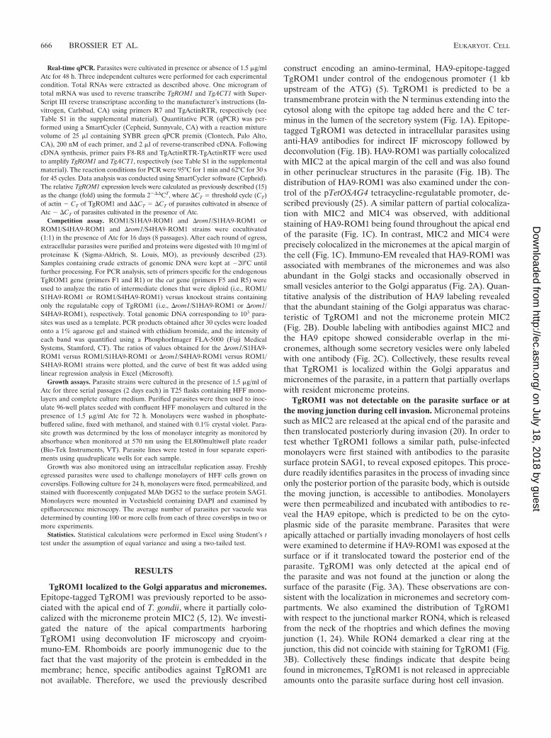
TgROM1 localized to the Golgi apparatus and micronemes.Epitope-tagged TgROM1 was previously reported to be asso-ciated with the apical end of T. gondii, where it partially colo-calized with the microneme protein MIC2 (5, 12). We investi-gated the nature of the apical compartments harboringTgROM1 using deconvolution IF microscopy and cryoim-muno-EM. Rhomboids are poorly immunogenic due to thefact that the vast majority of the protein is embedded in themembrane; hence, specific antibodies against TgROM1 arenot available. Therefore, we used the previously described
construct encoding an amino-terminal, HA9-epitope-taggedTgROM1 under control of the endogenous promoter (1 kbupstream of the ATG) (5). TgROM1 is predicted to be atransmembrane protein with the N terminus extending into thecytosol along with the epitope tag added here and the C ter-minus in the lumen of the secretory system (Fig. 1A). Epitope-tagged TgROM1 was detected in intracellular parasites usinganti-HA9 antibodies for indirect IF microscopy followed bydeconvolution (Fig. 1B). HA9-ROM1 was partially colocalizedwith MIC2 at the apical margin of the cell and was also foundin other perinuclear structures in the parasite (Fig. 1B). Thedistribution of HA9-ROM1 was also examined under the con-trol of the pTetOSAG4 tetracycline-regulatable promoter, de-scribed previously (25). A similar pattern of partial colocaliza-tion with MIC2 and MIC4 was observed, with additionalstaining of HA9-ROM1 being found throughout the apical endof the parasite (Fig. 1C). In contrast, MIC2 and MIC4 wereprecisely colocalized in the micronemes at the apical margin ofthe cell (Fig. 1C). Immuno-EM revealed that HA9-ROM1 wasassociated with membranes of the micronemes and was alsoabundant in the Golgi stacks and occasionally observed insmall vesicles anterior to the Golgi apparatus (Fig. 2A). Quan-titative analysis of the distribution of HA9 labeling revealedthat the abundant staining of the Golgi apparatus was charac-teristic of TgROM1 and not the microneme protein MIC2(Fig. 2B). Double labeling with antibodies against MIC2 andthe HA9 epitope showed considerable overlap in the mi-cronemes, although some secretory vesicles were only labeledwith one antibody (Fig. 2C). Collectively, these results revealthat TgROM1 is localized within the Golgi apparatus andmicronemes of the parasite, in a pattern that partially overlapswith resident microneme proteins.
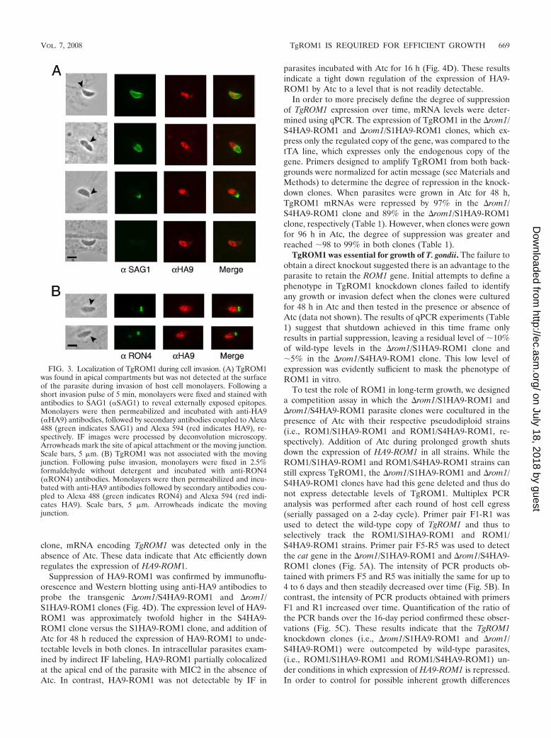
TgROM1 was not detectable on the parasite surface or atthe moving junction during cell invasion. Micronemal proteinssuch as MIC2 are released at the apical end of the parasite andthen translocated posteriorly during invasion (20). In order totest whether TgROM1 follows a similar path, pulse-infectedmonolayers were first stained with antibodies to the parasitesurface protein SAG1, to reveal exposed epitopes. This proce-dure readily identifies parasites in the process of invading sinceonly the posterior portion of the parasite body, which is outsidethe moving junction, is accessible to antibodies. Monolayerswere then permeabilized and incubated with antibodies to re-veal the HA9 epitope, which is predicted to be on the cyto-plasmic side of the parasite membrane. Parasites that wereapically attached or partially invading monolayers of host cellswere examined to determine if HA9-ROM1 was exposed at thesurface or if it translocated toward the posterior end of theparasite. TgROM1 was only detected at the apical end ofthe parasite and was not found at the junction or along thesurface of the parasite (Fig. 3A). These observations are con-sistent with the localization in micronemes and secretory com-partments. We also examined the distribution of TgROM1with respect to the junctional marker RON4, which is releasedfrom the neck of the rhoptries and which defines the movingjunction (1, 24). While RON4 demarked a clear ring at thejunction, this did not coincide with staining for TgROM1 (Fig.3B). Collectively these findings indicate that despite beingfound in micronemes, TgROM1 is not released in appreciableamounts onto the parasite surface during host cell invasion.
666 BROSSIER ET AL. EUKARYOT. CELL
on July 18, 2018 by guesthttp://ec.asm
.org/D
ownloaded from
Generation of TgROM1 knockdown parasites. To investi-gate the role of TgROM1 in T. gondii, we attempted to deletethe endogenous gene by double homologous crossover usingthe cat selectable marker flanked by upstream and downstreamsequences from TgROM1 as diagrammed in Fig. 4A. Despiterepeated attempts, this approach did not yield viable knock-outs (data not shown). This result suggested that the expres-sion of TgROM1 was critical for the survival of T. gondii.Consequently, the role of TgROM1 was investigated using thetetracycline (Tet)-repressible system that allows analysis ofessential genes in T. gondii (26). We used two regulatablepromoters that are known to have different strengths, to drivethe expression of HA9-ROM1, as reported previously (26).
The parental tTa strain of T. gondii, harboring a transacti-vator that binds to the Tet operator, was transfected with thevector pS4HA9-ROM1 to obtain parasites whose genomescontained both endogenous and tagged copies of TgROM1(referred to as ROM1/S4HA9-ROM1) (Fig. 4A). In a secondstep, a ROM1/S4HA9-ROM1 clone was transfected with theknockout vector p�R1YFP to generate knockouts that lackedthe endogenous gene but contained a regulatable HA9-taggedcopy of TgROM1 (referred to as �rom1/S4HA9-ROM1) (Fig.4A). A �rom1/S4HA9-ROM1 clone was obtained by limitingdilution and characterized at the genomic level. A similarstrategy was used to obtain a �rom1/S1HA9-ROM1 clone inwhich HA9-ROM1 was expressed under the control of thepTetOSAG1 promoter (25). The phenotypes reported belowwere similar for clones of both knockdowns (i.e., �rom1/S4HA9-ROM1 and �rom1/S1HA9-ROM1).
PCR was performed with primers designed to verify theintegration of the cat gene in place of TgROM1 at the correctlocus (Fig. 4A and B and see Table S1 in the supplementalmaterial). Primer pairs F2-R2, F2-R3, and F4-R4 can onlyamplify products if the cat gene replaced TgROM1 followinghomologous recombination. This pattern was observed whenPCR was performed with genomic DNA from �rom1/S4HA9-ROM1 but not with that from ROM1/S4HA9-ROM1. In con-trast, primers F1 and R1 hybridize within two separate intronsof the genomic copy of TgROM1. Since the plasmid pS4HA9-ROM1 was generated from HA9-ROM1 cDNA, primer setF1-R1 can only amplify the endogenous TgROM1 gene. Aproduct for the endogenous TgROM1 gene was observed whenPCR was performed with genomic DNA from ROM1/S4HA9-ROM1 but not with that from �rom1/S4HA9-ROM1. Takentogether, these data indicate that the endogenous TgROM1gene has been deleted in the �rom1/S4HA9-ROM1 clone.Similar results confirmed that the TgROM1 gene was disruptedin the �rom1/S1HA9-ROM1 clone (data not shown).
We determined the capacity of the nontoxic tetratcyclineanalogue, Atc, to down regulate the expression of HA9-ROM1in the transgenic ROM1/S4HA9-ROM1 and �rom1/S4HA9-ROM1 parasite clones following treatment for 48 h (Fig. 4C).The expression of TgROM1 was determined by RT-PCR, usingprimers that detect expression of both TgROM1 and HA9-
FIG. 1. Subcellular localization of HA9-tagged TgROM1 in in-tracellular parasites. (A) Model of the tagged construct of TgROM-1HA9. The protein is predicted to adopt a transmembrane topologywith the N terminus (Nt) in the cytosol along with the epitope tag.The C terminus (Ct) is predicted to extend into the lumen. Catalytictriad residues (N, asparagine; H, histidine; and S, serine), are shownin transmembrane domains 2, 6, and 4). (B) IF localization ofHA9-ROM1 driven by the endogenous promoter in transientlytransfected parasites. HA9-ROM1 was partially colocalized withMIC2 at the tip of intracellular parasites but also extended through-out the apical half of the parasite. At 16 h posttransfection, cellswere fixed, permeabilized, and incubated with anti-HA9 (� HA9) oranti-MIC2 (� MIC2) antibodies and revealed using secondary an-tibodies coupled to Alexa 594 (red), or Alexa 488 (green). IF imageswere processed by deconvolution microscopy, and a single Z-slice isshown in each example. Scale bar, 5 �m. (C) Transgenic parasitesexpressing HA9-TgROM1 under control of the TetOSAG4-regu-latable promoter showed partial colocalization with MIC2 andMIC4, again extending throughout the apical end of the parasite. Incontrast, MIC2 and MIC4 show almost perfect overlap at the apicalborder of the parasite. Cells were fixed; permeabilized; incubatedwith anti-HA9, anti-MIC2, or anti-MIC4 (� MIC4) antibodies; and
revealed using secondary antibodies coupled to Alexa 594 (red) orAlexa 488 (green). IF images were processed by deconvolution micros-copy, and a single Z-slice is shown in each example. Scale bar, 5 �m.
VOL. 7, 2008 TgROM1 IS REQUIRED FOR EFFICIENT GROWTH 667
on July 18, 2018 by guesthttp://ec.asm
.org/D
ownloaded from

ROM1 compared to actin (TgACT1) as an internal control.mRNA encoding TgROM1 was detected in absence or pres-ence of Atc in the ROM1/S4HA9-ROM1 clone, as expected,since it contains the endogenous copy as well as a regulatable
copy (Fig. 4C). In the absence of Atc, the signal correspondingto the TgROM1 transcript was more intense than that observedin presence of Atc, confirming that HA9-ROM1 was downregulated in the presence of Atc. In the �rom1/S4HA9-ROM1
FIG. 2. Cryoimmuno-EM localization of TgROM1 in intracellular parasites. (A) Immuno-EM revealed that TgROM1 was localized inmicronemes and the Golgi apparatus. Schematic representation of T. gondii showing dense granules (DG), rhoptries (ROP), micronemes (MIC),endoplasmic reticulum (ER), Golgi apparatus (Golgi), and nucleus (N). Cryoimmuno-EM was performed on extracellular parasites expressingHA9-ROM1 and detected with anti-HA9 antibody followed by secondary antibodies conjugated to 18-nm colloidal gold particles. Two separateimages show the middle (a) and the apical end (b) of the parasite, respectively. Two other images taken at higher magnification show the Golgiapparatus (c) and the micronemes (d). Scale bars, 200 nm. (B) Quantitative distribution of immunogold from representative images of the Golgiapparatus and apical regions as defined in panel A. The distribution of TgROM1 was determined from 10 representative negatives stained asdescribed above. The distribution of MIC2 was determined by staining with rabbit anti-C-domain followed by secondary antibodies conjugated togold. Values shown are means standard deviations. (C) Double labeling with HA9-ROM1 (18-nm gold; large arrows) and MIC2 (12-nm gold;small arrows) revealed costaining of some compartments, while others contained only one of the markers. Scale bars, 200 nm.
668 BROSSIER ET AL. EUKARYOT. CELL
on July 18, 2018 by guesthttp://ec.asm
.org/D
ownloaded from
clone, mRNA encoding TgROM1 was detected only in theabsence of Atc. These data indicate that Atc efficiently downregulates the expression of HA9-ROM1.
Suppression of HA9-ROM1 was confirmed by immunoflu-orescence and Western blotting using anti-HA9 antibodies toprobe the transgenic �rom1/S4HA9-ROM1 and �rom1/S1HA9-ROM1 clones (Fig. 4D). The expression level of HA9-ROM1 was approximately twofold higher in the S4HA9-ROM1 clone versus the S1HA9-ROM1 clone, and addition ofAtc for 48 h reduced the expression of HA9-ROM1 to unde-tectable levels in both clones. In intracellular parasites exam-ined by indirect IF labeling, HA9-ROM1 partially colocalizedat the apical end of the parasite with MIC2 in the absence ofAtc. In contrast, HA9-ROM1 was not detectable by IF in
parasites incubated with Atc for 16 h (Fig. 4D). These resultsindicate a tight down regulation of the expression of HA9-ROM1 by Atc to a level that is not readily detectable.
In order to more precisely define the degree of suppressionof TgROM1 expression over time, mRNA levels were deter-mined using qPCR. The expression of TgROM1 in the �rom1/S4HA9-ROM1 and �rom1/S1HA9-ROM1 clones, which ex-press only the regulated copy of the gene, was compared to thetTA line, which expresses only the endogenous copy of thegene. Primers designed to amplify TgROM1 from both back-grounds were normalized for actin message (see Materials andMethods) to determine the degree of repression in the knock-down clones. When parasites were grown in Atc for 48 h,TgROM1 mRNAs were repressed by 97% in the �rom1/S4HA9-ROM1 clone and 89% in the �rom1/S1HA9-ROM1clone, respectively (Table 1). However, when clones were gownfor 96 h in Atc, the degree of suppression was greater andreached �98 to 99% in both clones (Table 1).
TgROM1 was essential for growth of T. gondii. The failure toobtain a direct knockout suggested there is an advantage to theparasite to retain the ROM1 gene. Initial attempts to define aphenotype in TgROM1 knockdown clones failed to identifyany growth or invasion defect when the clones were culturedfor 48 h in Atc and then tested in the presence or absence ofAtc (data not shown). The results of qPCR experiments (Table1) suggest that shutdown achieved in this time frame onlyresults in partial suppression, leaving a residual level of �10%of wild-type levels in the �rom1/S1HA9-ROM1 clone and�5% in the �rom1/S4HA9-ROM1 clone. This low level ofexpression was evidently sufficient to mask the phenotype ofROM1 in vitro.
To test the role of ROM1 in long-term growth, we designeda competition assay in which the �rom1/S1HA9-ROM1 and�rom1/S4HA9-ROM1 parasite clones were cocultured in thepresence of Atc with their respective pseudodiploid strains(i.e., ROM1/S1HA9-ROM1 and ROM1/S4HA9-ROM1, re-spectively). Addition of Atc during prolonged growth shutsdown the expression of HA9-ROM1 in all strains. While theROM1/S1HA9-ROM1 and ROM1/S4HA9-ROM1 strains canstill express TgROM1, the �rom1/S1HA9-ROM1 and �rom1/S4HA9-ROM1 clones have had this gene deleted and thus donot express detectable levels of TgROM1. Multiplex PCRanalysis was performed after each round of host cell egress(serially passaged on a 2-day cycle). Primer pair F1-R1 wasused to detect the wild-type copy of TgROM1 and thus toselectively track the ROM1/S1HA9-ROM1 and ROM1/S4HA9-ROM1 strains. Primer pair F5-R5 was used to detectthe cat gene in the �rom1/S1HA9-ROM1 and �rom1/S4HA9-ROM1 clones (Fig. 5A). The intensity of PCR products ob-tained with primers F5 and R5 was initially the same for up to4 to 6 days and then steadily decreased over time (Fig. 5B). Incontrast, the intensity of PCR products obtained with primersF1 and R1 increased over time. Quantification of the ratio ofthe PCR bands over the 16-day period confirmed these obser-vations (Fig. 5C). These results indicate that the TgROM1knockdown clones (i.e., �rom1/S1HA9-ROM1 and �rom1/S4HA9-ROM1) were outcompeted by wild-type parasites,(i.e., ROM1/S1HA9-ROM1 and ROM1/S4HA9-ROM1) un-der conditions in which expression of HA9-ROM1 is repressed.In order to control for possible inherent growth differences
FIG. 3. Localization of TgROM1 during cell invasion. (A) TgROM1was found in apical compartments but was not detected at the surfaceof the parasite during invasion of host cell monolayers. Following ashort invasion pulse of 5 min, monolayers were fixed and stained withantibodies to SAG1 (�SAG1) to reveal externally exposed epitopes.Monolayers were then permeabilized and incubated with anti-HA9(�HA9) antibodies, followed by secondary antibodies coupled to Alexa488 (green indicates SAG1) and Alexa 594 (red indicates HA9), re-spectively. IF images were processed by deconvolution microscopy.Arrowheads mark the site of apical attachment or the moving junction.Scale bars, 5 �m. (B) TgROM1 was not associated with the movingjunction. Following pulse invasion, monolayers were fixed in 2.5%formaldehyde without detergent and incubated with anti-RON4(�RON4) antibodies. Monolayers were then permeabilized and incu-bated with anti-HA9 antibodies followed by secondary antibodies cou-pled to Alexa 488 (green indicates RON4) and Alexa 594 (red indi-cates HA9). Scale bars, 5 �m. Arrowheads indicate the movingjunction.
VOL. 7, 2008 TgROM1 IS REQUIRED FOR EFFICIENT GROWTH 669
on July 18, 2018 by guesthttp://ec.asm
.org/D
ownloaded from
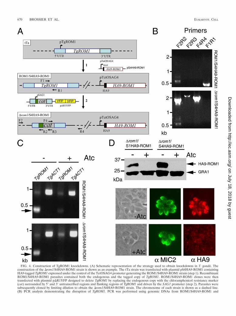
FIG. 4. Construction of TgROM1 knockdowns. (A) Schematic representation of the strategy used to obtain knockdowns in T. gondii. Theconstruction of the �rom1/S4HA9-ROM1 strain is shown as an example. The tTa strain was transfected with plasmid pS4HA9-ROM1 containingHA9-tagged TgROM1 expressed under the control of the TetOSAG4 promoter generating the ROM1/S4HA9-ROM1 strain (step 1). RecombinantROM1/S4HA9-ROM1 parasites contained both the endogenous and the tagged copy of TgROM1. ROM1/S4HA9-ROM1 clones were thentransfected with plasmid p�R1YFP designed to delete TgROM1 by replacing the endogenous copy with the chloramphenicol resistance marker(cat) surrounded by 5� and 3� untranscribed regions and flanking regions of TgROM1 and driven by the SAG1 promoter (step 2). Parasites weresubsequently cloned by limiting dilution to obtain the �rom1/S4HA9-ROM1 strain. The chromosome of each strain is shown as a dashed line.(B) PCR analysis demonstrating the disruption of TgROM1. PCR was performed using genomic DNAs from ROM1/S4HA9-ROM1 and
670 BROSSIER ET AL. EUKARYOT. CELL
on July 18, 2018 by guesthttp://ec.asm
.org/D
ownloaded from
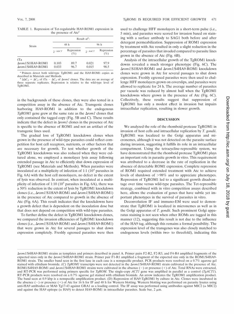
in the backgrounds of these clones, they were also tested in acompetition assay in the absence of Atc. Transgenic clonesharboring HA9-ROM1 in addition to the endogenousTgROM1 gene grew at the same rate as the �rom1 clones thatonly contained the tagged copy (Fig. 5B and C). These resultsindicate that the defect in �rom1 clones in the presence of Atcis specific to the absence of ROM1 and not an artifact of thetransgenic lines used.
The gradual loss of TgROM1 knockdown clones whengrown in the presence of wild-type parasites could reflect com-petition for host cell receptors, nutrients, or other factors thatare necessary for growth. To test whether growth of theTgROM1 knockdowns was also reduced when they were cul-tured alone, we employed a monolayer lysis assay followingextended passage in Atc to efficiently shut down expression ofTgROM1 (see Materials and Methods). When parasites wereinoculated at a multiplicity of infection of 1:1 (104 parasites inFig. 6A) with the host cell monolayers, no defect in the extentof lysis was observed. In contrast, when inoculated at a multi-plicity of infection of 1:10 (105 parasites in Fig. 6A), there wasa 50% reduction in the extent of lysis by TgROM1 knockdownclones (i.e., �rom1/S1HA9-ROM1 and �rom1/S4HA9-ROM1)in the presence of Atc compared to growth in the absence ofAtc (Fig. 6A). This result indicates that the knockdowns havea growth defect that is dependent on the inoculation dose butthat does not depend on competition with wild-type parasites.
To further define the defect in TgROM1 knockdown clones,we compared the invasion efficiencies of TgROM1 knockdownclones (i.e., �rom1/S1HA9-ROM1 and �rom1/S4HA9-ROM1)that were grown in Atc for several passages to shut downexpression completely. Freshly egressed parasites were then
used to challenge HFF monolayers in a short-term pulse (i.e.,5 min), and parasites were scored for invasion based on stain-ing with a surface antibody to SAG1 both before and afterdetergent permeabilization. Suppression of ROM1 expressionby treatment with Atc resulted in only a slight reduction in thepercentage of parasites that invaded compared to parasite linesgrown in the absence of Atc (Fig. 6B).
Analysis of the intracellular growth of the TgROM1 knock-downs revealed a much stronger phenotype (Fig. 6C). The�rom1/S1HA9-ROM1 and �rom1/S4HA9-ROM1 knockdownclones were grown in Atc for several passages to shut downexpression. Freshly egressed parasites were then used to chal-lenge HFF monolayers grown on coverslips, and parasites wereallowed to replicate for 24 h. The average number of parasitesper vacuole was reduced by almost half when the TgROM1knockdowns where grown in the presence of Atc (Fig. 6C).Collectively, these results suggest that suppression ofTgROM1 has only a modest effect in invasion but impairsintracellular replication, resulting in slower growth.
DISCUSSION
We analyzed the role of the rhomboid protease TgROM1 ininvasion of host cells and intracellular replication by T. gondii.TgROM1 was localized to the Golgi apparatus and mi-cronemes, although it was not detected on the parasite surfaceduring invasion, suggesting it fulfills its role in an intracellularcompartment. Using the tetracycline-repressible system, wehave demonstrated that the rhomboid protease TgROM1 playsan important role in parasite growth in vitro. This requirementwas attributed to a decrease in the rate of replication in theabsence of detectable ROM1 expression. Complete repressionof ROM1 required extended treatment with Atc to achievelevels of shutdown of �98% and to appreciate phenotypes.Repression of TgROM1 led to a significant growth disadvan-tage over time versus wild-type parasites. The Tet-repressiblestrategy, combined with in vitro competition assays describedhere, allows the evaluation of genes that have subtle yet im-portant phenotypes in the survival of parasites in vitro.
Deconvolution IF and immuno-EM were used to demon-strate that TgROM1 is localized in micronemes as well as inthe Golgi apparatus of T. gondii. Such prominent Golgi appa-ratus staining is not seen when other ROMs are tagged in thismanner (12), suggesting this result is not due to the influenceof the HA9 tag, although this remains a formal possibility. Theexpression level of the transgenes was also closely matched toendogenous levels (within two- to threefold), indicating this
�rom1/S4HA9-ROM1 strains as templates and primers described in panel A. Primer pairs F2-R2, F2-R3, and F4-R4 amplified fragments of theexpected sizes only in the �rom1/S4HA9-ROM1 strain. Primer pair F1-R1 amplified a fragment of the expected size only in the ROM1/S4HA9-ROM1 strain. The smaller band seen in the first lane in each case is a nonspecific product. PCR products were resolved on a 0.7% agarose gelstained with ethidium bromide. (C) TgROM1 transcripts were not detected in the �rom1/S4HA9-ROM1 strain cultivated in the presence of Atc.ROM1/S4HA9-ROM1 and �rom1/S4HA9-ROM1 strains were cultivated in the absence (�) or presence (�) of Atc. Total RNAs were extracted,and RT-PCR was performed using primers specific for TgROM. The single-copy ACT1 gene was amplified in parallel as a control (TgACT1).RT-PCR products were resolved on a 0.7% agarose gel stained with ethidium bromide. An arrow indicates the TgROM1 amplification product.The band seen at 0.9 kbp is a nonspecific amplification product. (D) Repression of HA9-TgROM1 by culture in Atc. Clones were incubated inthe absence (�) or presence (�) of Atc for 16 h for IF and 48 h for Western blotting. Western blotting was performed on parasite lysates usinganti-HA9 antibodies or MAb Tg17-43 against GRA1 as a loading control. The IF assay was performed using antibodies against MIC2 (� MIC2)and against the HA9 epitope (� HA9) to detect HA9-ROM1 in intracellular parasites. Scale bar, 2 �m.
TABLE 1. Repression of Tet-regulatable HA9-ROM1 expression inthe presence of Atca
Strain
Result atb:
48 h 96 h
2���CT Repression(%) 2���CT Repression
(%)
tTa�rom1/S1HA9-ROM1 0.103 89.7 0.021 97.9�rom1/S4HA9-ROM1 0.033 96.7 0.015 98.5
a Primers detect both wild-type TgROM1 and the HA9-ROM1 copies asdescribed in Materials and Methods.
b ��CT �CT of tTa � �CT of �rom1 clones. The data are an average ofthree separate replicates. Repression is relative to the wild-type level ofTgROM1.
VOL. 7, 2008 TgROM1 IS REQUIRED FOR EFFICIENT GROWTH 671
on July 18, 2018 by guesthttp://ec.asm
.org/D
ownloaded from
pattern is unlikely to be due to overexpression. TgROM1 waslargely localized to the Golgi apparatus, and only limited stain-ing of post-Golgi secretory vesicles was observed, other thanmature micronemes. This pattern differs from the post-Golgilocalization of pro-MIC2, which occupies a compartmentthought to be important for maturation of microneme proteins(17). Within the apical microneme organelles, the pattern ofROM1 staining only partially overlapped that of MIC2.Whether this indicates heterogeneity of secretory organelles orinefficient labeling is uncertain; however, it might represent afunctional separation of the protease from potential substrates.
Transmembrane microneme proteins are released from themicronemes and transiently occupy the cell surface before be-ing shed by proteolysis, which often occurs by intramembranecleavage by a rhomboid protease. The Tet-repressible systemhas previously been used to knock down the expression of twotransmembrane microneme proteins, MIC2 and TgAMA1 (19,28). Both proteins play crucial roles in the invasion of host cellsby parasites, although they act at different steps. MIC2 is re-quired for attachment and hence invasion, as well as helicalgliding motility of tachyzoites (19). TgAMA1 is required forthe tight apical interaction with the host cell that leads to
rhoptry secretion and invasion into the parasitophorous vacu-ole (28). As with other transmembrane MIC proteins,TgAMA1 is processed primarily within its transmembrane do-main, as shown by matrix-assisted laser desorption ionization–time of flight analysis of peptides generated from secretedTgAMA1 (18). Previous studies have failed to demonstrate arole for TgROM1 in processing MIC proteins in vitro (5, 12),and results described here suggest that ROM1 does not trafficto the cell surface and thus may not be involved in processingmicronemal proteins. In contrast, in vitro assays have demon-strated that TgROM5 is able to efficiently process TgAMA1(S. Urban, personal communication) and MIC2 (5), suggestingit is responsible for processing these key surface proteins in-volved in invasion by T. gondii.
In contrast to known transmembrane MICs, TgROM1 wasnot detected at the surface of the parasite upon invasion ofhost cells, although we cannot rule out the possibility that lowlevels of the protein might not be detected. Overall, our find-ings are most consistent with TgROM1 performing its functionwithin the secretory system, rather than on the cell surface.During intracellular growth, the parasite undergoes an unusualform of cell division termed endodyogeny (34). During this
FIG. 5. Competition assay demonstrating that TgROM1 was required for efficient growth of T. gondii. (A) Schematic representation of primersused for detection of the wild type (WT) and deleted �Tgrom1 loci. Primers F1 and R1 selectively amplify the TgROM1 gene. Primers F5 and R5selectively amplify the cat gene. (B) �rom1 clones were outcompeted by strains containing an endogenous copy of TgROM1 in a mixed cultureover time when the regulatable copy of HA9-ROM1 was repressed by continuous culture in Atc. In contrast, �rom1 clones showed no growthdefect when the regulatable copy was not repressed with Atc. Parasites were added at 1:1 at the starting time and cocultured in the absence (�)or presence (�) of Atc. Every 2 days, extracellular parasites were purified and genomic DNA was extracted for multiplex PCR analysis.ROM1/S1HA9-ROM1 and ROM1/S4HA9-ROM1 strains were tracked using primer pair F1-R1, and �rom1/S1HA9-ROM1 and �rom1/S4HA9-ROM1 strains were tracked using primer pair F5-R5. PCR products were resolved on a 1% agarose gels stained with ethidium bromide. (C) Theintensity of the bands seen in panel B was quantified using a PhosphorImager. The ratios of the values obtained for the knockdowns in the �rom1background versus the nondeleted strains are shown for each passage in the absence (solid squares) or presence (solid diamonds) of Atc. The linesof best fit (dashed lines) were determined by linear regression.
672 BROSSIER ET AL. EUKARYOT. CELL
on July 18, 2018 by guesthttp://ec.asm
.org/D
ownloaded from

process, maternal organelles are largely resorbed before form-ing de novo again in the daughter cells. While these processesare not well understood, it is possible that ROM1 plays a rolein recycling protein components within the secretory pathwayduring cell division. Despite suggesting a role for proteolyticprocessing during cell division, our studies do not identify theprecise targets of ROM1 within the cell. TgROM2 has alsobeen localized to the Golgi apparatus (12), raising the possi-bility that it performs an overlapping or redundant function.
Plasmodium spp. express orthologues of ROM1, althoughseveral features of their localization and potential activitiesdiffer from the results described here for T. gondii. While P.falciparum ROM1 (PfROM1) is capable of cleaving PfAMA1in a heterologous cellular assay (2), this is thought to be a rareevent in the parasite, based on mass spectrometric data (18).PfROM1 has been associated with the micronemes (29) andmore recently with a novel apical organelle called the monon-eme (33). Our results with TgROM1 do not reveal a similarorganelle in T. gondii, although TgROM1 was a prominentcomponent in the Golgi apparatus. Collectively, these differ-ences indicate that ROM1 may function differently betweenthese parasites rather than underlying a function(s) common tothe phylum.
While reverse genetics tools allow dissection of the role ofspecific genes, the normal definition of “essential” genes im-plies that they have a strong phenotype in a defined assay.TgROM1 is not essential for invasion, yet absence of thisprotein led to a decrease in replication efficiency. This effectwas somewhat modest in that it could be overcome by a highinoculum. However, the deficiency was cumulative and led to asignificant loss in a competitive assay against wild-type para-sites over time. In this regard, TgROM1 and similar proteinsare essential for fitness, since mutants lacking such geneswould rapidly be lost from the population. This may also ex-plain why parasites contain multiple ROM genes (5), whichmay encode isoforms with either different substrate specificityor with the potential to enhance the effectiveness of substrateprocessing.
Similarly to the growth defect observed with TgROM1knockdowns, a subtle growth phenotype was also obtained witha knockout of PP2C-hn in T. gondii (16). The PP2C-hn geneencodes a protein phosphatase 2C that is secreted from therhoptries and translocated to the nucleus of host cells uponinvasion. �pp2c-hn knockout parasites display a growth defect
FIG. 6. Decreased growth of TgROM1 knockdown clones was as-sociated with slower growth and less-efficient invasion. (A) Monolayerlysis assay revealed a dose-dependent defect in the TgROM knock-down clones following suppression of ROM1. Monolayers of HFF cellswere inoculated with �rom1/S1HA9-ROM1 and �rom1/S4HA9-ROM1 parasites in the presence (�) or absence (�) of Atc. Afterculture for 3 days, monolayers were rinsed, fixed, and stained withcrystal violet and the absorbance was read at 570 nm. Decreasedabsorbance (staining of host cells) was a result of lysis due to parasitegrowth, as confirmed by microscopic examination. The x axis shows thechallenge dose of parasites. Values are means standard deviations(n 4 wells per sample), representative of four experiments. ��, P �0.001 by Student’s t test. (B) Invasion assays revealed a modest reduc-tion in invasion by TgROM1 knockdown clones following suppressionof ROM1. Parasites were grown for several passages in the presence orabsence of Atc, harvested from freshly egressed cultures, and used tochallenge monolayers of HFF cells cultured on glass coverslips. Fol-lowing a 5-min pulse invasion, parasites were classified as intracellular
versus extracellular based on accessibility to staining of the surfaceantigen SAG1. Repression of ROM1 led to an �15% reduction ininvasion in both the �rom1/S1HA9-ROM1 and �rom1/S4HA9-ROM1strains. Values are means standard errors of the mean (n 3experiments). (C) Intracellular replication was more severely de-creased in TgROM1 knockdown clones following suppression ofTgROM1. Parasites were grown for several passages in the absence orpresence of Atc, harvested from freshly egressed cultures, and used tochallenge monolayers of HFF cells cultured on glass coverslips. Fol-lowing 24 h of incubation, parasites were fixed and stained for thesurface protein SAG and with DAPI to reveal nuclei, and the averagenumber of parasites per vacuole was determined by counting underepifluorescence microscopy. Values are means standard deviations(n 3 coverslips per group), representative of duplicate experiments.��, P � 0.001 by Student’s t test.
VOL. 7, 2008 TgROM1 IS REQUIRED FOR EFFICIENT GROWTH 673
on July 18, 2018 by guesthttp://ec.asm
.org/D
ownloaded from
that is only revealed by competition assay with a wild-typestrain. Taken together, it appears that two categories of genesare important for efficient survival of T. gondii: those whoseexpression is absolutely critical for events like motility and cellinvasion, such as MIC2 or TgAMA1, and those whose contri-bution is less dramatic yet still important for long-term sur-vival, such as TgROM1 and PP2C-hn. Given the complex strat-egies parasites have evolved for optimizing survival, it is likelythat a large proportion of genes will have such subtle pheno-types, which are nonetheless essential in a larger context.
ACKNOWLEDGMENTS
We thank Dominique Soldati (University of Geneva, Geneva, Swit-zerland) for the generous gift of rabbit anti-MIC4 polyclonal antibod-ies, the tTa strain, and the p7TetOS1 and p7TetOS4 plasmids; GaryWard (University of Vermont) for the MAb B3.90 against TgAMA1;Marie France Cesbron-Delauw (Universite Joseph Fourier, Grenoble,France) for the MAb to GRA1; Peter Bradley (University of Califor-nia, Los Angeles) for the mouse anti-RON4 polyclonal antibodies; andBoris Striepen (University of Georgia, Athens) for the TUBA-YF-PYFP plasmid.
This work was supported in part by National Institutes of Healthgrant AI 34036.
REFERENCES
1. Alexander, D. L., J. Mital, G. E. Ward, P. J. Bradley, and J. C. Boothroyd.2005. Identification of the moving junction complex of Toxoplasma gondii: acollaboration between distinct secretory organelles. PLoS Pathog. 1:137–149.
2. Baker, R. P., R. Wijetilaka, and S. Urban. 2006. Two Plasmodium rhomboidproteases preferentially cleave different adhesins implicated in all invasivestages of malaria. PLoS Pathog. 2:922–932.
3. Brecht, S., V. B. Carruthers, D. J. Ferguson, O. K. Giddings, G. Wang, U.Jaekle, J. M. Harper, L. D. Sibley, and D. Soldati. 2001. The Toxoplasmamicronemal protein MIC4 is an adhesin composed of six conserved appledomains. J. Biol. Chem. 276:4119–4127.
4. Brossier, F., T. J. Jewett, J. L. Lovett, and L. D. Sibley. 2003. C-terminalprocessing of the Toxoplasma protein MIC2 is essential for invasion intohost cells. J. Biol. Chem. 278:6229–6234.
5. Brossier, F., T. J. Jewett, L. D. Sibley, and S. Urban. 2005. A spatially-localized rhomboid protease cleaves cell surface adhesins essential for inva-sion by Toxoplasma. Proc. Natl. Acad. Sci. USA 102:4146–4151.
6. Carruthers, V. B., O. K. Giddings, and L. D. Sibley. 1999. Secretion ofmicronemal proteins is associated with Toxoplasma invasion of host cells.Cell. Microbiol. 1:225–236.
7. Carruthers, V. B., G. D. Sherman, and L. D. Sibley. 2000. The Toxoplasmaadhesive protein MIC2 is proteolytically processed at multiple sites by twoparasite-derived proteases. J. Biol. Chem. 275:14346–14353.
8. Carruthers, V. B., and L. D. Sibley. 1997. Sequential protein secretion fromthree distinct organelles of Toxoplasma gondii accompanies invasion of hu-man fibroblasts. Eur. J. Cell Biol. 73:114–123.
9. Cerede, O., J. F. Dubremetz, D. Bout, and M. Lebrun. 2002. The Toxoplasmagondii protein MIC3 requires pro-peptide cleavage and dimerization to func-tion as an adhesin. EMBO J. 21:2526–2536.
10. Dowse, T., and D. Soldati. 2004. Host cell invasion by the apicomplexans: thesignificance of microneme protein proteolysis. Curr. Opin. Microbiol. 7:388–396.
11. Dowse, T., and D. Soldati. 2005. Rhomboid-like proteins in Apicomplexa:phylogeny and nomenclature. Trends Parasitol. 21:254–258.
12. Dowse, T. J., J. C. Pascall, K. D. Brown, and D. Soldati. 2005. Apicomplexanrhomboids have a potential role in microneme protein cleavage during hostcell invasion. Int. J. Parasitol. 35:747–756.
13. Dubey, J. P. 1998. Advances in the life cycle of Toxoplasma gondii. Int. J.Parasitol. 28:1019–1024.
14. Dubremetz, J. F., A. Achbarou, D. Bermudes, and K. A. Joiner. 1993. Kineticsand pattern of organelle exocytosis during Toxoplasma gondii/host-cell interac-tion. Parasitol. Res. 79:402–408.
15. Fux, B., J. Nawas, A. Khan, D. B. Gill, C. Su, and L. D. Sibley. 2007.Toxoplasma gondii strains defective in oral transmission are also defective indevelopmental stage differentiation. Infect. Immun. 75:2580–2590.
16. Gilbert, L. K., S. Ravindran, J. M. Turetzky, J. C. Boothroyd, and P. J.Bradley. 2007. Toxoplasma gondii targets a protein phosphatase 2C to thenuclei of infected host cells. Eukaryot. Cell 6:73–83.
17. Harper, J. M., M. H. Huynh, I. Coppens, F. Parussini, S. N. Moreno, andV. B. Carruthers. 2006. A cleavable propeptide influences Toxoplasma in-fection by facilitating the trafficking and secretion of the TgMIC2-M2APinvasion complex. Mol. Biol. Cell 17:4551–4563.
18. Howell, S. A., F. Hackett, A. M. Jongco, C. Withers-Martinez, K. Kim, V. B.Carruthers, and M. J. Blackman. 2005. Distinct mechanisms govern proteo-lytic shedding of a key invasion protein in apicomplexan pathogens. Mol.Microbiol. 57:1342–1356.
19. Huynh, M. H., and V. B. Carruthers. 2006. Toxoplasma MIC2 is a majordeterminant of invasion and virulence. PLoS Pathog. 2:753–762.
20. Jewett, T. J., and L. D. Sibley. 2003. Aldolase forms a bridge between cellsurface adhesins and the actin cytoskeleton in apicomplexan parasites. Mol.Cell 11:885–894.
21. Jewett, T. J., and L. D. Sibley. 2004. The Toxoplasma proteins MIC2 andM2AP for a hexameric complex necessary for intracellular survival. J. Biol.Chem. 279:9362–9369.
22. Joynson, D. H., and T. J. Wreghitt. 2001. Toxoplasmosis: a comprehensiveclinical guide. Cambridge University Press, Cambridge, United Kingdom.
23. Khan, A., C. Su, M. German, G. A. Storch, D. Clifford, and L. D. Sibley.2005. Genotyping of Toxoplasma gondii strains from immunocompromisedpatients reveals high prevalence of type I strains. J. Clin. Microbiol. 43:5881–5887.
24. Lebrun, M., A. Michelin, H. El Hajj, J. Poncet, P. J. Bradley, H. J. Vial, andJ. F. Dubremetz. 2005. The rhoptry neck protein RON4 relocalizes at themoving junction during Toxoplasma gondii invasion. Cell. Microbiol. 7:1823–1833.
25. Meissner, M., S. Brecht, H. Bujard, and D. Soldati. 2001. Modulation ofmyosin A expression by a newly established tetracycline repressor basedinducible system in Toxoplasma gondii. Nucleic Acids Res. 29:E115.
26. Meissner, M., D. Schluter, and D. Soldati. 2002. Role of Toxoplasma gondiimyosin A in powering parasite gliding and host cell invasion. Science 298:837–840.
27. Messina, M., I. R. Niesman, C. Mercier, and L. D. Sibley. 1995. Stable DNAtransformation of Toxoplasma gondii using phleomycin selection. Gene 165:213–217.
28. Mital, J., M. Meissner, D. Soldati, and G. E. Ward. 2005. Conditionalexpression of Toxoplasma gondii apical membrane antigen-1 (TgAMA1)demonstrates that TgAMA1 plays a critical role in host cell invasion. Mol.Biol. Cell 16:4341–4349.
29. O’Donnell, R. A., F. Hackett, S. A. Howell, M. Treeck, N. Struck, Z. Krna-jski, C. Withers-Martinez, T. W. Gilberger, and M. J. Blackman. 2006.Intramembrane proteolysis mediates shedding of a key adhesin during eryth-rocyte invasion by the malaria parasite. J. Cell Biol. 174:1023–1033.
30. Opitz, C., M. Di Cristina, M. Reiss, T. Ruppert, A. Crisanti, and D. Soldati.2002. Intramembrane cleavage of the microneme proteins at the surface ofapicomplexan parasite Toxoplasma gondii. EMBO J. 21:1577–1585.
31. Rabenau, K. E., A. Sohrabi, A. Tripathy, C. Reitter, J. W. Ajioka, F. M.Tomley, and V. B. Carruthers. 2001. TgM2AP participates in Toxoplasmagondii invasion of host cells and is tightly associated with the adhesive proteinTgMIC2. Mol. Microbiol. 41:537–547.
32. Sibley, L. D. 2004. Invasion strategies of intracellular parasites. Science304:248–253.
33. Singh, S. B., M. Plassmeyer, D. Gaur, and L. H. Miller. 2007. Mononeme: anew secretory organelle in Plasmodium falciparum merozoites identified bylocalization of rhomboid-1 protease. Proc. Natl. Acad. Sci. USA 104:20043–20048.
34. Striepen, B., C. N. Jordan, S. Reiff, and G. G. van Dooren. 2007. Building theperfect parasite: cell division in Apicomplexa. PLoS Pathog. 3:691–698.
35. Urban, S. 2006. Rhomboid proteins: conserved membrane proteases withdivergent biological functions. Genes Dev. 20:3054–3068.
36. Urban, S., and M. Freeman. 2003. Substrate specificity of rhomboid in-tramembrane proteases is governed by helix-breaking residues in the sub-strate transmembrane domain. Mol. Cell 11:1425–1434.
37. Urban, S., J. R. Lee, and M. Freeman. 2001. Drosophila Rhomboid-1 definesa family of putative intramembrane serine proteases. Cell 107:173–182.
38. Urban, S., J. R. Lee, and M. Freeman. 2002. A family of rhomboid in-tramembrane proteases activates all Drosophila membrane-tethered EGFligands. EMBO J. 21:4277–4286.
39. Wan, K. L., V. B. Carruthers, L. D. Sibley, and J. W. Ajioka. 1997. Molecularcharacterisation of an expressed sequence tag locus of Toxoplasma gondiiencoding the micronemal protein MIC2. Mol. Biochem. Parasitol. 84:203–214.
40. Zhou, X. W., M. J. Blackman, S. A. Howell, and V. B. Carruthers. 2004.Proteomic analysis of cleavage events reveals a dynamic two-step mechanismfor proteolysis of a key parasite adhesive complex. Mol. Cell. Proteomics3:565–576.
674 BROSSIER ET AL. EUKARYOT. CELL
on July 18, 2018 by guesthttp://ec.asm
.org/D
ownloaded from