Micromorphology and phytochemistry of the foliar Stachys ...
131
Micromorphology and phytochemistry of the foliar secretory structures of Stachys natalensis Hochst. and development of an in vitro propagation protocol Benita Kalicharan A dissertation submitted in fulfilment of the academic requirements for the degree of Master of Science (Biological Science) in the School of Life Sciences, College of Agriculture, Engineering and Science, University of KwaZulu-Natal, Durban, South Africa March 2014
Transcript of Micromorphology and phytochemistry of the foliar Stachys ...
Micromorphology and phytochemistry of the foliar secretory structures of Stachys natalensis Hochst. and
development of an in vitro propagation protocol
Benita Kalicharan
A dissertation submitted in fulfilment of the academic requirements for the degree of
Master of Science (Biological Science)
in the School of Life Sciences, College of Agriculture, Engineering and Science,
University of KwaZulu-Natal, Durban, South Africa
March 2014

ABSTRACT
i
Many members of the genus Stachys have been used as ornamental plants, or as edible foods and, in a
number of cultures, as traditional medicine. One such species, Stachys natalensis Hochst., is a
perennial, straggling shrub with aromatic leaves that are covered with hairs. Despite its widespread
use for a number of reasons, little information has been available on this plant thus far, its foliar
secretory apparatus, or the nature and potential therapeutic value of these secretions. Hence, the aims
and objectives of the present study were to elucidate key micromorphological features of the leaf
secretory structures of S. natalensis, using both light and electron microscopy and to determine the
location and chemical composition of the exudates using various histochemical and phytochemical
techniques. An additional aim was to establish an in vitro micropropagation protocol for the sustained
and high-yielding production of this elusive and often difficult to cultivate species. Furthermore, the
foliar micromorphological fidelity between field and in vitro propagated material was compared.
Electron micrographs indicated the presence of glandular and non-glandular trichomes on both
abaxial and adaxial foliar surfaces of field grown plants. Greater trichome density was observed on
the abaxial surface with trichome distribution decreasing as the leaf developed (p<0.05). Uniseriate,
unbranched and striated non-glandular trichomes, present on the foliar surfaces of S. natalensis are
known to play a role in physical plant defence mechanisms against herbivory. The presence of an
elevated cellular pedestal and striated micro-ornamentation on the stalk served as characteristic
features of the non-glandular trichomes. Peltate and capitate glandular trichomes were also identified
on the foliar surfaces across all developmental stages. Peltate glands consisted of a short stalk and a
multicellular head containing two to eight secretory cells. Two types of capitate trichomes were
observed. Type I capitate trichomes consisted of a striated stalk, cutinized neck cell and bulbous head
which may be uni- or bi-cellular;Type II capitate trichomes were comprised of a wide base, long and
tapering, septate stalk, neck cell and a multicellular secretory head cell. The process of secretion
differed between the peltate and capitate glandular trichomes. The peltate and Type II capitate
trichomes included a porose cuticle which facilitated the release of secretion to the exterior. Cuticular
rupture at weak points of the equatorial plane of the secretory head was observed in Type I capitate
trichomes. Qualitative histochemical staining of leaf sections and preliminary phytochemical tests
revealed the presence of alkaloids, lipid components, terpenoids and complex polysaccharides
concentrated in the glandular trichome head cells and leaf crude extracts, respectively. The perceived
therapeutic benefits of this plant are likely to lie within this suite of secondary metabolites. Stachys
natalensis plant extracts also contained considerable levels of total phenolic compounds (3.43 ± 0.01
mg GAE/g dry material) and flavonoids (3.04 ± 0.01 mg QE/g dry material). The methanolic extracts
demonstrated significant free radical scavenging ability (49.49 ± 3.87 ug/ml) which indicates the
potential for its use as a natural antioxidant.

ii
In vitro propagation protocol using axillary bud explants was developed for this species. A multi-step
decontamination treatment involving explant immersion in 1% and 3% NaClO, followed by 0.1%
HgCl2 was the most efficient method for explant decontamination, resulting in overall explant
survival of 48%. All media preparations resulted in > 70% bud break within three weeks with cultures
initiated on Medium C ( MS supplemented with 0.5 mg/l BAP and 0.5 mg/l IBA) showing the highest
percentage of bud break. Growth medium B (0.5 mg/l kinetin and 0.5 mg/l IAA) showed the greatest
total shoot multiplication, number of shoots/explant (9.1 ± 3.6) and height/explant (50.2 ± 5.0 mm)
compared to other PGR combinations after 12 weeks. The addition of exogenous auxin (2 mg/l IAA)
to MS medium allowed for 64% of plantlets to produce adventitious roots in five weeks, after which
rooted plants were acclimatized. Acclimatized plantlets (92 ± 4.2 %) did not show any gross
morphological abnormalities compared to field-grown plants, apart from the presence of visibly
longer non-glandular trichomes. The peltate and both subtypes of capitate glandular trichomes of
acclimatized plants were morphologically similar to their field-grown counterparts. Trichome density
on acclimatized plants was greater on the abaxial surface of emergent leaves and this density
decreased with leaf maturity, as was observed with field-grown plants. This study appears to be the
first investigation of the micromorphology of the foliar structures of S. natalensis. Future studies on
morphological aspects of secretory structures should include cytochemical investigations to determine
the exact mechanism and origin of glandular secretions. Further analyses regarding the composition of
the glandular essential oils and its potential pharmacological efficacy are required. With an effective
in vitro propagation protocol being presently established, further optimisation with respect to the type
and concentration of exogenous PGRs, explant type or even various routes of organogenesis can be
investigated. This may provide a means of enhancing plantlet production, maintaining superior-
selected genotypes, and thus potentially maximising the yield of putative pharmacologically-
important secondary metabolites.

DECLARATION 1
iii
The experimental work described in this dissertation was carried out in the School of Life Sciences,
University of Kwa-Zulu Natal, Durban, from January 2012 to December 2013, under the supervision
of Dr Y. Naidoo and Dr M. Nakhooda.
These studies represent original work by the author and have not otherwise been submitted in any
form for any degree or diploma to any tertiary institution. Where use has been made of the work of
others it is duly acknowledged in the text.
As the candidate’s supervisor I have approved this dissertation for submission.
Signed: _______________ Name: Dr Yougasphree Naidoo Date: ____________ Supervisor
Signed: _______________ Name: Dr Muhammad Nakhooda Date: ____________ Co-supervisor

DECLARATION 2
iv
I, ………………………………………………………….. declare that
1. The research reported in this dissertation, except where otherwise indicated, is my
original research.
2. This dissertation has not been submitted for any degree or examination at any other
university.
3. This dissertation does not contain other persons’ data, pictures, graphs or other
information, unless specifically acknowledged as being sourced from other persons.
4. This dissertation does not contain other persons' writing, unless specifically
acknowledged as being sourced from other researchers. Where other written sources
have been quoted, then:
a. Their words have been re-written but the general information attributed to them has
been referenced.
b. Where their exact words have been used, then their writing has been placed in italics
and inside quotation marks, and referenced.
5. This dissertation does not contain text, graphics or tables copied and pasted from the
Internet, unless specifically acknowledged, and the source being detailed in the
dissertation and in the References sections.
Signed: …………………………………………………………
CONFERENCE/WORKSHOP CONTRIBUTIONS
v
1. Kalicharan, B., Naidoo, Y. and Nakhooda, M. (2012) The micromorphology and histochemical
characteristics of the secretory structures of Stachys natalensis Hochst. 50th Annual Conference of the
Microscopy Society of Southern Africa (MSSA), University of Cape Town, South Africa.
Funded by: Microscopy Society of Southern Africa (MSSA).
2. Central Analytical Facility: GC-MS Training Initiative (08-12 Jul 2013) Advanced training on aspects of gas chromatography and mass spectroscopy. Stellenbosch University, South Africa.

ACKNOWLEDGEMENTS
vi
This dissertation has been completed by the grace of the Lord Jesus Christ, through whom all things
are possible.
I would like to express my sincere appreciation to my supervisor, Dr Y Naidoo for her invaluable
support, guidance and encouragement throughout the duration of this study. I am very grateful for the
financial support she provided in the form of a postgraduate grantholder-linked bursary. Many thanks
to my co-supervisor, Dr M Nakhooda for his expertise, constructive advice and guidance especially in
aspects of plant biotechnology.
To all my friends and colleagues at UKZN Westville campus, especially Amanda Perumal and
Roxanne Wheeler, a heartfelt thank you for the many hours of lively debate, light-hearted luncheons
and support during difficult times. A special thank you is extended to a one-in-a-million friend and
sister, Jerusha Naidoo for her unwavering encouragement, friendship and assistance during the course
of this study and beyond.
Thank you to the Microscopy and Microanalysis Unit (MMU), UKZN, Westville campus for the use
of microscopy equipment, Dr C.T. Sadashiva for his assistance and expertise in phytochemical
aspects of the study and the National Research Foundation (NRF) for financial assistance.
Finally, I would like to thank my parents for their unconditional love and moral support. Without their
encouragement and patience, the completion of this dissertation may not have been possible.
TABLE OF CONTENTS
vii
ABSTRACT ............................................................................................................................................. i
DECLARATION 1 ................................................................................................................................ iii
DECLARATION 2 ................................................................................................................................ iv
CONFERENCE CONTRIBUTIONS ..................................................................................................... v
ACKNOWLEDGEMENTS ................................................................................................................... vi
TABLE OF CONTENTS ...................................................................................................................... vii
LIST OF FIGURES ................................................................................................................................ x
LIST OF TABLES ................................................................................................................................ xii
LIST OF ABBREVIATIONS .............................................................................................................. xiii
CHAPTER 1: INTRODUCTION ........................................................................................................ 1
1.1. TRADITIONAL MEDICINE IN SOUTH AFRICA .................................................................. 1
1.2. OVERVIEW OF THE GENUS STACHYS ................................................................................. 1
1.3. DESCRIPTION OF STACHYS NATALENSIS ............................................................................ 2
1.4. RESEARCH RATIONALE AND MOTIVATION .................................................................... 4
1.5. AIMS OF THE PRESENT STUDY ........................................................................................... 5
1.6. OBJECTIVES ............................................................................................................................. 5
CHAPTER 2: LITERATURE REVIEW ............................................................................................ 6
2.1. TRICHOME MORPHOLOGY ................................................................................................... 6
2.1.1. Non-glandular trichomes ..................................................................................................... 7
2.1.2. Glandular trichomes ............................................................................................................ 8
2.2. TRICHOME FUNCTIONS ........................................................................................................ 9
2.2.1. Plant defence against herbivory .......................................................................................... 9
2.2.2. Pollination ......................................................................................................................... 10
2.2.3. Protection against water loss, UV-B and light damage ..................................................... 10

viii
2.3. TRICHOME EVOLUTION & TAXONOMIC POTENTIAL WITHIN THE LAMIACEAE 11
2.4. STACHYS: HISTORY AND BIOLOGICAL ACTIVITY ........................................................ 12
2.5. BIODIVERSITY IN SOUTH AFRICA ................................................................................... 13
2.6. PLANT TISSUE CULTURE .................................................................................................... 15
2.7. PROCESS OF IN VITRO MICROPROPAGATION ................................................................ 17
2.7.1. Stage 0: Selection and maintenance of stock plant ........................................................... 17
2.7.2. Stage I: Initiation and establishment of an aseptic (sterile) culture .................................. 18
2.7.3. Stage II: Shoot multiplication ........................................................................................... 19
2.7.4. Stage III: Rooting .............................................................................................................. 21
2.7.5. Stage IV: Establishment of plantlets in soil ...................................................................... 22
CHAPTER 3: MATERIALS AND METHODS .............................................................................. 23
3.1. SAMPLE COLLECTION AND PREPARATION ................................................................... 23
3.2. STEREOMICROSCOPY.......................................................................................................... 23
3.3. SCANNING ELECTRON MICROSCOPY (SEM) ................................................................. 23
3.3.1. Freeze-drying .................................................................................................................... 23
3.3.2. Chemical fixation .............................................................................................................. 23
3.3.3. Cryo-SEM ......................................................................................................................... 24
3.4. LIGHT MICROSCOPY ........................................................................................................... 24
3.4.1. Wax embedding ................................................................................................................ 24
3.4.2. De-waxing protocol........................................................................................................... 24
3.5. HISTOCHEMISTRY ................................................................................................................ 24
3.6. PHYTOCHEMICAL PROFILE ............................................................................................... 26
3.6.1. Solvent extraction ............................................................................................................. 26
3.6.2. Qualitative phytochemical tests ........................................................................................ 26
3.6.3. Quantification of total phenolics and flavonoids .............................................................. 28
3.6.4. Antioxidant activity........................................................................................................... 30
3.7. MICROPROPAGATION OF STACHYS NATALENSIS .......................................................... 31
ix
3.7.1. Plant collection and decontamination ............................................................................... 31
3.7.2. Culture conditions and media ........................................................................................... 31
3.8. STATISTICAL ANALYSES ................................................................................................... 32
CHAPTER 4: RESULTS ................................................................................................................... 33
4.1. TRICHOME TYPES AND DISTRIBUTION .......................................................................... 33
4.2. TRICHOME MORPHOLOGY ................................................................................................. 43
4.3. HISTOCHEMISTRY AND PHYTOCHEMISTRY ................................................................. 52
4.4. MICROPROPAGATION OF STACHYS. NATALENSIS ......................................................... 57
4.4.1. Decontamination and culture establishment ..................................................................... 57
4.4.2. Shoot multiplication .......................................................................................................... 60
4.2.3. Rooting and acclimatization .............................................................................................. 60
4.5. MICROMORPHOLOGY OF SECRETORY STRUCTURES OF IN VITRO PROPAGATED S. NATALENSIS PLANTS ........................................................................................................ 65
4.5.1. Non-glandular trichomes ................................................................................................... 65
4.5.2. Glandular trichomes .......................................................................................................... 65
4.5.3. Trichome distribution and density .................................................................................... 69
CHAPTER 5: DISCUSSION ............................................................................................................. 76
5.1. TRICHOME DISTRIBUTION AND MICROMORPHOLOGY ............................................. 76
5.2. HISTOCHEMICAL AND PHYTOCHEMICAL PROFILE OF S. NATALENSIS ................... 79
5.3. MICROPROPAGATION OF STACHYS NATALENSIS HOCHST.......................................... 83
CHAPTER 6: CONCLUSION ........................................................................................................... 88
REFERENCES ..................................................................................................................................... 91

LIST OF FIGURES
x
CHAPTER 1
Figure 1.1 Stachys natalensis Hochst. growing in Reservoir Hills, Durban, South Africa.................... 3
CHAPTER 2
Figure 2.1 Early descriptions of the diverse types of non-glandular and glandular trichomes found on
plant surfaces. ......................................................................................................................................... 7
Figure 2.2 General overview of the stages involved in plant micropropagation. ................................ 17
Figure 2.3 The effect of auxin to cytokinin interaction on in vitro plant growth. ................................ 21
CHAPTER 4
Figure 4.1 Stereo micrographs showing overall surface morphology and trichome distribution on the
adaxial and abaxial foliar surfaces of S. natalensis............................................................................... 34
Figure 4.2 SEM micrograph showing an overview of the different types of non-glandular and
glandular trichomes on the abaxial surface of an emergent leaf ........................................................... 36
Figure 4.3 Transverse section of young S. natalensis leaf stained with fast green and safranin showing
overview of the foliar structures. .......................................................................................................... 37
Figure 4.4 SEM micrograph of the adaxial surface of an emergent S. natalensis leaf showing
miniscule peltate trichomes in epidermal depressions and numerous, bent non-glandular trichomes. . 38
Figure 4.5 SEM micrographs showing trichome distribution on the two foliar surfaces and across
three developmental stages of S. natalensis. ......................................................................................... 39
Figure 4.6 Mean non-glandular (A) and glandular (B) trichome density at three stages of leaf
development (emergent, young and mature) and on the abaxial and adaxial foliar surfaces of S.
natalensis. ............................................................................................................................................. 41
Figure 4.7 SEM micrographs of the non-glandular trichomes on the foliar surface of S. natalensis. . 44
Figure 4.8 SEM micrographs showing the morphology of the peltate trichomes of S. natalensis. ..... 46
Figure 4.9 SEM micrographs of two types of capitate trichomes found on the foliar surface of S.
natalensis. ............................................................................................................................................. 48
Figure 4.10 SEM micrograph showing secretory processes of the glandular trichomes of S.
natalensis. ............................................................................................................................................. 50
xi
Figure 4.11 Histochemical staining reactions using semi-thin fresh-hand (A,C,E and H) and dewaxed
(B,D, F and G) leaf sections. ................................................................................................................. 53
Figure 4.12 Dose-dependent DPPH free radical scavenging activity (%) of the methanolic extract of
S. natalensis. ......................................................................................................................................... 56
Figure 4.13 The effect of different decontamination treatments on S. natalensis explant survival. .... 58
Figure 4.14 Initiation of S. natalensis in in vitro culture. .................................................................... 59
Figure 4.15 Shoot multiplication of S. natalensis explants in media containing different combinations
of cytokinins and auxins after six weeks in culture. ............................................................................. 61
Figure 4.16 The effect of different PGR combinations, used in the multiplication stage on the mean
number of total shoots produced from all surviving explants over a period of 12 weeks. .................... 63
Figure 4.17 In vitro propagated plantlets after five weeks in culture before acclimatization. ............. 64
Figure 4.18 Acclimatized S. natalensis plantlets in the mist tent before transfer to the greenhouse. .. 64
Figure 4.19 SEM micrograph of elongated non-glandular trichome on abaxial surface of freeze-dried
emergent leaf of acclimatized S. natalensis plants................................................................................ 66
Figure 4.20 SEM micrographs of glandular trichomes on the foliar surface of acclimatized S.
natalensis plants. ................................................................................................................................... 67
Figure 4.21 SEM micrographs showing trichome distribution on the adaxial surface of acclimatized
S. natalensis leaves at three developmental stages. .............................................................................. 70
Figure 4.22 Mean non-glandular [A] and glandular [B] trichome density at three stages of leaf
development (emergent, young and mature) and on the abaxial and adaxial foliar surfaces of
acclimatized S. natalensis plants. .......................................................................................................... 72
Figure 4.23 Mean trichome density of field-grown and acclimatized S. natalensis plants. ................. 74
LIST OF TABLES
xii
CHAPTER 2
Table 2.1 Review of phytochemical studies on the essential oils and crude extracts of some Stachys
spp. (2000-2013). .................................................................................................................................. 13
Table 2.2 Early history and development of plant tissue culture. ........................................................ 16
CHAPTER 3
Table 3.1 Decontamination treatments for S. natalensis axillary bud explants. .................................. 31
CHAPTER 4
Table 4.1 Mean trichome density on the abaxial and adaxial leaf surfaces of S. natalensis plants ..... 43
Table 4.2 Preliminary phytochemical profile of the crude extracts (methanol, chloroform and hexane)
of powdered leaf material of S. natalensis. ........................................................................................... 55
Table 4.3 DPPH radical scavenging activity of S. natalensis methanolic leaf extracts. ...................... 56
Table 4.4 The effect of different plant growth regulators on successful initiation of axillary buds of S.
natalensis explants after two weeks in vitro. ........................................................................................ 59
Table 4.5 The effects of different plant growth regulators (PGR) on the mean number (± SE) of
shoots produced per explant and shoot height after six weeks. ............................................................ 63
Table 4.6 Mean trichome density between the abaxial and adaxial leaf surfaces of in vitro
micropropagated S. natalensis plants. ................................................................................................... 69
LIST OF ABBREVIATIONS
xiii
2-iP 6-γ-γ-[dimethylallylamino]-purine
BA (synonym. BAP) 6-benzyladenine or 6-benzylaminopurine
BSH Bi-cellular secretory head
CT Capitate trichome
CP Cellular pedestal
CR Cellular rupture
DPPH 2,2-diphenyl-1-picrylhydrazyl
ES Epidermal stalk
FAA Formaldehyde-acetic acid-ethanol
GAE Gallic acid equivalent
HgCl2 Mercuric chloride
IAA Indole-3-acetic acid
IBA Indole-3-butyric acid
MTH Multicellular trichome head
MS Murashige and Skoog
NC Neck cell
NAA 1-naphthaleneacetic acid
NaClO Sodium hypochlorite
NG Non-glandular trichome
PT Peltate trichome
PC Porose cuticle
PGRs Plant growth regulators
PSP Pre-secretory phase
QE Quercetin equivalent
S Stalk
SC Stalk cell
SEM Scanning electron microscopy
SO Secretory opening
spp. Species
SS Stalk septation
WHO World Health Organisation
CHAPTER 1: INTRODUCTION
1
1.1. TRADITIONAL MEDICINE IN SOUTH AFRICA
The socio-economic status of many African countries limits provision of basic healthcare to the
general population. According to the World Health Organisation (WHO), approximately 80% of
developing countries rely on traditional medicine as their primary source of medical care (WHO,
2002; Chan, 2003). Amongst rural populations, the use of traditional remedies sourced from
medicinal plants is high, mainly due to the inaccessibility, unavailability and cost of western
medicines (Patwardhan, 2005). In addition, the traditional methods for treating illness and disease
have existed for far longer than conventional methods, with information on plant use and preparation
being verbally passed down from generation to generation (Mander et al., 2007; Da Silva et al.,
2011). Studies conducted by Mander et al. (2007) indicate that in many instances, ethnomedicine is a
preferred choice over conventional drugs by both rural and urban consumers due to its combined use
with ritual and divination.
The traditional medicine trade in South Africa is a flourishing industry with over 27 million
consumers (Dold and Cocks, 2002). It is supported by the country’s rich plant biodiversity of
approximately 30 000 indigenous species, which represents 10% of the world’s floral diversity (Street
and Prinsloo, 2013). It is almost impossible to characterise all medicinally important plants in South
Africa, with current reports defining at least 4000 species as ethnobotanically significant and over 700
species as commercially utilised in ethnomedicines (Mander et al., 2007; Van Wyk et al., 2009). Due
to the growing interest in medicinal plants by researchers and those in industry, many indigenous
botanical resources have been screened for their pharmacologically important phytocompounds
(Street and Prinsloo, 2013). This preliminary data can be used for the scientific validation and
standardization of traditional medicines and to determine the efficacy and safety of herbal remedies
(Patwardhan, 2005). Additionally, it may facilitate the conservation of exploited plant species, which
remains a serious threat to biodiversity (Dold and Cocks, 2002; Firenzuoli and Gori, 2007).
1.2. OVERVIEW OF THE GENUS STACHYS
Stachys is one of the largest genera in the flowering family Lamiaceae, comprising of more than 300
different species (El Beyrouthy et al., 2009; Salimi et al., 2011). The distribution of the genus spans
parts of Europe, the Mediterranean and South-West Asia, North America and southern Africa (El-
Beyrouthy et al, 2009; Khanavi et al, 2009). Stachys, derived from the Greek word for “ear of grain”,
2
was named by Linnaeus in Species Plantarum in 1753 in recognition of its distinctive spike-like
inflorescence. Many members of this genus have been used as ornamental plants, edible food sources
and as therapeutic agents in traditional medicine (Govil et al., 2006; Piozzi & Bruno, 2011).
1.3. DESCRIPTION OF STACHYS NATALENSIS
Stachys natalensis Hochst. is a perennial, aromatic shrub that may be erect or straggling and up to two
metres in height (Figure 1.1). It is found in grassy and woody areas along the east coast of southern
Africa as well as in certain areas of Swaziland and Zimbabwe. Stachys natalensis retains familial
traits such as bilabiate flowers and thin, opposite leaves. The flowers are white with lilac markings
and are arranged in verticillasters (false whorls) (Hawke, 2005). The leaves are narrowly cordate with
crenate margins and are covered with hairs on both foliar surfaces as well as along the multiple,
branched stems (Pooley, 1998).

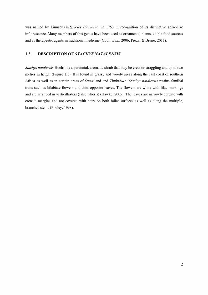
3
Figure 1.1 Stachys natalensis Hochst. growing in Reservoir Hills, Durban, South Africa.
[A] Grassland area in which S. natalensis parent material was obtained.
[B] Whole plant material.
[C] White flowers with lilac markings and distinct inflorescence of S. natalensis. Bar: 5 mm
A
B C
4
1.4. RESEARCH RATIONALE AND MOTIVATION
Foliar hairs, known as trichomes, originate from epidermal cells and may vary in appearance, size and
distribution among different plant species (Werker, 2000). Most glandular trichomes secrete
biologically active compounds which may be responsible for the curative properties of the plant. It is
imperative to categorize these therapeutic compounds and understand the link and mode of action of
the plant structures in which they are contained and secreted (Naidoo et al., 2009). Several studies
validate the use of Stachys spp. for the treatment of an assortment of ailments (Govil et al., 2006;
Goren et al., 2011). However, there appears to be no published reports on S. natalensis or its chemical
composition to date.
Many Stachys spp. are used in ethnomedicine in the form of extracts or decoctions made from the
aerial parts of the plant and roots (Piozzi and Bruno, 2011). Removal of these parts requires whole
plant destruction (Mander et al., 2007). The high demand for plant organs for traditional medicine and
novel drug development merits the use of plant tissue culture techniques for the production of
preferred plant genotypes or specific plant organs. Furthermore, in vitro propagation may alleviate the
pressure of over-harvesting medicinally important species (Legkobit and Khadeeva, 2004).
Micropropagation has many advantages over conventional propagation techniques including: rapid
mass multiplication of plants irrespective of climate or season, generation of pathogen free plants and
selective propagation of specific plant organs and metabolites (Tripathi and Tripathi, 2003; Rai,
2010). An efficient in vitro micropropagation protocol for S. natalensis is required as the plant is
rather inaccessible (mainly hidden or entwined with other similar straggling plants) and often difficult
to source due to poor seasonal growth. Additionally, there are many discrepancies in the taxonomic
evaluation of the genus Stachys due to their variable morphology and wide habitat (Dinc & Ozturk,
2008). Trichome morphology may be used as a useful, preliminary classification tool to resolve
complex phylogenetic relationships within the genus (Salmaki et al., 2009). Plant tissue culture is an
effective means to maintain the plant genotype and reduce the risk of taxonomic discrepancies.
Although the foliar characteristics and therapeutic properties of many Stachys spp. are well
documented, there have been no descriptive or experimental studies conducted on the morphology or
phytochemical constituents of S. natalensis. There are few reports on the micropropagation of Stachys
spp. (Legkobit and Khadeeva, 2004; Ghiorghita et al., 2011) but no reference to the propagation of S.
natalensis plantlets en masse.
5
1.5. AIMS OF THE PRESENT STUDY
This study was undertaken to describe key micromorphological features of the foliar structures of S.
natalensis, elucidate composition and localisation of leaf exudates and generate a reliable in vitro
micropropagation protocol for S. natalensis.
1.6. OBJECTIVES The objectives of this study were as follows:
a) Identify and describe the micromorphology of the secretory structures associated with S. natalensis
using stereomicroscopy, scanning electron microscopy (SEM) and light microscopy.
b) Quantitatively determine trichome density between the adaxial and abaxial surfaces of emergent,
young and mature leaf specimens of field-grown S. natalensis samples.
c) Identify the location of compounds of interest within the leaf tissue using various histochemical
stains.
d) Investigate the composition and nature of exudates utilising preliminary qualitative phytochemical
tests.
e) Assess the total phenolic content, total flavonoids content and antioxidant activity of the crude
extract of S. natalensis using the Folin–Ciocalteu method, aluminium chloride colorimetric assay
and DPPH (diphenyl-1-picrylhydrazyl) assay respectively.
f) Design and implement a reliable and efficient in vitro clonal propagation protocol using axillary
buds via direct organogenesis.
g) Determine trichome density between the adaxial and abaxial surfaces of emergent, young and
mature in vitro propagated plants and compare to that of field-grown plants.
CHAPTER 2: LITERATURE REVIEW
6
The plant foliar surface contains a plethora of secretory tissues and microstructures that vary in size,
shape, function and arrangement amongst different plant species (Werker, 2000). Secretory tissues are
important defining characteristics for many plant families and may be classified into two broad
categories based on their location and mechanism of secretion (Svoboda and Svoboda, 2001). The
first type, intercellular cavities or secretory ducts, exude substances within the plant body
(endogenous secretion) (Fahn, 1988). Examples of such tissues are the laticifers (latex secreting cell
system) present in members of the family, Apocynaceae (Vinca sardoa), Caricaceae (Carica papaya)
and Asclepiadaceae (Gomphocarpus physocarpus) to name a few (Agrawal and Konno, 2009; Konno,
2011). The second type is involved in exogenous secretion which is the release of substances to the
plant surface (Fahn, 1988). Examples of plant structures involved in exogenous secretion include:
hydathodes (salt-secreting glands), nectaries, gum and mucilage secreting glands and different types
of trichomes (Svoboda and Svoboda, 2001). The term “trichome”, from the Greek word ‘trikoma’
(growth of hair), refers to a bristle-like appendage that originates and extends outward from the
epidermal cells on plant vegetative and reproductive organs (Levin, 1973). They invariably differ in
size, morphology, location and composition amongst plant species, hence, classification is most often
based on a combination of characteristic features (Werker, 2000).
2.1. TRICHOME MORPHOLOGY
Plant trichomes may be separated into two distinct types: non-glandular and glandular (Wagner, 1991;
Werker, 2000). These trichome types have been described using various terminologies including
straight, hooked, dendritic and spiral (Levin 1973; Fahn, 1988; Dalin et al., 2008). Payne (1978) first
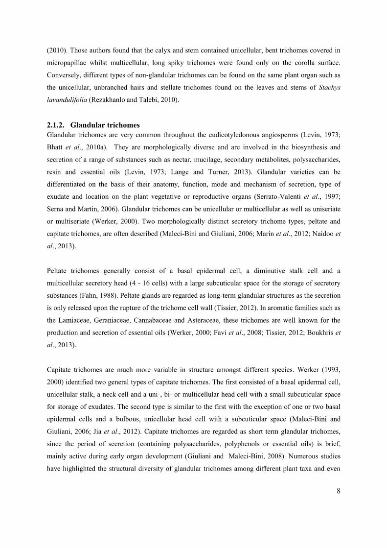
published a glossary of trichome terminology which described both non-glandular and glandular types
by virtue of their shape and appearance (Figure 2.1). Later reports elaborated upon this general
terminology and suggested that glandular trichomes may also be differentiated based on the nature
and localisation of substances that they secrete or store (Wagner, 1991).

7
Figure 2.1 Early descriptions of the diverse types of non-glandular and glandular trichomes found on
plant surfaces. These include: [1] attenuate, [2] uniseriate hair with osteolate cells, [3] acicular, [4]
ucinate, [5] furcate, [6] abietiform, [7] plumose, [8] pedate, [9] stellate, [10] penicillate, [11]
heliciform, [12] spiral, [13] dendritic, [14] peltate, [15] uniseriate capitate, [16] multiseriate capitate
and [17] bicellular capitate. Adapted from Payne (1978) and Roe (1971).
2.1.1. Non-glandular trichomes Non-glandular trichomes display great diversity with regard to their anatomy and structure (Wagner,
1991; Kolb and Muller, 2004). Most non-glandular trichomes are simple, branched or stellate (Choi
and Kim, 2013). They may be unicellular or multicellular. Multicellular trichomes may be further
classified as uniseriate (single row of cells), as seen in some Salvia and Teucrium spp., or multiseriate,
(several rows of cells) as noted in some Helianthus spp. (Werker, 2000; Grubesic et al., 2007;
Aschenbrenner et al., 2013). Non-glandular trichomes may differ in length and symmetry with the
apex tapering, rounded or hooked (Payne, 1978). The general soft or spiky appearance of non-
glandular trichomes can be attributed to the deposition of cell wall constituents such as lignin and
cutin (Werker, 2000). Trichomes may contain striations or warty micro-ornamentation that may
traverse the entire length or parts of their surface (Ascensao et al., 1995; Bhatt et al., 2010a).
Diverse morphologies of non-glandular trichomes may exist on different organs of the same plant
such as those on Dracocephalum moldavicum, reported by Dmitruk and Weryszko-Chmielewska
1
2
5 3
4
11
8
9
10
6
7
12
13
14
15 16
17
8
(2010). Those authors found that the calyx and stem contained unicellular, bent trichomes covered in
micropapillae whilst multicellular, long spiky trichomes were found only on the corolla surface.
Conversely, different types of non-glandular trichomes can be found on the same plant organ such as
the unicellular, unbranched hairs and stellate trichomes found on the leaves and stems of Stachys
lavandulifolia (Rezakhanlo and Talebi, 2010).
2.1.2. Glandular trichomes Glandular trichomes are very common throughout the eudicotyledonous angiosperms (Levin, 1973;
Bhatt et al., 2010a). They are morphologically diverse and are involved in the biosynthesis and
secretion of a range of substances such as nectar, mucilage, secondary metabolites, polysaccharides,
resin and essential oils (Levin, 1973; Lange and Turner, 2013). Glandular varieties can be
differentiated on the basis of their anatomy, function, mode and mechanism of secretion, type of
exudate and location on the plant vegetative or reproductive organs (Serrato-Valenti et al., 1997;
Serna and Martin, 2006). Glandular trichomes can be unicellular or multicellular as well as uniseriate
or multiseriate (Werker, 2000). Two morphologically distinct secretory trichome types, peltate and
capitate trichomes, are often described (Maleci-Bini and Giuliani, 2006; Marin et al., 2012; Naidoo et
al., 2013).
Peltate trichomes generally consist of a basal epidermal cell, a diminutive stalk cell and a
multicellular secretory head (4 - 16 cells) with a large subcuticular space for the storage of secretory
substances (Fahn, 1988). Peltate glands are regarded as long-term glandular structures as the secretion
is only released upon the rupture of the trichome cell wall (Tissier, 2012). In aromatic families such as
the Lamiaceae, Geraniaceae, Cannabaceae and Asteraceae, these trichomes are well known for the
production and secretion of essential oils (Werker, 2000; Favi et al., 2008; Tissier, 2012; Boukhris et
al., 2013).
Capitate trichomes are much more variable in structure amongst different species. Werker (1993,
2000) identified two general types of capitate trichomes. The first consisted of a basal epidermal cell,
unicellular stalk, a neck cell and a uni-, bi- or multicellular head cell with a small subcuticular space
for storage of exudates. The second type is similar to the first with the exception of one or two basal
epidermal cells and a bulbous, unicellular head cell with a subcuticular space (Maleci-Bini and
Giuliani, 2006; Jia et al., 2012). Capitate trichomes are regarded as short term glandular trichomes,
since the period of secretion (containing polysaccharides, polyphenols or essential oils) is brief,
mainly active during early organ development (Giuliani and Maleci-Bini, 2008). Numerous studies
have highlighted the structural diversity of glandular trichomes among different plant taxa and even
9
between species (Ascensao et al., 1999; Corsi and Bottega, 1999; Giuliani and Maleci-Bini, 2008;
Marin et al., 2012; Rusydi et al., 2013).
2.2. TRICHOME FUNCTIONS
The secretory functions of trichomes are often the defining characteristics in many plant genera such
as the salt glands of Avicennia, nectaries of Passiflora and oil secretory glands in Mentha (Wagner,
1991: Werker et al., 1993). Trichomes serve a variety of functions which are dependent on the
trichome type and location (Wagner, 2004).
2.2.1. Plant defence against herbivory Trichomes often function as sensors to detect leaf activity and prepare the plant for insect attack
(Werker, 2000). In many instances, trichomes serve as physical or chemical deterrents against insect
herbivory (Tian et al., 2012). Trichomes on the surface of Glycine max (soybeans) for example,
inhibit insect movement across the plant. Thus, larvae are unable to access the epidermis for
nourishment. Insects may eat through the trichome but suffer from little weight gain and subsequent
death due to the poor nutritional value conferred by the cellulose and lignin enriched trichomes (Dalin
et al., 2008). Trichome density also plays a factor in resistance to herbivory (Tian et al., 2012). In a
study by Riddick and Wu (2011), the survival rate of the lady beetle Stethorus punctillum was related
to the trichome density of Phaseolus lunatus (lima bean) leaves. The study indicated that younger
leaves had a dense distribution of hooked trichomes which impaled S. punctillum larvae resulting in
death due to starvation, dessication and numerous puncture wounds. Trichome production may also be
induced in many plant species as a direct response to insect damage (Levin, 1973; Howe and Jander,
2008).
Secondary metabolites are produced as a by-product of normal plant growth and development.
Glandular trichomes may store or induce production of these secondary metabolites such as
flavonoids, terpenoids, and alkaloids in response to insect or pathogen attack (Wittstock and
Gershenzon, 2002). The defensive compounds may poison, repel or immobilise insects. Tian et al.
(2012) determined that growth of Helicoverpa zea, an agricultural pest, was severely affected by the
presence of glandular trichomes on wildtype Solanum lycopersicum plants compared to its hairless
mutant. It was hypothesized that the glandular secretions contained toxic/sticky substances which
acted as chemical deterrants by entrapping H. zea. Other examples include the Sitophilus zeamais
weevil which showed significant toxicity to eucalyptol (1,8-cineole), isolated from the essential oil of

10
Vernonia amygdalina (Adeyemi, 2011) and Aphis nerii, an arthropod fatally immobilized in the sticky
exudate of the glandular trichomes from Sicana odorifera (Cucurbitaceae) (Kellog et al., 2002).
Plants may also employ indirect defence mechanisms by releasing volatile organic compounds (stored
in secretory structures) which attract predators of the insects that attack the plant surface (Pare and
Tumlinson, 1999; Howe and Jander, 2008). Examples include glandular structures of Zea mays and
Gossypium plants, which produce volatiles that attract predatory wasps when damaged by moth larvae
(Pare and Tumlinson, 1999).
2.2.2. Pollination Trichomes are involved in other plant-insect interactions in addition to plant defense. Many trichome-
derived compounds are utilized as attractants for species-specific pollination (Caissard et al., 2004).
Reis et al. (2004) described the pollination mechanism of Stanhopea lietzeii and S. insignis. These
orchid species exude fragrant oils which attract neotropical bees. Trichomes are also involved in
specialised mechanisms of insect capture for pollination. The downward-facing trapping flower
trichomes of Aristolochia spp. facilitate the easy entrance of insects into flowers but impede their
escape until the flowers modify their inner surface post-pollination (Oelschlägel et al., 2009).
2.2.3. Protection against water loss, UV-B and light damage Trichomes play an important role in maintaining plant physiology upon exposure to environmental
stress (Yan et al., 2012). In cases of drought or elevated temperatures, they reflect light, thus lowering
the temperature over the leaf surface, which reduces water loss through transpiration
(Abdulrahaman and Oladele, 2011). Perez-Estrada et al. (2000) investigated trichome density of
Wigandia urens under different seasonal and environmental parameters. These authors found that
trichome density was higher during the dry season and on plants that grew in sun exposed areas.
Futhermore, decreased trichome density was noted in response to irrigation and shade treatments.
These observations implied that trichomes were involved in limiting water loss through transpiration
and reflecting solar radiation. Trichomes play a role in protection against UV-B radiation by
accumulating flavonoids and other UV absorbing compounds (Yan et al., 2012). Studies indicated
that UV-B radiation induces an increase in trichome density in Nicotiana tabacum and Arabidopsis
thaliana which is important for the protection of underlying plant tissues against the detrimental
effects of UV-B radiation (Barnes et al., 1996; Yan et al., 2012).
11
2.3. TRICHOME EVOLUTION AND TAXONOMIC POTENTIAL WITHIN THE
LAMIACEAE
In terms of the evolution of secretory structures, many studies propose opposing hypotheses. Fahn
(1988, 2002) suggested that secretory structures originally developed within vascular plant organs (as
seen in early pteridosperms) and subsequently were scattered in cells of internal vascular tissues
(gymnosperms) and then found on the plant surface (present day angiosperms). Bearing this in mind,
the glandular trichomes would represent the most advanced and evolved form of the secretory
structures. Other studies indicate that glandular trichomes may have evolved independently from
stomata or non-glandular trichomes. Carpenter (2006) investigated the morphology of specialised
structures on the foliar surfaces of early angiosperms. That author detected the presence of
intermediate forms of trichomes and stomata and hypothesised that the specialized trichome structures
may be homologous to early stomatal complexes or derived from stomata over multiple evolutionary
events. Tissier (2012) provided an alternative hypothesis in that glandular trichomes may have
evolved from their non-glandular counterparts via the differentiation of apical cells into secretory
cells. The study described the capitate trichomes of Solanum lycopersicum and S. habrochaites which
differed morphologically from the non-glandular type by the presence of a single secretory cell at the
trichome tip. To date, there is little molecular support for these hypotheses. Moreover, a single
evolutionary occurrence cannot account for the extensive glandular trichome diversity amongst plant
families (Tissier, 2012).
The Lamiaceae is one of the largest families among the angiosperms, containing more than 240
genera (Giuliani and Maleci-Bini, 2008). Micromorphological studies with regard to epidermal
structures, in particular the trichomes, have been widely utilised to describe similarities and
differences amongst the various taxa (Ascensao et al., 1995; Gairola et al., 2009; Baran et al., 2010;
Dunkic et al., 2012). Both glandular (peltate and capitate) and non-glandular trichomes are
characteristic features of species of the family (El-Beyrouthy et al., 2009; Bhatt et al., 2010b; Dunkic
et al., 2012). The presence, type and morphology of trichomes and phytochemical characteristics of
their secretions may be used as discriminative markers at species or genera level (Ascensão et al.,
1995; Celep et al., 2011). Many members such as Ocimum, Stachys, Mentha and Salvia spp. are also
well known for the production of essential oils and various secondary metabolites within the glandular
trichomes which may be utilised as chemotaxonomic indicators for species classification (Maleci-Bini
and Giuliani, 2006; Naidoo et al., 2013). Stachys is considered as one of the largest genera within the
Lamiaceae, with many taxonomic discrepancies and problems in nomenclature (Giuliani and Maleci-
Bini, 2012). Trichome presence on vegetative and/or reproductive structures, coupled with its
12
typology has presented an efficient classification tool to delimit closely related species or subspecies
(Giuliani and Maleci-Bini, 2008) as well as differentiate between sections (Dinc and Ozturk, 2008).
2.4. STACHYS: HISTORY AND BIOLOGICAL ACTIVITY
The genus Stachys comprises of more than 270 species and is known by a variety of common names
including woundwort, betony and lamb’s ears (Piozzi and Bruno, 2011; Thomas, 2011). Many species
of the genus have been historically utilised by numerous cultures in folklore and for the treatment of a
wide variety of illnesses. The use of Stachys officinalis (betony) for the treatment of headaches,
wounds and abscesses is perhaps one of the earliest accounts of the genus (Thomas, 2011). The
Spanish adage, “He has as many virtues as betony” bears testament to its value. In fact, physicians
documented at least 47 medicinal uses of the plant including: anti-inflammatory, anti-bacterial, anti-
nephritic and anxiolitic effects. In addition, the early Anglo-Saxons believed that the plant warded off
evil spirits and therefore propagated it in churches (Thomas, 2011).
In recent times, the therapeutic use of Stachys spp. has increased worldwide. In Iranian traditional
medicine, herbal infusions of Stachys lavandulifolia and aerial parts of S. inflata and S. recta are used
to treat skin infections, rheumatism, indigestion and gastro-intestinal disorders (Khanavi et al., 2009;
Pirbalouti and Mohammadi, 2013). Stachys thunbergii and S. aethiopica are used in South African
traditional medicine for the treatment of hysteria and insomnia and to reduce fever caused by delirium
(Stafford et al., 2008). Stachys officinalis, known as ‘Ranilist’ and ‘Betonica’ in Bulgaria and Italy
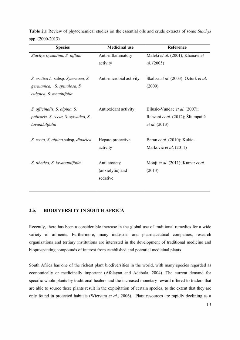
respectively, is used as an antiseptic and diuretic (Leporatti and Ivancheva, 2003). Table 2.1 indicates
the scientific support for the many medicinal uses of Stachys spp. In most cases, studies show a
diverse range of active compounds in the secretions or crude extracts including flavonoids,
glycosides, phenolic compounds, di-terpenoids and volatile and essential oils which support the use of
this genus in traditional medicine (Duman et al., 2005; Goren et al., 2011; Govil et al., 2006).
13
Table 2.1 Review of phytochemical studies on the essential oils and crude extracts of some Stachys
spp. (2000-2013).
Species Medicinal use Reference
Stachys byzantina, S. inflata
Anti-inflammatory
activity
Maleki et al. (2001); Khanavi et
al. (2005)
S. cretica L. subsp. Symrnaea, S.
germanica, S. spinulosa, S.
euboica, S. menthifolia
Anti-microbial activity Skaltsa et al. (2003); Ozturk et al.
(2009)
S. officinalis, S. alpina, S.
palustris, S. recta, S. sylvatica, S.
lavandulifolia
Antioxidant activity Bilusic-Vundac et al. (2007);
Rahzani et al. (2012); Šliumpaitė
et al. (2013)
S. recta, S. alpina subsp. dinarica. Hepato protective
activity
Baran et al. (2010); Kukic-
Markovic et al. (2011)
S. tibetica, S. lavandulifolia
Anti anxiety
(anxiolytic) and
sedative
Monji et al. (2011); Kumar et al.
(2013)
2.5. BIODIVERSITY IN SOUTH AFRICA
Recently, there has been a considerable increase in the global use of traditional remedies for a wide
variety of ailments. Furthermore, many industrial and pharmaceutical companies, research
organizations and tertiary institutions are interested in the development of traditional medicine and
bioprospecting compounds of interest from established and potential medicinal plants.
South Africa has one of the richest plant biodiversities in the world, with many species regarded as
economically or medicinally important (Afolayan and Adebola, 2004). The current demand for
specific whole plants by traditional healers and the increased monetary reward offered to traders that
are able to source these plants result in the exploitation of certain species, to the extent that they are
only found in protected habitats (Wiersum et al., 2006). Plant resources are rapidly declining as a
14
result of environmental changes to natural ecosystems and the illegal overharvesting of plants with
potential medicinal value (Afolayan and Adebola, 2004; Canter et al., 2005). This leads to reduced
endemic populations, loss of biodiversity, habitat loss and environmental degradation (Canter et al.,
2005). There are many initiatives for the assessment of threatened species as well as in situ and ex situ
conservation strategies under implementation. Although conventional cultivation for many plants is
desirable as it reduces harvesting pressure on threatened or rare species, there are several limitations
e.g. many medicinal plants do not produce seeds or seeds are too small and/or difficult to germinate
(Sharma et al., 2010). For instances where seeds are used for conventional propagation, plant yield is
highly heterozygous and shows variable growth (Sharma et al., 2010; Amujoyegbe et al., 2012).
Medicinal plants are not often acquiescent to vegetative propagation via cuttings, thus mass
propagation of cultivars is often limited due to low germination rates (Canter et al., 2005).
Futhermore, vegetative propagation may result in the presence of systemic bacteria and fungi which
may negatively affect the quality of the plant and any herbal end product derived from it
(Amujoyegbe et al., 2012). A viable option is the utilization of plant biotechnology to offset the loss
of significant species and for the efficient regeneration of medicinal and economically important plant
species (Canter et al., 2005). Plant tissue culture, or in vitro micropropagation, has been successfully
used for continuous, mass multiplication of elite and disease-free plants, obtained quickly and
economically (Sharma et al., 2010).
In vitro micropropagation has many advantages over conventional propagation methods. These
include enhanced rates of multiplication using relatively small amounts of starting plant material, a
shorter shoot cycle that yields an exponential increase in shoot production, rapid in vitro production
of secondary metabolites, plant regeneration irrespective of season or climate, maintenance of
germplasm stocks indefinitely and the rapid propagation of genotypes that are disease- and pathogen-
free and genetically identical to parent plants (Bhowmik and Matsuiz, 2001; Tripathi and Tripathi,
2003; Arikat et al., 2004). The method is also useful for the propagation of sexually sterile species
(triploids) and seedless plants such as bananas and to control experimental parameters such as light
intensity and temperature (Rai, 2010). Clonal propagation affords pharmaceutical and agricultural
industries an alternate and fast solution to the issue of procuring large amounts of plant matter
(Tasheva and Kosturkova, 2013). This simultaneously reduces the pressure of overharvesting plants
and maintaining natural resources.

15
2.6. PLANT TISSUE CULTURE
Tissue culture is a collective term referring to procedures used to maintain and grow plant cells,
tissues or organs on suitable media under aseptic conditions. It is based on the concept of totipotency,
whereby each single plant cell has the capability to express the full genetic potential of the plant (Rao
and Ravishanker, 2002). An overview of early developments in the field of plant cell culture is found
in Table 2.2. In 1902, Haberlandt developed the concept of in vitro cell culture by attempting to
cultivate various differentiated plant cells in a glucose-enriched salt solution (Rai, 2007). The
experiment garnered little success, possibly due to the relatively simple nutrients utilised and the lack
of sterile conditions (Sussex, 2008). In subsequent years, other researchers were successful in
maintaining plant growth via callus induction (White, 1939), introducing the use of plant growth
regulators (Went, 1935; Miller et al., 1955) and successfully inducing growth using different explant
material (Ball, 1946; Muir, 1954).
Micropropagation is the vegetative propagation of whole plants from explant material (small pieces of
plant tissue) using controlled in vitro tissue culture methods. Micropropagation techniques were
initially developed for the ornamental plant industry and later for the production of elite fruit and
vegetable crops (Tasheva and Kosturkova, 2013). Recently, much research has focused on
propagation of medicinal plant species en masse (Valizadeh and Valizadeh, 2011). Positive results
were obtained using shoot tip and meristems of medicinal plants such as Cinchona ledgeriana,
Digitalis spp., Rauvolfia serpentina, Glycyrrhiza glabra, Bacopa monnieri and Artemisia annua
(Tripathi and Tripath, 2003; Debnath et al., 2006; Sharma et al., 2008).
16
Table 2.2 Early history and development of plant tissue culture.
Year Progression Reference
1838 Proposal of cell theory Schleiden and Schwann
1902 First attempt at in vitro cell culture Haberlandt
1922 Successful culture of root and stem tips Kolte and Robbins
1935 Discovery of first plant growth hormone, indole
acetic acid (IAA)
Went
1939 Continuous growing of callus culture by
addition of vitamin B
White, Gautheret
1946 Whole plant of Lupinus regenerated by shoot
culture
Ball
1954 Single cells from callus culture Muir
1960 Isolation of protoplast cells Cocking
1962 Media development Murashige and Skoog
1970 Single cells to somatic embryos Backs-Husemann and Reinert
1974 Concept of developmental stages of in vitro
tissue culture
Murashige
1984 First industrial production of shikonin (dye and
medicinal compound) suspension cultures of
Lithospermum erythrorhizon
Mitsui petrochemical
1985 Development of gene transfer system using
Agrobacterium tumefaciens
Gheysen
1988 Development of photoautotrophic
micropropagation (sugar-free medium)
Kozai and Iwanami
* Adapted from Sussex, 2008; Rai, 2007

17
2.7. PROCESS OF IN VITRO MICROPROPAGATION
Micropropagation may proceed via two pathways: direct or indirect organogenesis. The former uses
axillary explants from donor plants for shoot induction, followed by rooting, whilst the latter requires
an intermediate callus induction stage (Rai, 2010). Murashige (1974) defined four stages for
successful in vitro plant tissue culture with an additional “stage 0” added by Debergh and Maene
(1981). The entire process illustrated in Figure 2.2 is discussed below.
Figure 2.2 General overview of the stages involved in plant micropropagation. Parent plants are
selected (stage 1) and explants are isolated and decontaminated before initiation onto suitable growth
media (stage 2). Indirect organogenesis results in callus formation with each new cell having the
potential for shoot and root formation (purple arrows). Direct organogenesis maximises shoot
multiplication over successive subcultures (red arrows), followed by elongation and rooting of
plantlets. Micropropagation results in the formation of multiple, acclimatized, true-to-type plantlets
(stage 4). Adapted from Medina et al. (2004).
2.7.1. Stage 0: Selection and maintenance of stock plant This primary step in the process requires the selection of stock material which should be cultivated
under optimum ex vitro conditions. Plant material may undergo pre-conditioning by exposure to anti-

18
microbial agents to reduce the risk of contamination. Studies showed that pre-treatment of Dryopteris
cristata rhizomes with Nystatin significantly reduced exogenous and endogenous fungal
contamination (Zenkteler and Kwasna, 2007) whilst Mng’omba et al. (2007) determined that the
application of a systemic fungicide to the Uapaca kirkiana parent plant before stock plant collection
effectively lessened the fungal infestation during in vitro culture. In the case of field-grown S.
natalensis plants, the hairy leaves are prone to grime accumulation, which may require the application
of a systemic fungicide to reduce microbial load on stock material.
2.7.2. Stage I: Initiation and establishment of an aseptic (sterile) culture This stage involves isolation of explants, surface sterilization and establishment of explants on
suitable growth medium (Murashige and Skoog, 1962).
a. Explant isolation Explants may be obtained from almost any part of the plant, including vegetative or
reproductive parts (Yildiz, 2012). Micropropagation of a number of plant species has been
successfully achieved using a range of explants, such as axillary buds (Coleus forskohlii,
Metha piperita), rhizome (Curcuma domestica, Costus speciosus) and cotyledon (Panax
ginseng) (Chaturvedi et al., 2007). Successful shoot regeneration of Withania somnifera, a
well known medicinal plant was obtained using different explant types such as the node,
internode, hypocotyl and embryo (Kulkarni et al., 2000). However, consideration must be
given to the route of organogenesis required in the study as explant source may often result in
different morphogenic responses. Komalavalli and Rao (2000) observed that shoot
multiplication was achieved using axillary node, cotyledonary node and shoot-tip leaf
explants whilst other explant types (leaf, petiole, and root) induced callus formation in
Gymnema sylvestre. The authors attributed these observations to the variation in levels of
plant hormones present in the excised explant before in vitro culture. Shoot tips and auxiliary
buds are extensively used as explant material for direct shoot induction in a variety of species
(Chauhan et al., 2012; Kosar and Mahmoud, 2012; Shahzad et al., 2012) and are viable
explant options in the present study as callus production is not required. Additional factors
such as the age of the donor plant and size of explant remain important considerations for
successful culture and may also affect the initiation potential of the culture. The general
consensus indicates that while larger explants may have better survival capabilities and are
able to withstand harsher modes of sterilization, smaller material limits viral and microbial
contamination (Bhojwani and Razdan, 1996; Yildiz, 2012). Studies also indicate that the
19
propagation potential of juvenile tissues is often higher compared to mature tissues (Le Roux
and Van Staden, 1991; Mohebodini et al., 2011).
b. Surface sterilization It is a pre-requisite for tissue culture techniques to be carried out under sterile conditions. In
many cases, treatment of stock plants at stage 0 is insufficient to eradicate surface
contaminants. Microbial growth rates far exceed plant cell division. Thus, surface sterilization
endeavours to remove all microorganisms that will thrive under in vitro conditions whilst still
maintaining explant viability. There are a variety of disinfectants such as ethanol, hydrogen
peroxide, bromine water, mercuric chloride, sodium hypochlorite and silver nitrate to name a
few. Sodium hypochlorite (NaClO) is a widely used disinfectant due to its potency against
microorganisms and some viruses (Oyebanji et al., 2009; Yildiz, 2012). In a study by
Mihaljevik et al. (2013) on the effectiveness of various decontaminating agents on axillary
buds of a cherry cultivar, sodium hypochlorite was shown to facilitate 80% aseptic explant
survival. In fact, many research articles have evaluated its use in place of sterilization
equipment (Peiris et al., 2012). Whilst mercuric chloride (HgCl2) is an effective
decontaminating agent, it is highly toxic to human health and to fragile plant material at high
concentrations (Moghaddam et al., 2011). However, it has been used for the explant
decontamination of Lamiaceaeous species such as Orthosiphun stamineus (Rashid et al.,
2012) and Ocimum gratissimum (Saha et al., 2012) and may potentially be of use for the
decontamination of S. natalensis.
2.7.3. Stage II: Shoot multiplication The multiplication stage follows that of initiation once explants have been successfully established on
suitable media and are flourishing. This stage involves repeated sub-culturing of in vitro material for
shoot multiplication until desired numbers of regenerated plantlets are obtained to continue to stage
III. Most often medium constituents for shoot multiplication remain the same as for initiation with the
only modification being that of plant growth regulators for maximum shoot induction (Diab et al.,
2011; Hussain et al., 2012). Vinterhalter and Vinterhalter (1999) observed that higher sucrose
concentrations in growth media also triggered morphogenesis by the formation of axillary buds and
adventitious root branching in Dracaena fragrans and Ceratonia siliqua. However, the effect of
carbon sources on shoot multiplication is beyond the scope of the current study.
20
a. Plant growth regulators (PGRs) Plant growth regulators are vital constituents in media preparation. There are four main
groups of plant regulators that are used in in vitro culture: auxins, cytokinins, gibberellins and
abscisic acid. They are involved in cell division and induce shoot or root production
depending on the proportion of cytokinins and auxins as shown in Figure 2.3.
Auxins, derived from the Greek word “auxien” meaning to grow or increase, often act
antagonistically with other growth regulators. There are many different auxins used in tissue
culture for induction of cell division and elongation, rooting and somatic embryogenesis
(Simon and Petrasek, 2011). Indole-3-acetic acid (IAA) and indole-3-butyric acid (IBA) are
natural auxins found at the growing shoot apex of plant tissues and are routinely used in the
micropropagation of many species (Arikat et al., 2004; Rai, 2010). The "direct inhibition
hypothesis" indicates that auxin from apical buds travels down shoots to inhibit axillary bud
growth. This encourages shoot growth and restricts lateral branching. Cytokinins move from
the roots into the shoots resulting in lateral bud growth. Removal of the apical bud results in
release of axillary bud inhibition which increases lateral growth (plants grow more densely)
(Booker et al., 2003).
The effect of cytokinins on plant tissue is variable, especially in collaboration with auxins.
Cytokinins are involved in the stimulation of cell division and direct morphogenic responses
(George et al., 2008). Although natural cytokinins such as zeatin and 2-iP are utilised in plant
tissue cultures, synthetic cytokinins such as 6-benzyloaminopurine (BAP) and kinetin are
more commonly used for their effectiveness in multiple shoot induction (Makunga and Van
Staden, 2008; Valizadeh and Valizadeh, 2011; Jana et al., 2013). The choice of specific
growth hormones in a study may require consideration of PGRs that are applicable to other
related species. For example, Zuzarte et al. (2010) and Kara and Baydar (2012) utilised
benzyladenine (BA) in combination with (IBA) for shoot multiplication of Lavandula
pedunculata and L. angustifolia respectively. Other examples include the use of kinetin for
shoot multiplication of Ocimum americanum (Kumar and Jhanwar, 2013) and Ocimum
basilicum (Gopal et al., 2014). Available literature on the micropropagation of Stachys spp. is
limited. In a study by Ghiorghita et al. (2011) the efficiency of BA in combination with IAA,
IBA and 1-naphthaleneacetic acid (NAA) for the propagation of Stachys sieboldii was
acknowledged. Thus the inclusion of synthetic cytokinin and exogenous auxin to plant culture
may prove beneficial for the micropropagation of S. natalensis.

21
Gibberellins are involved in growth regulation, breaking seed dormancy, enzyme induction,
callus induction and internode elongation. Of the almost twenty gibberellins known, GA3 is
most often used. Abscisic acid is used only for somatic embryogenesis and for culturing
woody species. Gibberellins share an antagonistic relationship with that of abscisic acid in
regulating plant growth (Razem et al., 2006; Rai, 2010). The current study focuses on the
generation of an efficient, simple propagation protocol via direct organogenesis. Therefore,
the effect of gibberellins and abscisic acid will not be evaluated in the current study.
Figure 2.3 The effect of auxin to cytokinin interaction on in vitro plant growth. Auxin alone
induces cell expansion but no cell division. Upon addition of cytokinin, cells expand and differentiate.
At equal levels of both hormones, parenchyma cells form undifferentiated callus. Higher cytokinin to
auxin levels induce shoot proliferation, whilst higher auxin levels induce root formation. Adapted
from Weber (2013).
2.7.4. Stage III: Rooting This stage involves the transferral of shoots obtained in stage II onto suitable rooting medium.
Rooting media may be slightly modified with regard to composition of basal medium and plant
growth regulators (increased levels of auxins) for root induction (Hussain et al., 2012). This is
however, dependant on the plant species. For example, the best response for root induction in
Achyranthes bidentata (Hossain et al., 2013) and Mentha spicata (Fadel et al., 2010) was achieved on

22
full strength MS medium. On the contrary, Baque et al. (2010) observed a significant inverse
relationship between MS strength and adventitious root growth in Morinda citrifolia. In fact, many
other studies have reported greater root induction on half-strength MS medium (Abou Dahab et al.,
2004; Goel et al., 2009; Asghari et al., 2012; Saha et al., 2012). It is generally accepted that the
addition of auxins play a role in root initiation and development (Pop et al., 2011). Studies report that
the addition of exogenous IAA to the culture medium of propagated Ocimum gratissimum (Gopi et
al., 2006) and Calendula officinalis (Victorio et al., 2012) significantly increased the rate of rooting.
Upon successful rooting, plantlets are evaluated for their survival potential and prepared for ex vivo
conditions.
2.7.5. Stage IV: Establishment of plantlets in soil In vitro tissue culture conditions are ideal for rapid and maximum growth of the plantlets. However,
these controlled conditions (ample light and high humidity) cause them to be vulnerable to disease,
soil pathogens, low moisture and nutrient levels and stress prevalent in natural environments (Kaur et
al., 2011; Panigrahi et al., 2013). Once plantlets have rooted sufficiently, they are removed from the
growth medium, rinsed and transferred to organic compost, potting soil, vermiculite or perlite (Kaur et
al., 2011; Hussain et al., 2012). The plantlets may also be placed in a mist chamber or exposed to
antitranspirants to control relative humidity and water loss thus ensuring that plants develop normally
and become autotrophic (Marin-Velazquez and Antonio, 2003).

CHAPTER 3: MATERIALS AND METHODS
23
3.1. SAMPLE COLLECTION AND PREPARATION Whole S. natalensis plants were collected from Durban, KwaZulu-Natal (29º 47´12.462; 30º 55´
45.0156). A voucher specimen was prepared and deposited at the Ward Herbarium, University of
KwaZulu-Natal, Westville Campus. Randomly selected leaf specimens at different developmental
stages i.e. emergent (<10 mm leaf length), young (10 mm – 25 mm leaf length) and mature (>30 mm
leaf length), were used for each sample preparation batch (n=10)
3.2. STEREOMICROSCOPY Entire leaf samples were used to investigate surface structures located on the adaxial and abaxial leaf
surfaces. A Nikon AZ100 Stereomicroscope and NIS-D Elements image software (Nikon Instruments
Inc., USA) were utilised to observe and capture images of different types of foliar trichomes and their
distribution.
3.3. SCANNING ELECTRON MICROSCOPY (SEM) Scanning electron microscopy was used to investigate trichome micromorphology and distribution.
Leaves were subjected to three different modes of preparation for SEM, i.e. freeze-drying, chemical
fixation and cryo-SEM.
3.3.1. Freeze- drying Fresh leaf sections of approximately 2 mm2 were quenched in liquid nitrogen and freeze-dried in an
Edwards’ Modulyo freeze-dryer for 72 hours (- 40 °C to -60 °C; 10-1 Torr). Samples were mounted
onto brass stubs using double-sided carbon adhesive tape and sputter- coated with gold in a Polaron
SC500 sputtercoater. Samples were viewed with a Zeiss UltraPlus Field Emission microscope. Areas
of interest were identified and images captured using SmartSEM image software (v.0.5 Zeiss,
Germany).
3.3.2. Chemical fixation Young and emergent leaves of S. natalensis were hand sectioned (± 2 mm2) and then chemically fixed
for two hours in 2.5% glutaraldehyde. The specimens were washed with phosphate buffer and post-
fixed with 0.5% osmium tetroxide in a dark cupboard for one hour. Samples were dehydrated by
exposure to increasing concentrations of acetone (30%, 50%, 75% and 100%). The samples were
critically point-dried using a Hitachi HPC1 critical point dryer, mounted onto brass stubs and sputter-
coated with gold. Images were captured as previously described.

24
3.3.3. Cryo-SEM Stachys natalensis leaf segments (± 1 mm2) were mounted on brass stubs with 50:50 colloidal
graphite & Tissue-Tek® O.C.T compound (Sakura® Finetek) mounting medium. Samples were
quenched in liquid N2, sublimed at -90 °C for 10 minutes and coated with Platinum (60 sec; 10 mA)
under vacuum. Samples were viewed and images captured with Zeiss UltraPlus Field Emission
microscope with an additional Quorum PP3000T Cryo-SEM system.
3.4. LIGHT MICROSCOPY Fresh hand as well as wax-embedded young leaf material was utilised for light microscopy. Sections
were prepared as outlined below, mounted onto glass slides and viewed using the Nikon Eclipse 80i
light microscope. Images were captured using the NIS-D elements software programme.
3.4.1. Wax embedding Young leaf tissue was sectioned and immersed immediately in 70% FAA (formaldehyde-acetic acid-
ethanol) fixative for 48 hours (Kladnik, 2006). Samples were then dehydrated using a graded ethanol
series involving immersion of samples in 70% ethanol for 30 minutes (repeated thrice), immersion in
80% ethanol for two hours, in 90% ethanol for four hours and finally in 100% ethanol for 90 minutes.
Four samples were then stained with fast green and safranin in 100% ethanol for 30 minutes. The
remaining slides were then placed overnight in 100% ethanol. Samples were then exposed to a
gradual xylene: ethanol infiltration (25:50, 50:50, 75:25) for an hour each before immersion in xylene,
twice for 15 minutes each time. Pellets of Paraplast Plus® tissue embedding media (McCormick
Scientific, USA) were added to the samples and left for three hours. These were replaced with new
wax pellets four times after three hour intervals. Hardened wax sections were sectioned using a
Reichhert Ultracut E rotary microtome to obtain semi-thin sections (1-2 microns).
3.4.2. De-waxing protocol Slides containing wax sections of leaf tissue were immersed in 100% xylene (x3 washes), 100%
ethanol (x3 washes), 70% ethanol (x2 washes) and distilled water for 2 minutes per wash.
3.5. HISTOCHEMISTRY De-waxed sections of leaf tissue were stained by the following reagents to detect the localization of
specific cellular compounds within the leaves:
25
3.5.1. Ferric trichloride – phenolic compounds Fresh leaf sections were placed in 10% ferric trichloride (10 g ferric trichloride pellets in 100 ml
distilled water, followed by filtration through Whatman™ No. 1 filter paper). A drop of sodium
carbonate was added and the sections were left at room temperature for 15 minutes. In this test, ortho-
dihydroxyphenols react with ferric ions to produce deep green or black deposits (Johansen, 1940).
3.5.2. Mercuric bromophenol blue – proteins Leaf sections were immersed in bromophenol blue (100 mg bromophenol blue with 10 g of HgCl2
and
95% ethyl alcohol) for 15 minutes. Stained samples were rinsed in 0.5% acetic acid for 20 minutes
and immersed in a 0.1 M sodium phosphate buffer (pH 7.0) for three minutes. A blue coloration was
indicative of total proteins (Mazia et al., 1953).
3.5.3. Nadi reagent – terpenoids and resins Fresh-hand leaf sections were immersed in two drops of nadi reagent (0.5 ml of a 1% alpha-naphthol
solution in 40% ethanol with 0.5 ml of 1% dimethyl-p-phenylenediamine HCl in water). Sections
were left at room temperature in the dark for 60 minutes. Sections were rinsed in sodium phosphate
buffer (0.1 M, pH 7.2), mounted and viewed. Essential oils appear blue whilst resin acids stain red
(David and Carde, 1964).
3.5.4. Nile blue – neutral and acidic lipids Leaf sections were placed in 1% nile blue for one minute at 37 ºC and then in 1% acetic acid for one
minute at 37 ºC before being rinsed in distilled water. Neutral lipids stained red whilst acidic lipids
stain blue (Cain, 1947).
3.5.5. Ruthenium red – mucilage and pectin De-waxed sections were placed in aqueous ruthenium red solution (1: 5000) for 10 minutes. Mucilage
and pectin stains red/ rose pink (Johansen, 1940).
3.5.6. Sudan III – lipids Fresh-hand sections were immersed in the stain (saturated solution) for 15 minutes and then rinsed
briefly with 70% ethanol to remove excess stain. Lipid inclusions stained orange/red (Pearse, 1985).
3.5.7. Toudine blue – general stain Leaf sections were immersed in a 0.05% aqueous solution of toludine blue for one minute. Excess
stain was washed with distilled water and stained sections were mounted in 70% glycerol and viewed.
26
This metachromatic stain (two sets of colour reactions) stains phosphate groups on molecules green-
blue whilst polyphenols (lignin) stain blue (O’Brien et al., 1964).
3.5.8. Wagner reagent & Dittmar reagent – alkaloids Fresh-hand leaf sections were immersed in Wagner reagent and Dittmar reagent for a period of ten
minutes each. The stained sections were rinsed with 70% ethanol and then mounted. Alkaloids were
detected by a brown colour change (Furr and Mahlberg, 1981).
Prepared slides stained with fast green and safranin was also viewed. Standard control slides were
carried out simultaneously. Each test was carried out in triplicate. Treated samples were viewed using
a Nikon Eclipse 80i light microscope. All images were captured using NIS-D Elements software.
3.6. PHYTOCHEMICAL PROFILE
3.6.1. Solvent extraction Leaves were harvested and allowed to air-dry for 72 hours. The dried material was then ground to a
fine powder. Approximately 8 g of ground material was dissolved in 100 ml of methanol and placed
in a round bottomed flask attached to a Soxhlet apparatus. The flask was heated and solvent extraction
proceeded for 60 minutes. The solution was filtered, the crude extract was retained and the process
was repeated thrice. Successive extractions using hexane followed by chloroform were carried out
twice after 30 minute intervals. The resultant liquid was used for qualitative phytochemical tests.
3.6.2. Qualitative phytochemical tests Phytochemical analyses on the crude extracts (methanol, chloroform and hexane) and negative
control (water) were conducted in accordance with standard protocols (Harbourne, 1973; Trease and
Evans, 1978; Philip et al., 2011; Damodaran and Manohar, 2012). All tests were replicated twice.
Detailed methodology was as follows:
a. Alkaloids Dragendroff’s test: Two drops of Dragendroff’s reagent were added to one ml of the extract.
A reddish- orange precipitate indicated the presence of alkaloids.
Hager’s test: One ml of extract was mixed with one ml of Hager’s reagent (1 g of
picric acid in 100 ml distilled water). The formation of a yellow precipitate indicated the
presence of alkaloids.
27
b. Amino acids Ninhydrin test: Two drops of Ninhydrin reagent were added to two ml of dilute extract. A
deep purple colour change indicated the presence of amino acids.
c. Anthraquinones A few drops of 2% hydrochloric acid were added to one ml of extract. The formation of a red
precipitate indicates the presence of anthraquinones.
d. Carbohydrates Benedict’s test: Benedict’s reagent (solution one made up of 173 g sodium citrate and 100 g
sodium carbonate in 800 ml water, filtered and made up to 850 ml added to a second solution
composed of 17.3 g copper sulphate and 100 ml distilled water) was added to one ml of
extract and heated for two minutes. A red-brown precipitate indicated the presence of
monosaccharides.
Molisch’s test: Molisch’s reagent (15 g α-naphthol dissolved in 100 ml ethanol) was added to
five ml crude plant extract. A brown-red colour reaction indicated the presence of
polysaccharides.
e. Cardiac glycosides Keller-Killiani test: Five ml of plant extract was mixed with two ml glacial acetic acid. Two
drops of ferric chloride were added, followed by the addition of concentrated sulphuric acid
such that the acid remains underneath. The presence of glycosides was indicated by the
formation of a brown ring at the junction of the two layers and a blue-green ring at the upper
surface.
f. Coumarins One ml of 10% NaOH was added to one ml extract. The development of a yellow colour
indicates a positive reaction for coumarins.
28
g. Flavonoids Lead acetate test: A 10% lead acetate solution (three ml) was added to two ml dilute extract.
The formation of a white precipitate indicated the presence of flavonoids.
h. Phenolic compounds/ tannins Ferric trichloride test: Two ml distilled water was added to one ml of the extract and a few
drops of 10% ferric trichloride. A dark green colour change indicated a positive result for the
presence of phenols and/or tannins.
i. Quinones One ml concentrated sulphuric acid was added to equal amount of extract. The development
of a red precipitate indicated the presence of quinones.
j. Saponins Foam test: The extract (five ml) was diluted with distilled water to 20 ml. The solution was
shaken in a graduated cylinder for 15 minutes. The presence of saponins in the extract was
identified by the formation of a persistent two cm foam layer.
k. Steroids Lieberman-Burchard test: Acetic anhydride (two ml) was added to the extract (five ml). One
ml of concentrated sulphuric acid was then carefully added to the solution. The formation of a
blue-green colour indicated the presence of steroids.
l. Terpenoids Salkowski’s test: Five ml of crude extract was mixed with two ml chloroform.
Concentrated sulphuric acid (three ml) was then carefully added to the mixture forming
distinct layers. The presence of terpenoids was indicated by the formation of a reddish-
brown colour at the interface between the two solutions.
3.6.3. Quantification of total phenolics and flavonoids The total phenolic content was determined using adapted protocols of the Folin-Ciocalteu method
(Singleton and Rossi, 1965; Moyo et al., 2013). Total flavonoid content was determined using an
adapted aluminium chloride colorimetric assay (Zhishen et al., 1999).
29
a. Preparation of plant extract Powdered leaf material (2 g) was extracted with 10 ml of 50% aqueous methanol in a
sonication bath containing ice-cold water for 60 minutes. The extract was filtered under
vacuum through Whatman™ No. 1 filter paper. The filtrate was immediately used for the
determination of total phenolics and flavonoids.
b. Total phenolic content: Folin–Ciocalteu method A stock solution of gallic acid was prepared by dissolving 0.5 g of dry gallic acid in 10 ml of
ethanol and diluting with distilled water in a 100 ml volumetric flask. Working solutions with
concentrations of 0, 50, 100, 150, 250, and 500 mg/l gallic acid were prepared. Gallic acid
(0–500 mg/l) was used as the effective range of the assay and for generation of the standard
curve.
The sample extract (0.5 ml) was added to 2.5 ml of 1:10 diluted Folin–Ciocalteu reagent.
After three minutes, 2 ml of saturated sodium carbonate solution (75 g/l) was added.
After two hours of incubation at room temperature in the dark, the absorbance of three
randomly selected samples were measured at 765 nm. The results were expressed as
milligram gallic acid equivalent (mg GAE)/g dry weight of plant matter.
c. Total flavonoid content: The aluminium chloride colorimetric assay
To quantify the total flavonoid content, quercetin was used as the reference. Quercetin stock
solution (0.25 mg/ml) was prepared by dissolving 10 mg quercetin in 40 ml of 80% ethanol.
Standard working solutions of 0, 25, 50, 75, and 100 ug/ml quercetin were prepared in 10 ml
volumetric flasks by adjusting the volume with 80% ethanol (Chang et al., 2002; Ozkok et al.,
2010)
Extract (1 ml) or quercetin standards were added to a 10 ml volumetric flask containing 4 ml
of distilled water. To the flask, 0.30 ml of 5% NaNO2 was added and after five minutes 0.3 ml
of 10% AlCl3 was added. After five minutes, 2 ml of 1M NaOH was added and the volume
was made up to 10 ml with distilled water. The solution was mixed and the absorbances of
three random samples were measured against an ethanol blank at 510 nm. The total flavonoid
content was expressed as mg quercetin equivalents (QE)/ g dry weight.
30
3.6.4. Antioxidant activity The antioxidant activity of the S. natalensis methanolic extract was determined using a modified
method of the DPPH radical scavenging assay (Blois, 1958; Karioti et al., 2004; Bilusic-Vundac et
al., 2007)
a. Principle of the DPPH (1,1-diphenyl-2-picrylhydrazyl) assay The assay is based on the reduction of 1,1-diphenyl-2-picrylhydrazyl, a stable free radical.
The presence of an odd electron provides a maximum absorption at 517 nm (purple colour).
In the presence of an antioxidant (or extract with radical scavenging potential), the electron is
paired off. As a consequence, absorption decreases and the degree of discolouration (yellow)
provides an indication of the extracts’ scavenging potential in accordance with its hydrogen
donating ability (Patel and Patel, 2011).
b. DPPH assay protocol A 0.1 mM methanolic DPPH solution and sample extracts consisting of final concentrations
of 0, 1, 2.5, 5, 10, 50, 100, 150 and 250 μg ml-1 were freshly prepared before the assay. One
ml of each plant extract concentration was added to one ml of methanolic DPPH solution and
incubated in the dark for 30 minutes. Thereafter, absorbance was measured at 517 nm using
methanol as a blank with a UV-visible spectrometer (Shimadzu, UV-1601; Japan). Ascorbic
acid at final concentrations of 1.0, 2.5, 5, 10 and 50 μg ml-1 were used as a positive control. A
solution of 50% methanol, instead of extract or antioxidant, was used as a negative control.
Extract colour which may adversely affect absorbance values was accounted for by
subtraction of sample absorbance from sample + DPPH absorbance. The assay was done in
triplicate using random extract samples.
The free radical scavenging activity of the extract, determined by the decolouration of the
DPPH solution, was calculated according to the formula:
The effective concentration of sample required to scavenge DPPH radical by 50%
(IC50 value) was calculated for the extract as well as ascorbic acid by linear regression
analysis between percentage inhibition and sample concentrations.
31
3.7. MICROPROPAGATION OF STACHYS NATALENSIS
3.7.1. Plant collection and decontamination Field-grown S. natalensis plants were obtained from Durban, South Africa (29º 47´12.462; 30º 55´
45.0156). The plants were pre-treated thrice over a two week period with an aerosol fungicide
(Wonder FUNGI GUNTM, Active ingredient: 3.0 g/kg Triforine). Axillary bud segments were used as
nodal explants (1.0 - 1.5 cm) and were initially surface decontaminated by intermittent washing with
tap water for 20 minutes. Due to high levels of plant contamination, samples were washed with
distilled water to remove residual media and microbial growth, followed by exposure to a number of
decontamination treatments, as outlined in Table 3.1. Samples were rinsed in distilled water after each
step of the procedure. Thereafter decontaminated samples were placed onto a suitable growth
medium. The experiment was conducted in a randomized design using 25 explants in each of the five
treatments.
Table 3.1 Decontamination treatments for S. natalensis axillary bud explants.
Treatment Immersion in
1% NaClO
Immersion in
3% NaClO
Immersion in
0.1% HgCl2
1 2 × 5 min - -
2 - 2 × 5 min 1 × 10 min
3 - - 1 × 20 min
4 1 × 7 min 1 × 5 min 1 × 10 min
5 1 × 5 min 1 × 2 min 1 × 7 min
3.7.2. Culture conditions and media
a. Initiation and shoot multiplication
Nodal explants (1-2 cm) were cultured on MS medium (Murashige and Skoog, 1962)
supplemented with 20.0 g/ l sucrose and 12 g/l nutrient agar. Media composition differed
with regard to the combination and concentration of plant growth regulators (PGRs) for
initiation, shoot multiplication and rooting. Media for bud break (initiation) was as follows:
medium A contained a combination of 0.5 mg/l BAP and 0.5 mg/l IAA, medium B contained
32
0.5 mg/l kinetin and 0.5 mg/l IAA and medium C contained 0.5 mg/l BAP and 0.5 mg/l IBA.
There were 50 explants per medium preparation. The same combinations of PGRs were used
for multiplication media in a cytokinin: auxin ratio of 2:1. Microshoots (8 - 10 cm) with a
minimum of six leaves were chosen for transfer into rooting medium which contained ½
strength MS medium supplemented with 2mg/l IAA as the only PGR. The percent rooting,
number of roots per shoot and root length were recorded over five weeks. The pH of all media
was adjusted to 5.7 ± 0.1 by the addition of 1 M HCl or 1 M NaOH before decontamination
by autoclaving at 121º C (1 kPa) for 20 minutes. Cultures were incubated in a growth room
with a 16 hour light/8 hour dark photoperiod, photon flux density of 200 µmol m-2 s-1 and
temperature of 26° C ± 0.1° C (day) and 23° C ± 0.1° C (night).
b. Acclimatization Once rooted, plantlets were removed from the medium, rinsed off to remove residual medium
and transferred to pots containing organic potting soil and kept in a mist tent (three second
spray per six minute interval) for acclimatization. The soil was supplemented with ¼ strength
MS salts for a week. After one month, acclimatized plants were transferred to a greenhouse
and irrigated daily. The percentage survival was recorded after six weeks.
3.8. STATISTICAL ANALYSES
Glandular and non-glandular trichome counts (per cm2 leaf surface) for field grown and in vitro plants
were obtained from SEM micrographs using the iTEM imaging software (Soft Imaging System
GmBH, Münster, Germany). A two-way ANOVA statistical test was conducted in IBM SPSS
Statistics (version 21.0, IBM®, USA) to determine trichome density across three developmental
stages viz. emergent, young and mature as well as between the abaxial and adaxial foliar surfaces.
Tukey’s post-hoc test was implemented to obtain multiple comparisons of factors investigated. All
quantitative data was reported as mean ± standard error of the mean (SE). Values were regarded as
significant at p<0.05. Regression analyses and calculation of EC50 values for antioxidant activity were
done using IBM SPSS Statistics.
CHAPTER 4: RESULTS
33
4.1. TRICHOME TYPES AND DISTRIBUTION
The leaf surfaces of Stachys natalensis are visibly hispid and bear numerous non-glandular and
glandular trichomes of different morphologies (Figure 4.1 A). Non-glandular trichomes primarily
occupy the midrib and lateral veins and are especially dense on the abaxial surface (Figure 4.1 B and
4.1 D). The adaxial surface contains a random distribution of colourless glands (Figure 4.1 C). In
addition, these glands are pronounced on the abaxial surface and display great structural diversity
(Figure 4.2). Transverse section of the leaves show the structural features of the leaf lamina including
multicellular peltate and bicellular capitate trichomes (Figure 4.3). The adaxial surface was furnished
with many bent non-glandular trichomes and peltate trichomes occurring in rows in epidermal
grooves (Figure 4.4).
Emergent leaves appeared to have a dense covering of trichomes which visibly decreased as the leaf
matured (Figure 4.5). Trichome density among three growth stages (emergent, young and mature) and
two foliar surfaces (abaxial and adaxial) were investigated using a two-way analysis of variance
(ANOVA). There were significant differences in both glandular (F(2, 0.678) = 7.408; p<0.05) and non-
glandular trichome density (F(2,0.324) = 10.544; p<0.05) among the three developmental stages and
between the upper and lower foliar surfaces (Figure 4.6 A and B). Multiple comparisons of the three
developmental stages using Tukey’s post-hoc test indicated significant differences in trichome density
between emergent and young leaves and between emergent and mature leaves (glandular and non-
glandular: p<0.05). The abaxial surface was found to contain a higher number of both glandular and
non-glandular trichomes (Table 4.1).

34
Plate One
Figure 4.1 Stereo micrographs showing overall surface morphology and trichome distribution on the
adaxial and abaxial foliar surfaces of S. natalensis.
[A] Hispid abaxial surface of emergent leaf.
[B] Abaxial surface of mature leaf showing non-glandular trichomes which appear frequently on the
central midrib and branching veins.
[C] Secretory glands randomly dispersed over the abaxial surface of a young leaf specimen.
[D] Mature leaf showing glandular (black arrow) and non-glandular (white arrow) trichomes on the
abaxial surface.
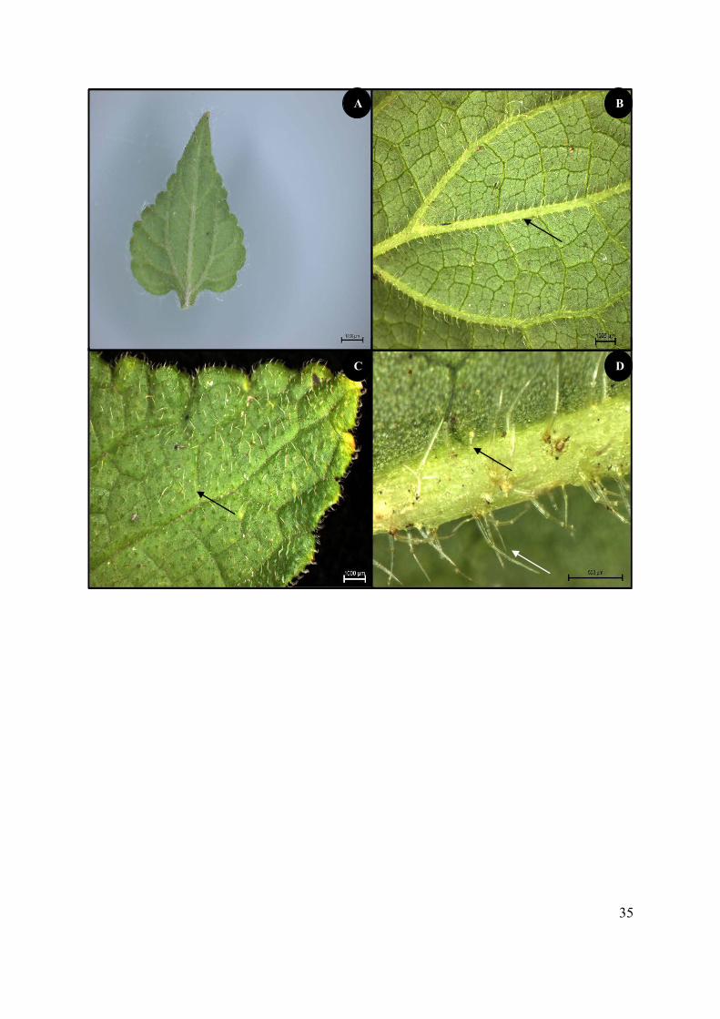
35
A
C
B
D
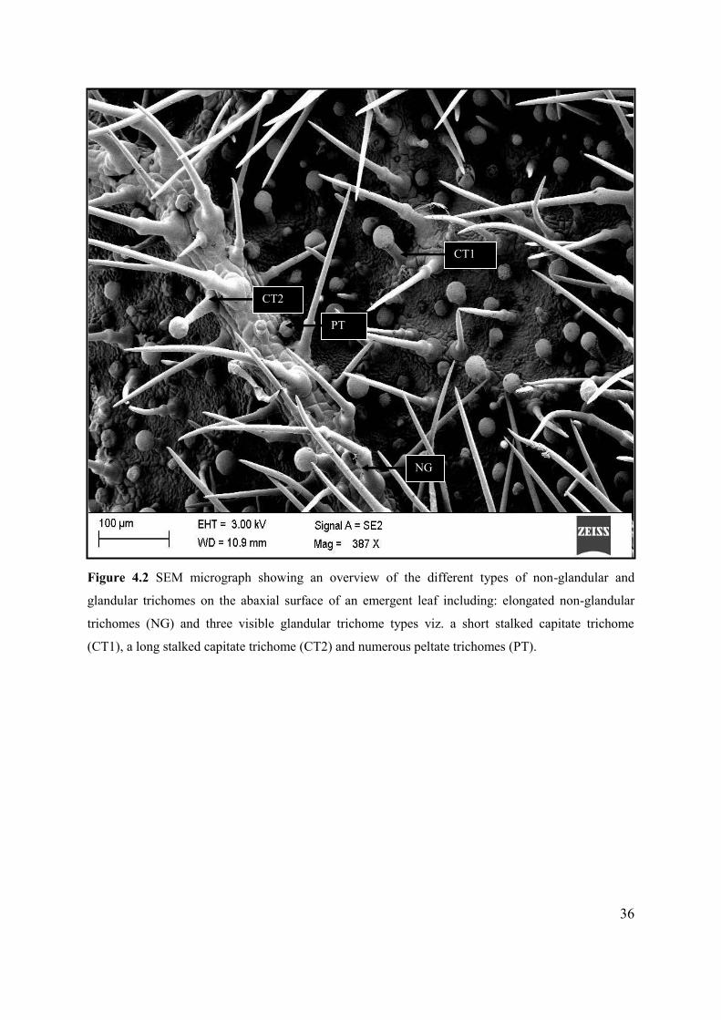
36
Figure 4.2 SEM micrograph showing an overview of the different types of non-glandular and
glandular trichomes on the abaxial surface of an emergent leaf including: elongated non-glandular
trichomes (NG) and three visible glandular trichome types viz. a short stalked capitate trichome
(CT1), a long stalked capitate trichome (CT2) and numerous peltate trichomes (PT).
CT2
CT1
NG
PT
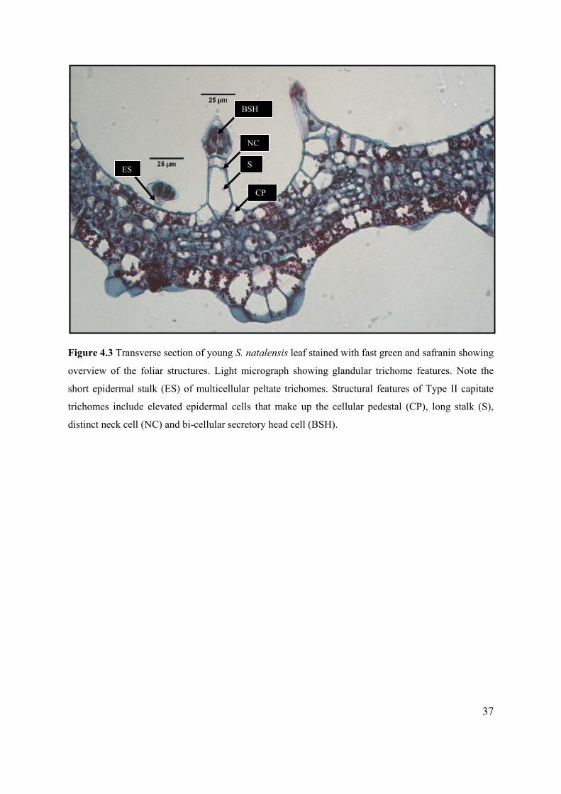
37
Figure 4.3 Transverse section of young S. natalensis leaf stained with fast green and safranin showing
overview of the foliar structures. Light micrograph showing glandular trichome features. Note the
short epidermal stalk (ES) of multicellular peltate trichomes. Structural features of Type II capitate
trichomes include elevated epidermal cells that make up the cellular pedestal (CP), long stalk (S),
distinct neck cell (NC) and bi-cellular secretory head cell (BSH).
BSH
NC
S
CP
ES
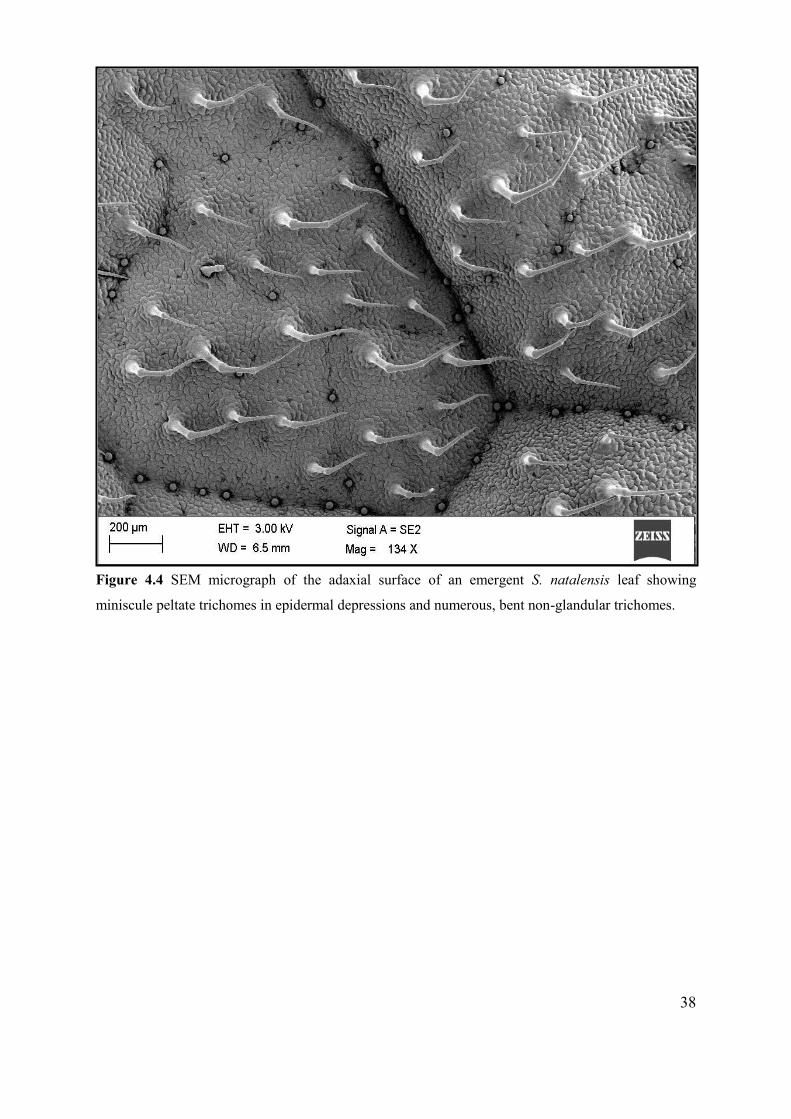
38
Figure 4.4 SEM micrograph of the adaxial surface of an emergent S. natalensis leaf showing
miniscule peltate trichomes in epidermal depressions and numerous, bent non-glandular trichomes.
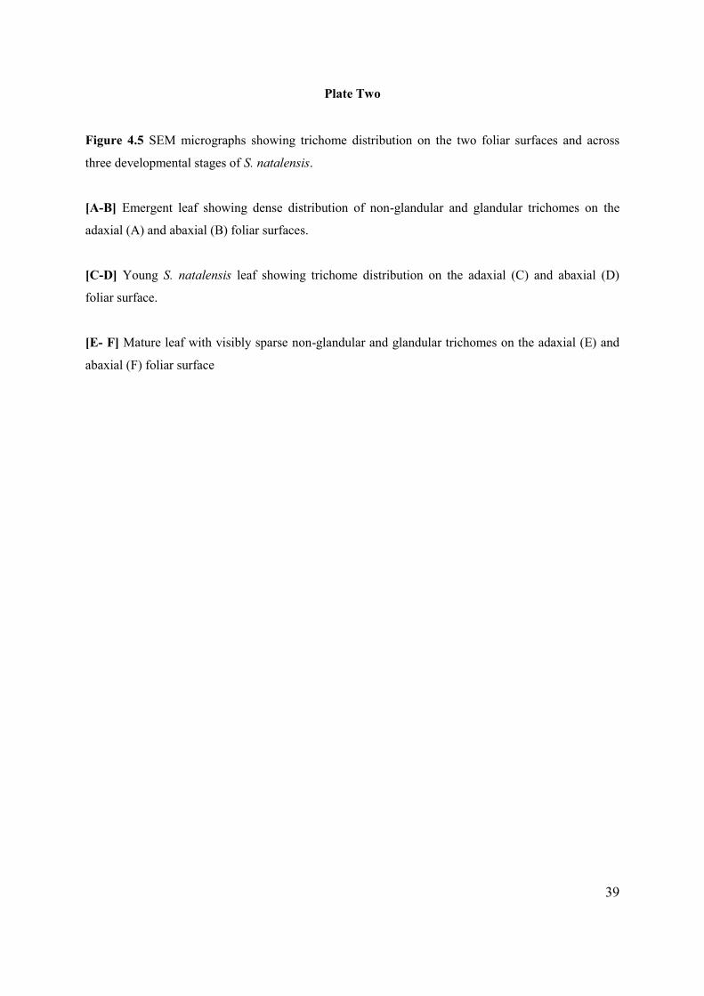
39
Plate Two
Figure 4.5 SEM micrographs showing trichome distribution on the two foliar surfaces and across
three developmental stages of S. natalensis.
[A-B] Emergent leaf showing dense distribution of non-glandular and glandular trichomes on the
adaxial (A) and abaxial (B) foliar surfaces.
[C-D] Young S. natalensis leaf showing trichome distribution on the adaxial (C) and abaxial (D)
foliar surface.
[E- F] Mature leaf with visibly sparse non-glandular and glandular trichomes on the adaxial (E) and
abaxial (F) foliar surface
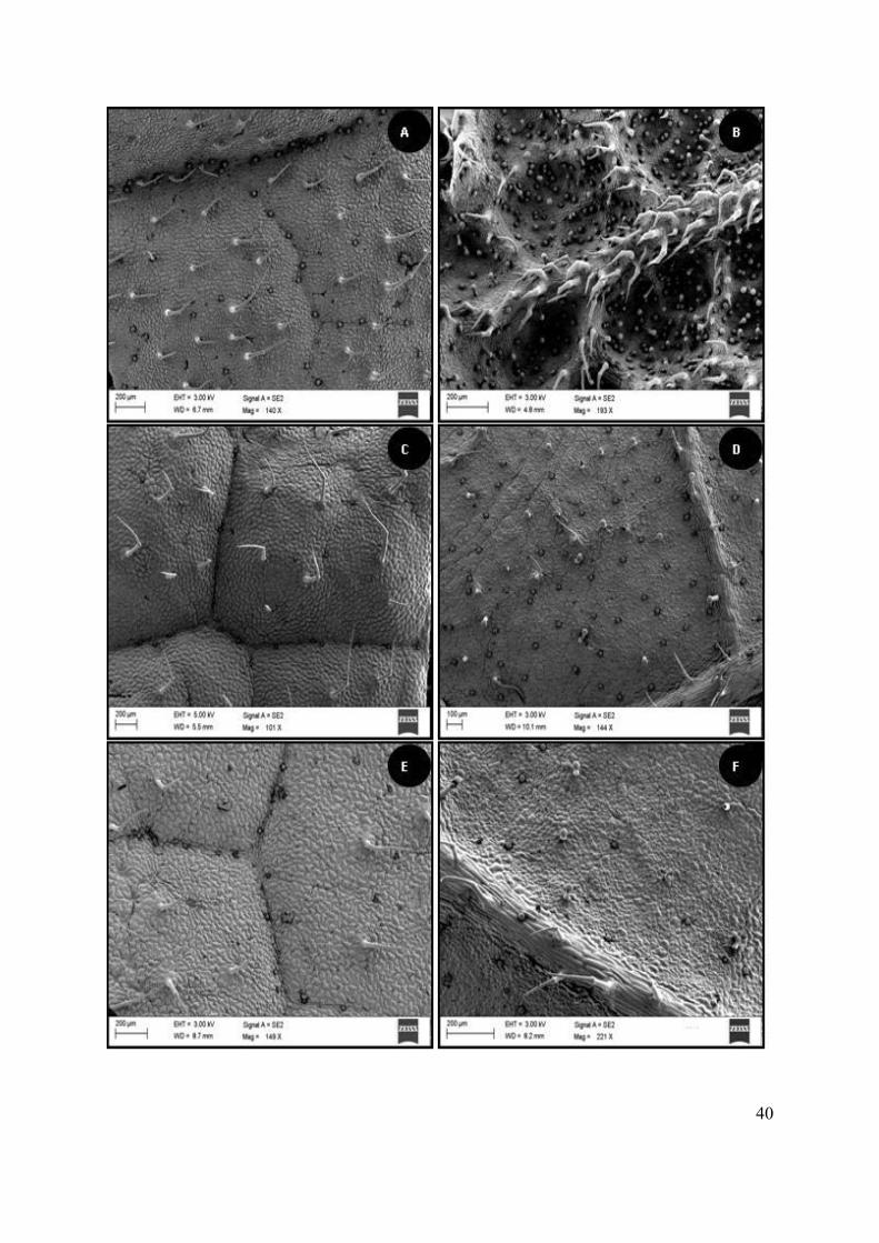
40
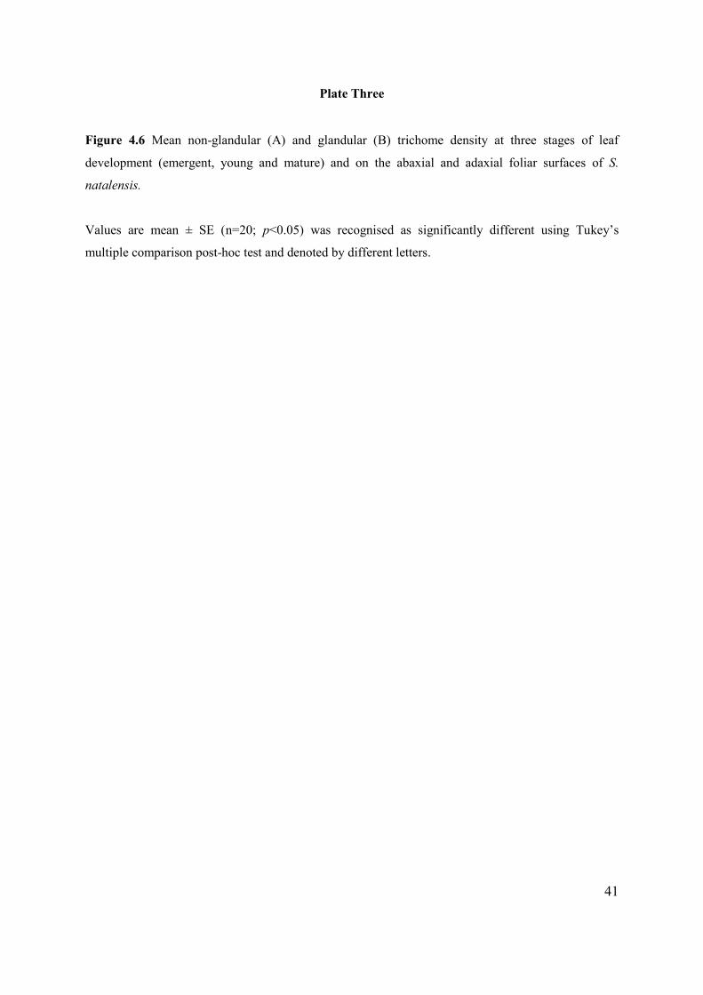
41
Plate Three
Figure 4.6 Mean non-glandular (A) and glandular (B) trichome density at three stages of leaf
development (emergent, young and mature) and on the abaxial and adaxial foliar surfaces of S.
natalensis.
Values are mean ± SE (n=20; p<0.05) was recognised as significantly different using Tukey’s
multiple comparison post-hoc test and denoted by different letters.
42
A
B
a
b b
a
b b

43
Table 4.1 Mean trichome density on the abaxial and adaxial leaf surfaces of S. natalensis plants.
Trichome type Mean trichome density (count/cm2)
Abaxial Adaxial
Non-glandular 0.631 ± 0.03 0.108 ± 0.02
Glandular 0.310 ± 0.03 0.139 ± 0.02
Data represents mean ± SE
4.2. TRICHOME MORPHOLOGY
Two morphological types of non-glandular trichome were observed on the foliar surfaces of S.
natalensis across all developmental stages. Structurally, the first type appeared acicular or acuminate,
with the stalk tapering gradually to a sharp point. The other morphological type appeared to be
arthrodachylous, jointed or highly septate (Payne, 1978). Both types of non-glandular trichomes were
uniseriate and unbranched (Figure 4.7). These trichomes have a wide base attached to the epidermal
layer via a multicellular (two-ten cells) epidermal cellular pedestal (Figure 4.7 A - D). The trichome
stalk which may be elongated tapering toward the apex or bent from the first or second stalk septation
facing the leaf apex has visible striations traversing the entire stalk length of the cellular pedestal
(Figure 4.7 E - F). These striations were also observed on glandular structures (Figure 4.9 A).
Glandular trichomes were divided into two types: peltate and capitate trichomes. Peltate trichomes
with a large, multicellular head appeared to contain between two to eight head cells (Figure 4.8). Two
types of capitate glandular trichomes were observed: Type I and Type II (Figure 4.9). Type I was
characterised by a distinctly striated stalk and a spherical shaped head that may be bi-cellular or
unicellular, according to Werker (2000) (Figure 4.9 A and Figure 4.11 A) and Type II was observed
primarily on the abaxial surfaces of emergent and young leaves. Type II capitate trichome was
composed of a bulbous base, long, tapering septate stalk, a distinctive neck cell and a multicellular
head cell (Figure 4.9 B). Glandular trichomes were observed in both pre- and post-secretory phases.
The former was characterised by accumulation of secretion in the subcuticular space in peltate head
cells (Figure 4.10 A). Preliminary observations indicate that septation of the secretory cells facilitates
the flow of secretion. Glandular trichomes in post-secretory phase showed a distinct contraction of
secretory head cells upon loss of secretion possibly via a porose cuticle (Figure 4.10 B). Two possible
mechanisms of secretion were observed in Type I and Type II capitate trichomes. Loss of secretion
via cuticular rupture was noted in Type I capitate trichomes (Figure 4.10 D) whilst Type II capitate
trichome was shown to secrete substances via pore-like openings on each of the multicellular head
cells (Figure 4.10 C).
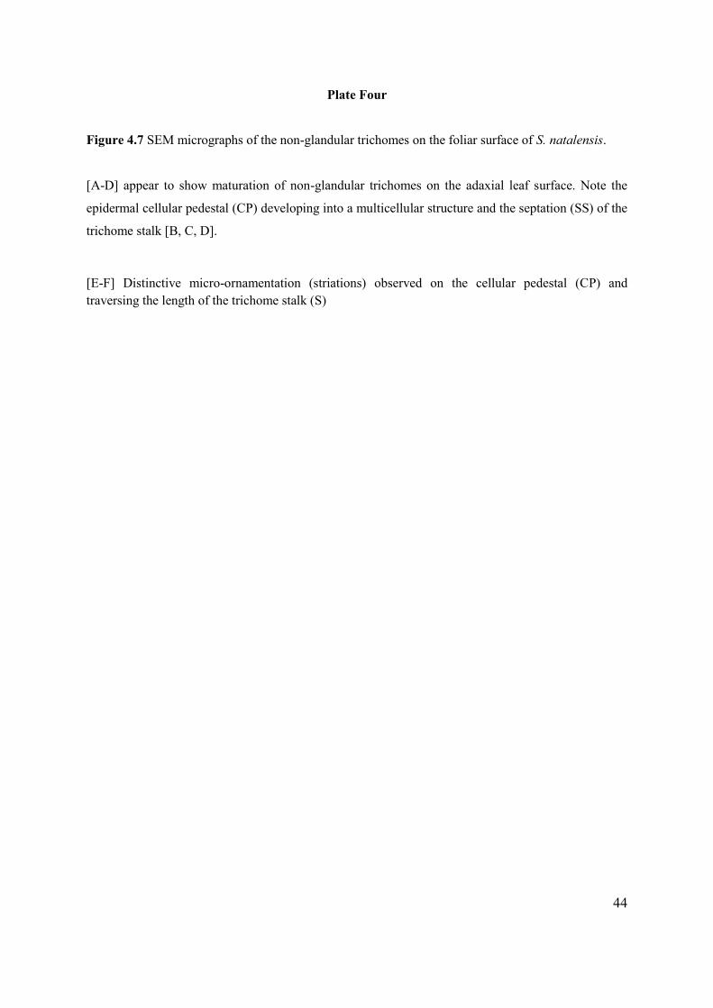
44
Plate Four
Figure 4.7 SEM micrographs of the non-glandular trichomes on the foliar surface of S. natalensis.
[A-D] appear to show maturation of non-glandular trichomes on the adaxial leaf surface. Note the
epidermal cellular pedestal (CP) developing into a multicellular structure and the septation (SS) of the
trichome stalk [B, C, D].
[E-F] Distinctive micro-ornamentation (striations) observed on the cellular pedestal (CP) and traversing the length of the trichome stalk (S)
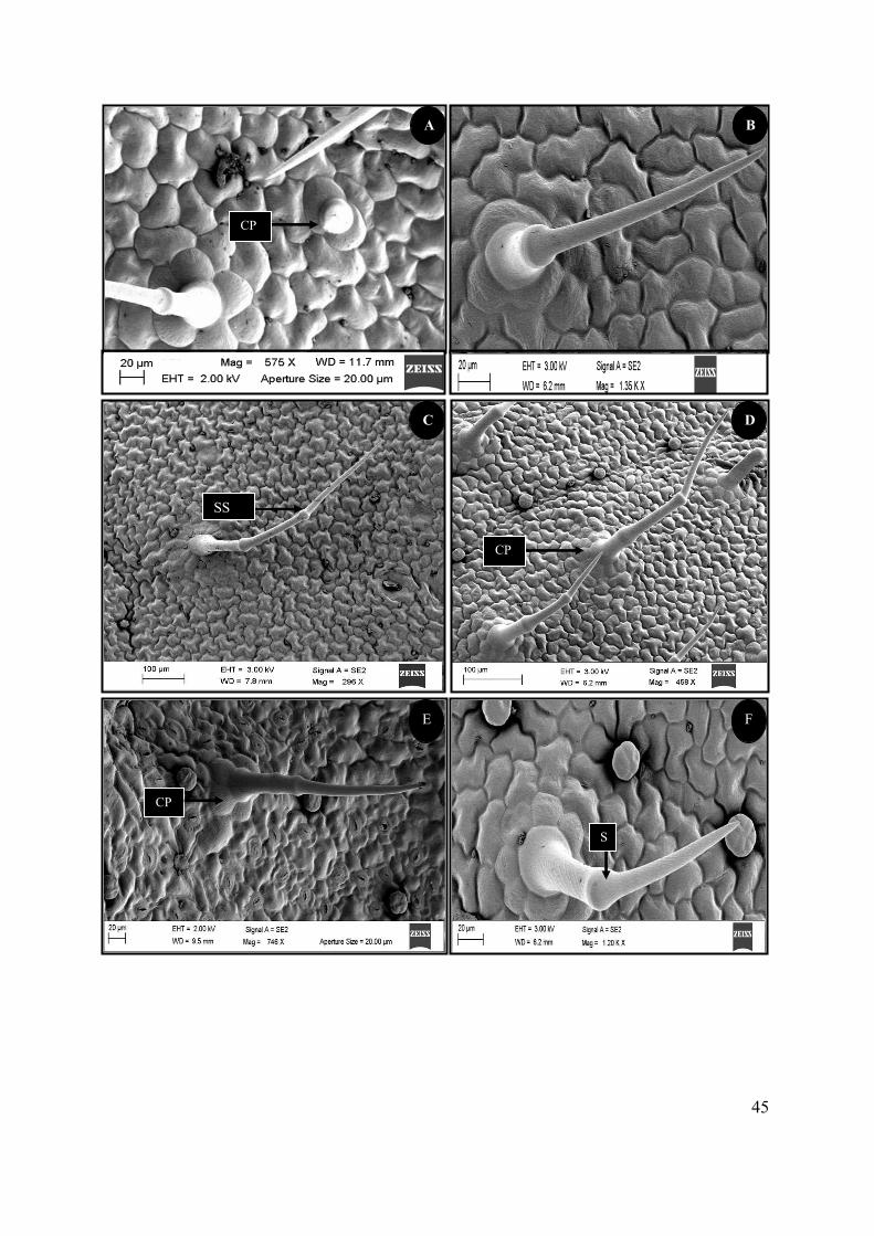
45
E F
CP
S
C
B
D
CP
CP
A
SS
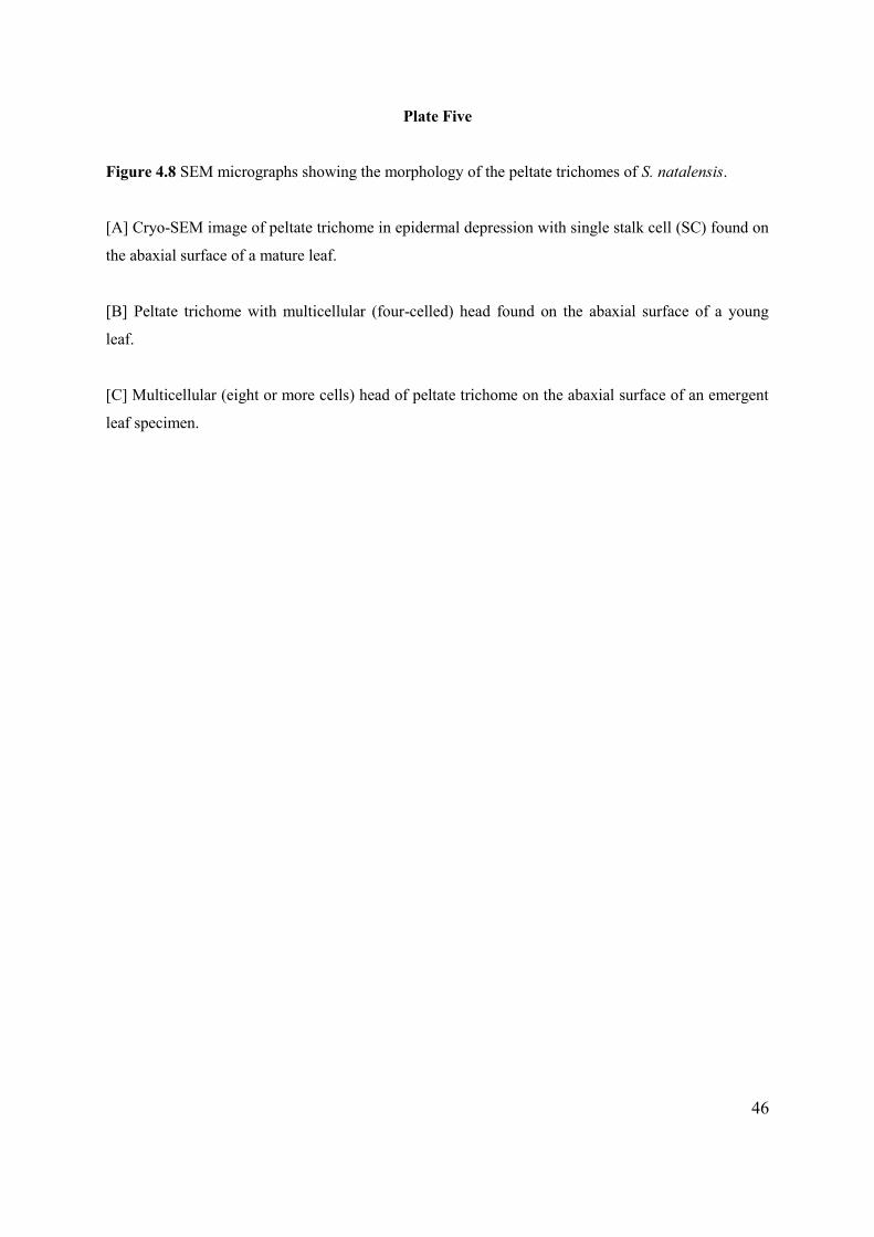
46
Plate Five
Figure 4.8 SEM micrographs showing the morphology of the peltate trichomes of S. natalensis.
[A] Cryo-SEM image of peltate trichome in epidermal depression with single stalk cell (SC) found on
the abaxial surface of a mature leaf.
[B] Peltate trichome with multicellular (four-celled) head found on the abaxial surface of a young
leaf.
[C] Multicellular (eight or more cells) head of peltate trichome on the abaxial surface of an emergent
leaf specimen.
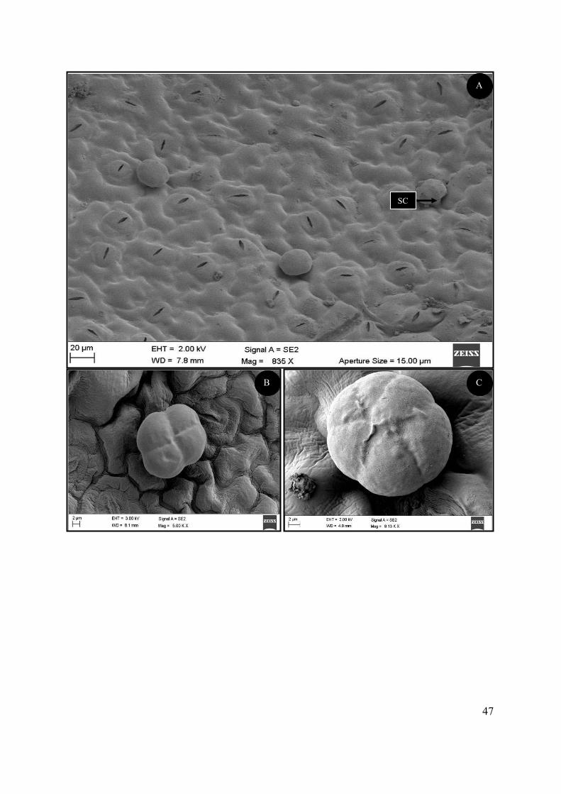
47
SC
A
C B
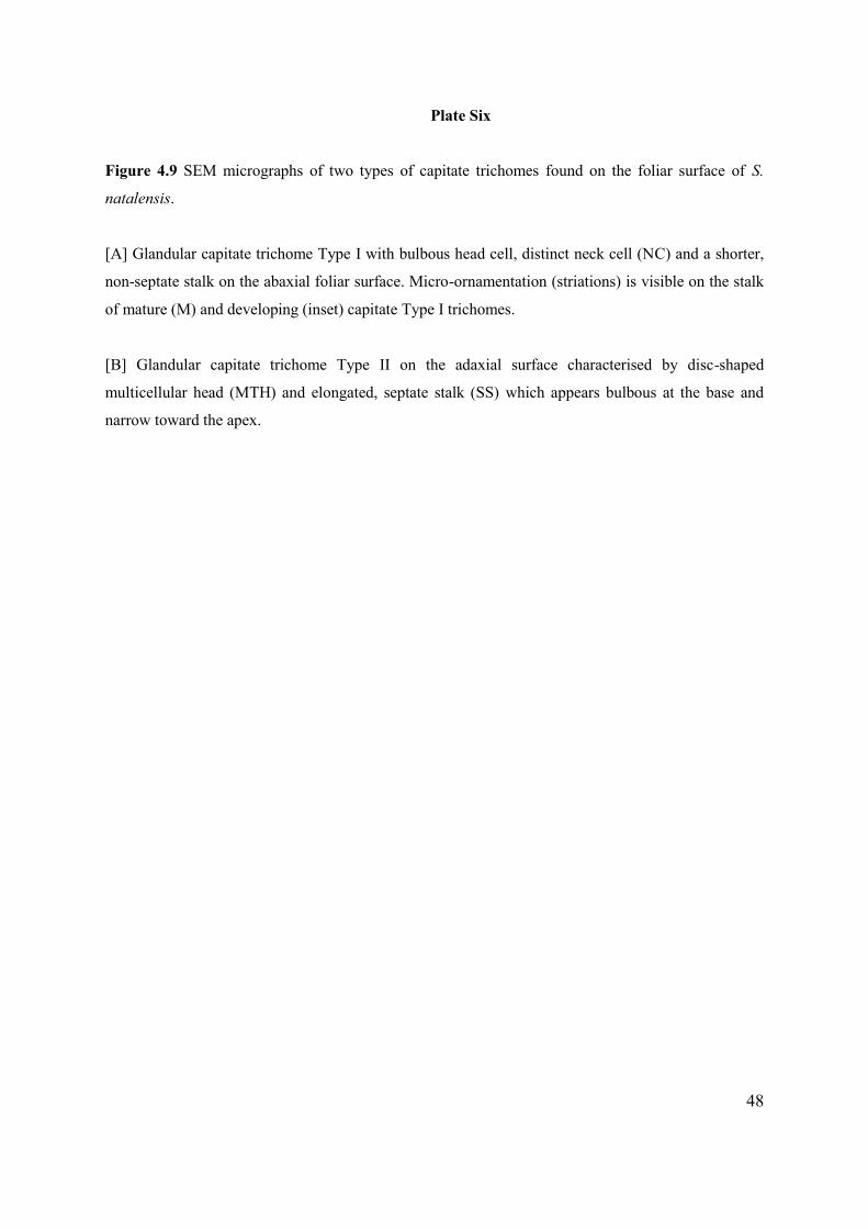
48
Plate Six
Figure 4.9 SEM micrographs of two types of capitate trichomes found on the foliar surface of S.
natalensis.
[A] Glandular capitate trichome Type I with bulbous head cell, distinct neck cell (NC) and a shorter,
non-septate stalk on the abaxial foliar surface. Micro-ornamentation (striations) is visible on the stalk
of mature (M) and developing (inset) capitate Type I trichomes.
[B] Glandular capitate trichome Type II on the adaxial surface characterised by disc-shaped
multicellular head (MTH) and elongated, septate stalk (SS) which appears bulbous at the base and
narrow toward the apex.
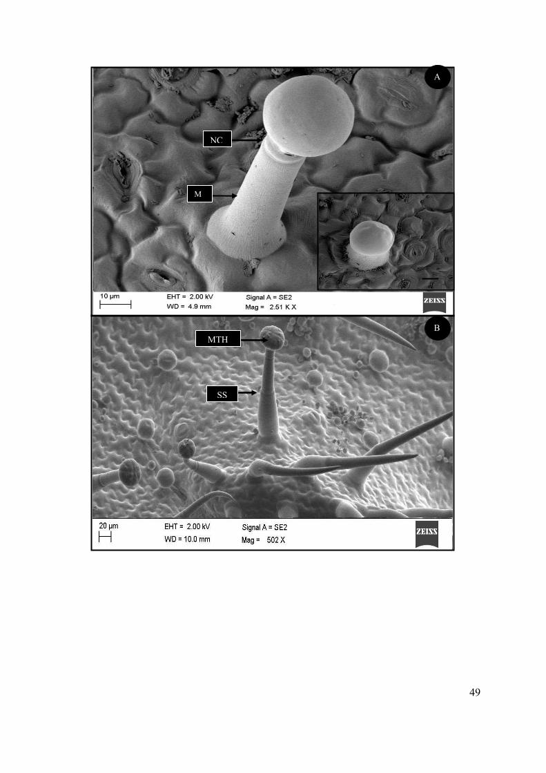
49
NC
M
A
B MTH
SS
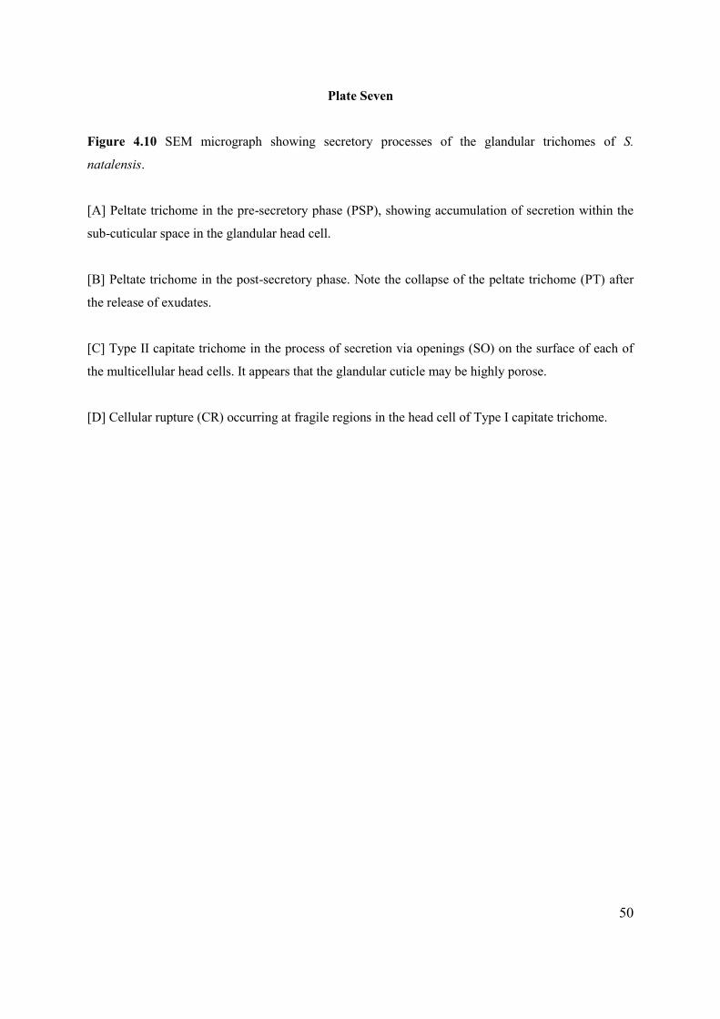
50
Plate Seven
Figure 4.10 SEM micrograph showing secretory processes of the glandular trichomes of S.
natalensis.
[A] Peltate trichome in the pre-secretory phase (PSP), showing accumulation of secretion within the
sub-cuticular space in the glandular head cell.
[B] Peltate trichome in the post-secretory phase. Note the collapse of the peltate trichome (PT) after
the release of exudates.
[C] Type II capitate trichome in the process of secretion via openings (SO) on the surface of each of
the multicellular head cells. It appears that the glandular cuticle may be highly porose.
[D] Cellular rupture (CR) occurring at fragile regions in the head cell of Type I capitate trichome.
51
A
C
B
PSP PT
SO CR
D
52
4.3. HISTOCHEMISTRY AND PHYTOCHEMISTRY Histochemical analyses were conducted on fresh-hand and de-waxed young leaf material showing the
localisation of a variety of secondary metabolites in the glandular head cells and cell wall constituents
(Figure 4.11).
Alkaloids were detected in the head cells of peltate and Type I capitate trichomes using the Wagner
and Dittmar stain (Figure 4.11 A). Positive results in the peltate glands include acidic polysaccharides
such as mucilage and pectin identified after staining with ruthenium red (Figure 4.11 B); ferric
trichloride positively stained phenolic compounds greenish-black (Figure 4.11 C); mercuric
bromophenol blue stained proteins (Figure 4.11 D); nadi reagent stained terpenoids (such as
oleoresins) red (Figure 4.11 E). Toluidine Blue stained carboxylated polysaccharides pink &
polyphenols blue-purple in glandular head cells (Figure 4.11 F). Acidic lipids were stained blue in
peltate glands by the nile blue stain (Figure 4.11 G). Sudan III was also used to detect lipid molecules
in the head cells of the capitate trichomes (Figure 4.11 H) and the presence of suberin or lignin in
trichome cell wall components.
Phytochemical tests were performed to determine the main classes of secondary metabolites present in
the crude extract of S. natalensis which may give a preliminary indication of the therapeutic potential
of the plant extract. Hexane was found to be the best solvent for phytochemical extraction as it
contained the greatest number of reactive compounds. The leaf extract of S. natalensis contained
many phytocompounds including: complex carbohydrates, alkaloids, coumarins, phenolics and
terpenoids which were detected at high intensities and quinones which showed a slightly weaker
reaction (Table 4.2). Total phenolic and flavonoid content in the leaves of S. natalensis were 3.43 ±
0.01 mg GAE/g dry material and 3.04 ± 0.01 mg QUE/g dry material, respectively. The radical
scavenging activity of S. natalensis extract is shown in Figure 4.12. Scavenging activity increases in a
dose-dependent manner. The IC50 value being the effective concentration to induce a 50% scavenging
activity of the DPPH radical of S. natalensis extract and ascorbic acid was determined (Table 4.3).
The IC50 value is negatively correlated with antioxidant potential.

53
Plate Eight
Figure 4.11 Histochemical staining reactions using semi-thin fresh-hand (A,C,E and H) and dewaxed
(B,D, F and G) leaf sections.
[A] Wagner/Dittmar reagent stained alkaloids brown in head cell of long stalked capitate trichome.
Bar: 50 µm.
[B] Ruthenium red stained positively for mucilage & pectin in multicellular head of peltate trichome
(pink-red). Bar: 25 µm.
[C] Ferric trichloride stained phenolic deposits greenish-black in peltate trichome head cell (arrow).
Bar: 25 µm.
[D] Mercuric bromophenol blue stained proteins in peltate head cells positively (blue). Bar: 50 µm.
[E] Positive Nadi reaction in head cells of peltate trichome (red) for the presence of oleoresins. Bar:
50 µm.
[F] Toluidine Blue stained carboxylated polysaccharides pink & polyphenols blue-purple in glandular
head cell. Bar: 50 µm.
[G] Positive detection of acidic lipids in peltate glands (blue) by nile blue stain. Bar: 20 µm.
[H] Sudan III stained lipids orange in head cell of long stalked capitate trichome. Bar: 50 µm.
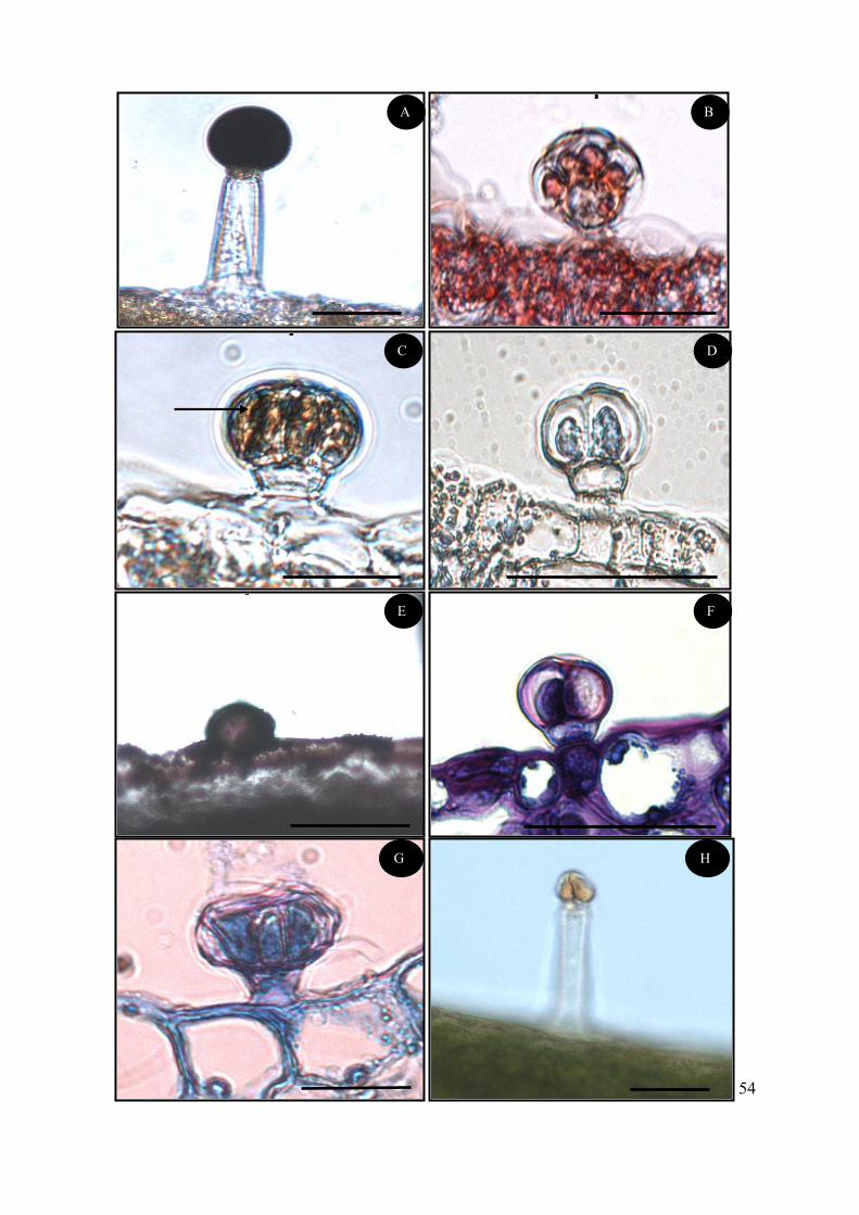
54
A B
D
E
F
C
G H
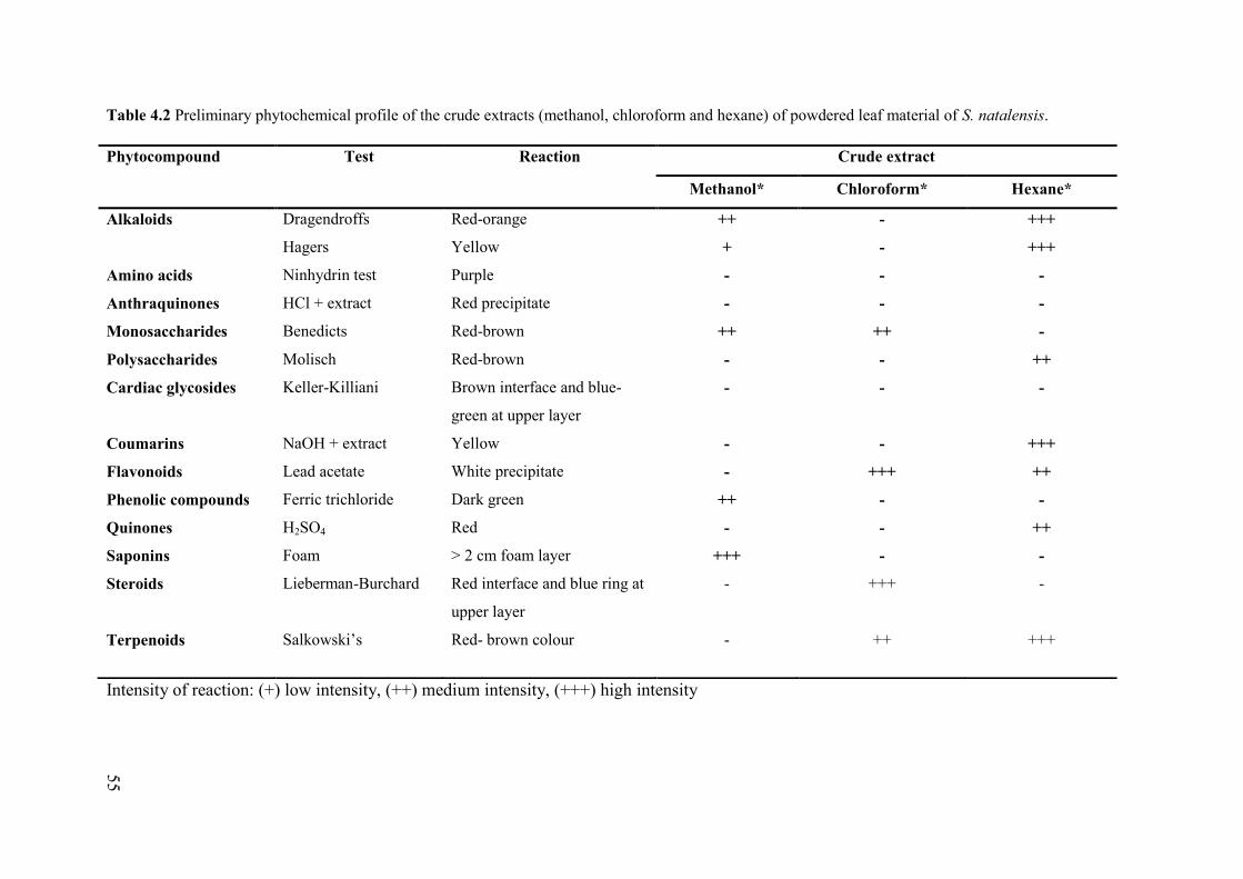
55
Table 4.2 Preliminary phytochemical profile of the crude extracts (methanol, chloroform and hexane) of powdered leaf material of S. natalensis.
Intensity of reaction: (+) low intensity, (++) medium intensity, (+++) high intensity
Phytocompound Test Reaction Crude extract
Methanol* Chloroform* Hexane*
Alkaloids Dragendroffs
Hagers
Red-orange
Yellow
++
+
-
-
+++
+++
Amino acids Ninhydrin test Purple - - -
Anthraquinones HCl + extract Red precipitate - - -
Monosaccharides Benedicts Red-brown ++ ++ -
Polysaccharides Molisch Red-brown - - ++
Cardiac glycosides Keller-Killiani Brown interface and blue-
green at upper layer
- - -
Coumarins NaOH + extract Yellow - - +++
Flavonoids Lead acetate White precipitate - +++ ++
Phenolic compounds Ferric trichloride Dark green ++ - -
Quinones H2SO4 Red - - ++
Saponins Foam > 2 cm foam layer +++ - -
Steroids Lieberman-Burchard Red interface and blue ring at
upper layer
- +++ -
Terpenoids Salkowski’s Red- brown colour - ++ +++
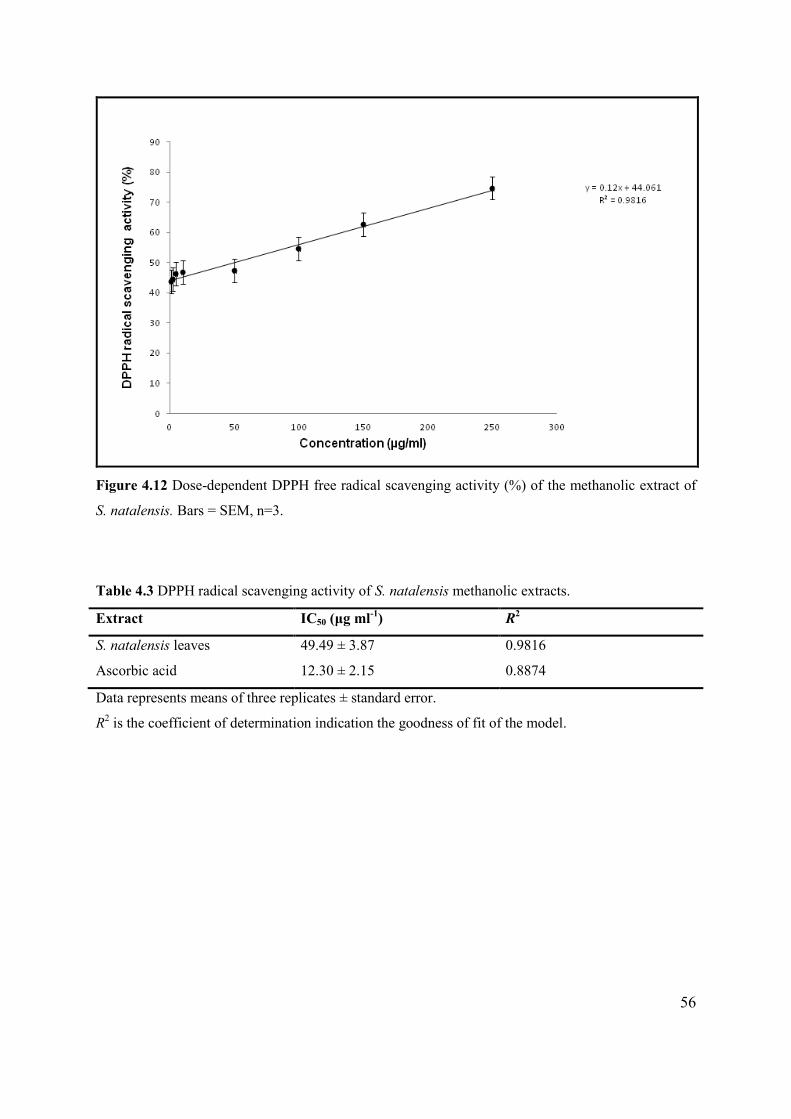
56
Figure 4.12 Dose-dependent DPPH free radical scavenging activity (%) of the methanolic extract of
S. natalensis. Bars = SEM, n=3.
Table 4.3 DPPH radical scavenging activity of S. natalensis methanolic extracts.
Extract IC50 (μg ml-1) R2
S. natalensis leaves 49.49 ± 3.87 0.9816
Ascorbic acid 12.30 ± 2.15 0.8874
Data represents means of three replicates ± standard error.
R2 is the coefficient of determination indication the goodness of fit of the model.
57
4.4. MICROPROPAGATION OF STACHYS NATALENSIS
In the present study, axillary bud segments (1 - 2 cm) were used as explant material for the in vitro
micropropagation of S. natalensis. Due to the high levels of explant infection, various
decontamination protocols were tested for the introduction of aseptic explants into culture. The effect
of different PGRs on initiation and multiplication of S. natalensis plantlets were evaluated and
microshoots from the most successful treatment were placed onto selected media for rooting and
eventually transferred to potting soil for acclimitization. The morphology and trichome density of in
vitro regenerated plantlets were compared to field-grown plants.
4.4.1. Decontamination and culture establishment Explant material in culture showed a high level of microbial (both fungal and bacterial) growth which
required stringent decontamination treatments (Table 3.1). Low explant survival was noted in
treatments that involved low concentrations of NaClO, short immersion intervals and only one type of
decontaminating agent. Explant decontamination was accomplished by immersion in 1% NaClO and
3% NaClO for seven minutes and five minutes respectively, followed by immersion in 0.1% HgCl2 for
ten minutes. This protocol resulted in 48 ± 5% survival of explants and was the only significantly
different treatment with regard to explant survival when compared to other decontamination
treatments (F(4,15) =18.353; p<0.05; Figure 4.13). This treatment was employed for the initiation of
new cultures as well as to revitalise established cultures showing early signs of contamination (± two
weeks post-initiation). The percent bud break of initiated cultures in each of the three media
preparations is shown in Table 4.4. All media preparations resulted in > 70 ± 6% successful bud break
within three weeks. Statistical analyses indicated no significant differences among the three media
preparations.
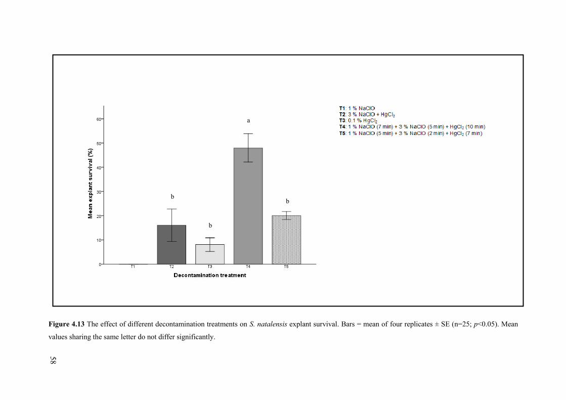
58
Figure 4.13 The effect of different decontamination treatments on S. natalensis explant survival. Bars = mean of four replicates ± SE (n=25; p<0.05). Mean
values sharing the same letter do not differ significantly.
b
b
b
a

59
Figure 4.14 Initiation of S. natalensis in in vitro culture.
[A] Healthy axillary nodal explants cultured on initiation medium to induce bud break. (Bar: 10 mm)
[B] Explants with several shoots formed on initiation medium (BAP + IBA) before subculture onto
multiplication media for shoot multiplication. (Bar: 5 mm)
Table 4.4 The effect of different plant growth regulators on successful initiation of axillary buds of S.
natalensis explants after two weeks in vitro, n=25.
Media PGR combination % Bud break ± SE
A 0.5 mg/l BAP + 0.5 mg/l IAA 78 ± 2a
B 0.5 mg/l Kinetin + 0.5 mg/l IAA 70 ± 6a
C 0.5 mg/l BAP + 0.5 mg/l IBA 80 ± 8a
Mean values sharing the same letter within a column do not differ significantly (p<0.05) according to
Tukey’s post-hoc test.
A B

60
4.4.2. Shoot multiplication Growth medium B (kinetin and IAA) showed the greatest total shoot multiplication and elongation
(following the bud break stage) compared to other PGR combinations after 12 weeks (Figure 4.15 and
4.16). Mean shoot production per explant and shoot height per explant was assessed using a one-way
ANOVA (Table 4.5). There were significant differences in the mean number of shoots produced per
explant between cultures in media A and B as well as between media B and C (F(2, 27)= 11.829;
p<0.05). Plantlets in multiplication medium B showed the greatest shoot production at 9.1 ± 3.6
shoots/explant. Significant differences were noted in the height of shoots per explant between cultures
in Media A and B as well as between Media B and C (F(2, 27) = 34.886; p< 0.05). The greatest mean
shoot height of 50.2 ± 5.0 mm was obtained in plantlets grown on medium B.
4.2.3. Rooting and Acclimatization Plantlets showed significant shoot multiplication and elongation on medium B compared to other
growth media and was transferred to medium containing only the PGR IAA (2 mg/l) in order to
promote adventitious root formation. Rooting occurred in 64% of plantlets after five weeks. The
average number of roots formed per shoot was 5.54 ± 0.43 and the mean root length per shoot was
66.13 ± 3.55 mm. Plantlets with a well-developed root system and root length > 30 mm were selected
for acclimatization (Figure 4.17). The 24 in vitro propagated S. natalensis plantlets placed under
greenhouse conditions showed 92 ± 4.2% survival after six weeks, indicating the efficiency of the
micropropagation protocol to form robust, easily acclimatized plantlets with no apparent
morphological abnormalities compared to field-grown plants (Figure 4.18).
61
Plate Nine
Figure 4.15 Shoot multiplication of S. natalensis axillary nodal explants in media containing different
combinations of cytokinins and auxins after six weeks in culture.
[A] Medium A supplemented with 2 mg/l BAP and 1 mg/l IAA showing relatively slow shoot
multiplication (Bar: 10 mm).
[B] Medium B supplemented with 2 mg/l kinetin and 1 mg/l IAA showing significant multiplication
and elongation of healthy microshoots (Bar: 10 mm).
[C] Medium C supplemented with 2 mg/l BAP and 1 mg/l IBA showing significant microshoot
multiplication in plantlets. Elongation of plantlets appears to be limited compared to medium B at the
same stage. (Bar: 10 mm)
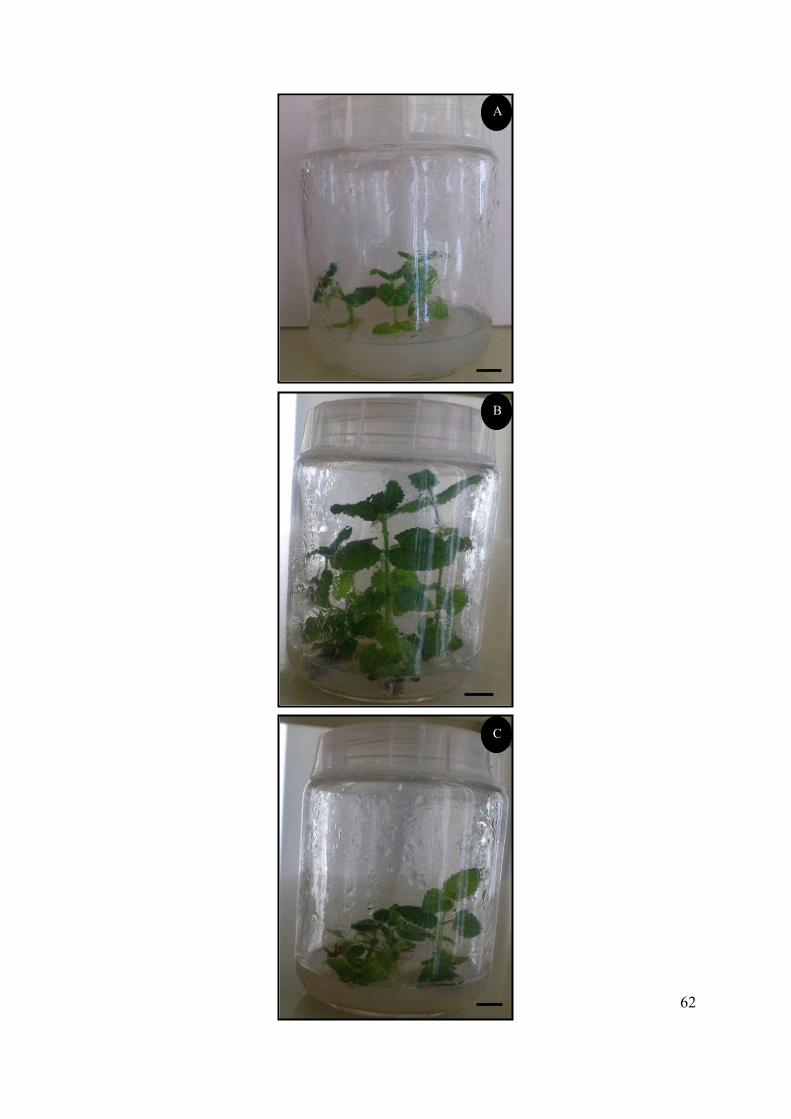
62
A
B
C
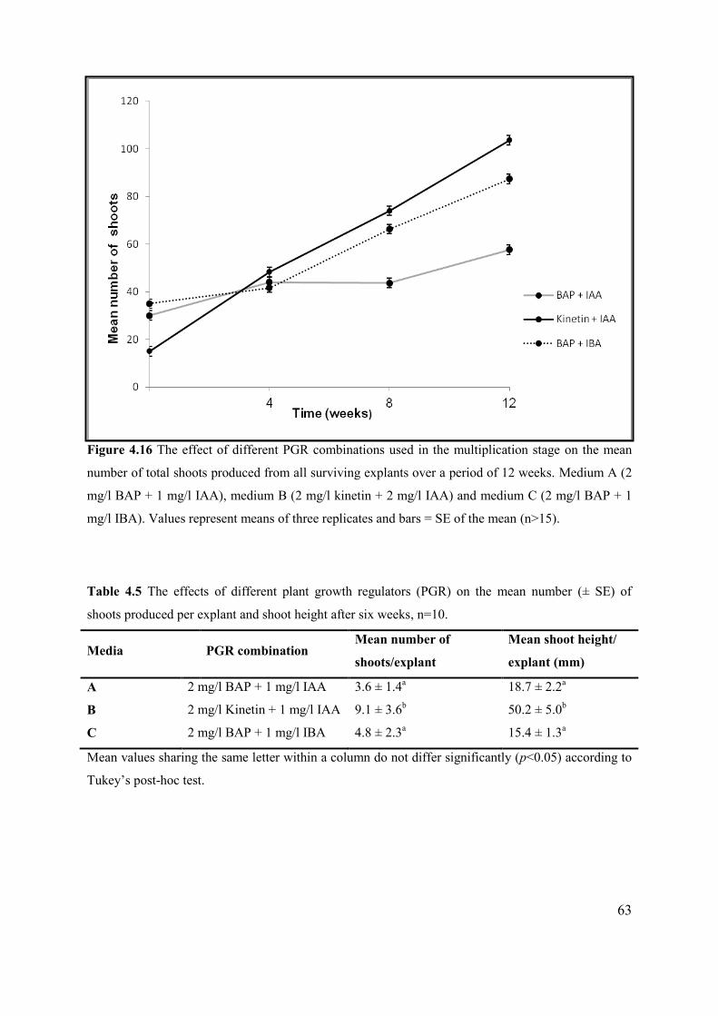
63
Figure 4.16 The effect of different PGR combinations used in the multiplication stage on the mean
number of total shoots produced from all surviving explants over a period of 12 weeks. Medium A (2
mg/l BAP + 1 mg/l IAA), medium B (2 mg/l kinetin + 2 mg/l IAA) and medium C (2 mg/l BAP + 1
mg/l IBA). Values represent means of three replicates and bars = SE of the mean (n>15).
Table 4.5 The effects of different plant growth regulators (PGR) on the mean number (± SE) of
shoots produced per explant and shoot height after six weeks, n=10.
Media PGR combination Mean number of
shoots/explant
Mean shoot height/
explant (mm)
A 2 mg/l BAP + 1 mg/l IAA 3.6 ± 1.4a 18.7 ± 2.2a
B 2 mg/l Kinetin + 1 mg/l IAA 9.1 ± 3.6b 50.2 ± 5.0b
C 2 mg/l BAP + 1 mg/l IBA 4.8 ± 2.3a 15.4 ± 1.3a
Mean values sharing the same letter within a column do not differ significantly (p<0.05) according to
Tukey’s post-hoc test.
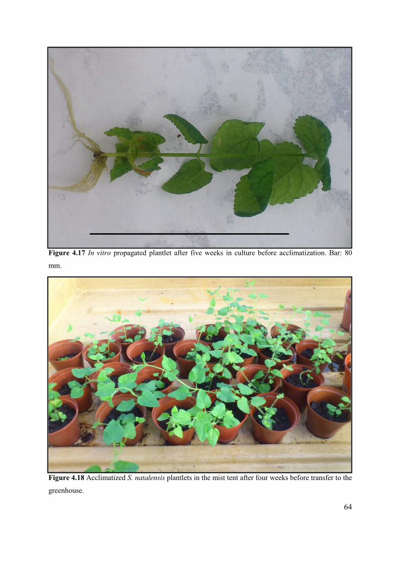
64
Figure 4.17 In vitro propagated plantlet after five weeks in culture before acclimatization. Bar: 80
mm.
Figure 4.18 Acclimatized S. natalensis plantlets in the mist tent after four weeks before transfer to the
greenhouse.
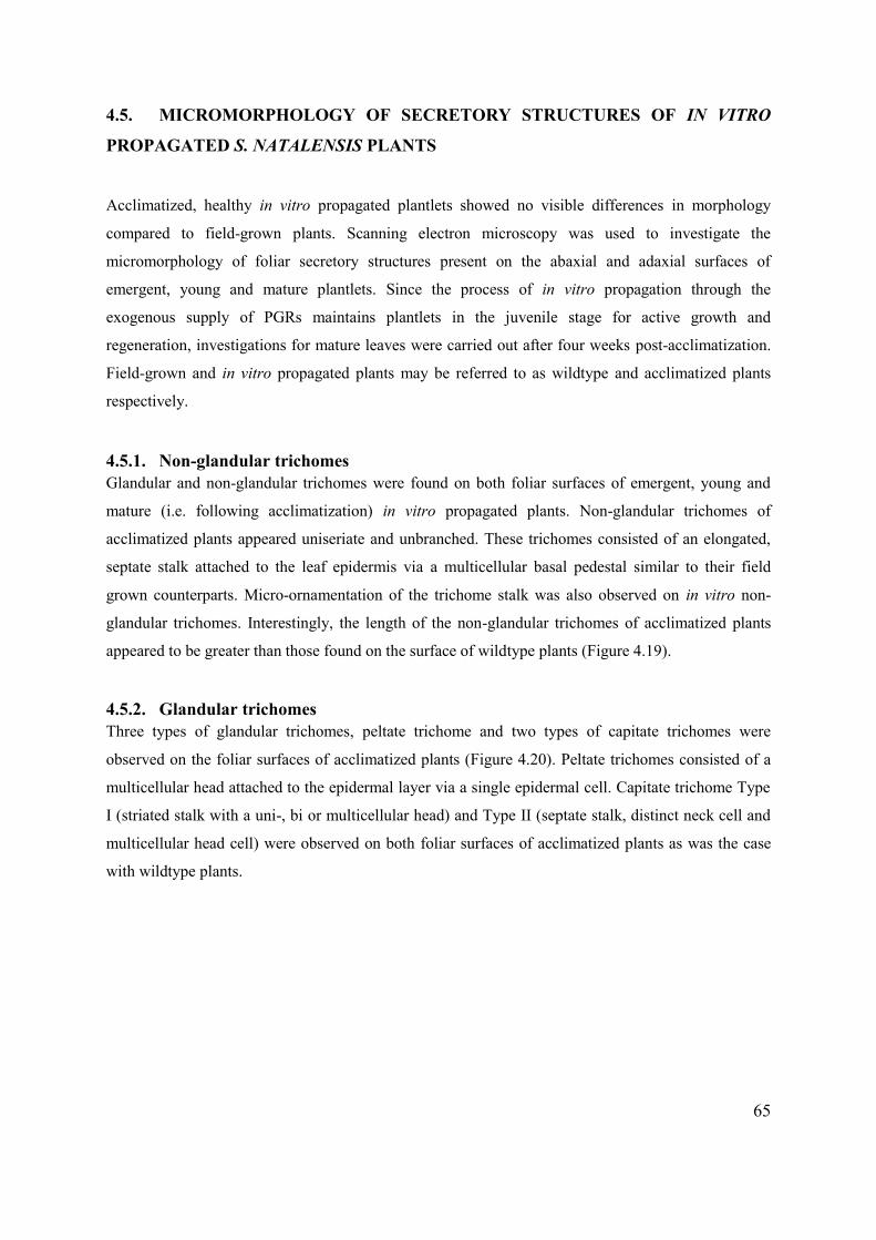
65
4.5. MICROMORPHOLOGY OF SECRETORY STRUCTURES OF IN VITRO
PROPAGATED S. NATALENSIS PLANTS
Acclimatized, healthy in vitro propagated plantlets showed no visible differences in morphology
compared to field-grown plants. Scanning electron microscopy was used to investigate the
micromorphology of foliar secretory structures present on the abaxial and adaxial surfaces of
emergent, young and mature plantlets. Since the process of in vitro propagation through the
exogenous supply of PGRs maintains plantlets in the juvenile stage for active growth and
regeneration, investigations for mature leaves were carried out after four weeks post-acclimatization.
Field-grown and in vitro propagated plants may be referred to as wildtype and acclimatized plants
respectively.
4.5.1. Non-glandular trichomes Glandular and non-glandular trichomes were found on both foliar surfaces of emergent, young and
mature (i.e. following acclimatization) in vitro propagated plants. Non-glandular trichomes of
acclimatized plants appeared uniseriate and unbranched. These trichomes consisted of an elongated,
septate stalk attached to the leaf epidermis via a multicellular basal pedestal similar to their field
grown counterparts. Micro-ornamentation of the trichome stalk was also observed on in vitro non-
glandular trichomes. Interestingly, the length of the non-glandular trichomes of acclimatized plants
appeared to be greater than those found on the surface of wildtype plants (Figure 4.19).
4.5.2. Glandular trichomes Three types of glandular trichomes, peltate trichome and two types of capitate trichomes were
observed on the foliar surfaces of acclimatized plants (Figure 4.20). Peltate trichomes consisted of a
multicellular head attached to the epidermal layer via a single epidermal cell. Capitate trichome Type
I (striated stalk with a uni-, bi or multicellular head) and Type II (septate stalk, distinct neck cell and
multicellular head cell) were observed on both foliar surfaces of acclimatized plants as was the case
with wildtype plants.
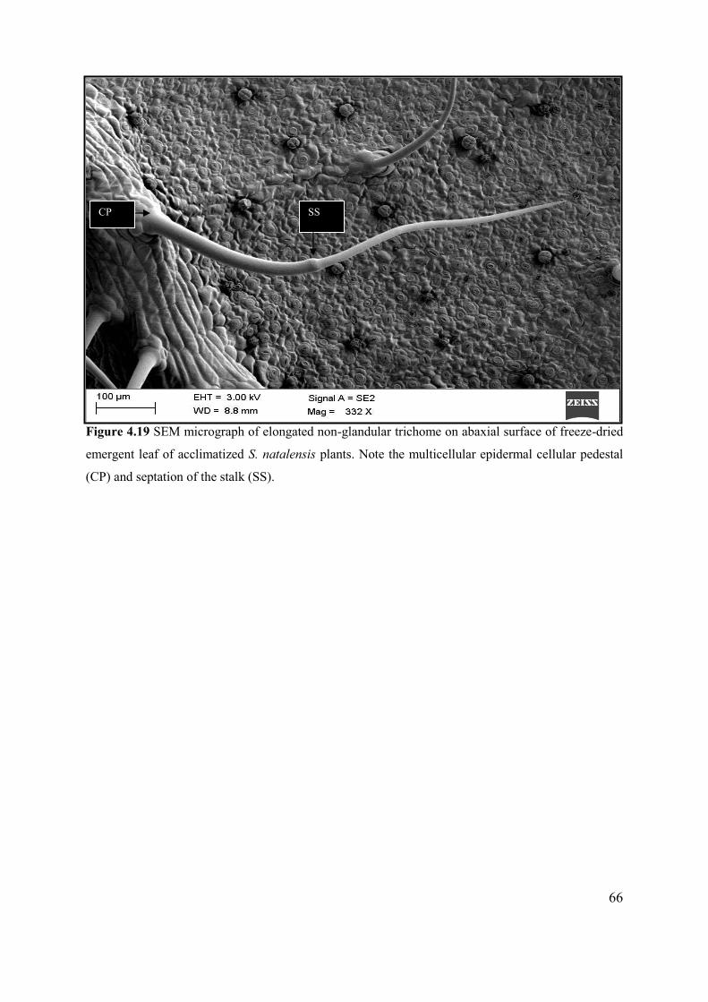
66
Figure 4.19 SEM micrograph of elongated non-glandular trichome on abaxial surface of freeze-dried
emergent leaf of acclimatized S. natalensis plants. Note the multicellular epidermal cellular pedestal
(CP) and septation of the stalk (SS).
CP SS
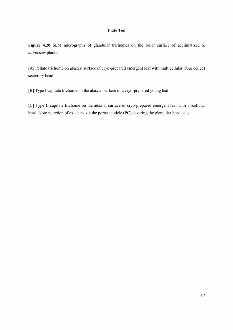
67
Plate Ten
Figure 4.20 SEM micrographs of glandular trichomes on the foliar surface of acclimatized S.
natalensis plants.
[A] Peltate trichome on abaxial surface of cryo-prepared emergent leaf with multicellular (four celled)
secretory head.
[B] Type I capitate trichome on the abaxial surface of a cryo-prepared young leaf.
[C] Type II capitate trichome on the adaxial surface of cryo-prepared emergent leaf with bi-cellular
head. Note secretion of exudates via the porose cuticle (PC) covering the glandular head cells.
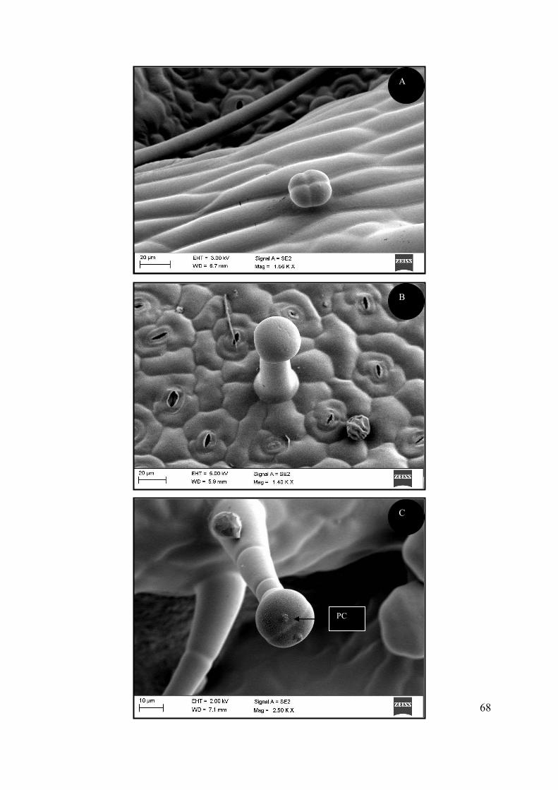
68
PC
C
A
B
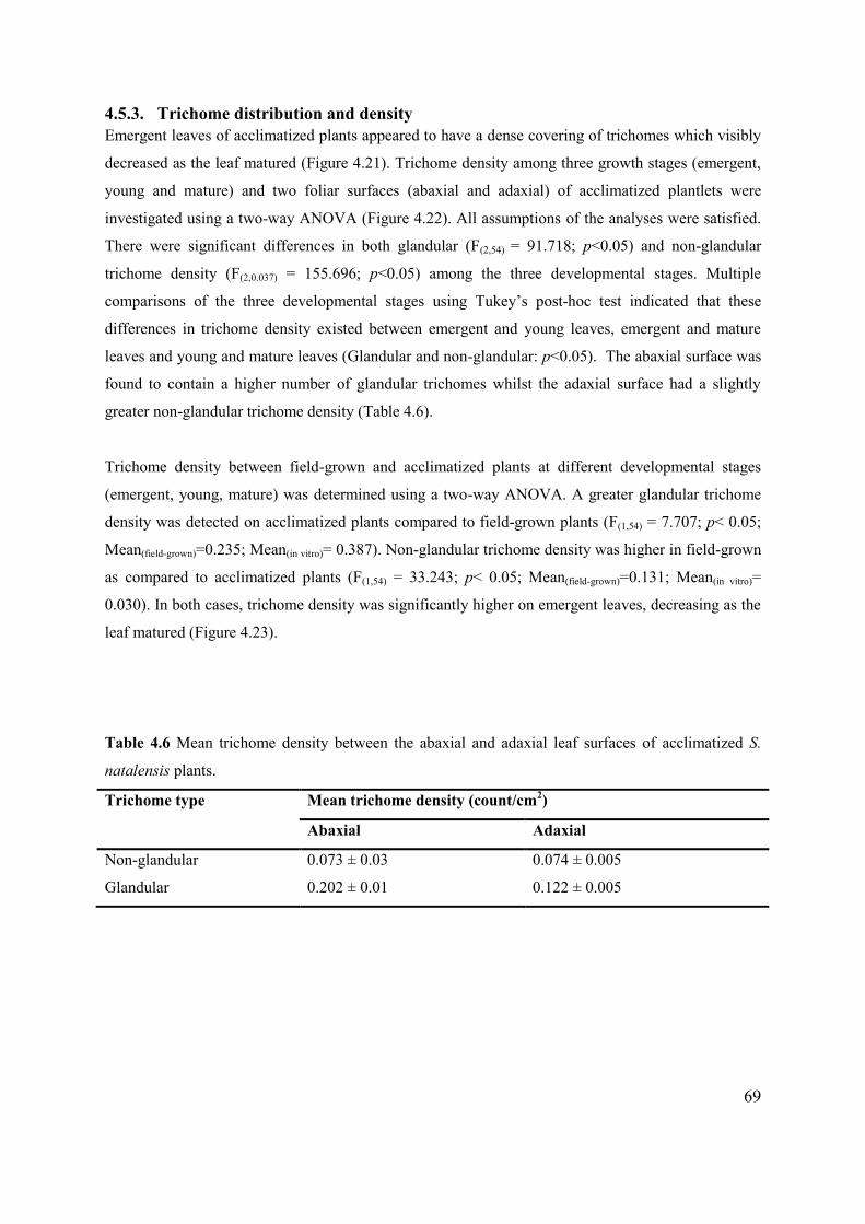
69
4.5.3. Trichome distribution and density Emergent leaves of acclimatized plants appeared to have a dense covering of trichomes which visibly
decreased as the leaf matured (Figure 4.21). Trichome density among three growth stages (emergent,
young and mature) and two foliar surfaces (abaxial and adaxial) of acclimatized plantlets were
investigated using a two-way ANOVA (Figure 4.22). All assumptions of the analyses were satisfied.
There were significant differences in both glandular (F(2,54) = 91.718; p<0.05) and non-glandular
trichome density (F(2,0.037) = 155.696; p<0.05) among the three developmental stages. Multiple
comparisons of the three developmental stages using Tukey’s post-hoc test indicated that these
differences in trichome density existed between emergent and young leaves, emergent and mature
leaves and young and mature leaves (Glandular and non-glandular: p<0.05). The abaxial surface was
found to contain a higher number of glandular trichomes whilst the adaxial surface had a slightly
greater non-glandular trichome density (Table 4.6).
Trichome density between field-grown and acclimatized plants at different developmental stages
(emergent, young, mature) was determined using a two-way ANOVA. A greater glandular trichome
density was detected on acclimatized plants compared to field-grown plants (F(1,54) = 7.707; p< 0.05;
Mean(field-grown)=0.235; Mean(in vitro)= 0.387). Non-glandular trichome density was higher in field-grown
as compared to acclimatized plants (F(1,54) = 33.243; p< 0.05; Mean(field-grown)=0.131; Mean(in vitro)=
0.030). In both cases, trichome density was significantly higher on emergent leaves, decreasing as the
leaf matured (Figure 4.23).
Table 4.6 Mean trichome density between the abaxial and adaxial leaf surfaces of acclimatized S.
natalensis plants.
Trichome type Mean trichome density (count/cm2)
Abaxial Adaxial
Non-glandular 0.073 ± 0.03 0.074 ± 0.005
Glandular 0.202 ± 0.01 0.122 ± 0.005

70
Plate Eleven
Figure 4.21 SEM micrographs showing trichome distribution on the adaxial surface of acclimatized
S. natalensis leaves at three developmental stages.
[A] Emergent, [B] young and [C] mature leaves show the presence of both non-glandular and
glandular trichomes. Trichomes appear to be more densely distributed in younger leaves, compared
with mature leaves.

71
C
A
B

72
Plate Twelve
Figure 4.22 Mean non-glandular [A] and glandular [B] trichome density at three stages of leaf
development (emergent, young and mature) and on the abaxial and adaxial foliar surfaces of
acclimatized S. natalensis plants.
Values are mean ± SE (n= 20; p< 0.05 was recognised as significantly different using Tukey’s
multiple comparison post-hoc test and denoted by different letters).
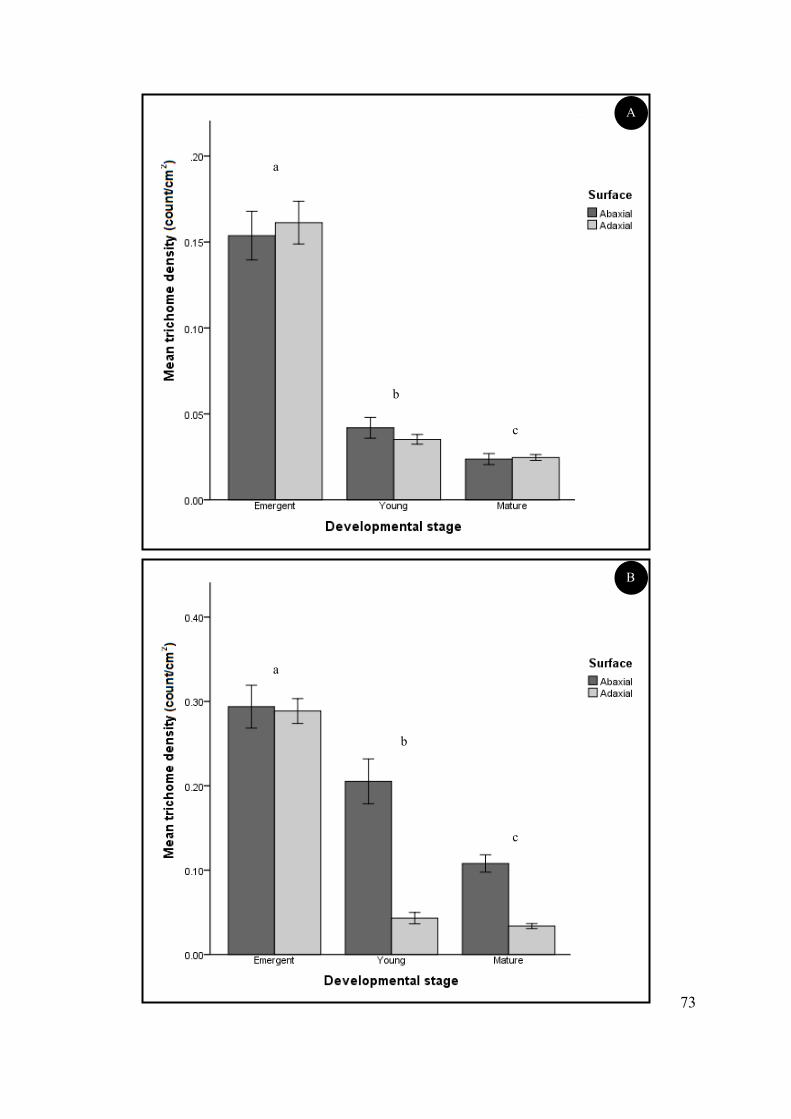
73
A
B
a
b
c
a
c
b
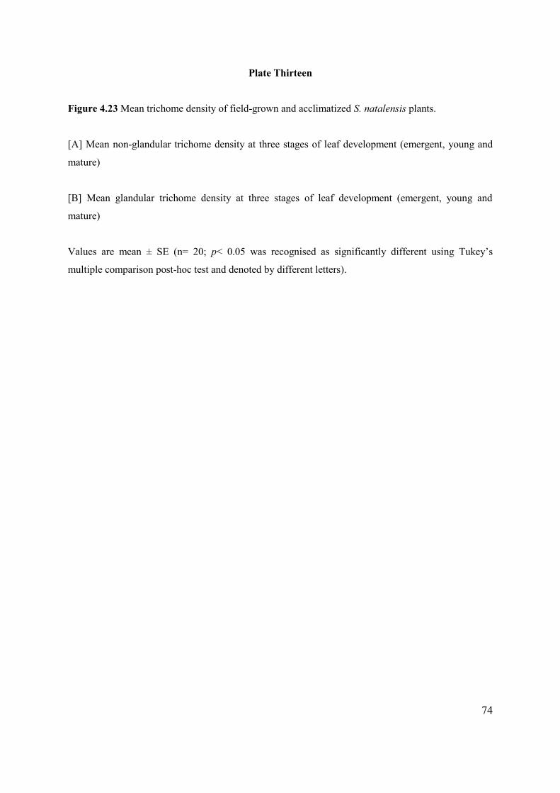
74
Plate Thirteen
Figure 4.23 Mean trichome density of field-grown and acclimatized S. natalensis plants.
[A] Mean non-glandular trichome density at three stages of leaf development (emergent, young and
mature)
[B] Mean glandular trichome density at three stages of leaf development (emergent, young and
mature)
Values are mean ± SE (n= 20; p< 0.05 was recognised as significantly different using Tukey’s
multiple comparison post-hoc test and denoted by different letters).
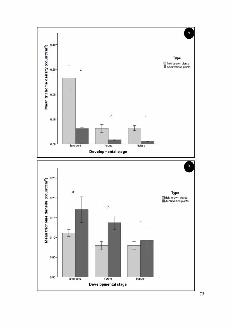
75
A
B
a
b b
a
a,b
b

CHAPTER 5: DISCUSSION
76
This study was undertaken to investigate the micromorphology of secretory structures found on the
foliar surfaces of Stachys natalensis Hochst. and the histo- and phytochemical characteristics of the
exudates. In addition, an efficient micropropagation protocol for the species was developed and the
micromorphology of in vitro propagated plants was assessed.
5.1. TRICHOME DISTRIBUTION AND MICROMORPHOLOGY
The indumentum of Stachys natalensis Hochst contains a spread of both glandular (secretory) and
non-glandular trichomes on both foliar surfaces, as seen in most species belonging to the Lamiaceae
family (Werker et al., 1993; Bhatt et al., 2010a; Rusydi et al., 2013). In the present study, glandular
and non-glandular trichomes were more densely distributed on the abaxial surface compared to the
adaxial surface with trichome density progressively decreasing with leaf maturation. Significantly
higher glandular trichome densities on the abaxial surface have also been observed in Trichosanthes
cucumerina (Adebooye et al., 2012), Artemisia annua (Kjaer et al., 2012) and Mentha spicata (Choi
and Kim, 2013) whilst higher non-glandular densities were noticed in Lamiaceaeous species such as
Dracocephalum moldavicum (Dmitruk and Weryszko-Chmielewska, 2010), Plectranthus laxiflorus
(Bhatt et al., 2010a) and Isodon rubescens (Liu and Liu, 2012), amongst others.
The phenomenon of leaf expansion has been used in the literature to account for the gradual decrease
in trichome density from young leaves to older specimens and is often reported in species of the
Lamiaceae (Ascensao et al., 1995; Valkama et al., 2004). It refers to the increase in the leaf surface
area as it matures whilst trichome density remains more or less constant. This results in a greater
dispersal of trichomes across the leaf surface. Valkama et al. (2004) reported that foliar trichomes of
some plants are present very early during leaf development, with trichome density remaining constant
throughout leaf growth. In other plants, new trichomes are produced throughout leaf development
(Maffei et al., 1989). Werker et al. (1993) hypothesized that trichome production would continue for
however long there are meristematic regions present. The question of trichome functionality at
different developmental stages is addressed by many contrasting studies. Werker (2000) showed that
trichomes are still functional in mature leaves whilst Gairola et al. (2009) reported that trichomes
often senesce and are lost in later stages of leaf development as they have little or no functional role.
However, the functional role of trichomes may be species-specific or depend on the location of
trichomes on different plant organs (Werker, 2000). The non-glandular trichomes of S. natalensis
were erect or bent toward the leaf apex with a high trichome density occurring on the mid-rib and
margin. The greater trichome distribution on younger leaves of S. natalensis suggested that trichomes
77
serve a protective function over their glandular counterparts during early development which may or
may not be required at later stages. Many studies show significant increases in trichome density upon
exposure to adverse conditions such as mechanical damage or herbivory (Molina-Montenegro et al.,
2006; Jaime et al., 2012; Loughner et al., 2012), water loss (Abdulrahaman and Oladele, 2011) and
environmental stresses (Bhatt et al., 2010b; Adebooye et al., 2012).
The non-glandular trichomes of S. natalensis were uniseriate and unbranched, appearing acuminate
(long, slender and tapering toward the apices) to arthrodachylous (jointed) in form. These were typical
of the non-glandular trichomes of other Stachys spp. found worldwide including: Stachys cretica L.
subsp. vacillans, S. ehrenbergii and S. distans from Lebanon (El-Beyrouthy et al., 2009), S.
ballotiformis and S. megalodonta from Iran (Salmaki et al., 2009) and S. recta from Italy (Giuliani et
al., 2008). Non-glandular trichomes of S. natalensis contained two distinctive morphological
attributes. The first was a multicellular basal pedestal which is composed of approximately two to ten
epidermal cells depending on the stage of differentiation. These raised cells serve to support the
trichome (Ascensao et al., 1999) and to provide a point of attachment that “anchors” the trichome to
the epidermal surface (Bhatt et al., 2010a). It is a characteristic trichome feature in many plants such
as Ceratotheca triloba (Naidoo et al., 2012a), Plectranthus tomentosa (Kim, 2013) and Stachys
alopecuros (Venditti et al., 2013a). Trichome development generally proceeds from a single, enlarged
and protruding protodermal cell to form uniseriate multicellular trichomes like those of S. natalensis,
after a series of periclinal divisions (Werker, 2000). The latter are a result of cell divisions that occur
parallel to the length of the pre-existing cell (Werker, 2000).
The second characteristic refers to unique striations occurring on the trichome surface of S. natalensis
from the basal pedestal up to and including the stalk. These are referred to as “micro-ornamentation”
and may appear as warts, papillae or striations arising from the cell wall or cuticle on the trichome
surface (Werker, 2000; Gairola et al., 2009; Rusydi et al., 2013). Interestingly, studies conducted on
trichome morphology of Stachys spp. by Salmaki et al. (2009) and El-Beyrouthy et al. (2009) indicate
that in most cases papillate micro-ornamentation is a characteristic feature. The striated trichomes of
S. natalensis may serve as a defining characteristic but further research is required to evaluate its
usefulness as a taxonomic feature. The function of such ornamentation remains unclear. Werker
(2000) suggested that micro-ornamentation serves as an indicator of trichome maturation. Striated
micro-ornamentation has been identified in trichomes on the young and mature S. natalensis leaf
surface which suggests that trichome production is maintained throughout leaf development but due to
leaf expansion trichome density decreases overall (Werker, 2000; Liu and Liu, 2012). Literature also
reports on the involvement of cuticular micro-ornamentation in a self-cleaning mechanism known as
78
the “Lotus Effect” which is attributed to the hydrophobic nature and structure of trichome surfaces
(Barthlott and Neinhuis, 1997). Trichome micro-ornamentation may aid in the removal of grime,
water or pathogenic microorganisms and compensate for the accumulation of particles due to the
rough leaf surfaces (Barthlott and Neinhuis, 1997). Naidoo et al. (2014) hypothesized that cuticular
micro-ornamentation may also facilitate the flow of exudates from secretory glands.
The structural and functional aspects of glandular trichomes within the Lamiaceae is well documented
(Werker et al., 1993; Serato-Valenti et al., 1997; Ascensao et al., 1998; Ascensao et al., 1999;
Giuliani and Maleci-Bini, 2008: Marin et al., 2012; Rusydi et al., 2013). The glandular hairs observed
on the foliar surfaces of S. natalensis are of two distinct types: peltate and capitate. Many Stachys
spp., much like other species within the family, do not display any uniformity with regard to the type
of glandular trichomes they possess. Foliar surfaces may be covered by both peltate and capitate
trichomes, only peltate, only capitate or in rare cases neither type (Huang et al., 2008). In fact, S.
plumosa, S. sylvatica, S. byzantina and S. recta subsp. recta have capitate trichomes only (Maleci-
Bini and Giuliani, 2006; El-Beyrouthy et al., 2009) whilst Stachys subgenus. Betonica possesses only
peltate glands (Giuliani and Maleci-Bini, 2012).
The peltate trichomes of S. natalensis were uniseriate and comprised of a basal cell, a very short
unicellular stalk and a multicellular secretory head (approximately 50 µm in diameter) that contains
two to eight secretory cells. Morphologically similar peltate trichomes were noted in other Stachys
spp. including; S. cretica, , S. neurocalycina, S. nivea and S. alopecuros (El-Beyrouthy et al., 2009;
Venditti et al., 2013a). Other species differed in terms of the number of secretory head cells (4-16
cells in S. officinalis) and the development of a fairly elongated (40 - 60 µm length) stalk cell, as seen
in S. germanica and related species Prasium majus, Sideritis romana and Scutellaria galericulata
(Giuliani and Maleci-Bini, 2008).
The Type I capitate trichomes of S. natalensis, widespread within the family Lamiaceae and genus
Stachys, are composed of a uniseriate stalk, neck cell and a bulbous secretory head (Werker, 2000).
They occur on both foliar surfaces across all developmental stages of S. natalensis and other species
including: Stachys palaestina (El-Beyrouthy et al., 2009), Salvia smyrnea (Baran et al., 2010) and
Ballota undulata (Osman, 2012). The Type II capitate trichome with an elongated striated stalk and
distinct neck cell bearing a variable number of secretory cells identified in this study shares
similarities with the capitate trichomes described in many species of Stachys, Sideritis, Prasium and
Scutellaria (Giuliani and Maleci-Bini, 2008). This Type II capitate trichome was generally found only
on the plant reproductive organs of the Lamiaceae (Giuliani and Maleci-Bini, 2008). However,

79
Giuliani et al. (2011) later identified the exception in the multicellular capitate trichomes of Sideritis
italica which occurred on the leaves. In the present study, S. natalensis was found to also possess this
capitate trichome on the foliar surfaces. Further investigations of Stachys, Sideritis and other related
species are required to re-assess the occurrence and distribution of this capitate trichome as a potential
taxonomic marker.
Both peltate and capitate glandular trichomes have been shown to accumulate secretory material in a
subcuticular space of the glandular secretory head in the family Lamiaceae (Ascensão et al., 1995,
Turner et al., 2000; Ventrella and Marinho, 2008; Naidoo et al., 2013). The subcuticular space is
formed by the detachment of the cuticle from the cell wall (Ascensao et al., 1999; Tissier, 2012). The
exact mechanism of secretion accumulation within the subcuticular space is currently unknown and
requires ultrastructural investigations of the glandular trichomes of S. natalensis, which has not been
included in the scope of this study. The release of secretion in glandular trichomes can occur via
cuticular rupture at fragile regions of the secretory head (Jia et al., 2012; Naidoo et al., 2013) through
a singular apical pore or multiple micropores (Ascensão et al., 1999; Naidoo et al., 2012b) or even by
means of diffusion through the cuticle (Caissard et al., 2012). Release of secretion in S. natalensis
seems to occur via different mechanisms depending on the type of glandular trichome (Figure 4.11).
The peltate and Type II capitate trichomes appear to have a porose cuticle which facilitates release of
droplets of secretions to the exterior. This has also been observed in S. galericulata, S. germanica
(Giuliani and Maleci-Bini, 2008) and S. smyrnea (Baran et al., 2010). In the present study, cuticle
elevation resulting from the build-up of secretion in the sub-cuticular space, followed by cuticular
rupture at weak points at the equatorial plane of the secretory head cell was observed in Type I
capitate trichomes. Cuticle rupture may also be due to external factors such as high temperature, low
humidity or mechanical damage (Ascensao et al., 1995; Ventrella and Marinho, 2008; Baran et al.,
2010). A similar phenomenon of cuticular rupture was noted in Plectranthus grandidentatus (Mota et
al., 2013) and Teucrium salviastrum (Cavaleiro et al., 2002).
5.2. HISTOCHEMICAL AND PHYTOCHEMICAL PROFILE OF S. NATALENSIS
Histochemical tests are useful for the in situ localization of the major chemical classes of metabolites
present in plant secretions. The histochemical results in this study indicate that the secretion in both
the peltate and Type II capitate trichomes of S. natalensis include an oleoresin containing terpenoid
and lipid inclusions positively stained by the nadi reagent and sudan III. Carboxylated and acidic
polysaccharides and small amounts of alkaloids, phenolics and proteins were detected by Wagner and
80
Dittmar reagent, ferric trichloride and mercuric bromophenol blue, respectively (Figure 4.12). The
Type I capitate trichome contained smaller amounts of terpenoids and a mixture of polysaccharides
and phenolic compounds. With regard to secretion composition, literature indicates that peltate
trichomes typically produce and store the bulk of terpenoids in the form of essential oils (Ascensao et
al., 1997; Tissier, 2012) whilst capitate trichomes often contain a complex mixture of polysaccharide,
lipid and protein substances (Ascensao and Pais, 1998; Huang et al., 2008). Some exceptions have
been identified within the genus Stachys and in other plant families which is in agreement with the
results obtained in this study, e.g. polyphenols and polysaccharides are present in the peltate
trichomes of S. officinalis and S. alopecuros (Giuliani and Maleci-Bini, 2008), Micromeria thymifolia
(Marin et al., 2013) and Thymus quinquecostatus (Jia et al., 2012).
The presence of lignin, cutin and pectin was detected in the thickened cell walls of non-glandular
trichomes using toluidine blue, sudan III and ruthenium red, respectively. They were also identified in
the trichome head, neck cell and stalk of S. natalensis capitate trichomes. Neck cells with cutinized
walls are also present in members of the Boraginaceae (Cordia verbenacea, Ventrella and Marinho,
2008), Pedaliaceae (C. triloba, Naidoo et al., 2012a) and other Lamiaceae (S. italica, Giuliani et al.,
2011). The thickened neck cells of capitate trichomes are hypothesized to prevent apoplastic backflow
of secretions, the potentially phytotoxic composition of which may pose a threat to the rest of the
plant if released (Serrato-Valenti et al., 1997; Werker, 2000; Baran et al., 2010). Cutin, a versatile
cuticular component, is involved in numerous cellular functions required for plant growth and
development, whilst pectins are a substantial source of oligosaccharides that are involved in chemical
defensive response mechanisms to insect herbivory or pathogenic attack on the plant (Curvers et al.,
2010). Non-cellulosic polysaccharides such as lignin provide a frame-work of strength to trichomes to
tolerate high levels of mechanical stress (Bischoff et al., 2010).
The crude extracts and essential oils of Stachys species contain many biologically active compounds
that exert various pharmacological effects and provide a basis for the use of Stachys spp. in
ethnomedicine (Kukic et al., 2006a). Preliminary qualitative phytochemical tests on crude extracts of
S. natalensis indicated the presence of complex carbohydrates, alkaloids, saponins, steroids,
terpenoids, phenolics and flavonoids as the main constituents (Table 4.2). In a study by Maa et al.
(2012) complex carbohydrates were extracted from Stachys floridana and tested for its
hepatoprotective activity. Results demonstrated that extracted material significantly protected against
acute hepatotoxicity induced in mice by carbon tetrachloride (CCl4).

81
The presence of alkaloids within the genus Stachys is variable. In most species such as Stachys
aethiopica, S. inflata, and S. byzantina, alkaloids are either absent, or present in low quantities that are
undetected using phytochemical tests (Khanavi et al., 2009). In the case of S. natalensis, the converse
is true as shown by the strong positive reaction for alkaloids with Wagner and Dittmar reagent. In
other Stachys spp. e.g. S. palustris and S. officinalis, the alkaloid-like compounds, stachydrine and
betonicine have been detected (Kartsev et al., 1994). Saponins, part of a diverse range of plant
glycosides, have been investigated for their significant anti-cancer and anti-cholesterol activity
(Guclu-Ustundag and Mazza, 2007). Rezazadeh et al. (2009) identified saponins, flavonoids and
terpenoids in the aerial parts of Stachys athorecalyx which may be responsible for the alleviation of
acute and chronic induced inflammation in Wister rat models. It has also been suggested that the
anxiolytic effect of extracts of S. lavandulifolia may be attributed to the phenylpropanoid or terpenoid
content in the aerial parts of the plant (Monji et al., 2011). Terpenoids are the largest class of
secondary metabolites and are implicated in plant chemical defence mechanisms (Wittstock and
Gershenzon, 2002). They have received scientific interest due to their involvement in a variety of
biological activities including: anti-cancer, anti-microbial, anti-spasmodic, anti-inflammatory and
anti-hyperglycemic activities (Thoppil and Bishayee, 2011; Lange and Akhami, 2013). Stachys
parviflora, S. alpina subsp. dinarica, S. balansae and S. germanica subsp. salviifolia, amongst other
species recognised for their medicinal value, all contain terpene hydrocarbons as the dominant
constituent in the plant crude extract and essential oil (Khanavi et al., 2005: Kukic et al., 2006a;
Venditti et al., 2013b). The presence of these secondary metabolites in the crude extract of S.
natalensis has been established by preliminary phytochemical methods. Further research is justified to
determine the quantitative total of these compounds in both crude extract and essential oil in
comparison to other related species.
Phenolic compounds and flavonoids were detected within the leaf extract of S. natalensis by both
preliminary qualitative and quantitive tests. Phenolic compounds include simple phenols, phenolic
acids, coumarins, flavonoids and condensed tannins, amongst others, which are involved in
pollination mechanisms, plant pigmentation and plant defence strategies (Blainski et al., 2013).
Phenolic compounds present in glandular secretory structures may be implicated in plant organ
protection against UV-B radiation resulting from sunlight exposure (Combrinck et al., 2007).
Flavonoids are involved in inflammatory processes and possess antibacterial and antifungal properties
(de Sousa-Araujo et al., 2008). The total phenolic content (3.43 ± 0.01 mg/g dry weight) in S.
natalensis was substantially lower in comparison to species such as S. officinalis (61.2 ± 5.6 mg/g dry
weight, Sliumpaite et al., 2013) and S. inflata (92.2 ± 0.04 mg/g dry weight, Sohi et al., 2011).
Conversely, studies on S. recta, S. salviifolia and S. pilifera detected low phenolic content of < 3 mg/g
82
dry weight (Bilusic-Vundac et al., 2007; Moein et al., 2012). The Folin-Ciocalteu assay is a routinely
employed method to estimate total phenolic compounds in samples. However, it is non-specific to
phenolics and detects other readily oxidised samples which may account for elevated or reduced
levels of phenolic compounds (Ainsworth and Gillespie, 2007). Greater total flavonoid content (3.04
± 0.01 mg/g dry weight) was obtained in S. natalensis leaf extract compared to S. lavandulifolia (4.02
± 0.02 mg/g dry weight, Bouayed et al., 2007) and other medicinally important herbs Melissa
officinalis (0.45 mg/g dry weight), Salvia officinalis (0.26 mg/g dry weight) and Mentha piperita
(0.25 mg/g dry weight) (Atanassova et al., 2011).
Many studies have shown that phenolics, especially flavonoids, are potent antioxidants by way of
their capacity for free radical scavenging, inhibition of peroxidation and chelation of transition metals
(Bilusic-Vundac et al., 2007; Tibiri et al., 2010; Amari et al., 2014). Cai et al. (2006) verified that
increased phenolic content (phenolic acids, flavonoids, coumarins, quinines and curcuminoids)
corresponded to an increased antioxidant activity in 112 selected Chinese medicinal plants known for
their anti-cancer potential. In the present study, S. natalensis methanolic extract showed a propensity
to quench DPPH radicals in a dose-dependent manner as demonstrated by the increase in radical
scavenging activity (Figure 4.13). The IC50 value obtained for the free radical scavenging activity of S.
natalensis methanolic extract (49.49 ± 3.87 μg ml-1) was approximately four-fold higher than the
standard ascorbic acid antioxidant (12.30 ± 2.15 μg ml-1), indicating the considerable antioxidant
activity of the S. natalensis extract. In comparison to S. natalensis, variable results were obtained for
other Stachys spp. Stachys palustris, S. recta and S. alpina possessed substantial antioxidant activity
(Bilusic-Vundac et al., 2007) whilst S. byzantina and S. plumosa demonstrated a larger IC50 value
(640 μg ml-1 and 101.61 μg ml-1 resp.) and by extension reduced antioxidant potential (Erdemoglu et
al., 2006; Kukic et al. 2006b). In a study on the antioxidant activity of selected Stachys taxa, Bilusic-
Vundac et al. (2007) determined that the ethanolic extract of S. salviifolia (IC50 = 9.87 µg ml-1) was
slightly more effective than the methanolic extract (IC50 = 10.7 µg ml-1) in terms of antioxidant
potential. Therefore, it is of future interest to investigate the effect of different solvent extracts on the
antioxidant activity of S. natalensis. A major drawback exists in the comparison of the antioxidant
activity of related plant species. There are many types of antioxidant assays (DPPH, FRAP and
ABTS) employed using different solvent extracts (methanol, ethanol, dichloromethane) or essential
oils, diverse standard antioxidants (quercetin, ascorbic acid, butylated hydroxyanisole) or varying
units of measurement (Háznagy-Radnai et al., 2006; Wojdylo et al., 2007; Conforti et al., 2009;
Khanavi et al., 2009; Sliumpaite et al., 2013).
83
The histo- and phytochemical profile of different taxa within the genus Stachys has intensified over
recent years, indicating the multitude of pharmacological and biological effects exerted by the plant
extracts and the glandular trichome-derived essential oils (Kukic et al., 2006b; Giuliani and Maleci-
Bini, 2012; Venditti et al., 2013b). The preliminary phytochemical profile of S. natalensis shares
similar morphological and phytochemical characteristics with other medicinally important species
within the genus which suggests that S. natalensis leaves may be good source of phytocompounds.
However, S. natalensis is difficult to procure due to its poor seasonal growth. Thus, the introduction
of this species into aseptic culture and development of an efficient protocol for its propagation en
masse is undeniably important in order to provide a continuous supply of plant material.
5.3. MICROPROPAGATION OF STACHYS NATALENSIS. HOCHST.
Currently, there appears to be limited published literature concerning the in vitro propagation of
Stachys spp. and no protocol for S. natalensis, to our knowledge. Thus, a micropropagation protocol
was developed for the species using axillary nodal explants. Various decontamination procedures and
different combinations of PGRs were tested for their usefulness in initiating shoot and root production
in aseptic plant cultures. Acclimatized S. natalensis plantlets were evaluated for their
micromorphological fidelity compared to field-grown S. natalensis plants at three developmental
stages (emergent, young and mature).
Tissue culture initiation and production of aseptic S. natalensis nodal explants was severely hampered
by the onset of persistently high levels of fungal and bacterial infection. Explant source is an
important consideration for in vitro aseptic culture and growth. Stachys natalensis explants were
derived from field-grown plant material, which may account for the high levels of microbial
contamination and the inefficiency of general decontamination protocols that were successful in other
related species (Legkobit and Khadeeva, 2004). Microbial contamination in plant cultures may arise
due to inefficient surface sterilisation of the explant as observed in S. natalensis, resilience of
endophytic microorganisms, or poor aseptic techniques in the laminar flow (Kumar and Shekawat,
2009; Altan et al., 2010; Mihaljevic et al., 2013). There are many reports on the use of sodium
hypochlorite or mercuric chloride (HgCl2) for explant decontamination in plant tissue culture (Avato
et al., 2005; Diab et al., 2011; Boix et al., 2012; Saha et al., 2012; Sen et al., 2013). However,
prolonged exposure to HgCl2, in addition to being hazardous and carcinogenic to human health, has
potential negative effects on explant material including; explant browning, loss of viability and
explant destruction (Sharifkhani et al., 2011; Moghaddam et al., 2011). This has been demonstrated in
84
the present study by the low explant survival rate upon exposure to HgCl2 for extended periods of
time.
Sodium hypochlorite is a very effective bactericidal agent, significantly reducing bacterial populations
even at micromolar concentrations (Oyebanji et al., 2009: Sen et al., 2013). In a study by Thompson
et al. (2009) decontamination of shoot apex explants by dilute NaClO (1:3) treatment was not
significantly different to a 0.2% HgCl2 treatment. Moreover, shoot development was more
pronounced after the NaClO treatment. Hailu et al. (2013) determined that the use of
1.5% NaClO treatment for 20 minutes was most favourable for the sterilisation of nodal explants of
Artemisia annua. In contrast, the use of 1% and 3% NaClO treatments, alone and in combination with
each other, were insufficient to significantly reduce contamination in S. natalensis explants. Singh et
al. (2011) suggested the need for balance between the concentration of the decontaminating agent and
time in order to overcome the issues of phytotoxicity and low explant survival. Eeswara et al. (1999)
resorted to more rigorous sterilisation methods by combining HgCl2 (0.1%, five minutes) and NaClO
(10%, ten minutes) for Azadirachta indica explants derived from trees in the wild. The use of
different concentrations of sodium hypochlorite (NaClO) in combination with mercuric chloride
(HgCl2) over a shorter time interval appeared to be the best method for the decontamination of S.
natalensis explants whilst maintaining explant viability. Multi-step procedures involving both NaClO
and HgCl2 for surface decontamination has also been noted in Orthosiphon stamineus (Rashid et al.,
2012), Alpinia calcarata (Sudha et al., 2012) and Saccharum officinarum (Danso et al., 2011).
In the present study, successful initiation was achieved in all growth media. However, the highest
percentage of bud-break was seen in media supplemented with BAP + IBA. Similar results have been
obtained in a study by Koul et al. (2010), whereby shoot buds were induced directly on mature leaf
tissue growing in MS media supplemented with equal concentrations of BAP and IBA. Many studies
support the efficiency of different concentrations of BAP in combination with IBA for initiation of
shoots (Laribi et al., 2012; Chae et al., 2013; Shukla et al., 2013). In a study by Chae et al. (2013) it
was determined that the number of initiated shoots per explant was higher in BAP enriched media as
opposed to media containing kinetin and that the combined use of BAP and auxins had a significant
influence on shoot initiation and development. Medium C (BAP + IBA) and Medium A (BAP + IAA)
achieved similar success for initiation of S. natalensis shoots. Similarly, Ghiorghita et al. (2011)
acknowledged that media containing PGRs in the same combination as media A and C in the current
study, was the most efficient at shoot initiation in Stachys sieboldii MIQ.

85
Shoot initiation and multiplication is regulated by the concentration ratio of cytokinin to auxin within
the growth medium (Das et al., 2013). Successful multiplication of S. natalensis microshoots was
achieved in all media preparations (Figure 4.16) but the greatest shoot multiplication was obtained in
media supplemented with kinetin + IAA (2:1) (Medium B). Similar results for shoot multiplication
have been seen in nodal cultures of Ocimim americanum and Solidago virgaurea by Kumar and
Jhanwar (2013) and Yuvaraj (2013) respectively, and by Nalini (2012) in the shoot tip explants of
Chrysanthemum morifolium. Conversely, prolific shoot growth was detected in many
micropropagated Stachys spp. cultured in BAP alone, or in combination with IAA, IBA or NAA
compared to other PGR combinations (Legkobit and Khadeeva, 2004: Ghiorghita et al., 2011). This
suggests that response to different PGRs is variable within the genus Stachys. Nathar and Yathoo
(2014) advocated the selection of cytokinins for shoot multiplication according to the following
descending order of efficiency: BAP > kinetin > zeatin > adenine. The synthetic cytokinin BAP has
been involved in effective shoot multiplication in many species (Bhojwani and Razdan, 1996;
Muhammad et al., 2007; Abdelmageed et al., 2011; Aicha et al., 2013). However, in the present
study, kinetin + IAA showed greatest success for mass shoot production with the highest number of
shoots (9.1 ± 3.6 per explant) and greatest shoot height (50.2 ± 5.0 mm) compared to BAP in
combination with IAA or IBA (Table 4.5). Medium A (2 mg/l BAP + 1mg/l IAA) and Medium C (2
mg/l BAP+ 1mg/l IBA) showed significantly reduced shoot multiplication of S. natalensis plantlets,
compared with Medium B (2 mg/l kinetin + 1 mg/l IAA). At high concentrations, BAP is known to
induce callus and arrest growth or promote mass shoot formation and limit plant elongation
(Bhojwani and Razdan, 1996; El-Finti et al., 2013; Gonbad et al., 2014; Nathar and Yatoo, 2014).
This may be the basis for the observed results in the present study but further investigations of the
effect of different concentrations of PGRs on shoot multiplication is required. Plant responses under
in vitro conditions are thus dependent on plant genotype, explant origin and culture conditions
(including media constituents and the combination and concentration of PGRs), all of which may be
adapted for each species within the same genus (Namli et al., 2010; El-Finti et al., 2013; Gonbad et
al., 2014).
Phytohormones have been implicated in many aspects of root development, especially auxins which
are known to play a pivotal role in root initiation, patterning, elongation and gravitropism (Pop et al.,
2011; Sevik and Guney, 2013). Auxins are regularly applied exogenously to encourage lateral and
adventitious root formation in tissue culture (Gutierrez et al., 2012; Sevik and Guney, 2013). The first
identified natural auxin, IAA in combination with ½ strength MS media was used in the present study
to promote rooting of S. natalensis shoots. This combination has previously been responsible for the
greatest percent rooting in a variety of species such as Hypericum perforatum (Goel et al., 2009),
86
Calendula officinalis (Victorio et al., 2012), Ocimum basilicum (Asghari et al., 2012), M. piperita
(Manik et al., 2012) and Dioscorea alata (Das et al., 2013). Many studies have indicated that in vitro
rooting occurs faster and more effectively (in terms of root length and number of roots produced per
shoot) in the presence of IBA. This has been attributed to its higher stability and persistence in plant
tissues compared to IAA (Ludwig-Muller et al., 2005; Nakhooda et al., 2011; Pop et al., 2013).
However, Echeverrigaray et al. (2010) showed that media supplemented with IAA significantly
increased the number of roots and root length in Salvia guaranitica plantlets as compared to BAP and
NAA which did not affect rooting parameters. Futhermore, Asghari et al. (2012) discovered that
certain BAP concentrations led to decreased rooting potential and increased shoot hyperhydricity.
Hyperhydricity or vitrification is a physiological and morphological condition whereby shoots appear
brittle or water-logged, and the in vitro plantlets develop poor vascular systems and stomatal
abnormalities (Echeverrigaray et al., 2010; Asghari et al., 2012). Root formation may be induced by
any type of auxin, however, the mechanism of auxin action at different concentrations and the effect
on morphological and physiological development of in vitro plantlets should be considered
(Nakhooda et al., 2011). Rooting under the influence of other auxins or in PGR-free media was not
evaluated within the scope of the present study and may be of interest for further investigations.
In vitro cultured plantlets exist in microenvironments in which all growth parameters occur at
optimum levels. Thus, these plantlets are exposed to severe stress during acclimatization which may
result in lack of growth and subsequent plantlet destruction. This may be circumvented by the
addition of nutrients during early acclimatization and increasing relative humidity (Hazarika, 2003).
The high survival rate of S. natalensis ex vitro may be attributed to the optimal growth and
developmental conditions of plantlets in vitro or the supplementation of ½ strength MS medium to the
potting soil. Additionally, plantlets were exposed to gradual decreases in relative humidity in the mist
tent which may have promoted adaptation to the harsher environment and maintained a high survival
rate of S. natalensis under greenhouse and field conditions. All acclimatized plantlets (92 ± 4.2 %) in
this study did not show any morphological abnormalities compared to field-grown plants. However, it
has been reported that in vitro propagated plants often exhibit genetic or micromorphological
variation, the occurrence of which is dependent on the plant genotype (Legkobit and Khadeeva,
2004).
An examination of the trichomes of in vitro propagated S. natalensis plants showed no gross
anatomical differences compared to field-grown plants with the exception of observably longer non-
glandular trichomes on the in vitro plant epidermis. Similarly, Abbaspour et al. (2012) determined
that the average length of leaf trichomes in transgenic tobacco plants were significantly higher than
87
those in the non transgenic plants. That study suggested that phytohormones in the culture medium
were responsible for the observed differences in trichome length. Further optimisation of the in vitro
protocol of S. natalensis is required to determine the effect, if any, of different PGRs on trichome
biosynthesis. Both types (peltate and capitate) and subtypes (capitate Type I and II) of glandular
trichomes were observed on the foliar surfaces of acclimatized S. natalensis plants across all
developmental stages. Morphological characteristics of each type such as; stalk septation and micro-
ornamentation, number of secretory head cells and mode of secretion were maintained in both field-
grown and acclimatized plants showing the success of micropropagation and the mass production of
S. natalensis plants.
Stachys natalensis leaves grown in vitro showed higher gland densities concentrated on the abaxial
surface in accordance with their field-grown counterparts. Similar results were observed for the
glandular trichomes of Origanum vulgare (Morone-Fortunato and Avato, 2008). The density of non-
glandular and glandular trichomes of acclimatized plants decreased as the leaf matured which is in
keeping with observations of field grown plants (Figure 4.23). Avato et al. (2005) also determined
that glandular trichome density of in vitro regenerated S. officinalis plants changed with the
physiological age of the leaf. Additionally, the study mentioned a correlation between the
development of efficient micropropagation protocols of certain Salvia spp. and the production of
secondary metabolites (triterpenes and phenolic compounds). If secondary metabolites are available
and extractable in high quantities from a plant part, in vitro propagation affords the opportunity of
mass producing only that specific organ. In the present study, the efficiency of the micropropagation
protocol for S. natalensis was supported by the morphological fidelity between field-grown and
acclimatized plants. However, it is uncertain if the phytochemical fidelity is retained. This may be the
basis for future research on the secondary metabolites of acclimatized plants.
CHAPTER 6: CONCLUSION
88
This study was undertaken to examine the micromorphology of foliar secretory structures found
across the three developmental stages of leaves of Stachys natalensis Hochst, i.e. emergent, young
and mature. The histo- and phytochemical characteristics of the secretion was evaluated for its
potential medicinal properties. In addition, an efficient micropropagation protocol for the species was
developed and the micromorphology of in vitro propagated plants was assessed and compared to that
of field-grown plants.
Non-glandular and glandular trichomes were found on both leaf surfaces and across all developmental
stages of S. natalensis as seen in many members of the Lamiaceae. The non-glandular trichomes
appeared to be uniseriate, unbranched and acuminate to arthrodachylous in structure. These trichomes
were characterised by the presence of an elevated cellular pedestal, which served to support the
trichome and attach it to the leaf epidermis. Two morphologically distinct types of glandular
trichomes were identified in the present study: capitate and peltate trichomes. The micromorphology
of S. natalensis foliar trichomes was found to be similar to other Stachys spp. and related genera in
terms of structure and location. An interesting observation was the striated micro-ornamentation on
the stalk of the non-glandular and Type I capitate trichomes of S. natalensis. In contrast, the majority
of Stachys spp. that have been studied bear papillate micro-ornamentation. Furthermore, Type II
capitate trichomes identified on the foliar surface of S. natalensis have appeared only on the
reproductive organs of other related Stachys spp. and on the foliar surface of Sideritis italica (Giuliani
et al., 2011). The observations in this study may serve as basic distinguishing features in the
taxonomic evaluation of S. natalensis in comparison to other species and other closely related genera,
such as Sideritis. Moreover, the use of phylogenetic analyses derived from DNA sequence data of
Stachys and related species and genera may provide a viable avenue for further research.
Histochemical tests in conjunction with preliminary phytochemical analyses of S. natalensis leaves
indicated the presence of various phytocompounds such as terpenoids containing oleoresin, saponins,
alkaloids and complex polysaccharides within the glandular trichomes. Stachys natalensis plant
extracts also contained appreciable levels of total phenolic compounds and flavonoids and
demonstrated significant free radical scavenging abilities. This indicates the potential for its use as a
natural antioxidant. Although the use of S. natalensis in traditional medicine has not been
documented, it is significant that the plant contains similar classes of secondary metabolites analogous
to well established medicinal plants of the genus.
89
This study appears to be the first investigation of the foliar structures of S. natalensis, thus there is a
wide scope for future research in this field. Future studies on the morphological aspects of S.
natalensis should include ultrastructural investigations of the secretory structures to determine the
exact mechanism of secretion and an in-depth evaluation of trichome micromorphology to resolve
taxonomic discrepancies across related genera. The preliminary phytochemical results obtained in this
study validate the need for an intensive evaluation of the crude extracts for other pharmacological
activity such as anti-microbial and anti-inflammatory and acetylcholinesterase (AChE) inhibition. In
addition, it will be of interest to isolate and characterise the chemical constituents of the essential oils
(possibly by gas chromatography-mass spectroscopy) and compare pharmacological efficacy to the
plant crude extract.
If potential medicinal phytocompounds are concentrated in a specific plant organ, in vitro propagation
affords the opportunity for the mass production of that exact plant part (eg. leaves) and the potential to
extract concentrated amounts of phytocompounds. Futhermore, in vitro micropropagation circumvents
the need to harvest mass amounts of plant material thereby facilitating the conservation of indigenous
plant resources. The present study sought to establish an in vitro propagation protocol for S. natalensis
by evaluating the effect of different decontamination procedures and combinations of PGRs for
efficient initiation and shoot multiplication. High levels of microbial infection were observed in
initiated cultures, probably due to the fact that explants were derived from field-grown stock plants.
Decontamination using NaClO and HgCl2 for short intervals, followed by copious washing with
distilled water proved to be the most effective decontamination procedure for the axillary nodal
explants of S. natalensis. This was in contrast to many established protocols for related species in
which NaClO or HgCl2 was utilised as a singular decontaminating agent. The empirical application of
cytokinins and auxins induced shoot multiplication and subsequent rooting. Acclimatized S.
natalensis plantlets (92 ± 4.2 % survival after six weeks) formed robust plants with no apparent
morphological variation compared to field-grown plants. The current study has highlighted the need
for the control of parameters such as explant origin and culture conditions for effective plant response
under in vitro conditions. It also provides a basis for future optimization of this protocol involving
manipulation of PGR concentrations, different routes of organogenesis and explants derived from
other plant organs.
Acclimatized S. natalensis plants appeared to be morphologically analogous to their field-grown
counterparts in terms of foliar trichome structure. The only observable differences in acclimatized
plants were the presence of slightly more elongated non-glandular trichomes on the leaf epidermis.
This difference in trichome length was attributed to the action of PGRs in the culture system.

90
However, the exact PGR responsible for the change and its mechanism of action requires further
investigation. Further research is required to determine efficient methods for isolation and extraction
of useful secondary metabolites from glandular trichomes of acclimatized plants.
Although an efficient micropropagation protocol via axillary shoot proliferation has been established
for S. natalensis, it is well known that in vitro cultivation of plants often cause genetic alterations or
mutations in progeny which may be hereditary. It is important that the genetic integrity of regenerated
plants be assessed by molecular techniques (possibly RAPD markers) which may form part of
optimizing the current established protocol for future research.
REFERENCES
91
Abbaspour, J., Ehsanpouri, A. A. and Amini, F. (2012) The role of gibberellic acid on some
physiological responses of transgenic tobacco (Nicotiana tabacum L.) plant carrying Ri T-DNA.
Journal of Cell and Molecular Research. 3 (2): 75-80.
Abdelmageed, A. H. A., Faridah, Q. Z., Norhana, F. M. A., Julia, A. A. and Kadir, M. A. (2011)
Micropropagation of Etlingera elatior (Zingiberaceae) by using axillary bud explants. Journal of
Medicinal Plants Research. 5 (18): 4465-4469.
Abdulrahaman, A. A. and Oladele, F. A. (2011) Response of trichomes to water stress in two species
of Jatropha. Insight Botany. 1 (2): 15-21.
Abou Dahab, A. M., Habib, A. M. A., Hosni, Y. A. and Gabr, A. M. M. (2004) Effect of MS-salt
strength, sucrose and IBA concentration and acclimatization media on Ruscus hypoglossum L.
micropropagation. Arab Journal of Biotechnology. 8 (1): 141-154.
Adebooye, O. C., Hunsche, M., Noga, G. and Lankes, C. (2012) Morphology and density of
trichomes and stomata of Trichosanthes cucumerina (Cucurbitaceae) as affected by leaf age and
salinity. Turkish Journal of Botany. 36: 328-335.
Adeyemi, M. M. H. (2011) A review of secondary metabolites from plant materials for post harvest
storage. International Journal of Pure and Applied Sciences and Technology. 6 (2): 94-102.
Afolayan, A. J. and Adebola, P. O. (2004) In vitro propagation: a biotechnological tool capable of
solving the problem of medicinal plants decimation in South Africa. African Journal of
Biotechnology. 3 (12): 683-687.
Agrawal, A. A. and Konno, K. (2009) Latex: a model for understanding mechanisms, ecology and
evolution of plant defense against herbivory. Annual Review of Ecology, Evolution, and
Systematics. 40: 311–31.
Aicha, N., Rachida, T. C. and Abdelmalek, E. L. M. (2013) Micropropagation of Thymus satureioides
Coss. an endangered medicinal plant of Morocco. Journal of Agricultural Technology. 9 (2): 487-501.

92
Ainsworth, E. A. and Gillespie, K. M. (2007) Estimation of total phenolic content and other oxidation
substrates in plant tissues using Folin–Ciocalteu reagent. Nature Protocols. 2 (4): 875- 877.
Altan, F., Burun, B. and Sahin, N. (2010) Fungal contaminants observed during micropropagation of
Lilium candidum L. and the effect of chemotherapeutic substances applied after sterilization. African
Journal of Biotechnology. 9 (7): 991-995.
Amujoyegbe, B. J., Agbedahunsi, J. M. and Amujoyegbe, O. O. (2012) Cultivation of medicinal
plants in developing nations: means of conservation and poverty alleviation. International Journal of
Medicinal and Aromatic Plants. 2 (2): 345-353.
Arikat, N. A., Jawad, F. M., Karam, N. S. and Shibli, R. A. (2004) Micropropagation and
accumulation of essential oils in wild sage (Salvia fruticosa Mill.). Scientia Horticulturae. 100: 193–
202.
Ascensao, L., Marques, N. and Pais, M.S. (1995) Glandular trichomes on vegetative and reproductive
organs of Leonotis leonurus (Lamiaceae). Annals of Botany. 75: 619-626.
Ascensao, L., Marques, N. and Pais, M.S. (1997) Peltate glandular trichomes of Leonotis leonurus
leaves: ultrastructure and histochemical characterisation of secretion. International Journal of Plant
Science. 158: 249-258.
Ascensao, L., Figueiredo, A.C., Barroso, J.G., Pedro, L.G., Schripsema, J., Deans, S.G., Scheffer,
J.J.C. (1998) Plectranthus madagascariensis: morphology of the glandular trichomes, essential oil
composition, and its biological activity. International Journal of Plant Sciences. 159 (1): 31-38.
Ascensao, L. and Pais, M. S. (1998) The leaf capitate trichomes of Leonotis leonurus: histochemistry,
ultrastructure and secretion. Annals of Botany. 81: 263-271.
Ascensao, L., Mota, L. and Castro, M. (1999) Glandular trichomes on the leaves and flowers of
Plectranthus ornatus: morphology, distribution and histochemistry. Annals of Botany 84: 437-447.
Aschenbrenner, A. K., Horakh,S. and Spring, O. (2013) Linear glandular trichomes of Helianthus
(Asteraceae) - Morphology, localization, metabolite activity and occurrence. Annals of Botany. 1-23.

93
Asghari, F., Hossieni, B., Hassani, A. and Shirzad, H. (2012) Effect of explants source and different
hormonal combinations on direct regeneration of basil plants (Ocimum basilicum L.). Australian
Journal of Agricultural Engineering. 3 (1): 12-17.
Amari, N. O., Bouzouina, M., Berkani, A. and Lotmani, B. (2014) Phytochemical screening and
antioxidant capacity of the aerial parts of Thymelaea hirsuta L. Asian Pacific Journal of Tropical
Disease. 4 (2): 104-109.
Atanassova, M., Georgieva, S. and Ivancheva, K. (2011) Total phenolic and total flavonoid contents,
antioxidant capacity and biological contaminants in medicinal herbs. Journal of the University of
Chemical Technology and Metallurgy. 46 (1): 81-88.
Avato, P., Morone-Fortunato, I., Ruta, C. and D’Elia, R. (2005) Glandular hairs and essential oils in
micropropagated plants of Salvia officinalis L. Plant Science. 169: 29–36.
Ball. E. (1946) Development in sterile culture of stem tips and subjacent regions of Tropaeolum majus
L. and of Lupinus albus L. American Journal of Botany. 33 (5): 301-318.
Baque, M. A., Lee, E. J. and Paek, K. Y. (2010) Medium salt strength induced changes in growth,
physiology and secondary metabolite content in adventitious roots of Morinda citrifolia: the role of
antioxidant enzymes and phenylalanine ammonia lyase. Plant Cell Report. 29 (7): 685-94.
Baran, P., Aktas, K. and Özdemir, C. (2010) Structural investigation of the glandular trichomes of
endemic Salvia smyrnea L. South African Journal of Botany. 76: 572–578.
Barnes, J. D., Percy, K. E., Paul, N. D., Jones, P., McLaughlin, C. K., Mullineaux, P. M. et al. (1996)
The influence of UV-B radiation on the physicochemical nature of tobacco (Nicotiana tabacum L.)
leaf surfaces. Journal of Experimental Botany. 47 (294): 99-109.
Barthlott, W. and Neinhuis, C. (1997) Purity of the sacred lotus, or escape from contamination in
biological surfaces. Planta. 202: 1-8.
Bhatt, A., Naidoo, Y. and Nicholas, A. (2010a) The foliar trichomes of Hypoestes aristata (Vahl) Sol.
ex Roem. & Schult var aristata (Acanthaceae) a widespread medicinal plant species in tropical sub-
94
Saharan Africa: with comments on its possible phylogenetic significance. Biological Research. 43:
403-409.
Bhatt, A., Naidoo, Y. and Nicholas, A. (2010b) The foliar trichomes of Plectranthus laxiflorus Benth
[Lamiaceae]: an important medicinal plant. New Zealand Journal of Botany. 48 (2): 55-61.
Bhojwani, S. S. and Razdan, M. K. (1996) Plant tissue Culture: theory and practice. A revised edition.
Elsevier Science, Amsterdam, The Netherlands. ISBN: 0-44481623-2.
Bhowmik, P. K. and Matsuiz, T. (2001) Novel Micropropagtion system: a review. Pakistan Journal of
Biological Sciences. 4 (2): 117-120.
Bilusic-Vundac, V., Brantner, A. H. and Plazibat, M. (2007) Content of polyphenolic constituents and
antioxidant activity of some Stachys taxa. Food Chemistry. 104: 1277–1281.
Bischoff, V., Nita, S., Neumetzler, L., Schindelasch, D., Urbain, A. and Eshed, R. (2010) Trichome
birefringence and its homolog AT5G01360 encode plant-specific DUF231 proteins required for
cellulose biosynthesis in Arabidopsis. Plant Physiology. 153: 590–602.
Blainski, A., Lopes, G. C. and de Mello, J. C. P. (2013) Application and analysis of the Folin
Ciocalteu method for the determination of the total phenolic content from Limonium Brasiliense L.
Molecules. 18: 6852-6865.
Blois, M.S. (1958) Antioxidant determinations by the use of a stable free radical. Nature. 181: 1199-
1200.
Boix, Y. F., Arruda, R. C. O., Defaveri, A. C. A., Sato, A., Lage, C. L. S. and Victorio, C. P. (2012)
Callus in Rosmarinus officinalis L. (Lamiaceae): a morphoanatomical, histochemical and volatile
analysis. Plant Biosystems. iFirst article. 1–7.
Booker, J., Chatfield, S. and Leyser, O. (2003) Auxin acts in xylem-associated or medullary cells to
mediate apical dominance. The Plant Cell. 15: 495–507.
Bouayed, J., Piri, K., Rammal, H., Dicko, A., Desor, F., Younos, C. et al. (2007) Comparative
evaluation of the antioxidant potential of some Iranian medicinal plants. 104 (1): 364–368.
95
Boukhris, M., Nasri-Ayachi, M. B., Mezghani, I., Bouaziz, M., Boukhris, M. and Sayadi, S. (2013)
Trichomes morphology, structure and essential oils of Pelargonium graveolens L’Hér. (Geraniaceae).
Industrial Crops and Products 50: 604– 610.
Cai, Y.Z., Sun, M., Xing, J., Luo, Q. and Corke, H. (2006) Structure–radical scavenging activity
relationships of phenolic compounds from traditional Chinese medicinal plants. Life Sciences. 78:
2872- 2888.
Cain, A. J. (1947) The use of Nile Blue in the examination of lipiods. The Quarterly Journal of
Microscopical Science. 88: 383-92.
Caissard, J. C., Joly, C., Bergougnoux, V., hugueney, P., Mauriat, M. and Baudino, S. (2004)
Secretion mechanisms of volatile organic compounds in specialized cells of aromatic plants. Recent
Research Developments in Cell Biology. 2: 1-15.
Caissard, J. C., Olivier, T., Delbecque, C., Palle, S., Garry, P. P., Audran, A. et al. (2012)
Extracellular localization of the diterpene sclareol in Clary Sage (Salvia sclarea L., Lamiaceae). PLoS
ONE. 7 (10): 1-8.
Canter, P. H., Thomas, H. and Ernst, E. (2005) Bringing medicinal plants into cultivation:
opportunities and challenges for biotechnology. Trends in Biotechnology. 23 (4): 180-185.
Carpenter, K. J. (2006) Specialised structures in the leaf epidermis of basal angiosperms: morphology,
distribution and homology. American Journal of Botany. 93 (5): 665–681.
Cavaleiro, C., Salgueiro, L. R., Antunes, T., Sevinate-Pinto, I. and Barroso, J. G. (2002) Composition
of the essential oil and micromorphology of trichomes of Teucrium salviastrum, an endemic species
from Portugal. Flavour and Fragrance Journal. 17: 287–291.
Celep, F., Kahraman, A., Atalay, Z. and Dogan, M. (2011) Morphology, anatomy and trichome
properties of Lamium truncatum Boiss. (Lamiaceae) and their systematic implications. Australian
Journal of Crop Science. 5(2):147-153.
Chae, S. C., Chung, S. O. and Park, S. U. (2013) Shoot organogenesis and plant regeneration of Aloe
saponaria. Life Science Journal. 10 (3): 575-578.
96
Chan, K. (2003) Some aspects of toxic contaminants in herbal medicines. Chemosphere. 52 (9):
1361–1371.
Chang, C. C., Yang, M. H., Wen, H. M. and Chern, J. C. (2002) Estimation of total flavonoid content
in propolis by two complementary colorimetric methods. Journal of Food and Drug Analysis. 10 (3):
178-182.
Chaturvedi, H. C., Jain. M. and Kidwai, N. R. (2007) Cloning of medicinal plants through tissue
culture-A review. Indian Journal of Environmental Biology. 45 :937-948.
Chauhan, D. K., Thakur, A. K., Dass, A., Lima, J. M. and Malik, S. K. (2012) Direct organogenesis
from leaf explants of Garcinia indica Choisy: an important medicinal plant. Indian Journal of
Biotechnology. 11: 215-219.
Choi, J. S. and Kim, E. S. (2013) Structural features of glandular and non-glandular trichomes in three
species of Mentha. Applied Microscopy. 43 (2):47-53.
Combrinck, S., du Plooy, G. W., McCrindle, R. I. and Botha, B. M. (2007) Morphology and
histochemistry of the glandular trichomes of Lippia scaberrima (Verbenaceae). Annals of Botany. 99:
1111–1119.
Conforti, F., Menichini, F., Formisano, C., Rigano, D., Senatore, F., Arnold, N. A. et al. (2009)
Comparative chemical composition, free radical-scavenging and cytotoxic properties of essential oils
of six Stachys species from different regions of the Mediterranean Area. Food Chemistry. 116 (4):
898-905.
Corsi, G. and Bottega, S. (1999) Glandular Hairs of Salvia officinalis: new data on morphology,
localization and histochemistry in relation to function. Annals of Botany. 84: 657-664.
Curvers, K., Seifi, H., Mouille, G., de Rycke, R., Asselbergh, B., Van Hecke, A. et al. (2010) Abscisic
acid deficiency causes changes in cuticle permeability and pectin composition that influence tomato
resistance to Botrytis cinerea. Plant Physiology. 154: 847–860.
Dalin, P., Gren, J. A., Bjorkman, C., Huttunen, P. and Karkkainen, K. (2008) Chapter 4: Leaf
trichome formation and plant resistance to herbivory. Induced Plant Resistance to Herbivory. 89-105.
97
Damodaran, A. and Manohar, S. (2012) Qualitative screening for phytochemicals of various solvent
extracts of Cassia alata Linn. Leaves. Journal of Herbal Science and Technology. 9 (1): 11-13.
Danso, K. E. Azu, E., Elegba, W., Asumeng, A., Amoatey, H. M. and Klu, G. Y. P. (2011) Effective
decontamination and subsequent plantlet regeneration of sugarcane (Saccharum officinarum L.) in
vitro. International Journal of Integrative Biology. 11 (2): 90-96.
Das, S., Choudhury, M. D. and Mazumder, P. B. (2013) In vitro propagation of genus Dioscorea – A
critical review. Asian Journal of Pharmaceutical and Clinical research. 6(3): 26-30.
Da Silva, G., Serrano, R. and Silva, O. (2011) Maytenus heterophylla and Maytenus senegalensis, two
traditional herbal medicines. Journal of Natural Science, Biology and Medicine. 2 (1): 59-65.
David, R. and Carde, J.P. (1964) Coloration differentielle des inclusions lipidiqueet terpniques des
pseudophylles de Pin maritime au moyen du reactif Nadi. Comptes Rendus de lst Academie des
Sciences, Paris 258, 1338-1340.
Debnath, M., Malik, C. P. and Bisen, P. S. (2006) Micropropagation: a tool for the production of high
quality plant-based medicines. Current Pharmaceutical Biotechnology. 7: 33-49.
Debergh, P. C. and Maene, L. J. (1981) A scheme for commercial propagation of ornamental plants
by tissue culture. Scientia Horticulturae. 14 (4): 335–345.
De Sousa-Araújo, T. A., Alencar, N. L., de Amorim, E. L. C., de Albuquerque, U. P. (2008) A new
approach to study medicinal plants with tannins and flavonoids contents from the local knowledge.
Journal of Ethnopharmacology. 120: 72–80.
Diab, A. A., Khalil, S.M. and Ismail, R. M. (2011) Regeneration and micropropagation of Grapevine
(Vitis vinifera L.) through shoot tips and axillary buds. International Journal of Advanced
Biotechnology and Research. 2 (4): 484-491.
Dinc, M. and Ozturk, M. (2008) Comparative morphological, anatomical, and palynological studies
on the genus Stachys L. sect. Ambleia Bentham (Lamiaceae) species in Turkey. Turkish Journal of
Botany. 32: 113-121.
98
Dmitruk, M. and Weryszko-Chmielewska, E. (2010) Morphological differentiation and distribution
of non-glandular and glandular trichomes on Dracocephalum moldavicum L. shoots. Acta
Agrobotanica. 61 (3): 11-22.
Dold, A.P. and Cocks, M.L. (2002) The trade in medicinal plants in the Eastern Cape Province, South
Africa. South African Journal of Science. 98: 589-597.
Duman, H., Kartal, M., Altun, L., Demirci, B. and Husnu Can-Baser, K. (2005) The essential oil of
Stachys laetivirens Kotschy & Bois. ex Rech. fil., endemic in Turkey. Flavour and Fragrance Journal.
20: 48-50.
Dunkic, V., Kremer, D., Muller, I. D., Stabentheiner, E., Kuzmic, S. and Grubesic, R. J. (2012)
Chemotaxonomic and micromorphological traits of Satureja montana L. and S. subspicata Vis.
(Lamiaceae). Chemistry and Biodiversity. 9: 2825-2842.
Echeverrigaray, S., Carrer, R. P. and Andrade, L. B. (2010) Micropropagation of Salvia guaranitica
Benth. through axillary shoot proliferation. Brazilian Archives of Biology and Technology. 53 (4):
883-888.
Eeswara, J. P., Allan, E. J., Mordue, A. J. and Stuchbury, T. (1999) A procedure for sterilising
explants collected from wild Neem (Azadirachta indica) trees. Journal of the National Science
Foundation of Sri Lanka. 27 (2): 131-136.
El-Beyrouthy, M., Arnold-Apostolides, N. and Dupont, F. (2009) Trichome morphology of six
Lebanese species of Stachys (Lamiaceae). Flora Mediterranean. 19: 129-139.
El-Finti, A., El-Boullani, R., Aait Aabd, N., Msanda, F., Serghini, M. A. and El- Mousadik, A. (2013)
In Vitro propagation of three Moroccan prickly pear cactus Opuntia and plant establishment in soil.
Notulae Scientia Biologicae. 5 (1): 39-44.
Erdemoglu, N., Turan, N. N., Cakici, I., Sener, B. and Aydın, A. (2006) Antioxidant activities of
some Lamiaceae plant extracts. Phytotherapy Research. 20: 9–13.
99
Fadel, D., Kintzios, S., Economou, S. E., Moschopoulou, G. and Constantinidou, H. I. A. (2010)
Effect of different strength of medium on organogenesis, phenolic accumulation and antioxidant
Activity of Spearmint (Mentha spicata l.). The Open Horticulture Journal. 3: 31-35.
Fahn, A. (1988) Secretory tissues in plants. New Phytologist . 108: 229-257.
Fahn, A. (2002) Functions and locations of secretory tissues in plants and their possible evolutionary
trends. Israel Journal of Plant Sciences. 50: 59-64.
Favi, F., Cantrall, C. L., Mebrahtu, T. and Kramer, M. E. (2008) Leaf peltate trichomes of Vernonia
Galamensis ssp. Galamensis var. Ethiopica Gilbert: development, ultrastructure and chemical
composition. International Journal of Plant Sciences. 169: 605-614.
Firenzuoli, F. and Gori, L. (2007) Herbal medicine today: clinical and research issues. Evidence based
Complementary and Alternative Medicine. 4:37-40.
Furr, M. and Mahlberg, P.G. (1981) Histochemical analyses of laticifers and glandular trichomes in
Cannabis sativa. Journal of Natural Products. 44: 153-159.
Gairola, S., Naidoo, Y., Bhatt, A. and Nicholas, A. (2009) An investigation of the foliar trichomes of
Tetradenia riparia (Hochst.) Codd [Lamiaceae]: an important medicinal plant of Southern Africa.
Flora. 204: 325-330.
George, E. F., Hall, M. A. and de Klerk, G. J. (2008) Plant Growth Regulators II: cytokinins, their
analogues and antagonists. Chapter 6 in Plant Propagation by Tissue Culture. 3rd Edition. 205-226.
Springer. ISBN: 978-1-4020-5005-3.
Ghiorghita, G., Maftei, D. I., Nicuta, D., Maftei, D. E. and Badaluta, N. (2011) The study of several
morpho-physiological indices of in vitro regenerants of Rhodiola rosea and Stachys sieboldii Miq.
Biologie Vegetala. 57: 53-60.
Giuliani, G. and Maleci-Bini, L. (2008) Insight into the structure and chemistry of glandular
trichomes of Labiatae, with emphasis on subfamily Lamioideae. Plant Systematics and Evolution.
276: 199–208.
100
Giuliani, C., Maleci-Bini, L., Papa, F., Cristalli, G., Sagratini, G.,Vittori, S. et al. (2011) Glandular
trichomes and essential oil composition of endemic Sideritis italica (Mill.) Greuter et Burdet from
Central Italy. Chemistry and Biodiversity. 8: 2179-2194.
Giuliani, G. and Maleci-Bini, L. (2012) Glandular trichomes as further differential characters between
Stachys subgenus Betonica (L.) Bhattacharjee and Stachys subgenus Stachys. Plant Biosystems – An
International Journal Dealing with all Aspects of Plant Biology: Official Journal of the Societa
Botanica Italiana. 146 (1): 1-8.
Goel, M. K., Kukreja, A. K. and Bisht, N. S. (2009) In vitro manipulations in St. John’s wort
(Hypericum perforatum L.) for incessant and scale up micropropagation using adventitious roots in
liquid medium and assessment of clonal fidelity using RAPD analysis. Plant Cell, Tissue and Organ
Culture. 96: 1–9.
Gonbad, R. A., Sinniah, U. R., Aziz, M. A. and Mohamad, R. (2014) Influence of cytokinins in
combination with GA3 on shoot multiplication and elongation of tea clone Iran 100 (Camellia
sinensis (L.) O. Kuntze). The Scientific World Journal. 1-9.
Gopal, G. V., Repalle, S., Talluri, V. R., Ronda, S. R. and Allu, P. R. (2014) In vitro propagation and
GC-MS studies of Ocimum basilicum Linn. var. pilosum (Willd.) Benth. British Biotechnology
Journal. 4 (1): 96-107.
Gopi, C., Sekhar, N. Y. and Ponmurugan, P. (2006) In vitro multiplication of Ocimum gratissimum L.
through direct regeneration. African Journal of Biotechnology. 5 (9): 723-726.
Goren, A. C., Piozzi, F., Akcicek, E., Kilic, T., Carikci, S., Mozioglu, E. et al. (2011) Essential oil
composition of twenty-two Stachys species (mountain tea) and their biological activities.
Phytochemistry Letters. 4 (4): 448-453.
Govil, J. N., Singh, V. K., Arunachalam, C. (2006) Search for natural drugs. Recent Progress in
Medicinal Plants. 13: 436-442.
Grubesic, R. J., Vladimir-Knezevic, S., Kremer, D., Kalodera, K. and Vukovic, J. (2007) Trichome
micromorphology in Teucrium (Lamiaceae) species growing in Croatia. Biologia. 62 (2): 148-156.
101
Guclu-Ustundag, O and Mazza, G. (2007) Saponins: properties, applications and processing. Critical
Reviews in Food Science and Nutrition. 47 (3): 231-258.
Gutierrez, L., Mongelard, G., Floková, K., Pacurar, D. I., Novák, O. R., Staswick, P. et al. (2012)
Auxin controls Arabidopsis adventitious root initiation by regulating jasmonic acid homeostasis. The
Plant Cell. 24: 2515–2527.
Hailu, T., Abera, B. and Mariam, E. G. (2013) In vitro mass propagation of Artemisia (Artemisia
annua L.) cv: Anamed. Plant Tissue Culture and Biotechnology. 23 (2): 165‐176.
Harborne, J. B. (1973) Phytochemical methods, London. Chapman and Hall, Ltd. 49-188.
Hawke, R.G. (2005) A comparative study of cultivated Stachys. Chicago Botanic Garden. Plant
evaluation notes. 27:1-4.
Hazarika, B. N. (2003) Acclimatisation of tissue cultured plants. Current Science. 85 (12): 1704-1712.
Haznagy-Radnai, E., Czigle, S., Zupkó, I., Falkay, G. and Mathe, I. (2006) Comparison of
antioxidant activity in enzyme-independentsystem of six Stachys species.Fitoterapia. 77: 521–524.
Hossain, M, J., Khaleda, L. and Al-Forkan, M. (2013) Development of an efficient in vitro
micropropagation protocol for medicinally important plant Achyranthes bidentata Blume. Journal of
Pharmacognosy and Phytochemistry. 2 (4): 6-13.
Howe, G. A. and Jander, G. (2008) Plant immunity to insect herbivores. Annual Review of Plant
Biology. 59: (49-66).
Huang, S. S., Kirchoff, B. K. and Liao, J. P. (2008) The capitate and peltate glandular trichomes of
Lavandula pinnata L. (Lamiaceae): histochemistry, ultrastructure and secretion. Journal of the Torrey
Botanical Society. 135: 155–167.
Hussain, A., Qarshi, I. A., Nazir, H. and Ullah, I. (2012) Plant tissue culture: current status and
opportunities. Chapter 1 in Recent advances in plant in vitro culture. 1-27.
102
Jaime, R., Rey, P. J., Alcantara, J. M. and Bastida, J. M. (2012) Glandular trichomes as an
inflorescence defence mechanism against insect herbivores in Iberian columbines. Oecologia. 172 (4):
1051-60.
Jana, S., Sivanesan, I. and Jeong, B. R. (2013) Effect of cytokinins on in vitro multiplication of
Sophora tonkinensis. Asian Pacific Journal of Tropical Biomedicine. 3 (7): 549-553.
Jia, P., Gao, T. and Xin, H. (2012) Changes in structure and histochemistry of glandular trichomes of
Thymus quinquecostatus Celak. The Scientific World Journal. 1-7.
Johansen, D. A. (1940). Plant Microtechnique. McGraw-Hill, New York.
Kara, N. and Baydar, H. (2012) Effects of different explant sources on micropropagation in Lavender
(Lavandula sp.). Journal of Essential Oil Bearing Plants. 15 (2): 250-255.
Karioti, A., Hadjipavlou-Litina, D., Mensah, M. L. K., Fleischer, T. C. and Skaltsa, H. (2004)
Composition and antioxidant activity of the essential oils of Xylopia aethiopica (Dun) A. Rich.
(Annonaceae) leaves, stem bark, root bark, and fresh and dried Fruits, growing in Ghana. Journal of
Agricultural and Food Chemistry. 52: 8094-8098.
Kartsev, V. G., Stepanichenko, N. N. and Auelbekov, S. A. (1994) Chemical composition and
pharmacological properties of plants of the genus Stachys. Chemistry of Natural Compounds. 30 (6):
645-654.
Kaur, H., Anand, M. and Goyal., D. (2011) Optimisation of potting mixture for hardening of in vitro
raised plants of Tylophora indica to ensure high survival percentage. International Journal of
Medicinal and Aromatic Plants. 1 (2): 83-88.
Kellogg, D. W., Taylor, T. N. and Krings, M. (2002) Effectiveness in defense against phytophagous
arthropods of the cassabanana (Sicana odorifera) glandular trichomes. Entomologia Experimentalis et
Applicata. 103: 187–189.
Khanavi, M., Sharifzadeh, M., Hadjiakhoondi, A. and Shafiee, A. (2005) Phytochemical investigation
and anti-inflammatory activity of aerial parts of Stachys byzantina C. Koch. Journal of
Ethnopharmacology. 97 (3): 463-468.
103
Khanavi, M., Hajimahmoodi, M., Cherghi-Niroomand, M., Kargar, Z., Ajani, Y., Hadjiakhoondi, A.
et al. (2009) Comparison of the antioxidant activity and total phenolic contents in some Stachys
species. African Journal of Biotechnology. 8 (6): 1143-1147.
Kim, K. W. (2013) Ambient variable pressure field emission scanning electron microscopy for
trichome profiling of Plectranthus tomentosa by secondary electron imaging. Applied Microscopy. 43
(1): 34-39.
Kjaer, A., Grevsen, K. and Jensen, M. (2012) Effect of external stress on density and size of glandular
trichomes in full-grown Artemisia annua, the source of anti-malarial artemisinin. Annals of Botany
Plants. 1-12.
Kladnik, A. 2006. Fixation, embedding in wax and sectioning protocol. Laboratory for Experimental
Botany. 1-4. http://botanika.biologija.org/exp. Date accessed: 09/10/2012.
Kolb, D. and Muller, M. (2004) Light, conventional and environmental scanning electron microscopy
of the trichomes of Cucurbita pepo subsp. pepo var. styriaca and histochemistry of glandular
secretory products. Annals of Botany. 94: 515–526.
Komalavalli, N. and Rao, M. V. (2000) In vitro micropropagation of Gymnema sylvestre – a
multipurpose medicinal plant. Plant Cell, Tissue and Organ Culture. 61: 97-105.
Konno, K. (2011) Plant latex and other exudates as plant defense systems: roles of various defence
chemicals and proteins contained therein. Phytochemistry. 72 : 1510–1530.
Kosar, M. and Mahmoud, O. (2012) Trichomes and regeneration by direct organogenesis of medicinal
plant Dracocephalum kotschyi L. using shoot tips (Lamiaceae). Journal of Crop Science and
Biotechnology. 15 (3): 251-257.
Koul, S., Sambyal, M., Kitchlu, S. K., Bakshi, S. K. and Kaul, M. K. (2010) Development,
micropropagation and characterisation of colchiploid of Echinacea purpurea (L.) Moench. Indian
Journal of Biotechnology. 9: 221-224.
104
Kukic-Markovic, J., Dobric, S., Jacevic, V., Topic, A., Petrovic, S. and Marin, P. (2011) Influence of
selected Stachys extracts on carbon tetrachloride- induced liver damage in rats. Digest Journal of
Nanomaterials and Biostructures. 6 (3): 1035-1041.
Kukic, J., Petrovic, S., Pavlovic, M., Couladis, M., Tzakou, O., Niketic, M. et al. (2006a)
Composition of essential oil of Stachys alpina L. ssp. dinarica Murb. Flavour and Fragrance Journal.
21: 539–542.
Kukic, J., Petrovic, S. and Niketic, M. (2006b) Antioxidant activity of four endemic Stachys taxa.
Biological and Pharmaceutical Bulletin. 29 (4): 725-729.
Kulkarni, A. A., Thengane, S. R. and Krishnamurthy, K. V. (2000) Direct shoot regeneration from
node, internode, hypocotyl and embryo explants of Withania somnifera. Plant Cell, Tissue and Organ
Culture. 62: 203–209.
Kumar, D., Bhat, Z. A., Kumar, V., Raja, W. Y. and Shah, M. Y. (2013) Anti-anxiety activity of
Stachys tibetica Vatke. Chinese Journal of Natural Medicines.11 (3): 0240−0244.
Kumar, A. and Jhanwar, B. (2013) In vitro micropropagation of Ocimum Americanum (L.): a wild but
important medicinal plant. Journal of Pharmaceutical and Bioanalytical Science. 2 (3): 29-32.
Kumar, A. and Shekhawat, N. S. (2009) Plant tissue culture and molecular markers. Their role in
improving crop productivity. IK International Publishing House, New Delhi, India. ISBN: 81-89866-
10-9.
Lange, B. M. and Ahkami, A. (2013) Metabolic engineering of plant monoterpenes, sesquiterpenes
and diterpenes-current status and future opportunities. Plant Biotechnology Journal. 11: 169–196.
Lange, B. M. and Turner, G. W. (2013) Terpenoid biosynthesis in trichomes—current status and
future opportunities. Plant Biotechnology Journal. 11: 2–22.
Laribi, B., Rouatbi, N., Kouki, K. and Bettaieb, T. (2012) In vitro propagation of Stevia rebaudiana
(Bert.) - A non caloric sweetener and antidiabetic medicinal plant. International Journal of Medicinal
and Aromatic Plants. 2 (2): 333-339.
105
Legkobit, M. P. and Khadeeva, N. V. (2004) Variation and morphogenetic characteristics of different
Stachys species during microclonal propagation. Russian Journal of Genetics. 40 (7): 743-750.
Leporatti, M. L. and Ivancheva, S. (2003) Preliminary comparative analysis of medicinal plants used
in the traditional medicine of Bulgaria and Italy. Journal of Ethnopharmacology. 87: 123–142.
Le Roux, J. J. and Van Staden, J. (1991) Micropropagation and tissue culture of Eucalyptus- A
review. Tree Physiology. 9: 435-477.
Levin, D.A. (1973) The role of trichomes in plant defence. Quarterly Review of Biology. 48: 3-15.
Liu, M. and Liu, J. (2012) Structure and histochemistry of the glandular trichomes on the leaves of
Isodon rubescens (Lamiaceae). African Journal of Biotechnology. 11 (17): 4069-4078.
Loughner, R., Wentworth, K., Loeb, G. and Nyrop, J. (2012) Influence of leaf trichomes on predatory
mite density and distribution in plant assemblages and implications for biological control. Biological
Control. 54: 255–262.
Ludwig-Muller, J., Vertocnik, A. and Town, C. D. (2005) Analysis of indole-3-butyric acid-induced
adventitious root formation on Arabidopsis stem segments. Journal of Experimental Botany. 56 (418):
2095–2105.
Maa, L., Gana, D., Wanga, M., Zhanga, Z., Jianga, C. and Zenga, X. (2012) Optimization of
extraction, preliminary characterization and hepatoprotective effects of polysaccharides from Stachys
floridana Schuttl. ex Benth. Carbohydrate Polymer. 87 (2): 1390–1398.
Maffei, M., Chialva, F. and Sacco, T. (1989) Glandular trichomes and essential oils in developing
peppermint leaves. New Phytologist. 111: 707–716.
Makunga, N. P. and van Staden, J. (2008) An efficient system for the production of clonal plantlets of
the medicinally important aromatic plant: Salvia africana-lutea L. Plant cell, Tissue and Organ
Culture. 92: 63–72.
Maleci-Bini, L. and Giuliani, C. (2006) The Glandular Trichomes of the Labiatae. A Review. Acta
Horticulturae. 723: 85-90.
106
Maleki, N., Garjani, A., Nazemiyeh, H., Nilfouroushan, N., Sadat, A. T. E., Allameh, Z. et al (2001)
Potent anti-inflammatory activities of hydroalcoholic extract from aerial parts of Stachys inflata on
rats. Journal of Ethnopharmacology. 75: 213–218.
Mander, M., Ntuli, L., Diederichs, N. and Mavundla, K. (2007) Economics of the traditional medicine
trade in South Africa. South African Health Review. 189-196.
Manik, S. R., Yatoo, G. M., Ahmad, Z. and Nathar, V. N. (2012) Direct organogenesis of Mentha
piperata L. from shoot tip, nodal and sucker explants. Journal of Agricultural Technology. 8(2): 663-
669.
Marin-Velazquez, J. A. and Antonio, J. (2003) High survival rates during acclimatization of
micropropagated fruit tree rootstocks by increasing exposures to low relative humidity. Acta
horticulturae. 616:139-142.
Marin, M., Ascensao, L. and Lakusic, B. (2012) Trichomes of Satureja horvathii Silic (Lamiaceae) –
Micromorphology and histochemistry. Archives of Biological Science Belgrade. 64 (3): 995-1000.
Marin, M., Jasnic, N. and Ascensao, L. (2013) Histochemical, micromorphology and ultrastructural
investigation in glandular trichomes of Micromeria thymifolia. Botanica Serbika. 37 (1): 49-53.
Mazia, D., Brewer, P. A. and Alfert, M. (1953) The cytochemical staining and measurement of
protein with Mercuric Bromophenol blue. Marine Biological Laboratory. 104 (1):57-67.
Medina, R., Faloci, M., Marassi, M. A. and Mroginski, L. A. (2004) Genetic stability in rice
micropropagation. Biocell. 28 (1): 13-20.
Mihaljevic, I., Dugalic, K., Tomas, V., Viljevac, M., Pranjic, A., Cmelik, Z. et al. (2013) In vitro
sterilisation procedures for micropropagation of ‘Oblancinska’ sour cherry. Journal of Agricultural
Sciences. 58 (2): 117-126.
Miller, C. O., Skoog, F., Von Saltza, M. H. and Strong, F. M. (1955) Kinetin, a cell division factor
from dioxyribonucleic acid. Journal of the American Chemical Society. 77 (5): 1392–1392.
107
Mng’omba, S. A., du Toit, E. S., Akinnifesi, F. K. and Venter, H. M. (2007) Effective preconditioning
methods for in vitro propagation of Uapaca kirkiana Müell Arg. tree species. African Journal of
Biotechnology. 6 (14): 1670-1676.
Mohebodini, M., Javaran, M. J., Mahboudi, F., Alizadeh, H. (2011) Effects of genotype, explant age
and growth regulators on callus induction and direct shoot regeneration of Lettuce (Lactuca sativa L.).
Australian Journal of Crop Science. 5 (1): 92-95.
Moein, S., Moein, M., Khoshnoud, M. J. and Kalanteri, T. (2012) In vitro antioxidant properties
evaluation of 10 Iranian medicinal plants by different methods. Iranian Red Crescent Medical Journal.
14 (12): 771-775.
Moghaddam, S. S., Jaafar, H. B., Aziz, M. A., Ibrahim, R., Rahmat, A. B. and Philip, E. (2011)
Optimization of an efficient semi-solid culture protocol for sterilization and plant regeneration of
Centella asiatica (L.) as a medicinal herb. Molecules. 11: 8981-8991.
Molina-Montenegro, M. A., Avila, P., Hurtado, R., Valdivia, A. I. and Gianoli, E. (2006). Leaf
trichome density may explain herbivory patterns of Actinote sp. (Lepidoptera: Acraeidae) on Liabum
mandonii (Asteraceae) in a montane humid forest (Nor Yungas, Bolivia) Acta Oecologica. 30: 147-
150.
Monji, F., Tehrani, H. H., Halvaei, Z. and Bidgoli, S. A. (2011) Acute and subchronic toxicity
assessment of the hydroalcoholic extract of Stachys lavandulifolia in mice. Acta Medica Iranica. 49
(12): 769-775.
Morone-Fortunato, I. and Avato, P. (2008) Plant development and synthesis of essential oils in
micropropagated and mycorrhiza inoculated plants of Origanum vulgare L. ssp. hirtum (Link)
Ietswaart. Plant Cell, Tissue and Organ Culture. 93:139–149.
Mota,L., Figueiredo, A. C., Pedro, L. G., Barroso, J. G. and Ascensao, L. (2013) Glandular trichomes,
histochemical localization of secretion, and essential oil composition in Plectranthus grandidentatus
growing in Portugal. Flavour and Fragrance Journal. 28: 393–401.

108
Moyo, M., Amoo, S. O., Ncube, B., Ndhlala, A.R., Finnie, J. F. and Van Staden, J. (2013)
Phytochemical and antioxidant properties of unconventional leafy vegetables consumed in southern
Africa. South African Journal of Botany. 84: 65-71.
Muhammad, A., Rashid, H. and Hussain, I. (2007) Proliferation-rate effects of BAP and kinetin on
Banana (Musa spp. AAA Group) ‘Basrai’. HortScience. 42 (5): 1253-1255.
Muir, W.H., Hildebrandt, A. C. and Riker, A. J. (1954) Plant tissue cultures produced from single
isolated cells. Science. 119 (3103): 877-878.
Murashige, T. and Skoog, F. (1962) A revised medium for rapid growth and bio assays with tobacco
tissue cultures. Physiologia Plantarum. 15 (3): 473-497.
Murashige, T. (1974) Plant propagation through tissue cultures. Annual Review of Plant Physiology.
25: 135-166.
Naidoo, G., Kaliamoorthy, S., and Naidoo, Y. (2009) The secretory apparatus of Xerophyta viscosa
(Velloziaceaea): epidermis anatomy and chemical composition of the secretory product. Flora. 204:
561-568.
Naidoo, Y., Karim, T., Heneidak, S., Sadashiva, C. T. and Naidoo, G. (2012a) Glandular trichomes of
Ceratotheca triloba (Pedaliaceae): morphology, histochemistry and ultrastructure. Planta. 236: 1215–
1226.
Naidoo, Y., Heneidak, S., Gairola, S., Nicholas, A. and Naidoo, G. (2012b) The leaf secretory scales
of Combretum molle (Combretaceae): Morphology, ultrastructure and histochemistry. Plant
Systematics and Evolution. 298: 25–32.
Naidoo, Y., Kasim, N., Heneidak, S., Nicholas, A. and Naidoo, G. (2013). Foliar secretory trichomes
of Ocimum obovatum (Lamiaceae): Micromorphological structure and histochemistry. Plant
Systematics and Evolution. 299: 873–885.
Naidoo, Y., Heneidak, S., Bhatt, A., Kasim, N. and Naidoo, G. (2014) Morphology, histochemistry,
and ultrastructure of foliar mucilage-producing trichomes of Harpagophytum procumbens
(Pedaliaceae). Turkish Journal of Botany. 38 (1): 60.

109
Nakhooda, M., Watt, M. P. and Mycock, D. (2011) Auxin stability and accumulation during in vitro
shoot morphogenesis influences subsequent root induction and development in Eucalyptus grandis.
Plant Growth Regulation. 65: 263–271.
Nalini, R. (2012) Micropropagation of Chrysanthemum (Chrysanthemum morifolium) using shoot tip
as explant. International Journal of Food, Agriculture and Veterinary Sciences. 2 (2): 62-66.
Namli, S., Akbas, F., Isikalan, C., Tilkat, E. A. and Basaran, D (2010) The effect of different plant
hormones (PGRs) on multiple shoots of Hypericum retusum Aucher. Plant Omics Journal. 3 (1): 12-
17.
Nathar, V. N. and Yatoo, G. M. (2014) Micropropagation of an antidiabetic medicinal plant,
Artemisia pallens. Turkish Journal of Botany. 38:1-8.
O’ Brien, T. P., Feder, N. and McCully, M. E. (1964) Polychromatic staining of plant cell walls by
Toluidine Blue O. Protoplasma. 59 (2): 368-373.
Oelschlägel, B., Gorb, S., Wanke, S. and Neinhuis, C. (2009) Structure and biomechanics of trapping
flower trichomes and their role in the pollination biology of Aristolochia plants (Aristolochiaceae).
New Phytologist. 184 (4): 988-1002.
Osman, A. K. (2012) Trichome micromorphology of Egyptian Ballota (Lamiaceae) with emphasis on
its systematic implication. Pakistan Journal of Botany. 44 (1): 33-46.
Oyebanji, O. B., Nweke, O., Odebunmi, O., Galadima, N. B., Idris, M. S., Nnodi, U. N. et al. (2009)
Simple, effective and economical explant-surface sterilization protocol for cowpea, rice and sorghum
seeds. African Journal of Biotechnology. 8 (20): 5395-5399.
Ozkok, A., Darcy, B. and Sorkun, K. (2010) Total phenolic acid and total flavonoid content of
Turkish pine honeydew honey. Journal of ApiProduct and ApiMedical Science. 2 (2): 65 – 71.
Ozturk, M., Duru, M. E., Aydogmus-Ozturk, F., Harmandar, M., Mahlicli, M., Kolak, U., et al.
(2009) GC-MS analysis and antimicrobial activity of essential oil of Stachys cretica subsp
Smyrnaea. Natural Product Communications. 4: 109–114.

110
Panigrahi, S., Lakshmi, K. A. and Bathina, S. (2013) A biological approach to harden the
micropropagated plants using the soil microorganisms- micro and macro nutrient analysis. Helix. 3:
324-327.
Pare, P. W. and Tumlinson, J. H. (1999) Plant volatiles as a defense against insect herbivores. Plant
Physiology. 121: 325–331.
Patel, R. M. and Patel, N. J. (2011) In vitro antioxidant activity of coumarin compounds by DPPH,
super oxide and nitric oxide free radical scavenging methods. Journal of Advanced Pharmacy
Education & Research. 1: 52-68.
Patwardhan, B. (2005) Traditional medicine: Modern approach for affordable global health: Final
Report. World Health Organization, Geneva. 74: 1-172.
Payne, W. W. (1978) A glossary of plan hair terminology. Brittonia. 30 (2): 239-255.
Pearse, A. G. E. (1985) Histochemistry: theoretical and applied. Vol. II, 4th ed. Longman.
Peiris, S. E., De Silva, E. D. U. D., Edussuriya, M., Attanayake, A. M. U. R. K. and Peiris, B. C. N.
(2012) CSUP technique: a low cost sterilization method using sodium hypochlorite to replace the use
of expensive equipment in micropropagation. Journal of the National Science Foundation of Sri
Lanka. 40 (1): 49-54.
Perez-Estrada, L. B., Cano-Santana, Z. and Oyama, K. (2000) Variation in leaf trichomes of Wigandia
urens: environmental factors and physiological consequences. Tree Physiology. 20: 629–632.
Philip, D., Valivittan, K. K. and Kumar, C. P. G. (2011) Phytochemical screening and antimicrobial
activity of Sansevieria roxburghiana Schult. and Schult. F. Middle-East Journal of Scientific
Research. 10 (4): 512-518.
Piozzi, F. and Bruno, M. (2011) Diterpenoids from roots and aerial parts of the genus Stachys.
Records of Natural Products. 5 (1): 1-11.
111
Pirbalouti, A. G. and Mohammadi, M. (2013) Phytochemical composition of the essential oil of
different populations of Stachys lavandulifolia Vahl. Asian Pacific Journal of Tropical Biomedicine.
3(2): 123-128.
Pooley, E.S. (1998) A field guide to wild flowers of KwaZulu-Natal & the Eastern Region.
Natal Flora Publications Trust. 1-632.
Pop, T. I., Pamfil, D. and Bellini, C. (2011) Auxin control in the formation of adventitious roots.
Notulae Botanicae Horti Agrobotanici Cluj-Napoca. 39 (1): 307-316.
Rahzani, K., Malekirad, A. A., Zeraatpishe, A., Hosseini, N., Seify, S. M. R. and Abdollahi, M.
(2012) Anti-oxidative stress activity of Stachys lavandulifolia aqueous extract in human. Cell Journal.
14 (4): 314-317.
Rai, R. (2007) Genetics and plant breeding: introduction to plant biotechnology. 1-42.
http://nsdl.niscair.res.in/bitstream/123456789/668/1. Date accessed: 09/06/2013.
Rai, M. K. (2010) Review: biotechnological strategies for conservation of rare and endangered
medicinal plants. Biodiversitas. 11 (3):157-166.
Rao, S. R. and Ravishankar, G. A. (2002) Plant cell cultures: chemical factories of secondary
metabolites. Biotechnology Advances. 20: 101–153.
Rashid, K., Nezhadahmadi, A., Mohsin, R., Azhar, S. and Efzueni, S. (2012) In Vitro propagation of
medicinal plant Orthosiphun Stamineus (Misai Kucing) through axillary branching and callus culture.
Life Sciences Journal. 5 (4): 5283-5294.
Razem, F. A., Baron, K. and Hill, R. D. (2006) Turning on gibberellin and abscisic acid signalling.
Current Opinion in Plant Biology. 9: 454–459.
Reis, M. G., Pansarim, E. R., da Silva, U. F., Amaral, M. C. E. and Marsaioli, A. J. (2004) Pollinator
attraction devices (floral fragrances) of some Brazilian orchids. Arkivoc. 6: 103-111.
Rezakhanlo, A. and Talebi, S. M. (2010) Trichomes morphology of Stachys lavandulifolia vahl.
(Labiatae) of Iran. Procedia Social and Behavioral Sciences. 2: 3755–3763.
112
Rezazadeh, S. H., Zaringhalam, J., Manaheji, H. and Kebryaeezadeh, H. (2009) Anti-inflammatory
and anti–hyperalgesic activities of Stachys athorecalyx extracts on CFA-induced inflammation.
Journal of Medicinal Plants Research. 3(5): 368-376.
Riddick, E. W. and Wu, Z. (2011) Lima bean–lady beetle interactions: hooked trichomes affect
survival of Stethorus punctillum larvae. Biocontrol. 56:55–63.
Roe, K. E. (1971) Terminology of hairs in the genus Solanum. International Association for Plant
Taxonomy. 20 (4): 501-508.
Rusydi, A., Talip, N., Latip, J., Rahman, R. A. and Sharif, I. (2013) Morphology of trichomes in
Pogostemon cablin Benth. (Lamiaceae). Australian Journal of Crop Science. 7 (6):744-749.
Saha, S., Kader, A. M., Sengupta, C. and Ghosh, P. (2012) In Vitro propagation of Ocimum
gratissimum L. (Lamiaceae) and its evaluation of genetic fidelity using RAPD marker. American
Journal of Plant Sciences. 3: 64-74.
Salimi, F., Shafaghat, F., Sahebalzamani, H. and Kiyani, F. (2011) GC-MS analysis of constituents of
essential oil from Stachys pubescens growing wild in north-west of Iran. Archives of Applied Science
Research. 3 (5): 245-247.
Salmaki, Y., Zarre, S., Jamzad, Z. and Brauchler, C. (2009) Trichome micromorphology of Iranian
Stachys (Lamiaceae) with emphasis on its systematic implication. Flora. 371-381.
Sen, M. K., Jamal, M. A. H. M. and Nasrin, S. (2013) Sterilization factors affect seed germination and
proliferation of Achyranthes aspera cultured in vitro. Environmental and Experimental Biology. 11:
119-123.
Serna, L. and Martin, C. (2006) Trichomes: different regulatory networks lead to convergent
structures. Trends in Plant Science. 11: 274–280.
Serrato-Valenti, G., Bisio, A., Cornara, L. and Ciarallo, G. (1997) Structural and histochemical
investigation of the glandular trichomes of Salvia aurea L. leaves, and chemical analysis of the
essential oil. Annals of Botany. 79: 329-336.

113
Sevik, H. and Guney, K. (2013) Effects of IAA, IBA, NAA, and GA3 on rooting and morphological
features of Melissa officinalis L. stem cuttings. The Scientific World Journal. 1-5.
Shahzad, A., Faisal, M., Ahmad, N., Anis, M., Alatar, A. and Hend, A. A. (2012) An efficient system
for in vitro multiplication of Ocimum basilicum through node culture. African Journal of
Biotechnology. 11(22): 6055-6059.
Sharifkhani, A., Saud, H. M. and Aziz, M. B. A. (2011) An alternative safer sterilization method for
explants of Aloe vera barbadensis Mill. 2011 2nd International Conference on Chemical Engineering
and Applications. 23: 32-36.
Sharma, A., Yadav, A. S., Bajaj, A. and Rai, A. (2008) Cost effective in vitro micropropagation
protocol for conservation of plant resources with special reference to important medicinal plants.
Journal of Environmental Research and Development. 2 (3): 357-364.
Sharma, S., Rathi, N., Kamal, B., Pundir, D., Kaur, B. and Arya, S. (2010) Conservation of
biodiversity of highly important medicinal plants of India through tissue culture technology- A
review. Agriculture and Biology Journal of North America. 1 (5): 827-833.
Shukla, P., Makwana, V., Bhatt, D. and Robin, P. (2013) Efficient method for direct and indirect
organogenesis in biofuel crop Jatropha curcus. International Journal of Pharma and Bio Sciences. 4
(1): 673-682.
Simon, S. and Petrasek, J. (2011) Why plants need more than one type of auxin? Plant Science. 180:
454–460.
Singh, V., Tyagi, A., Chauhan, P. K., Kumari, P. and Kaushal, S. (2011) Identification and prevention
of bacterial contamination on explant used in tissue culture labs. International Journal of Pharmacy
and Pharmaceutical Sciences. 3 (4): 160-163.
Singleton, V. L. and Rossi, J. A. (1965) Colorimetry of total phenolics with phosphomolybdic-
phosphotungstic acid reagents. American Journal of Enology and Viticulture. 16 (3): 144-158.
Skaltsa, H. D., Demetzos, C., Lazari, D. and Sokovic, M. (2003) Essential oil analysis and
antimicrobial activity of eight Stachys species from Greece. Phytochemistry. 64: 743–752.
114
Sliumpaite, I., Venskutonis, P. R., Murkovic, M. and Ragazinskiene, O. (2013) Antioxidant properties
and phenolic composition of wood betony (Betonica officinalis L., syn. Stachys officinalis L.).
Industrial Crops and Products. 50: 715– 722.
Sohi, S. M. H., Eghdami, A. and Sadeghi, F. (2011) Antioxidant activity and high performance liquid
chromatography analyzation of methanolic and aqueous extract of Stachys Inflata. Organic Chemistry
Journal. 1: 36-43.
Stafford, G. I., Pedersen, M. E., Van Staden, J. and Jäger, A. K. (2008) Review on plants with CNS-
effects used in traditional South African medicine against mental diseases. Journal of
Ethnopharmacology. 119: 513–537.
Street, R.A. and Prinsloo, G. (2013) Commercially important medicinal plants of South Africa: A
Review. Journal of Chemistry. 1-16.
Sudha, C. G., George, M., Rameshkumar, K. B. and Nair, G. M. (2012) Improved clonal propagation
of Alpinia calcarata Rosc., a commercially important medicinal plant and evaluation of chemical
fidelity through comparison of volatile compounds. American Journal of Plant Sciences. 3: 930-940.
Sussex, I. M. (2008) The scientific roots of modern plant biotechnology. The Plant Cell. 20: 1189–
1198.
Svoboda, K. P. and Svoboda, T. G. (2001) Secretory structures of aromatic and medicinal plants: a
review and atlas of micrographs. HerbalGram. 53: 35-43.
Tasheva, K. and Kosturkova, G. (2013) Role of biotechnology for protection of endangered medicinal
plants. Chapter 11: Environmental Biotechnology - New approaches and prospective applications.
ISBN: 978-953-51-0972-3. 235-285.
Thomas, V. (2011) Do modern-day medical herbalists have anything to learn from Anglo-Saxon
medical writings? Journal of Herbal Medicine. 1:42-52.
Thompson, I. M., Laing, M. D., Beck-Pay, S. L. and Fossey, A. (2009) Screening of topical sterilants
for shoot apex culture of Acacia mearnsii. Southern Forests: A Journal of Forest Science. 71 (1): 37-
40.
115
Thoppil, R. J. and Bishayee, A. (2011) Terpenoids as potential chemopreventive and therapeutic
agents in liver cancer. World Journal of Hepatology. 3(9): 228-249.
Tian, D., Tooker, J., Peiffer, M., Chung, S. H. and Felton, G. W. (2012) Role of trichomes in defense
against herbivores: comparison of herbivore response to woolly and hairless trichome mutants in
tomato (Solanum lycopersicum). Planta. 236: 1053–1066.
Tibiri, A., Sawadogo, R. W. and Ouedraogo, N. (2010) Evaluation of antioxidant activity, total
phenolic and flavonoid contents of Entada africana Guill. et Perr. (Mimosaceae) organ extracts.
Research Journal of Medical Sciences. 4: 81-87.
Tissier, A. (2012) Glandular trichomes: what comes after expressed sequence tags?. The Plant
Journal. 70, 51–68.
Trease, G.E. and Evans, W. C (1978) A Text Book of Pharmacognosy. 14th Edition. Bailliere Tindall
Ltd. London, UK.
Tripathi, L. and Tripathi, J. N. (2003) Role of biotechnology in medicinal plants. Tropical Journal of
Pharmaceutical Research. 2 (2): 243-253.
Turner, G. W., Gershenzon, J. and Croteau, R. B. (2000) Distribution of peltate glandular trichomes
on developing leaves of Peppermint. Plant Physiology.124: 655–663.
Valizadeh, J. and Valizadeh, M. (2011) Development of efficient micropropagation protocol for
Withania coagulans (Stocks) Dunal. African Journal of Biotechnology. 10 (39): 7611-7616.
Valkama, E., Salminen, J. P., Koricheva, J. and Pihlaja, K. (2004) Changes in leaf trichomes and
epicuticular flavonoids during leaf development in three Birch taxa. Annals of Botany. 94: 233-242.
Van Wyk, B.E., Van Oudtshoorn, B. and Gericke, N. (2009) Medicinal plants of South Africa (2nd
edition) Briza Publications, Pretoria, South Africa. Hardcover. ISBN 978-1-875093-37-3.366.
Venditti, A., Bianco, A., Nicoletti, M., Quassinti, L., Bramucci, M., Lupidi, G. et al. (2013a)
Phytochemical analysis, biological evaluation and micromorphological study of Stachys alopecuros
(L.) Benth. subsp. divulsa (Ten.) Grande endemic to central Apennines, Italy. Fitoterapia. 90: 94–103.
116
Venditti, A., Serrilli, A. M., Di Cecco, M., Ciaschetti, G., Andrisano, T. and Bianco, A. (2013b)
Phytochemical composition of polar fraction of Stachys germanica L. subsp. salviifolia (Ten.) Gams,
a typical plant of Majella National Park. Natural Product Research: Formally Natural Product Letters.
27: (2): 190-193.
Ventrella, M.C. and Marinho, C.R. (2008) Morphology and histochemistry of glandular trichomes of
Cordia verbenaceae (Boraginaceae) leaves. Revista Brasilia Botanica. 3(3): 457-467.
Victório, C. P., Lage, C. L. S. and Sato, A. (2012) Tissue culture techniques in the proliferation of
shoots and roots of Calendula officinalis. Revista Ciência Agronômica. 43 (3): 539-545.
Vinterhalter, D. V. and Vinterhalter, B. S. (1999) Hormone-like effects of sucrose in plant in vitro
cultures. Phyton (Austria) special issue: Plant Physiology. 39 (3): 57-60.
Wagner, G. J. (1991) Secreting glandular trichomes: more than just hairs. Plant Physiology. 96: 675-
679.
Wagner, G. J., Wang, E. and Shepard, R. W. (2004) New approaches for studying and exploiting an
old protuberance, the plant trichome. Annals of Botany. 93: 3-11.
Weber, G. (2013) From Genes to Transgenic Plants. Plant Breeding and Biotechnology. 37-42.
https://www.uni-hohenheim.de/biotechnologie. Date accessed: 05/12/2013.
Went, F. W. (1935) Auxin, the plant growth hormone. The Botanical Review. 1 (5): 162-182.
Werker, E., Putievsky, S., Ravid, U., Dudai, N. and Katzir, L. (1993) Glandular hairs and essential oil
in developing leaves of Ocimum basilicum L. (Lamiaceae). Annals of Botany. 71: 43-45.
Werker, E. (2000) Trichome diversity and development. Advanced Botany Research. 31: 1-35.
White, P. R. (1939) Controlled differentiation in a plant tissue culture. Bulletin of the Torrey
Botanical Club. 66 (8): 507-513.
WHO. (2002) WHO Traditional Medicine Strategy 2002-2005. World Health Organization, Geneva.
74.

117
Wiersum, K. F., Dold, A. P., Husselman, M. and Cocks, M. (2006) Cultivation of medicinal plants as
a tool for biodiversity conservation and poverty alleviation in the Amatola region, South Africa. In
R.J. Bogers, L.E. Craker and D.Lange (eds), Medicinal and Aromatic Plants, 43 – 57. Springer,
Netherlands.
Wittstock, U. and Gershenzon, J. (2002) Constitutive plant toxins and their role in defence against
herbivores and pathogens. Current Opinion in Plant Biology. 5: 1-7.
Wojdylo, A., Oszmianski, J. and Czemerys, R. (2007) Antioxidant activity and phenolic compounds
in 32 selected herbs. Food Chemistry. 105: 940-949.
Yan, A., Pan, J., An, L., Gan, Y. and Feng, H. (2012) The responses of trichome mutants to enhanced
ultraviolet-B radiation in Arabidopsis thaliana. Journal of Photochemistry and Photobiology B:
Biology 113: 29–35.
Yildiz, M. (2012) The pre-requisite of the success in plant tissue culture: high frequency shoot
regeneration. Chapter 4 in Recent advances in plant in vitro culture. 63-90.
Yuvaraj, P. (2013) Multiple shoot induction of Solidago virgaurea L. an important medicinal plant
through nodal culture. International Research Journal of Plant Sciences. 1 (1): 1-6.
Zenkteler, E. and Kwasna, H. (2007) Pre-treatment of Dryopteris cristata (L.) A grey rhizome as a
method of elimination of contaminents and explant browning micropropagation. Biodiversity
Conservation. 5 (8): 81-86.
Zhishen, J., Mengcheng, T. and Jianming, W. (1999) The determination of flavonoid contents in
mulberry and their scavenging effect on superoxide radicals. Food Chemistry. 65: 555-559.
Zuzarte, M. R., Dinisa, A. M., Cavaleiro, C., Salgueirob, L. R. and Canhoto, J. M. (2010) Trichomes,
essential oils and in vitro propagation of Lavandula pedunculata (Lamiaceae). Industrial Crops and
Products. 32: 580–587.