Microencapsulation of enriched extracts of two Satureja ...
32
The following manuscript was accepted for publication in Pharmaceutical Sciences. It is assigned to an issue after technical editing, formatting for publication and author proofing. Citation: Fathi F, Ebrahimi SN, Pereira DM, Estevinho BN, Rocha F. Microencapsulation of enriched extracts of two Satureja species by spray drying, evaluation of the controlled release mechanism and cytotoxicity, Pharm Sci. 2021, doi:10.34172/PS.2021.54 Pharmaceutical Sciences (Indexed in ISI and Scopus) https://ps.tbzmed.ac.ir Microencapsulation of enriched extracts of two Satureja species by spray drying, evaluation of the controlled release mechanism and cytotoxicity Faezeh Fathi 1 , Samad N. Ebrahimi*1, David M. Pereira 2 , Berta N. Estevinho 3 , Fernando Rocha 3 Authors 1 Department of Phytochemistry, Medicinal Plants and Drugs Research Institute, Shahid Beheshti University, Evin, 1983969411 Tehran, Iran. 2 REQUIMTE/LAQV, Laboratório de Farmacognosia, Departamento de Química, Faculdade de Farmácia, Universidade do Porto, R. Jorge Viterbo Ferreira, no 228, 4050-313 Porto, Portugal. 3 LEPABE - Laboratory for Process Engineering, Environment, Biotechnology and Energy, Faculty of Engineering, University of Porto, Rua Dr. Roberto Frias, 4200-465 Porto, Portugal. * Corresponding Author Dr. Samad N. Ebrahimi Department of Phytochemistry, Medicinal Plants and Drugs Research Institute, Shahid Beheshti University, Evin, 1983969411 Tehran, Iran. Email: [email protected]
Transcript of Microencapsulation of enriched extracts of two Satureja ...

The following manuscript was accepted for publication in Pharmaceutical Sciences. It is assigned to
an issue after technical editing, formatting for publication and author proofing.
Citation: Fathi F, Ebrahimi SN, Pereira DM, Estevinho BN, Rocha F. Microencapsulation of enriched
extracts of two Satureja species by spray drying, evaluation of the controlled release mechanism and
cytotoxicity, Pharm Sci. 2021, doi:10.34172/PS.2021.54
Pharmaceutical Sciences (Indexed in ISI and Scopus)
https://ps.tbzmed.ac.ir
Microencapsulation of enriched extracts of two Satureja species by
spray drying, evaluation of the controlled release mechanism and
cytotoxicity
Faezeh Fathi1, Samad N. Ebrahimi*1, David M. Pereira2, Berta N. Estevinho3, Fernando
Rocha3
Authors
1Department of Phytochemistry, Medicinal Plants and Drugs Research Institute, Shahid Beheshti
University, Evin, 1983969411 Tehran, Iran.
2REQUIMTE/LAQV, Laboratório de Farmacognosia, Departamento de Química, Faculdade de
Farmácia, Universidade do Porto, R. Jorge Viterbo Ferreira, no 228, 4050-313 Porto, Portugal.
3LEPABE - Laboratory for Process Engineering, Environment, Biotechnology and Energy, Faculty
of Engineering, University of Porto, Rua Dr. Roberto Frias, 4200-465 Porto, Portugal.
* Corresponding Author
Dr. Samad N. Ebrahimi
Department of Phytochemistry, Medicinal Plants and Drugs Research Institute, Shahid Beheshti
University, Evin, 1983969411 Tehran, Iran.
Email: [email protected]

Pharmaceutical Sciences (Indexed in ISI and Scopus)
https://ps.tbzmed.ac.ir
Abstract
Background: Phenolic compounds are one of the main groups of secondary metabolites responsible
for multiple biological and pharmacological properties that play a vital role in improving human
health quality. Encapsulation by spray dryer creates protection toward the phenolic compounds as an
efficient way for increasing product performance.
Method: The phenolic compounds of Satureja khuzistanica Jamzad (SKH) and S. rechingeri Jamzad
(SRH) were enriched based on adsorbent resin column chromatography and the enrichment index
was confirmed by HPLC-UV analysis. Gum Arabic, carboxylated chitosan, and pectin with the
optimum percentage of 1% w/w used to encapsulate SKH and SRH by the spray drying technique.
Result: Encapsulation yield was 38.18 – 59.00 %, particle size ranged 2.278 - 4.689 µm, and release
time was between 4.08 - 82.08 min. The gum Arabic-based capsules showed the fastest and pectin-
based revealed the slowest release time. The best statistical model explained a release mechanism
was Korsmeyer model. Anomalous transport was observed from all formulas except SKH-gum
Arabic (case-I transport), SKH-pectin, and SRH-carboxylated-chitosan (super case-II transport). The
cytotoxic activity of encapsulate SKH’s revealed reducing the viability of AGS evaluated by the MTT
compared with SRH’s.
Conclusion: Encapsulation by spray drying has proven to be a promising technique to improve the
bioavailability, release time, and mechanism of functional polyphenolic compounds as medicines,
food supplements, and food additives.
Keywords: Encapsulation, Spray drying, Satureja, Release mechanism, Kinetics models.
Introduction

Pharmaceutical Sciences (Indexed in ISI and Scopus)
https://ps.tbzmed.ac.ir
Nowadays, concerns related to whether a product is healthy, efficient, or not are always present. The
demand for high-quality products with high nutritional and health value relates to the advances in
quality improvements. Encapsulation by spray dryer converts a mixture of biopolymer and natural
component/drugs to powder in a millisecond and produced the biopolymer-based encapsulated
natural component/drugs. This technique improved stability and bioavailability of the natural product
such as polyphenols, vitamins, and flavors or drugs like doxorubicin, diazepam, progesterone in a
capsulated structure. Encapsulation protects the core substrate against decomposition by UV, heat,
oxidation, evaporation and kept pharmacological properties at storage time or uptake by human cells.
Furthermore, encapsulation technology could program delivery to target, enhance the releasing time,
cover unpleasant tastes/flavors, reduce dosage, improve storage stability, and ease the handling.1,2
The following methodologies have been developed for drying an active ingredient with various
methods and several applications like pharmaceutical, food, cosmetics, and chemical industries.2,3
Meantime, the encapsulating agent (biopolymers) plays a critical role in the efficiency of the final
product. Biopolymers are typically used as carriers in simple, dual, or matrix-based structures,4 in
which they are non-toxic, biodegradable, biocompatible and safe for human consumption. Also, these
agents are selected according to the required site at gastronomical media and functionality of the final
product.5 Carboxylated chitosan (modified chitosan), gum Arabic, and pectin are three biopolymers
used as natural encapsulation agents in current studies. Chitosan and modified-Chitosan (different
types) are the second most abundant cationic natural-based biopolymers after cellulose. They are non-
toxic, biodegradable, biocompatible, and have high film building capacity commonly used as an
encapsulation agent.6 Apple pectin is a linear non-toxic polysaccharide. It is characterized by
extended retention time capacity, mainly due to the resistance to gastrointestinal conditions and lower
solubility in acidic conditions. Generally, pectin improves the physicochemical stability of particles

Pharmaceutical Sciences (Indexed in ISI and Scopus)
https://ps.tbzmed.ac.ir
and enhances the release of natural compounds in a sustained or controlled way. These effects are
notable about lipophilic compounds.7 Gum Arabic extracted from Acacia Senegal (L.) with high
molecular weight generally functions as a delivery carrier due to good film former, high encapsulation
efficiency especially in high dosage, moisture stabilizer, suitable porous and easy to find and utilized.8
Besides, phenolic compounds are the secondary metabolites responsible for multiple biological and
pharmacological properties. Consequently, these compounds exert antioxidative, anticarcinogenic,9
antibacterial, antiviral, and anticancer activities.10,11 The Satureja species belongs to the Lamiaceae
family is a rich source of polyphenolic compounds.12 Satureja khuzistanica Jamzad and S. rechingeri
Jamzad are among the nine endemic species in fourteen indigenous Satureja species were found in
Iran.13 Furthermore, Satureja species are rich in specific metabolites, like terpenes, tannins, caffeic
acid, and fatty acids.14 These families are an abundant source of essential oils, particularly phenolic
monoterpenes like carvacrol, thymol, and rosmarinic acid.15
Folk medical practices consider that S. khuzistanica can reduce cholesterol and blood pressure,
control heart rate, rheumatic pain, and be a useful additive in food to reduce weight. Many researchers
point towards the antimicrobial, anti-parasitic, and antioxidant properties of S. khuzistanica essential
oil and its relation to the presence of carvacrol.16 S. khuzistanica possesses antifungal, antimicrobial,17
antioxidant, antidiabetic activities as well as hypoglycemic, anti-hyperlipidemic, anti-choleretic, anti-
parasitic, anti-inflammatory, scolicidal, lysozyme activities and hematological factors,18,19
Meanwhile, S. rechingeri20 traditionally used to treat various diseases such as analgesic and antiseptic
properties on healing activities.13 S. rechingeri has been used in folk medicine to treat multiple
diseases such as respiratory tract infections, diarrhea, urinary tract infections, and wound healing
activities.21 Moreover, many structural and biological similarities have been found between S.

Pharmaceutical Sciences (Indexed in ISI and Scopus)
https://ps.tbzmed.ac.ir
khuzistanica, and S. rechingeri13, regarding anti-nociceptive, antioxidant, antidiabetic, anti-
hyperlipidemic, anti-inflammatory activities of S. rechingeri 22, and S. khuzistanica.23
Concludingly, considering the structural and biological similarities between these two species, similar
therapeutic effects should be observed, as well. Furthermore, the encapsulation of polyphenolic rich
extracts (PEE) of S. khuzistanica and S. rechingeri by the spray drying technique could be good,
protected candidate for a polyphenolic-rich additive in the food supplement or spice.
MATERIAL AND METHODS
Materials
Rosmarinic acid was purchased from Sigma Aldrich. Ethanol was supplied from Valente e Ribeiro,
Lda® (Alcanena, Portugal) with a purity of (99%). Apple pectin was purchased from Sigma Aldrich,
with CAS N9000-69-5, from Switzerland. Gum Arabic deviated from the acacia tree (51201-
1315371-24606P04) was supplied by Fluka (Germany). Modified chitosan (carboxylated chitosan )
pharmaceutical-grade was purchased from China Eastar Group (Dongchen, China) (Batch no.
SH20091010). Mettler Toledo AG245 analytical balance (Columbus, OH, USA) was used for
technical weight measurement. The polystyrene adsorption resin, Diaion® HP-20, was purchased
from Supelco (Bellefonte, PA, USA). HPLC grade solvents for chromatography were purchased from
Scharlau (Barcelona, Spain). HPLC grade water was obtained by an EASY-pure II water purification
system (Barnstead, Dubuque IA, USA).
Plants Material and extraction procedure
Both species of Satureja were collected from the farm of Vasha Herbals Company at the flowering
stage, Dezful, Khuzestan Province, Iran, and dried at ambient temperature. Plant materials have been
characterized and voucher specimen (No. 58416 for Satureja khuzestanica Jamzad and No. 75587 for

Pharmaceutical Sciences (Indexed in ISI and Scopus)
https://ps.tbzmed.ac.ir
Satureja rechingeri Jamzad) were deposite at the Medicinal Plants and Drugs Research Institute
(MPDRI), Shahid Beheshti University, Tehran, Iran.
A total of 300 g of dried leaves of each species were powdered and added to a 5L glass flask
separately. The extraction was performed with a 3 L ethanol/water mixture (70:30 v/v). It was
macerated for 24 hours and three times. Each time a new solvent was introduced into the maceration
system, the process was repeated for three cycles. The solution was filtered after the maceration stage
and concentrated at 40°C using a rotating vacuum evaporator. The dried extract was stored at 4°C
before the next step.
The polyphenolic enrichment was performed by the Diaion® HP-20 adsorbent resin column (70 cm ˟
8 cm). The dried hydroethanolic extracts (50 g) were dissolved in distilled water (500 ml) and loaded
on the resin column at a 5 ml/min flow rate. The polyphenolic compound were adsorbed by HP-20
resin; the column was washed with 5L of distilled water and the obtained solution was discarded. In
the next step, the column of resin was eluted with ethanol (2 L) for desorption compounds. The
desorbed solutions evaporated by a rotary evaporator in a vacuum and called polyphenolic enriched
extracts (PEE). The PEEs were lyophilized and stored in the refrigerator for HPLC-UV analysis
before proceeding to the next stage.24,25
The phytochemical analysis
The HPLC analysis was performed using a Waters liquid chromatography device consisting of a 2695
Separations Module (Milford, Massachusetts), an autosampler equipped with a 100 μl loop, and
Photodiode Array Detectors (PDA) using 4.6 × 150 mm Sunfire TM 3.5 µm C18 column. The mobile
phase used to monitor a PEE was Methanol + 0.02% TFA / H2O + 0.02% TFA. The following gradient
was applied: 100% (A) and 0% (B) in time 0; 20% to 30% (A) in 10 min; 30% to 60% (A) in 30 min;
60% to 80% (A) in 10 min; 80% to 100% in 5 min; 100% (B) in 5 min; 100% to 20% in 10 min; all

Pharmaceutical Sciences (Indexed in ISI and Scopus)
https://ps.tbzmed.ac.ir
process took about 55 min, flow rate was 0.5 ml/min and the injection volume was 20 µL. The
detection was performed at UV 254 and 366 nm. For all analytical HPLC analyses, the samples were
dissolved in H2O (10 mg/mL).
Rosmarinic acid was used as standard validation for HPLC analyses of PEE from S. khuzistanica and
S. rechingeri. The calibration curve (y = 163038 x – 587108) was considered in the linear range (2-
1000 µg/ml), detection limit (LOD, 0.02 µg/ml), quantification limit (LOQ, 0.2 µg/ml) with a
correlation coefficient (r²) of 0.9983.
The LC-ESIMS analysis was performed by A HPLC System with a quadruple pump connected to
detector Photodiode (Agilent Waldbronn, Germany) coupled to the Bruker Esquire 3000, ion trap
mass spectrometer with electrospray ionization (ESI) (Bruker Daltonic, Bremen, Germany). The
HPLC separation condition was similar to the condition mentioned above.
Feed solutions of the spray-dryer
Gum Arabic, carboxylated chitosan, and apple pectin were used to encapsulate S. khuzistanica and S.
rechingeri PEE by the spray drying technique. Each solution was prepared with an optimum
percentage of 10% (w/v) of biopolymer, and an optimum 1% (w/v) of PEE in 100 mL of distilled
water and stirred for two hours at room temperature. Two other solutions were prepared with a
mixture of three biopolymers in which was added S. khuzistanica- and S. rechingeri-PEE, separately.
Blank were prepared with varity of biopolymer (seperatly and complex) and used for comparison
with experimental data. Biopolymer and PEEs were dissolved in distilled water and mixed by a
magnetic stirrer to form a homogenized solution.
Besides, all solutions were prepared and injected into a spray dryer. The active ingredient
concentration was selected based on the amount of active compound in the extracts and the
restrictions of the encapsulation procedure. The list of ingredients and the sample code were provided

Pharmaceutical Sciences (Indexed in ISI and Scopus)
https://ps.tbzmed.ac.ir
in table 1. The characterization of microparticles was performed in terms of morphology, particle size
and release profile.
Spray-drying conditions
Mini spray-dryer B-290 BÜCHI (Flawil, Switzerland) with a standard 0.5 mm nozzle was used for
the spray drying technique. The encapsulation procedure was optimized beforehand based on
previously reported data.26 All solutions were prepared in deionized water, under adequate stirring
conditions, at room temperature. The airflow rate was 35 m3/h (90%), the solution flow rate was 3.5
mL/min, the inlet temperature was 115 °C, the air pressure was set to 5.0 bar, and 100% aspiration
rate. The nozzle washer was set on three. Outlet temperature was changed due to several factors, such
as the character of the solution and experimental conditions (typically around 60°C). The powders
were collected in falcon tubes, protected in aluminum foil, and stored at 4°C.27 The product yield (%)
was calculated by total weight of microcapsule obtained from the spray dryer and the total mass of
the initial feeding mixture, Equation 1.28
Equation 1 𝑻𝒐𝒕𝒂𝒍 𝒘𝒆𝒊𝒈𝒉𝒕 𝒐𝒇 𝒈𝒂𝒊𝒏 𝒑𝒐𝒘𝒅𝒆𝒓 (𝒎𝒈)
𝑻𝒐𝒕𝒂𝒍 𝒘𝒆𝒊𝒈𝒉𝒕 𝒐𝒇 𝒇𝒆𝒆𝒅 𝒔𝒐𝒍𝒖𝒕𝒊𝒐𝒏 (𝒎𝒈)× 100
Particle size distribution
Particle size was analyzed by laser granulometry using a Coulter counter-LS 230 Particle Size
Analyser (Miami, FL, USA). The particle size distribution was analyzed in relative volume and
relative numbers. By avoiding the unwanted agglomeration, the appropriate number of particles was
suspended in ethanol before each measurement: the measurements were made in triplicate. The
measurement method by Laser Diffraction and Polarization Intensity Differential Scattering (PIDS)
took approximately 7 minutes for each run. The number distribution shows the most abundant
particles with a specific size, while the volume distribution shows the particle size that contributes
more strongly to the solution volume.

Pharmaceutical Sciences (Indexed in ISI and Scopus)
https://ps.tbzmed.ac.ir
Scanning electron microscopy (SEM)
The scanning electron microscopy technique (SEM) was used to investigate the morphological
analysis of the particles. The investigation was carried out in a JEOL JFC 100 apparatus at Centro de
Materiais da Universidade do Porto (CEMUP). The process was performed under vacuum at 15 kV
with a magnification of 100,000 – 30,000 for surface morphology investigation.
Controlled release experiments
Rosmarinic acid was used as a reference compound to validate the amount of PEEs release from
microcapsule. The validation consisted of monitoring the release at 324 nm with a Thermo Scientific
NanoDrop™ One C UV-vis spectrophotometer. The calibration curve was considered in the linear
range, detection limit (LOD), quantification limit (LOQ). The evaluating of the release mechanism
of Satureja species microcapsule provided essential data on microcapsules features. The release
profile is a criterion for the active component is released from the capsule in the simulation area.
Therefore, the control release experiment was performed in the water at a pH of 5.6. The rosmarinic
acid calibration curve was y = 0.0298 + 26.11 x with a correlation coefficient (r2) of 0.9979 and
detection limit of 0.0011. By the way, 2 mg of encapsulated powder was suspended in water during
a specific time. The specified amount of standard released in a specified time was considered as a
criterion amount of active ingredient entrapped in a capsule. The operation was done in triplicate
under stirred at 60 rpm at room temperature and monitored at 324 nm.29 The release mechanism was
adapted according to the features of biopolymers30 in which three different methods perform under
the magnetic stirring condition at 25 ± 2ºC. Therefore, the method's performance was evaluated by
specific absorption of rosmarinic acid released from the biopolymer capsule over time. The
mathematical models for release were designed by excel within a linear range of standards.

Pharmaceutical Sciences (Indexed in ISI and Scopus)
https://ps.tbzmed.ac.ir
Release mechanism
Several parameters such as the preparation technique, the drying conditions, and the amount and
feature of the biopolymer affected the release profile. A different statistical release model was
designed to clarify the release profile of natural products.31 Mathematical models are often valuable
for predict tendencies, and some used repeated-measurement factors due to variance and standard
deviation analysis. The literature reports three main categories of models to classify the release
mechanism. Those are statistical, dependent, and independent models.29 Depended models include
several mathematical functions describing the release profile. The most popular statistical models are
the zero-order, first-order, Hixson–Crowell, Baker–Lonsdale, Higuchi, Korsmeyer-Peppas, and
Weibull models.31 The ideal release mechanism of action follows a zero, half, or first-order kinetic
model. Model-dependent methods are often used to access releasing mechanisms. A good releasing
process may follow zero-order if the core contains single compounds, so a release occurs at a slow
and constant rate.32
Usually, the releasing mechanisms are dependent on several factors. Therefore, the release of the core
material should be more complicated than zero, half, and first orders. More sophisticated
mathematical models like Korsmeyer-Peppas are frequently related to the kinetic of potent
compounds and used to validate the current sample in this study.
Equation 1 𝑸𝒕𝑸∞⁄ =KK 𝒕𝒏
Qt⁄Q∞ presents the amount of active compound released until time t, KK is the Korsmeyer constant, n
clarify a drug transport mechanism, and t issued a rate as a function of time. The microcapsules
assume a spherical geometry; when n < 0.43, drug transport occurs by diffusion following Fick law
(case-I transport). When 0.43 < n < 0.85, drug transport is considered anomalous, involving a

Pharmaceutical Sciences (Indexed in ISI and Scopus)
https://ps.tbzmed.ac.ir
combination of diffusion and swelling release. If n = 0.85, drug transport follows a zero-order release
(case-II transport). Super case-II transport (polymer matrix relaxation) occurs when n > 0.85.32,33
Cytotoxicity assay
Extracts were prepared in culture media and tested in the concentration range of 31.25 - 500 µg/ml.
Cells were exposed to the extracts for 24 hours, after which the MTT reduction assay was performed.
The test was done on human gastric cancer cell line AGS (1.5 x 10⁴ cells/well) and lung cancer cell
line A549 (1.5 x 10⁴ cells/well), as evaluated by the MTT assay. *p<0.05, **p<0.01, ***p<0.001,
****p<0.0001 34,35. Several researchers investigated the safety assessment of these samples in normal
cell lines and it was concluded that S. khuzistanica and S. rechingeri had been assessed in vivo in
multiple species, with no adverse effects have been described.36,37
RESULTS
Phytochemical profiling
The analysis of the phytochemical profile of PEEs of S. khuzistanica and S. rechingeri is represented
in Figure 1. Rosmarinic acid, one of the principal compounds, showed a peak in the HPLC
chromatogram (retention time of 29.5 minutes) in both species (Figure 1). The LC-MS analysis in
negative mode and UV spectrum confirmed the presence of rosmarinic acid in both species with m/z
at 359 [M-H]- and 719 [2M-H]- , the two other major compounds in the extrat identified by ESI-MS
showed a quasi-molecular ion peak at m/z 593 [M-H]- for apigenin-7-O-gentiobioside and m/z 717
[M-H]─ for epi-salvianolic acid B. (Figure 2). The HPLC chromatograms for extracts before and after
the enrichment process with a resin have an identical retention time. The data indicate that enrichment
does not modify the phytochemical profile of the extracts qualitatively. Diaon HP-20 resin column
chromatography significantly adsorbed and enriched polyphenolic compounds and suppressed
undesirable compounds such as sugars and chlorophyll. The rosmarinic acid contents in S.

Pharmaceutical Sciences (Indexed in ISI and Scopus)
https://ps.tbzmed.ac.ir
khuzistanica crude extract (CE) and PEE were quantified as 29.90 mg/L and 54.03 mg/L,
respectively. Regarding S. rechingeri the rosmarinic acid content was 51.27 mg/L and 98.04 mg/L in
the CE and PEE, respectively. The enrichment process resulted in an increase from 1.8 and 1.9 –
related to S. khuzistanica and S. rechingeri, respectively (Figure 1).
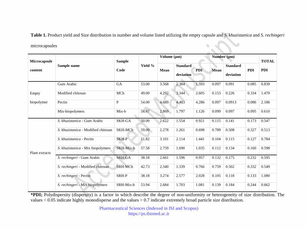
Product yield
Particle yield depends on several parameters influencing the spray drying process, such as the solution
features, inlet, outlet temperature, and pressure.38 The product yield ranged between 38.18% - 59.00
%, including full and free microparticles (Table 1). The unloaded capsule presented a higher result,
around 49.00– 58.67 %, while the loaded one presented a yield of approximately 38.18 – 59.00 %.
Finally, the lowest and the highest yields were reported for SRH-P and SKH-MCh microparticles.
Particle yield directly depended on the condition of the equipment and or loading conditions. It was
not related to the biopolymer features. Table 1 shows the product of all samples in detail.
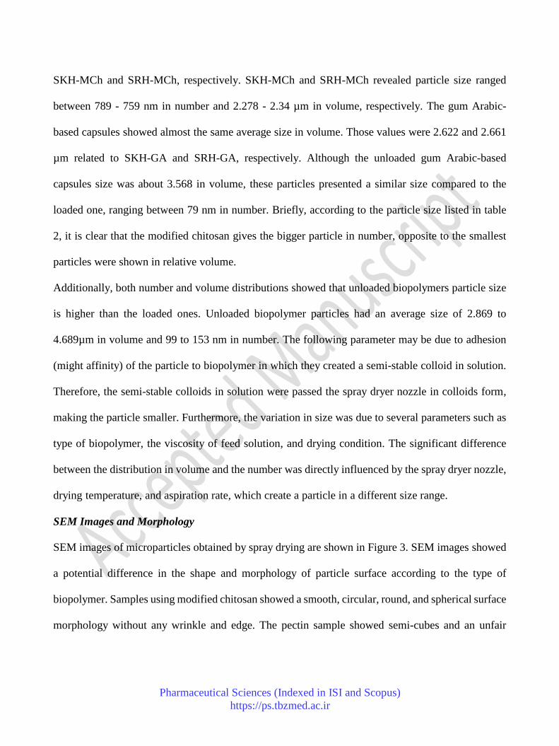
Microcapsules Size Distribution
The evaluation of particle size distribution in relative volume and number carried out in samples
dispersed in ethanol (Table 2). The particle size distributions in the relative of number (independently)
and volume (independently) were similar and uniform regarding the feature of related biopolymer
and heterogeneous in terms of comparison between the type of biopolymer, Table 2. According to
each biopolymer's relative volume and relative number, the monodisperse in size distribution was
noticeable. The volume distribution of these biopolymers was compared to other biopolymers as
follows; the largest particle size was observed for the pectin capsules, about 3.101 and 3.274 µm to
SKH-P and SRH-P, respectively. Whether in empty or full capsules, pectin-based particles showed a
size distribution ranging between 97 - 105 nm in number. Modified chitosan-based capsules also
exhibited a similar size for loaded particles. In this case, the averages were 2.278 and 2.34 µm to
Pharmaceutical Sciences (Indexed in ISI and Scopus)
https://ps.tbzmed.ac.ir
SKH-MCh and SRH-MCh, respectively. SKH-MCh and SRH-MCh revealed particle size ranged
between 789 - 759 nm in number and 2.278 - 2.34 µm in volume, respectively. The gum Arabic-
based capsules showed almost the same average size in volume. Those values were 2.622 and 2.661
µm related to SKH-GA and SRH-GA, respectively. Although the unloaded gum Arabic-based
capsules size was about 3.568 in volume, these particles presented a similar size compared to the
loaded one, ranging between 79 nm in number. Briefly, according to the particle size listed in table
2, it is clear that the modified chitosan gives the bigger particle in number, opposite to the smallest
particles were shown in relative volume.
Additionally, both number and volume distributions showed that unloaded biopolymers particle size
is higher than the loaded ones. Unloaded biopolymer particles had an average size of 2.869 to
4.689µm in volume and 99 to 153 nm in number. The following parameter may be due to adhesion
(might affinity) of the particle to biopolymer in which they created a semi-stable colloid in solution.
Therefore, the semi-stable colloids in solution were passed the spray dryer nozzle in colloids form,
making the particle smaller. Furthermore, the variation in size was due to several parameters such as
type of biopolymer, the viscosity of feed solution, and drying condition. The significant difference
between the distribution in volume and the number was directly influenced by the spray dryer nozzle,
drying temperature, and aspiration rate, which create a particle in a different size range.
SEM Images and Morphology
SEM images of microparticles obtained by spray drying are shown in Figure 3. SEM images showed
a potential difference in the shape and morphology of particle surface according to the type of
biopolymer. Samples using modified chitosan showed a smooth, circular, round, and spherical surface
morphology without any wrinkle and edge. The pectin sample showed semi-cubes and an unfair

Pharmaceutical Sciences (Indexed in ISI and Scopus)
https://ps.tbzmed.ac.ir
morphology with lots of pores and cavities. Gum Arabic particle shape was similar to pectin, but with
more wrinkle and edge, with a gap between winkle on the cubic surface of the particle (Figure 3).
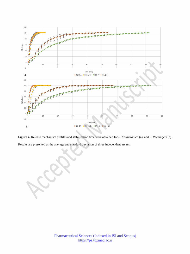
Stabilization/ Release Time
Rosmarinic acid was used as a reference compound for the monitoring release process. The amount
of rosmarinic acid released from the capsules during a specific time was used as the standard
percentage of the extract released from capsules. The stabilization time of three different biopolymers
is shown in Figure 4. Pectin-based microcapsule revealed a similar release behavior in both species,
with stabilization times around 82.08 and 81.08 for SKH-P and SRH-P, respectively (Table 2), which
was presented as the most extended release times among other samples. Gum Arabic-based offers the
fast release of about 7.33 and 4.08 min for SKH-GA and SRH-GA, respectively. The stabilization
time of active compounds loaded on the modified chitosan was 8.58 and 7.58, belong to SKH-MCh
and SRH-MCh, respectively.
In conclusion, modified chitosan and gum Arabic-based microparticles presented a similar and
slowest stabilization time, approximately less than 8.58 min. In contrast, pectin-based macroparticles
had the longest stabilization time (more than 82.08 min) Table 2, Figure 4. It must conclude that the
type of biopolymer directly affected the release time of encapsulated Satureja species.
Kinetic Models
New generations of delivery systems directly depended on the type of carriers and dosage of active
part. The mathematical models are applied using repeated-measurement factors to estimate the release
profile by the statistical model. Korsmeyer-PPEEas model was introduced as the most suitable kinetic
model to describe the release mechanism of particles loaded with natural active compounds on a
matrix-based and straightforward delivery system. Matrix-based particles loaded with herbal extracts
seem to present an excellent release mechanism.31 Thus, the correlation coefficients (r2) obtained
Pharmaceutical Sciences (Indexed in ISI and Scopus)
https://ps.tbzmed.ac.ir
from the Korsmeyer-PPEEas model involving microcapsules containing S. khuzistanica and S.
rechingeri coated by gum Arabic, modified chitosan, and pectin as a carrier, represented between
0.9665 and 0.9965. The release mechanisms are shown in detail in table 2. The value of the parameter
(n) in the Korsmeyer-PPEEas model shows that the release of Satureja species from the capsule is
controlled mainly by anomalous transport, involving a combination of diffusion and swelling release
named SKH-MCh, SRH-GA, and SRH-P. Mix-SKH and Mix-SRH (mix of three biopolymers).
Furthermore, SKH-GA release occurs by diffusion following Fick law (case-I transport), and SKH-P
and SRH-MCh release follow super case-II transport (polymer matrix relaxation) (Table 2).
Cytotoxicity Activity
Polyphenolic extracts were prepared in culture media and tested in the concentration range of 31.25
- 500 µg/mL. Cells were exposed to the different extracts for 24 hours, after which the MTT reduction
assay was performed. S. khuzistanica elicited a significant decrease in viability towards the human
gastric cancer cell line AGS in a concentration as low as 125 µg/mL, the same being true for S.
rechingeri (Figure 6). In the A549 cell line, S. khuzistanica exhibited a similar capacity, albeit to a
lower extent. In contrast, only the highest concentration of S. rechingeri, 500 µg/mL, affected the
viability adversely. (Figure 6).
DISCUSSION
Natural compounds play an essential role in human health with beneficial effects on the recovery of
several diseases. Flavonoids and caffeic acids are the principal polyphenol structural groups identified
in S. khuzistanica PEE. Rosmarnic acid was used as a standard and was detected at a retention time
of 29.5 min on the HPLC chromatogram previously identified by this species.39,40 Meanwhile, the
m/z at 359 [M-H]- and 719 [2M-H]- in the mass spectra determined the existence of rosmarnic acid
with a molecular weight of 360, which is in agreement with Fatemi and coworkers 2019.41 Due to the
Pharmaceutical Sciences (Indexed in ISI and Scopus)
https://ps.tbzmed.ac.ir
probable decomposition of the phytoconstituent in the human body, a spray drying encapsulation
technique has been developed to improve the protection of the phytoconstituent.42 Gum Arabic,
modified chitosan, and pectin were chosen due to inherent characteristics such as process stability,
low hygroscopicity, appropriate film-forming, and an appropriate release profile.32 As a result, the
critical parameter like choosing a right encapsulating agent, optimized preparation method and
optimize drying condition was presented in this study, which was consistent with the fact of delivery
in literature.48 As mentioned in the result, the highest yield was observed from modified chitosan,
within a range of values reported by the literature in nearly the same condition with a spray dryer (30
- 75%).43 Besides, our findings are in acceptance with other studies in the area of surface morphology.
For instance, it was reported that the modified carboxylation chitosan (water-soluble) had a surface
round and spherical morphology.44 The smooth and spherical morphology has been reported from
modified hydrosoluble carboxylated chitosan loaded with vitamin B12.51 A semi-cubic unfair surface
was observed for pectin while an irregular cubic surface with numerous wrinkles and spherical edges
was observed from gum Arabic particles before.26
Finally, it was concluded that the type of biopolymer directly influences the form and morphology of
microcapsules. Due to the stability of the microparticles, several studies were conducted using a
similar kind of particles but containing vitamins (B1, B12, A) and retinoic acid. These studies
confirmed that the particles stored for at least four months were stable. Therefore, it was concluded
that the bioactive compounds within the particulates remain active after this storage period, without
degradation for at least four months.26,45
The effects of biopolymers on the release time have already been reported in other studies.46 Olive
leaf extracts are a source of polyphenolic compounds, when encapsulated by chitosan, present a
similar stabilization release time compared to our results.47 Moreover, research has shown that when
Pharmaceutical Sciences (Indexed in ISI and Scopus)
https://ps.tbzmed.ac.ir
pectin is used as a carrier, the release time of curcumin nanoparticles increases significantly.48
However, a kinetic model was used to describe the release of carriers on a routine basis. For instance,
a gelatine microsphere loaded with trans-retinoic followed a zero-order or Higuchi kinetics.49
Korsmeyer model was used to explain the release mechanism of hydrogel pH-sensitive particles.50
As well, the Korsmeyer model was used to describe the release profile of chitosan-based
nanoparticles.51 In a convincing way, different kinetic models open a wise view of the release
functionality. Therefore, researchers have found appropriate and controllable delivery systems and
release mechanisms to be validated for the application in pharmaceutics medicine, chemotherapy,
food, and supplement, and cosmetic and skincare product with the help of a kinetic model in case of
in vitro release assay.
CONCLUSION
Conclusionally, our study suggests a new delivery system for two encapsulated Iranian endemic
Satureja species, S. khuzistanica and S. rechingeri, as a function of food spice and dietary food
supplements. The pectin-based formula showed the largest particle size between 3.101 and 3.274 µm
with a product yield of 51.82 and 38.18, belong to SKH-P and SRH-P, respectively. Moreover, the
slowest stabilize time was observed from pectin-based microcapsule around 82.08 min to SKH-P and
81.08 min to SRH-P. The fastest stabilize time was observed from SRH-GA microcapsule in 4.08
min with 2.661 µm particle size, 38.18% particle yield, and anomalous (combination of diffusion and
swelling release) release mechanism. The mathematical model which best matched the release
mechanism profile was the Korsmeyer model.
Credit authorship contribution statement
Faezeh Fathi: Experimental part, writing the manuscript; Samad N. Ebrahimi: Supervision,
experimental validation in phytochemical part, developing the draft of the paper; David Pereira:
Pharmaceutical Sciences (Indexed in ISI and Scopus)
https://ps.tbzmed.ac.ir
Biological assessment Berta N. Estevinho and Fernando Rocha: supervision of encapsulation part
and developing the draft of the article.
Conflict of interest
The authors declare no conflict of interest.
Funding
This work was financially supported by: the research council of Shahid Beheshti University, Vasha
herbal company for providing plant material and supporting extraction part, and Base Funding -
UIDB/00511/2020 of the Laboratory for Process Engineering, Environment, Biotechnology, and
Energy – LEPABE - funded by national funds through the FCT/MCTES (PIDDAC); Project POCI-
01-0145-FEDER-028715 (MicroDelivery - Development of controlled delivery functional systems
by microencapsulation of natural and active compounds with therapeutic, nutritional and
technological interest), funded by FEDER funds through COMPETE2020 – Programa Operacional
Competitividade e Internacionalização (POCI) and by national funds (PIDDAC) through
FCT/MCTES. Additional funding by UIDB/50006/2020 (REQUIMTE) and project POCI-01-0145-
FEDER-30154 (FEDER/FCT).
REFERENCES
1. Comunian TA, Favaro-Trindade CS. Microencapsulation using biopolymers as an alternative
to produce food enhanced with phytosterols and omega-3 fatty acids: A review. Food
Hydrocoll .2016;61:442–57. doi: 10.1016/j.foodhyd.2016.06.003
2. Ré M-I. Formulating drug delivery systems by spray drying. Dry Technol. 2006;24(4):433–
46. doi:10.1080/07373930600611877
3. Jafari SM, He Y, Bhandari B, Jafari SM, He Y, Bhandari B. Encapsulation of Nanoparticles
of d-Limonene by Spray Drying: Role of Emulsifiers and Emulsifying Techniques. Dry

Pharmaceutical Sciences (Indexed in ISI and Scopus)
https://ps.tbzmed.ac.ir
Technol. 2010; 3937: 1069–79. doi: 10.1080/07373930701396758
4. Robert P, Gorena T, Romero N, Sepulveda E, Chavez J, Saenz C. Encapsulation of
polyphenols and anthocyanins from pomegranate (Punica granatum) by spray drying. Int J
Food Sci Technol. 2010;45(7):1386–94. doi: 10.1111/j.1365-2621.2010.02270.x
5. Rui Xiong, Anise M.Grant, Ruilong Ma, Shuaidi Zhang, Vladimir V. Tsukruk. Naturally-
derived biopolymer nanocomposites: Interfacial design, properties and emerging applications.
Mater. Sci. Eng. R Rep. 2018;125:1–41. doi: 10.1016/j.mser.2018.01.002
6. Bernkop-Schnürch A, Dünnhaupt S. Chitosan-based drug delivery systems. Eur J Pharm
Biopharm. 2012 Aug;81(3):463–9. doi: 10.1016/j.ejpb.2012.04.007
7. Chang C, Wang T, Hu Q, Zhou M, Xue J, Luo Y. Alginate/chitosan nanoparticles for
encapsulation and controlled release of vitamin B2. Food Hydrocoll. 2017; 70: 143–51. doi:
10.1016/j.foodhyd.2017.03.033
8. Arepally D, Goswami TK. Effect of inlet air temperature and gum Arabic concentration on
encapsulation of probiotics by spray drying. LWT-Food Sci Technol. 2019; 99: 583–93. doi:
10.1016/j.lwt.2018.10.022
9. Vaher M, Koel M. Separation of polyphenolic compounds extracted from plant matrices using
capillary electrophoresis. J Chromatogr A. 2003;990(1–2):225–30. doi: 10.1016/S0021-
9673(02)02013-7
10. Soobrattee MA, Bahorun T, Aruoma OI. Chemopreventive actions of polyphenolic
compounds in cancer. BioFactors. 2006;27(1–4):19–35. doi: 10.1002/biof.5520270103
11. Cai Y, Evans FJ, Roberts MF, Phillipson JD, Zenk MH, Gleba YY. Polyphenolic compounds
from Croton lechleri. Phytochemistry. 1991;30(6):2033–40. doi: 10.1016/0031-
9422(91)85063-6
Pharmaceutical Sciences (Indexed in ISI and Scopus)
https://ps.tbzmed.ac.ir
12. Dardioti A, Karousou R, Lanaras T, Kokkini S. Diversity of Satureja pilosa subsp. origanita
essential oils: A new “ Oregano” from East Mediterranean. Biochem Syst Ecol. 2012; 40: 178–
83. doi: 10.1016/j.bse.2011.10.015
13. Nooshkam A, Mumivand H, Hadian J, Alemardan A, Morshedloo MR. Drug yield and
essential oil and carvacrol contents of two species of Satureja (S. khuzistanica Jamzad and S.
rechingeri Jamzad) cultivated in two different locations. J Appl Res Med Aromat Plants. 2017;
6: 126–30. doi: 10.1016/j.jarmap.2017.04.002
14. Momtaz S, Abdollahi M. An Update on Pharmacology of Satureja Species; From Antioxidant,
Antimicrobial, Antidiabetes and Anti-hyperlipidemic to Reproductive Stimulation. Int J
Pharmacol. 2010;1;6(4):346–53. doi: 10.3923/ijp.2010.346.353
15. Hajimehdipoor H, Saeidnia S, Gohari A, Hamedani M, Shekarchi M. Comparative study of
rosmarinic acid content in some plants of Labiatae family. Pharmacogn Mag. 2012;8(29):37–
41. Doi: 10.4103/0973-1296.93316
16. Haeri S, Minaie B, Amin G, Nikfar S, Khorasani R, Esmaily H, Alinazar S, Abdollahi M.
Effect of Satureja khuzestanica essential oil on male rat fertility. Fitoterapia. 2006;77(7–
8):495–9. doi: 10.1016/j.fitote.2006.05.025
17. Mazarei Z, Rafati H. Nanoemulsification of Satureja khuzestanica essential oil and pure
carvacrol; comparison of physicochemical properties and antimicrobial activity against food
pathogens. LWT-Food Sci Technol. 2019; 100: 328–34. doi: 10.1016/j.lwt.2018.10.094
18. Jafari F, Ghavidel F, Zarshenas MM. A Critical Overview on the Pharmacological and Clinical
Aspects of Popular Satureja Species. JAMS J Acupunct Meridian Stud. 2016;9(3):118–27. doi:
10.1016/j.jams.2016.04.003
19. Giannouli E, Athanasopoulos D, Panagopoulos G, Tsiara S, Karageorgiou CE.
Pharmaceutical Sciences (Indexed in ISI and Scopus)
https://ps.tbzmed.ac.ir
Paraneoplasmatic encephalitis due to Merkel cell neuroendocrine skin carcinoma. J Neurol
Sci. 2013; 333: e703. doi: 10.1016/j.jns.2013.07.2427
20. Jamzad Z. Satureja rechingeri (Labiatae)—a new species from Iran. Ann des
Naturhistorischen Museums Wien Ser B für Bot und Zool. 1996;75–7.
21. Hadian J, Esmaeili H, Nadjafi F, Khadivi-Khub A. Essential oil characterization of Satureja
rechingeri in Iran. Ind Crops Prod. 2014; 61: 403–9. doi: 10.1016/j.indcrop.2014.07.034
22. Alizadeh A. Essential oil composition, phenolic content, antioxidant, and antimicrobial activity
of cultivated Satureja rechingeri Jamzad at different phenological stages. J Biosci. 2015; 70:
51–8. doi: 10.1515/znc-2014-4121
23. Mirjalili MH, Kanani MR, Salehnia A, Ganjipoor P, Phytochemical and Morphological
Characterization of Satureja khuzistanica Jamzad Populations from Iran. Chem Biodivers.
2011; 8: 902–15. doi: https://doi.org/10.1002/cbdv.201000249
24. Ebrahimi SN, Gafner F, Dell’Acqua G, Schweikert K, Hamburger M. Flavone 8-C-glycosides
from Haberlea rhodopensis Friv. (Gesneriaceae). Helv Chim Acta. 2011;94(1):38–45. doi:
10.1002/hlca.201000378
25. Shehzad O, Jin Ha I, Park Y, Wan Ha Y, Shik Kim Y. Development of a rapid and convenient
method to separate eight ginsenosides from Panax ginseng by high-speed counter-current
chromatography coupled with evaporative light scattering detection. J Sep Sci. 2011; 34(10):
1116–22. doi: 10.1002/jssc.201000932
26. Gonçalves A, Estevinho BN, Rocha F. Design and characterization of controlled-release
vitamin A microparticles prepared by a spray-drying process. Powder Technol. 2017; 305:
411–7. doi: 10.1016/j.powtec.2016.10.010
27. Kha TC, Nguyen MH, Roach PD, Stathopoulos CE. Microencapsulation of Gac oil:

Pharmaceutical Sciences (Indexed in ISI and Scopus)
https://ps.tbzmed.ac.ir
Optimisation of spray drying conditions using response surface methodology. Powder
Technol. 2014; 264: 298–309. doi: 10.1016/j.powtec.2014.05.053
28. Santana AA, Oliveira RA de, Kurozawa LE, Park KJ. Microencapsulation of pequi pulp by
spray drying: use of modified starches as encapsulating agent. Eng Agrícola. 2014;34(5):980–
91. doi: 10.1590/S0100-69162014000500017
29. Ito R, Golman B, Shinohara K. Controlled release with coating layer of permeable particles. J
Control Release. 2003; 92: 361–8. doi: 10.1016/S0168-3659(03)00363-8
30. Ziaee M, Moharramipour S, Mohsenifar A. MA-chitosan nanogel loaded with Cuminum
cyminum essential oil for efficient management of two stored product beetle pests. J Pest Sci.
2014; 87(4): 691–9. doi: 10.1007/s10340-014-0590-6
31. Dash S, Murthy PN, Nath L, Chowdhury P. Kinetic modeling on drug release from controlled
drug delivery systems. Acta Pol Pharm - Drug Res. 2010;67(3):217–23.
32. Pothakamury UR, Barbosa-Cánovas G V. Fundamental aspects of controlled release in foods.
Trends Food Sci Technol. 1995;6(12):397–406. doi: 10.1016/S0924-2244(00)89218-3
33. Unagolla JM, Jayasuriya AC. Drug transport mechanisms and in vitro release kinetics of
vancomycin encapsulated chitosan-alginate polyelectrolyte microparticles as a controlled drug
delivery system. Eur J Pharm Sci. 2018; 114: 199–209. doi: 10.1016/j.ejps.2017.12.012
34. Videira RA, Andrade PB, Monteiro LS, Valentão P, Ferreira PMT, Pereira DM. Toxicity and
structure-activity relationship (SAR) of α,β-dehydroamino acids against human cancer cell
lines. Toxicol Vitr. 2018; 47: 26–37. doi: 10.1016/j.tiv.2017.10.027
35. Figueiredo-González M, Valentão P, Pereira DM, Andrade PB. Further insights on tomato
plant: Cytotoxic and antioxidant activity of leaf extracts in human gastric cells. Food Chem
Toxicol. 2017; 109: 386–92. doi: 10.1016/j.fct.2017.09.018

Pharmaceutical Sciences (Indexed in ISI and Scopus)
https://ps.tbzmed.ac.ir
36. Rasooli A, Fatemi F, Akbarzadeh K, Dini S, Bahremand S. Synergistic Protective Activity of
Deuterium Depleted Water (DDW) and Satureja rechingeri Essential Oil on Hepatic Oxidative
Injuries Induced by Acetaminophen in Rats. J Essent Oil Bear Plants. 2016 3;19(5):1086–101.
doi: 10.1080/0972060X.2015.1111776
37. Sahraei H, Gharzi A, Amiri H, Abbasi M, Gholami M. Wound Healing Effect of Satureja
Khuzistanica and Satureja Rechingeri Ethanolic Extracts in NMRI Adult Mice. Zahedan J Res
Med Sci. 2016 16; 18(5): e6665. doi: 10.17795/zjrms-6665
38. Gharsallaoui A, Roudaut G, Chambin O, Voilley A, Saurel R. Applications of spray-drying in
microencapsulation of food ingredients: An overview. Food Res Int. 2007; 40(9): 1107–21.
doi:10.1016/j.foodres.2007.07.004
39. Davoodi M, Rustaiyan A, Ebrahimi SN. Monoterpene flavonoid from aerial parts of Satureja
khuzistanica. Rec Nat Prod. 2017;12(2):175–8. doi: 10.25135/rnp.19.17.06.109
40. Siavash Saei-Dehkordi S, Fallah AA, Heidari-Nasirabadi M, Moradi M. Chemical
composition, antioxidative capacity and interactive antimicrobial potency of Satureja
khuzestanica Jamzad essential oil and antimicrobial agents against selected food-related
microorganisms. Int J Food Sci Technol. 2012; 47(8): 1579–85. doi: 10.1111/j.1365-
2621.2012.03006.x
41. Fatemi F, Abdollahi MR, Mirzaie-asl A, Dastan D, Garagounis C, Papadopoulou K.
Identification and expression profiling of rosmarinic acid biosynthetic genes from Satureja
khuzistanica under carbon nanotubes and methyl jasmonate elicitation. Plant Cell Tissue Organ
Cult. 2019; 136(3): 561–73. doi: 10.1007/s11240-018-01537-8
42. Raman RK, Santhalakshmy S, Bosco SJD, Deshwal GK. Effect of encapsulating agents on
antioxidative properties of spray dried Jamun juice powder. Int J Chem Stud. 2020; 8(3): 1807–
Pharmaceutical Sciences (Indexed in ISI and Scopus)
https://ps.tbzmed.ac.ir
1810. doi: 10.22271/chemi.2020.v8.i3y.9468
43. Mourya VK, Inamdar NN. Chitosan-modifications and applications: Opportunities galore.
React Funct Polym. 2008;68(6):1013–51. doi: 10.1016/j.reactfunctpolym.2008.03.002
44. Estevinho B N. Damas A M., Martins P. Rocha F. The Influence of Microencapsulation with
a Modified Chitosan (Water Soluble) on βGalactosidase Activity. Dry. Technol. 2014; 32:
1575–1586. doi: 10.1080/07373937.2014.909843
45. Carlan IC, Estevinho BN, Rocha F. Study of microencapsulation and controlled release of
modified chitosan microparticles containing vitamin B12. Powder Technol. 2017; 318: 162–9.
doi: 10.1016/j.powtec.2017.05.041
46. Ersus S, Yurdagel U. Microencapsulation of anthocyanin pigments of black carrot (Daucus
carota L.) by spray drier. J Food Eng. 2007; 80(3): 805–12. doi:
10.1016/j.jfoodeng.2006.07.009
47. Kosaraju SL, D’ath L, Lawrence A. Preparation and characterisation of chitosan microspheres
for antioxidant delivery. Carbohydr Polym. 2006;64(2): 163–7. doi:
10.1016/j.carbpol.2005.11.027
48. Nguyen AT, Winckler P, Loison P, Wache Y, Chambin O. Physico-chemical state influences
in vitro release profile of curcumin from pectin beads. Colloids Surfaces B Biointerfaces. 2014;
121: 290–8. doi: 10.1016/j.colsurfb.2014.05.023
49. Dinarvand R, Rahmani E, Farbod E. Gelatin Microspheres for the Controlled Release of All-
trans-Retinoic Acid Topical Formulation and Drug Delivery Evaluation. Iran J Pharm Res
IJPR. 2010;2(1):47–50. doi: 10.22037/ijpr.2010.35
50. Gupta P, Vermani K, Garg S. Hydrogels: from controlled release to pH-responsive drug
delivery. Drug Discov Today. 2002; 7(10): 569–79. doi: 10.1016/S1359-6446(02)02255-9

Pharmaceutical Sciences (Indexed in ISI and Scopus)
https://ps.tbzmed.ac.ir
51. Soares PIP, Isabel A, Carvalho J, Ferreira IMM, Novo CMM, Paulo J. Chitosan-based
nanoparticles as drug delivery systems for doxorubicin: Optimization and modelling.
Carbohydr Polym. 2016; 147: 304–12. doi: 10.1016/j.carbpol.2016.03.028.
Pharmaceutical Sciences (Indexed in ISI and Scopus)
https://ps.tbzmed.ac.ir
Table 1. Product yield and Size distribution in number and volume listed utilizing the empty capsule and S. khuzistanica and S. rechingeri
microcapsules
Microcapsule
content
Sample name
Sample
Code
Yield %
Volume (µm) Number (µm)
TOTAL
PDI Mean
Standard
deviation
PDI Mean
Standard
deviation
PDI
Empty
biopolymer
Gum Arabic GA 53.00 3.568 2.384 1.593 0.097 0.091 0.085 0.839
Modified chitosan MCh 49.00 4.292 3.344 2.605 0.153 0.226 0.334 1.470
Pectin P 54.00 4.689 4.483 4.286 0.097 0.0913 0.086 2.186
Mix-biopolymers Mix-b 58.67 2.869 1.797 1.126 0.099 0.097 0.095 0.610
Plant extracts
S. khuzistanica - Gum Arabic SKH-GA 50.00 2.622 1.554 0.921 0.115 0.141 0.173 0.547
S. khuzistanica - Modified chitosan SKH-MCh 59.00 2.278 1.261 0.698 0.789 0.508 0.327 0.513
S. khuzistanica - Pectin SKH-P 51.82 3.101 2.114 1.441 0.104 0.115 0.127 0.784
S. khuzistanica - Mix biopolymers SKH-Mix-b 57.58 2.759 1.690 1.035 0.112 0.134 0.160 0.598
S. rechingeri - Gum Arabic SRH-GA 38.18 2.661 1.596 0.957 0.132 0.175 0.232 0.595
S. rechingeri - Modified chitosan SRH-MCh 42.73 2.340 1.339 0.766 0.759 0.502 0.332 0.549
S. rechingeri - Pectin SRH-P 38.18 3.274 2.577 2.028 0.105 0.118 0.133 1.080
S. rechingeri - Mix biopolymers SRH-Mix-b 53.94 2.684 1.703 1.081 0.139 0.184 0.244 0.662
*PDI; Polydispersity (dispersity) is a factor in which describe the degree of non-uniformity or heterogeneity of size distribution. The
values < 0.05 indicate highly monodisperse and the values > 0.7 indicate extremely broad particle size distribution.

Pharmaceutical Sciences (Indexed in ISI and Scopus)
https://ps.tbzmed.ac.ir
Table 2. Parameters and correlation coefficients (Korsmeyer kinetics) were applied to the
experimental release profiles.
Sample
Release
Stable Time
(min)
R2 Kk (min-n)
n
n < 0.43 0.43 < n < 0.85 n = 0.85 n > 0.85
SKH-GA 7.330 0.9821 0.9086 0.0186 - - -
SKH-MCh 8.580 0.9846 0.6132 - 0.3204 - -
SKH-P 82.08 0.9950 0.0480 - - - 0.9527
Mix-SKH 44.08 0.9665 0.1386 - 0.6150 - -
SRH-GA 4.080 0.9965 0.7121 - 0.5358 - -
SRH -MCh 7.580 0.9536 0.1912 - - - 1.2031
SRH -P 81.08 0.9856 0.0694 - 0.7824 - -
Mix-SRH 35.08 0.9841 0.2256 - 0.4853 - -
*Stable time is a time when the release start to be stable and the curve change to linear.

Pharmaceutical Sciences (Indexed in ISI and Scopus)
https://ps.tbzmed.ac.ir
Figure 1. Comparison of crude and polyphenolic enrich HPLC chromatograms of S. khuzistanica (A), S. rechingeri (B)
at a wavelength of 324 nm. Retention time 20.00; apigenin-7-O-gentiobioside (1), retention time 29.50; rosmarniric acid
(2), retention time 36.00; epi-salvianolic acid B (3).

Pharmaceutical Sciences (Indexed in ISI and Scopus)
https://ps.tbzmed.ac.ir
Figure 2. The UV spectrum of rosmarinic acid (above) and mass spectra of rosmarinic acid in negative mode (below).
Image (a); apigenin-7-O-gentiobioside, (b); rosmarniric acid, (c); epi-salvianolic acid B.

Pharmaceutical Sciences (Indexed in ISI and Scopus)
https://ps.tbzmed.ac.ir
Figure 3. SEM images of microcapsule of SKH-GA (a). SKH-MCh (b), SKH-P (c), and MIX of three Mix-SKH (d),
SRH-GA (e). SRH -MCh (f), SRH -P (g), and MIX of three “Mix-SRH“(h), and empty biopolymers gum Arabic (l),
modified Chitosan (m) and Pectin (n) and mix of three biopolymers (o), Magnification 30000, beam intensity (HV) 15.00
kV, the distance between the sample and the lens (WD) around 10 mm.
Pharmaceutical Sciences (Indexed in ISI and Scopus)
https://ps.tbzmed.ac.ir
Figure 4. Release mechanism profiles and stabilization time were obtained for S. Khuzistanica (a), and S. Rechingeri (b).
Results are presented as the average and standard deviation of three independent assays.

Pharmaceutical Sciences (Indexed in ISI and Scopus)
https://ps.tbzmed.ac.ir
Figure 5. Effect of aqueous extracts upon the viability of the human gastric cancer cell line (AGS) and the human lung
cancer cell line (A549) are evaluated by the MTT assay. *p<0.05, **p<0.01, ***p<0.001, ****p<0.0001