MICROBIOLOGY, VIROLOGY, IMMUNOLOGY
83
MICROBIOLOGY, VIROLOGY, IMMUNOLOGY Laboratory workbook Student ____ group of dental faculty ___________________________________________________ MINSK BSMU 2017 . . .
Transcript of MICROBIOLOGY, VIROLOGY, IMMUNOLOGY

MICROBIOLOGY, VIROLOGY, IMMUNOLOGY
Laboratory workbook
Student ____ group of dental faculty ___________________________________________________
MINSK BSMU 2017
.
.
.

МИНИСТЕРСТВО ЗДРАВООХРАНЕНИЯ РЕСПУБЛИКИ БЕЛАРУСЬ
БЕЛОРУССКИЙ ГОСУДАРСТВЕННЫЙ МЕДИЦИНСКИЙ УНИВЕРСИТЕТ
КАФЕДРА МИКРОБИОЛОГИИ, ВИРУСОЛОГИИ, ИММУНОЛОГИИ
МИКРОБИОЛОГИЯ, ВИРУСОЛОГИЯ, ИММУНОЛОГИЯ
MICROBIOLOGY, VIROLOGY, IMMUNOLOGY
Лабораторный практикум
Минск БГМУ 2017

УДК 579+578+612.017.1(076.5)(075.8)-054.6 ББК 52.64я73 М59
Рекомендовано Научно-методическим советом университета в качестве практикума 17.05.2017 г., протокол № 9
А в т о р ы: канд. мед. наук, доц. В. В. Кочубинский; канд. мед. наук, доц. Т. А. Канашкова; канд. мед. наук, доц. Д. А. Черношей; канд.
мед. наук, доц. И. А. Гаврилова Р е ц е н з е н т ы: д-р мед. наук, проф., зав. каф. клинической микробиологии Витебского государственного ордена Дружбы народов
медицинского университета И. И. Генералов; канд. мед. наук, доц. каф. биологии Белорусского государственного медицинского университе-та В. Э. Бутвиловский М59
Микробиология, вирусология, иммунология = Microbiology, virology, immunology : лабораторный практикум / В. В. Кочубинский [и др.]. – Минск : БГМУ, 2017. – 82 с.
ISBN 978-985-567-812-1.
Отражены вопросы общей и частной медицинской микробиологии, вирусологии и иммунологии. Даны алгоритмы, схемы, некоторые справочные сведения, методики выполнения лабораторных работ по дисциплине «Микробиология, вирусология и иммунология».
Предназначен для студентов стоматологического факультета, обучающихся на английском языке.
УДК 579+578+612.017.1(076.5)(075.8)-054.6 ББК 52.64я73
ISBN 978-985-567-812-1 © УО «Белорусский государственный
медицинский университет», 2017

Учебное издание
Кочубинский Валентин Витальевич Канашкова Татьяна Александровна Черношей Дмитрий Александрович Гаврилова Ирина Александровна
МИКРОБИОЛОГИЯ, ВИРУСОЛОГИЯ, ИММУНОЛОГИЯ
MICROBIOLOGY, VIROLOGY, IMMUNOLOGY
Лабораторный практикум
Ответственная за выпуск Т. А. Канашкова Переводчик В. В. Кочубинский
Компьютерный набор В. В. Кочубинского Компьютерная верстка Н. М. Федорцовой
Подписано в печать 28.08.17. Формат 6084/8. Бумага писчая «Снегурочка». Ризография. Гарнитура «Calibri». Усл. печ. л. 9,76. Уч.-изд. л. 5,56. Тираж 120 экз. Заказ 635.
Издатель и полиграфическое исполнение: учреждение образования
«Белорусский государственный медицинский университет». Свидетельство о государственной регистрации издателя, изготовителя,
распространителя печатных изданий № 1/187 от 18.02.2014. Ул. Ленинградская, 6, 220006, Минск.

3
Glossary aerobic - Using oxygen for growth and metabolism. agar - A gelling agent used in bacterial growth media that allows liquids to become a gel-like solid. anaerobic - Not requiring any oxygen for growth. antigen - Part of an organism that is foreign to our bodies and stimulates an immune response. asexual organisms - Living creatures (usually bacteria) that are neither male nor female, and therefore
do not reproduce by exchanging genetic material. biofilm - A complex community of microorganisms living together and attached to a surface. capsule - A structure that surrounds or encapsulates many bacteria and may serve to protect them from
harsh conditions or to assist with adherence to surfaces. cariology - The study of cavities. collagenase - An enzyme produced by some bacteria that breaks down the connective tissue collagen. colonies - Masses of bacteria that arise from a single cell on solid growth media. colonization - The act of attaching to and inhabiting a surface. conjugation - The process of DNA transfer from one bacterial cell to another. culturing - The act of growing bacteria in a laboratory. cytokine - Proteins that are made by cells that alter the properties and behavior of other cells. cytosol - The interior of a cell that contains the cell’s inner components, or “guts” . dissemination - The process by which a pathogen is transmitted from one host to another. DNA fingerprint - A characteristic sequence of nucleic acid bases (A, G, C, T) that is unique to and defines
a given bacterial species. endodontic infections - Infections that occur within the pulp of the tooth. endoplasmic reticulum - In a eukaryotic cell, the structure on which ribosomes reside. extracellular - The environment outside of a cell. flagella - Flexible rope-like structures that help bacteria swim and move in different environments. genome - The complete DNA material of an organism. genus - The designation for a group of organisms highly related to each other. gingivitis - Gum disease. glucan - A general term for sugar or polysaccharide. Gram negative - Bacteria that appear pink after the Gram stain procedure due to their thin
peptidoglycan cell wall. Gram positive - Bacteria that appear purple after the Gram stain procedure due to their thick
peptidoglycan cell wall. growth media - The food and nutrients on which bacteria grow in the laboratory. Hemagglutination - The clumping together of red blood cells. hemolysin - A bacterial toxin that is able to destroy red blood cells. hemolysis - The act of lysing, or killing, a red blood cell. host - The organism, usually a human, that a pathogen lives in or on. immuno-compromised - A state where an individual’s immune system is weakened, usually by an
infection or disease. incubate - To allow microorganisms to grow in the lab under favorable growth conditions. inflammation - The process whereby immune cells and chemicals accumulate at the site of infection and
result in swelling and redness.
inner membrane - The phospholipid-containing structure around a Gramnegative cell. invasin - A protein that a pathogen uses to enter into a host cell. lectin - A protein that binds to a specific type of sugar. leukotoxin - A bacterial toxin that is able to destroy white blood cells. lipid A - The innermost portion of lipopolysaccharide (LPS) that anchors it into the outer membrane of
Gram-negative bacteria; composed of lipid. lipopolysaccharide (LPS) - The outer part of the outer membrane of Gram-negative bacteria; composed
of lipid and sugars. localized - Found only at a specific location. macroscopic - Large enough to be seen with the naked eye. metabolize - To utilize a nutrient source for growth and maintenance. microbiologist - A professional who studies organisms too small to be seen with the naked eye. migration - The act of moving throughout the body and occupying a new environment. mucins - Large proteins in saliva that give it hydrating properties. normal flora - The community of microorganisms that is found in an environment during good health. nucleoid - The region of the bacterial cell cytosol that contains the chromosome. O-antigen - The outermost portion of lipopolysaccharide; composed of sugars linked together in chains. oligosaccharide core - The central portion of lipopolysaccharide that links the O-antigen to lipid A;
composed of sugars. organelles - Discrete structures that carry out specific functions within a cell. outer membrane - The outermost layer of a Gram-negative cell that contains both phospholipids and
lipopolysaccharide. pathogen - An organism that can cause disease. peptide - A short sequence of amino acids linked together in a chain. peptidoglycan - Chemical that makes up a bacterial cell wall; composed of a mixture of amino acids and
sugars. persistent - A state where a pathogen remains in an environment for a prolonged period of time. pH - The measure of how acidic or basic a substance is; acids have low pH values and bases have high pH
values. phagocytes - Cells of the immune system that are able to engulf pathogens and parts of them. phospholipid bilayer - The composition of cell membranes, made up of phosphate groups attached to
lipid molecules. pili - Bacterial hair-like projections that are made of protein and aid in attachment to surfaces and other
bacteria. plaque - The bacterial biofilm that accumulates on teeth. polymerase chain reaction (PCR) - The method by which the amount of genetic material (DNA) can be
selectively increased. polymicrobial infection - An infection caused by more than one microorganism. resolution - The ability to distinguish two objects as separate entities. ribosome - The structure on which amino acids are synthesized into a protein. saliva - The liquid produced in our mouths by the salivary glands that helps to maintain good oral health. salivary antibody - Proteins in the mouth that are directed against specific pathogens. salivary glands - The organs in the mouth that produce saliva.

4
secretion systems - Components that bacteria use to export material from the inside of their cells to the outside.
sialidase - An enzyme produced by some bacteria that breaks apart specific types of sugars. species - The designation for organisms that are biologically identical to each other. transpeptidation - Linking together sugar chains with peptides. vaccine - A substance that can boost the immune response and protect us from subsequent infection by
a specific pathogen. virulence - The ability to cause disease.
Laboratory safety procedures 1. Place all extra clothing, unnecessary books, purses, backpacks, and paraphernalia in an
appropriate place. Racks are provided for these materials. The laboratory work area must be kept free of articles not actually in use.
2. Eating, drinking, and smoking are forbidden at all times in the laboratory. 3. Keep your locker or laboratory door clean. Do not allow your locker drawer to become
filled with cultures that have no value in your current work. 4. Return all reagents, cultures, and glassware to their appropriate places. 5. Wear a laboratory coat, smock, or lab apron when working in the laboratory. This will
protect clothing from contamination or accidental discoloration by staining solutions. 6. Do not place anything in your mouth while in the laboratory. This includes pencils, food,
and fingers. Learn to keep your hands away from your mouth and eyes. 7. Avoid contamination of benches, floor, and wastebaskets. 8. Clean your work area (laboratory bench) with a phenolic disinfectant such as 5% Lysol or
5% phenol or a quaternary compound such as cetylpyridinium (Ceepyrn) before and after each laboratory period. This standard procedure lessens the chance for accidental infection as well as for contamination of cultures.
9. Special receptacles will be provided for infectious materials and used glass slides. Place all discarded cultures and contaminated glassware into these receptacles. Do not let unwanted and unneeded materials accumulate. Tall jars filled with a solution such as 5% Lysol or special receptacles will be provided for pipettes.
10. When infectious material is accidentally spilled, cover it immediately with a disinfectant such as 5% Lysol or 5% phenol and notify your instructor at once.
11. Flame wire loops and needles before and immediately after transfer of cultures. Do not move through the laboratory with a loop or pipette containing infectious material.
12. Wash your hands thoroughly before and after each experiment, using disinfecting soap if possible.
13. Label all experimental material with your: a. Name ____________________ b. Date ___/___/_____ c. Exercise number Ex. 5
14. Telephone number to call in case of an emergency 101, 103.

5
Practical class 1. Methods in diagnostic microbiology. Microscopic method of examination (MME). Basic morphological forms of bacteria. Simple methods of staining Suggested reading for self-study:
History of the microbiology, virology, immunology department; main spheres of activity and trends in research. Design and equipment of microbiological laboratory, biosafety levels. Basic rules of work in microbiological laboratory (biosafety in work with class II biohazards). Universal precautions in work with burners and electric supplies.
Taxonomy of microorganisms: classification and nomenclature. Modern approaches to taxonomy of microorganisms. Taxonomic ranks. Vars (types), strains, clones, pure cultures.
Basic morphological forms of bacteria. Morphological characteristics of cocci, rods and spiral-shaped bacteria. Microscopic method of examination: tasks, procedure, method evaluation. Bright-field light microscope: components and proper
use of the microscope. Smear preparation and fixation. Simple methods of staining. The technique of oil immersion microscopy.
Signature of the tutor _______________
Oral quiz Laboratory
work Individual
work Tests
Total results
Laboratory work
Laboratory exercises Laboratory report
1. Prepare heat-fixed slide of Escherichia coli, cultured on agar medium, stain with methylene blue, examine under the oil immersion lens and complete the report.
2. Prepare heat-fixed slides of Staphylococcus spp., cultured on liquid medium, stain with basic fuchsin, examine under the oil immersion lens and complete the report.
3. Complete the drawings of slides seen in demonstration room:
- Streptococcus spp., pure culture, stained with crystal violet;
- Vibrio spp., pure culture, stained with basic fuchsin;
- Bacillus spp., pure culture, stained with crystal violet.
1 Smear __________________
Stain _____________
2 Smear ____________________
Stain _____________
3 Smear ____________
Stain ____________
4 Smear ____________
Stain ____________
5 Smear ____________
Stain ____________
6
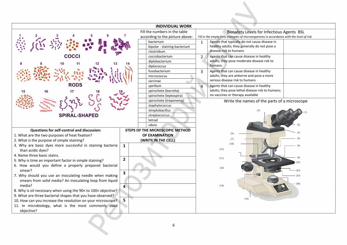
INDIVIDUAL WORK
Fill the numbers in the table according to the picture above:
Biosafety Levels for Infectious Agents BSL Fill in the empty cells examples of microorganisms in accordance with the level of risk
bacterium 1 Agents that typically do not cause disease in healthy adults; they generally do not pose a disease risk to humans
bipolar - staining bacterium
clostridium
coccobacterium 2 Agents that can cause disease in healthy adults; they pose moderate disease risk to humans
diplobacterium
diplococcus
fusobacterium 3 Agents that can cause disease in healthy adults; they are airborne and pose a more serious disease risk to humans
micrococcus
sarcinae
spirillum 4 Agents that can cause disease in healthy adults; they pose lethal disease risk to humans; no vaccines or therapy available
spirochete (borrelia)
spirochete (leptospira)
spirochete (treponema) Write the names of the parts of a microscope
staphylococcus
streptobacillus
streptococcus
tetrad
vibrio
Questions for self-control and discussion: 1. What are the two purposes of heat fixation? 2. What is the purpose of simple staining? 3. Why are basic dyes more successful in staining bacteria
than acidic dyes? 4. Name three basic stains. 5. Why is time an important factor in simple staining? 6. How would you define a properly prepared bacterial
smear? 7. Why should you use an inoculating needle when making
smears from solid media? An inoculating loop from liquid media?
8. Why is oil necessary when using the 90× to 100× objective? 9. What are three bacterial shapes that you have observed? 10. How can you increase the resolution on your microscope? 11. In microbiology, what is the most commonly used
objective?
STEPS OF THE MICROSCOPIC METHOD OF EXAMINATION
(WRITE IN THE CELL)
1
2
3
4
5

7
Practical class 2. MME. The morphology and fine structure of bacteria. Differential methods of staining Suggested reading for self-study:
Distinctive features of prokaryotic and eukaryotic cells. Basic bacterial cell structure: components of bacterial cell. The composition, function, detection methods of bacterial cell wall. Gram stain: medical application, principles, procedure for Gram stain.
The composition, function of capsule, flagella, pili (fimbriae) and methods for their detection. Detection of capsule using negative staining.
The cytoplasmic membrane: structure, function. The most important bacterial cytoplasmic membrane proteins. Bacterial core: cytoplasm, cytoplasmic structures (nucleoid, plasmids, ribosomes, and mesosomes). Inclusion bodies - storage granules (starch, fat, sulfur, polymetaphosphate (volutin)). Methods for nucleoid and volutin detection. Loeffler and Neisser stain for volutin granules.
Acid-fast bacteria and unique properties of their cell wall. Ziehl-Neelsen acid-fast staining: medical application, principle, procedure.
Signature of the tutor
_______________
Oral quiz Laboratory
work Individual
work Tests
Total results
Laboratory work
Laboratory exercises Laboratory report 1. Prepare heat-fixed slide of the mixed culture
of Escherichia coli (gram-negative) and Staphylococcus aureus (gram-positive), Gram stain, examine under oil immersion and complete the report.
2. Complete the drawings of slides seen in demonstration room:
- slide with capsule of Klebsiella pneumoniae, negative staining;
- slide with mixture of Escherichia coli (gram-negative) and Staphylococcus aureus (gram-positive), Gram stain;
- slide with volutin granules of Corynebacterium diphtheriae, Loeffler staining;
- slide with volutin granules of Corynebacterium diphtheriae, Neisser staining;
- slide of the mixed culture of asid-fast and asid-liable microorganisms, staing Ziehl-Neelsen.
1 Smear ___________
Stain ____________
2 Smear ___________
Stain ___________
3 Smear ___________
Stain ____________
4 Smear ___________
Stain ____________
5 Smear ___________
Stain ____________
6 Smear ___________
Stain ____________
7 Smear ___________
Stain ____________

8
INDIVIDUAL WORK (See continued on page 18)
A 1 2 3 4 5
B 4 5
Write the component name of the wall
1
2
3
4
5
A
B
Paint bacteria by Gram‘ stage:
Enter the cell names of
structures
1 -
2 -
3 -
4 -
5 -
6 -
7 - -
8 -
9 -
10 -
11 -
12 -
13 -

9
Practical class 3. MME. The morphology of the spirochetes, actinomyces, rickettsia, chlamydia, mycoplasmas Suggested reading for self-study:
Bacterial forms with defective cell wall (protoplasts, spheroplasts and L forms): factors inducing cell wall removal, medical importance of L-forms.
Resting forms of microorganisms. Bacterial endospores: medical importance, properties of endospore, the periods of endospore formation, detection methods. Spore stain using Ozheshko method: principle, procedure.
Taxonomy, morphology, medical significance of the Spirochetes, Actinomyces, Rickettsiae, Chlamydiae, Mycoplasmas.
Romanowsky-Giemsa stain. Dark-field light microscopy. Phase-contrast light microscopy. Fluorescence microscopy.
Signature of the tutor
_______________
Oral quiz Laboratory
work Individual
work Tests
Total results
Laboratory work
Laboratory exercises Laboratory report
1. Prepare slide of Rickettsia spp., stain with fuschin, examine under the microscope, complete the report.
2. Complete the drawings of slides seen in demonstration room:
- slide with Treponema denticola in dental plaque, Gram stain;
- Leptospira spp., dark-field microscopy;
- Borrelia recurrentis in the blood of patient with relapsing fever, Romanowsky-Giemsa stain;
- Chlamydia inclusions in cytoplasm of host-cell, Romanowsky-Giemsa stain;
- slide with Actinomyces spp., pure culture, Gram stain;
- slide with spores of Bacillus anthracis, Ozheshko staining;
- slide with E. coli, pure culture, acridine orange stain.
1 Smear ___________
Stain ____________
2 Smear ___________
Stain ____________
3 Smear ___________
Stain ____________
4 Smear ___________
Stain ____________
5 Smear ___________
Stain ____________
6 Smear ___________
Stain ____________
7 Smear ___________
Stain ____________
8 Smear ___________
Stain ____________

10
INDIVIDUAL WORK
Morphology of Spirochetes (write in cells names of structures) Endoflagella (axial filaments) beneath outer membrane, Basal body, Outer membrane, Endoflagella, Periplasm, Cell wall (peptidoglycan), Inner (cell/plasma) membrane, DNA in nucleoid, cytoplasm
Confront Gram-positive and Gram-negative bacteria
1 Characteristic Gram-Positive Gram-Negative
2 Number of peptidoglycan layers
3 Overall thickness in nm
4 Specific compounds
5 Interbridges between tetra peptides of neighbor glycan chains
6 Outer membrane
7 Periplasmic space
8 Porin proteins
9 Permeability
The technique of Gram stain (write the component and exposure time)
Component: crystal violet, tag water, basic fuchsine or safranin, ethanol, iodine
Secretion systems
Flagella fixation in cell envelope
component exposure time, sec
Main mechanisms of genetic exchange
1 Cell wall deficient forms in vitro
2 Ability to produce spores
3 Ability to produce long filamentous
4 Susceptibility to Lysozyme
5 Adhesion by pili
6 Pathogenicity islands
7 Tag water (wash slide thoroughly) 5 Gram stain (fill)

11
INDIVIDUAL WORK
Questions for self-control and discussion (Practical class 2) Questions for self-control and discussion (Practical class 3) What is the function of the iodine solution in the Gram stain? If it were omitted, how would staining results be affected?
result For what diseases would you use an acid-fast stain?
What is the purpose of the alcohol solution in the Gram stain?
What chemical is responsible for the acid-fast property of mycobacteria?
What counterstain is used? Why is it necessary? Could colors other than red be used? What is the advantage of the Gram stain over the simple stain?
result How should the acid-fast stain of a sputum specimen from a patient with suspected pulmonary Nocardia infection be performed?
Describe at least two conditions in which an organism might stain gram variable.
Is a Gram stain an adequate substitute for an acid-fast stain? Why?
Which step is the most crucial or most likely to cause poor results in the Gram stain? Why?
Are acid-fast bacteria gram positive or gram negative? Explain your answer.
Why must young cultures be used when doing a Gram stain? What is meant by gram variable?
Why is it important to know whether bacterial cells possess flagella, or endospores?
What part of the bacterial cell is most involved with Gram staining, and why?
What do endospore stains have in common with the Ziehl-Neelsen acid-fast stain? Is bacterial sporulation a reproductive process? Explain.
What is an advantage of negative staining?
What is the purpose of the heat during the acid-fast staining procedure?
Why is negative staining also called either indirect or background staining?
Why are endospores so difficult to stain?

12
Practical class 4. Ecology of microorganisms. Asepsis. Methods of sterilization, disinfection and antisepsis Suggested reading for self-study:
Ecology of microorganisms. Interspecific and intraspecific relations. Symbiosis, its variants. Antagonistic microbial relationships, its background and medical importance. Bacteriocins.
Definition of terms asepsis, sterilization, disinfection, antisepsis. Methods of sterilization: physical, chemical, mechanical. Differences between sterilization and disinfection. Types and methods of disinfection. Types and methods of antisepsis. Practical antisepsis. Classification of antiseptics, origin and characteristics of groups. Mechanisms of action on microorganisms. Antimicrobial management in dentistry.
Signature of the tutor
_______________
Oral quiz Laboratory
work Individual
work Tests
Total results
Laboratory work
Laboratory exercises Laboratory report
1. Test the effectiveness of hygienic and surgical hand antisepsis. The result is taken into account in the next practical class.
1. Divide a nutrient agar plate into 4 sections with a marking pen or pencil. Mark each section of the plate with numbers 1, 2, 3, 4. 2. Mark each plate with your group number and your name. 3. On the surface of agar medium at section N 1 make a fingerprint of skin untreated with any antiseptic (control). 4. Wash your hands with soap as you do it usually at home and make a fingerprint on the surface of the agar medium at section N2. 5. Wash your hands with soap twice and then your fingers with antiseptic (1% solution of iodopyron) – 2 minutes, neutralize
iodopyron with neutralizer (1% solution of sodium thiosulfate) for 2 minutes and make a fingerprint on the surface of agar medium at section N 3.
6. Do not wash your hands and fingers with antiseptic (1% of iodopyron) – 2 minutes, neutralize iodopyron with neutralizer (1% of sodium thiosulfate) for 2 minutes and make a fingerprint on the surface of agar medium at section N 4.
7. Incubate Petri dishes at 37°C for 24 hours. 8. After incubation count the amount of colonies grown at each section and fill in the table. Formulate the conclusion regarding
effectiveness of hygienic and surgical hand antisepsis.
Section Experiment description Quantity of CFU
1 Control
2 Hygienic hand antisepsis (washing with soap)
3 Surgical hand antisepsis
4 Antisepsis with iodopyron
Conclusion:

13
2. Test the effectiveness of hygienic oral antisepsis. The result is taken into account in the next practical class.
1. Mark the Petri plate "Experience" and "Control". 2. Rinse mouth with sterile saline 45 seconds, and spit in
the plate "Control". 3. Rinse the mouth with 1% solution of boric acid 45
seconds and spit into the sink. 4. Rinse mouth with sterile saline, and spit in the plate of
"Experience". 5. Using a sterile pipette and spray bulb make breeding
materials: a) prepare 4 test tubes with 4,5 ml of sterile saline, label
1C, 2C, 3C, 4C; - dial 0,5 ml of material from the plate "Control" and
release into the tube 1C. Reset the pipette into a porcelain cup;
- other pipette to mix the contents of the tube 1С, type 0,5 ml tube and release in 2C. Reset the pipette into a porcelain cup. Do this with the other tubes.
b) analogous prepare "Experience" material. 6. Use a glass pipette and spray bulb produce seed
dilutions on sugar broth: - prepare 4 tubes with Sugar broth sign 1C, 2C, 3C, 4C; - sterile pipette to stir the contents of the tube 4C gain of
diluted material 0,5 ml in a test tube and release 4C broth;
- without changing the pipette, transfer 0,5 ml of the diluted material from the tube into the tube 3C broth; do this with the other tubes.
7. Analogous prepare "Experience" material. 8. Incubate all tubes at 37°C for 24 hours. After
incubation observe each tube for growth (+) or absence of growth (–). Complete the table by recording your own results and formulate the conclusion regarding effectiveness of oral antisepsis.
"Experience" / "Control"
1 0,5 ml 2 0,5 ml 3 0,5 ml 4
Saline, 4,5 ml
0,5 ml 0,5 ml 0,5 ml 0,5 ml
Sugar broth, 4,5 ml
Result Experience
Control
Conclusion:
14
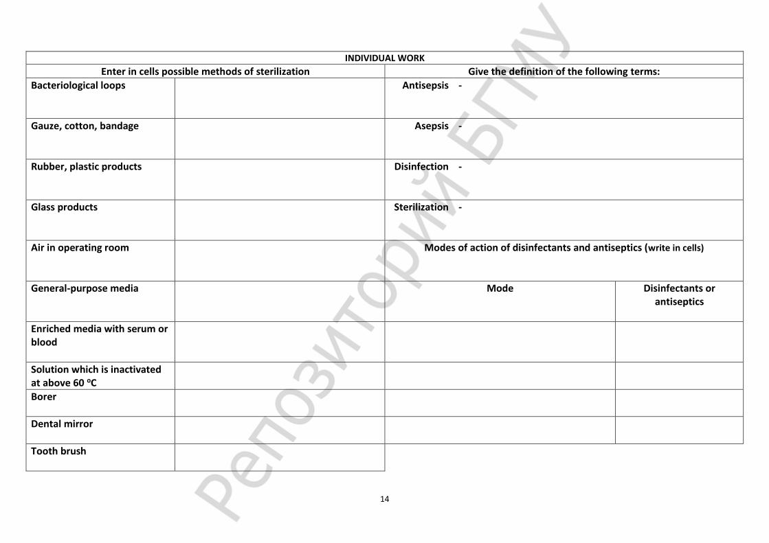
INDIVIDUAL WORK
Enter in cells possible methods of sterilization Give the definition of the following terms:
Bacteriological loops Antisepsis
-
Gauze, cotton, bandage Asepsis
-
Rubber, plastic products Disinfection
-
Glass products Sterilization
-
Air in operating room
Modes of action of disinfectants and antiseptics (write in cells)
General-purpose media
Mode Disinfectants or antiseptics
Enriched media with serum or blood
Solution which is inactivated at above 60 oC
Borer
Dental mirror
Tooth brush

15
Practical class 5. Bacteriological method of laboratory diagnosis of infectious diseases. Techniques for pure culture isolation and maintenance Suggested reading for self-study:
Metabolism and energy exchange in microbes. Constructive and energy metabolism. Types and methods of feeding, nutrient transport through the membrane. Breathing microbes, breathing apparatus, ways of biological oxidation. Aerobic, anaerobic, facultative anaerobes.
Cultivation of microorganisms. Conditions required for growth. Nutrient media for culturing bacteria: classification and characteristics. Culture media ingredients, procedure of preparation and sterilization. General requirements to bacteriologic nutrient media. Incubator.
Bacteriological method of laboratory diagnosis: tasks, procedure, evaluation of the method. Methods of aerobic and anaerobic microorganisms isolation in pure culture. Bacterial colony characteristics.
Signature of the tutor
_______________
Oral quiz
Laboratory work
Individual work
Tests Total
results
Laboratory work
Laboratory exercises Laboratory report
1. Register the results of experiment on antisepsis (see class N 4).
2. Perform the 2nd period of bacteriological diagnosis (inspection and accumulation of aerobic microorganisms pure cultures isolation ):
- characterize morphology of colonies two different types present on agar medium;
- determine morphology and purity of colonies two different types using Gram stain;
- use aseptic technique and transfer the colony of Gram-negative microorganisms for subculturing on a surface of agar slant for microbial biomass accumulation.
The 2ND PERIOD OF BACTERIOLOGICAL DIAGNOSIS
Nutrient agar with isolated colonies
Incubation 24 hours, 37 оС Inoculation of slant media with isolated colony of gram-negative bacteria
Morphology of culture 1
Stain ____________
Morphology of culture 2
Stain ____________
Morphology of colony Colony of culture 1 Colony of culture 2
Shape
Size
Surface
Edge
Color
Consistency
Transparency
Gram stain

16
INDIVIDUAL WORK
Questions for self-control and discussion: Define a pure culture, a mixed culture.
Define a bacterial colony. List four characteristics by which bacterial colonies may be distinguished.
Why should a Petri dish not be left open for any extended period?
Why does the streaking method of plates inoculation result in isolated colonies?
Why are culture media sterilized before use?
Discuss the relative value of broth and agar media in isolating bacteria from mixed cultures.
At what temperature does agar solidify? At what temperature does agar melt?
Define a culture medium.
Discuss some of the physical and chemical factors involved in the composition, and in the preparation, of a culture medium.
Why is it necessary to isolate individual colonies from a mixed growth?
Are the large numbers of microorganisms found in the mouth cause for concern? Explain.
Why are plate cultures incubated in the inverted position?
How do you decide which colonies should be picked from a plate culture of a mixed flora?
Why is it necessary to make pure subcultures of organisms grown from clinical specimens?
How can you determine whether a culture or subculture is pure?
What kinds of clinical specimens may yield a mixed flora in bacterial cultures?
When more than one colony type appears in a pure culture, what are the most likely sources of the extraneous organisms?

17
Practical class 6. Bacteriological method of infectious diseases laboratory diagnosis. Techniques for pure culture identification Suggested reading for self-study:
Identification of microorganisms: approaches and methods. Bacterial species: term definition, species criteria and methods for discovering bacterial species.
Biochemical activities of bacteria and methods for the biochemical properties detection of microorganisms. Enzymes of microorganisms: classification, importance for identification: a) proteolytic (proteases, peptidases, decarboxylases, deaminases, cysteine desulfurase, urease, tryptophanase); b) carbohydrate hydrolyses (carbohydralyses, amylase); c) lipolytic (lipases,
lecithinase); d) oxidative- reductive (dehydrohenase, oxidase, catalase); e) hemolysins; -, β-, -, -hemolysis. Rapid multitest systems for microorganisms identification. Automatic bacteriological analyzers: structure and principle of
bacterial identification.
Signature of the tutor
_______________
Oral quiz Laboratory
work Individual
work Tests
Total results
Laboratory work
Laboratory exercises Laboratory report 1. Perform the 3rd period of bacteriological
diagnosis (identification of aerobic microorganisms pure cultures):
- determine morphology and confirm purity of agar slant culture;
- using stab technique inoculate Hiss media with sucrose, maltose, mannitol for the determination of bacterial carbohydrate hydrolyses;
- using stab and streaking technique inoculate Kligler Iron agar for the determination of bacterial carbohydrate hydrolyses and H2S production;
- using stab technique inoculate semisolid tube medium to detect motility;
- inoculate nutrient broth and test the culture for the indole production.
2. Demonstration: - semisolid and liquid Hiss media with different
pH indicators; - hemolysis on blood agar medium, lecitinase
activity, indol detection; - differentiate among members of the family
Enterobacteriaceae using Kligler Iron agar; - rapid multitest systems for identification of
microorganisms.
Smear ___________ Stain ____________
Key YELLOWY 6,8< RED<8,2 CRIMSON
phenol red
Triple sugar iron agar Semiliquid nutrient medium
Hiss medium sucrose
Hiss medium maltose
Hiss medium mannitol
Nutrient bullion
glucose,
lactose H2S
production Carbo
hydrases cysteinedesulfu
rase
motility
detection
Carbo
hydrase
Carbo
hydrase
Carbo
hydrase
indole
detection tryptophanase
18
INDIVIDUAL WORK
BACTERIOLOGICAL METHOD OF LABORATORY DIAGNOSIS – 5 I’s
1 2 3 4

19
Practical class 7. Molecular Basis of Bacterial Genetics. Molecular methods of infectious diseases diagnosis and bacterial genetic investigations Suggested reading for self-study:
The structure of bacterial genetic apparatus. Regulation of gene expression. General properties and varieties of plasmids. Detection of plasmids. Bacterial variability: phenotypic and genetic. Practical significance of bacterial variability. Mechanisms of genetic variability: Mutation and recombination. Classification of mutations. Methods of mutant bacteria selection.
Molecular methods: tasks, specimens for investigation, advantages of the methods. Molecular hybridization: test materials, DNA extraction, components of DNA hybridization reaction, molecular probes,
detection of DNA hybrid duplexes, interpretation of results. Equipment. Practical application of molecular hybridization method. Polymerase chain reaction (PCR): test materials, principle, DNA extraction, components of PCR reaction mixture, primers,
PCR thermal cycle, detection of amplicons, interpretation of results. Equipment for PCR. Practical application of PCR.
Signature of the tutor
_______________
Oral quiz Laboratory
work Individual
work Tests Total results
Laboratory work
Laboratory exercises Laboratory report
1. Identify isolated pure culture and complete the final report:
- register the biochemical properties of tested pure culture in the table;
- analyze the results and determine the species of tested pure culture.
Species Morphology Biochemical characteristics Conclusion:
According to morphological, cultural, biochemical properties X-microbe is attributed to ______________ * “A” – acid, “G” - gas
Glu
cose
Lact
ose
Mal
tose
Man
nit
o
l Sucr
ose
Н2S
Ind
ole
Mo
tilit
y
E. coli Gram- rods AG AG AG AG - - + +
S. Typhi Gram- rods A* - A A - + - +
S. Paratyphi A Gram- rods AG - AG AG - - - +
S. Schottmuelleri Gram- rods AG - AG AG - + - +
Х-microbe
2. Perform PCR for the detection of M.tuberculosis in the sputum of the patient with tuberculosis suspected. Identification of М.tuberculosis in sputum is based on the detection of gen MPB64 unique for M. tuberculosis and M. bovis. PCR amplifies the fragment with the size 357 bp. of this gene.
Procedure of PCR DNA extraction: Mark the tubes with the volume 1,5 ml with letters S (sputum) and NC (control). Add 100 µl of the sputum to the tube with letter S and 100 µl of negative control to the tube marked with letter NC. Shake the tubes thoroughly and boil in the water bath for 10 minutes (in room 507). PCR cocktail preparation: Mark the tubes with the volume 0,5 ml with letters S (sputum) and NC (control). These tubes contain primers, dNTPs, MgCl2. Add 10 µl of prepared DNA and 10 µl of liquid into PCR' tube. Amplification in special equipment - thermocycler – for approximately 1 hour. Detection of PCR products: Electrophoresis of PCR products in agarose gel. UV detection of specific PCR-products in gel with ethidium bromide.
Report: Specific products sized 357 bp were / not detected. Sputum is positive / negative for Mycobacterium tuberculosis.
20
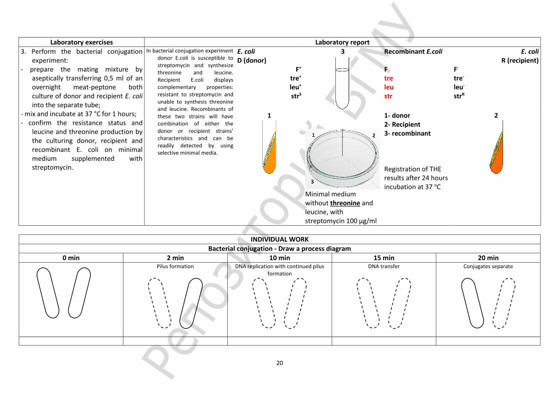
Laboratory exercises Laboratory report
3. Perform the bacterial conjugation experiment:
- prepare the mating mixture by aseptically transferring 0,5 ml of an overnight meat-peptone both culture of donor and recipient E. coli into the separate tube;
- mix and incubate at 37 °C for 1 hours; - confirm the resistance status and
leucine and threonine production by the culturing donor, recipient and recombinant E. coli on minimal medium supplemented with streptomycin.
In bacterial conjugation experiment donor E.coli is susceptible to streptomycin and synthesize threonine and leucine. Recipient E.coli displays complementary properties: resistant to streptomycin and unable to synthesis threonine and leucine. Recombinants of these two strains will have combination of either the donor or recipient strains’ characteristics and can be readily detected by using selective minimal media.
E. coli D (donor)
F+ tre+ leu+ strS
3
Recombinant E.coli F tre leu str
E. coli R (recipient)
F- tre- leu- strR
1
Minimal medium without threonine and leucine, with streptomycin 100 µg/ml
1- donor 2- Recipient 3- recombinant Registration of THE results after 24 hours incubation at 37 оС
2
INDIVIDUAL WORK
Bacterial conjugation - Draw a process diagram
0 min 2 min 10 min 15 min 20 min
Pilus formation
DNA replication with continued pilus formation
DNA transfer
Conjugates separate

21
INDIVIDUAL WORK
The polymerase chain reaction (PCR), complete cells
Stages
Amplification
Evaluation of method
Practical application

22
Practical class 8. Infections. Application of laboratory animals in microbiology. Antibiotic susceptibility testing of microorganisms Suggested reading for self-study:
Defenition of infection. Classification of infections. Bacterial pathogenicity and virulence. Measurements of virulence: ID50, LD50, DLM. The genetics of bacterial pathogenicity. Pathogenicity islands. Pathogenicity factors: adhesins, invasins, impedins, agressins, modulins. The role of bacterial biofilms. Methods of adhesins, capsule, invasins, toxigenicity detection.
Biological method (application of laboratory animals in microbiology): tasks, phases, evaluation of the method. Animal models for infectious diseases. Routs for animal infection. Ethical, humane and legal considerations involved in the use of laboratory animals.
Sources of antibiotics. Spectrum of action. Chemical classification of antibiotics. Mechanisms of action. Side effects. Principles for rational antimicrobial therapy. The problem of resistance to antimicrobials: definitions (intrinsic, acquired resistance), incidence, significance. Resistance mechanisms: non-genetic and genetic origin of drug resistance. Antibiotic susceptibility testing of microorganisms: methods and principles.
Signature of the tutor
_______________
Oral quiz
Laboratory work
Individual work
Tests Total
results
Laboratory work
Laboratory exercises Laboratory report
1. Perform the disk diffusion method (Kirby-Bauer) for determination of antibiotic susceptibility of four different microorganisms which often infect humans - Staphylococcus aureus, Escherichia coli, Pseudomonas aeruginosa, and Klebsiella pneumoniae.
2. Determine antibiotic susceptibility of
microorganisms by agar dilution test. Complete the report.
Pure culture
1,0 ml of inoculum of
microorganisms
Inoculation on Müeller-Hinton agar
Müeller-Hinton agar (composition):
meat extract – 2,0 g; casein hydrolysate – 17,5 g;
corn starch – 1,5 g; agar – 17,0 g;
aqua distillate – 1 l; рН 7,4±0,2
Müeller-Hinton agar
application of antimicrobial discs to the surface of the inoculated agar plate
Incubation at 35оС-24 h
Registration of results
Petri dishes with serial doubled dilutions of Ampicillin in agar media
Interpretation of results, MIC, mcg/l
antibiotic resistant susceptible
Ampicillin ≥32 ≤8 Microbial culture MIC, mcg/ml Interpretation of results
Culture 1
Conclusion: Culture 2 Culture 3 Culture 4

23
3. Determine antibiotic susceptibility of microorganisms by disk diffusion method, complete the report (perform it at classes N 9).
4. Demonstration: - agar disk diffusion test for antibiotic
susceptibility testing of microorganisms; - rapid test for antibiotic susceptibility testing
of microorganisms; - slide of Bacillus anthracis in tissues of white
mouse, Gram stain; - slide of Y.pestis in tissues of white mouse,
Gram stain; - slide of Klebsiella pneumoniae
rhinoscleromatis in tissues of white mouse, Gram stain.
Results of pure culture ________________testing by disc diffusion method
Antibiotic Diameter of inhibition zones (mm)
Antibiotic Diameter of inhibition
zone , mm Interpretation of results
resistant susceptible
Staphylococcus spp. Penicillin ≤28 ≥29
Oxacillin S.aureus ≤10 ≥13
CNS ≤17 ≥18
Canamycine ≤13 ≥18
Gentamicin ≤12 ≥15
Ciprofloxacin ≤15 ≥21
Tetracycline ≤14 ≥19
Erythromycine ≥23 ≥23
Lincomycine ≤13 ≥21 0,5
µg/ml 1,0
µg/ml 2,0
µg/ml 4,0
µg/ml 8,0
µg/ml 16,0
µg/ml 32,0
µg/ml Control Chloramphenicol <17 ≥18
Enterobacteriaceae
Ampicillin ≤13 ≥17
Cefazolin ≤14 ≥18
Cefotaxime ≤14 ≥23
Canamycine ≤13 ≥18
Gentamicin ≤12 ≥15
Ciprofloxacin ≤15 ≥21
Lomefloxacin ≤18 ≥22
Tetracycline ≤14 ≥19
Doxicycline ≤12 ≥16
Chloramphenicol ≤12 ≥18
DDM report (formulate what antibiotics can be recommended for the
therapy):
BDT report: minimal inhibitory concentration of antibiotic is ___________ μg/ml.
4-1 Smear ____________
Stain ____________
4-2 Smear ____________
Stain _____________

24
INDIVIDUAL WORK
Define the target action of antibiotics Mechanisms of action of antimicrobial drugs (write in cells)
Side effects of antimicrobial drugs (write in cells)
Pathogenicity factors’ groups (write in cells)
Mechanisms of resistance of bacteria to an antimicrobial agents (write in cells)
DNA-directed RNA polymerase, Cell wall synthesis, RNA elongation, protein synthesis (50S inhibitors), protein synthesis (30S inhibitors), Folic acid metabolism, Cytoplasmic membrane structure, protein synthesis (tRNA)

25
INDIVIDUAL WORK
Interacting factors of antimicrobial therapy (write in circle)
Characteristics of ideal antimicrobial drug: Analyze the circuit in the picture (in the middle) and answer next. Which of the resistance mechanisms are shown in the figure?
Give the definition of the following terms:
Methods of the antibiotic susceptibility testing (write methods and indicate possibility to determine MIC)
Antibiotic -
Specific antibacterial
therapy
-
Minimal inhibitory
concentration
-
Multiple resistance
-
Pathogenicity -

26
Practical class 9. Credit “Morphology and physiology of microorganisms”
List of questions Oral quiz Script Tests Total results
1. History of microbiology as a science. Periods. The founders of microbiology main routs. 2. Microscopic method of examination: tasks, procedure, evaluation of the method. 3. Bright-field light microscope: components and proper use of the microscope. Dark-field light microscopy: the
principle behind dark-field microscopy. Phase-contrast light microscope: basic principles behind phase-contrast microscopy. Fluorescence microscopy: principles behind the fluorescence microscopy. The technique of oil immersion microscopy.
4. Type of microscopic preparations. Smear preparation and fixation. Simple methods of staining. 5. Differential stains of microorganisms. Gram stain: medical application, principles, procedure for Gram stain. 6. Morphology of bacteria. Distinctive features of prokaryotic and eukaryotic cells. Basic morphological forms of
bacteria. Morphological characteristics of cocci, rods and spiral-shaped bacteria. Motility of bacteria, methods of detection.
7. Structure and function of cell envelope and appendages. Capsule. Detection methods of the capsule. 8. The composition, function, detection methods of bacterial cell wall. The cell wall of gram-positive bacteria. The
cell wall of gram-negative bacteria. Bacterial forms with defective cell wall. Factors inducing cell wall removal, medical importance of L-forms.
9. Bacterial core: cytoplasm, cytoplasmic structures; their functions and detection methods. Acid-fast bacteria and unique properties of their cell wall. Methods of acid-fast staining: medical application, principle, procedure.
10. Resting forms of microorganisms. Bacterial endospores: medical importance, properties of endospore, the periods of endospore formation, detection methods (principles, procedures).
11. Taxonomy of microorganisms: classification and nomenclature. Modern approaches to taxonomy of microorganisms. Taxonomic ranks. Vars (types), strains, clones, pure cultures.
12. Taxonomy, morphology, medical significance of the spirochetes. Methods for spirochetes detection. 13. Taxonomy, morphology, medical significance of Actinomyces. 14. Taxonomy, morphology, medical significance of Mycoplasmas. Methods for Mycoplasmas investigations. 15. Taxonomy, morphology, medical significance of Chlamydiae and Rickettsiacea. 16. Nutrition of microorganisms. Source of macro- and micronutrients, growth factors. Nutritional types. Transport
mechanisms for nutrient absorption. 17. Energy strategies in microorganisms. Aerobic and anaerobic respiration. Structures involved in respiration in
microorganisms. 18. Reproduction of microorganisms. Mechanisms and phases of bacterial division. 19. Bacteriological method of laboratory diagnosis: tasks, procedure, evaluation of the method. 20. Cultivation of microorganisms. Conditions required for growth. Nutrient media for culturing bacteria: classification
and characteristics. Culture media ingredients, procedure of preparation and sterilization. General requirements to bacteriologic nutrient media.
21. Methods of aerobic microorganisms isolation in pure culture. 22. Methods of anaerobic microorganisms isolation in pure culture. Cultivation of anaerobic bacteria: culture media,
techniques, equipment. 23. Identification of microorganisms: morphological, cultural, serologic, biological, genetic. 24. Biochemical identification of microorganisms. Detection of: a) proteolytic enzymes; b) carbohydrate hydrolyses
enzymes; c) lipolytic enzymes; d) oxidative- reductive enzymes; e) hemolysins. Automatic stations for identification of bacteria.
25. The structure of bacterial genetic apparatus. Phenotype, genotype, genome, genes. Regulation of gene expression. General properties and varieties of plasmids. Detection of plasmids.
26. Bacterial variability: phenotypic and genetic. Practical significance of bacterial variability. Population variability. 27. Molecular methods in diagnosis of infection diseases: aims, methods, advantages. Molecular hybridization and
polymerase chain reaction: principles of the methods. 28. Doctrine regarding infections. Terms for emergence of infectious disease. Basic terminology of infectology.
Classification of infections. 29. Role of microorganisms in infection emergence. Bacterial pathogenicity and virulence. The genetics of bacterial
pathogenicity. Pathogenicity islands. Pathogenicity factors: adhesins, invasins, impedins, agressins, modulins. 30. Role of microorganisms, social and physical factors in infection emergence. 31. Biological method (application of laboratory animals in microbiology): tasks, phases, evaluation of the method. 32. Chemoprophylaxis and chemotherapy; antimicrobial chemotherapeutic agents and antibiotics. Sources of
antibiotics. Especially the use of antibiotics in dentistry. 33. Mechanisms of antibiotics action. Side effects of antibiotics. Principles for rational antimicrobial therapy. 34. The problem of resistance to antimicrobials: definitions (intrinsic, acquired resistance), incidence, significance.
Resistance mechanisms. 35. Antibiotic susceptibility testing of microorganisms: methods and principles. 36. Ecology of microorganisms. Basic terminology of ecology. 37. Asepsis: definition, surgical, medical asepsis, asepsis in microbiological laboratory. 38. Sterilization: definition, methods of sterilization (physical, chemical, mechanical), quality control. 39. Disinfection: definition, methods of disinfection. 40. Antisepsis: definition, methods of antisepsis. Disinfectant and antiseptics: classification and modes of action. List of practice. 1. Prepare heat-fixed slide of bacteria, cultured on agar medium, stain with methylene blue. 2. Prepare heat-fixed slides of bacteria, cultured on liquid medium, stain with basic fuchsin. 3. Prepare heat-fixed slides of bacteria, cultured on liquid medium, stain by Gram. 4. Technology immersion microscopy. 5. Determine the morphology of Staphylococcus, pure culture, Gram stain. 6. Determine the morphology of E. coli, pure culture, Gram stain. 7. Determine the morphology of Gram+ and Gram- bacteria into the mix, Gram stain. 8. Determine the morphology of the culture in smear colored by Ginsu-Burri. 9. Define streptobacill pure culture morphology, Gram stain coloring. 10. Determine antibiotic susceptibility of microorganisms by disk diffusion method. 11. Characterize morphology of two different types of colonies present on agar medium.

27
Practical class 10. Immune system. Innate immunity. Methods for innate immunity factors evaluation Suggested reading for self-study:
Human immune system: organs, cells, molecules (CD; receptors; MHC I, II, III; cytokines, adhesion molecules etc.). Immunity, types of immunity. Innate immunity. Immune and not-immune factors. Complement system: composition, way of activation, functions.
Methods for estimation of complement system activity. Lysozyme, b-lysins. Polynuclear and mononuclear phagocytes systems. Phagocytosis: phases, intracellular killing mechanisms, outcomes.
Dendritic cells. Methods for estimation of phagocytosis. Natural killer cells. Antigen-presenting cells. TOLL-like receptors.
Signature of the tutor
_______________
Oral quiz Laboratory
work Individual
work Tests Total results
Laboratory work
Laboratory exercises Laboratory report
1. Determine phagocytosis parameters in prepared slides stained by Gimza method.
2. Complete the drawings of slides seen in demonstration room:
- incomplete phagocytosis of N. gonorrhoea.
- incomplete phagocytosis of K. rhinoscleromatis.
3. Register the complement system
activity by 50% hemolysis method. Serum is diluted and added in wells from 0,05 to
0,5 ml. Then saline solution is added to the final volume of 1,5 ml. 1,5 ml of hemolytic system is added to each well. Reaction is incubated at 37oC for 45 min, cooled at 4 oC and centrifuged at 1500 rpm for 5 min. The well in which 50% hemolysis occurred is determined visually. This means the volume of patient’s serum that contains one unit of CH50. Then the CH50 for the whole serum is calculated.
Staphylococci are mixed with leucocytes (50:1) and incubated at 37 oC for 15-120 min. Then slides are prepared and stained by Gimza method. Under oil immersion the phagocyting leucocytes and phagocyted staphylococci are counted and phagocytosis parameters calculated.
PI (Phagocytosis index) = Number of phagocyting leucocytes / All leucocytes counted Norma* - 40-60 %. PN (Phagocytosis number) = Number of phagocyted staphylococci / Number of phagocyting leucocytes Norma* - 4-7.
Smear ________________ Stain _________________
Smear ________________ Stain _________________
Volume of diluted (1:10) serum, ml 1 СН50 – in ______ ml serum Х СН50 – in 1 ml serum N 40 – 60 СН50
0,05 0,1 0,15 0,2 0,25 0,3 0,35 0,4 0,45 0,5 50% hemolysis
Results:

28
INDIVIDUAL WORK
Fill cells with types of immunity
immunity, adoptive, passive, natural, artificial, immune factors, humoral, cellular, non-immune factors, active
Fill with sample of
Organs of immune system Cells of immune system Molecules of immune system
Write in cells ligand of receptors Associate the scientist and his discovery Pattern
Recognition Receptors
Ligand pathogen-associated molecular patterns
Edward Anthony Jenner
Phagocytosis, Cell-mediated immunity
TLR1 Élie Metchnikoff
Chemical structure of antibodies
TLR2 Polly Celine Eveline Matzinger
Smallpox vaccine, vaccination
TLR3 Charles Alderson Janeway
side chains, humoral immune response
TLR4 Rodney Robert Porter
Gerald M. Edelman
Diphtheria antitoxin
TLR5 Karl Landsteiner
Danger model, danger theory
TLR6 Paul Ehrlich
Immune tolerance
TLR7 Jules Jean-Baptiste Vincent
Bordet
pattern recognition theory
TLR8 Emil Adolf von Behring
complement
TLR9 Frank Macfarlane Burnet
blood group system, Rh factor, poliovirus

29
INDIVIDUAL WORK
Compare Nose-Associated Lymphoid Tissue 1 – 2 – 3 – 4 – 5 –
INNATE IMMUNITY ADOPTIVE/ACQUIRED IMMUNITY
Complement system Phases of phagocytosis (write in cells)
Activation pathway
activators
C3-convertase
C5-convertase
MAC development
The illustration shows the process of phagocytosis. Draw a picture of the possible outcomes of the process in adjacent cells and named them.

30
Practical class 11. Antigens. Antibodies. Immune response Suggested reading for self-study:
Immune response, definition, main factors. Antigens: definition, main features, classification. B-lymphocytes system. B cells genesis. B cell receptor (BCR). B-cell activation, proliferation, differentiation to plasmocyte,
immunoglobulin production. Humoral immune response. Primary and secondary humoral response. Immunoglobulins: structure, functions. Classes and subclasses of immunoglobulins. Monoclonal immunoglobulins.
Methods of B-lymphocytes evaluation: quantitative and functional tests. T lymphocyte system. T-cell markers. TCR. Genetic control of TCR diversity. T-lymphocytes subpopulations: helpers, killers,
DTH-effectors, regulators. T helpers of 1, 2, 3 and 17 types. Cellular immune response and its phenomena. Interaction and control of the immune system. Methods for evaluation of T- and B-lymphocytes system: quantitative and functional tests.
Signature of the tutor
_______________
Oral quiz Laboratory
work Individual
work Tests
Total results
Laboratory work
Laboratory exercises Laboratory report
1. Determine the quantity of B-cells by immune rosettes methods in ready-made slides.
2. Complete the drawings of slides seen
in demonstration room: - immune rosettes method for T-cell quantity
determination (Romanowsky-Giemsa stain);
- blast transformation of lymphocytes (Romanowsky-Giemsa stain);
- determine an IgG, A, M concentration in serum by Manchini method (simple radial gel immunodiffusion).
N Count N Count N Count The method reveals CD20 antigen on B-cell surface; Normal В-cells count by CD20 = 8-20% total blood lymphocytes.
BCD20= rosette’s Cell/30=
Conclusion:
Smear ________________ Stain _________________
Smear ________________ Stain _________________
1 11 21
2 12 22
3 13 23
4 14 24
5 15 25
6 16 26
7 17 27
8 18 28
9 19 29
10 20 30

31
INDIVIDUAL WORK
Write figures for elements of an immunoglobulin molecule indicated on scheme
Enter the names of structures of bacteria, which are antigens
Light chain (L)
Variable domen of the light chain
Constant domen of the light chain
Heavy chain (H)
Variable domen of the heavy chain
Constant domen of the heavy chain
Hinge fragment
Fc- fragment
Fab- fragment
Active center
Fc-receptor ligand
Write the main cells and molecules that are involved in the humoral immune response
Write down the characteristics of immunoglobulin according to class and molecule structure
structure characteristics class
cells molecules
Ig A
Ig D
Ig E
Ig G
Ig M

32
INDIVIDUAL WORK
According to the following diagram, draw a graph of dynamics of immunoglobulins G and M classes for primary and secondary immune responses.
Write methods of the study of cellular immunity
Draw the B-lymphocyte
CD4 CD8 CD40b BCR
sIgM CD3 TCR TCRα,β
sIgD CD19 IL4r ACR
CD52 CD20 ILR HLA
CD45 CD23 CD37 CD11C
CD79a CD79b CD38

33
Practical class 12. Serological method Suggested reading for self-study:
Serological method, characteristics. Antibody titre. Diagnostic titre. Diagnosticum. Diagnostic serum. Agglutination, passive agglutination, reversed passive agglutination, latex agglutination. Precipitation. Ring precipitation test, double immunodiffusion in a gel (by Ouchterlony), simple radial
immunodiffusion in a gel (by Mancini), immunoelectrophoresis, electroimmunodiffusion. Immune lysis reactions. Complement fixation test: ingredients, implementation, characteristics. Immunofluorescence test: direct and indirect variants. Immunoenzyme test. ELISA. Radioimmune test.
Signature of the tutor _______________
Oral quiz Laboratory
work Individual
work Tests Total results
Laboratory work
Laboratory exercises Laboratory report
1. Perform slide agglutination test to identify an X-bacteria.
1. antiserum S.Typhi
2. antiserum E.coli
3. Saline X-bacteria Conclusion: X-microbe is _________________
2. Determine the result of the complement fixation test.
3. Determine the result of passive
hemagglutination reaction.
CFT 1:20 1:40 1:80 1:160 1:320 SC AC
Key “+” “-“
Assess: Conclusion:
PASSIVE BLOOD AGGLUTINATION TEST
Key 1/10 1/20 1/40 1/80 1/160 1/320 1/640 SC AC
“+”
“-“
Assess:
Conclusion:
34
Laboratory exercises Laboratory report
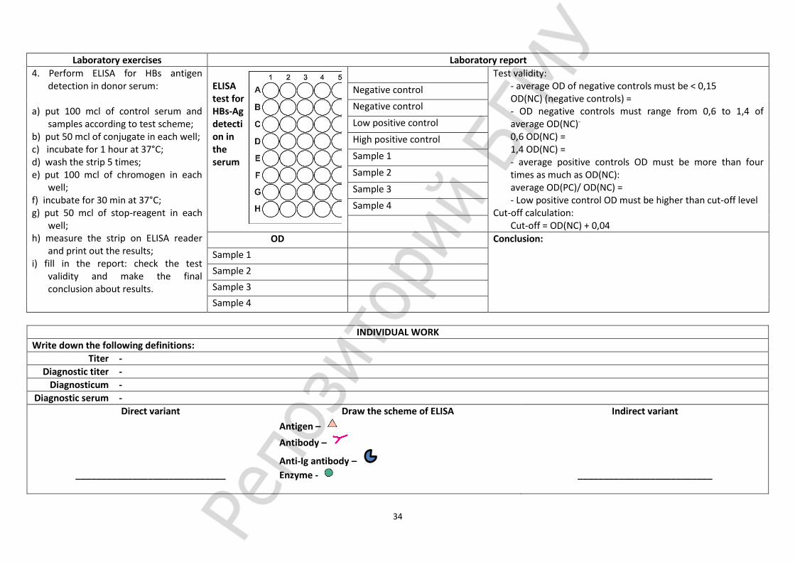
4. Perform ELISA for HBs antigen detection in donor serum:
a) put 100 mcl of control serum and
samples according to test scheme; b) put 50 mcl of conjugate in each well; c) incubate for 1 hour at 37°С; d) wash the strip 5 times; e) put 100 mcl of chromogen in each
well; f) incubate for 30 min at 37°С; g) put 50 mcl of stop-reagent in each
well; h) measure the strip on ELISA reader
and print out the results; i) fill in the report: check the test
validity and make the final conclusion about results.
ELISA test for HBs-Ag detection in the serum
Test validity: - average OD of negative controls must be < 0,15 OD(NC) (negative controls) = - OD negative controls must range from 0,6 to 1,4 of average OD(NC)- 0,6 OD(NC) = 1,4 OD(NC) = - average positive controls OD must be more than four times as much as OD(NC): average OD(PC)/ OD(NC) = - Low positive control OD must be higher than cut-off level
Cut-off calculation: Cut-off = OD(NC) + 0,04
Negative control
Negative control
Low positive control
High positive control
Sample 1
Sample 2
Sample 3
Sample 4
OD Conclusion:
Sample 1
Sample 2
Sample 3
Sample 4
INDIVIDUAL WORK
Write down the following definitions:
Titer -
Diagnostic titer -
Diagnosticum -
Diagnostic serum -
Direct variant
_____________________________
Draw the scheme of ELISA
Antigen –
Antibody –
Anti-Ig antibody –
Enzyme -
Indirect variant
__________________________

35
INDIVIDUAL WORK
Write name of reaction’ type in the first and in the second case:
What type of serologic test is depicted? Give explanations and result for both variant:
Write labels for next types of assays:
Immunofluorescence ELISA
Radioimmune test

36
Practical class 13. Immunoprophylaxis and immunotherapy. Immunopathology and clinical immunology Suggested reading for self-study:
Immunoprophylaxis and immunotherapy. Vaccines, classification, essential characteristics. Vaccinal immunity, factors affecting its development. Methods of vaccinal immunity evaluation. Passive immunoprophylaxis. Immune sera and serum preparations; methods of its production and application. Allergy, periods, types. Immediate type of hypersensitivity mechanisms: mediator type (I), cytotoxic type (II), immune complex type (III). Delayed type
of hypersensitivity mechanism (IV). Drug allergy. Allergens in dentistry. Methods for allergic conditions diagnostics. Clinic immunology: definition. Immune status. Immunogram. Primary and secondary immunodeficiency. Autoimmune disease. Causes, manifestation. Autoantibodies, diagnostic value, methods of determination. Antitumor immunity. Methods of immune
status correction. Immunosuppression. Immunostimulation. Immunomodulators. Thymus, spleen, bone marrow substances. Interleukins, interferons.
Signature of the tutor
_______________
Oral quiz Laboratory
work Individual
work Tests
Total results
Laboratory work
Laboratory exercises Laboratory report 1. Perform the passive hemagglutination test
for the detection of rheumatoid factor. Diagnosticum = armed bull erythrocytes
coated with human IgG. Rheumatoid factor is an autological antibody
(IgM) to IgG. It is found in certain autoimmune diseases (SLE, RA etc.) and is useful for diagnostics.
2. Perform the LA test to detect autoantibodies to thyreoglobulin
Latex diagnosticum = latex microsphera coated with thyreoglobulin molecules
3. Demonstration: - degranulation of mast cells, Romanowsky-
Giemsa stain;
- Allergens;
- Medicine for correction.
1. Saline
2. Patient’s serum
3. ER Diagnosticum
1. Saline
2.Patient’s serum
3. Latex Diagnosticum
Smear ________________ Stain _________________
Conclusion:
Conclusion:
INDIVIDUAL WORK
Write down the types of allergy by P.G.H.Gell and P.R.A.Coombs (1964):

37
INDIVIDUAL WORK
What type of allergy phenomena is depicted? Give explanations.
The vaccines for active immunization can be divided into four groups:
What are the two phenomena are depicted in the diagram. Give explanations.
Write major allergens of drug allergy:

38
Practical class 14. Test “Immunology. Immunity. Allergy”
List of questions Oral quiz Script Tests Total results
1. Immunology. Definition, tasks, methods. History of immunology. 2. Immune system. Characteristics. Organs, cells, molecules of the immune system. 3. Cytokines. Definition, classification. Biological importance. 4. Immunity: definition, classification. Characteristics of anti-infection immunity. 5. Innate immunity: definition, immune and non-immune factors, characteristics. 6. Complement system: definition, ways of activation, functions. Medical importance.
Methods of complement activity evaluation. 7. Phagocytosis. Phagocytes. Phagocytosis phases. Phagocytosis outcome (complete,
incomplete). Chemotaxins, opsonins: origin and medical importance. 8. Phagocytosis evaluation methods. 9. Immune response and factors influencing its strength. 10. B-lymphocytes, characteristics, main markers. Humoral immune response, periods. 11. Methods for B-lymphocytes quantity and functional activity evaluation. 12. Antigens: structure, classification, characteristics. 13. Bacteria antigenic structure. Cross-reacting antigens. 14. Antibodies, structure-functional organization of immunoglobulin molecule, characteristics.
Antiidiotypic and monoclonal antibodies. 15. Classes of immunoglobulins, characteristics. 16. Mechanisms of antigens and antibodies interactions. Specificity. Phases. Affinity. Avidity. 17. Serology reactions, characteristics. Tasks, periods, clinical importance. 18. Agglutination reaction. Methods of conduction and result registration. Medical
importance. 19. Passive hemagglutination, ingredients. Methods of conduction and result registration.
Medical importance. Reversed passive agglutination test. Latex agglutination. 20. Precipitation reaction. Methods of conduction and result registration. Medical importance. 21. Immunofluorescence test. Medical importance. 22. Immunoenzyme analysis. ELISA. Ingredients, methods of conduction, results registration,
characteristics. Medical importance. 23. Immune lysis reactions. Hemolysis. 24. Complement fixation test. Ingredients, methods of conduction, results registration,
characteristics. Medical importance. 25. T-lymphocytes system, characteristics. Cellular immune response, dynamics. 26. Methods for T-lymphocytes quantity and functional activity evaluation. 27. Allergy: definition, classification. Allergy phases and types. 28. Allergens: definition, classification, characteristics.
29. Allergic reaction of immediate type, clinical phenomena. 30. Mediator type of ITH: definition, mechanisms, clinical phenomena, approaches for
prophylaxis. 31. Cytotoxic (II) and immunocomplex (III) ITH types: definitions, mechanisms, clinical
phenomena. 32. Hypersensitivity of delayed type (IY): definition, classification, clinical phenomena. 33. Methods for ITH diagnostics (in vivo and in vitro). 34. Methods for DTH diagnostics (in vivo and in vitro). 35. Immune tolerance: definition, mechanisms, medical importance. 36. Transplantation immunity. MHC antigens of I, II, III types, role for an immune response
development. Transplantological reactions. Mechanisms of transplant rejection. Prophylaxis. 37. Clinical immunology: definition, aims. 38. Primary and secondary immunodeficiencies: definitions, classification, medical
importance. 39. Immune status: definition, methods for evaluation. Influence of life way on the immune
system function. 40. Autoimmune diseases, classification. Autoantigens. Mechanisms of autoimmunity. 41. Immunoprophylaxis and immunotherapy of infections. Achievements and problems. 42. Vaccines, main demands. Classification, characteristics, approaches to development. New
vaccines. 43. Vaccinal immunity. Factors influencing vaccinal immunity. 44. Passive immunoprophylaxis. Antisera for therapy and prophylaxis, medical importance. 45. Immunocorrection. Methods for suppression and stimulation of the immune response,
drugs for immunocorrection. List of practice.
1. Register the result of agglutination test. 2. Register the result of gel immunoprecipitation test. 3. Register the result of complement fixation test. 4. Register the result of passive hemagglutination test. 5. Perform the slide agglutination test 6. Determine the immunoglobulins concentration. 7. Determine T-lymphocytes quantity in ready slide by immune rosettes method. 8. Determine phagocytosis indices in ready slides

39
Practical class 15. Microbiological diagnostics of diseases caused by Staphylococci, Streptococci, Neisseria Suggested reading for self-study:
Staphylococci, general characteristics. Pathogenicity factors. Staphylococcal infection, including dentistry. Staphylococci as causative agents of nosocomial infections. Methods of staphylococcal infections microbiological diagnostics. The material for the research depending on the infection form. Scheme of pure culture isolation (from pus, mucus, blood, etc.). Identification methods, phagetyping of Staphylococci. Specific prevention and treatment of staphylococcal infections.
Streptococci, systematics, general characteristics. Antigenic structure. S.pyogenes, S.pneumoniae, S.mutans and other spp of the oral cavity. The role in the health and pathology of the oral cavity. Acute and chronic diseases, pathogenesis, immunity. Methods for streptococcal infections diagnosis. Bacteriological method, study design. Material for studies depending on the form of the infection, the rules and methods for taking material. Principles of therapy and prevention streptococcal infections.
Neisseria. Systematics, general characteristics. The role in the health and pathology of the oral cavity. Meningococcus, gonococcus. Pathogenicity factors. Pathogenesis and immunity. Microbiological diagnostics, material for studies. Specific prevention and treatment.
Signature of the tutor
___________
Oral quiz Laboratory
work Individual
work Tests
Total results
Laboratory work - practical class’ duration in second semester is 2 academic hours 15 minutes
Laboratory exercises Laboratory report
1. Microbiological diagnostics of staphylococcal infection, 2nd period:
- macro- and microscopic examination of the colonies on YSA;
- plasmacoagulase test (stabilized rabbit plasma, 37оС, 2-4-24 h).
Smear ________________ Stain _________________
Conclusion: according to morphological, cultural and biochemical properties unknown bacterium is identified as _______________________
Staphylococcal colonies
shape (form)
size/elevation
surface (appearance)
edge (margin)
pigmentation
consistency
transparency
lecithinase
2. Microbiological diagnostics of streptococcal infection, 3rd period:
- the description of Streptococci growth in serum broth;
- determining the morphology of streptococci, Gram staining;
- determination of streptococcus serogroups by ring precipitation test.
Smear ________________ Stain _________________
Conclusion: according to morphological, cultural and biochemical properties unknown bacterium is identified as _______________________

40
Laboratory exercises Laboratory report
3.Demonstration: - Staphylococcus aureus in pus, Gram staining; - Streptococcus pneumonia, pure culture,
Gram staining; - S.pneumoniae, white mice, Gram staining; - Neisseria gonorrhoeae in pus, Gram staining; - Neisseria meningitidis in cerebrospinal fluid,
methylene blue; - the growth of staphylococci on YSA, blood
agar, broth; - the growth of streptococci on blood agar and
broth; - coagulase test (plasma); - anaerobic mannitol fermentation; - phage typing of staphylococci.
Smear ________________ Stain _________________
Smear ________________ Stain _________________
Smear ________________ Stain _________________
Smear ________________ Stain _________________
Smear ________________ Stain _________________
INDIVIDUAL WORK

41
Practical class 16. Microbiological diagnostics of acute enteric infections caused by Enterobacteria. Methods for food poisoning diagnostics Suggested reading for self-study:
General characteristics of Enterobacteriaceae family. Escherichia, general characteristics. The biological role of Escherichia coli in health and pathology. Salmonella, classification and general characteristics. The role in the pathology, the pathogenesis of typhoid,
manifestations in the oral cavity. Shigella, classification, general characteristics. The role in pathology. Common principle of microbiological diagnosis of acute intestinal infection. Etiology of food poisoning. Principles of microbiological diagnostics.
Signature of the tutor
___________
Oral quiz Laboratory
work Individual
work Tests Total results
Laboratory work
Laboratory exercises Laboratory report
1. Demonstration: - E. coli, pure culture, Gram staining; - Salmonella typhi pure culture, Gram staining; - Shigella flexneri pure culture, Gram staining; - clean media: Endo, Levin, Ploskirev, bismuth
sulfite agar, Rapoport, magnesium, Kliglera; - the same media with the growth of E. coli,
Salmonella, Shigella; - biochemical activity of E. coli and Salmonella;
2. Slide agglutination test with diagnostic O and H-serum for identification of Salmonella.
Smear ________________ Stain _________________
Smear ________________ Stain _________________
Smear ________________ Stain _________________
Slide agglutination test
Conclusion:

42
Practical class 17. Microbiological diagnostics of diseases caused by Klebsiella, Campylobacter, Helicobacter and Pseudomonada Suggested reading for self-study:
Klebsiella, classification and general characteristics, main diseases caused. Campylobacter, general characteristics, role in human pathology. Mechanisms of pathogenesis. Diagnosis of
campylobacteriosis. Helicobacter. Pseudomonas aeruginosa, general characteristics, role in human pathology.
Signature of the tutor
___________
Oral quiz Laboratory
work Individual
work Tests
Total results
Laboratory work
Laboratory exercises Laboratory report
1. Microbiological diagnostics of Klebsiellosis, 3rd period:
- determine the biochemical properties of Klebsiella;
- perform slide agglutination test with anti-capsule diagnostic sera and determine the K-antigen;
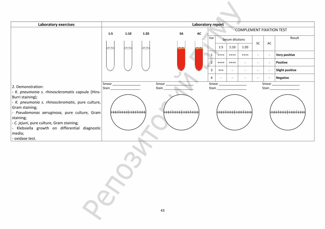
- determine the titer of CFT for serological diagnosis of Scleroma.
Russell
2
3
4
5
6
7
Smear ________________ Stain _________________
Slide agglutination test with anti-capsule serum
K3 K4 CA Conclusion:________________________
Biochemical properties
K. pneumoniae
s. rhinoscleromatis s. ozaenae s. pneumoniae
1, 2 Glucose (A+G) - +/- +
1, 3 Lactose - +/- +
4 Saccharose (4th day) - +/- +
5 Citrate - +/- +
6 Urea - -/+ +
7 Malonate + - +
8 Antigens O2a:K3 O2b:K4 O1,3-5:K1-3
43
Laboratory exercises Laboratory report
2. Demonstration: - K. pneumonia s. rhinoscleromatis capsule (Hins-Burri staining); - K. pneumonia s. rhinoscleromatis, pure culture, Gram staining; - Pseudomonas aeruginosa, pure culture, Gram staining; - C. jejuni, pure culture, Gram staining; - Klebsiella growth on differential diagnostic media; - oxidase test.
1:5
1:10
1:20
SA
AC
COMPLEMENT FIXATION TEST
Var Serum dilutions SC AC
Result
1:5 1:10 1:20
1 ++++ ++++ ++++ - - Very positive
2 ++++ ++++ - - - Positive
3 +++ - - - - Slight positive
4 - - - - - Negative
Smear ________________ Stain _________________
Smear ________________ Stain _________________
Smear ________________ Stain _________________
Smear ________________ Stain _________________
44
Practical class 18. Final test “General microbiology. Immunology”
List of questions Oral quiz Script Tests Total results
1. Microbiology: definition, area and fields of microbiology, methods of investigation. Dental microbiology: goals,
objectives, role in the dentist’s practice.
2. Milestones (periods) in microbiology. Work of Louis Pasteur, Robert Koch, Ilya Mechnikov. Evolution of microorganisms and infectious diseases.
3. Common with other organisms and the unique features of microorganisms. Principles of microorganisms systematics . Classification and nomenclature of microorganisms. The term of “species” in bacteria: group of traits for species identification (criteria for speciation).
4. Morphology of bacteria. Basic morphological forms of bacteria. The bacterial cell structure. Functions of the surface and cytoplasmic structures of the bacterial cell. Mechanism of Gram staining. Forms of bacteria with the cell wall defects.
5. Unique features of metabolism in prokaryotes. Nutrition of bacteria: types, requirements of bacteria, nutrients and pathways of nutrients penetration into the bacterial cell. Nutrient media: specification (what they should be to provide the best growth of bacteria), classification.
6. Respiration of microorganisms: types, pathways of energy production. Enzymes and cell structures involved into the process of respiration. Classification of bacteria regarding their oxygen requirements.
7. Growth and reproduction of bacteria. The mechanism of simple division and its phases. Dormant forms of microorganisms: general characteristics, factors inducing their formation, medical importance.
8. Sampling for microbiological studies: types of samples, the rules of sampling, storage, transportation. Principles of organization, equipment and levels of biosafety in microbiological laboratories.
9. Microscopic (bacterioscopic) method of diagnosing the infectious diseases: definition, aim and tasks, steps and evaluation of specificity, sensitivity, disadvantages of the method. Types of microscopic preparations. Staining of microorganisms: methods. Types of microscopes.
10. The bacteriological method of the infectious diseases diagnosing: aim, tasks, phases, and evaluation of specificity, sensitivity, disadvantages of the method.
11. Methods for isolation identification of aerobic and anaerobic bacteria pure cultures. Identification of microorganisms without pure culture isolation.
12. Genetic apparatus of bacteria (nucleoid, plasmids, transposons, IS-elements) characteristics, functions, effect and importance. The concept of genetic engineering and biotechnology.
13. Inheritance and variability of microorganisms. Types of variability. Mutations. The genetic recombination of bacteria. Phenotypic variability. The practical significance of the variability of microorganisms in the diagnosis, treatment and prevention of infectious diseases.
14. Molecular biological method of diagnosing the infectious diseases (molecular hybridization, polymerase chain reaction): definition, the principle of the methods, application in dentistry.
15. Effect of physical and chemical factors on microorganisms. Disinfection: definition of the term, aim and tasks, types, disinfectants, methods of disinfection quality control.
16. Sterilization: the term definition, methods, quality control. Sterilization of instruments and medical devices. Consequences of sterilization errors.
17. Infection (infection process): the term definition, causes and conditions of infectious diseases emergence. Differences in communicable and non-communicable diseases. Periods of infectious diseases. Infectious disease classification and outcomes.
18. The role of microorganisms in the infectious process. Pathogenicity and virulence. Factors of pathogenicity of microorganisms. Pathogenicity Island. Microbial toxins. Types of exotoxins and their biological properties. Mechanisms of microbial persistence and latency in host’s organisms.
19. The role of host, social, environmental factors in the infectious process.
20. The biological (experimental) method of diagnosing the infectious diseases: definition of the term, aim, tasks, phases, evaluation. Disbiosis: causes, consequences, prevention. Gnotobiology.
21. The ecology of microorganisms. Types of ecological relationships in microorganisms. The role of microorganisms in the genesis and development of the Biosphere (the concept of the microbial dominance). Spread of microorganisms in the nature.
22. The chemotherapy and chemoprophylaxis of infectious diseases. Groups of antimicrobial chemotherapeutic agents, mechanisms and spectrum of action on microbial cells. Chemotherapeutic index.
23. The antisepsis: definition of the term, types, categories, methods of application. Antiseptic agents: classification, mechanism of action, side effects. Principles of rational antisepsis in dental practice.
24. The antibiotics: characteristics, classification, mechanisms of action. The rational antibiotic therapy principles. Antibiotics for bacterial complications prophylaxis. Side effects of antibiotics.
25. Natural and acquired resistance of microorganisms to antibiotics. The genetic and biochemical mechanisms of microorganisms resistance. Genotypic and phenotypic methods for determining the microorganisms susceptibility to antibiotics.
26. Immunology: definition of the term, aim and task, methods, history of development, branches. Immunity: definition, types of immunity.
27. The immune system. Central and peripheral organs of the immune system. Immunocompetent cells: classification, function, molecules.
28. Innate immunity. Innate immunity versus acquired immunity. Immune and non-immune factors of innate immunity. Mechanisms of recognition in the innate immune system.
29. The complement system: definition, main components, activators and activation pathways, functions of components and their fragments. Methods of the complement system activity evaluation.
30. Natural killer cells and mechanisms of cytotoxicity. Phagocytes, classification. Phagocytosis reaction: phases, mechanisms of intracellular microorganisms killing, outcomes. Methods of phagocytosis evaluation. Phagocytic reaction indexes, definition and importance in clinical practice.
31. Antigens: structure, properties, classification. T-dependent and T- independent antigens. Superantigens.
32. Antigens of microorganisms. Antigenic structure of bacteria. Type, species, group antigens. Protective antigens. Cross- reactive antigens, medical importance.
33. Immune response: definition, conditions for development. Humoral immune response: definition, development. Activation, proliferation, differentiation and interactions of cells involved. T-dependent and T- independent response. Primary and secondary humoral immune response characteristics.
34. B cells: development, markers, antigen-specific B cell receptor. Methods for B-lymphocytes quantity and functional activity assaying.
35. Antibodies (immunoglobulins): structure, properties, classification, immunoglobulins biosynthesis. The mechanism of interaction of antibodies with antigens: specificity, phases, manifestations. Affinity and avidity.
36. Methods for the immunoglobulins concentration detection: simple radial immunodiffusion, ELISA, nephelometry. Monoclonal antibody: principles of production, application.
45
37. Serological method of investigation: general definition of the term, objectives, basic concepts (diagnosticum, diagnostic serum, titer, diagnostic titer, paired sera). Samples for serological examination. General characteristics of the method. Use of serological method for infectious and non-infectious diseases diagnostics.
38. Agglutination: ingredients, main variants of performance, registration, evaluation, application. Indirect (passive) and reverse passive agglutination: ingredients, mechanism, methodology, registration of results, practical use.
39. Immunoprecipitation reaction: ingredients, mechanism, main methods of performance, application. Reaction of the immune lysis. Complement fixation test: ingredients, mechanism, registration of results.
40. Solid phase immunoassay reactions. Immunofluorescence (fluorescent antibodies test, FAT), main variants, ingredients, mechanisms, registration of results, practical use. ELISA: ingredients, mechanisms, registration of results, practical use. Immunoblotting (IB). Radioimmunoassay (RIA).
41. T cells: development, markers, subpopulations. Helper T-cells, main types (Th1, Th2, Th3, Th17), spectrum of cytokines produced. Control of the immune response of T lymphocytes (Th3, T- regulators, CD4+CD25+Tcells). Methods for assaying of the amount and functional activity of T - lymphocytes.
42. T-cell receptor: structure, types, genetic control, variety. T-dependent antigens. T- cell epitopes. T-cell restriction.
43. Cellular immune response: definition, development, main periods, manifestation. The model of two (three) signals: the response, anergy, apoptosis. Manifestation of cellular immune response. Immunological memory.
44. Anti-infection immunity and its types depending on pathogen nature. Innate and acquired defines mechanisms. Protective immunity. Mechanisms of antitoxic, antibacterial, antifungal, antiparasite immunity. Maternal immunity: mechanisms, significance.
45. Immunoprophylaxis and immunotherapy for infectious diseases. Active immunoprophylaxis. Vaccines: requirements, characteristics of main vaccines types (live, inactivated (corpuscular, chemical, conjugated, split, subunit), toxoids, genetic engineered). The concept of "ideal vaccine." Adjuvants mechanisms of action. New approaches for the vaccine development. Side effects of vaccination: sever vaccinal reaction, post-vaccination complications.
46. Post-vaccination immunity: mechanisms and factors influencing its development. Indications and contraindications to vaccination. Immunization schedule. Expanded Programme on immunization. Collective immunity to infectious diseases, importance.
47. Passive immunoprophylaxis and immunotherapy of infectious diseases: indications, principles, complications. Classification of serum preparations (specificity, the manufacturing method, object of the antibodies action, purpose).
48. Allergology: the definition, objectives. Allergens. Allergy: the periods, types of reactions.
49. Allergic reaction in the oral cavity. Allergic method of investigation: definition, objectives, general characteristics, periods, evaluation.
50. Immediate type hypersensitivity (ITH). Mediator type (I) ITH: allergens, mechanism, development, manifestation, prevention of anaphylaxis. Cytotoxic (II) type ITH: allergens, development, mechanisms, manifestations. Immunocomplex (III) type ITH: allergens, development, mechanisms, manifestations.
51. Delayed type of hypersensitivity (IV): allergens, development, mechanism, manifestation (infection and contact allergy), importance in oral cavity.
52. Drug allergy: major allergens, the mechanisms and types of allergic reactions, methods for diagnostics and prevention.
53. Food allergy. Main allergens. Prevention of food allergy. Paraallergy. Idiosyncrasy.
54. Autoantibodies: origin, role in the pathology. Autoimmune diseases: definition, classification, etiology, mechanisms of tissue damage, manifestations. Principles of treatment. Prophylaxis.
55. Transplantation immunity. Histocompatibility antigens. Graft reaction types, mechanisms of development, prevention. Immunological tolerance: mechanisms, significance.
56. Clinical Immunology: definition, objectives, main concepts. Immune status: principle and methods of examination. Immunogram. Immunodeficiency conditions: classification, causes of development, methods for detection, principles for correction. Antitumor immunity. The concept of immune surveillance. Mechanisms of tumour escape from immune surveillance.
MICROBIOLOGY IMMUNOLOGY
INDIVIDUAL WORK TEST
PRACTICAL SKILLS
AVERAGE GRADE
ABSENCE FROM PLACTICAL CLASS
ABSENCE FROM LECTURE
RATING
Credit (CROSS ) «PASSED» «NOT PASSED»

46
Practical class 1 (19). Microbiological diagnosis methods of diseases caused by Corynebacteria, Bordetella Suggested reading for self-study:
Corynebacterium diphtheria, general characteristics of the pathogen. Types of Corynebacterium diphtheria, their distinctive features. Diphtheria toxin and antitoxic serum. The pathogenesis of diphtheria. Diphtheria in the oral cavity. Methods of diphtheria microbiological and molecular biological diagnosis. Principles of diphtheria therapy and prevention.
Bordetella pertussis and parapertussis. Characteristics of the pathogen, pathogenicity factors. The pathogenesis of pertussis, manifestation in the oral cavity, immunity, diagnostics. Principles of pertussis therapy and prevention.
Oral quiz Laborator
y work Individual
work Tests
Total results
Signature of the tutor
_____________________
Laboratory work
Laboratory exercises Laboratory report
1. Bacteriological diagnosis of diphtheria, the 2nd period:
- describe the colonies Corynebacterium on potassium tellurite serum agar;
- seed bacteria from typical colonies into Hiss media (glucose, sucrose, starch).
Smear ________________ Stain _________________
Feature Colonies on serum tellurite agar
Gl
Sa
Starch
Urea
H2S
Shape
Size
Surface
Edge
Color
Consistency
Biochemical properties of sertain corynobacteria
Corynobacteria spp.
Enzymatic activity
with Acid production Cysteinase
Ureasa Glucose Sucrose Starch
C. diphtheriae gravis + - + + -
C. diphtheriae mitis + - - + -
C. pseudodiphtheriae (hofmani)
- - - - +
C. xerosis + + - - +
C. ulcerans + - + + +
Х-microbe
Conclusion: according to morphological, cultural and biochemical properties unknown bacterium is identified as __________________________

47
2. Demonstration: - Corynebacterium diphtheria stained by Neisser; - C.diphtheria stained by Leffler; - Bordetella pertussis, Gram staining; - test for Corynebacterium diphtheria toxigenicity; - preparations for specific prevention and
treatment of diphtheria and pertussis; - Growth of Bordetella pertussis and parapertussis
on CCA, NA with tyrosine, urease test; - assessment of antidiphtheria immunity intensity.
Smear ________________ Stain _________________
Smear ________________ Stain _________________
Smear ________________ Stain _________________
Smear ________________ Stain _________________
Practical class 2 (20). Microbiological diagnosis methods of diseases caused by Mycobacteria and Actinomycetes Suggested reading for self-study:
Actinomycetes, systematic position, general characteristics, prevalence, role in the oral cavity pathology. Etiology, pathogenesis, microbiological diagnostics principles of the head and neck tissues actinomycosis.
Mycobacteria, general characteristics, resistance to acids. The causative agents of tuberculosis, species composition, morphology, nutritional needs, pathogenicity factors, differences from non-tuberculosis mycobacteria. The pathogenesis of tuberculosis, infectious granuloma, immunity, allergy, anergy. Principles of microbiological diagnostics of tuberculosis, immunoprophylaxis. TB chemotherapeutic drugs. TB symptoms in the oral cavity.
Oral quiz Laborator
y work Individual
work Tests
Total results
Signature of the tutor _____________________
Laboratory work
Laboratory exercises Laboratory report 1. Bacteriological diagnosis of diphtheria, the 3rd period: - the assessment of Corynobacteria enzymatic activity,
identification, conclusion. 2. Demonstration: - Cord factor of M.tuberculosis, Ziehl-Neelsen staining; - Actinomycetes spp., pure culture, Gram staining; - M. leprae, Ziehl-Neelsen staining; - M.tuberculosis in sputum, Ziehl-Neelsen staining; - Mycobacteria growth on nutrient media; - Flotation method; - determination of M. tuberculosis drug resistance.
Smear ________________ Stain _________________
Smear ________________ Stain _________________
Smear ________________ Stain _________________
Smear ________________ Stain _________________
48
Practical class 3 (21). Methods of anaerobic infections microbiological diagnostics Suggested reading for self-study:
Anaerobes, classification, general characteristics. Non-spore anaerobes of the oral cavity (streptococci, bacteroides, fusobacteria, peptococci, peptostreptococci,
veillonella, fusobacterial, leptotrichi, prevotella, bilophila), role in pathology. Causative agents of gas gangrene, tetanus, botulism, general characteristics. Pathogenicity factors, exotoxins.
Clostridium role in dentistry. General principles and methods for anaerobic infections diagnosis. Molecular biological diagnostics - PCR. Principles of anaerobic infections therapy and prevention.
Oral quiz Laboratory
work Individual
work Tests
Total results
Signature of the tutor _____________________
Laboratory work
Laboratory exercises Laboratory report
1. Bacteriological diagnosis of diphtheria, the 3rd period:
- the assessment of Corynobacteria enzymatic activity, identification, conclusion.
2. Demonstration: - Clostridium, Gram staining; - Bacteroides, Gram staining; - veillonella spp., Gram staining; - fusobacterial spp., Gram staining; - anaerobes growth on nutrient media.
Smear ________________ Stain _________________
Smear ________________ Stain _________________
Smear ________________ Stain _________________
Smear ________________ Stain _________________

49
Practical class 4 (22). Microbiological diagnostics of diseases caused by Spirochetes, Rickettsia, Chlamydia, Mycoplasma Suggested reading for self-study:
Spirochetes, classification, general characteristics. Treponema. Systematics and general characteristics. Pathogenesis and immunity in syphilis, manifestations in
the oral cavity. Methods of syphilis microbiological diagnosis. Principles of syphilis therapy and prevention. Fusospirochetosis pathogens.
Leptospira, Borrelia. Role in human pathology. The causative agent of Lyme borreliosis. Rickettsiae, systematic position, classification, general characteristics, role in human pathology. Rickettsia
typhii, pathogenesis, immunity and methods of microbiological diagnostics. Other pathogenic rickettsia. Chlamydia, systematics and general characteristics, role in human pathology. Mycoplasma, systematics and general characteristics, role in human pathology.
Oral quiz Laboratory
work Individual
work Tests
Total results
Signature of the tutor
_____________________
Laboratory work
Laboratory exercises Laboratory report
1. Demonstration: - Leptospires spp., dark field microscopy; - Borrelia recurentis in blood, Romanovsky-Giemsa
staining; - Treponema spp. in dental plaque, Gram staining; - Treponema pallidum, pure culture; Romanovsky-
Giemsa staining; - Chlamydia spp. in cell culture, Romanovsky-
Giemsa staining; - R.prowazeki, pure culture, Zdrodovski staining; - Wasserman test (ELISA).
Smear ________________ Stain _________________
Smear ________________ Stain _________________
Smear ________________ Stain _________________
Smear ________________ Stain _________________
Smear ________________ Stain _________________
Smear ________________ Stain _________________

50
Laboratory exercises Laboratory report
2. Assess CFT for the epidemic typhus diagnostics. 3. Passive blood agglutination test for differential
diagnostics of epidemic and residual typhus. 4. Perform the slide microprecipitation reaction
(VDRL) for the syphilis serodiagnosis. 5. Assess ELISA (Wasserman test) for the syphilis
diagnostics.
4. CFT 1:20 1:40 1:80 1:160 1:320 SC AC
Key “+” “-“
Assess:
Conclusion:
PASSIVE BLOOD AGGLUTINATION TEST
1/10 1/20 1/40 1/80 1/160 1/320 1/640 SC1 AC
SC2
Conclusion: Slide microprecipitation reaction (VDRL) for the syphilis serodiagnosis Conclusion:
Assess ELISA (Wasserman test) for the syphilis diagnostics. Conclusion:
1. Patient serum 1:20
2. Saline sol.
3. Cardiolipin Ag
2 1 3

51
Practical class 5 (23). Test “Special bacteriology”
List of questions Oral quiz Script Tests Total results
1. Staphylococci, classification, general characteristics. Staphylococcal infections, pathogenesis and
immunity. Role in in oral cavity pathology. Microbiological diagnosis. Principles of staphylococcal infections treatment and prevention.
2. Streptococci, classification, general characteristics, antigenic structure. Acute and chronic streptococcal infections. Oral streptococci. The role of streptococci in oral pathology. Methods of streptococcal infections diagnostics. Principles of therapy and prophylaxis.
3. Classification of Neisseria. Meningococcus, general characteristics. Meningococcal infections, mechanisms of pathogenesis, immunity, methods of diagnosis, prevention.
4. Gonococci, general characteristics. Mechanisms of pathogenesis and immunity. Microbiological diagnosis of acute and chronic gonorrhea. Principles of therapy and prophylaxis. Gonorrheal stomatitis.
5. General characteristics of the family. Enterobacteriaceae. 6. General Principles of acute intestinal infections (AII) bacteriological diagnosis. E. coli, common
characteristic. The biological role of Escherichia coli. Diseases caused by Escherichia. 7. Salmonella. General characteristics. Members of the genus. Diseases caused by Salmonella. 8. Pathogens of typhoid, paratyphoid A and B, general characteristic. Pathogenesis, immunity, prophylaxis
and methods of microbiological diagnosis of typhoid and paratyphoid. 9. The etiology of bacterial origin food poisoning and intoxication. Materials and methods of diagnosis. 10. Shigella. Classification. Characteristics. Pathogenesis, immunity of dysentery. 11. Klebsiella, general characteristics. Role in human pathology. Methods of klebsiellosis microbiological
diagnostics. 12. Pseudomonas aeruginosa, general characteristics, pathogenicity factors. Role in human pathology. 13. C.diphtheria, general characteristics. Pathogenesis of diphtheria. Manifestation of diphtheria in oral
cavity. Immunity in diphtheria. Methods of microbiological diagnostics, principles of diphtheria therapy and prevention.
14. The causative agent of whooping cough, general characteristics. Differentiation with parapertussis agent. Pathogenesis, immunity. Microbiological diagnosis, principles of pertussis treatment and prevention.
15. Actinomycetes, general characteristics. Role in the oral cavity pathology. Actinomycosis, characteristic of pathogen diagnostic techniques.
16. Classification of Mycobacteria. General characteristics of the tuberculosis causative agents. Pathogenesis, immunity, diagnostic, principles of tuberculosis therapy and prophylaxis. Manifestations of tuberculosis in the oral cavity.
17. Quarantine infection. Classification mode. Basic rules of infectious material sampling, sending and transportation. General principles of diagnosis.
18. V.cholera, general characteristics. Pathogenesis, immunity, principles of treatment and prevention. 19. Classification and general characteristics of anaerobes. Clostridia. Nonspore anaerobes. Role in the oral
cavity pathology.
20. The causative agent of tetanus, general characteristics. Pathogenesis, immunity, principles of tetanus treatment and prevention. Gas gangrene pathogens, general characteristics. Pathogenesis, principles of gas gangrene treatment and prevention.
21. The causative agent of botulism, general characteristic. Pathogenesis, principles of botulism prevention and therapy.
22. Methods of anaerobic infections diagnosis. 23. Classification and general characteristics of spirochetes. Borreliosis and leptospirosis agents. 24. Classification of treponemes and treponemal diseases. Characteristics of syphilis causative agent.
Pathogenesis, immunity, principles of syphilis therapy and prophylaxis, manifestations in the oral cavity. Methods of syphilis diagnosis.
25. Oral spirochetes. Fusospirochaetosis. 26. Rickettsia. Role in human pathology. Pathogenesis, immunity, methods of typhus diagnosis. 27. Chlamydia. Role in human pathology. Pathogenesis, immunity, methods of diagnosis. 28. Mycoplasma. Role in human pathology. Pathogenesis, immunity, methods of diagnosis.
Practical skills:
1. Determine the morphology of Staphylococcus, pure culture, Gram stain. 2. Determine the morphology of Streptococcus, pure culture, Gram stain. 3. Determine the morphology of Gonococci in pus, Gram stain. 4. Determine the morphology of Enterobacteria, pure culture, Gram stain. 5. Determine the morphology of the mixture of S.aureus and Escherichia coli, Gram stain. 6. Determine the morphology of B.anthracis, pure culture, Gram stain. 7. Determine the morphology Vibrio, pure culture, Gram stain. 8. Determine the morphology of Brucella, a pure culture, Gram stain. 9. Determine the morphology Corynebacteria, pure culture, Leffler stain. 10. Determine the morphology of Klebsiella, pure culture, Hins-Burri stain. 11. Determine the morphology of Mycobacteria in sputum, Ziehl-Neelsen stain. 12. Determine the biochemical properties of enterobacteria on Kligler iron agar medium.

52
Practical class 6 (24). Methods of investigations in virology. Bacteriophages Suggested reading for self-study:
Viruses. Taxonomy and morphology of viruses. Mechanisms of reproduction. Strict parasitism and cytotropism of viruses.
The types of viral infection. The mechanisms of antiviral immunity. Principles for the prevention of viral infections in the dental practice. Methods of viral infections diagnostics. Culturing of viruses.
Viruses of bacteria (bacteriophages), characteristics of bacteriophages. Use of bacteriophages in medical practice.
Oral quiz Laboratory
work Individual
work Tests
Total results
Signature of the tutor
_____________________
Laboratory work
Laboratory exercises Laboratory report
1. Chicken embryo inoculation with influenza virus in allantois cavity.
1.Study the structure of hen embryo (8-11 days) 2. Examine hen embryo in ovoscope and determine the vitality signs: а) the dimensions of the embryo shape b) presence of the developed blood vessels pattern c) active mobility of the embryo d) mark the air cavity border 3. Set embryo on the egg rack and work with the shell as follows: а) 70% alcohol b) 5% iodine 4. Inoculate embryo as follows: а) flame scissors b) carefully pierce the shell for 3-5 mm above the air cavity border c) introduce 0,2 ml of viral material (live influenza vaccine) into the syringe d) put the needle into the embryo (25 mm) vertically and introduce the material. 5. Repeat the shell manipulations according to p.3. 6. Seal the shell with tape or melted wax. Mark the embryo (group number).
Inoculation of the Allantois cavity: 1. Use cotton wool and 70 percent alcohol to swab the eggs end to be inoculated. Allow the alcohol to evaporate. 2. Swab the eggshell punch with 70 percent of alcohol solution. Place used cotton wool in discard tray. 3. Pierce a hole in the end of the egg at the marked inoculation site. 4. Attach needle to 1 mL syringe. 5. Draw inoculum into 1 mL syringe. 6. Keeping the needle and syringe vertically, run through the eggshell hole approximately for 16 mm into the egg to reach the allantois cavity. 7. Inject 0.1 mL of inoculum into the egg. 8. Take the needle out from the egg. 9. Seal the hole in the shell with stationery tape or melted wax. 10. Discard the used needles and syringes. 11. Put the inoculated eggs into an incubator.
1. Shell membrane 2. Air sac 3. Chorioallantoic membrane 4. Allantois cavity 5. Amnion cavity 6. Yolk sac 7. Albumin 8. Extraembryonic cavity 9. Embryo
1 2
3
4
5
6
7 8
9

53
2. Virus titration by color test.
10-1 10-2 10-3 10-4 10-5 10-6 10-7 CC VC
KEY
рН>=7,2 рН<7,2
Conclusion:
3. Demonstration: - chicken fibroblasts, eosin stain; - Нер2 cell line, normal, eosin stain; - cytopathic effect of adenoviruses, eosin stain; - hemadsorption test.
Smear ________________
Stain _________________
Smear ________________
Stain _________________
Smear ________________
Stain _________________
Smear ________________
Stain _________________
INDIVIDUAL WORK
According to Baltimore classification, viruses are divided into the following seven classes (fill table)
class I II III IV V VI VII Description of genome and replication strategy
tip T-C-A-G A-G-T-C
T-C-A-G U-C-A-G A-G-U-C
U-C-A-G U-C-A-G- U-C-A-G T-C-A-G A-G-T-C

54
Practical class 7 (25). Virology diagnostics of diseases caused by Orthomyxoviruses, Paramyxoviruses. Togaviruses Suggested reading for self-study:
Orthomyxoviruses. Taxonomy and characteristics of the family. Influenza viruses, morphology, antigenic structure and antigenic diversity (shift and drift) and its consequences. Methods for influenza diagnostics. Principles of therapy and prophylaxis.
Paramyxoviruses. Taxonomy and characteristics of the family. Differentiation with Orthomyxoviruses, Parainfluenza viruses, Mumps virus, Morbilivirus, HRSV. Pathogenesis, immunity, specific prophylaxis.
Rubella virus. General characteristics. Role in pathology. Manifestations of rubella in the maxillofacial region. Prevention of rubella.
Oral quiz Laboratory
work Individual
work Tests
Total results
Signature of the tutor
_____________________
Laboratory work
Laboratory exercises Laboratory report
1. Chicken embryo autopsy. 2. Virus indication by slide HT. 3. Evaluation of HIT for influenzavirus
identification.
1. Before autopsy embryo should be cooled for 2-3 hours at 4–6° С for blood vessels constriction. 2. Treat the eggshell with 70%-alcohol and flamed. Repeat it once more. 3. Open the shell by sterile scissors 2-3 mm above air sack border. Remove shell membrane and aspirate 1 ml of allantois cavity liquid. 4. Amnion cavity liquid can also be taken (0,5-1,5 ml). 5. Remove an embryo on the Petri plate. Allantois membrane should be carefully examined by eyes. Usually influenza viruses produce no CPE. 6. Perform slide HT for virus indication
1
2
3
SLIDE HT Put two drops of 5% chicken erythrocytes suspension onto glass slide. Add and mix one drop of allantois liquid (experiment) and saline (negative control) with each drop. The test is positive if flakes of erythrocytes are developed. The test is negative if erythrocytes remain in suspension after 5-7 min.
1. Allantois liquid. 2. Saline. 3. 5% chicken erythrocytes.
Smear ________________ Stain _________________

55
Laboratory work
Laboratory exercises Laboratory report
4. Evaluation of HIT for influenza virus identification
L patient’s virus Anti H1N1 Anti H3N2 Anti H5N1 EC VC КантиС1 КантиС2 КантиС3
D patient’s virus
Conclusion:
INDIVIDUAL WORK
Fill the table 1. Hemagglutinin 2. Neuraminidase 3. Lipid bilayer membrane 4. Matrix protein М1 5. Ion channel protein М2 6. Nucleoprotein 7. Nuclear export protein 8. Polymerase complex
Virion of ___________________virus (identify numerals virion structure) Baltimore Group ________________
Host Tropism Diseases Trans-
mission Vaccine
Antiviral drugs
Samples Laboratory diagnostics
Influenza A virus
Measles virus

56
Practical class 8 (26). Virologic diagnostics of diseases caused by picornaviruses and hepatitis viruses Suggested reading for self-study:
Picornaviruses. Characteristics of the family, importance for human pathology. Etiology, pathogenesis, immunity, diagnostics and immunoprophylaxis of poliomyelitis. Coxsackieviruses and ECHOviruses. Stomatitis in diseases caused by RNA-viruses.
Hepatitis viruses А, В, C, D, Е. Taxonomy and characteristics, role in human pathology. Pathogenesis and immunity in hepatitis B. Laboratory diagnostics. Specific and non- specific prophylaxis in dentistry.
Oral quiz Laboratory
work Individual
work Tests
Total results
Signature of the tutor
_____________________
Laboratory work
Laboratory exercises Laboratory report
1. Performance of ELISA for VHC diagnostics. The protocol is based on the commercial ELISA kit for VHC
diagnostics “RecombiBest anti-HCV” by VectorBest, RF. The method reveals antibodies (IgG and IgM) to HCV antigens.
Antibodies from patients’ serum bind to recombinant antigens adsorbed on the well of a plate. Specific immune complexes then detected by conjugate antibody-enzyme and respective enzymatic reaction. Colored product developed is measured by ELISA reader. Reaction scheme: а) HCV antigens are adsorbed on the strip wells as follows: rows A, E – core
rows B,F – NS3 rows C,G - NS4 rows D, H - NS5
b) put 100 µl of control sera and samples according to the plate layout’s) close strip with adhesive tape and incubate for 1 hour at 37оС; d) wash wells 5 times; e) put 100 µл of conjugate in each well; f) seal strip with tape and incubate for 30 min at 37оС; g) wash 5 times; h) put 100 µl of substrate in each well; i) incubate for 30 min at 37оС; j) put 50 µl of stop solution in each well; k) measure the plate by ELISA reader; l) evaluate results.
C- - negative control; С+ - positive control; Х1- serum patient 1; Х2 – serum patient 2; «1», «2» – plate vertical rows; А-Н - plate horizontal rows; Card STATEMENT
1 2
Core A C- X1
NS3 B C- X1
NS4 C C- X1
NS5 D C- X1
Core E C+ X2
NS3 F C+ X2
NS4 G C+ X2
NS5 H C+ X2
Antigens Row OD control
OD probe
Cut-off Results 1. Test results validation: Negative control OD < 0,2 Mean negative control OD = Mean positive control OD >0,8 Mean positive control OD = 2. Cut-off level for each antigen: Cut-off (core-Ag) = NC ODО(core) + 0,2 = Cut-off (NS3-Ag) = NC OD (NS3) + 0,2 = Cut-off (NS4-Ag) = NC OD (NS4) + 0,2 = Cut-off (NS5-Ag) = NC OD (NS5) + 0,2 = 3. Positivity index determination for each antigen:
PI(core-Ag) = OD sample(core)/ Cut-off(core-Ag) = PI(NS3-Ag) = OD sample (NS3)/Cut-off(NS3-Ag) = PI(NS4-Ag) = OD sample (NS3)/Cut-off(NS4-Ag) = PI(NS5-Ag) = OD sample (NS3)/Cut-off(NS5-Ag) = 4. Results evaluation: а) If PI less than 1, sample is considered negative; б) the results are considered positive if IP exceeds 1 for: core-Ag any two antigens в) result is considered uncertain if IP exceeds 1 for one nonstructural protein only.
Core A
NS3 B
NS4 C
NS5 D
Core E
NS3 F
NS4 G
NS5 H

57
2. Neutralization test on cell culture in paired sera for poliomyelitis serodiagnostics – accounting of reaction.
NT IN PAIRED SERA FOR POLIOMYELITIS SERODIAGNOSTICS
1/10 1/20 1/40 1/80 1/160 SC1 VC CC Patient Z’ serum
SC2
Signature of the tutor___________
Patient X’ serum
Conclusion:
INDIVIDUAL WORK
Fill the table 1. DNA 2. DNA Polymerase 3. Lipid bilayer membrane 4. Large HBsAg 5. Medium HBsAg 6. Small HBsAg 7. Core HBcAg 8. HBeAg
Virion of ___________________ virus (identify numerals virion structure) Baltimore Group ________________
Host Tropism Diseases Trans-
mission Vaccine
Antiviral drugs
Samples Laboratory diagnostics
Hepatitis B virus
Hepatitis C virus

58
INDIVIDUAL WORK
Fill the table
1. RNA 2. Capsid polypeptides 3. VPg
Virion of ___________________virus (identify numerals virion structure)
Baltimore Group ________________
Host Tropism Diseases Trans-
mission Vaccine
Antiviral drugs
Samples Laboratory diagnostics
Hepatitis E virus
Hepatovirus A
Virus Family-Genus-Species Genome The structure, size of the virion, nm High-risk group
HAV Picornaviridae – Hepatovirus - Hepatitis A virus HBV Hepadnaviridae – Orthohepadnavirus - Hepatitis B virus HCV Flaviviridae – Hepacivirus - Hepatitis C virus HDV Unassigned - Deltavirus - Hepatitis delta virus HEV Hepeviridae- Hepevirus - Hepatitis E virus

59
Practical class 9 (27). Methods of diagnostics for diseases caused by Retroviruses and Rabdoviruses Suggested reading for self-study:
Retroviruses. Taxonomy and characteristics of the family. Human immunodeficiency virus (HIV-1, HIV-2). Pathogenesis. AIDS-associated diseases. Manifestations in the oral cavity. HIV diagnostics, prophylaxis, treatment. HIV in Belarus.
Rabdoviruses. Taxonomy and characteristics of rabdoviruses. Pathogenesis, immunity and specific prophylaxis of rabies.
Oral quiz
Laboratory work
Individual work
Tests Total
results
Signature of the tutor _____________________
Laboratory work
Laboratory exercises Laboratory report
1. Demonstration: - Negry bodies in mouse brain
homogenate, Muromtcev stain.
Smear ________________ Stain _________________
Practical class 10 (28). Methods of diagnostics for diseases caused by herpes- and adenoviruses diseases in oral cavity Suggested reading for self-study:
Herpes viruses. Taxonomy and family characteristics. HSV-1, HSV-2, properties, role in human pathology, pathogenesis, immunity, diagnostics, chemo and immunotherapy. Herpetic stomatitis, keratoconjunctivitis, facial skin lesions and red lip rims. A virus of chicken pox and herpes zoster. Cytomegalovirus, properties, forms of infection. Cytomegalovirus parotitis. Epstein-Barr virus, properties, role in human pathology. Infectious mononucleosis. Herpesviruses of human 6, 7, 8 types, role in human pathology. Immunity, diagnosis, chemotherapy and immunotherapy of herpetic infections.
Adenoviruses. Characteristics. Human adenoviruses. Virions structures, pathogenesis, immunity, laboratory diagnostics.
Oral quiz Laboratory
work Individual
work Tests
Total results
Signature of the tutor _____________________
Laboratory work
Laboratory exercises Laboratory report
1. Demonstration: - CPE of adenoviruses.
Smear ________________ Stain _________________

60
INDIVIDUAL WORK
Virion of ___________________virus (identify numerals virion structure)
Baltimore Group ________________
Fill the table
Host Tropism Diseases Trans-
mission Vaccine
Antiviral drugs
Samples Laboratory diagnostics
Human adenovirus
1. Envelope proteins 2. Outer tegument 3. Inner tegument 4. Major capsid protein 5. Triplex 6. Portal vertex 7. DNA Virion of ___________________virus (identify numerals virion structure)
Baltimore Group ________________
Human herpesvirus 1, 2
Human herpesvirus 4
Human herpesvirus 5
Human herpesvirus 6, 7
Human herpesvirus 8

61
Practical class 11 (29). Dental microbiology. Methods of oral cavity normal flora investigation. Etiology and pathogenesis of caries Suggested reading for self-study:
Dental microbiology, goals and objectives. Normal microflora of the oral cavity, characteristic. Ontogeny of normal microflora. Influence of genetic and non-genetic factors on the composition of the oral cavity microflora (which regulates the role of saliva, teeth, soft tissue, contact with alien microorganisms, diet and oral hygiene). The value of normal microflora. Methods of study.
Dysbacteriosis of the mouth, causes, diagnostic methods. The etiology of caries. Causal importance of microorganisms. S. mutans, properties. Subsidiary germs. Pathogenesis. Conditions
conducive to the caries development. Prophylaxis and therapy of caries. Rules and methods of sampling for the study of cariesogenic microflora. Criteria for assessment of the isolated microorganisms etiological significance.
Oral quiz Laboratory
work Individual
work Tests
Total results
Signature of the tutor _____________________
Laboratory work
Laboratory exercises Laboratory report
1. Perform isolation of normal flora from mucus of oral cavity membrane surfaces to gain the microorganisms diversity understanding at these body locations and exclude/confirm dysbacteriosis.
- Divide agar plates into four sections with a marking pen or pencil. Mark each section with 1, 2, 3, 4. - Mark each plate with group number and your name. - Add sterile isotonic solution to the Petri dish with sterile filter paper squares (1×1 cm); - Use flamed forceps to cover the squares of the various body sites in which normal flora is to be investigated (saliva, lips, gum, mucus membranes of tong, cheeks) with filter paper for 30 sec. - Put the squares of filter paper for 60 sec on the surface of blood and MacConkey agar. - Fill in the table with the sites in which the microbial flora is under study. Incubate the plates at 37 оС for 24-48 hours.
Blood agar MacConkey agar 1 2 3
2. Register the results of experiment on normal flora isolation from mucus membrane surfaces, Gram stain different types of colonies, explore under microscope, complete the report. (The task will be given at the next lesson). 3. Prepare heat-fixed smear from dental plaque, Gram stain, explore under microscope, complete the report. 4. Demonstration: - slide with dental plaque, Gram stain; - methods for detection of pathogenicity factors (capsule, hemolysins, lecithinase, cougulase).
Results of registration of dysbacteriosis: Conclusion: ___________________________ _____________________________________ _____________________________________
Body site 1 - 2 - 3 -
Amount of colonies and their description
3 Smear _____________
Stain ________________
1 – 2 – 3 – 4 – 5 – 6 – 7 – 8 – 9 – 10 –
Gram stain Smear _______________ Stain ________________
Smear _______________ Stain ________________
Smear _______________ Stain ________________
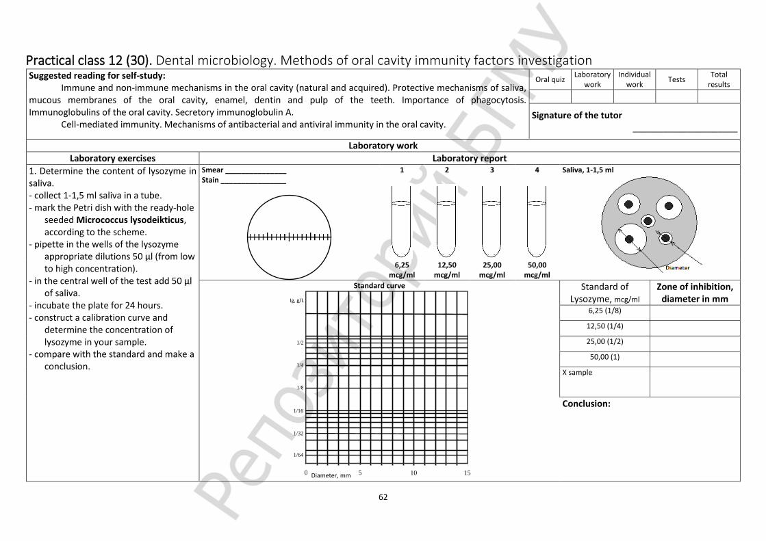
62
Practical class 12 (30). Dental microbiology. Methods of oral cavity immunity factors investigation Suggested reading for self-study:
Immune and non-immune mechanisms in the oral cavity (natural and acquired). Protective mechanisms of saliva, mucous membranes of the oral cavity, enamel, dentin and pulp of the teeth. Importance of phagocytosis. Immunoglobulins of the oral cavity. Secretory immunoglobulin A.
Cell-mediated immunity. Mechanisms of antibacterial and antiviral immunity in the oral cavity.
Oral quiz Laboratory
work Individual
work Tests
Total results
Signature of the tutor _____________________
Laboratory work
Laboratory exercises Laboratory report
1. Determine the content of lysozyme in saliva. - collect 1-1,5 ml saliva in a tube. - mark the Petri dish with the ready-hole
seeded Micrococcus lysodeikticus, according to the scheme.
- pipette in the wells of the lysozyme appropriate dilutions 50 µl (from low to high concentration).
- in the central well of the test add 50 µl of saliva.
- incubate the plate for 24 hours. - construct a calibration curve and
determine the concentration of lysozyme in your sample.
- compare with the standard and make a conclusion.
Smear _______________ Stain ________________
1 2 3 4 Saliva, 1-1,5 ml
6,25
mcg/ml 12,50
mcg/ml 25,00
mcg/ml 50,00
mcg/ml
Standard curve
0 5 10 15
Ig, g/L
Diameter, mm
1/2
1/4
1/8
1/32
1/64
1/16
Standard of Lysozyme, mcg/ml
Zone of inhibition, diameter in mm
6,25 (1/8)
12,50 (1/4)
25,00 (1/2)
50,00 (1)
X sample
Conclusion:

63
Laboratory exercises Laboratory report
2. Determine the IgA concentration in saliva by Manchini method (simple radial gel immunodiffusion). sIgA standard – 2,0 g per liter. 3. Register the experiment results on normal flora isolation from mucus membrane surfaces, Gram stain different types of colonies, explore under the microscope, complete the report.
0 5 10 15
Ig, g/L
Diameter, mm
1/2
1/4
1/8
1/32
1/64
1/16
Standart curve Standard sIgA = 2 g/l
titer concentrtion, g/l
Diameter, mm
Point 1 1 2,000
Point 2 ½ 1,000
Point 3 ¼ 0,500
Point 4 1/8 0,250
Point 5 1/16 0,125
Х-sample
As a normal sIgA ranger is 0,3-0,4 g/l
Conclusion:

64
Practical class 13 (31). Dental microbiology. Microbiology of periodontal and peri-implantitis diseases Suggested reading for self-study:
Plaque: stages of formation, microorganisms-colonizers. Plaque as a biofilm. Periodontal diseases: classification, etiology, risk factors. Theories of the pathogenesis of periodontitis. Properties of periodontopathogenic microorganisms, mechanisms of invasion and persistence. Microbial complexes (Socransky, 1998). Immune mechanisms in diseases of the tissues of the periodioth. Principles of prevention and treatment of periodontitis. Dynamics of microflora with successful and complicated dental implantation.
Oral quiz Laboratory
work Individual
work Tests
Total results
Signature of the tutor _____________________
Laboratory work
Laboratory exercises Laboratory report
1. Determine the content of lysozyme in saliva – ending (see practical class 12).
INDIVIDUAL WORK
SUBLINGVAL MICROBIAL COMPLEXES by SOCRANSKY (enter the names of the species of bacteria in each complex according to color)

65
INDIVIDUAL WORK
BIOFILM FORMATION (write stage of formation of biofilm)
A – B – C – D – E –
detachment and recolonization – multiplication – co-adhesion – colonization – reversible adhesion

66
Practical class 14 (32). Dental microbiology. Methods of microbiological diagnostics of stomatitis. Microbiological diagnostics of fungal infections Suggested reading for self-study:
Inflammatory diseases of the oral mucosa. Bacterial stomatitis: specific (gonococcal, typhoid fever, antrax stomatitis, manifestations in the oral cavity of syphilis, tuberculosis, actinomycosis, scarlet fever) and nonspecific. Viral stomatitis.
Classification and general characteristics of fungi. Classification of mycosis. Candida, general characteristics. Role in human pathology. Soor. General principles of fungal infections diagnostics.
Oral quiz Laboratory
work Individual
work Tests
Total results
Signature of the tutor _____________________
Laboratory work
Laboratory exercises Laboratory report 1. Research of the sample of the patient’s
pus with an abscess subcutaneous tissue of maxillofacial area, the 1st period:
- microscopy of pus (smear, Gram stain); - preparation of inverse hundredfold
dilutions material in sterile saline (1:100; 1:10000; 1:1000000);
- quantitative (50 mcl) streak respective sectors dilutions of pus on solid nutrient media (MSA, Endo, blood agar, NA with furagin) depending on the results of microscopy.
2. Research of the blood sample from the patient with stomatogenic sepsis, the 1st period:
- microscopy of blood, smear "thick drop", methylene blue stain or Romanovsky; - Crop material in the liquid medium of the primary crop (enrichment) in a ratio of 1: 10-60;
- Incubation of cultivation in an incubator at 37 0C - 18-24 hours and up to 14 days.
All inoculations are placed in a incubator for 24 hours, then transferred to a refrigerator for 14 days.
Sample of pus from
Serial dilution of the sample Smear 2.1 _____________ Stain __________
Blood sample examination, 1st period
10 ml:60 ml
Burnt wound
10-2 10-4 10-6
0,1 ml
0,1 ml 9,9 ml
Saline sol.
0,1 ml 9,9 ml
Saline sol.
9,9 ml
Saline sol.
Steak respective sector with 0,05 ml (1 drop)
Smear 3.1 _______________ Stain __________
Medium
Blood agar Levin Nutrient agar with
10–2
10–4
10–6 10–2
10–4
10–6 10–2
10–4
10–6

67
Laboratory exercises Laboratory report 3. Snyder’s caries susceptibility test The degradation of enamel and dentin in the
formation of tooth decay (dental caries) occurs as a result of the production of lactic acid by bacteria (Streptococcus mutans and others) in the presence of sucrose high levels. Of the various methods that have been devised to determine one’s susceptibility to tooth decay, M. L. Snyder’s caries susceptibility test is a relatively simple test that has been shown to have a high reliability correlation.
This method relies on the rapidity of organisms in saliva to lower the pH in the medium that contains 2% dextrose (Snyder test agar). Since decalcification of enamel begins at pH of 5.5, and progresses rapidly as the pH is lowered to 4.4 and less, the demonstration of pH lowering becomes evidence of susceptibility to caries.
To indicate the presence of acid production in the medium, the indicator bromcresol green is incorporated in it. This indicator is green at pH 4.8 and becomes yellow at pH 4.4, remaining yellow below 4.4.
Figure illustrates the procedure that is used in the Snyder caries susceptibility test. Note that 0.2ml of saliva is added to the tube of liquefied Snyder test agar (50° C) and mixed well by rotating the tube between the palms of both hands. After the medium has solidified, the tube is incubated at 37° C for a period of 24–72 hours. If the medium turns yellow in 24–48 hours, the individual is said to be susceptible to caries.
Although we will be performing this test only once, it should be noted that test reliability is enhanced by performing the test on three consecutive days at the same time each day. If the test is performed correctly after tooth brushing, it is not as reliable as if 2 or 3 hours have elapsed after brushing.
1. Liquefy a tube of Snyder test agar and cool it to 50° C. 2. After allowing a piece of paraffin to soften under the tongue for a few minutes, start chewing it. Chew it for 3 minutes, moving it from one side of the mouth to the other. Do not swallow the saliva. As it accumulates, deposit it in the small sterile beaker. 3. Vigorously shake the sample in the beaker from side to side for 30 seconds to disperse the organisms. 4. With a 1 ml pipette transfer 0.2 ml of saliva to the tube of agar. Do not allow the pipette to touch the side of the tube or agar.
5. Before the medium solidifies, mix the contents of the tube by rotating the tube vigorously between the palms of the hands. 6. Write your name on a gummed label and attach it to the tube. 7. Incubate the tube at 37° C. Examine the tube every 24 hours to see if the bromcresol green indicator has changed to yellow. If it has, the test is positive. The degree of caries susceptibility is determined from the table below. 8. Record your results on the Laboratory Report.
Materials: 1 tube of Snyder test agar (5 ml in 15 mm dia tube) 1 30 ml sterile beaker 1 piece of paraffin (1/4″ 1/4″ 1/8″) 1 ml pipette 1 gummed label
CARIES SUSCEPTIBILITY
MEDIUM TURNS YELLOW IN:
24 HOURS 48 HOURS 72 HOURS
Marked Positive
Moderate Negative Positive
Slight Negative Negative Positive
Negative Negative Negative Negative

68
Practical class 15 (33). Test “General and special virology. Dental microbiology”
List of questions Oral quiz Script Tests Total results
1. Virology, tasks and methodologies. The systematic position and classification of viruses. 2. Forms of viruses existence. The morphology of virions. The interaction of viruses with susceptible cells. 3. Features of infection and immunity in viral infections. 4. Methods of virus cultivation (cell culture, chicken embryo, laboratory animals). 5. General principles of viral infections diagnostics. 6. Influenza viruses. General characteristics. Pathogenesis, specific and non-specific treatment and prevention, influenza laboratory diagnosis. Manifestations in the oral cavity. 7. Paramyxoviruses, general characteristics. Mumps virus, respiratory-syncytial virus, measles virus, parainfluenza viruses. Manifestations in the oral cavity. 8. Enteroviruses, general characteristics, role in human pathology. Poliovirus, pathogenesis and laboratory diagnostics, specific prevention. Manifestations of enteroviruses infection in oral cavity. 9. Classification of hepatitis viruses. Characterization of hepatitis A, B, C virus. Pathogenesis, immunity, laboratory diagnosis, prevention. 10. Retroviruses. Human immunodeficiency virus (HIV-1, HIV-2). Pathogenesis. AIDS-associated diseases in dentistry. HIV diagnostics, prophylaxis. 11. Adenoviruses, general characteristics. Pathogenesis, laboratory diagnostics of adenoviral infections. Manifestations in oral cavity. 12. Herpes viruses. Classification. General characteristics, disease. Herpetic stomatitis. 13. Bacterial viruses (bacteriophages), properties, classification. The practical use of bacteriophages. 14. The microflora of the oral cavity (indigenous, transient). Ontogeny of normal oral flora. 15. Representatives of the normal oral flora: Gram-positive and Gram -negative cocci (streptococci, peptostreptococci, staphylococci, veillonella, Neisseria), their role. 16. Representatives of the normal oral flora: Gram-positive (propionjbacterium, lactobacillus, actinomyces, corynebacterium) and Gram-negative rods (bacteroides, prevotella, porphyromonas, fusobacterium, leptotrichia), their role. 17. Representatives of the normal oral flora spiralshaped bacteria (vibrio, wolinella, centipedia, selenomonas, campylobacter, spirochetes), mycoplasma, protozoa, fungi, and their role. 18. Microflora of specific areas of the mouth: saliva, dorsum of the tongue, dental pocket, mucous membranes. Methods of study of oral microflora. 19. Influence of environmental factors and physiological features on oral flora. The role of the oral cavity normal microflora (positive and negative). Disbacteriosis of the oral cavity: causes, outcome, prevention, principles of correction. 20. Antigens and the immune system of the oral cavity. Citrullinated antigens. Immune mechanisms in the oral cavity. Antimicrobial factors of saliva: defensins, cathelicidin, mucins, histatin, statherin, cystatins. Proinflammatory cytokines. 21. Nonspecific mechanisms of defense of the mucous membranes, saliva, gingival fluid, tooth enamel, normal microflora’s. 22. Factors and mechanisms of acquired immunity of oral cavity. Local Immunity of the oral cavity. Immunological aspects of relationship of inflammatory periodontal diseases, cardiovascular and rheumatic diseases. 23. Types of inflammatory processes of the oral cavity, their characteristics. Cytokines of early and late phase of inflammation: cell producers, properties. Methods of cytokines detection: obtaining of specimens, storage, methods of determination (ELISA, genetic).
24. The etiology of dental caries. Features of cariogenic microorganisms. Cariogenic streptococci. Characteristics of S.mutans. Characteristics of lactobacilli. Associative (additional) microorganisms. The role of the microorganism in the development of caries. 25. Cariogenesis: mechanisms of streptococci adhesion to teeth and their role in dental plaque formation. Role of glucans and their characteristics. Factors responsible for caries development. Resistance to caries. Prevention of dental caries. 26. Odontogenic infections: etiology, types. The role of microorganisms in the etiology and pathogenesis of gingivitis. Dynamics of the microflora of implants in case of successful implantation and complicated. 27. The role of microorganisms in the pathogenesis of pulpitis, acute and chronic periodontitis ray, periostitis, osteomyelitis, abscesses and soft tissue abscesses. 28. Periodontal diseases: classification, risk factors. General properties of periodontopathogenic microorganisms. Red complex microorganisms: Porphyromonas gingivalis, Tannerella forsythia, Treponema denticola. Characterization, pathogenicity factors and their role in the pathogenesis of periodontitis. Characteristics of Aggregatibacter actinomycetemcomitans and role in the development of aggressive periodontitis. 29. Dental Plaque: microflora, formation stages. The role of dental plaque in the development of periodontitis. Microorganisms of orange and yellow complexes, their role in the development of periodontal disease. Plaque as a biofilm. The role of quorum sensing factors in the formation of plaque. New approaches to reduce the bioburden of plaque. 30. Immune mechanisms in the development of periodontal diseases. Factors contributinging invasion of microorganisms. Mechanisms to protect tissues from microbial invasion. Principles of prevention and treatment of periodontitis 31. The role of microorganisms in the formation of dental calculus. Pathogenesis of the carie dental calculus formation. 32. Inflammatory diseases of the oral mucosa: classification, the role of microorganisms in their development. Specific and nonspecific stomatitis. 33. Stomatitis caused by obligate pathogens and opportunistic bacteria. 34. Fusospirochetal diseases: etiology, characteristics of pathogens, pathogenesis, clinical forms. 35. Actinomyces spp.: systematics, classification, characteristics, antigenic structure, factors of pathogeneity. Cervico- maxillo-facial actinomycosis: pathogenesis, immunity, microbiological diagnosis, prevention. 36. Viral stomatitis. 37. Candida: systematics, properties, pathogenicity factors. Candidosis: factors responsible for the developement, methods of diagnosis and prevention. 38. Methods of studying the normal oral flora. Methods of sampling for dental diseases diagnosis. 39. Manifestations of allergic and immunodeficiency conditions in the oral cavity. Recurrent viral aphthous stomatitis. 40. Types and etiology of stomatogenic infections. 41. Dental Clinical Microbiology. Opportunistic pathogens. Specific features opportunistic pathogens and infections caused by them. Specific features of pathogenesis and diagnosis of opportunistic diseases. Criteria of Etiological significance of isolated bacteria from a specimen.

69
Practical class 16 (34). Dental microbiology. Method of microflora investigation in diseases of the teeth and oral cavity soft tissues Suggested reading for self-study:
Odontogenic inflammation. Microflora, pathogenesis, microbiological diagnosis of pulpitis, periodontitis, periostitis, osteomyelitis, odontogenic abscesses and phlegmon.
Purulent-inflammatory dental diseases of soft tissues and bones of the maxillofacial area. Pathogens, pathogenesis, methods of microbiological diagnostics (material for research, rules and methods of sampling, a scheme for bacteriological examination of pus, criteria for the etiological role of isolated microorganisms). Determination of sensitivity to antibiotics. Dental sepsis. Pathogens, methods of microbiological diagnosis.
Oral quiz Laboratory
work Individual
work Tests
Total results
Signature of the tutor ____________________
Laboratory work
Laboratory exercises Laboratory report
1. Research of the sample of the patient’s pus with an abscess subcutaneous tissue of maxillofacial area, 2nd period:
- microscopy of slides prepared from all types of colonies;
- the study of microbial growth on the media;
- determination of the pathogen quantity per ml/g (CFU) of the sample with formula;
- oxidase test; - coagulase test; - seeding the pure culture for
accumulation and biochemical identification, incubation in an incubator at 37 oC - 18-24 hours.
Colonies characteristics
Medium _______ Medium _______ Smear _____________ Stain _____________
Shape Size Surface Edge Color Consistency
Conclusion:
Transparency
Determination of CFU Calculation of bacteria quality per ml/g of the sample: N(CFU/ml) = n × 20 × 10x, n – colonies quantity in respective sector, 20 – conversion factor for 1 ml, 10x – the degree of the sample dilution. N(CFU/ml) =
Coagulase test Sample control
Stabilized rabbit plasm: 37 °С – 2, 4, 24 h
Oxidase test
Sample
control
2. Research of the blood sample from the patient with stomatogenic sepsis, the 2nd period:
- the study of microbial growth on the media; - microscopy of slides prepared from the media; - seeding on the blood and Yolk-salt agar for the
pure culture.
10-2
10-4
10-6 10-2
10-4
10-6 10-2
10-4
10-6

70
Laboratory exercises Laboratory report 3. Research of the sample of the patient’s
pus with an abscess subcutaneous tissue
of maxillofacial area, 3rd period (The task will be given at the next lesson):
- microscopy of slides prepared from pure culture;
- the study of microbial growth on the media; - seeding the pure culture for accumulation
and biochemical identification, incubation in an incubator at 37 oC - 18-24 hours;
- seeding the pure culture for determination of antibiotic resistance.
4. Research of the sample of the patient’s
pus with an abscess subcutaneous tissue
of maxillofacial area, 4th period (The task will be given at the next lesson):
- microscopy of slides prepared from pure culture;
- the study of microbial growth on the media; - determination of antibiotic resistance; - conclusion: identification and typing results,
antibioticgramm.
Smear _____________
Stain _____________
Antibiotic Diameter of inhibition zones (mm)
resistant susceptible
Staphylococcus spp.
Penicillin ≤28 ≥29
Oxacillin CNS S.aureus
≤17 ≥18
≤10 ≥13
Canamycine ≤13 ≥18
Gentamicin ≤12 ≥15
Ciprofloxacin ≤15 ≥21
Tetracycline ≤14 ≥19
Erythromycine ≥23 ≥23
Lincomycine ≤13 ≥21
Chloramphenicol <17 ≥18
Enterobacteriacea spp.
Ampicillin ≤13 ≥17
Cefazolin ≤14 ≥18
Cefotaxime ≤14 ≥23
Canamycine ≤13 ≥18
Gentamicin ≤12 ≥15
Ciprofloxacin ≤15 ≥21
Lomefloxacin ≤18 ≥22
Tetracycline ≤14 ≥19
Doxicycline ≤12 ≥16
Chloramphenicol ≤12 ≥18
antibioticgramm
Smear _____________
Stain _____________
Conclusion:
Antibiotic Diameter of inhibition zone, mm Interpretation of results
DDM
- make standard inoculum with saline solution (0,5 unit MacFarlane);
- microscopy of slides prepared from inoculum culture)
- seeding of 1,0 ml of inoculum on MH agar; - incubation 18-20 hours 35оС.

71
Practical class 17 (35). Clinical microbiology. Microbiological diagnostics of purulent infections of bronchi and lungs. Hospital-acquired infection Suggested reading for self-study:
Dental bronchopulmonary diseases. Pathogens. Pathogenesis. Conditions of occurrence. Methods of microbiological diagnosis (materials for research, rules and methods of sampling, a scheme for bacteriological sputum examination, bronchial washings, criteria for the etiological role of isolated microorganisms).
Determination of sensitivity to antibiotics. Nosocomial infections. Pathogens, features in the practice of a dentist, principles of diagnosis. Anti-epidemic regime in
dental practice. Principles of microbiological diagnosis. Prevention.
Oral quiz Laboratory
work Individual
work Tests
Total results
Signature of the tutor _____________________
Laboratory work
Laboratory exercises Laboratory report 1. Research of the blood sample from the patient with stomatogenic sepsis, the 3rd period: - the study of microbial growth on the
medium; - microscopy of slides prepared from all types
of colonies; - oxidase test; - coagulase test; - seeding the pure culture for accumulation
and biochemical identification, incubation in an incubator at 37 0C - 18-24 hours.
- incubation at 37 0C - 18-24 hours. 2. Research of the blood sample from the patient with stomatogenic sepsis, the 4th period: - the study of tests used for identification of
cultures and antimicrobial sensitivity level in DDM.
Blood agar
Hemolyses_____
YSA
Lecithinase ____
MH agar
Kirby-Bauer method
Coagulase test Exp Control
Stabilized rabbit plasm: 37 °С – 2, 4, 24 h
Glucose and mannitol fermentation (anaerobic)
Smear _____________
Stain __________
Colonies characteristics
Medium _______ Medium _______ Conclusion:
Shape
Size
Surface
Edge
Color
Consistency
Transparency
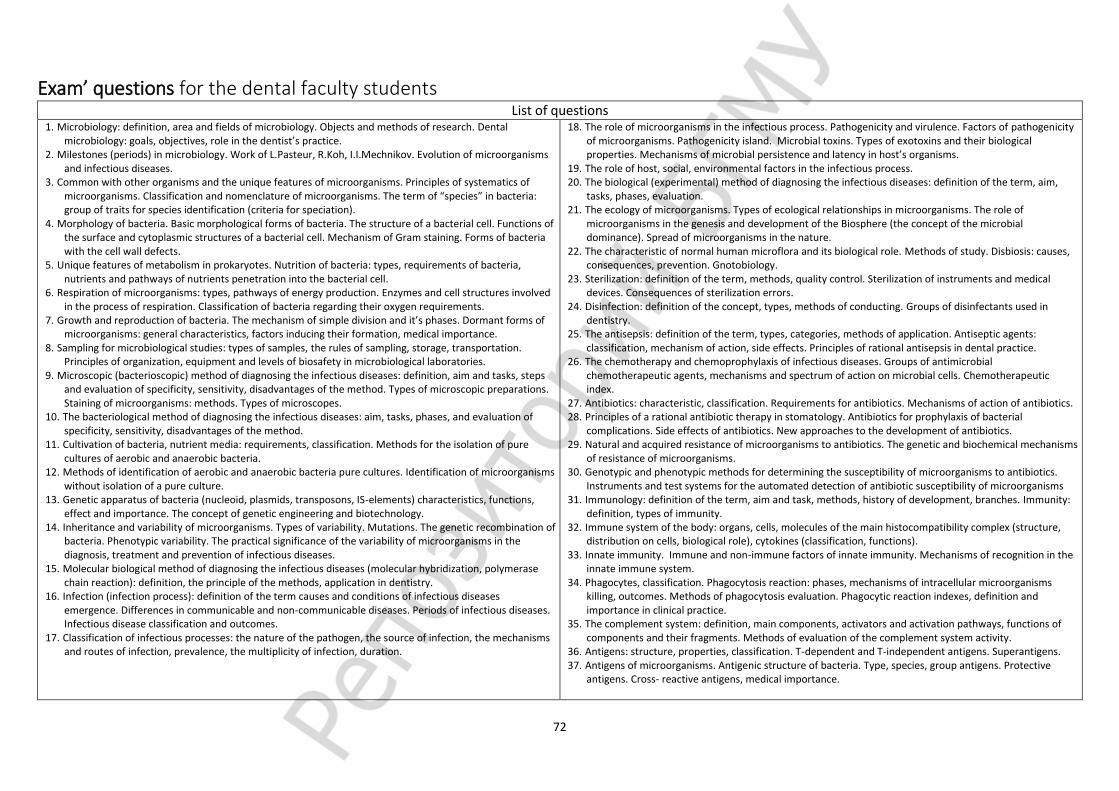
72
Exam’ questions for the dental faculty students List of questions
1. Microbiology: definition, area and fields of microbiology. Objects and methods of research. Dental microbiology: goals, objectives, role in the dentist’s practice.
2. Milestones (periods) in microbiology. Work of L.Pasteur, R.Koh, I.I.Mechnikov. Evolution of microorganisms and infectious diseases.
3. Common with other organisms and the unique features of microorganisms. Principles of systematics of microorganisms. Classification and nomenclature of microorganisms. The term of “species” in bacteria: group of traits for species identification (criteria for speciation).
4. Morphology of bacteria. Basic morphological forms of bacteria. The structure of a bacterial cell. Functions of the surface and cytoplasmic structures of a bacterial cell. Mechanism of Gram staining. Forms of bacteria with the cell wall defects.
5. Unique features of metabolism in prokaryotes. Nutrition of bacteria: types, requirements of bacteria, nutrients and pathways of nutrients penetration into the bacterial cell.
6. Respiration of microorganisms: types, pathways of energy production. Enzymes and cell structures involved in the process of respiration. Classification of bacteria regarding their oxygen requirements.
7. Growth and reproduction of bacteria. The mechanism of simple division and it’s phases. Dormant forms of microorganisms: general characteristics, factors inducing their formation, medical importance.
8. Sampling for microbiological studies: types of samples, the rules of sampling, storage, transportation. Principles of organization, equipment and levels of biosafety in microbiological laboratories.
9. Microscopic (bacterioscopic) method of diagnosing the infectious diseases: definition, aim and tasks, steps and evaluation of specificity, sensitivity, disadvantages of the method. Types of microscopic preparations. Staining of microorganisms: methods. Types of microscopes.
10. The bacteriological method of diagnosing the infectious diseases: aim, tasks, phases, and evaluation of specificity, sensitivity, disadvantages of the method.
11. Cultivation of bacteria, nutrient media: requirements, classification. Methods for the isolation of pure cultures of aerobic and anaerobic bacteria.
12. Methods of identification of aerobic and anaerobic bacteria pure cultures. Identification of microorganisms without isolation of a pure culture.
13. Genetic apparatus of bacteria (nucleoid, plasmids, transposons, IS-elements) characteristics, functions, effect and importance. The concept of genetic engineering and biotechnology.
14. Inheritance and variability of microorganisms. Types of variability. Mutations. The genetic recombination of bacteria. Phenotypic variability. The practical significance of the variability of microorganisms in the diagnosis, treatment and prevention of infectious diseases.
15. Molecular biological method of diagnosing the infectious diseases (molecular hybridization, polymerase chain reaction): definition, the principle of the methods, application in dentistry.
16. Infection (infection process): definition of the term causes and conditions of infectious diseases emergence. Differences in communicable and non-communicable diseases. Periods of infectious diseases. Infectious disease classification and outcomes.
17. Classification of infectious processes: the nature of the pathogen, the source of infection, the mechanisms and routes of infection, prevalence, the multiplicity of infection, duration.
18. The role of microorganisms in the infectious process. Pathogenicity and virulence. Factors of pathogenicity of microorganisms. Pathogenicity island. Microbial toxins. Types of exotoxins and their biological properties. Mechanisms of microbial persistence and latency in host’s organisms.
19. The role of host, social, environmental factors in the infectious process. 20. The biological (experimental) method of diagnosing the infectious diseases: definition of the term, aim,
tasks, phases, evaluation. 21. The ecology of microorganisms. Types of ecological relationships in microorganisms. The role of
microorganisms in the genesis and development of the Biosphere (the concept of the microbial dominance). Spread of microorganisms in the nature.
22. The characteristic of normal human microflora and its biological role. Methods of study. Disbiosis: causes, consequences, prevention. Gnotobiology.
23. Sterilization: definition of the term, methods, quality control. Sterilization of instruments and medical devices. Consequences of sterilization errors.
24. Disinfection: definition of the concept, types, methods of conducting. Groups of disinfectants used in dentistry.
25. The antisepsis: definition of the term, types, categories, methods of application. Antiseptic agents: classification, mechanism of action, side effects. Principles of rational antisepsis in dental practice.
26. The chemotherapy and chemoprophylaxis of infectious diseases. Groups of antimicrobial chemotherapeutic agents, mechanisms and spectrum of action on microbial cells. Chemotherapeutic index.
27. Antibiotics: characteristic, classification. Requirements for antibiotics. Mechanisms of action of antibiotics. 28. Principles of a rational antibiotic therapy in stomatology. Antibiotics for prophylaxis of bacterial
complications. Side effects of antibiotics. New approaches to the development of antibiotics. 29. Natural and acquired resistance of microorganisms to antibiotics. The genetic and biochemical mechanisms
of resistance of microorganisms. 30. Genotypic and phenotypic methods for determining the susceptibility of microorganisms to antibiotics.
Instruments and test systems for the automated detection of antibiotic susceptibility of microorganisms 31. Immunology: definition of the term, aim and task, methods, history of development, branches. Immunity:
definition, types of immunity. 32. Immune system of the body: organs, cells, molecules of the main histocompatibility complex (structure,
distribution on cells, biological role), cytokines (classification, functions). 33. Innate immunity. Immune and non-immune factors of innate immunity. Mechanisms of recognition in the
innate immune system. 34. Phagocytes, classification. Phagocytosis reaction: phases, mechanisms of intracellular microorganisms
killing, outcomes. Methods of phagocytosis evaluation. Phagocytic reaction indexes, definition and importance in clinical practice.
35. The complement system: definition, main components, activators and activation pathways, functions of components and their fragments. Methods of evaluation of the complement system activity.
36. Antigens: structure, properties, classification. T-dependent and T-independent antigens. Superantigens. 37. Antigens of microorganisms. Antigenic structure of bacteria. Type, species, group antigens. Protective
antigens. Cross- reactive antigens, medical importance.

73
38. Antigen presenting cells: types, characteristics. B-lymphocytes: development, markers, antigen-specific B-cell receptor.
39. Humoral immune response: definition, development. Activation, proliferation, differentiation and interactions of cells involved. T-dependent and T-independent response. Primary and secondary humoral immune response characteristics.
40. Antibodies (immunoglobulins): structure, properties, classification, Immunoglobulins biosynthesis. The mechanism of interaction of antibodies with antigens: specificity, phases, manifestations. Affinity and avidity. Monoclonal antibody: principles of production, application.
41. Serological method of investigation: general definition of the term, objectives, basic concepts (diagnosticum, diagnostic serum, titer, diagnostic titer, paired sera). Samples for serological examination. General characteristics of the method. Use of serological method for infectious and noninfectious diseases diagnostics.
42. Agglutination: ingredients, main variants of performance, registration, evaluation, application. Indirect (passive) and reverse passive agglutination: ingredients, mechanism, methodology, registration of results, practical use.
43. Immunoprecipitation reaction: ingredients, mechanism, main methods of performance, application. Reaction of the immune lysis. Complement fixation test: ingredients, mechanism, registration of results.
44. Immunofluorescence (fluorescent antibodies test, FAT), main variants, ingredients, mechanisms, registration of results, practical use. ELISA: ingredients, mechanisms, registration of results, practical use. Immunoblotting (IB). Radioimmunoassay (RIA).
45. T cells: development, markers, subpopulations. Helper T-cells, main types (Th1, Th2, Th3, Th17), spectrum of cytokines produced. T-cell receptor: structure, types, genetic control, variety.
46. Cellular immune response: definition, development, main stages, manifestation. The model of two (three) signals: the response, anergy, apoptosis. Manifestation of cellular immune response. Immunological memory.
47. Anti-infection immunity and its types depending on pathogen nature. Mechanisms of antitoxic, antibacterial, antifungal, antiparasite immunity.
48. Immunoprophylaxis and immunotherapy for infectious diseases. Active immunoprophylaxis. Vaccines: requirements, characteristics of main types of vaccines. Adjuvants mechanisms of action. Side effects of vaccination: sever vaccinal reaction, post-vaccination complications.
49. Post-vaccination immunity: mechanisms and factors influencing its development. Indications and contraindications to vaccination. Immunization schedule. Expanded Programme on immunization. Collective immunity to infectious diseases, importance.
50. Passive immunoprophylaxis and immunotherapy of infectious diseases: indications, principles, complications.
51. Allergology: the definition, objectives. Allergens. Allergy: the stages, types of reactions. Classification of allergens. Allergens in dentistry.
52. Immediate type hypersensitivity (ITH). Mediator type (I) ITH: allergens, mechanism, development, Manifestations in the oral cavity, ways to prevent anaphylaxis.
53. Cytotoxic (II) type ITH: allergens, development, mechanisms, manifestations. Immunocomplex (III) type ITH: allergens, development, mechanisms. Manifestations of allergic reactions II and III types in the oral cavity.
54. Delayed type of hypersensitivity (IV): allergens, development, mechanism, manifestation (infection and contact allergy), importance in oral cavity.
55. Drug allergy: major allergens, the mechanisms and types of allergic reactions, methods for diagnostics and prevention. Food allergy. Main allergens. Prevention of food allergy. Idiosyncrasy.
56. Methods of diagnosing allergic diseases. Prevention of allergy. 57. Antitumor immunity. The concept of immune surveillance. Mechanisms of tumor escape from immune
surveillance. 58. Clinical Immunology: definition, objectives, main concepts. Immune status: principle and methods of
examination. Methods for determining the amount and functional activity of T-and B-lymphocytes. 59. Autoantibodies: origin, role in pathology. Autoimmune diseases: definition, classification, aetiology,
mechanisms of tissue damage, manifestations. 60. Immunodeficiency conditions: classification, causes of development, methods for detection, principles for
correction. 61. Staphylococci: classification, characterization, antigenic structure, pathogenicity factors. Staphylococcal
infections: pathogenesis, immunity, microbiological diagnosis and principles of prevention, immunotherapy. Staphylococcal carriage: diagnosis, significance. Staphylococcus aureus: MRSA, antibiotics of choice for their therapy.
62. Streptococci: classification, characterization, antigenic structure, pathogenicity factors. Streptococcal disease: pathogenesis, immunity, microbiological diagnosis, and prevention. Pneumococci: classification, characterization, antigenic structure, pathogenicity factors. Pneumococcal infections.
63. Neisseria meningitidis: systematics, characterization, antigenic structure, pathogenicity factors. Meningococcal infections: pathogenesis, immunity, microbiological diagnosis, prophylaxis.
64. Neisseria gonorrhoeae: systematics, characterization, antigenic structure, pathogenicity factors. Pathogenesis, immunity, microbiological diagnosis of acute and chronic gonorrhoea, prophylaxis. Prevention of gonorrhoea and gonorrhoeal conjunctivitis, stomatitis.
65. Family of Enterobacteria: classification, characterization, pathogenicity factors. Principle of microbiological diagnosis of GIT diseases caused by Enterobacteria. Principles of identification of enterobacteria.
66. Escherichia: systematics, characterization, antigenic structure, pathogenicity factors. Pathogenic and opportunistic Escherichia coli. The biological role of Escherichia coli. Escherichiosis: pathogenesis, immunity, microbiological diagnosis and prevention.
67. Salmonella: systematics and classification, characterization, antigenic structure, pathogenicity factors, role Salmonella in pathology. Salmonellosis and Typhoid fever: pathogenesis, immunity, prevention.
68. Shigella: classification, characteristics, antigenic structure, pathogenicity factors. Bacterial dysentery: pathogenesis, immunity, microbiological diagnosis, prophylaxis.
69. Food poisoning of microbial aetiology: classification, etiology, pathogenesis, principles of microbiological diagnosis, prophylaxis.
70. Klebsiella: classification, characteristics, antigenic structure, pathogenicity factors, Klebsiella diseases. Pseudomonas: characteristics, antigenic structure, pathogenicity factors, role in the pathology.
71. Campylobacter, Helicobacter: characteristics, role in pathology. 72. Corynebacterium: classification, characteristics, antigenic structure, pathogenicity factors. Diphtheria:
pathogenesis, immunity, microbiological diagnostics, immunotherapy and aetiological therapy of diphtheria, prophylaxis. Manifestation of diphtheria in oral cavity.
73. Bordetella: classification, characteristics, antigenic structure, pathogenicity factors. Whooping cough: pathogenesis, immunity, microbiological diagnosis, prophylaxis. Haemophilus spp.: characteristics, role in pathology, prophylaxis Hib-infections.
74. Actinomyces: classification, characterization, antigenic structure, pathogenicity factors. Cervico-maxillofacial actinomycosis: pathogenesis, immunity, microbiological diagnosis, prevention.

74
75. Mycobacteria: classification, characteristics, antigenic structure, pathogenicity factors. Tuberculosis: pathogenesis, immunity, methods of diagnosis, principle of prevention and treatment. Mycobacterioses. Manifestation of tuberculosis in oral cavity.
76. Obligate anaerobes. Classification and characteristics. Clinical signs of anaerobic infection. Features of taking the material in case of suspected anaerobic infection.
77. Gas gangrene Clostridia spp.: classification, characteristics, antigenic structure, pathogenicity factors. Anaerobic myonecrosis: pathogenesis, immunity, microbiological diagnostics and prophylaxis, aetiological treatment.
78. Clostridium tetani: systematics, characterization, antigenic structure, pathogenicity factors. Tetanus: pathogenesis, immunity, microbiological diagnosis, prevention, aetiological treatment.
79. Nonsporforming anaerobes: classification, characteristics, role in pathology of oral cavity. Principles of sampling in anaerobic bacteriology. Principle of bacteriological diagnosis of infections caused by nonsporforming anaerobes.
80. Quarantine diseases: characteristics, classification. Principles of collection, transportation and investigation of specimens with pathogens of 3d and 4th biosafety levels.
81. Vibrio: classification, characteristics, antigenic structure, pathogenicity factors. Cholera: pathogenesis, immunity, microbiological diagnosis, prophylaxis.
82. Classification and characteristics of causative agents of plague, tularemia, pathogenicity factors, microbiological diagnosis, prophylaxis.
83. Classification and characteristics of causative agents of brucellosis, anthrax, pathogenicity factors, microbiological diagnosis, prophylaxis.
84. Spirochetes: classification, characteristics, antigenic structure, pathogenicity factors. Role of Borrelia spp. in human pathology. Lyme borreliosis: aetiology, pathogenesis, immunity, microbiological diagnosis, prophylaxis. Role of Leptospira in human pathology, prophylaxis of leptospirosis.
85. Treponema: classification, characteristics, antigenic structure, pathogenicity factors. Syphilis: pathogenesis, immunity, microbiological diagnosis, prophylaxis. Manifistation of Syphilis in oral cavity.
86. Treponema of oral cavity and their role in pathology. Fusospirochetozes: etiology, characteristics of pathogens, pathogenesis, clinical forms.
87. Chlamydia: classification, characterization, development cycle, antigenic structure, pathogenicity factors, role in pathology. Microbiological diagnostics and prevention.
88. Mycoplasma spp.; classification, characteristics, role in pathology. 89. Rickettsia: classification, characteristics, role in pathology. 90. Pathogenic fungi: classification, characteristics. Fungal infections promoting factors and conditions. Role
microfungi in human pathology. Prophylaxis of mycoses. 91. Virology: definition, objectives, methods. Systematic position and classification of viruses. History.
D.Ivanovski works importance. Forms of existence of viruses. Morphology and biochemical structure of virions. Structure, function and properties of virion nucleic acid, proteins, lipids and carbohydrates. Prions, role in human pathology.
92. Interaction of the virus and susceptible cell. Strict parasitism and cytotropism of viruses. Cell receptors for viruses. Viral genome organization. Reproduction strategy of DNA and RNA viruses.
93. Types of viral infection of cell. Changes in the host cells in the process of a viral infection. Peculiarities of viral infections of an organism. Acute, chronic and slow infection. Local and systemic mechanisms of antiviral immunity. Factors of innate and adaptive antiviral immunity. Interferons: classes, properties, mechanisms of antiviral activity.
94. Principles of etiologic diagnostics of viral infections. Rapid methods. Serological diagnostics: principles, criteria for diagnosis. Principles of viral infections chemotherapy. Groups of antiviral drugs.
95. Cultivation of viruses. Indication and identification of viruses. 96. The aetiology of acute respiratory viral infections. Influenza viruses: classification, characteristics, antigenic
properties. Influenza: pathogenesis, immunity, prevention, etiologic diagnostics of influenza, chemotherapy and chemoprophylaxis of influenza.
97. Paramyxoviruses: classification, characteristics, role in pathology. Prevention of infection caused by paramyxoviruses
98. Rabies virus: classification, characteristics, specific inclusion. Rabies: pathogenesis, etiologic diagnosis, prevention.
99. Rubella virus. General characteristics. Role in pathology. Prevention of rubella. 100. Enteroviruses: classification, characteristics. Enterovirus infections: pathogenesis, prevention. Role in
pathology of oral cavity. 101. Viral hepatitis A: pathogenesis, immunity, etiologic diagnosis, prevention. 102. Parenteral hepatitis viruses: classification, characteristics. Parenteral hepatitis: pathogenesis, immunity,
etiologic diagnostics, prevention. 103. Retroviruses. Human immunodeficiency virus (HIV). HIV infection: pathogenesis, immunity, etiologic
diagnostics, principles of therapy, prophylaxis. AIDS - related illnesses. HIV-associated diseases in oral cavity.
104. Herpesviruses: classification, characterization, role in pathology. Herpetic stomatitis. Chickenpox. Herpes viruses of 4-8 types, their role in human pathology.
105. Adenoviruses: classification, characteristic. Adenoviral infections: pathogenesis, immunity, etiological diagnosis. Papillomaviruses: characteristics, role in pathology, disease prevention.
106. Dental microbiology: definition, goals, objectives. General principles of microbiological diagnosis of dental diseases.
107. The microflora of the oral cavity (indigenous, transient). Ontogeny of normal oral flora. 108. The role of normal microflora of the oral cavity (positive and negative). Dysmicrobiosis of the oral cavity:
causes, effects, prevention, principles of correction. Influence of environmental factors, physiological features of the oral cavity and other factors of the microorganism on the microflora of the oral cavity.
109. Representatives of the normal microflora of the oral cavity: aerobes and facultative anaerobes (streptococci, corynebacteria, staphylococci, Neisseria), their role. General characteristics of streptococci of the oral cavity.
110. Representatives of the normal oral flora: anaerobes (velonella, propionjbacterium, lactobacillus, actinomyces, bacteroides, prevotella, porphyromonas, fusobacterium, leptotrichia), their role.
111. Representatives of the normal oral flora spiralshaped bacteria (vibrio, wolinella, centipedia, selenomonas, campylobacter, spirochetes), mycoplasma, protozoa, fungi, and their role.
112. Microflora of specific areas of the mouth: saliva, dorsum of the tongue, dental pocket, mucous membranes. Features of these biotopes, affecting microorganisms.
113. Methods of study of oral microflora. Methods of sampling material for dental diseases. Environments for the isolation of cariogenic streptococci, lactobacilli.
114. Nonspecific mechanisms of defense of the mucous membranes, saliva, gingival fluid, tooth enamel, normal microflora’s, system of polymorphonuclear leukocytes.
115. Functions of saliva. Antimicrobial factors of saliva: defensins, cathelicidin, mucins, histatin, statherin, cystatins, peroxidase.

75
116. The role of factors and mechanisms of acquired immunity of the oral cavity. Local immunity of the oral cavity. Functions of secretory immunoglobulins A.
117. Dental plaque: the stages of formation, microorganisms-colonizers. Plaque as a biofilm. The role of factors in the quorum of sensing in the formation of plaque. New approaches to reducing the bioburden of plaque.
118. Etiology of caries. Criteria of cariogenicity. Cariesogenic streptococci. Characteristic of S. mutans et sobrinus. Characteristics of lactobacilli. Associative (auxiliary) microorganisms. The role of the macroorganism in the development of caries.
119. Pathogenesis of caries: mechanisms of adhesion (carbohydrate-dependent and carbohydrate-independent) streptococci and mechanisms of destruction of tooth tissues. The role of streptococci in coaggregation. Glukans. Conditions for the development of caries. Caries resistance. Prophylaxis of caries. Fluorides and their influence are microorganisms.
120. Odontogenic inflammation: etiology, types and phases of inflammation. Significance in pathology of foci of chronic odontogenic infection. Immunological aspects of the relationship between inflammatory periodontal diseases, cardiovascular and rheumatic diseases.
121. Types of microorganisms and their role in the origin and pathogenesis of pulpitis, acute and chronic apical periodontitis, periostitis, osteomyelitis, abscesses and phlegmon soft tissues.
122. Periodontal disease: classification, risk factors for development. The role of microorganisms in the etiology and pathogenesis of gingivitis. Dynamics of microflora of implants in case of successful and complicated implantation.
123. The role of dental plaque in the development of periodontitis. The role of microorganisms in the formation of dental plaque. Pathogenetic importance of dental plaque.
124. General properties of periodontopathogenic microorganisms. Microorganisms of the red complex: Porphyromonas gingivalis, Tannerella forsythia, Treponema denticola. Characteristics, pathogenicity factors, their role in the pathogenesis of periodontitis.
125. Microorganisms of orange, green and yellow complexes, their role in the development of periodontal diseases. Characteristics Aggregatibacter actinomycetemcomitans, pathogenicity factors, the mechanism of invasion and persistence, a role in the development of periodontitis.
126. Immune mechanisms in diseases of periodontal tissues. Factors contributing to the invasion of microorganisms. Mechanisms of tissue protection from microbial invasion. Principles of prevention and treatment of periodontitis.
127. Inflammatory diseases of the oral mucosa: specific and nonspecific bacterial stomatitis. 128. Viral stomatitis. 129. Candida: systematics, properties, pathogenicity factors. Candidosis: factors responsible for the
developement, methods of diagnosis and prevention. 130. Manifestations of allergic and immunodeficiency conditions in the oral cavity. Recurrent viral aphthous
stomatitis. 131. Dental Clinical Microbiology. Opportunistic pathogens. Specific features opportunistic pathogens and
infections caused by them. Specific features of pathogenesis and diagnosis of opportunistic diseases. Criteria of Etiological significance of isolated bacteria from a specimen.
132. Etiology and principles of microbiological diagnosis of opportunistic diseases of skin and subcutaneous tissue of stomatogenic origin.
133. Etiology and principles of microbiological diagnosis of opportunistic diseases of bronchopulmonary tract of stomatogenic origin.
134. Etiology and principles of microbiological diagnosis of bacteremia, sepsis of stomatogenic origin. 135. Nosocomial infections: definition of the term, etiology, incidence and spread, principles of microbiological
diagnosis, prevention. Antiepidemic control in stomatology.
PRACTICAL SKILLS FOR DEMONSTRATION (PRE-EXAM)
1. Prepare a smear from bullion culture of bacteria and stain by Gram method.
2. Prepare a smear from agar medium culture of bacteria and stain by Gram method.
3. Identify Staphylococcus spp.
4. Identify Streptococcus spp.
5. Identify Neisseria gonorrhoeae.
6. Identify Escherichia coli.
7. Identify a mixture of Staphylococcus spp. and Escherichia coli.
8. Identify a causative agent of anthrax – Bacillus anthracis.
9. Identify Vibrio spp.
10. Identify Brucella spp.
11. Identify Candida spp.
12. Identify Corynebacterium diphtheria (Loffler stain).
13. Identify capsule of Klebsiella spp. (negative contrasting)
14. Identify Mycobacterium in sputum (Ziehl–Neelsen stain stain)
15. Demonstrate inoculation technique on plated agar medium from slant media.
16. Demonstrate inoculation technique on slant agar medium from plated medium.
17. Demonstrate inoculation technique on slant medium from slant medium.
18. Register and assess the results antibiotic susceptibility testing by disc diffusion method.
19. Assess the results of agglutination reaction in tubes.
20. Assess the results of Complement fixation test.
21. Assess the results of Indirect (passive) agglutination test.
22. Assess the results of haemagglutination inhibition test.
23. Demonstrate the technique of slide agglutination testing.

76
References TEXTBOOK 1. Medical microbiology / F. H. Kayser [et al.]. 10th ed., 2005. 742 p. 2. Manual of Clinical Microbioilogy / ed. in chief, J. H. Jorgensen. 11th ed. American Society for Microbiology, 2015. P. 2892. 3. Samaranayake, L. Essential microbiology for dentistry / L. Samaranayake. 3rd ed., 2005. P. 389. 4. Khaitov, R. M. Immunology : textbook / R. M. Khaitov. Moscow : GEOTAR-Media, 2008. 256 p. PRACTICAL BOOK 5. Harley, J. P. Laboratory Exercises in Microbiology / J. P. Harley, L. M. Prescott. 5th ed., 2002. P. 445. 6. Alexander, S. V. Lab Exercises in Organism and Molecular Microbiology / S. V. Alexander, D. Strete, M. J. Niles. 2004. 384 p. 7. Benson, D. Microbiological Applications Lab Manual / D. Benson. 8th ed. 2001. P. 438. COMPLEMENTARY LITERATURE 8. Rabson, A. Really essential medical immunology / A. Rabson, I. M. Roitt, P. J. Delves. 2nd ed., 2005. 242 p. 9. Lippincott's Illustrated Reviews : Microbiology. 2nd ed., e-book. 10. Kachlany, S. C. Infectious diseases of the mouth / S. C. Kachlany ; foreword by D. Heyman. 2007. 92 p. 11. Paul, W.,E. Fundamental Immunology / W. E. Paul. 6th ed. Lippincott Williams & Wilkins, 2008. P. 1555. 12. Abbas, A. K. Cellular and molecular immunology / A. K. Abbas, A. H. Lichtman. 4th ed. Elsevier Inc., 2007. P. 476. 13. Immunobiology : immune system in health and disease / Ch. A. Janeway [et al.]. 5th ed. Garland Publishing, 2001. P. 735. 14. Keogan, M. T. Concise Clinical Immunology for Health Care Professionals / M. T. Keogan, E. M. Wallace, P. O’ Leary. Routledge, 2006. P. 426. INTERNET SOURCE http://www.bsmu.by Belarusian State Medical University This site provides important information
http://www.ada.org American Dental Association This site provides important information about practicing good oral hygiene
http://www.asm.org American Society for Microbiology This organization provides valuable resources about bacteria and microorganisms.
http://www.forsyth.org The Forsyth Institute This institute is a leader in oral biology research.
http://www.iadr.org International Association for Dental Research This association provides valuable resources about oral care and research in dentistry.
http://www.nidcr.nih.gov The National Institute of Dental and Craniofacial Research This site provides information about dental research funding in America.
http://www.nih.gov The National Institutes of Health The site provides information about grants and research funding in America.

77
Appendix 1. Classification of bacteria PROCARIOTE by Bergy, 2001 DOMAIN BACTERIA
PHYLUM CLASS ORDER FAMILY GENUS SPECIES
Pro
teo
ba
cter
ia
Alphaproteo-bacteria
Rickettsiales Rickettsiaceae Rickettsia R.prowazekii, R.typhi, R.felis,R.rickettsii, R.conorii, R.australis,R.akari, R.sibirica, R.japonica, R.honei
Orientia O.tsutsugamushi
Ehrlichiaceae Ehrlichia E.chaffeensis, E.sennetsu, E.equilike (E.phagocytophila)
Rhizobiales Bartonellaceae Bartonella B.quintana, B.henselae, B.bacilliformis, B.chlaridgeae, B.elizabethae
Brucellaceae Brucella B.melitensis, B.abortus, B.suis и др.
Betaproteo-bacteria
Burkholderiales Burkholderiaceae Burkholderia B.mallei, B.pseudomallei, B.cepacia и др.
Alcaligenaceae Alcaligenes A.faecales и др.
Bordetella B.pertussis, B.parapertussis, B.bronchiseptica и др.
Neisseriales Neisseriaceae Neisseria N.gonorrhoeae, N.meningitidis, N.sicca, N.subflava и др.
Eikenella E.corrodens
Kingella K.kingae и др.
Nitrozomonadales Spirillaceae Spirillum S.minus и др.
Ga
mm
ap
rote
ob
act
eria
Thiotrichales Francisellaceae Francisella F.tularensis
Legionellales Legionellaceae Legionella L.pneumophila и др.
Coxiellaceae Coxiella C.burnetii
Pseudomonadales Pseudomonadaceae Pseudomonas P.aeruginosa и др.
Moraxellaceae Moraxella Подрод Moraxella (M.lacunata и др.); Подрод Branhamella (B.catarralis и др.)
Acinetobacter A.сalcoaceticus и др.
Vibrionales Vibrionaceae Vibrio V.cholerae (биовары: cholerae, eltor), V.parahaemolyticus, V.vulnificus, V.sputorum и др.
Aeromonadales Aeromonadaceae Aeromonas A.hydrophilia
Enterobacteriales Enterobacteriaceae Enterobacter E.cloacae, E.sakazakii, E.agglomerans, E.gergoviae и др.
Calymmatobacterium C.granulomatis
Citrobacter C.freundii, C.amalonaticus, C.diversus и др.
Edwardsiella E.tarda и др.
Erwinia E.amylovora и др.
Escherichia E.coli, E.fergusonii, E.germannii, E.vulneris, E.blattae
Hafnia H.alvei
Klebsiella K.pneumoniae (подвиды: ozaenae, rhinoscleromae, pneumoniae), K.oxytoca, K.planticola, K.terrigena
Morganella M.morganii
Plesiomonas P.shigelloides
Proteus P.vulgaris, P.mirabilis, и др.
Providencia P.alcallifaciens и др.
Salmonella S.enterica, S.bongori. Вид S.enterica состоит из 6 подвидов (subsp.: arizonae, diarizonae, enterica, houtenae, indica, salamae). Серовары: S.Typhi, S.Paratyphi A, S.Schottmuelleri, S.Enteritidis, S.Typhimurium, S.Choleraesuis и др.
Serratia S.marcescens и др.
Shigella S.dysenteriae, S.flexneri, S.boydii, S.sonnei
Yersinia Y.pestis, Y.enterocolitica, Y.pseudotuberculosis и др.
Pasteurellales Pasteurellaceae Haemophilus H.influenzae, H.ducreyi и др.
Epsilonpro-teobacteria
Campylobacteriales Campylobacteriaceae Campylobacter C.jejuni, C.fetus, C.coli и др.
Helicobacteriaceae Helicobacter H.pylori, H.heilmanii и др.
Wolinella W.succinogenes

78
PHYLUM CLASS ORDER FAMILY GENUS SPECIES
Firm
icu
tes
Clostridia Clostridiales Clostridiaceae Clostridium C.botulinum, C.perfringens, C.novyi, C.histolyticum, C.septicum, C.tetani, C.defficile и др.
Peptostreptococcaceae Peptostreptococcus P.anaerobius и др.
Peptococcaceae Peptococcus P.niger
Centipeda C.periodontii
Mitsuokella M.dentalis
Acidaminococcaceae Selenomonas S.sputigena
Veillonella V.parvula и др.
Mollicutes Mycoplasmatales Mycoplasmataceae Mycoplasma M.pneumoniae, M.hominis, M.fermentans, M.salivarum, M.orale, M.artritidis и др.
Ureaplasma U.urealiticum и др.
Bacilli Bacillales Bacillaceae Bacillus B.anthracis, B.cereus и др.
Listeriaceae Listeria L.monocytogenes и др.
Staphylococcaceae Staphylococcus S.aureus, S.epidermidis, S.saprophyticus и др.
Lactobacillales Lactobacillaceae Lactobacillus L.caseii, L.fermentum, и др.
Enterococcaceae Enterococcus E.faecalis, E.faecium и др.
Leuconostoccaceae Leuconostoc L.mesenteroides
Streptococcaceae Streptococcus S.pyogenes, S.pneumoniae, S.agalactiae, S.anginosus, S.bovis, S.mutans, S.mitis, S.salivarius, S.sanguis, S.milleri и др.
Lactococcus L.lactis и др.
Actino-bacteria
Actinobacteria Actinomycetales Actinomycetaceae Actinomyces A.israelii, A.naeslundii, A.viscosus, A.odontolyticus, A.pyogenes,
Micrococcaceae Micrococcus M.lysodeicticum, M.luteus и др.
Corynebacteriaceae Corynebacterium C.diphtheriae, C.ulcerans, C.urealyticum, C.xerosis и др.
Mycobacteriaceae Mycobacterium M.tuberculosis, M.bovis, M.africanum, M.leprae, M.kasasii, M.avium, M.ulcerans, M.fortuitum и др.
Nocardiaceae Nocardia N.asteroides, N.farcinica и др.
Propionibacteriaceae Propionibacterium P.acnes, P.propionicus и др.
Bifidobacteriales Bifidobacteriaceae Bifidobacterium B.bifidum и др.
Gardnerella G.vaginalis
Chlamydiae Chlamydiae Chlamydiales Chlamydiaceae Chlamydia C.trachomatis
Chlamydophila C.psittaci, C.pneumoniae
Spirochaetes Spirochaetes Spirochaetales Spirochaetaceae Borrelia B.recurrentis, B.burgdorferi, B.duttoni, B.persica и др.
Treponema T.pallidum (подвиды – pallidum, endemicum, pertenue), T.carateum, T.denticola, T.minutum, T.refringens, T.scoliodontum, T.vincentii и др.
Leptospiraceae Leptospira L.interrogans, L.biflexa
Bacteroidetes Bacteroidetes Bacteroidales Bacteroidaceae Bacteroides B.fragilis, B.gingivalis и др.
Porphyromonadaceae Porphyromonas P.gingivalis, P.endodontales и др.
Prevotellaceae Prevotella P.melaninogenica, P.denticola и др.
Flavobacteria Flavobacteriales Flavobacteriaceae Flavobacterium F.meningosepticum, F.breve и др.
Fusobacteria Fusobacteria Fusobacteriales Fusobacteriaceae Fusobacterium F.nucleatum, F.necroforum, F.vincentii и др.
Leptotrichia L.buccalis и др.
Streptobacillus S.moniliformis

79
Appendix 2. Classification of viruses (updates approved during EC 48, Budapest, Hungary, August 2016; Email ratification 2017) Genome Order Family Subfamily Genus Species
dsDNA Herpesvirales Herpesviridae Alphaherpesvirinae Simplexvirus Human alphaherpesvirus 1, 2
dsDNA Herpesvirales Herpesviridae Alphaherpesvirinae Varicellovirus Human alphaherpesvirus 3
dsDNA Herpesvirales Herpesviridae Betaherpesvirinae Cytomegalovirus Human betaherpesvirus 5
dsDNA Herpesvirales Herpesviridae Betaherpesvirinae Roseolovirus Human betaherpesvirus 6A, 6B, 7
dsDNA Herpesvirales Herpesviridae Gammaherpesvirinae Lymphocryptovirus Human gammaherpesvirus 4
dsDNA Herpesvirales Herpesviridae Gammaherpesvirinae Rhadinovirus Human gammaherpesvirus 8
dsDNA Unassigned Adenoviridae Mastadenovirus Human mastadenovirus A-F
dsDNA Unassigned Iridoviridae Alphairidovirinae Lymphocystivirus Lymphocystis disease virus 1
dsDNA Unassigned Papillomaviridae Alphapapillomavirus Alphapapillomavirus 1-72
dsDNA Unassigned Papillomaviridae Betapapillomavirus Betapapillomavirus 1
dsDNA Unassigned Papillomaviridae Deltapapillomavirus Deltapapillomavirus 1
dsDNA Unassigned Papillomaviridae Gammapapillomavirus Gammapapillomavirus 1
dsDNA Unassigned Polyomaviridae Alphapolyomavirus Human polyomavirus 12
dsDNA Unassigned Polyomaviridae Betapolyomavirus Human polyomavirus 1
dsDNA Unassigned Polyomaviridae Deltapolyomavirus Human polyomavirus 6
dsDNA Unassigned Poxviridae Chordopoxvirinae Molluscipoxvirus Molluscum contagiosum virus
dsDNA Unassigned Poxviridae Chordopoxvirinae Orthopoxvirus Vaccinia virus
dsDNA Unassigned Poxviridae Chordopoxvirinae Orthopoxvirus Variola virus
dsDNA Unassigned Poxviridae Chordopoxvirinae Orthopoxvirus Monkeypox virus
ssDNA(-) Unassigned Anelloviridae Alphatorquevirus Torque teno virus 1
ssDNA(-) Unassigned Anelloviridae Betatorquevirus Torque teno mini virus 1
ssDNA(-) Unassigned Anelloviridae Gammatorquevirus Torque teno midi virus 1
ssDNA(+/-) Unassigned Circoviridae Circovirus Human associated circovirus 1
ssDNA(+/-) Unassigned Genomoviridae Gemykibivirus Human associated gemykibivirus 1
ssDNA(+/-) Unassigned Genomoviridae Gemyvongvirus Human associated gemyvongvirus 1
ssDNA(+/-) Unassigned Parvoviridae Parvovirinae Bocaparvovirus Ungulate bocaparvovirus 1
dsDNA-RT Unassigned Hepadnaviridae Orthohepadnavirus Hepatitis B virus
ssRNA(-) Bunyavirales Nairoviridae Orthonairovirus Crimean-Congo hemorrhagic fever orthonairovirus
ssRNA(-) Bunyavirales Peribunyaviridae Orthobunyavirus Bunyamwera orthobunyavirus
ssRNA(-) Bunyavirales Peribunyaviridae Orthobunyavirus California encephalitis orthobunyavirus
ssRNA(-) Mononegavirales Bornaviridae Bornavirus Mammalian 1 bornavirus
ssRNA(-) Mononegavirales Filoviridae Ebolavirus Bundibugyo/Reston/Sudan/Taï Forest/Zaire ebolavirus
ssRNA(-) Mononegavirales Filoviridae Marburgvirus Marburg marburgvirus
ssRNA(-) Mononegavirales Paramyxoviridae Henipavirus Hendra henipavirus
ssRNA(-) Mononegavirales Paramyxoviridae Morbillivirus Measles morbillivirus
ssRNA(-) Mononegavirales Paramyxoviridae Respirovirus Human respirovirus 1, 3
ssRNA(-) Mononegavirales Paramyxoviridae Rubulavirus Human rubulavirus 2, 4
ssRNA(-) Mononegavirales Paramyxoviridae Rubulavirus Mumps rubulavirus
ssRNA(-) Mononegavirales Pneumoviridae Metapneumovirus Human metapneumovirus
ssRNA(-) Mononegavirales Pneumoviridae Orthopneumovirus Human orthopneumovirus
ssRNA(-) Mononegavirales Rhabdoviridae Lyssavirus Rabies lyssavirus
ssRNA(-) Mononegavirales Rhabdoviridae Vesiculovirus Indiana vesiculovirus
ssRNA(-) Unassigned Orthomyxoviridae Influenzavirus A Influenza A virus
ssRNA(-) Unassigned Orthomyxoviridae Influenzavirus B Influenza B virus
ssRNA(-) Unassigned Orthomyxoviridae Influenzavirus C Influenza C virus
ssRNA(-) Unassigned Orthomyxoviridae Influenzavirus D Influenza D virus

80
Genome Order Family Subfamily Genus Species ssRNA(-) Unassigned Orthomyxoviridae Quaranjavirus Quaranfil virus
ssRNA(-) Unassigned Orthomyxoviridae Thogotovirus Thogoto virus
ssRNA(-) Unassigned Unassigned Deltavirus Hepatitis delta virus
ssRNA(+/-) Bunyavirales Phenuiviridae Phlebovirus Rift Valley fever phlebovirus
ssRNA(+/-) Bunyavirales Phenuiviridae Phlebovirus Uukuniemi phlebovirus
ssRNA(+/-) Unassigned Arenaviridae Mammarenavirus Junín mammarenavirus
ssRNA(+/-) Unassigned Arenaviridae Mammarenavirus Lassa mammarenavirus
ssRNA(+/-) Unassigned Arenaviridae Mammarenavirus Lymphocytic choriomeningitis mammarenavirus
ssRNA(+/-) Unassigned Arenaviridae Mammarenavirus Machupo mammarenavirus
ssRNA(+) Nidovirales Coronaviridae Coronavirinae Alphacoronavirus Human coronavirus 229E, NL63
ssRNA(+) Nidovirales Coronaviridae Coronavirinae Betacoronavirus Human coronavirus HKU1
ssRNA(+) Nidovirales Coronaviridae Torovirinae Torovirus Human torovirus
ssRNA(+) Picornavirales Picornaviridae Aphthovirus Foot-and-mouth disease virus
ssRNA(+) Picornavirales Picornaviridae Cardiovirus Cardiovirus A
ssRNA(+) Picornavirales Picornaviridae Cosavirus Cosavirus A
ssRNA(+) Picornavirales Picornaviridae Enterovirus Enterovirus C
ssRNA(+) Picornavirales Picornaviridae Enterovirus Rhinovirus A
ssRNA(+) Picornavirales Picornaviridae Hepatovirus Hepatovirus A
ssRNA(+) Picornavirales Picornaviridae Kobuvirus Aichivirus A
ssRNA(+) Picornavirales Picornaviridae Parechovirus Parechovirus A, В, С
ssRNA(+) Picornavirales Picornaviridae Rosavirus Rosavirus A
ssRNA(+) Picornavirales Picornaviridae Salivirus Salivirus A
ssRNA(+) Unassigned Astroviridae Mamastrovirus Mamastrovirus 1
ssRNA(+) Unassigned Caliciviridae Norovirus Norwalk virus
ssRNA(+) Unassigned Caliciviridae Sapovirus Sapporo virus
ssRNA(+) Unassigned Flaviviridae Flavivirus Dengue virus
ssRNA(+) Unassigned Flaviviridae Flavivirus Japanese encephalitis virus
ssRNA(+) Unassigned Flaviviridae Flavivirus Murray Valley encephalitis virus
ssRNA(+) Unassigned Flaviviridae Flavivirus Omsk hemorrhagic fever virus
ssRNA(+) Unassigned Flaviviridae Flavivirus Tick-borne encephalitis virus
ssRNA(+) Unassigned Flaviviridae Flavivirus West Nile virus
ssRNA(+) Unassigned Flaviviridae Flavivirus Yellow fever virus
ssRNA(+) Unassigned Flaviviridae Flavivirus Zika virus
ssRNA(+) Unassigned Flaviviridae Hepacivirus Hepacivirus C
ssRNA(+) Unassigned Flaviviridae Pegivirus Pegivirus H
ssRNA(+) Unassigned Hepeviridae Orthohepevirus Orthohepevirus A
ssRNA(+) Unassigned Togaviridae Alphavirus Chikungunya virus
ssRNA(+) Unassigned Togaviridae Alphavirus O'nyong-nyong virus
ssRNA(+) Unassigned Togaviridae Alphavirus Semliki Forest virus
ssRNA(+) Unassigned Togaviridae Alphavirus Sindbis virus
ssRNA(+) Unassigned Togaviridae Alphavirus Venezuelan equine encephalitis virus
ssRNA(+) Unassigned Togaviridae Rubivirus Rubella virus
dsRNA Unassigned Picobirnaviridae Picobirnavirus Human picobirnavirus
dsRNA Unassigned Reoviridae Sedoreovirinae Rotavirus Rotavirus A-G
dsRNA Unassigned Reoviridae Spinareovirinae Coltivirus Colorado tick fever virus
ssRNA-RT Unassigned Retroviridae Orthoretrovirinae Deltaretrovirus Primate T-lymphotropic virus 1
ssRNA-RT Unassigned Retroviridae Orthoretrovirinae Lentivirus Human immunodeficiency virus 1, 2
ssRNA-RT Unassigned Retroviridae Spumaretrovirinae Spumavirus Simian foamy virus

81
TABLE OF CONTENTS Practical class 1. Methods in diagnostic microbiology. Microscopic method of examination (MME). Basic morphological forms of bacteria. Simple methods of staining......................................................... 5 Practical class 2. MME. The morphology and fine structure of bacteria. Differential methods of staining ............................................................................................................................................................ 7 Practical class 3. MME. The morphology of the spirochetes, actinomyces, rickettsia, chlamydia, mycoplasmas ................................................................................................................................................... 9 Practical class 4. Ecology of microorganisms. Asepsis. Methods of sterilization, disinfection and antisepsis ......................................................................................................................................................... 12 Practical class 5. Bacteriological method of laboratory diagnosis of infectious diseases. Techniques for pure culture isolation and maintenance .............................................................................................. 15 Practical class 6. Bacteriological method of infectious diseases laboratory diagnosis. Techniques for pure culture identification................................................................................................................................................................ 17 Practical class 7. Molecular Basis of Bacterial Genetics. Molecular methods of infectious diseases diagnosis and bacterial genetic investigations ............................................................................................. 19 Practical class 8. Infections. Application of laboratory animals in microbiology. Antibiotic susceptibility testing of microorganisms ................................................................................................................... 22 Practical class 9. Credit “Morphology and physiology of microorganisms” ............................................................................................................................................................................................................. 26 Practical class 10. Immune system. Innate immunity. Methods for innate immunity factors evaluation ............................................................................................................................................................... 27 Practical class 11. Antigens. Antibodies. Immune response .................................................................................................................................................................................................................................... 30 Practical class 12. Serological method ..................................................................................................................................................................................................................................................................... 33 Practical class 13. Immunoprophylaxis and immunotherapy. Immunopathology and clinical immunology ........................................................................................................................................................... 36 Practical class 14. Test “Immunology. Immunity. Allergy” ....................................................................................................................................................................................................................................... 38 Practical class 15. Microbiological diagnostics of diseases caused by Staphylococci, Streptococci, Neisseria ........................................................................................................................................................ 39 Practical class 16. Microbiological diagnostics of acute enteric infections caused by Enterobacteria. Methods for food poisoning diagnostics .................................................................................................. 41 Practical class 17. Microbiological diagnostics of diseases caused by Klebsiella, Campylobacter, Helicobacter and Pseudomonada .................................................................................................................... 42 Practical class 18. Final test “General microbiology. Immunology” ......................................................................................................................................................................................................................... 44 Practical class 1 (19). Microbiological diagnosis methods of diseases caused by Corynebacteria, Bordetella ........................................................................................................................................................ 46 Practical class 2 (20). Microbiological diagnosis methods of diseases caused by Mycobacteria and Actinomycetes ............................................................................................................................................. 47 Practical class 3 (21). Methods of anaerobic infections microbiological diagnostics .............................................................................................................................................................................................. 48 Practical class 4 (22). Microbiological diagnostics of diseases caused by Spirochetes, Rickettsia, Chlamydia, Mycoplasma .................................................................................................................................. 49 Practical class 5 (23). Test “Special bacteriology” .................................................................................................................................................................................................................................................... 51 Practical class 6 (24). Methods of investigations in virology. Bacteriophages ......................................................................................................................................................................................................... 52 Practical class 7 (25). Virology diagnostics of diseases caused by Orthomyxoviruses, Paramyxoviruses. Togaviruses ........................................................................................................................................... 54 Practical class 8 (26). Virologic diagnostics of diseases caused by picornaviruses and hepatitis viruses ................................................................................................................................................................ 56 Practical class 9 (27). Methods of diagnostics for diseases caused by Retroviruses and Rabdoviruses .................................................................................................................................................................. 59 Practical class 10 (28). Methods of diagnostics for diseases caused by herpes- and adenoviruses diseases in oral cavity .................................................................................................................................... 59 Practical class 11 (29). Dental microbiology. Methods of oral cavity normal flora investigation. Etiology and pathogenesis of caries.................................................................................................................. 61 Practical class 12 (30). Dental microbiology. Methods of oral cavity immunity factors investigation .................................................................................................................................................................... 62 Practical class 13 (31). Dental microbiology. Microbiology of periodontal and peri-implantitis diseases ............................................................................................................................................................... 64 Practical class 14 (32). Dental microbiology. Methods of microbiological diagnostics of stomatitis. Microbiological diagnostics of fungal infections ......................................................................................... 66 Practical class 15 (33). Test “General and special virology. Dental microbiology” .................................................................................................................................................................................................. 68 Practical class 16 (34). Dental microbiology. Method of microflora investigation in diseases of the teeth and oral cavity soft tissues ................................................................................................................. 69 Practical class 17 (35). Clinical microbiology. Microbiological diagnostics of purulent infections of bronchi and lungs. Hospital-acquired infection ........................................................................................... 71 Exam’ questions for the dental faculty students ..................................................................................................................................................................................................................................................... 72 References ................................................................................................................................................................................................................................................................................................................ 76 Appendix 1. Classification of bacteria....................................................................................................................................................................................................................................................................... 77 Appendix 2. Classification of viruses ........................................................................................................................................................................................................................................................................ 79