Microbial profile of symptomatic pericoronitis lesions: a ...

J Appl Oral Sci.
Abstract
Submitted: April 26, 2019Modification: July 11, 2019
Accepted: July 22, 2019
Microbial profile of symptomatic pericoronitis lesions: a cross-sectional study
Objective: The microbial composition of pericoronitis (Pc) is still controversial; it is not yet clear if the microbial profile of these lesions is similar to the profile observed in periodontitis (Pd). Therefore, the aim of the present study was to describe the microbial profile of Pc lesions and compare it directly with that of subjects with Pd. Methodology: Subjects with Pc and Pd were selected, and subgingival biofilm samples were collected from (i) third molars with symptomatic Pc (Pc-T), (ii) contralateral third molars without Pc (Pc-C) and (iii) teeth with a probing depth >3 mm from subjects with Pd. Counts and proportions of 40 bacterial species were evaluated using a checkerboard DNA-DNA hybridization technique. Results: Twenty-six patients with Pc and 18 with Pd were included in the study. In general, higher levels of microorganisms were observed in Pd. Only Actinomyces oris and Eubacterium nodatum were present in higher mean counts in the Pc-T group in comparison with the Pc-C and Pd-C groups (p<0.05). The microbiota associated with Pc-T was similar to that found in Pc-C. Sites with Pc lesions had lower proportions of red complex in comparison with the Pd sites. Conclusion: The microbiota of Pc is very diverse, but these lesions harbour lower levels of periodontal pathogens than Pd.
Keywords: Pericoronitis. Periodontitis. Microbiota.
Marcus Heleno Borges RIBEIRO1
Paulo Cesar RIBEIRO1
Belén RETAMAL-VALDES²
Magda FERES²
Antonio CANABARRO1,3,4
Original Articlehttp://dx.doi.org/10.1590/1678-7757-2019-0266
1Universidade Iguaçu, Curso de Odontologia, Departamento de Cirurgia Oral, Nova Iguaçu, Rio de Janeiro, Brasil.2Universidade Guarulhos, Divisão de Pesquisa Odontológica, Departamento de Periodontia, Guarulhos, São Paulo, Brasil.3Universidade Veiga de Almeida, Programa de Pós-Graduação em Odontologia, Rio de Janeiro, Rio de Janeiro, Brasil.4Universidade Estado do Rio de Janeiro, Departamento de Procedimentos Clínicos Integrados, Rio de Janeiro, Rio de Janeiro, Brasil.
Corresponding address:Antonio Canabarro.
Universidade Veiga de Almeida - Rua Ibituruna 108 - 20271-901 - Maracanã - Rio de Janeiro - RJ - Brasil.
Phone: +55 21 25748871e-mail: [email protected].
2020;28:e201902661/7
J Appl Oral Sci. 2020;28:e201902662/7
Introduction
Pericoronitis (Pc) is an infectious condition involving
the soft tissue around the crown of a partially erupted
tooth.1 Thus, a high prevalence of Pc during the
eruption of primary and permanent dentition could
be expected. However, this condition rarely occurs
in primary dentition; it is mainly associated with the
eruption of the mandibular third molars2 and is more
commonly reported in females.3 Although Pc may
affect the patient’s quality of life because it is often
followed by discomfort, pain, bleeding, halitosis or
even trismus, this condition is often neglected in daily
clinical practice.1
Third-molar eruption normally occurs in people
between 18 and 25 years old, but problems with the
eruption process are frequently observed.2 A study that
evaluated 245 cases of Pc found 35% of these cases
occurred in patients between 20 and 29 years old. The
occlusal surface of the affected tooth is often covered
by gingival tissue, which favors the accumulation
of food and biofilm, promoting the development of
an infectious process.2 Vertically impacted molars
are more likely to present Pc.4 Severe cases have
an associated risk of systemic dissemination of the
infection.5
Very few studies to date have analyzed the
microbial composition associated with Pc.2,6-11 Overall,
studies have shown that periodontal pathogens
are common in third-molar periodontal sites in
subjects without periodontal diseases.9-11 Previous
studies have demonstrated the presence of gram-
negative anaerobes and mobile forms of spirochetes
in periodontal sites with Pc,6,7 and concluded that
the composition of the biofilm associated with Pc
seems to be similar to that found in periodontitis
(Pd).8-10 However, these studies evaluated only a few
biofilm samples and microbial species6,7 or were not
Pc patients and Pd patients comparative studies,8-11
which could preclude a complete understanding on the
microbial profile of Pc. Therefore, this study aims to
describe the microbial profile of Pc lesions and compare
it directly with the microbial profile of subjects with Pd.
Methodology
Study design and settingsThis is a bicentric cross-sectional study with a
control-to-case ratio of 0.7. This study was conducted
at Veiga de Almeida University (Universidade Veiga
de Almeida – UVA, Rio de Janeiro, RJ, Brazil) and
Iguaçu University (Universidade Iguaçu - UNIG, Nova
Iguaçu, RJ, Brazil) from January to March 2015. The
study protocol was previously approved by the Human
Research Ethics Committee of Veiga de Almeida
University, Rio de Janeiro, RJ, Brazil (protocol number
962399).
ParticipantsSystemically healthy volunteers diagnosed with
untreated Pc or Pd were selected from the population
that searched for dental treatment. Subjects who
fulfilled the inclusion criteria were invited to participate
in the study. All eligible subjects were thoroughly
informed of the nature, potential risks, and benefits
of their participation in the study and then signed an
Informed Consent Form. Detailed medical and dental
histories were obtained, and a full-mouth periodontal
examination was performed.
The inclusion criteria for the study groups were:
(I) Pericoronitis test (Pc-T): at least one mandibular
third molar with partial eruption and gingival tissue
covering the crown of the tooth with one or more of the
following symptoms: pain, edema and spontaneous
bleeding; (II) Pericoronitis control (Pc-C): at least
one contralateral third molar of the same Pc patient
with no periodontal pocket depth (PPD<3 mm) and
no bleeding on probing. If this tooth was absent,
data of contralateral first or second molar were used;
and (III) Periodontitis control (Pd-C): adults (>35
years old) with ≥2 teeth with ≥1 detectable buccal
or interproximal site with a clinical attachment level
(CAL) ≥ 3 mm and a PPD > 3 mm.12
The exclusion criteria were: presence of Pd (for
Pc groups), traumatic injury of the soft tissues,
periodontal treatment in the previous six months,
smoking, pregnancy, lactation, use of prostheses, all
systemic conditions that could affect the periodontal
microbial composition (for example HIV, diabetics,
etc.), treatment with nonsteroidal anti-inflammatory
drug and antibiotic medications or mouthwashes in
the previous six months.
A total of 44 volunteers participated in the study,
26 with Pc (16 females and 10 males, aged between
19 and 29 years old) and 18 with Pd (12 females and 6
males, aged between 35 and 67 years old). The mean
ages (±standard deviation) of the Pc and Pd groups
Microbial profile of symptomatic pericoronitis lesions: a cross-sectional study

J Appl Oral Sci. 2020;28:e201902663/7
were 25.46±2.87 and 48.89±13.02, respectively
(p>0.05). Pd also showed a mean PPD of 4.72±1.40
mm.
Microbiological monitoring - sample collectionSubgingival biofilm samples were collected from
two periodontal sites per volunteer, including:
Pc groups:
(I) Pc-T – the deepest periodontal site of the
mandibular third molar with Pc and (II) Pc-C - one site
with a PPD < 3 mm and no bleeding on probing of the
mandibular third molar in the contralateral quadrant.
Pd group:
(III) the site with the deepest periodontal pocket
in the mouth (PPD≥4 mm).
After the clinical examination, all the teeth
were dried and isolated using cotton rolls. After
supragingival plaque removal, subgingival biofilm
samples were collected using sterile paper points
(size 45) (Dentsply Sirona, Pirassununga, SP, Brazil)
inserted into each site for 30 seconds, as previously
described.13,14 The samples were immediately placed
into individual tubes containing 150 μl of TE buffer
solution (10 mM Tris-HCL; Life Technologies, Carlsbad,
CA, USA) and 1 mM of EDTA (Labsynth, Diadema, SP,
Brazil; pH 7.6). 100 μl of 0.5 M NaOH (Labsynth) was
added to each tube to preserve the bacterial DNA. All
the tubes were stored under refrigeration at -20°C until
the samples were analyzed using checkerboard DNA-
DNA hybridization at the Laboratory of Microbiology,
Immunology and Molecular Biology of Universidade
de Guarulhos (Guarulhos, SP, Brazil).
Checkerboard DNA–DNA hybridizationCounts and proportions of 40 bacterial species were
determined in each sample, using the checkerboard
DNA–DNA hybridization technique.15,16 The samples
were boiled for 10 min and neutralized using 0.8 mL
of 5 M ammonium acetate. The released DNA was
then placed into the extended slots of a Minislot 30
apparatus (Immunetics, Marlborough, MA, USA),
concentrated on a 15x15 cm positively charged nylon
membrane (Boehringer Mannheim, Indianapolis, IN,
USA), and were fixed to the membrane by baking it
at 120°C for 20 min. The membrane was then placed
RIBEIRO MH, RIBEIRO PC, RETAMAL-VALDÉS B, FERES M, CANABARRO A
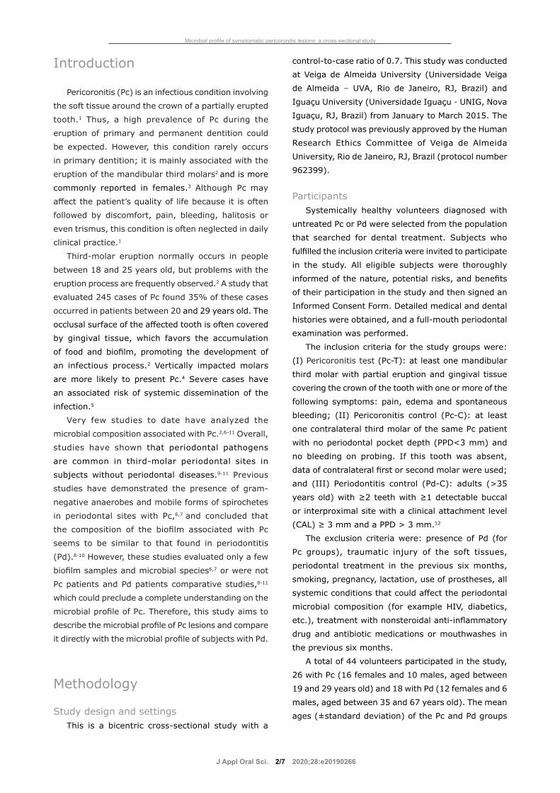
Figure 1- Bacterial strains used for the preparation of DNA probes. Species are grouped according to the microbial complexes18

J Appl Oral Sci. 2020;28:e201902664/7
in a Miniblotter 45 (Immunetics) with the lanes of
DNA at 90° to the lanes of the device. Digoxigenin-
labelled whole genomic DNA probes for 40 bacterial
species (Figure 1) were hybridized in individual
lanes of the Miniblotter. After the hybridization, the
membranes were washed at high stringency, and
the DNA probes were detected using the antibody to
digoxigenin conjugated with alkaline phosphatase.
Chemiluminescence detection was then performed.
The last two lanes in each run contained standards at
concentrations of 105 and 106 cells of each species.
Signals were evaluated visually by comparison with
the standards at the 105 and 106 bacterial cells for the
test species on the same membrane by a calibrated
examiner. The sensitivity of this assay was adjusted
to allow detection of 104 cells of a given species by
adjusting the concentration of each DNA probe.
This procedure was carried out to provide the same
sensitivity of detection for each species.
Statistical methodsThe mean counts (x105 cells) of the individual
bacterial species were averaged within each subject
and then across the subjects in the different clinical
groups. Similarly, the percentage of the total DNA
probe counts was determined initially in each site,
then per subject and averaged across the subjects in
the three groups. The individual proportions of each
species were added to determine the proportions
of each microbial complex.17 The significance of the
differences between the groups was assessed using
one-way ANOVA test. In addition, a t-test was used
to determine significant differences between the pairs
of groups. Adjustments for multiple comparisons
were performed when the 40 bacterial species were
evaluated simultaneously.17 All the analyses of this
study were conducted using a statistical program
developed by Sigmund Socransky (The Forsyth
Institute, Cambridge, MA, USA). The significance level
was set at 5%.
Results
Figure 2 shows the mean counts (x105 cells) of
the sites colonized by the 40 species evaluated in
the subgingival plaque samples from the Pc-C, Pc-T
and Pd-C groups. The species present in the highest
levels in Pc-T were Actinomyces oris, Eikenella
corrodens, Eubacterium nodatum, Fusobacterium
nucleatum spp. nucleatum, Treponema denticola and
Eubacterium saburreum. A. oris and E. nodatum were
present in higher mean counts in the Pc-T group in
comparison with the Pc-C and Pd-C groups (p<0.05).
The microbiota associated with the Pc-T group
was very similar to that found in Pc-C. Most of the
bacterial species evaluated in the study were found
in higher counts in the Pd-C group, and 20 of them
were significantly higher in this group compared with
the Pc-T group, including Actinomyces gerencseriae,
A. oris, Veillonella parvula, Streptococcus sanguinis,
Capnocytophaga gingivalis, Capnocytophaga ochracea,
Capnocytophaga sputigena, Campylobacter rectus,
Campylobacter showae, E. nodatum, Fusobacterium
nucleatum. spp. polymorphum, Fusobacterium
nucleatum. spp. vicentii, Parvimonas micra, Prevotella
intermedia, Porphyromonas gingivalis, Treponema
denticola, Leptotrichia buccalis, Propionibacterium
acnes, Streptococcus anginosus and Treponema
socranskii (p<0.05).
The mean proportions of the microbial complexes
in the different groups are described in Figure 3.
The red complex pathogens were higher in Pd-C
than Pc-T and Pc-C groups (p<0.05). A similar trend
was also observed for the green complex (p<0.05).
However, a tendency towards a higher proportion of
yellow complex species in the PC groups was noticed
(p=0.09).
Discussion
The results of this study showed Pc sites harbored
a quite diverse microbiota; nonetheless, with a lower
degree of dysbiosis than that observed in Pd lesions.
Pc biofilm samples had lower levels and proportions
of putative and traditional periodontal pathogens
and a tendency towards higher levels of the health-
associated yellow complex species than Pd lesions.
The red complex, which harbors the three most
traditional periodontal pathogens (P. gingivalis, T.
denticola and Tannerella forsythia),18 was present in
higher counts in Pd patients, compared with both the
Pc groups. Although previous studies have shown a
high number of T. forsythia in Pc patients,11,19 this
study could not confirm these findings. It is important
to highlight that P. gingivalis, an anaerobic gram-
negative bacteria, and T. denticola, an anaerobic
Microbial profile of symptomatic pericoronitis lesions: a cross-sectional study

J Appl Oral Sci. 2020;28:e201902665/7
Figure 2- Mean counts (105) of 40 subgingival species in each study group. The species were ordered according to the microbial complexes described by Socransky, et al.18 (1988). The significance of differences between groups was assessed using one-way ANOVA test. Different letters indicate significant differences between pairs of groups (t-test, p<0.05). Letters were color coded to indicate the different groups: green for Pc-C, red for Pc-T, and blue for Pd-C. Pc-C: Pericoronitis control group; Pc-T: Pericoronitis test group; Pd-C: Periodontitis control group
RIBEIRO MH, RIBEIRO PC, RETAMAL-VALDÉS B, FERES M, CANABARRO A

J Appl Oral Sci. 2020;28:e201902666/7
gram-negative spirochete, have been considered
key periodontal pathogens.20 Those are frequently
found to co-exist in deep periodontal pockets.21
Such interaction between them can contribute to
Pd progression.22 T. socranskii, another anaerobic
gram-negative spirochete, is also considered a
periodontal pathogen23,24 associated with Pc25 and
was also found in higher proportions and levels in
the Pd group. Interestingly, these 3 microorganisms
together (P. gingivalis, T. denticola, and T. socranskii)
were correlated with abnormal periodontal clinical
parameters and have been associated with periodontal
tissue loss.23
In fact, few published studies compared putative
pathogens in healthy and Pc sites. Some of these
studies have used conventional culture-dependent
methods that many times fail to detect strict anaerobe
pathogens,6,7 at least one study neglected to include a
control group without Pc, hampering the interpretation
of the results.8 Another study compared healthy and
symptomatic Pc sites, and the results supported
the hypothesis that the pericoronal region harbors
putative periodontal pathogens,10 and may provide a
favored niche for periodontal pathogens in a healthy
oral environment.9
Discussing the clinical findings of this study in
relation to the microbial profiles observed in the
various lesions is important. First, these findings
suggest the biofilm associated with Pc apparently
does not have a strong potential to trigger irreversible
periodontal destruction since it does not harbor high
levels of major periodontal pathogens and maintains
good levels of host-compatible microorganisms.
As Pc is an acute disease, one could hypothesize
that time between the development of the lesion
and its treatment for the massive growth of key
periodontal pathogens was insufficient. Besides that,
the prophylactic surgical removal of teeth with Pc is
frequently performed in dental practice.26 In addition,
antibiotic treatment must be considered in patients
whose Pc infections were disseminated and have
invaded deeper oral spaces.26 Most Pc lesions are
associated with a subgingival anaerobic niche created
by the overgrowth of gingival tissues. Nonetheless,
the differences between the microbial load of Pd and
Pc shown in this study suggest the antibiotic protocols
required to treat these conditions may not be the
same. Future studies addressing this topic would help
to guide clinical practice.
The main strength of this study is that, to the
best of our knowledge, this is the first study to
comprehensively assess the microbial composition of
Pc lesions and to compare this profile to that found in
Pd. One limitation of the study design is the relatively
small sample size, due to the difficulty in selecting Pc
cases in daily clinical practice. Furthermore, this study
shows results for the 40 bacterial species proposed
by Socransky, et al.18 (1988) as it is well established
that the periodontal microbiome comprises more
taxa than those included in this group of bacterial
species.27 Nevertheless, this panel of species has been
successfully used as a biological marker for many
studies of periodontal disease risk and treatment.28,29 A
comprehensive study showed it covers approximately
60% of the bacterial genera present in the oral cavity.30
Few studies have identified periodontal bacteria in
pericoronitis samples, but it seems that pericoronal
sites can harbor several pathogens. A recent
study showed some periodontopathic bacteria and
herpesviruses occurred concomitantly in pericoronitis
Figure 3- Mean proportions of the microbial complexes in each study group. The colors represent different microbial complexes18 and Actinomyces species (blue). The significance of differences between groups was sought using the one-way ANOVA. The differences were only found for red and green complexes. Different letters indicate significant differences between pairs of groups (t-test, p<0.05). Pc-C: Pericoronitis control group; Pc-T: Pericoronitis test group; Pd-C: Periodontitis control group
Microbial profile of symptomatic pericoronitis lesions: a cross-sectional study

J Appl Oral Sci. 2020;28:e201902667/7
samples.11 Such herpesviral-bacterial interaction could
be an important feature of pericoronitis and should be
further studied.
In conclusion, Pc microbiota is diverse, but these
lesions harbor lower levels of periodontal pathogens
than those of Pd.
AcknowledgementThe authors thank FAPERJ (grant number
E-26/101.454/2010), CAPES, CNPq and LAOHA for
financial support.
References1- Magraw CB, Golden B, Phillips C, Tang DT, Munson J, Nelson BP, et al. Pain with pericoronitis affects quality of life. J Oral Maxillofac Surg. 2015;73(1):7-12.2- Nitzan DW, Tal O, Sela MN, Shteyer A. Pericoronitis: a reappraisal of its clinical and microbiologic aspects. J Oral Maxillofac Surg. 1985;43(7):510-6.3- Singh P, Nath P, Bindra S, Rao SS, Reddy KV. The predictivity of mandibular third molar position as a risk indicator for pericoronitis: a prospective study. Natl J Maxillofac Surg. 2018;9(2):215-21.4- Katsarou T, Kapsalas A, Souliou C, Stefaniotis T, Kalyvas D. Pericoronitis: a clinical and epidemiological study in greek military recruits. J Clin Exp Dent. 2019;11(2):e133-7.5- Douglass AB, Douglass JM. Common dental emergencies. Am Fam Physician. 2003;67(3):511-6.6- Labriola D, Mascaro J, Alpert B. The microbiologic flora of orofacial abscess. J Oral Maxillofac Surg. 1983;41(11):711-4.7- Leung WK, Theilade E, Comfort MB, Lim PL. Microbiology of the pericoronal pouch in mandibular third molar pericoronitis. Oral Microbiol Immunol. 1993;8(5):306-12.8- White RP Jr, Madianos PN, Offenbacher S, Phillips C, Blakey GH, Haug RH, et al. Microbial complexes detected in the second/third molar region in patients with asymptomatic third molars. J Oral Maxillofac Surg. 2002;60(11):1234-40.9- Mansfield JM, Campbell JH, Bhandari AR, Jesionowski AM, Vickerman MM. Molecular analysis of 16S rRNA genes identifies potentially periodontal pathogenic bacteria and archaea in the plaque of partially erupted third molars. J Oral Maxillofac Surg. 2012;70(7):1507-14.10- Rajasuo A, Sihvonen OJ, Peltola M, Meurman JH. Periodontal pathogens in erupting third molars of periodontally healthy subjects. Int J Oral Maxillofac Surg. 2007;36(9):818-21.11- Jakovljevic A, Andric M, Knezevic A, Milicic B, Beljic-Ivanovic K, Perunovic N, et al. Herpesviral-bacterial co-infection in mandibular third molar pericoronitis. Clin Oral Investig. 2017;21(5):1639-46.12- Papapanou PN, Sanz M, Buduneli N, Dietrich T, Feres M, Fine DH, et al. Periodontitis: Consensus report of workgroup 2 of the 2017 World Workshop on the Classification of Periodontal and Peri-Implant Diseases and Conditions. J Periodontol. 2018;89(Suppl 1):S173-82.
13- Hallström H, Persson GR, Strömberg U, Twetman S, Renvert S. Reproducibility of subgingival bacterial samples from patients with peri-implant mucositis. Clin Oral Investig. 2015;19(5):1063-8.14- Socransky SS, Haffajee AD, Smith C, Martin L, Haffajee JA, Uzel NG, et al. Use of checkerboard DNA-DNA hybridization to study complex microbial ecosystems. Oral Microbiol Immunol. 2004;19(6):352-62.15- Mestnik MJ, Feres M, Figueiredo LC, Duarte PM, Lira EA, Faveri M. Short-term benefits of the adjunctive use of metronidazole plus amoxicillin in the microbial profile and in the clinical parameters of subjects with generalized aggressive periodontitis. J Clin Periodontol. 2010;37(4):353-65.16- Socransky SS, Smith C, Martin L, Paster BJ, Dewhirst FE, Levin AE. “Checkerboard” DNA-DNA hybridization. Biotechniques. 1994;17(4):788-92.17- Socransky SS, Haffajee AD, Smith C, Dibart S. Relation of counts of microbial species to clinical status at the sample sites. J Clin Periodontol. 1991;18(10):766-75.18- Socransky SS, Haffajee AD, Cugini MA, Smith C, Kent RL Jr. Microbial complexes in subgingival plaque. J Clin Periodontol. 1988;25(2):134-44.19- Sencimen M, Saygun I, Gulses A, Bal V, Acikel CH, Kubar A. Evaluation of periodontal pathogens of the mandibular third molar pericoronitis by using real time PCR. Int Dent J. 2014;64(4):200-5.20- Dashper SG, Seers CA, Tan KH, Reynolds EC. Virulence factors of the oral spirochete Treponema denticola. J Dent Res. 2011;90(6):691-703.21- Ng HM, Kin LX, Dashper SG, Slakeski N, Butler CA, Reynolds EC. Bacterial interactions in pathogenic subgingival plaque. Microb Pathog. 2016;94:60-9.22- Zhu Y, Dashper SG, Chen YY, Crawford S, Slakeski N, Reynolds EC. Porphyromonas gingivalis and Treponema denticola synergistic polymicrobial biofilm development. PLoS One. 2013;8(8):e71727.23- Takeuchi Y, Umeda M, Sakamoto M, Benno Y, Huang Y, Ishikawa I. Treponema socranskii, Treponema denticola, and Porphyromonas gingivalis are associated with severity of periodontal tissue destruction. J Periodontol. 2001;72(10):1354-63.24- Mineoka T, Awano S, Rikimaru T, Kurata H, Yoshida A, Ansai T, et al. Site-specific development of periodontal disease is associated with increased levels of Porphyromonas gingivalis, Treponema denticola, and Tannerella forsythia in subgingival plaque. J Periodontol. 2008;79(4):670-6.25- Dabu B, Mironiuc-Cureu M, Jardan D, Szmal C, Dumitriu S. Identification of four Treponema species in subgingival samples by nested-PCR and their correlation with clinical diagnosis. Roum Arch Microbiol Immunol. 2012;71(1):43-7.26- Adeyemo WL. Do pathologies associated with impacted lower third molars justify prophylactic removal? A critical review of the literature. Oral Surg Oral Med Oral Pathol Oral Radiol Endod. 2006;102(4):448-52.27- Pérez-Chaparro PJ, Gonçalves C, Figueiredo LC, Faveri M, Lobão E, Tamashiro N, et al. Newly identified pathogens associated with periodontitis: a systematic review. J Dent Res. 2014;93(9):846-58.28- Teles R, Teles F, Frias-Lopez J, Paster B, Haffajee A. Lessons learned and unlearned in periodontal microbiology. Periodontol 2000. 2013;62(1):95-162.29- Feres M, Figueiredo LC, Soares GM, Faveri M. Systemic antibiotics in the treatment of periodontitis. Periodontol 2000. 2015;67(1):131-86.30- Socransky SS, Haffajee AD, Smith C, Martin L, Haffajee JA, Uzel NG, et al. Use of checkerboard DNA–DNA hybridization to study complex microbial ecosystems. Oral Microbiol Immunol. 2004;19(6):352-62.
RIBEIRO MH, RIBEIRO PC, RETAMAL-VALDÉS B, FERES M, CANABARRO A