Microarray analysis shows that some microRNAs downregulate large numbers of target mRNAs LEE P. LIM,...
15
Microarray analysis shows that some microRNAs downregulate large numbers of target mRNAs LEE P. LIM, NELSON C. LAU, PHILIP GARRETT-ENGELE ANDREW GRIMSON, JANELL M. SCHELTER, JOHN CASTLE, DAVID P. BARTEL, PETER S. LINSLEY & JASON M. JOHNSON Presented by Li-Chen Wu
-
date post
20-Dec-2015 -
Category
Documents
-
view
216 -
download
0
Transcript of Microarray analysis shows that some microRNAs downregulate large numbers of target mRNAs LEE P. LIM,...

Microarray analysis shows that some microRNAs downregulate large numbers of target mRNAs
LEE P. LIM, NELSON C. LAU, PHILIP GARRETT-ENGELE ANDREW GRIMSON, JANELL M. SCHELTER, JOHN CASTLE, DAVID P. BARTEL, PETER S. LINSLEY & JASON M. JOHNSON
Presented by Li-Chen Wu

What are MicroRNAs (miRNAs)?
MicroRNAs are a growing family of small non-protein-coding regulatory genes (~ 22 nucleotides) that regulate the expression of homologous target-gene transcripts.
Bioinformatics studies predict that there are ~250 miRNAs encoded in the human genome. About a quarter of human miRNA genes are located in introns of pre-mRNAs; the remaining miRNAs are clustered in the genome, predicting a multi-cistronic transcript.
miRNAs are processed by the RNA-mediated interference machinery. They have been implicated in the control of cell death and proliferation in flies, haematopoietic lineage differentiation in mammals, neuronal patterning in nematodes, and leaf and flower development.

Model of microRNA biogenesis
Step 1. Transcription of a miRNA gene produces a primary nuclear transcript (pri-miRNA).
Step 2. The nuclear RNase III enzme Drosha cleaves pri-miRNA to generate a stem-loop pre-miRNA. (~70 nucleotides pre-miRNA intermediate)
Step 3. The pre-miRNA directly associates with the exportin Exp5 and Ran-GTP, and this complex exits the nucleus. Hydrolysis of Ran-GTP to Ran-GDP causes dissociation of the export complex.
Step 4. The pre-miRNA associates with the cytoplasmic RNase III enzyme Dicer, which cleaves it to form a 22-nucleotide miRNA intermediate.
Step 5. The miRNA intermediate rapidly unwinds as it assembles into a miRNP complex, and one miRNA strand is retained in the miRNP.
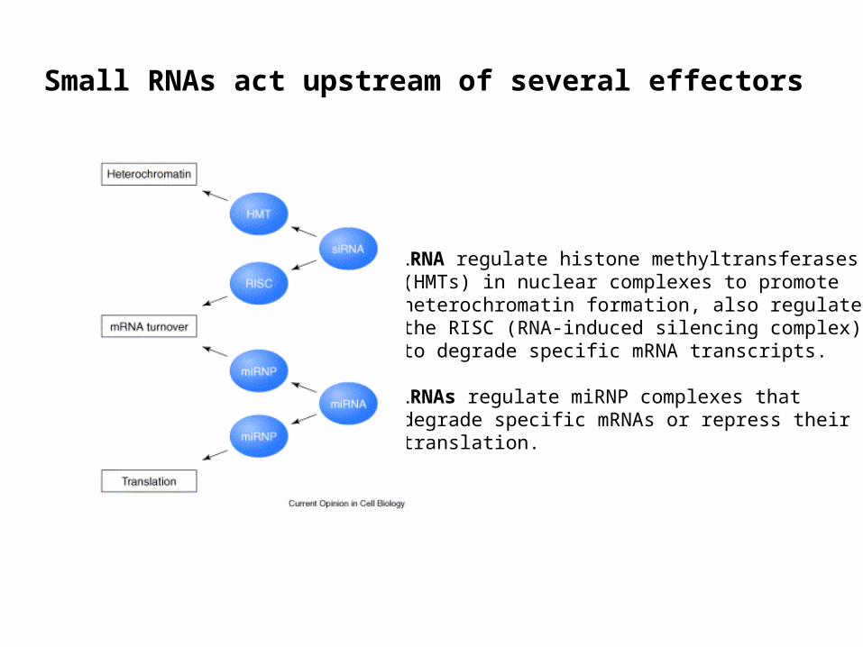
Small RNAs act upstream of several effectors
siRNA regulate histone methyltransferases (HMTs) in nuclear complexes to promote heterochromatin formation, also regulate the RISC (RNA-induced silencing complex) to degrade specific mRNA transcripts.
miRNAs regulate miRNP complexes that degrade specific mRNAs or repress their translation.

Direct Labeling and Hybridization of MicroRNA Sample

Quantitative northern blot analysis of miR-1 and miR-124 expression
A. Northern blot of a human total RNA tissue panel. B. Molecular abundance of upregulated miRNAs and corresponding U6 snRNA in
tissue types, based on quantitation of signals from the Northern blot in A).
Supplementary Figure 2.

Figure 1. Tissue-specific gene expression rankings for downregulated genes
a. Microarray signature12h after miR-124 transfection. (Significantly downregulated probes are in green)
b.c. Cerebral cortex rankings for LocusLink genes and the set of genes downregulated at both 12 and 24h following transfection of miR124.
The 174 genes downregulated by miR-124 transfection are significantly enriched for genes that are expressed at lower levels in cerebal cortex relative to other tissues.

d. e. Log (base 10) P values are plotted for each of the 46 tissues for the miR124 (d) and miR-1 (e) downregulated sets.
Tissue-specific gene expression rankings for downregulated genes

Figure 2. Over-represented motifs in the 3' UTRs of downregulated genes.
a. b. miR-1 (a) and miR-124 (b) downregulated genes. Motif nucleotides that were complementary to the transfected miRNA are shown in red;
base-pairing to the miRNA sequence is shown in blue.
*These results imply that knockdown of the transcripts is caused mainly by direct binding of the transfected miRNAs to the 3’UTRs, with binding at the 5’ end of the miRNA being particularly vital.*The 5’ region, and particularly seed positions 2-8, is the most conserved region of metazoan miRNAs and has long been thought to play a key role in the target recognition.

Figure 3 Microarray analysis of the effects of miRNA mutations.
a. Sequences of 124mut9-10 duplex after transfected into HeLa cells, gave a downregulated signature 89% shared with that of the wild type, whereas for 124mut5-6 was almost completely distinct.

Supplementary Figure 2. As in Figure 1c and 1d, log p-values are plotted for each of the 46 tissues for the downregulated sets from the a. 124mut9-10, b. 124mut5-6, c. chimiR-124/1, and d. chimiR-1/124 transfections.
Tissue analysis for mutant microRNAs
These results demonstrate that, for the mRNA knockdown observed in this assay, positions 5 and 6 are much more crucial than positions 9 and 10.

a. Sequences of 124mut9-10 duplex after transfected into HeLa cells, gave a downregulated signature 89% shared with that of the wild type, whereas for 124mut5-6 was almost completely distinct.
b. Sequences of wild type and chimaeric miRNA after transfected into HeLa cells, showed that the 5’ ends of the miRNAs were sufficient for generating tissue-specific signatures.
Figure 3 Microarray analysis of the effects of miRNA mutations.

Figure 4. MicroRNA-directed repression of renilla luciferase reporter genes bearing 3' UTR segments from predicted target genes.
a. b. MiR-1(a) and miR-124(b) target genes. wild type (open bars), mutant plasmids (filled bars)
Results: When cotransfected with the cognate miRNA, six of the ten wild-type reports exhibited significant repression relative to the corresponding constructs with mutant seed matches; cotransfection of the noncognate miRNA typically had no effect.
Conclusion: Pairing to the miRNA seed region contributes directly to mRNA repression

Expected and observed seed match counts in different regions of miR-1 or miR-124 downregulated genes
Supplementary Figure 3.
Conclusion: MiRNAs interact more frequently or more effectively with 3’ UTRs than with other regions of mRNAs.

Conclusion
1. Metazoan miRNAs are predominantly negative regulators of gene expression, they can reduce the levels of many of their target transcripts, not just the amount of protein deriving from these transcripts.
2. MiR-1 and miR-124. and presumably other tissue-specific miRNAs, seem to downregulate a far greater number of targets than previously appreciated, thereby helping to define tissue-specific gene expression in humans.
3. Microarrays can be used to detect physiologically relevant miRNA.