
Michael P. Murphy and Richard C. Hartley
58
1 Invited review for Nature Reviews Drug Discovery Mitochondria as a therapeutic target for common pathologies Michael P. Murphy 1 and Richard C. Hartley 2 1 MRC Mitochondrial Biology Unit, University of Cambridge, Hills Road, Cambridge CB2 0XY, UK 2 WestCHEM School of Chemistry, University of Glasgow, Glasgow G12 8QQ, UK. Emails: [email protected] : [email protected] Abstract | Although the development of mitochondrial therapies has largely focused on diseases caused by mutations in mitochondrial DNA or in nuclear genes encoding mitochondrial proteins, it has emerged that mitochondrial dysfunction also contributes to the pathology of many common disorders, including neurodegeneration, metabolic disease, heart failure, ischaemia-reperfusion injury and protozoal infections. Mitochondria therefore represent an important drug target for these highly prevalent diseases. Several strategies aimed at therapeutically restoring mitochondrial function are emerging and a small number of agents have entered clinical trials. This review will discuss the opportunities and challenges faced for the further development of a mitochondrial pharmacology for common pathologies. Introduction Mitochondria perform many key roles in the cell, most notably oxidative phosphorylation, central carbon metabolism and the biosynthesis of intermediates for cell growth, but they are also responsible for several other essential processes that determine cell function and fate 1,2 3-6 7 (FIG. 1 and Box 1). Consequently, mutations in nuclear or mtDNA genes that disrupt mitochondrial function lead to devastating “primary” mitochondrial diseases 3,8-10 1,11 . Our knowledge of how mitochondria function in the cell has expanded dramatically. It is now clear that mitochondria participate in nearly all aspects of cell function, affecting processes not traditionally linked with the organelle, including cancer, inflammation, metabolic
Transcript of Michael P. Murphy and Richard C. Hartley

1
Invited review for Nature Reviews Drug Discovery
Mitochondria as a therapeutic target for common pathologies
Michael P. Murphy1 and Richard C. Hartley2
1MRC Mitochondrial Biology Unit, University of Cambridge, Hills Road, Cambridge CB2
0XY, UK
2WestCHEM School of Chemistry, University of Glasgow, Glasgow G12 8QQ, UK.
Emails: [email protected] : [email protected]
Abstract | Although the development of mitochondrial therapies has largely focused on
diseases caused by mutations in mitochondrial DNA or in nuclear genes encoding
mitochondrial proteins, it has emerged that mitochondrial dysfunction also contributes to the
pathology of many common disorders, including neurodegeneration, metabolic disease, heart
failure, ischaemia-reperfusion injury and protozoal infections. Mitochondria therefore
represent an important drug target for these highly prevalent diseases. Several strategies
aimed at therapeutically restoring mitochondrial function are emerging and a small number of
agents have entered clinical trials. This review will discuss the opportunities and challenges
faced for the further development of a mitochondrial pharmacology for common pathologies.
Introduction
Mitochondria perform many key roles in the cell, most notably oxidative phosphorylation,
central carbon metabolism and the biosynthesis of intermediates for cell growth, but they are
also responsible for several other essential processes that determine cell function and fate 1,2
3-6 7 (FIG. 1 and Box 1). Consequently, mutations in nuclear or mtDNA genes that disrupt
mitochondrial function lead to devastating “primary” mitochondrial diseases 3,8-10 1,11. Our
knowledge of how mitochondria function in the cell has expanded dramatically. It is now
clear that mitochondria participate in nearly all aspects of cell function, affecting processes
not traditionally linked with the organelle, including cancer, inflammation, metabolic

2
signalling, and cell death, transformation and fate 5,6 7. Consequently, mitochondrial
dysfunction has been found to contribute to many common disorders, including
neurodegeneration, metabolic disease and heart failure 4,5,12,13. These “secondary”
mitochondrial diseases can arise even if the proximal cause is not mitochondrial, for example
when the initiating disease process disrupts mitochondrial function as a downstream effect
6,10,12,14-16 7. Thus, drugs designed to act on mitochondria may be effective therapies for a
range of common diseases, and could be more effective than when applied to the notoriously
hard to treat diseases that arise due to mutations in mitochondrial genes 3,12 14 7 10.
Importantly, drugs designed to affect mitochondrial function can be applied to many highly
prevalent diseases and pathological processes, with important social, medical and economic
impacts 2,17,18. In many cases progress in developing new therapeutic approaches for these
common diseases has been dispiritingly slow, as is illustrated by the lack of new drugs
coming to market for stroke or neurodegenerative diseases. Focusing on mitochondria offers
a promising alternative approach to developing new therapeutic options for these disorders
14,19,20. Examples of mitochondrial agents that are currently being, or have recently been,
assessed in humans include agents to replenish NAD+ pools such as nicotinamide
mononucleotide (NMN) 21, mitochondria-targeted protective compounds such as MitoQ 22,23
and Bendavia 24, antioxidants such as Coenzyme Q10 25 and Cyclosporin A, an inhibitor of
the mitochondrial permeability transition pore 26 27. Given that the development and
application of drugs designed to affect mitochondria is still in its infancy, this review will
focus on the general principles, vast potential and ongoing challenges for intervening at the
mitochondrial level.
Rationale for targeting mitochondria
Disruption to mitochondrial bioenergetic and metabolic function can lead to many secondary
mitochondrial disorders (FIG. 1). Interestingly, common patterns regarding how
mitochondria contribute to the aetiology of disparate pathologies have emerged 5,14,28.
Important among these are: the aberrant production of reactive oxygen species (ROS),
calcium dyshomeostasis, defective mitochondrial biogenesis, disruption to mitochondrial
dynamics and quality control, necrotic cell death through induction of the permeability
transition pore (MPTP), inappropriate activation or suppression of apoptosis, lowered cellular
ATP/ADP ratio, decreased NAD+ levels and alterations to mitochondrial signaling pathways
(FIG. 1) 14,28,29 7 . In many cases these different types of organelle dysfunction are linked

3
mechanistically, hence are often found together, and in addition they may contribute to
disease by acute, irreversible cell death, long term disruption to the role of mitochondria as
signaling hubs, or to the life-long accumulation of environmental damage that leads to a
degenerative disorder 15. The details of how mitochondrial dysfunction leads to specific
pathologies are discussed below.
In short, there are three factors supporting the pursuit of mitochondria as a therapeutic
target for common pathologies. First, many prevalent diseases are “secondary” mitochondrial
disorders in that mitochondrial dysfunction contributes to the disease process or clinical
progression. Hence, targeting the organelle can improve patient outcome, even though
mitochondrial dysfunction may not be the primary driver of pathology. Second, mitochondria
contribute to diverse pathologies through common pathways 10,14, therefore a single
therapeutic approach may apply to multiple disorders. Finally, the common diseases where
targeting mitochondria show promise are of increasing medical, social and economic impact
in our aging population. Given that the development of new drugs for these disorders has
been frustratingly slow, new approaches are needed 30 31 32.
Therapeutic approaches to mitochondria
There are a number of approaches aimed at modulating mitochondrial function in primary
and secondary mitochondrial diseases 3,9. These include: behavioural interventions, such as
changes in diet or exercise 33; exposure to hypoxia 34; stem cell therapies 35; replacing
defective mtDNA in an oocyte 36; and supplementation of a tissue with exogenous
mitochondria 37. Furthermore, there are many potential therapeutic strategies utilising gene
therapies to deliver corrected versions of a defective gene, or to ectopically express proteins
designed to degrade mutated mtDNA 38 or alter metabolism 39. While all these approaches
could lead to potential treatments for common pathologies, their coverage is beyond the
scope of this review, which will focus on the general strategies for the development of small
molecule therapies that can modulate mitochondrial function.
Drugs can act directly on the mitochondria themselves, or affect the organelle
indirectly by binding to regulatory targets in the cytosol or nucleus 14,40. An important aspect
of drugs that affect the organelle directly, is the ability to selectively target bioactive moieties
to mitochondria in vivo by conjugation to lipophilic cations or to peptides, which facilitates
drug effectiveness by enhancing potency, avoiding side effects and accelerating delivery
14,20,41,42 (Box 2).

4
There are five broad therapeutic strategies in which small molecules can be used to
affect mitochondria directly or indirectly in secondary mitochondrial diseases. These are: (i)
repairing or preventing damage to the organelle; (ii) inducing mitochondrial biogenesis; (iii)
enhancing organelle quality control by stimulating degradation of damaged mitochondria or
organelle components; (iv) co-opting mitochondrial function to induce cell death; or (v)
altering mitochondrial signaling pathways or metabolic processes. Below, we expand on
these, but of course it is important to note that many of these types of damage are linked and
that treating one mode of mitochondrial dysfunction often has a positive impact on others.
Protecting mitochondria
Mitochondrial dysfunction in diseases can arise from sustained damage to the organelle’s
protein, DNA and lipids 2,43-45. Oxidative damage is frequently considered, due to the
relatively high level of ROS production by the mitochondrial respiratory chain and the
susceptibility of the organelle to oxidative damage 46,47. Carbon stress is another disruptor of
mitochondrial function that arises due to the high levels of activated acyl-CoAs in the
mitochondrial matrix that lead to non-enzymatic protein acylation, typically on lysine
residues, that affects protein function and proteostasis 44,45,48.
A related common pathway of mitochondrial damage in many scenarios is the
depletion of NAD+, which can occur by activation of pathways that use up cellular and
mitochondrial NAD+ pools, such as activation of poly (ADP-ribose) polymerases (PARPs),
mono ADP ribosyl transferases, and the cyclic ADP-ribose hydrolase CD38 49,50 51 52. One
consequence of NAD+ depletion is disruption of bioenergetic pathways. In addition, NAD+ is
required for the reversal of lysine acylation by sirtuins, hence NAD+ depletion also
contributes to an elevation of protein lysine acylation, disrupting signalling pathways that are
altered by lysine acylation and also contributing to carbon stress leading to the accumulation
of damaged and misfolded proteins. Of course, many other forms of damage occur, for
example disruption due to formation of the mitochondrial permeability transition pore
(MPTP), a large conductance channel in the inner membrane that is activated following
calcium accumulation in the presence of oxidative stress, leading to mitochondrial swelling
and subsequent cell death 53-55.
5
Defects in mitochondrial proteostasis is another important form of mitochondrial
damage that contributes to a wide range of pathologies 7 56 57. Normally the proteins within
the mitochondria are folded correctly and when they become damaged or miss-folded are
either refolded or rapidly degraded 7 56 57. Thus, when correctly functioning, proteostasis
prevents the accumulation and aggregation of defective proteins within mitochondria, which
would severely disrupt organelle function. Mitochondria face a number of challenges in
maintaining proteostasis and maintaining the correct folding of proteins that are either
imported into, or translated within the organelle 57. A further complication is that four of the
mitochondrial oxidative phosphorylation complexes contain polypeptides encoded by both
the nuclear and mitochondrial genomes, hence the relative levels of these polypeptides have
to be carefully matched to correctly assemble these complexes 57. Finally, the mitochondrial
matrix is exposed to high levels of both oxidative and carbon stress, that can damage
proteins, rendering them less stable 57. In dealing with these challenges the mitochondria
does not have a proteasome, nor the same heat shock protein complement as the cytosol.
Instead, it has its own repertoire of chaperones and proteases to maintain organelles
proteostasis 57 7 56. The mitochondrial chaperones include mitochondrial heat shock protein
70 and 90 and the matrix chaperonin complex composed of mitochondrial heat shock protein
60 and 10 that help fold nascent proteins, or refold misfolded ones. In addition, mitochondria
contain a wide range of proteases that degrade misfolded proteins 58 7 56. Mutations in these
mitochondrial proteases lead to the accumulation of misfolded proteins and dysfunctional
mitochondria in a number of diseases 58. Furthermore, excessive oxidative damage, or protein
acylation due to carbon stress, cause protein missfolding and aggregation within
mitochondria. Thus factors such as replenishing the NAD+ pool to counteract carbon stress
by enhancing the activity of sirtuins, or preventing oxidative damage with antioxidants all
help maintain proteostasis. Due to the contribution of defective protostasis to common
diseases there is considerable interest in activating chaperones or proteases at the level of the
organelle. Related to this, the mitochondrion has an unfolded protein response (mtUPR) that
upregulates the expression of chaperones within the mitochondrial matrix 57 56 and
enhancing the activity of the mtUPR is protective in a number of model organisms 56.
Many drugs protect the organelle directly by affecting a specific process following
selective binding to a particular target site. Some drugs target matrix proteins, for example,
cyclosporin A binds to the matrix protein cyclophilin D (CyD) and thereby prevents cell
death caused by formation of the MPTP 59. Other compounds such as suppressors of site IQ
electron leak (S1QELs) and suppressors of site IIIQo electron leak (S3QELs) bind directly to

6
respiratory chain complexes I and III, respectively, in the mitochondrial inner membrane to
inhibit ROS production 60,61. Conversely, there are many protective molecules that act on
general processes within mitochondria, rather than by binding to specific targets 14,20. These
include antioxidants designed to lower mitochondrial oxidative damage 62, molecules that
enable electrons to bypass respiratory complexes in order to sustain oxidative
phosphorylation in spite of respiratory chain damage 63. A related intervention is the use of
small molecule uncouplers such as dinitrophenol (DNP) which decrease the protonmotive
force (p) across the mitochondrial inner membrane thereby making oxidative
phosphorylation less efficient, which helps to burn off excess fat and also to decrease
mitochondrial ROS production 64 65. The depletion of NAD+, which can lead to both
bioenergetic defects and to inappropriate protein acylation, can be counteracted by
compounds such as nicotinamide (NAM), nicotinamide riboside (NR) and nicotinamide
mononucleotide (NMN) which act by replenishing NAD+ levels 50-52,66-70. Restoring NAD+
levels has a number of protective effects, in part by enhancing the activity of sirtuins which
act as NAD+-dependent lysine deacylases. As protein acylation is thought to have a
regulatory role in a number of metabolic processes, the positive effects of NAD+ modulators
are often ascribed to changes in regulation 66,69. However, as lysine acylation is also a carbon
stress that can lead to protein dysfunction and aggregation, it is also likely that some of the
positive effects of elevating NAD+ levels and activating sirtuins are to counteract carbon
stress 44,45.
Altering mitochondrial biogenesis
Instead of directly affecting mitochondria, an important alternative therapeutic strategy is to
alter organelle amount or activity by enhancing mitochondrial biogenesis 15,71-73. This raises
the possibility of pharmacologically increasing the mitochondrial content of the cell, the
surface area of the inner membrane or the content of the oxidative phosphorylation
machinery in order to increase mitochondrial ATP output, just as occurs in response to
exercise 72. This could be achieved by pharmacologically intervening at the level of the
transcription factors and related regulatory proteins that control mitochondrial biogenesis
15,72,73. There are a large number of nuclear-encoded transcriptional factors which control the
expression of those genes involved in mitochondrial biogenesis. For example, Nuclear
Respiratory Factors (NRF) 1 and 2 determine the expression of multiple nuclear genes that
encode proteins targeted to mitochondria, such as DNA polymerase (POLG) and the DNA

7
helicase Twinkle which are essential for mtDNA replication 74 and Transcription Factor A
(Mitochondrial) (TFAM) which regulates expression of the 37 genes encoded by mtDNA
6,15. There are many other transcription factors that affect mitochondrial biogenesis, such as
Peroxisome Proliferator-Activated Receptors (PPARs), Estrogen-Related Receptors (ERRs),
and cAMP response element-binding protein (CREB1) and Forkhead box-O (FOXO) 7,15,72,
however a detailed consideration of these is beyond the scope of this review and is covered
elsewhere 7. Transcription factor activity is further affected by the transcriptional coactivators
such as peroxisome proliferator-activated receptor-γ coactivator-1α (PGC-1α) and
corepressors such as nuclear receptor corepressor 1(NCOR1), receptor interacting protein 140
(RIP140) and retinoblastoma proteins (pRb) which helps to coordinate organelle biogenesis
and oxidative metabolism in response to changes in cell metabolic requirements (reviewed in
7,15 75). These responses are often transmitted through post translational modifications (PTMs)
for example, phosphorylation of PGC-1α by the energy sensor AMP-activated protein kinase
(AMPK) increases mitochondrial biogenesis in response to energy demand 13, while PGC-
1α deacetylation by Sirtuin1 (SIRT1) enables responses to metabolic challenges 75.
A number of drugs interact with these pathways to regulate mitochondrial biogenesis
by altering the activity of transcription factors 72,73. For example, the PPARγ transcription
factor can be activated directly by the anti-diabetic drugs pioglitazone and rosiglitazone, as
well as by the lipid metabolism modifiers bezafibrate and thiazolideindiones, which increase
PGC-1α expression and upregulate mitochondrial biogenesis 15,76 7
77. Mitochondrial biogenesis can also be enhanced by drugs that alter PGC-1α activity
indirectly 15 75. For example, AMPK agonists such as 5-aminoimidazole-4-carboxamide
ribonucleotide (AICAR) activate PGC-1, mimicking the enhancement of mitochondrial
biogenesis by energy demand 78. Another approach is to use the SIRT 1 activators resveratrol
and viniferin, which activate PGC-1 by reversing acetylation 15. A parallel approach to
enhancing mitochondrial biogenesis is to inhibit pathways that repress mitochondrial
biogenesis, such as hypoxia-inducible factor-1α (HIF-1α) 79,80.
Modulating mitochondrial dynamics
Mitochondria do not exist as isolated organelles in the cell, but instead undergo a continual
cycle of fusing together to form larger mitochondria that then undergo fission to break up into
smaller bodies 81,82. The protein machinery that leads to these processes comprises fission
proteins such as dynamin related protein 1 (DRP-1), while fusion is determined by proteins
8
such as mitofusins (MFN 1 & 2) on the outer membrane and Optic Atrophy 1 (OPA1) on the
inner membrane 82 81. Small molecules have been developed such as mitochondrial division
inhibitor-1 (Mdivi-1), which decrease DRP-1 activity and thus slow mitochondrial fission
77,83, however their specificity is unclear hence some effects may not be due to affecting
organelle division 84,85. Modulating mitochondrial dynamics is thought to have a number of
beneficial impacts on mitochondrial function and activity, although in many cases the
mechanism and significance of these effects are not clear 77. However, it is evident that one
important aspect of mitochondrial dynamics is that it is intimately linked to mitochondrial
quality control, discussed below.
Enhancing mitochondrial quality control
A major reason for continual mitochondrial fission/fusion is that it facilitates the degradation
of damaged organelles by mitophagy, because small mitochondrial particles can be easily
engulfed by the mitophagy machinery 71 81,82. This requires a means of recognizing that
mitochondria moving through the small particulate stage are damaged. One way in which this
may be done is by their lowered protonmotive force (p) which leads to accumulation of the
kinase PINK on their surface, PINK in turn recruits the PARKIN E3 ligase which
ubiquitinylates damaged mitochondria and thereby targets them for degradation by
mitophagy 86. While the role of this pathway in vivo is less clear 87, pathways that recognise
damaged mitochondria and target them for mitophagy are a central part of mitochondrial
quality control. Thus, drugs that enhance mitochondrial division may increase the clearance
of defective organelles 71 81,82. One example is AMPK activation which can increase DRP-1
recruitment to mitochondria by direct phosphorylation of the mitochondrial adaptor,
mitochondrial fission factor (MFF), and thus enhances fission and subsequent autophagy of
damaged mitochondria 13,88. Increasing the removal of damaged mitochondria by mitophagy
has many positive effects, such as decreasing inflammation 71, thus activating mitophagy is
an appealing therapeutic strategy and this has been explored with promising results using
natural compounds such as Urolithin A which enhances muscle function in rodents with
possible relevance to sarcopenia 89.
There are many other ways in which mitochondria quality control can happen at the
sub-mitochondrial level in parallel to mitophagy. Correct mitochondrial proteostasis protects
against the accumulation of damaged and unfolded proteins within mitochondria 57 56.
Prevention of mitochondrial protein aggregation can be enhanced by upregulating the mtUPR
9
response, which increases the expression of a series of chaperones within the mitochondria
and activating this response is protective in a number of model organisms 56. 7. Mitochondria
can compartmentalize oxidized protein and lipid into mitochondria-derived vesicles (MDVs)
that bud off from the organelle and are then targeted for degradation in lysosomes 90-92.
There are also multiple proteases, nucleases and lipases within the mitochondria that degrade
damaged molecules 58. Among these are the proteases ATPases associated with diverse
cellular activities (AAA) proteases, mutations to which contribute to degenerative diseases 58.
Finally, the myriad of potentially disruptive small molecules generated within mitochondria
by oxidative damage and carbon stress can be conjugated to glutathione by glutathione S-
transferases and the resulting conjugate exported by ATP Binding Cassette (ABC) proteins 93.
Thus enhancing the clearance of damaged mitochondria and the organelle’s components is a
promising strategy for future development.
Harnessing mitochondria to kill cells
The central role of mitochondria in cell death by apoptosis or necrosis makes them a good
target when aiming to kill a particular cell 55,94, such as a cancer cell or a protozoan parasite.
While it is easy to kill cells non-selectively by targeting mitochondria, the challenge is to do
so selectively. Mitochondria are similar in most cells, consequently any small differences in
the mitochondrial function of target cells makes an appealing target 95. Therefore, using
mitochondria to kill cancer cells necessitates focussing on how they differ from non-
transformed cells, or selectively activating a toxic pro-drug within the target cell. For
example, many cancer cells have ineffective mitochondrial apoptosis that can be re-activated
96. Another approach is to deplete antioxidant defences 97, or to increase mitochondrial ROS
production 98, and combine these cell stressors with another cancer drug to induce synthetic
lethality 97.
Altering mitochondrial signaling
A rapidly expanding area of mitochondrial biology is the role of the organelles as signaling
hubs that respond to and influences processes throughout the cell 6,99,100. The signals that
emanate from mitochondria to the rest of the cell include changes in ATP/ADP ratio, Ca2+,
NAD+, metabolites and ROS, but our understanding of their nature, targets and physiological
roles is still developing 99. Redox signaling by the production of ROS such as hydrogen
peroxide, that modify protein activity through the reversible oxidation of redox sensitive
cysteine residues has been a long standing focus 99,101,102. More recently there has been
10
considerable interest in how citric acid cycle (CAC) metabolites are transmitted back and
forth between the mitochondrion and the cytosol as a way of regulating cell function and fate
103,104. For example, histone acetylation is sensitive to acetyl CoA levels that are determined
by citrate export from the mitochondria 105. Furthermore, there are numerous 2-oxoglutarate-
dependent dioxygenases, including: prolyl hydroxylases in the Hif-1 oxygen sensing
pathway; Ten Eleven Translocation (TET) DNA demethylase; and the histone lysine
demethylase Jumonji C 106. These enzymes utilise 2-oxoglutarate as a substrate and are
inhibited by succinate, hence providing a link between mitochondrial CAC activity and the
regulation of oxygen sensing and the formation of epigenetic marks on the genome 106 106 107.
Thus, the manipulation of CAC metabolite transfer between the mitochondrial matrix and the
rest of the cell may be a useful therapeutic approach 108.
Treating pathologies via mitochondria
The general principles of how and why to treat mitochondria in common pathologies have
been outlined above. Here we consider some concrete examples of the common pathologies
ischemia-reperfusion injury, inflammation, the metabolic syndrome, neurodegeneration, heart
failure and protozoal infection where therapies focussed on mitochondria are likely to be
effective, discuss the approaches used and suggest future directions. Mitochondria are also
proving to be an interesting therapeutic target in cancer therapies, however the diversity of
this field puts it beyond the scope of this review but a few key points are considered in Box 3.
Mitochondria are also emerging as potential targets in many other common pathologies
including muscular dystrophies 69, sarcopenia 109, lung diseases 110 and colitis 111 and the
reader is referred to the cited papers and reviews for more detail.
Ischemia-reperfusion injury
Ischemia ensues when the blood flow to an organ is disrupted, depriving it of oxygen and its
supply of external metabolites, while also causing a build-up of metabolic products such as
lactate and succinate 112 113,114 115 (FIG. 2). The lack of oxygen and respiratory substrates
stops oxidative phosphorylation causing the ATP/ADP ratio to fall, which in turn leads to
adenine nucleotide breakdown 116. The obvious remedy for ischemia is to restore blood flow
as quickly as possible to the affected tissue. For example, the standard of care for the most
damaging form of heart attack, ST-Elevation Myocardial Infarction (STEMI) is to remove the
11
blockage from the cardiac artery by Primary Percutaneous Coronary Intervention (PPCI) 30.
Despite prompt reperfusion by PPCI, extensive tissue damage known as ischemia-reperfusion
(IR) injury is still a major cause of morbidity and mortality 117, thus, a major unmet need is a
treatment that can be administered to the patient at the same time as PPCI 30 117. Similarly, in
ischemic stroke the standard of care is to restore blood flow through thrombolysis by infusion
of tissue plasminogen activator (TPA) 118 or by angiographic revascularisation 119. These
interventions rapidly restore blood flow, but paradoxically the restoration of oxygenated
blood to the ischemic tissue itself leads to IR injury 112 113,114 115,120. IR injury is a key driver
of pathology in heart attack and stroke 112,114,115, but also in many other pathologies, including
acute kidney injury 121, muscle injury 122 and the organ damage associated with organ
transplantation and elective surgery 123. While there has been considerable clinical progress in
minimising the duration of ischemia in many pathologies, there is now increasing interest in
developing therapies that decrease the inevitable IR injury that occurs on reperfusion of
ischemic tissues 115.
Mitochondrial ROS production in IR injury. The initiating factor of IR injury is a
burst of the ROS, superoxide from the mitochondrial respiratory chain upon reperfusion that
initiates a cascade of tissue damage 114,115. This process had long been tacitly assumed to be a
random consequence of the reperfusion of ischemic tissue, however, recent work suggests
that IR injury occurs as a result of specific processes and is not just a catastrophic breakdown
of cell function 114,124 (FIG. 2). During ischemia, the CAC metabolite succinate builds up
dramatically, then upon reperfusion the accumulated succinate is rapidly oxidised driving
superoxide production at complex I by reverse electron transport (RET) (FIG. 2) 114. The
superoxide production results in oxidative damage that disrupts mitochondrial function, and
in conjunction with calcium accumulation within mitochondria during ischemia, leads to
induction of the MPTP 125-127. The cell death and organ dysfunction caused by induction of
the MPTP leads to the release of mitochondrial and cell contents, resulting in the activation
of an inflammatory response that can further damage tissue and will ultimately give rise to
tissue scarring and remodelling 128. Whether or not this model of IR injury stands the test of
time, it seems to account for much of the confusing literature in the field, and can be used to
generate rational therapies and provides a useful framework for discussing mitochondrial
therapies for IR injury 114 (FIG. 2).
Metabolic changes in IR injury. Succinate accumulation during ischemia and its
oxidation during reperfusion are key drivers of IR injury 129-131. Malonate is a potent inhibitor
of succinate dehydrogenase (SDH) and its cell-permeable form dimethyl malonate (DMM)
12
both decreases succinate accumulation during ischemia and its oxidation upon reperfusion
129. Furthermore addition of malonate upon reperfusion is also protective 130,131. In addition,
some succinate is released from the ischemic tissue into the circulation upon reperfusion 132
and can activate the pro-inflammatory succinate receptor (SUNCR1) which is expressed in
immune cells, thereby stimulating inflammatory damage 133-135. These findings suggest that
inhibitors of succinate accumulation during ischemia, and its oxidation and release during
reperfusion are promising therapeutic agents 136.
Complex I as a target in IR injury. Succinate oxidation upon reperfusion generates
ROS at complex I by RET and this ROS production can be blocked with the complex I
inhibitors rotenone 137, with S1QELs 61, or by the mild uncoupling of mitochondria in order
to lower p, a driving force for RET 138. These findings suggest that inhibiting RET at
complex I transiently during reperfusion blocks the ROS burst, with complex I activity
returning to normal when the accumulated succinate during ischemia has been oxidised. For
example, inhibiting complex I temporarily during reperfusion with the mitochondria-targeted
S-nitrosating agent MitoSNO decreases cardiac IR injury in mice 139-141. The reversible
inhibition of complex I is brought about by S-nitrosating a particular cysteine residue that is
only exposed during ischemia when complex I undergoes a conformational shift to a deactive
state 141. S-nitrosation temporarily locks complex I in the deactive state, preventing RET
upon reperfusion, but as the modification is reversible, the activity of complex I is restored to
normal a few minutes after reperfusion 141. It is likely that many other agents that protect
against IR injury, such as hydrogen sulfide 142,143, act in a similar way to decrease ROS
production upon reperfusion 114.
The next point of intervention is to protect mitochondria from oxidative damage
during IR injury 144. Exogenous antioxidants are protective against IR injury 144, and
mitochondria-targeted antioxidants have also shown protection against cardiac 145 146 and
kidney 147 IR injury. However, a limitation is that the antioxidant was administered prior to
IR injury, and it may not be taken up rapidly enough to be effective when added upon
reperfusion to treat heart attack or stroke. Even so, mitochondria-targeted antioxidants may
be useful for situations where IR injury is predictable, such as elective surgery or organ
transplantation.
The MPTP in IR injury. Blocking MPTP induction is the next point to protect
mitochondria during IR injury 125-127. While the nature of the MPTP is still not definitively
13
established, it is clear that the mitochondrial cis-trans prolyl isomerase CyD is required for
induction of the MPTP under pathological conditions 125-127.
The MPTP can be blocked by infusion of the CyD inhibitor CsA at reperfusion 148,
immediately suggesting a drug treatment for IR injury in humans. When CsA was
administered at the same time as PPCI in a Phase II trial of STEMI patients it showed
promising results 149. However, when extended to Phase III in the CIRCUS 26 and CYCLE
trial 27, it was unsuccessful. The drug TRO40303, which binds to mitochondrial outer
membrane translocator protein (TSPO) and is thereby thought to inhibit the MPTP, was also
unsuccessful against STEMI in the MITOCARE study 150 151. The mitochondria-targeted
peptide Bendavia (SS31) showed promising results against IR injury in animal studies 152,
although its mechanism of action is unknown, but it too was unsuccessful when administered
to STEMI patients during PPCI in the EMBRACE STEMI study 24.
Translation of IR therapies to the clinic. While treatment of IR injury with
mitochondrial therapeutics is well justified by animal studies, when it was attempted in a
well-defined clinical scenario – PPCI of STEMI patients – the outcome has so far been
disappointing 153. There are several factors contributing to this 30: the animals used were
young and healthy, lacking the co-morbidities of old and unhealthy patients; patients are on
multiple medications that may act on the same pathways as the drugs being assessed, offering
little scope for further protection; the duration of ischemia prior to treatment may have been
too short, so the tissue will fully recover anyway, or too long, making salvage of the organ
impossible; the uptake of drugs such as CsA into mitochondria may have been too slow to
stop the cell damage, hence the need to administer the drugs very rapidly to the tissue. For
many of the drugs investigated so far, administration must occur at the time of or very shortly
after the onset of reperfusion. Clinical trials should be designed more carefully to address
these pitfalls 117,154. Despite the disappointments we believe that therapies targeted at
preventing ROS production upon reperfusion 141 129 130,131 have potential in humans, either
alone or as part of a combination therapy targeted to multiple nodes of mitochondrial damage
during IR injury.
In summary, preventing mitochondrial damage during IR injury remains a promising
treatment strategy and the hope is that treatments focussed on mitochondria will lead to new
therapies for a range of pathologies. The common mitochondrial pathway for IR injury
suggests that many of the therapies under development can be applied to other clinical
situations when IR injury arises, such as elective surgery, organ transplantation, acute trauma,
or stroke. Using mitochondrial therapies to treat stroke is particularly appealing as such
14
treatments can be given safely to patients prior to a brain scan in hospital, which is
mandatory before thrombolysis or thrombectomy to determine if it is an ischemic or
hemorrhagic stroke. IR injury in stroke is far less investigated than in myocardial infarction
(MI) and the translation of protective strategies has been frustratingly slow. Furthermore,
while mortality and morbidity for MI has declined in recent years due to early reperfusion,
this is not the case for stroke, so focussing on mitochondria may help address this unmet
need.
Pathological Inflammation
Inappropriate activation of inflammation contributes to the aetiology of many common
disorders, ranging from the acute inflammatory response in sepsis, to the chronic
autoimmune diseases multiple sclerosis (MS), lupus and rheumatoid arthritis 120 4,155.
Mitochondria contribute to inflammation by contributing to the tissue damage that leads to
inflammation and also by their role as signaling hubs in key immune cells such as T cells and
macrophages 4,156. Resting monocytes/macrophages and lymphocytes rely on oxidative
phosphorylation but following immune activation, their metabolism is reprogrammed to
aerobic glycolysis and glutaminolysis to support cell proliferation 110,157 4. Thus, new
therapies targeted to mitochondria are a promising way to intervene in disorders associated
with inflammation 110 4,157.
Mitochondria play an important role in the activation of innate immune signaling
29,110. Due to their endosymbiotic origin from -proteobacteria, mitochondria can be
considered as ancient ‘enemies within’ that only reveal themselves as such when their
contents are released 29. These mitochondrial components are then recognized as Damage
Associated Molecular Patterns (DAMPs) by the innate immune system, akin to the Pathogen
Associated Molecular Patterns (PAMPs) that activate the innate immune system in response
to bacterial or viral infections 29. DAMPS released by mitochondria include N-formyl
peptides, which are made during mitochondrial (and bacterial) protein synthesis, but not by
eukaryotic cytoplasmic ribosomes 158. Another important DAMP is mtDNA, on which CpG
islands are hypomethylated compared to those on eukaryotic nuclear DNA, but again is
similar to bacterial and virus DNA 158. Mitochondrial DAMPs also provide a signal to initiate
repair following tissue injury by binding to receptors of the innate immune system, they 29.
These mitochondrial DAMPs can act both within the cell, or following their release into the
circulation 159. In many disorders this immune activation by tissue damage contributes to the
15
pathology. Hence, many approaches that protect against mitochondrial damage, such as
antioxidants or CsA, exert some of their clinical benefit by decreasing immune activation
through limiting the release of mitochondrial DAMPS 114. Mitochondria contribute to the
initiation of inflammatory signaling pathways within cells in a number of ways. One way is
through the assembly of the NOD LRR and pyrin domaincontaining protein 3 (NLRP3)
inflammasome on the surface of the mitochondrial outer membrane in response to
mitochondrial damage and elevated ROS levels, leading to the maturation of pro-
inflammatory cytokines such as IL-1 and Il-18 160 4 161. These inflammatory pathways can
also be activated in response to viral infection through the mitochondrial antiviral signaling
(MAVS) pathway on the mitochondrial outer membrane 29,162. Thus mitochondria are
involved in many ways in the activation of innate immune signalling in a number of ways.
Mitochondria also play an important role in the adaptive immune response, for
example CD4+ helper T cells and cytotoxic CD8+ T cells reprogram their metabolism away
from oxidative phosphorylation to aerobic glycolysis and glutaminolysis, which supports
elevated mitochondrial ROS production and cytokine production that enables subsequent T
cell proliferation, and is sustained through epigenetic changes 4,156,163. Mitochondrial
metabolism in macrophages is also reprogrammed in a similar way when they shift from the
anti-inflammatory M2 phenotype to the pro-inflammatory M1 phenotype in response to
infection and tissue damage, subsequently returning to the M2 phenotype to help resolve the
inflammation 4. The shift of macrophages to the M1 phenotype is associated with elevated
succinate generation by mitochondria, which stabilizes Hif-1, and also generates
mitochondrial ROS by RET at complex I 104,164. Together these signals activate downstream
transcriptional pathways that sustain macrophage proliferation and cytokine production in
response to infection or tissue damage 104,164. In addition, upon its release from cells into the
plasma succinate acts as a pro-inflammatory signal, by binding to SUNCR1, a G-protein
coupled receptor on the surface of cells in the retina, kidney and immune system which
responds to extracellular succinate to activate a proinflammatory signaling pathway 135,165.
In summary, mitochondrial damage, elevated ROS production and succinate
generation are frequently associated with inflammation. Therefore, pharmacological
interventions that decrease mitochondrial damage or alter signaling pathways by decreasing
mitochondrial ROS production and succinate generation/oxidation may prevent an excessive
immune response. Supporting this, animal models of sepsis have shown that mitochondria-
targeted antioxidants 166,167 and inhibitors of succinate oxidation 164 are protective.

16
Furthermore, mitochondria-targeted antioxidants have also shown efficacy in animal models
of autoimmune diseases such as multiple sclerosis and tumor necrosis factor receptor periodic
disease (TRAPs) 157,168. While these approaches have yet to be translated to the clinic, they
suggest that therapies focussed on mitochondria are an emerging way of limiting pathological
inflammation.
The metabolic syndrome
The metabolic syndrome comprises a cluster of symptoms including central obesity, insulin
resistance, elevated blood pressure and raised levels of circulating glucose, triglycerides and
cholesterol 169,170. The metabolic syndrome is at epidemic levels in both the developed and
developing world, greatly increasing the risk of pathologies including type 2 diabetes, heart
attack, stroke, fatty liver and heart failure, with considerable economic, social and medical
consequences 169. Although lifestyle changes could address many cases of the metabolic
syndrome, there remains a huge unmet need for better treatments to, ideally, address the
underlying pathology, or at least ameliorate the symptoms. As over-nutrition and lack of
physical activity are frequently associated with the metabolic syndrome it is unsurprising that
mitochondrial dysfunction is central to its development 170,171.
Obesity. Central obesity is a key component of the metabolic syndrome, and
decreasing obesity by bariatric surgery is an effective treatment for the metabolic syndrome
172, hence reducing obesity pharmacologically is appealing medically, as well as aesthetically
65. An obvious way to decrease adipose tissue is to burn off stored fat as heat 173. Uncoupling
protein 1 (UCP1) in brown adipose tissue releases the chemical potential energy stored in fat
as heat rather than as a high ATP/ADP ratio 65. This occurs because UCP1 facilitates
increased proton movement through the mitochondrial inner membrane, thereby making
oxidative phosphorylation less efficient 173. Small molecule protonophoric uncouplers such as
dinitrophenol (DNP) are very effective at decreasing obesity in humans in this way 65,174.
However, in 1938, the FDA banned use of DNP as a slimming agent because its narrow
therapeutic window led to cases of fatal hyperthermia 64,65,174. Thus, a safe mitochondrial
protonophore with a far wider therapeutic index than DNP has considerable appeal for
treating the metabolic syndrome 65. One promising approach is through a DNP methyl ether
that is preferentially metabolised to DNP by cytochrome P450s in the liver, selectively
releasing DNP and decreasing fatty liver disease, hyperlipidemia and insulin resistance with
far less toxicity than DNP 174 175,176. An alternative approach is to use a self-limiting
protonophore that would only induce proton leak in mitochondria with a high p, but which

17
would then inactivate itself once the p decreased systems 177,178. It may also be possible to
enhance uncoupling by activating endogenous mitochondrial proteins to dissipate the p, for
example by cysteine modification of UCP1 in brown adipose tissue 179. Mitochondrial
oxidative phosphorylation could also be made less efficient by allowing electrons to bypass
proton pumping respiratory chain complexes, as is achieved by the direct transfer of electrons
from the CoQ pool to oxygen using the alternative oxidase (AOX) in plants and protozoans
39. However, replicating this process with small molecules without generating ROS is a major
challenge. Oxidative phosphorylation can also be rendered less efficient by degrading ATP
non-productively in a futile cycle, which is how shivering generates heat. There are
interesting recent reports that creatine phosphate can be hydrolysed in this way 180,181, but
whether this process can be pharmacologically manipulated is not yet known. It may also be
possible to enhance ATP hydrolysis more directly, the potential of which is illustrated by
arsenate which substitutes for phosphate during mitochondrial ATP synthesis to form ADP-
arsenate which hydrolyses spontaneously 182 183. In summary, decreasing mitochondrial
efficiency is an appealing strategy to treat the metabolic syndrome which has been tainted by
its past association with the unregulated use of DNP as a slimming pill 65. Promising new
approaches with enhanced selectivity are emerging, so it should be possible to gradually
decrease obesity without dangerously disrupting energy metabolism 175,176.
Insulin resistance. Another hallmark of the metabolic syndrome is insulin resistance
whereby tissues, notably skeletal muscle, are less effective at taking up glucose in response to
insulin and liver glucose output is not shut down 16. Metformin is a widely used drug for type
II diabetes which inhibits complex I, elevates the ADP/ATP ratio and thereby activates liver
AMPK to slow liver gluconeogenesis 184. Mitochondrial dysfunction has long been associated
with insulin resistance, however the mechanism is not known and it is unclear whether
defective mitochondrial function is a cause or consequence of insulin resistance 171. Even so,
there is considerable circumstantial evidence linking elevated mitochondrial ROS production
and organelle dysfunction with insulin resistance, as well as with ectopic lipid accumulation
and chronic inflammation 185 171,186. This is supported by studies where decreasing
mitochondrial ROS production and oxidative damage by the use of mitochondria-targeted
antioxidants restored insulin sensitivity and attenuated associated factors such as
hyperlipidemia 187 188,189. Chronically elevated blood glucose leads to a range of
complications in both type I and II diabetes, including microvascular disease damaging small
blood vessels that particularly affects the retina, peripheral neurons and the kidney 190 16.

18
Increased mitochondrial ROS production is thought to be one consequence of the elevated
glucose 190 191. Consistent with this, mitochondria-targeted antioxidants have shown promise
in decreasing diabetic complications 16. Furthermore, in mouse models of type 2 diabetes
there is depletion of the NAD+ pool and ameliorating this with NMN has shown efficacy 70,
suggesting that the bioenergetic and proteostatic defects associated with NAD+ depletion
contribute to the metabolic syndrome and that restoring the NAD+ pool is a promising
therapeutic approach.
Hypertension. Mitochondrial oxidative damage and elevated production of
superoxide in endothelial cells is a contributing factor to the elevated blood pressure seen in
the metabolic syndrome 192. This elevation in blood pressure is thought to occur due to
mitochondrial superoxide reacting with and thus sequestering the vasorelaxant NO 193. In
addition, the elevated production of ROS leads to oxidative damage to extracellular elastase
192, which also contributes to hypertension. These findings suggest that decreasing
mitochondrial ROS production and preventing the associated oxidative damage is a potential
therapy for hypertension. Supporting this view, the administration of mitochondria-targeted
antioxidants to rodents was shown to lower blood pressure 193,194 195. These studies also
indicated that the positive effects on hypertension were associated with less mitochondrial
ROS production, consistent with a key role for mitochondrial oxidative stress in
hypertension. One limitation to these studies was that the mitochondria-targeted antioxidants
were given while the hypertension developed in the animals, rather than reversing the
hypertension once it was established. This was addressed in a study with old (~ 27 month
old) mice with established aortic stiffness where treatment for 4 weeks with MitoQ reversed
this 196. This was then extended to older human volunteers (60 – 79 years of age) with
impaired endothelial function indicated by impaired brachial artery flow-mediated dilation
197. In this placebo-controlled crossover design study it was found that 6 weeks of oral
supplementation with MitoQ improved brachial artery flow-mediated dilation 197. These
studies suggest that mitochondria are a promising therapeutic target for hypertension.
Non Alcoholic Fatty liver disease (NAFLD). NAFLD is frequently associated with
the metabolic syndrome, both as a consequence and as a contributor to the pathology 198.
NAFLD comprises a range of pathologies, beginning with fatty liver, or steatosis, and
progressing to nonalcoholic steatohepatitis (NASH), which in turn often leads on to liver
fibrosis and finally to cirrhosis 198,199. NAFLD is the most common form of chronic liver
disease in the western world and is strongly associated with obesity. Treatment options for
19
NAFLD are limited, with liver transplantation being the only possibility for cirrhosis 198. The
accumulation of fat in the liver is the key driver of NAFLD and this can be addressed directly
by enhancing mitochondrial fat oxidation by inducing selective mitochondrial uncoupling in
the liver using DNP derivatives 175,199. In addition, mitochondrial damage is intimately linked
to the development of NAFLD, with elevated oxidative stress and NAD+ depletion 51,200.
Consequently in animal models of NAFLD there have been demonstrations of efficacy with
mitochondria-targeted antioxidants such as MitoQ 187 188,189 as well as with NMN which
replenished the NAD+ pool 51. Thus treatments aimed at enhancing mitochondrial fat
oxidation, or protecting mitochondria against damage are both appealing strategies for
treating NAFLD.
Neurodegenerative diseases
Most current treatments for neurodegenerative disorders are aimed at alleviating symptoms,
therefore therapies that slow or stop the progression of the neurodegeneration are desperately
needed 120,201-203 120 204 205 15. However, the search for disease modifying treatments is
hampered by our limited knowledge of neuronal cell death mechanisms in these disorders,
even when the gene responsible is established, as in Huntington’s disease (HD) and familial
Parkinson’s Disease (PD). Even so, there is a long standing and robust consensus that
mitochondrial dysfunction is strongly associated with a wide range of neurodegenerative
diseases, including PD, Alzheimer’s Disease (AD), Amyotrophic Lateral Sclerosis, HD and
Friedreich’s Ataxia 77,120,202,203 120. This association between mitochondrial dysfunction and
neurodegeneration is supported by in vitro studies, genetic and toxin animal models, post
mortem human brain tissue and human genetic studies 206,207 120,202,203 120 204. Many types of
mitochondrial dysfunction have been associated with neurodegeneration, including oxidative
damage, defective ATP synthesis, NAD+ depletion, limited mitochondrial dynamics and
quality control, disrupted calcium homeostasis and the association of protein or peptide
aggregates with mitochondria 202,203 120 202.
Thus, there is a clear consensus that mitochondrial dysfunction is closely associated
with neurodegeneration, but whether organelle dysfunction is a cause, a consequence or part
of a self-sustaining vicious cycle of damage is difficult to deconvolute. However, resolving
these issues is not essential for drug development, as therapies that protect mitochondria
work in genetic and toxin animal models of neurodegenerative disorders 202,206 31 208. Among
the treatments that are protective against mitochondrial damage and have shown efficacy in
animals are antioxidants such as CoQ10, mitochondria-targeted antioxidants such as MitoQ,
20
and mitochondria-targeted peptides such as Bendavia (SS31) 209,210. Therapies that enhance
mitochondrial biogenesis by increasing the activity of transcription factors such as PGC1
and NRF2, or of AMPK are also effective in animals models 203. In addition, replenishing
NAD+ pools with molecules such as NMN 67 or altering mitochondrial dynamics 211 212, have
also shown benefit in animal models. Of particular interest is the potential to use these
interventions to address defects in mitochondrial proteostasis, which contribute to a range of
neurodegenerative diseases 68 213.
Despite these promising data in animals, the translation of mitochondrial therapies to
the clinic has been disappointing 31. For example, creatine, CoQ10 and NRF2 were ineffective
in PD or AD 202 214,215 77 and the mitochondria-targeted antioxidant, MitoQ showed no effect
in PD 23. Why the lack of success? In our view the extensive animal and human data indicate
that targeting mitochondria is a good strategy that should slow the progression of
neurodegenerative diseases. A likely factor contributing to the lack of success to date is that
by the time a patient with a neurodegenerative disease is recruited to a clinical trial, the
pathology is already too firmly established to be treated. In contrast, in many animal studies,
therapies are given before the onset of clinically evident symptoms. Related to this, many
neurodegenerative disease processes may constitute a vicious spiral, such that once the cell
damage is initiated, other factors, such as inflammation and vascular damage contribute to a
feed forward spiral of death. Thus, by the time the disease is symptomatic it may already be
too late to intervene at the level of the mitochondria.
Possible ways to improve the translation of mitochondrial drugs to the clinic are to
screen compounds in animal models after neurological symptoms are well established, to
determine if the drug can slow progression before moving to human trials. A corollary is the
urgent need for early diagnosis in as-yet-asymptomatic patients so that clinical trials can be
initiated well before irreversible damage has occurred. In the absence of presymptomatic
diagnosis, we can focus trials on patients with a strong likelihood of developing a
neurodegenerative disease, such as those with HD 216, Down’s syndrome 217, familial forms
of PD 218, or subjects predisposed to AD due to the presence of the homozygous 4 allele of
apolipoprotein E 219. We remain optimistic about the potential of mitochondrial therapies for
the treatment of neurodegenerative diseases, particularly those designed to prevent
mitochondrial damage, increase organelle biogenesis or enhance mitochondrial quality
control. However, these developments require advances in early diagnosis, the development
21
of clinically relevant biomarkers and improved trial design to enable the faster evaluation of
compounds in the clinic.
Retinal dysfunction. An important subset of neurological diseases that have a strong
mitochondrial component are those due to retinal defects 220 221. Damage or loss of retinal
photoreceptor cells (RPCs) is the most common cause of sight loss in the western world, with
the most prevalent form being age-related macular degeneration (AMD) 220 221. The most
common, “dry” form of AMD is caused by loss of retinal pigment epithelia (RPE) cells that
sustain photoreceptor cells 221. In addition, there are a number of inherited conditions that
predispose to photoreceptor loss, the most common of which is retinitis pigmentosa (RP)
220. The RPCs, RPE and Müller glial cells all contain large amounts of mitochondria making
the retina one of the most oxidatively active tissues 222 223. In addition, the retina is exposed to
high levels of oxidative stress due to light exposure 224. The dependence on oxidative
phosphorylation and high levels of oxidative stress make the retina very susceptible to
mitochondrial dysfunction and suggests that treatments focussed on this organelle may be
beneficial 222. This is supported by findings in animal models showing that PRC death is
associated with NAD+ depletion, leading to decreased sirtuin 3 activity, and that NAD+
repletion with NMN decreases this cell loss 52. Furthermore, treatment with a mitochondria-
targeted antioxidant in an animal model of AMD decreased oxidative stress and inflammation
225. While a number of challenges remain, such as the selective delivery of molecules to the
retina, preliminary data and the importance of mitochondria in retinal pathologies suggest
that this is an important area for future development.
Heart failure
There are multiple causes and variants of chronic heart failure (HF) 226, but in all cases it
leads to progressive cardiac dysfunction and inadequate blood pumping 227-229 230. Current
treatments for HF include beta blockers, angiotensin converting enzyme inhibitors,
vasorelaxants and diuretics, which predominantly act by lowering the work load on the
failing heart 231 232. Drugs capable of improving heart contractility and blood pumping in HF
without the adverse effects associated with positive inotropic therapy are needed 32.
The energy-demanding blood pumping by the heart relies on mitochondrial ATP
production to both drive cardiomyocyte contraction and redistribute the calcium released to
initiate this process 233. Metabolic supply and demand are closely matched so that the heart
can adapt rapidly to the 5-6-fold increase in workload required for maximum physical
22
activity 233. Hence, it is unsurprising that mitochondrial dysfunction is a key component of
HF 234 230 227-229. This is illustrated by the metabolic remodelling in the failing heart which
shifts from fatty acid oxidation towards glucose utilisation because it produces more ATP per
oxygen consumed than fat 235. There are multiple factors leading to mitochondrial
dysfunction in HF, but elevated ROS production and oxidative damage 227-229, and defective
mitochondrial biogenesis 236 are recurring themes, although whether these are causes or
consequences of HF is less clear 237.
Mitochondrial dysfunction in HF could be targeted by preventing mitochondrial
damage, increasing mitochondrial biogenesis or enhancing the ATP output of the remaining
mitochondria 230 238 32,226 239 230. As mitochondrial ROS production and oxidative damage has
been found repeatedly in HF, the use of antioxidants to prevent this damage is an appealing
strategy. While this approach has worked in animal trials of HF, on translation to humans the
results have generally been disappointing 230,239. One way to enhance antioxidant
effectiveness may be to target them to mitochondria 230,239, and supporting this possibility,
MitoQ 193 and the mitochondria targeted peptide Bendavia (SS31) have shown efficacy in
animal models of HF 238,240,241. More positively, the Q-SYMBIO trial showed that using
CoQ10 as an antioxidant improved heart function 25, although larger trials are required.
Upregulating mitochondrial biogenesis, for example by activating PGC-1 242, are further
potentially interesting approaches 226. Thus, therapies targeted at protecting mitochondria or
increasing their biogenesis in HF are promising areas for future development 230.
Protozoal infections
Protozoal infections are responsible for a number of medically, socially and economically
important diseases including malaria (Plasmodium falciparum), African sleeping sickness
(Trypanosoma brucei) and Chagas’ disease (Trypanosoma cruzi) 243,244, which are common
in Africa and South America. Given the lack of vaccines, drug toxicity and the emergence of
resistance, the development of new therapies for such parasitic diseases represents an area of
urgent unmet need 95. Protozoan mitochondria are an attractive drug target because their
mitochondria are not only essential for survival but are also quite different from those of their
mammalian hosts 244 95 245. Therefore, although the application of mitochondria-based
therapies in this setting is quite different to the indications discussed above, in that they do
not target human mitochondria, given the significant unmet need and promising therapeutic
potential, strategies targeted to plasmodium and trypanosome mitochondria will be discussed

23
below (FIG. 3). In addition, it should be noted that in contrast to the approaches discussed for
other diseases, the potential therapeutic strategies outlined below aim to impair, rather than
restore, mitochondrial function.
Plasmodium falciparum, the protozoan that underlies malaria, infects 200 million
people world-wide and kills some 0.4 million per year, but remains somewhat neglected by
drug developers. Plasmodium undergoes dramatic changes in mitochondrial metabolism and
function depending on the stage in its life cycle and its host 244 95. Within the human red
blood cell the protozoan contains a single, large mitochondrion, which is essential for
survival 95. The P. falciparum mitochondrion contains a stripped-down respiratory chain
comprising a non-proton pumping NADH dehydrogenase (ND2) that oxidises NADH in the
cytosol, as well as conventional cytochrome bc1 and cytochrome oxidase complexes which
contain subunits that are encoded by mtDNA 244. The bloodstream form of P. falciparum
relies entirely on glycolysis for ATP production, but its mitochondrion nevertheless contains
an active FoF1-ATP synthase that acts in reverse as a proton pump to help sustain a
mitochondrial p that is essential for mitochondrial protein import and viability 95,244. The
major role of the respiratory chain is to pass electrons from NADH to O2 to resupply NAD+
in order to sustain glycolysis 244. As P. falciparum lacks pyrimidine salvage pathways they
rely on the mitochondrial enzyme dihydroorotate dehydrogenase (DHODH) for pyrimidine
biosynthesis 245 95,244. DHODH is thus itself a potential drug target. Furthermore, as DHODH
activity reduces CoQ to CoQH2 an active respiratory chain is also essential for pyrimidine
biosynthesis by recycliing CoQH2 to CoQ 245 95,244.
The distinct and essential mitochondrial metabolism of P. falciparum immediately
suggests that it should be a good drug target 95. This is illustrated by the malaria drug
atovaquone, which inhibits the P. falciparum cytochrome bc1 complex more effectively than
the mammalian complex 244. However, rapid resistance to atovoquone occurs because its
binding site on the cytochrome bc1 complex is encoded by a gene on mtDNA which is more
susceptible to oxidative damage and mutation, thereby facilitating the evolution of resistance
95,246. This has led to the search for other plasmodium selective cytochrome bc1 complex
inhibitors and for ND2 inhibitors, with the latter less likely to generate resistance due to the
nuclear location of its gene 246 95 95 244,246. The requirement for pyrimidine biosynthesis in
plasmodium has also led to the development of DHOD inhibitors 245 246. One further
interesting point to consider is that while the mode of action of the anti-plasmodium drug
artemisinin is unclear, it may act by disrupting mitochondrial respiration 247.

24
The Trypanosomatid protozoa that underlies African (sleeping sickness) and
American (Chagas’ disease) tropanosomiasis are widespread in Africa and South America,
but as with malaria these devastating diseases are relatively neglected by drug developers.
The mitochondria of Trypanosomatids are an attractive drug target, because they have
different modes of metabolism, depending on host and stage of the life cycle, and are distinct
from human mitochondria 243,248,249. The T. brucei trypomastigote stage in the blood stream of
infected humans, which relies on glycolysis for ATP production, contains a single
mitochondrion that has an unconventional respiratory chain that is essential for regenerating
NAD+ from NADH to sustain glycolysis 249. NAD+ is regenerated from NADH by reduction
of dihydroxyacetone phosphate to glycerol 3-phosphate by cytosolic glycerol 3-phosphate
dehydrogenase 249. The glycerol 3-phosphate is then reoxidised by mitochondrial glycerol 3-
phosphate dehydrogenase (mG3PDH), thereby reducing mitochondrial CoQ to CoQH2 which
in turn is reoxidised by oxygen, catalysed by the alternative oxidase (AOX) in the
mitochondrial respiratory chain 243,250. Trypansomatid mitochondria also contain an active
FoF1-ATP synthase which acts in reverse as a proton pump to maintain the p that is essential
to maintain mitochondrial protein import and biogenesis 251. The lack of AOX in humans
makes it an appealing drug target 250 243, for example the AOX inhibitor ascofuranone has
been shown to be effective against T. brucei in mice in vivo 252.
There are likely to be many other potential targets in protozoan mitochondria distinct
from those in human mitochondria, for example some protozoans have unique metabolite
transporters 108 and trypanosomatid mitochondria organise their mtDNA in concatenated
chains, which makes them particularly sensitive to topoisomerase inhibitors 248.
Challenges
The development of mitochondrial therapies for common diseases faces considerable
challenges. A key issue is the difficulty in assessing mitochondrial function and damage non-
invasively in patients 253. Currently, it can be difficult to know when to treat a patient with a
mitochondrial therapy, or to determine whether the putative therapy acts on mitochondria or
elsewhere 253. There is an urgent need for biomarkers that are specific, sensitive over short
periods of time and clinically meaningful 253.
To assess mitochondrial function, the most direct approach is to isolate mitochondria
and assess their activity ex vivo, for example as is done in muscle biopsies in the assessment
of mitochondrial disease patients. However, this is too invasive for repeated use and hence
25
there has been considerable effort devoted to assessing mitochondrial activity in blood
leukocytes and platelets 254-256. In these approaches, the full range of assessments of
mitochondrial function or damage could be applied 254, but often now the approach is to
subject the cells to bioenergetic profiling by respirometry to infer mitochondrial function 257
254,255. This could in principle be applied directly to the assessment of mitochondrial function
in these cell types, but more usually the analysis of mitochondrial function in the blood is
used as a surrogate marker for changes in mitochondrial activity in other, less accessible
tissues, or as an indicator that a drug designed to affect mitochondria is effective in patients.
These approaches are a current area of considerable interest, with the hope that measurements
of mitochondrial function in the blood can be used to infer mitochondrial function and drug
impact in other tissues.
Overall mitochondrial function in the whole body can be assessed by changes in the
blood or urine of the lactate/pyruvate ratio 258, and occasionally of changes of other
metabolites, or by measuring markers of oxidative damage such as F2-isoprostanes 259. This
can be extended to link particular metabolic signatures in plasma and urine, which shows
promise in some situations 260. We may also be able to assess mitochondrial stress responses,
such as changes in one carbon metabolism that affect the release of fibroblast growth factor
21 (FGF21) or growth differentiation factor 15 (GDF 15) into the circulation 6. Other
possibilities are the measurement of the release of mtDNA or mitochondrial derived
exosomes and microvesicles into the circulation 261. However, a generic problem with these
approaches is the difficulty of inferring the site of the tissue damage that led to release of the
damage markers into the circulation. The ability to assess mitochondrial function in vivo has
been approached in animals by targeting molecules to mitochondria to generate exomarkers
262,263, but as this requires the isolation of the tissue its application to patients is currently
limited to biopsy material 264.
Imaging technologies can be used to infer mitochondrial function within the tissues of
interest in vivo. 31P-magnetic resonance spectroscopy (MRS) reports on ATP and creatine
phosphate levels, which can be used to assess mitochondrial dysfunction in muscles and the
brain 265 266. Related to this, is the endogenous assessment of mitochondrial oxygen
consumption which can be done in vivo with near infrared spectroscopy measurements 267.
Alternatively, mitochondrial function can be assessed by administering compounds to the
patient and visualising their distribution and metabolism. For example, positron emission
tomography (PET) can be used to follow changes in mitochondrial in vivo by injecting a
26
TPP cation tagged with a PET-visible atom 268. Alternatively, the transformations of 13C-
labelled metabolites can be assessed in vivo using magnetic resonance spectroscopy (MRS)
266, and the sensitivity can be greatly enhanced by hyperpolarization of the 13C-labelled
metabolites prior to infusion 269. The development of these and related approaches to assess
mitochondrial function in vivo is central to the development of mitochondrial pharmacology.
Another major challenge in targeting drugs to mitochondria is how to achieve tissue
selectivity, so that the drug is only delivered to mitochondria in the tissue or cell type of
interest, minimising off-target effects. This can be addressed by the tissue-selective activation
of a drug, as was done for DNP 176. A related goal is to activate drugs only within
mitochondria, or to confine them there in order to minimise side effects 42. These concerns
are particularly acute when the intention is to kill cells such as protozoal parasites. There are
a number of chemical biology approaches that suggest pathways towards these goals, such as
selective activation of pro-drugs by enzymes, co-administration of multiple mitochondria-
targeted compounds that react together within the organelle 270, or combination with other
factors such as light or radiotherapy 271.
An appealing opportunity is raised by the repeated finding that mitochondria
contribute to pathology by elevated ROS production, oxidative damage, carbon stress,
disruption to calcium homeostasis, induction of the MPTP, the accumulation of protein
aggregates and elevated inflammation. This suggests that a similar pattern of mitochondrial
damage underlies disparate pathologies, enabling “mitochondrial” drugs to be applied to
many pathologies. A particularly intriguing corollary is that these same hallmarks of
mitochondrial dysfunction are also found in organismic ageing and cell senescence 272. This
raises the possibility that mitochondrial drugs may increase overall healthspan. For example,
the National Institute on Aging (NIA) intervention testing programme (ITP) 273 showed that
metformin in conjunction with rapamycin increased healthy lifespan 274 275, and now other
mitochondrial drugs such as MitoQ are being assessed in the NIA-ITP
(https://www.nia.nih.gov/research/dab/interventions-testing-program-itp). It will be
interesting to see how these interventions affect “normal” aging and healthspan, raising the
possibility of extending any promising findings with mitochondrial therapies in animals to
prophlyactic treatments to enhance the wellbeing of our aging populations 276.
Outlook
Mitochondrial dysfunction can contribute to the pathology of many “common” disorders and
discussed general strategies by which small molecule therapies targeting mitochondria may
27
be used to treat these “secondary” mitochondrial diseases are emerging. This raises the
prospect of treating common pathologies of considerable social, medical, and economic
importance with novel mitochondria-targeted therapies.
Of course, we have only considered a small number of the many possible diseases and
indications for which mitochondrial therapies may be useful. For example, a major issue with
many drugs is mitochondrial toxicity, which leads to the hepatotoxicity of acetaminophen 277,
the heart damage caused by some cancer drugs 278 and the damage associated with
antiretroviral therapies 74. Co-administration of compounds designed to protect mitochondria
may enable the wider use of drugs that are currently too toxic for routine use 279. As well as
the many common disorders discussed throughout this review which have a relatively clear
“physical” aetiology, a further intriguing possibility is that mitochondrial dysfunction may
also contribute to psychological and psychiatric disorders such as anxiety and depression
280,281. How mitochondrial dysfunction can impact on mental processes is obscure at present,
but raises the prospect that intervening at the mitochondrial level may impact psychological
and psychiatric disorders 280,281. Time will tell whether focussing on mitochondria will
provide new approaches to treat these and other common pathologies beyond the scope of
this review.
In conclusion, we have shown how we can think anew about therapies for common
pathologies. Our view is that focussing on mitochondria and developing the field of
mitochondrial pharmacology offers hope for new therapies in many of the most important
pathologies facing humanity.
Acknowledgements
We thank Nils Burger, Patrick Chinnery, Christian Frezza, Elizabeth C. Hinchy, Thomas
Krieg, Hiran A. Prag, Julien Prudent and Kourosh Saeb-Parsy, for critical comments and
suggestions. We apologise to our many colleagues whose primary papers we were unable to
cite due to our frequent citing of reviews, due to the wide scope of the topic. M.P.M.’s
laboratory is supported by a grant from the Medical Research Council UK
(MC_U105663142) and by a Wellcome Trust Investigator award (110159/Z/15/Z). R.C.H.’s
laboratory is supported by a Wellcome Trust Investigator award (110158/Z/15/Z).
Competing interests statement
The authors declare competing interests. See Web version for details.
28
References
1 Koopman, W. J., Willems, P. H. & Smeitink, J. A. Monogenic mitochondrial
disorders. The New England journal of medicine 366, 1132-1141,
doi:10.1056/NEJMra1012478 (2012).
2 Wallace, D. C., Fan, W. & Procaccio, V. Mitochondrial energetics and therapeutics.
Annu Rev Pathol 5, 297-348, doi:10.1146/annurev.pathol.4.110807.092314 (2010).
3 Pfeffer, G., Majamaa, K., Turnbull, D. M., Thorburn, D. & Chinnery, P. F. Treatment
for mitochondrial disorders. Cochrane Database Syst Rev 4, CD004426,
doi:10.1002/14651858.CD004426.pub3 (2012).
4 Mehta, M. M., Weinberg, S. E. & Chandel, N. S. Mitochondrial control of immunity:
beyond ATP. Nat Rev Immunol 17, 608-620, doi:10.1038/nri.2017.66 (2017).
5 Nunnari, J. & Suomalainen, A. Mitochondria: in sickness and in health. Cell 148,
1145-1159, doi:10.1016/j.cell.2012.02.035 (2012).
6 Suomalainen, A. & Battersby, B. J. Mitochondrial diseases: the contribution of
organelle stress responses to pathology. Nature reviews. Molecular cell biology,
doi:10.1038/nrm.2017.66 (2017).
7 Sorrentino, V., Menzies, K. J. & Auwerx, J. Repairing Mitochondrial Dysfunction in
Disease. Annual review of pharmacology and toxicology 58, 353-389,
doi:10.1146/annurev-pharmtox-010716-104908 (2018).
8 Gorman, G. S. et al. Mitochondrial diseases. Nat Rev Dis Primers 2, 16080,
doi:10.1038/nrdp.2016.80 (2016).
9 Hassani, A., Horvath, R. & Chinnery, P. F. Mitochondrial myopathies: developments
in treatment. Curr Opin Neurol 23, 459-465, doi:10.1097/WCO.0b013e32833d1096
(2010).
10 Koopman, W. J., Distelmaier, F., Esseling, J. J., Smeitink, J. A. & Willems, P. H.
Computer-assisted live cell analysis of mitochondrial membrane potential,
morphology and calcium handling. Methods 46, 304-311,
doi:10.1016/j.ymeth.2008.09.018 (2008).
11 Wallace, D. C. Mitochondrial DNA mutations in disease and aging. Environmental
and molecular mutagenesis 51, 440-450, doi:10.1002/em.20586 (2010).
12 Andreux, P. A., Houtkooper, R. H. & Auwerx, J. Pharmacological approaches to
restore mitochondrial function. Nature reviews. Drug discovery 12, 465-483,
doi:10.1038/nrd4023 (2013).
13 Herzig, S. & Shaw, R. J. AMPK: guardian of metabolism and mitochondrial
homeostasis. Nature reviews. Molecular cell biology, doi:10.1038/nrm.2017.95
(2017).
14 Smith, R. A., Hartley, R. C., Cocheme, H. M. & Murphy, M. P. Mitochondrial
pharmacology. Trends in pharmacological sciences 33, 341-352,
doi:10.1016/j.tips.2012.03.010 (2012).
15 Whitaker, R. M., Corum, D., Beeson, C. C. & Schnellmann, R. G. Mitochondrial
Biogenesis as a Pharmacological Target: A New Approach to Acute and Chronic
Diseases. Annual review of pharmacology and toxicology 56, 229-249,
doi:10.1146/annurev-pharmtox-010715-103155 (2016).
16 Sivitz, W. I. & Yorek, M. A. Mitochondrial dysfunction in diabetes: from molecular
mechanisms to functional significance and therapeutic opportunities. Antioxidants &
redox signaling 12, 537-577, doi:10.1089/ars.2009.2531 (2010).
17 Finkel, T. Opinion: Radical medicine: treating ageing to cure disease. Nat Rev Mol
Cell Biol 6, 971-976 (2005).
29
18 Picard, M., Wallace, D. C. & Burelle, Y. The rise of mitochondria in medicine.
Mitochondrion 30, 105-116, doi:10.1016/j.mito.2016.07.003 (2016).
19 Logan, A. & Murphy, M. P. Using chemical biology to assess and modulate
mitochondria: progress and challenges. Interface Focus 7, 20160151,
doi:10.1098/rsfs.2016.0151 (2017).
20 Jean, S. R., Ahmed, M., Lei, E. K., Wisnovsky, S. P. & Kelley, S. O. Peptide-
Mediated Delivery of Chemical Probes and Therapeutics to Mitochondria. Accounts
of chemical research 49, 1893-1902, doi:10.1021/acs.accounts.6b00277 (2016).
21 Tsubota, K. The first human clinical study for NMN has started in Japan. NPJ Aging
Mech Dis 2, 16021, doi:10.1038/npjamd.2016.21 (2016).
22 Gane, E. J. et al. The mitochondria-targeted anti-oxidant mitoquinone decreases liver
damage in a phase II study of hepatitis C patients. Liver Int 30, 1019-1026,
doi:LIV2250 [pii] 10.1111/j.1478-3231.2010.02250.x (2010).
23 Snow, B. J. et al. A double-blind, placebo-controlled study to assess the
mitochondria-targeted antioxidant MitoQ as a disease-modifying therapy in
Parkinson's disease. Movement disorders : official journal of the Movement Disorder
Society 25, 1670-1674, doi:10.1002/mds.23148 (2010).
24 Gibson, C. M. et al. EMBRACE STEMI study: a Phase 2a trial to evaluate the safety,
tolerability, and efficacy of intravenous MTP-131 on reperfusion injury in patients
undergoing primary percutaneous coronary intervention. Eur Heart J 37, 1296-1303,
doi:10.1093/eurheartj/ehv597 (2016).
25 Mortensen, S. A. et al. The effect of coenzyme Q10 on morbidity and mortality in
chronic heart failure: results from Q-SYMBIO: a randomized double-blind trial.
JACC Heart Fail 2, 641-649, doi:10.1016/j.jchf.2014.06.008 (2014).
26 Cung, T. T. et al. Cyclosporine before PCI in Patients with Acute Myocardial
Infarction. The New England journal of medicine 373, 1021-1031,
doi:10.1056/NEJMoa1505489 (2015).
27 Ottani, F. et al. Cyclosporine A in Reperfused Myocardial Infarction: The
Multicenter, Controlled, Open-Label CYCLE Trial. J Am Coll Cardiol 67, 365-374,
doi:10.1016/j.jacc.2015.10.081 (2016).
28 Sun, N., Youle, R. J. & Finkel, T. The Mitochondrial Basis of Aging. Molecular cell
61, 654-666, doi:10.1016/j.molcel.2016.01.028 (2016).
29 Galluzzi, L., Kepp, O. & Kroemer, G. Mitochondria: master regulators of danger
signalling. Nature reviews. Molecular cell biology 13, 780-788, doi:10.1038/nrm3479
(2012).
30 Heusch, G. & Gersh, B. J. The pathophysiology of acute myocardial infarction and
strategies of protection beyond reperfusion: a continual challenge. Eur Heart J 38,
774-784, doi:10.1093/eurheartj/ehw224 (2017).
31 Onyango, I. G., Dennis, J. & Khan, S. M. Mitochondrial Dysfunction in Alzheimer's
Disease and the Rationale for Bioenergetics Based Therapies. Aging Dis 7, 201-214,
doi:10.14336/AD.2015.1007 (2016).
32 Downey, J. M. & Cohen, M. V. Why do we still not have cardioprotective drugs?
Circ J 73, 1171-1177 (2009).
33 Parikh, S. et al. A modern approach to the treatment of mitochondrial disease. Curr
Treat Options Neurol 11, 414-430 (2009).
34 Jain, I. H. et al. Hypoxia as a therapy for mitochondrial disease. Science (New York,
N.Y 352, 54-61, doi:10.1126/science.aad9642 (2016).
35 Ma, H. et al. Metabolic rescue in pluripotent cells from patients with mtDNA disease.
Nature 524, 234-238, doi:10.1038/nature14546 (2015).
30
36 Hyslop, L. A. et al. Towards clinical application of pronuclear transfer to prevent
mitochondrial DNA disease. Nature 534, 383-386, doi:10.1038/nature18303 (2016).
37 McCully, J. D., Levitsky, S., Del Nido, P. J. & Cowan, D. B. Mitochondrial
transplantation for therapeutic use. Clin Transl Med 5, 16, doi:10.1186/s40169-016-
0095-4 (2016).
38 Minczuk, M., Papworth, M. A., Kolasinska, P., Murphy, M. P. & Klug, A. Sequence-
specific modification of mitochondrial DNA using a chimeric zinc finger methylase.
Proc Natl Acad Sci U S A 103, 19689-19694 (2006).
39 Fernandez-Ayala, D. J. et al. Expression of the Ciona intestinalis alternative oxidase
(AOX) in Drosophila complements defects in mitochondrial oxidative
phosphorylation. Cell metabolism 9, 449-460, doi:10.1016/j.cmet.2009.03.004 (2009).
40 Nightingale, H., Pfeffer, G., Bargiela, D., Horvath, R. & Chinnery, P. F. Emerging
therapies for mitochondrial disorders. Brain 139, 1633-1648,
doi:10.1093/brain/aww081 (2016).
41 Smith, R. A., Hartley, R. C. & Murphy, M. P. Mitochondria-targeted small molecule
therapeutics and probes. Antioxidants & redox signaling 15, 3021-3038,
doi:10.1089/ars.2011.3969 (2011).
42 Yousif, L. F., Stewart, K. M. & Kelley, S. O. Targeting mitochondria with organelle-
specific compounds: Strategies and applications. Chembiochem 10, 1939-1950
(2009).
43 Balaban, R. S., Nemoto, S. & Finkel, T. Mitochondria, oxidants, and aging. Cell 120,
483-495 (2005).
44 Wagner, G. R. et al. A Class of Reactive Acyl-CoA Species Reveals the Non-
enzymatic Origins of Protein Acylation. Cell metabolism 25, 823-837 e828,
doi:10.1016/j.cmet.2017.03.006 (2017).
45 Wagner, G. R. & Hirschey, M. D. Nonenzymatic protein acylation as a carbon stress
regulated by sirtuin deacylases. Molecular cell 54, 5-16,
doi:10.1016/j.molcel.2014.03.027 (2014).
46 Finkel, T. & Holbrook, N. J. Oxidants, oxidative stress and the biology of ageing.
Nature 408, 239-247 (2000).
47 Murphy, M. P. How mitochondria produce reactive oxygen species. Biochem J 417,
1-13, doi:BJ20081386 [pii] 10.1042/BJ20081386 (2009).
48 James, A. M. et al. Non-enzymatic N-acetylation of Lysine Residues by AcetylCoA
Often Occurs via a Proximal S-acetylated Thiol Intermediate Sensitive to Glyoxalase
II. Cell Rep 18, 2105-2112, doi:10.1016/j.celrep.2017.02.018 (2017).
49 Ying, W. NAD+ and NADH in cellular functions and cell death. Frontiers in
bioscience : a journal and virtual library 11, 3129-3148 (2006).
50 Camacho-Pereira, J. et al. CD38 Dictates Age-Related NAD Decline and
Mitochondrial Dysfunction through an SIRT3-Dependent Mechanism. Cell
metabolism 23, 1127-1139, doi:10.1016/j.cmet.2016.05.006 (2016).
51 Gariani, K. et al. Eliciting the mitochondrial unfolded protein response by
nicotinamide adenine dinucleotide repletion reverses fatty liver disease in mice.
Hepatology (Baltimore, Md 63, 1190-1204, doi:10.1002/hep.28245 (2016).
52 Lin, J. B. et al. NAMPT-Mediated NAD(+) Biosynthesis Is Essential for Vision In
Mice. Cell Rep 17, 69-85, doi:10.1016/j.celrep.2016.08.073 (2016).
53 Giorgio, V., Guo, L., Bassot, C., Petronilli, V. & Bernardi, P. Calcium and regulation
of the mitochondrial permeability transition. Cell calcium,
doi:10.1016/j.ceca.2017.05.004 (2017).
31
54 Carraro, M. & Bernardi, P. Calcium and reactive oxygen species in regulation of the
mitochondrial permeability transition and of programmed cell death in yeast. Cell
calcium 60, 102-107, doi:10.1016/j.ceca.2016.03.005 (2016).
55 Rasola, A. & Bernardi, P. Mitochondrial permeability transition in Ca(2+)-dependent
apoptosis and necrosis. Cell Calcium 50, 222-233, doi:10.1016/j.ceca.2011.04.007
(2011).
56 Jensen, M. B. & Jasper, H. Mitochondrial proteostasis in the control of aging and
longevity. Cell metabolism 20, 214-225, doi:10.1016/j.cmet.2014.05.006 (2014).
57 Moehle, E. A., Shen, K. & Dillin, A. Mitochondrial Proteostasis in the Context of
Cellular and Organismal Health and Aging. The Journal of biological chemistry,
doi:10.1074/jbc.TM117.000893 (2018).
58 Quiros, P. M., Langer, T. & Lopez-Otin, C. New roles for mitochondrial proteases in
health, ageing and disease. Nature reviews. Molecular cell biology 16, 345-359,
doi:10.1038/nrm3984 (2015).
59 Halestrap, A. P. & Davidson, A. M. Inhibition of Ca2(+)-induced large-amplitude
swelling of liver and heart mitochondria by cyclosporin is probably caused by the
inhibitor binding to mitochondrial-matrix peptidyl-prolyl cis-trans isomerase and
preventing it interacting with the adenine nucleotide translocase. Biochem J 268, 153-
160 (1990).
60 Orr, A. L. et al. Suppressors of superoxide production from mitochondrial complex
III. Nature chemical biology 11, 834-836, doi:10.1038/nchembio.1910 (2015).
61 Brand, M. D. et al. Suppressors of Superoxide-H2O2 Production at Site IQ of
Mitochondrial Complex I Protect against Stem Cell Hyperplasia and Ischemia-
Reperfusion Injury. Cell metabolism 24, 582-592, doi:10.1016/j.cmet.2016.08.012
(2016).
62 Kelso, G. F. et al. Selective targeting of a redox-active ubiquinone to mitochondria
within cells: antioxidant and antiapoptotic properties. The Journal of biological
chemistry 276, 4588-4596, doi:10.1074/jbc.M009093200 (2001).
63 Eleff, S. et al. 31P NMR study of improvement in oxidative phosphorylation by
vitamins K3 and C in a patient with a defect in electron transport at complex III in
skeletal muscle. Proc Natl Acad Sci U S A 81, 3529-3533. (1984).
64 Simkins, S. Dinitrophenol and dessicated thyroid in the treatment of obesity. Jama
108, 2210-2217 (1937).
65 Harper, J. A., Dickinson, K. & Brand, M. D. Mitochondrial uncoupling as a target for
drug development for the treatment of obesity. Obes Rev 2, 255-265 (2001).
66 Yang, S. J. et al. Nicotinamide improves glucose metabolism and affects the hepatic
NAD-sirtuin pathway in a rodent model of obesity and type 2 diabetes. J Nutr
Biochem 25, 66-72, doi:10.1016/j.jnutbio.2013.09.004 (2014).
67 Long, A. N. et al. Effect of nicotinamide mononucleotide on brain mitochondrial
respiratory deficits in an Alzheimer's disease-relevant murine model. BMC Neurol 15,
19, doi:10.1186/s12883-015-0272-x (2015).
68 Sorrentino, V. et al. Enhancing mitochondrial proteostasis reduces amyloid-beta
proteotoxicity. Nature 552, 187-193, doi:10.1038/nature25143 (2017).
69 Ryu, D. et al. NAD+ repletion improves muscle function in muscular dystrophy and
counters global PARylation. Sci Transl Med 8, 361ra139,
doi:10.1126/scitranslmed.aaf5504 (2016).
70 Yoshino, J., Mills, K. F., Yoon, M. J. & Imai, S. Nicotinamide mononucleotide, a key
NAD(+) intermediate, treats the pathophysiology of diet- and age-induced diabetes in
mice. Cell metabolism 14, 528-536, doi:10.1016/j.cmet.2011.08.014 (2011).

32
71 Suliman, H. B. & Piantadosi, C. A. Mitochondrial Quality Control as a Therapeutic
Target. Pharmacological reviews 68, 20-48, doi:10.1124/pr.115.011502 (2016).
72 Komen, J. C. & Thorburn, D. R. Turn up the power - pharmacological activation of
mitochondrial biogenesis in mouse models. British journal of pharmacology 171,
1818-1836, doi:10.1111/bph.12413 (2014).
73 Valero, T. Mitochondrial biogenesis: pharmacological approaches. Current
pharmaceutical design 20, 5507-5509 (2014).
74 Arnaudo, E. et al. Depletion of muscle mitochondrial DNA in AIDS patients with
zidovudine- induced myopathy. Lancet 337, 508-510. (1991).
75 Scarpulla, R. C. Metabolic control of mitochondrial biogenesis through the PGC-1
family regulatory network. Biochimica et biophysica acta 1813, 1269-1278,
doi:10.1016/j.bbamcr.2010.09.019 (2011).
76 Yatsuga, S. & Suomalainen, A. Effect of bezafibrate treatment on late-onset
mitochondrial myopathy in mice. Human molecular genetics 21, 526-535,
doi:10.1093/hmg/ddr482 (2012).
77 Chaturvedi, R. K. & Flint Beal, M. Mitochondrial diseases of the brain. Free radical
biology & medicine 63, 1-29, doi:10.1016/j.freeradbiomed.2013.03.018 (2013).
78 Viscomi, C. et al. In vivo correction of COX deficiency by activation of the
AMPK/PGC-1alpha axis. Cell metabolism 14, 80-90, doi:10.1016/j.cmet.2011.04.011
(2011).
79 Semenza, G. L. Oxygen-dependent regulation of mitochondrial respiration by
hypoxia-inducible factor 1. The Biochemical journal 405, 1-9,
doi:10.1042/BJ20070389 (2007).
80 Zhang, H. et al. HIF-1 inhibits mitochondrial biogenesis and cellular respiration in
VHL-deficient renal cell carcinoma by repression of C-MYC activity. Cancer Cell 11,
407-420, doi:10.1016/j.ccr.2007.04.001 (2007).
81 Wai, T. & Langer, T. Mitochondrial Dynamics and Metabolic Regulation. Trends
Endocrinol Metab 27, 105-117, doi:10.1016/j.tem.2015.12.001 (2016).
82 Sebastian, D., Palacin, M. & Zorzano, A. Mitochondrial Dynamics: Coupling
Mitochondrial Fitness with Healthy Aging. Trends in molecular medicine 23, 201-
215, doi:10.1016/j.molmed.2017.01.003 (2017).
83 Kim, H., Lee, J. Y., Park, K. J., Kim, W. H. & Roh, G. S. A mitochondrial division
inhibitor, Mdivi-1, inhibits mitochondrial fragmentation and attenuates kainic acid-
induced hippocampal cell death. BMC Neurosci 17, 33, doi:10.1186/s12868-016-
0270-y (2016).
84 Smith, G. & Gallo, G. To mdivi-1 or not to mdivi-1: Is that the question? Dev
Neurobiol 77, 1260-1268, doi:10.1002/dneu.22519 (2017).
85 Bordt, E. A. et al. The Putative Drp1 Inhibitor mdivi-1 Is a Reversible Mitochondrial
Complex I Inhibitor that Modulates Reactive Oxygen Species. Dev Cell 40, 583-594
e586, doi:10.1016/j.devcel.2017.02.020 (2017).
86 Narendra, D. P. & Youle, R. J. Targeting mitochondrial dysfunction: role for PINK1
and Parkin in mitochondrial quality control. Antioxidants & redox signaling 14, 1929-
1938, doi:10.1089/ars.2010.3799 (2011).
87 McWilliams, T. G. et al. Basal Mitophagy Occurs Independently of PINK1 in Mouse
Tissues of High Metabolic Demand. Cell metabolism 27, 439-449 e435,
doi:10.1016/j.cmet.2017.12.008 (2018).
88 Toyama, E. Q. et al. Metabolism. AMP-activated protein kinase mediates
mitochondrial fission in response to energy stress. Science (New York, N.Y 351, 275-
281, doi:10.1126/science.aab4138 (2016).
33
89 Ryu, D. et al. Urolithin A induces mitophagy and prolongs lifespan in C. elegans and
increases muscle function in rodents. Nat Med 22, 879-888, doi:10.1038/nm.4132
(2016).
90 Soubannier, V. et al. A vesicular transport pathway shuttles cargo from mitochondria
to lysosomes. Current biology : CB 22, 135-141, doi:10.1016/j.cub.2011.11.057
(2012).
91 Soubannier, V., Rippstein, P., Kaufman, B. A., Shoubridge, E. A. & McBride, H. M.
Reconstitution of mitochondria derived vesicle formation demonstrates selective
enrichment of oxidized cargo. PloS one 7, e52830, doi:10.1371/journal.pone.0052830
(2012).
92 Sugiura, A., McLelland, G. L., Fon, E. A. & McBride, H. M. A new pathway for
mitochondrial quality control: mitochondrial-derived vesicles. The EMBO journal 33,
2142-2156, doi:10.15252/embj.201488104 (2014).
93 Zutz, A., Gompf, S., Schagger, H. & Tampe, R. Mitochondrial ABC proteins in health
and disease. Biochimica et biophysica acta 1787, 681-690,
doi:10.1016/j.bbabio.2009.02.009 (2009).
94 Vakifahmetoglu-Norberg, H., Ouchida, A. T. & Norberg, E. The role of mitochondria
in metabolism and cell death. Biochemical and biophysical research communications
482, 426-431, doi:10.1016/j.bbrc.2016.11.088 (2017).
95 Goodman, C. D., Buchanan, H. D. & McFadden, G. I. Is the Mitochondrion a Good
Malaria Drug Target? Trends in parasitology 33, 185-193,
doi:10.1016/j.pt.2016.10.002 (2017).
96 Lopez, J. & Tait, S. W. Mitochondrial apoptosis: killing cancer using the enemy
within. British journal of cancer 112, 957-962, doi:10.1038/bjc.2015.85 (2015).
97 Rocha, C. R. et al. Glutathione depletion sensitizes cisplatin- and temozolomide-
resistant glioma cells in vitro and in vivo. Cell Death Dis 5, e1505,
doi:10.1038/cddis.2014.465 (2014).
98 Robb, E. L. et al. Selective superoxide generation within mitochondria by the targeted
redox cycler MitoParaquat. Free radical biology & medicine 89, 883-894,
doi:10.1016/j.freeradbiomed.2015.08.021 (2015).
99 Chandel, N. S. Evolution of Mitochondria as Signaling Organelles. Cell metabolism
22, 204-206, doi:10.1016/j.cmet.2015.05.013 (2015).
100 Murphy, M. P. Mitochondrial thiols in antioxidant protection and redox signaling:
distinct roles for glutathionylation and other thiol modifications. Antioxidants & redox
signaling 16, 476-495, doi:10.1089/ars.2011.4289 (2012).
101 Murphy, M. P. et al. Unraveling the biological roles of reactive oxygen species. Cell
metabolism 13, 361-366, doi:10.1016/j.cmet.2011.03.010 (2011).
102 Holmstrom, K. M. & Finkel, T. Cellular mechanisms and physiological consequences
of redox-dependent signalling. Nature reviews. Molecular cell biology 15, 411-421,
doi:10.1038/nrm3801 (2014).
103 Sciacovelli, M. et al. Fumarate is an epigenetic modifier that elicits epithelial-to-
mesenchymal transition. Nature 537, 544-547, doi:10.1038/nature19353 (2016).
104 Tannahill, G. M. et al. Succinate is an inflammatory signal that induces IL-1beta
through HIF-1alpha. Nature 496, 238-242, doi:10.1038/nature11986
nature11986 [pii] (2013).
105 Morrish, F. et al. Myc-dependent mitochondrial generation of acetyl-CoA contributes
to fatty acid biosynthesis and histone acetylation during cell cycle entry. The Journal
of biological chemistry 285, 36267-36274, doi:10.1074/jbc.M110.141606 (2010).
106 Salminen, A., Kauppinen, A. & Kaarniranta, K. 2-Oxoglutarate-dependent
dioxygenases are sensors of energy metabolism, oxygen availability, and iron
34
homeostasis: potential role in the regulation of aging process. Cellular and molecular
life sciences : CMLS 72, 3897-3914, doi:10.1007/s00018-015-1978-z (2015).
107 Kaelin, W. G., Jr. & McKnight, S. L. Influence of metabolism on epigenetics and
disease. Cell 153, 56-69, doi:10.1016/j.cell.2013.03.004 (2013).
108 Palmieri, F. The mitochondrial transporter family SLC25: identification, properties
and physiopathology. Molecular aspects of medicine 34, 465-484,
doi:10.1016/j.mam.2012.05.005 (2013).
109 Calvani, R. et al. Mitochondrial pathways in sarcopenia of aging and disuse muscle
atrophy. Biological chemistry 394, 393-414, doi:10.1515/hsz-2012-0247 (2013).
110 Yue, L. & Yao, H. Mitochondrial dysfunction in inflammatory responses and cellular
senescence: pathogenesis and pharmacological targets for chronic lung diseases.
British journal of pharmacology 173, 2305-2318, doi:10.1111/bph.13518 (2016).
111 Dashdorj, A. et al. Mitochondria-targeted antioxidant MitoQ ameliorates
experimental mouse colitis by suppressing NLRP3 inflammasome-mediated
inflammatory cytokines. BMC Med 11, 178, doi:10.1186/1741-7015-11-178 (2013).
112 Sanderson, T. H., Reynolds, C. A., Kumar, R., Przyklenk, K. & Huttemann, M.
Molecular mechanisms of ischemia-reperfusion injury in brain: pivotal role of the
mitochondrial membrane potential in reactive oxygen species generation. Mol
Neurobiol 47, 9-23, doi:10.1007/s12035-012-8344-z (2013).
113 Hausenloy, D. J. & Yellon, D. M. Myocardial ischemia-reperfusion injury: a
neglected therapeutic target. The Journal of clinical investigation 123, 92-100,
doi:10.1172/JCI62874 (2013).
114 Chouchani, E. T. et al. A Unifying Mechanism for Mitochondrial Superoxide
Production during Ischemia-Reperfusion Injury. Cell metabolism 23, 254-263,
doi:10.1016/j.cmet.2015.12.009 (2016).
115 Lesnefsky, E. J., Chen, Q., Tandler, B. & Hoppel, C. L. Mitochondrial Dysfunction
and Myocardial Ischemia-Reperfusion: Implications for Novel Therapies. Annual
review of pharmacology and toxicology 57, 535-565, doi:10.1146/annurev-pharmtox-
010715-103335 (2017).
116 Jennings, R. B. et al. Relation between high energy phosphate and lethal injury in
myocardial ischemia in the dog. The American journal of pathology 92, 187-214
(1978).
117 Hausenloy, D. J. et al. Targeting reperfusion injury in patients with ST-segment
elevation myocardial infarction: trials and tribulations. Eur Heart J 38, 935-941,
doi:10.1093/eurheartj/ehw145 (2017).
118 Adeoye, O., Hornung, R., Khatri, P. & Kleindorfer, D. Recombinant tissue-type
plasminogen activator use for ischemic stroke in the United States: a doubling of
treatment rates over the course of 5 years. Stroke 42, 1952-1955,
doi:10.1161/STROKEAHA.110.612358 (2011).
119 Zaidat, O. O. et al. Recommendations on angiographic revascularization grading
standards for acute ischemic stroke: a consensus statement. Stroke 44, 2650-2663,
doi:10.1161/STROKEAHA.113.001972 (2013).
120 Dawson, T. M. & Dawson, V. L. Mitochondrial Mechanisms of Neuronal Cell Death:
Potential Therapeutics. Annual review of pharmacology and toxicology 57, 437-454,
doi:10.1146/annurev-pharmtox-010716-105001 (2017).
121 Bonventre, J. V. & Yang, L. Cellular pathophysiology of ischemic acute kidney
injury. The Journal of clinical investigation 121, 4210-4221, doi:10.1172/JCI45161
(2011).
122 Wilson, R. J. et al. Mitochondrial protein S-nitrosation protects against ischemia
reperfusion-induced denervation at neuromuscular junction in skeletal muscle. Free
35
radical biology & medicine 117, 180-190, doi:10.1016/j.freeradbiomed.2018.02.006
(2018).
123 Kosieradzki, M. & Rowinski, W. Ischemia/reperfusion injury in kidney
transplantation: mechanisms and prevention. Transplantation proceedings 40, 3279-
3288, doi:10.1016/j.transproceed.2008.10.004 (2008).
124 Pell, V. R., Chouchani, E. T., Murphy, M. P., Brookes, P. S. & Krieg, T. Moving
Forwards by Blocking Back-Flow: The Yin and Yang of MI Therapy. Circulation
research 118, 898-906, doi:10.1161/CIRCRESAHA.115.306569 (2016).
125 Schinzel, A. C. et al. Cyclophilin D is a component of mitochondrial permeability
transition and mediates neuronal cell death after focal cerebral ischemia. Proc Natl
Acad Sci U S A 102, 12005-12010 (2005).
126 Nakagawa, T. et al. Cyclophilin D-dependent mitochondrial permeability transition
regulates some necrotic but not apoptotic cell death. Nature 434, 652-658 (2005).
127 Baines, C. P. et al. Loss of cyclophilin D reveals a critical role for mitochondrial
permeability transition in cell death. Nature 434, 658-662 (2005).
128 Lutz, J., Thurmel, K. & Heemann, U. Anti-inflammatory treatment strategies for
ischemia/reperfusion injury in transplantation. J Inflamm (Lond) 7, 27,
doi:10.1186/1476-9255-7-27 (2010).
129 Chouchani, E. T. et al. Ischaemic accumulation of succinate controls reperfusion
injury through mitochondrial ROS. Nature 515, 431-435, doi:10.1038/nature13909
(2014).
130 Valls-Lacalle, L. et al. Succinate dehydrogenase inhibition with malonate during
reperfusion reduces infarct size by preventing mitochondrial permeability transition.
Cardiovascular research 109, 374-384, doi:10.1093/cvr/cvv279 (2016).
131 Valls-Lacalle, L. et al. Selective Inhibition of Succinate Dehydrogenase in
Reperfused Myocardium with Intracoronary Malonate Reduces Infarct Size. Sci Rep
8, 2442, doi:10.1038/s41598-018-20866-4 (2018).
132 Kohlhauer, M. Metabolomic Profiling in Acute ST Elevation Myocardial Infarction
Identifies Succinate as an Early Marker of Human Ischemia-Reperfusion Injury. J Am
Heart Assoc (2018).
133 Peruzzotti-Jametti, L. et al. Macrophage-Derived Extracellular Succinate Licenses
Neural Stem Cells to Suppress Chronic Neuroinflammation. Cell Stem Cell 22, 355-
368 e313, doi:10.1016/j.stem.2018.01.020 (2018).
134 Hamel, D. et al. G-Protein-Coupled Receptor 91 and Succinate Are Key Contributors
in Neonatal Postcerebral Hypoxia-Ischemia Recovery. Arterioscler Thromb Vasc
Biol, doi:10.1161/ATVBAHA.113.302131 (2013).
135 Littlewood-Evans, A. et al. GPR91 senses extracellular succinate released from
inflammatory macrophages and exacerbates rheumatoid arthritis. The Journal of
experimental medicine 213, 1655-1662, doi:10.1084/jem.20160061 (2016).
136 Ariza, A. C., Deen, P. M. & Robben, J. H. The succinate receptor as a novel
therapeutic target for oxidative and metabolic stress-related conditions. Front
Endocrinol (Lausanne) 3, 22, doi:10.3389/fendo.2012.00022 (2012).
137 Lesnefsky, E. J. et al. Blockade of electron transport during ischemia protects cardiac
mitochondria. The Journal of biological chemistry 279, 47961-47967,
doi:10.1074/jbc.M409720200 (2004).
138 Hoerter, J. et al. Mitochondrial uncoupling protein 1 expressed in the heart of
transgenic mice protects against ischemic-reperfusion damage. Circulation 110, 528-
533, doi:10.1161/01.CIR.0000137824.30476.0E (2004).
139 Chouchani, E. T. et al. Identification of S-nitrosated mitochondrial proteins by S-
nitrosothiol difference in gel electrophoresis (SNO-DIGE): implications for the
36
regulation of mitochondrial function by reversible S-nitrosation. Biochem J 430, 49-
59, doi:BJ20100633 [pii]
10.1042/BJ20100633 (2010).
140 Prime, T. A. et al. A mitochondria-targeted S-nitrosothiol modulates respiration,
nitrosates thiols, and protects against ischemia-reperfusion injury. Proc Natl Acad Sci
U S A 106, 10764-10769, doi:0903250106 [pii]
10.1073/pnas.0903250106 (2009).
141 Chouchani, E. T. et al. Cardioprotection by S-nitrosation of a cysteine switch on
mitochondrial complex I. Nat Med 19, 753-759, doi:10.1038/nm.3212 (2013).
142 Elrod, J. W. et al. Hydrogen sulfide attenuates myocardial ischemia-reperfusion
injury by preservation of mitochondrial function. Proceedings of the National
Academy of Sciences of the United States of America 104, 15560-15565,
doi:10.1073/pnas.0705891104 (2007).
143 Karwi, Q. G. et al. AP39, a mitochondria-targeting hydrogen sulfide (H2 S) donor,
protects against myocardial reperfusion injury independently of salvage kinase
signalling. British journal of pharmacology 174, 287-301, doi:10.1111/bph.13688
(2017).
144 Dhalla, N. S., Elmoselhi, A. B., Hata, T. & Makino, N. Status of myocardial
antioxidants in ischemia-reperfusion injury. Cardiovascular research 47, 446-456
(2000).
145 Adlam, V. J. et al. Targeting an antioxidant to mitochondria decreases cardiac
ischemia-reperfusion injury. Faseb J 19, 1088-1095 (2005).
146 Dare, A. J. et al. The mitochondria-targeted anti-oxidant MitoQ decreases ischemia-
reperfusion injury in a murine syngeneic heart transplant model. J Heart Lung
Transplant 34, 1471-1480, doi:10.1016/j.healun.2015.05.007 (2015).
147 Dare, A. J. et al. Protection against renal ischemia-reperfusion injury in vivo by the
mitochondria targeted antioxidant MitoQ. Redox Biol 5, 163-168,
doi:10.1016/j.redox.2015.04.008 (2015).
148 Skyschally, A., Schulz, R. & Heusch, G. Cyclosporine A at reperfusion reduces
infarct size in pigs. Cardiovascular drugs and therapy 24, 85-87,
doi:10.1007/s10557-010-6219-y (2010).
149 Piot, C. et al. Effect of cyclosporine on reperfusion injury in acute myocardial
infarction. N Engl J Med 359, 473-481 (2008).
150 Atar, D. et al. Effect of intravenous TRO40303 as an adjunct to primary percutaneous
coronary intervention for acute ST-elevation myocardial infarction: MITOCARE
study results. Eur Heart J 36, 112-119, doi:10.1093/eurheartj/ehu331 (2015).
151 Schaller, S. et al. TRO40303, a new cardioprotective compound, inhibits
mitochondrial permeability transition. The Journal of pharmacology and experimental
therapeutics 333, 696-706, doi:10.1124/jpet.110.167486 (2010).
152 Cho, J. et al. Potent mitochondria-targeted peptides reduce myocardial infarction in
rats. Coron Artery Dis 18, 215-220, doi:10.1097/01.mca.0000236285.71683.b6
(2007).
153 Campo, G. et al. Clinical benefit of drugs targeting mitochondrial function as an
adjunct to reperfusion in ST-segment elevation myocardial infarction: A meta-
analysis of randomized clinical trials. Int J Cardiol 244, 59-66,
doi:10.1016/j.ijcard.2017.06.040 (2017).
154 Hausenloy, D. J. et al. Ischaemic conditioning and targeting reperfusion injury: a 30
year voyage of discovery. Basic Res Cardiol 111, 70, doi:10.1007/s00395-016-0588-8
(2016).
37
155 Sadeghian, M. et al. Mitochondrial dysfunction is an important cause of neurological
deficits in an inflammatory model of multiple sclerosis. Sci Rep 6, 33249,
doi:10.1038/srep33249 (2016).
156 Chao, T., Wang, H. & Ho, P. C. Mitochondrial Control and Guidance of Cellular
Activities of T Cells. Front Immunol 8, 473, doi:10.3389/fimmu.2017.00473 (2017).
157 Pelletier, M., Lepow, T. S., Billingham, L. K., Murphy, M. P. & Siegel, R. M. New
tricks from an old dog: mitochondrial redox signaling in cellular inflammation. Semin
Immunol 24, 384-392, doi:10.1016/j.smim.2013.01.002 (2012).
158 Nakahira, K., Hisata, S. & Choi, A. M. The Roles of Mitochondrial Damage-
Associated Molecular Patterns in Diseases. Antioxidants & redox signaling 23, 1329-
1350, doi:10.1089/ars.2015.6407 (2015).
159 Hu, Q., Wood, C. R., Cimen, S., Venkatachalam, A. B. & Alwayn, I. P.
Mitochondrial Damage-Associated Molecular Patterns (MTDs) Are Released during
Hepatic Ischemia Reperfusion and Induce Inflammatory Responses. PloS one 10,
e0140105, doi:10.1371/journal.pone.0140105 (2015).
160 Zhou, R., Yazdi, A. S., Menu, P. & Tschopp, J. A role for mitochondria in NLRP3
inflammasome activation. Nature 469, 221-225, doi:10.1038/nature09663 (2011).
161 Jo, E. K., Kim, J. K., Shin, D. M. & Sasakawa, C. Molecular mechanisms regulating
NLRP3 inflammasome activation. Cell Mol Immunol 13, 148-159,
doi:10.1038/cmi.2015.95 (2016).
162 Tal, M. C. et al. Absence of autophagy results in reactive oxygen species-dependent
amplification of RLR signaling. Proceedings of the National Academy of Sciences of
the United States of America 106, 2770-2775, doi:10.1073/pnas.0807694106 (2009).
163 Sena, L. A. et al. Mitochondria are required for antigen-specific T cell activation
through reactive oxygen species signaling. Immunity 38, 225-236,
doi:10.1016/j.immuni.2012.10.020 (2013).
164 Mills, E. L. et al. Succinate Dehydrogenase Supports Metabolic Repurposing of
Mitochondria to Drive Inflammatory Macrophages. Cell 167, 457-470 e413,
doi:10.1016/j.cell.2016.08.064 (2016).
165 Rubic-Schneider, T. et al. GPR91 deficiency exacerbates allergic contact dermatitis
while reducing arthritic disease in mice. Allergy 72, 444-452, doi:10.1111/all.13005
(2017).
166 Lowes, D. A., Thottakam, B. M., Webster, N. R., Murphy, M. P. & Galley, H. F. The
mitochondria-targeted antioxidant MitoQ protects against organ damage in a
lipopolysaccharide-peptidoglycan model of sepsis. Free Radic Biol Med 45, 1559-
1565 (2008).
167 Supinski, G. S., Wang, W. & Callahan, L. A. Caspase and Calpain Activation Both
Contribute to Sepsis Induced Diaphragmatic Weakness. J Appl Physiol,
doi:00341.2009 [pii]
10.1152/japplphysiol.00341.2009 (2009).
168 Bulua, A. C. et al. Mitochondrial reactive oxygen species promote production of
proinflammatory cytokines and are elevated in TNFR1-associated periodic syndrome
(TRAPS). The Journal of experimental medicine 208, 519-533,
doi:10.1084/jem.20102049 (2011).
169 Kaur, J. A comprehensive review on metabolic syndrome. Cardiol Res Pract 2014,
943162, doi:10.1155/2014/943162 (2014).
170 James, A. M., Collins, Y., Logan, A. & Murphy, M. P. Mitochondrial oxidative stress
and the metabolic syndrome. Trends Endocrinol Metab 23, 429-434,
doi:10.1016/j.tem.2012.06.008 (2012).
38
171 Martin, S. D. & McGee, S. L. The role of mitochondria in the aetiology of insulin
resistance and type 2 diabetes. Biochimica et biophysica acta 1840, 1303-1312,
doi:10.1016/j.bbagen.2013.09.019 (2014).
172 Batsis, J. A. et al. Effect of bariatric surgery on the metabolic syndrome: a
population-based, long-term controlled study. Mayo Clin Proc 83, 897-907,
doi:10.4065/83.8.897 (2008).
173 Brand, M. D. The proton leak across the mitochondrial inner membrane. Biochim
Biophys Acta 1018, 128-133 (1990).
174 Childress, E. Journal of medicinal chemistry (2017).
175 Perry, R. J. et al. Reversal of hypertriglyceridemia, fatty liver disease, and insulin
resistance by a liver-targeted mitochondrial uncoupler. Cell metabolism 18, 740-748,
doi:10.1016/j.cmet.2013.10.004 (2013).
176 Perry, R. J., Zhang, D., Zhang, X. M., Boyer, J. L. & Shulman, G. I. Controlled-
release mitochondrial protonophore reverses diabetes and steatohepatitis in rats.
Science (New York, N.Y 347, 1253-1256, doi:10.1126/science.aaa0672 (2015).
177 Lou, P. H. et al. Mitochondrial uncouplers with an extraordinary dynamic range.
Biochem J 407, 129-140 (2007).
178 Severin, F. F. et al. Penetrating cation/fatty acid anion pair as a mitochondria-targeted
protonophore. Proceedings of the National Academy of Sciences of the United States
of America 107, 663-668, doi:10.1073/pnas.0910216107 (2010).
179 Chouchani, E. T. et al. Mitochondrial ROS regulate thermogenic energy expenditure
and sulfenylation of UCP1. Nature 532, 112-116, doi:10.1038/nature17399 (2016).
180 Kazak, L. et al. A creatine-driven substrate cycle enhances energy expenditure and
thermogenesis in beige fat. Cell 163, 643-655, doi:10.1016/j.cell.2015.09.035 (2015).
181 Kazak, L. et al. Genetic Depletion of Adipocyte Creatine Metabolism Inhibits Diet-
Induced Thermogenesis and Drives Obesity. Cell metabolism 26, 660-671 e663,
doi:10.1016/j.cmet.2017.08.009 (2017).
182 Moore, S. A., Moennich, D. M. & Gresser, M. J. Synthesis and hydrolysis of ADP-
arsenate by beef heart submitochondrial particles. The Journal of biological chemistry
258, 6266-6271 (1983).
183 Long, J. W. & Ray, W. J., Jr. Kinetics and thermodynamics of the formation of
glucose arsenate. Reaction of glucose arsenate with phosphoglucomutase.
Biochemistry 12, 3932-3937 (1973).
184 Owen, M. R., Doran, E. & Halestrap, A. P. Evidence that metformin exerts its anti-
diabetic effects through inhibition of complex 1 of the mitochondrial respiratory
chain. The Biochemical journal 348 Pt 3, 607-614 (2000).
185 Houstis, N., Rosen, E. D. & Lander, E. S. Reactive oxygen species have a causal role
in multiple forms of insulin resistance. Nature 440, 944-948,
doi:10.1038/nature04634 (2006).
186 Hoehn, K. L. et al. Insulin resistance is a cellular antioxidant defense mechanism.
Proceedings of the National Academy of Sciences of the United States of America
106, 17787-17792, doi:10.1073/pnas.0902380106 (2009).
187 Ni, R. et al. Therapeutic inhibition of mitochondrial reactive oxygen species with
mito-TEMPO reduces diabetic cardiomyopathy. Free radical biology & medicine 90,
12-23, doi:10.1016/j.freeradbiomed.2015.11.013 (2016).
188 Mercer, J. R. et al. The mitochondria-targeted antioxidant MitoQ decreases features
of the metabolic syndrome in ATM+/-/ApoE-/- mice. Free radical biology &
medicine 52, 841-849, doi:10.1016/j.freeradbiomed.2011.11.026 (2012).

39
189 Jeong, E. M. et al. Role of Mitochondrial Oxidative Stress in Glucose Tolerance,
Insulin Resistance, and Cardiac Diastolic Dysfunction. J Am Heart Assoc 5,
doi:10.1161/JAHA.115.003046 (2016).
190 Blake, R. & Trounce, I. A. Mitochondrial dysfunction and complications associated
with diabetes. Biochimica et biophysica acta 1840, 1404-1412,
doi:10.1016/j.bbagen.2013.11.007 (2014).
191 Brownlee, M. Biochemistry and molecular cell biology of diabetic complications.
Nature 414, 813-820 (2001).
192 Dikalov, S. I. & Dikalova, A. E. Contribution of mitochondrial oxidative stress to
hypertension. Curr Opin Nephrol Hypertens 25, 73-80,
doi:10.1097/MNH.0000000000000198 (2016).
193 Graham, D. et al. Mitochondria-targeted antioxidant MitoQ10 improves endothelial
function and attenuates cardiac hypertrophy. Hypertension 54, 322-328,
doi:10.1161/HYPERTENSIONAHA.109.130351 (2009).
194 McLachlan, J. et al. Combined therapeutic benefit of mitochondria-targeted
antioxidant, MitoQ10, and angiotensin receptor blocker, losartan, on cardiovascular
function. J Hypertens 32, 555-564, doi:10.1097/HJH.0000000000000054 (2014).
195 Dikalova, A. E. et al. Therapeutic targeting of mitochondrial superoxide in
hypertension. Circulation research 107, 106-116, doi:CIRCRESAHA.109.214601
[pii] 10.1161/CIRCRESAHA.109.214601 (2010).
196 Gioscia-Ryan, R. A. Mitochondria-targeted antioxidant therapy with MitoQ
ameliorates aortic stiffening in old mice. Journal of Applied Physiology (2018).
197 Rossman, M. Chronic supplementation with a mitochondrial antioxidant (MitoQ)
improves vascular function in healthy late middle-aged and older adults Hypertension
(2018).
198 Younossi, Z. & Henry, L. Contribution of Alcoholic and Nonalcoholic Fatty Liver
Disease to the Burden of Liver-Related Morbidity and Mortality. Gastroenterology
150, 1778-1785, doi:10.1053/j.gastro.2016.03.005 (2016).
199 Samuel, V. T. & Shulman, G. I. Nonalcoholic Fatty Liver Disease as a Nexus of
Metabolic and Hepatic Diseases. Cell metabolism 27, 22-41,
doi:10.1016/j.cmet.2017.08.002 (2018).
200 Nassir, F. & Ibdah, J. A. Role of mitochondria in nonalcoholic fatty liver disease. Int
J Mol Sci 15, 8713-8742, doi:10.3390/ijms15058713 (2014).
201 Bezard, E., Yue, Z., Kirik, D. & Spillantini, M. G. Animal models of Parkinson's
disease: limits and relevance to neuroprotection studies. Movement disorders : official
journal of the Movement Disorder Society 28, 61-70, doi:10.1002/mds.25108 (2013).
202 Moreira, P. I. et al. Mitochondria: a therapeutic target in neurodegeneration.
Biochimica et biophysica acta 1802, 212-220, doi:10.1016/j.bbadis.2009.10.007
(2010).
203 Johri, A. & Beal, M. F. Mitochondrial dysfunction in neurodegenerative diseases. The
Journal of pharmacology and experimental therapeutics 342, 619-630,
doi:10.1124/jpet.112.192138 (2012).
204 Kumar, A. & Singh, A. A review on mitochondrial restorative mechanism of
antioxidants in Alzheimer's disease and other neurological conditions. Front
Pharmacol 6, 206, doi:10.3389/fphar.2015.00206 (2015).
205 Schapira, A. H. Mitochondria in the aetiology and pathogenesis of Parkinson's
disease. The Lancet. Neurology 7, 97-109, doi:10.1016/S1474-4422(07)70327-7
(2008).
40
206 Chaturvedi, R. K. & Beal, M. F. Mitochondria targeted therapeutic approaches in
Parkinson's and Huntington's diseases. Mol Cell Neurosci 55, 101-114,
doi:10.1016/j.mcn.2012.11.011 (2013).
207 Coskun, P. et al. A mitochondrial etiology of Alzheimer and Parkinson disease.
Biochimica et Biophysica Acta - General Subjects 1820, 553-564 (2012).
208 Dulovic, M. et al. The protective role of AMP-activated protein kinase in alpha-
synuclein neurotoxicity in vitro. Neurobiology of disease 63, 1-11,
doi:10.1016/j.nbd.2013.11.002 (2014).
209 Miquel, E. et al. Neuroprotective effects of the mitochondria-targeted antioxidant
MitoQ in a model of inherited amyotrophic lateral sclerosis. Free radical biology &
medicine 70, 204-213, doi:10.1016/j.freeradbiomed.2014.02.019 (2014).
210 McManus, M. J., Murphy, M. P. & Franklin, J. L. The Mitochondria-Targeted
Antioxidant MitoQ Prevents Loss of Spatial Memory Retention and Early
Neuropathology in a Transgenic Mouse Model of Alzheimer's Disease. The Journal
of neuroscience : the official journal of the Society for Neuroscience 31, 15703-
15715, doi:10.1523/JNEUROSCI.0552-11.2011 (2011).
211 Bido, S., Soria, F. N., Fan, R. Z., Bezard, E. & Tieu, K. Mitochondrial division
inhibitor-1 is neuroprotective in the A53T-alpha-synuclein rat model of Parkinson's
disease. Sci Rep 7, 7495, doi:10.1038/s41598-017-07181-0 (2017).
212 Wang, W. et al. Inhibition of mitochondrial fragmentation protects against
Alzheimer's disease in rodent model. Human molecular genetics 26, 4118-4131,
doi:10.1093/hmg/ddx299 (2017).
213 Bingol, B. et al. The mitochondrial deubiquitinase USP30 opposes parkin-mediated
mitophagy. Nature 510, 370-375, doi:10.1038/nature13418 (2014).
214 Moreira, P. I., Carvalho, C., Zhu, X., Smith, M. A. & Perry, G. Mitochondrial
dysfunction is a trigger of Alzheimer's disease pathophysiology. Biochimica et
biophysica acta 1802, 2-10, doi:10.1016/j.bbadis.2009.10.006 (2010).
215 Chaturvedi, R. K. et al. Impaired PGC-1alpha function in muscle in Huntington's
disease. Hum Mol Genet 18, 3048-3065, doi:ddp243 [pii]10.1093/hmg/ddp243
(2009).
216 Damiano, M., Galvan, L., Deglon, N. & Brouillet, E. Mitochondria in Huntington's
disease. Biochimica et biophysica acta 1802, 52-61, doi:10.1016/j.bbadis.2009.07.012
(2010).
217 Lott, I. T. & Dierssen, M. Cognitive deficits and associated neurological
complications in individuals with Down's syndrome. The Lancet. Neurology 9, 623-
633, doi:10.1016/S1474-4422(10)70112-5 (2010).
218 Klein, C. & Westenberger, A. Genetics of Parkinson's disease. Cold Spring Harb
Perspect Med 2, a008888, doi:10.1101/cshperspect.a008888 (2012).
219 Liu, C. C., Liu, C. C., Kanekiyo, T., Xu, H. & Bu, G. Apolipoprotein E and
Alzheimer disease: risk, mechanisms and therapy. Nat Rev Neurol 9, 106-118,
doi:10.1038/nrneurol.2012.263 (2013).
220 Wright, A. F., Chakarova, C. F., Abd El-Aziz, M. M. & Bhattacharya, S. S.
Photoreceptor degeneration: genetic and mechanistic dissection of a complex trait.
Nat Rev Genet 11, 273-284, doi:10.1038/nrg2717 (2010).
221 Terluk, M. R. et al. Investigating mitochondria as a target for treating age-related
macular degeneration. The Journal of neuroscience : the official journal of the Society
for Neuroscience 35, 7304-7311, doi:10.1523/JNEUROSCI.0190-15.2015 (2015).
222 Lefevere, E. et al. Mitochondrial dysfunction underlying outer retinal diseases.
Mitochondrion 36, 66-76, doi:10.1016/j.mito.2017.03.006 (2017).
41
223 Okawa, H., Sampath, A. P., Laughlin, S. B. & Fain, G. L. ATP consumption by
mammalian rod photoreceptors in darkness and in light. Current biology : CB 18,
1917-1921, doi:10.1016/j.cub.2008.10.029 (2008).
224 Yau, K. W. & Hardie, R. C. Phototransduction motifs and variations. Cell 139, 246-
264, doi:10.1016/j.cell.2009.09.029 (2009).
225 Tarallo, V. et al. DICER1 loss and Alu RNA induce age-related macular degeneration
via the NLRP3 inflammasome and MyD88. Cell 149, 847-859,
doi:10.1016/j.cell.2012.03.036 (2012).
226 Bayeva, M., Gheorghiade, M. & Ardehali, H. Mitochondria as a therapeutic target in
heart failure. J Am Coll Cardiol 61, 599-610, doi:10.1016/j.jacc.2012.08.1021 (2013).
227 Rosca, M. G. & Hoppel, C. L. Mitochondria in heart failure. Cardiovascular research
88, 40-50, doi:10.1093/cvr/cvq240 (2010).
228 Rosca, M. G. & Hoppel, C. L. Mitochondrial dysfunction in heart failure. Heart Fail
Rev 18, 607-622, doi:10.1007/s10741-012-9340-0 (2013).
229 Rosca, M. G., Tandler, B. & Hoppel, C. L. Mitochondria in cardiac hypertrophy and
heart failure. Journal of molecular and cellular cardiology 55, 31-41,
doi:10.1016/j.yjmcc.2012.09.002 (2013).
230 Brown, D. A. et al. Expert consensus document: Mitochondrial function as a
therapeutic target in heart failure. Nat Rev Cardiol 14, 238-250,
doi:10.1038/nrcardio.2016.203 (2017).
231 Swedberg, K. et al. Guidelines for the diagnosis and treatment of chronic heart
failure: executive summary (update 2005): The Task Force for the Diagnosis and
Treatment of Chronic Heart Failure of the European Society of Cardiology. Eur Heart
J 26, 1115-1140, doi:10.1093/eurheartj/ehi204 (2005).
232 Barrese, V. & Taglialatela, M. New advances in beta-blocker therapy in heart failure.
Front Physiol 4, 323, doi:10.3389/fphys.2013.00323 (2013).
233 Balaban, R. S. Domestication of the cardiac mitochondrion for energy conversion.
Journal of molecular and cellular cardiology 46, 832-841,
doi:10.1016/j.yjmcc.2009.02.018 (2009).
234 Wu, F., Zhang, J. & Beard, D. A. Experimentally observed phenomena on cardiac
energetics in heart failure emerge from simulations of cardiac metabolism.
Proceedings of the National Academy of Sciences of the United States of America
106, 7143-7148, doi:10.1073/pnas.0812768106 (2009).
235 Stanley, W. C., Recchia, F. A. & Lopaschuk, G. D. Myocardial substrate metabolism
in the normal and failing heart. Physiological reviews 85, 1093-1129,
doi:10.1152/physrev.00006.2004 (2005).
236 Pisano, A. et al. Impaired mitochondrial biogenesis is a common feature to
myocardial hypertrophy and end-stage ischemic heart failure. Cardiovasc Pathol 25,
103-112, doi:10.1016/j.carpath.2015.09.009 (2016).
237 Ide, T. et al. Mitochondrial electron transport complex I is a potential source of
oxygen free radicals in the failing myocardium. Circulation research 85, 357-363
(1999).
238 Sabbah, H. N. Targeting mitochondrial dysfunction in the treatment of heart failure.
Expert Rev Cardiovasc Ther 14, 1305-1313, doi:10.1080/14779072.2016.1249466
(2016).
239 Okonko, D. O. & Shah, A. M. Heart failure: mitochondrial dysfunction and oxidative
stress in CHF. Nat Rev Cardiol 12, 6-8, doi:10.1038/nrcardio.2014.189 (2015).
240 Eirin, A. et al. Mitochondrial targeted peptides attenuate residual myocardial damage
after reversal of experimental renovascular hypertension. J Hypertens 32, 154-165,
doi:10.1097/HJH.0b013e3283658a53 (2014).
42
241 Sabbah, H. N. et al. Chronic Therapy With Elamipretide (MTP-131), a Novel
Mitochondria-Targeting Peptide, Improves Left Ventricular and Mitochondrial
Function in Dogs With Advanced Heart Failure. Circ Heart Fail 9, e002206,
doi:10.1161/CIRCHEARTFAILURE.115.002206 (2016).
242 Rowe, G. C., Jiang, A. & Arany, Z. PGC-1 coactivators in cardiac development and
disease. Circulation research 107, 825-838, doi:10.1161/CIRCRESAHA.110.223818
(2010).
243 Menzies, S. K., Tulloch, L. B., Florence, G. J. & Smith, T. K. The trypanosome
alternative oxidase: a potential drug target? Parasitology, 1-9,
doi:10.1017/S0031182016002109 (2016).
244 Vaidya, A. B. & Mather, M. W. Mitochondrial evolution and functions in malaria
parasites. Annu Rev Microbiol 63, 249-267,
doi:10.1146/annurev.micro.091208.073424 (2009).
245 Phillips, M. A. et al. A long-duration dihydroorotate dehydrogenase inhibitor
(DSM265) for prevention and treatment of malaria. Sci Transl Med 7, 296ra111,
doi:10.1126/scitranslmed.aaa6645 (2015).
246 Stocks, P. A. et al. Novel inhibitors of the Plasmodium falciparum electron transport
chain. Parasitology 141, 50-65, doi:10.1017/S0031182013001571 (2014).
247 Wang, J. et al. Artemisinin directly targets malarial mitochondria through its specific
mitochondrial activation. PloS one 5, e9582, doi:10.1371/journal.pone.0009582
(2010).
248 Fidalgo, L. M. & Gille, L. Mitochondria and trypanosomatids: targets and drugs.
Pharmaceutical research 28, 2758-2770, doi:10.1007/s11095-011-0586-3 (2011).
249 van Hellemond, J. J., Opperdoes, F. R. & Tielens, A. G. The extraordinary
mitochondrion and unusual citric acid cycle in Trypanosoma brucei. Biochemical
Society transactions 33, 967-971, doi:10.1042/BST20050967 (2005).
250 May, B., Young, L. & Moore, A. L. Structural insights into the alternative oxidases:
are all oxidases made equal? Biochemical Society transactions 45, 731-740,
doi:10.1042/BST20160178 (2017).
251 Nolan, D. P. & Voorheis, H. P. The mitochondrion in bloodstream forms of
Trypanosoma brucei is energized by the electrogenic pumping of protons catalysed by
the F1F0-ATPase. European journal of biochemistry 209, 207-216 (1992).
252 Yabu, Y. et al. The efficacy of ascofuranone in a consecutive treatment on
Trypanosoma brucei brucei in mice. Parasitol Int 52, 155-164 (2003).
253 Steele, H. E., Horvath, R., Lyon, J. J. & Chinnery, P. F. Monitoring clinical
progression with mitochondrial disease biomarkers. Brain 140, 2530-2540,
doi:10.1093/brain/awx168 (2017).
254 Tyrrell, D. J., Bharadwaj, M. S., Jorgensen, M. J., Register, T. C. & Molina, A. J.
Blood cell respirometry is associated with skeletal and cardiac muscle bioenergetics:
Implications for a minimally invasive biomarker of mitochondrial health. Redox Biol
10, 65-77, doi:10.1016/j.redox.2016.09.009 (2016).
255 Tyrrell, D. J. et al. Blood-Based Bioenergetic Profiling Reflects Differences in Brain
Bioenergetics and Metabolism. Oxid Med Cell Longev 2017, 7317251,
doi:10.1155/2017/7317251 (2017).
256 Zharikov, S. & Shiva, S. Platelet mitochondrial function: from regulation of
thrombosis to biomarker of disease. Biochemical Society transactions 41, 118-123,
doi:10.1042/BST20120327 (2013).
257 Chacko, B. K. et al. The Bioenergetic Health Index: a new concept in mitochondrial
translational research. Clinical science 127, 367-373, doi:10.1042/CS20140101
(2014).
43
258 Robinson, B. H. Lactic acidemia and mitochondrial disease. Mol Genet Metab 89, 3-
13, doi:10.1016/j.ymgme.2006.05.015 (2006).
259 Milne, G. L., Musiek, E. S. & Morrow, J. D. F2-isoprostanes as markers of oxidative
stress in vivo: an overview. Biomarkers 10 Suppl 1, S10-23 (2005).
260 Thompson Legault, J. et al. A Metabolic Signature of Mitochondrial Dysfunction
Revealed through a Monogenic Form of Leigh Syndrome. Cell Rep 13, 981-989,
doi:10.1016/j.celrep.2015.09.054 (2015).
261 Ingelsson, B. et al. Lymphocytes eject interferogenic mitochondrial DNA webs in
response to CpG and non-CpG oligodeoxynucleotides of class C. Proceedings of the
National Academy of Sciences of the United States of America 115, E478-E487,
doi:10.1073/pnas.1711950115 (2018).
262 Cocheme, H. M. et al. Using the mitochondria-targeted ratiometric mass spectrometry
probe MitoB to measure H2O2 in living Drosophila. Nature protocols 7, 946-958,
doi:10.1038/nprot.2012.035 (2012).
263 Logan, A. et al. Using exomarkers to assess mitochondrial reactive species in vivo.
Biochimica et biophysica acta 1840, 923-930, doi:10.1016/j.bbagen.2013.05.026
(2014).
264 Pun, P. B. et al. A mitochondria-targeted mass spectrometry probe to detect glyoxals:
implications for diabetes. Free radical biology & medicine 67, 437-450,
doi:10.1016/j.freeradbiomed.2013.11.025 (2014).
265 Iotti, S., Lodi, R., Frassineti, C., Zaniol, P. & Barbiroli, B. In vivo assessment of
mitochondrial functionality in human gastrocnemius muscle by 31P MRS. The role of
pH in the evaluation of phosphocreatine and inorganic phosphate recoveries from
exercise. NMR Biomed 6, 248-253 (1993).
266 Befroy, D. E., Falk Petersen, K., Rothman, D. L. & Shulman, G. I. Assessment of in
vivo mitochondrial metabolism by magnetic resonance spectroscopy. Methods in
enzymology 457, 373-393, doi:10.1016/S0076-6879(09)05021-6 (2009).
267 Willingham, T. B. & McCully, K. K. In Vivo Assessment of Mitochondrial
Dysfunction in Clinical Populations Using Near-Infrared Spectroscopy. Front Physiol
8, 689, doi:10.3389/fphys.2017.00689 (2017).
268 Alpert, N. M. et al. Quantitative in vivo mapping of myocardial mitochondrial
membrane potential. PloS one 13, e0190968, doi:10.1371/journal.pone.0190968
(2018).
269 Dodd, M. S. et al. Impaired in vivo mitochondrial Krebs cycle activity after
myocardial infarction assessed using hyperpolarized magnetic resonance
spectroscopy. Circ Cardiovasc Imaging 7, 895-904,
doi:10.1161/CIRCIMAGING.114.001857 (2014).
270 Logan, A. et al. Assessing the Mitochondrial Membrane Potential in Cells and In
Vivo using Targeted Click Chemistry and Mass Spectrometry. Cell metabolism 23,
379-385, doi:10.1016/j.cmet.2015.11.014 (2016).
271 Chalmers, S. et al. Selective uncoupling of individual mitochondria within a cell
using a mitochondria-targeted photoactivated protonophore. Journal of the American
Chemical Society 134, 758-761, doi:10.1021/ja2077922 (2012).
272 Lopez-Otin, C., Blasco, M. A., Partridge, L., Serrano, M. & Kroemer, G. The
hallmarks of aging. Cell 153, 1194-1217, doi:10.1016/j.cell.2013.05.039 (2013).
273 Miller, R. A. et al. An Aging Interventions Testing Program: study design and interim
report. Aging cell 6, 565-575, doi:10.1111/j.1474-9726.2007.00311.x (2007).
274 Strong, R. et al. Longer lifespan in male mice treated with a weakly estrogenic
agonist, an antioxidant, an alpha-glucosidase inhibitor or a Nrf2-inducer. Aging cell
15, 872-884, doi:10.1111/acel.12496 (2016).

44
275 Harrison, D. E. et al. Rapamycin fed late in life extends lifespan in genetically
heterogeneous mice. Nature 460, 392-395, doi:10.1038/nature08221 (2009).
276 Barzilai, N., Crandall, J. P., Kritchevsky, S. B. & Espeland, M. A. Metformin as a
Tool to Target Aging. Cell metabolism 23, 1060-1065,
doi:10.1016/j.cmet.2016.05.011 (2016).
277 McGill, M. R. et al. The mechanism underlying acetaminophen-induced
hepatotoxicity in humans and mice involves mitochondrial damage and nuclear DNA
fragmentation. The Journal of clinical investigation 122, 1574-1583,
doi:10.1172/JCI59755 (2012).
278 Kalivendi, S. V. et al. Doxorubicin activates nuclear factor of activated T-
lymphocytes and Fas ligand transcription: role of mitochondrial reactive oxygen
species and calcium. Biochem J (2005).
279 Chandran, K. et al. Doxorubicin inactivates myocardial cytochrome c oxidase in rats:
cardioprotection by Mito-Q. Biophys J 96, 1388-1398 (2009).
280 Picard, M. et al. Mitochondrial functions modulate neuroendocrine, metabolic,
inflammatory, and transcriptional responses to acute psychological stress.
Proceedings of the National Academy of Sciences of the United States of America
112, E6614-6623, doi:10.1073/pnas.1515733112 (2015).
281 Nussbaumer, M. et al. Selective Mitochondrial Targeting Exerts Anxiolytic Effects In
Vivo. Neuropsychopharmacology 41, 1751-1758, doi:10.1038/npp.2015.341 (2016).
282 Nicholls, D. G. & Ferguson, S. J. Bioenergetics 4. (Academic Press, 2013).
283 Smith, R. A., Porteous, C. M., Gane, A. M. & Murphy, M. P. Delivery of bioactive
molecules to mitochondria in vivo. Proceedings of the National Academy of Sciences
of the United States of America 100, 5407-5412, doi:10.1073/pnas.0931245100
(2003).
284 Stewart, K. M., Horton, K. L. & Kelley, S. O. Cell-penetrating peptides as delivery
vehicles for biology and medicine. Org Biomol Chem 6, 2242-2255,
doi:10.1039/b719950c (2008).
285 Horton, K. L., Stewart, K. M., Fonseca, S. B., Guo, Q. & Kelley, S. O. Mitochondria-
penetrating peptides. Chem Biol 15, 375-382, doi:S1074-5521(08)00126-9 [pii]
10.1016/j.chembiol.2008.03.015 (2008).
286 Szeto, H. H. & Schiller, P. W. Novel therapies targeting inner mitochondrial
membrane--from discovery to clinical development. Pharmaceutical research 28,
2669-2679, doi:10.1007/s11095-011-0476-8 (2011).
287 DeBerardinis, R. J. & Chandel, N. S. Fundamentals of cancer metabolism. Sci Adv 2,
e1600200, doi:10.1126/sciadv.1600200 (2016).
288 Hanahan, D. & Weinberg, R. A. Hallmarks of cancer: the next generation. Cell 144,
646-674, doi:10.1016/j.cell.2011.02.013 (2011).
289 Ward, P. S. & Thompson, C. B. Metabolic reprogramming: a cancer hallmark even
warburg did not anticipate. Cancer Cell 21, 297-308, doi:10.1016/j.ccr.2012.02.014
(2012).
290 Pavlova, N. N. & Thompson, C. B. The Emerging Hallmarks of Cancer Metabolism.
Cell metabolism 23, 27-47, doi:10.1016/j.cmet.2015.12.006 (2016).
291 Vander Heiden, M. G., Cantley, L. C. & Thompson, C. B. Understanding the
Warburg effect: the metabolic requirements of cell proliferation. Science (New York,
N.Y 324, 1029-1033, doi:10.1126/science.1160809 (2009).
292 Warburg, O. On the origin of cancer cells. Science (New York, N.Y 123, 309-314
(1956).
293 Zu, X. L. & Guppy, M. Cancer metabolism: facts, fantasy, and fiction. Biochemical
and biophysical research communications 313, 459-465 (2004).
45
294 Fan, J. et al. Glutamine-driven oxidative phosphorylation is a major ATP source in
transformed mammalian cells in both normoxia and hypoxia. Molecular systems
biology 9, 712, doi:10.1038/msb.2013.65 (2013).
295 Weinberg, F. et al. Mitochondrial metabolism and ROS generation are essential for
Kras-mediated tumorigenicity. Proceedings of the National Academy of Sciences of
the United States of America 107, 8788-8793, doi:10.1073/pnas.1003428107 (2010).
296 Chandel, N. S. & Tuveson, D. A. The promise and perils of antioxidants for cancer
patients. The New England journal of medicine 371, 177-178,
doi:10.1056/NEJMcibr1405701 (2014).
297 Weinberg, S. E. & Chandel, N. S. Targeting mitochondria metabolism for cancer
therapy. Nature chemical biology 11, 9-15, doi:10.1038/nchembio.1712 (2015).
298 Rideout, D. C., Calogeropoulou, T., Jaworski, J. S., Dagnino, R. & McCarthy, M. R.
Phosphonium salts exhibiting selective anti-carcinoma activity in vitro. Anti-Cancer
Drug Design 4, 265-280 (1989).
299 Patel, J. et al. Antineoplastic activity, synergism and antagonism of
trialkylphsphonium salts and their combinations. Anticancer Research 14, 21-28
(1994).
300 Manetta, A. et al. Novel phosphonium salts display in vitro and in vivo cytotoxic
activity against human ovarian cancer cell lines. Gynecologic Oncology 60, 203-212
(1996).
301 Chen, L. B. Mitochondrial membrane potential in living cells. Annual Review of Cell
Biology 4, 155-181 (1988).
302 Davis, S., Weiss, M. J., Wong, J. R., Lampidis, T. J. & Chen, L. B. Mitochondrial and
plasma membrane potential cause unusual accumulation and retention of rhodamine
123 by human breast adenocarcinoma-derived MCF-7 cells. Journal of Biological
Chemistry 260, 13844-13850 (1985).
303 Madar, I. et al. Enhanced uptake of [11C]TPMP in canine brain tumor: a PET study. J
Nucl Med 40, 1180-1185 (1999).
304 Madar, I. et al. Characterization of uptake of the new PET imaging compound 18F-
fluorobenzyl triphenyl phosphonium in dog myocardium. J Nucl Med 47, 1359-1366
(2006).
305 Ravert, H. T., Madar, I. & Dannals, R. F. Radiosynthesis of 3-
[<sup>18</sup>F]fluoropropyl and 4-[ <sup>18</sup>F]fluorobenzyl
triarylphosphonium ions. Journal of Labelled Compounds and Radiopharmaceuticals
47, 469-476 (2004).
306 Tan, A. S. et al. Mitochondrial genome acquisition restores respiratory function and
tumorigenic potential of cancer cells without mitochondrial DNA. Cell metabolism
21, 81-94, doi:10.1016/j.cmet.2014.12.003 (2015).
307 Schumacker, P. T. Reactive oxygen species in cancer cells: live by the sword, die by
the sword. Cancer Cell 10, 175-176, doi:10.1016/j.ccr.2006.08.015 (2006).
308 Sabharwal, S. S. & Schumacker, P. T. Mitochondrial ROS in cancer: initiators,
amplifiers or an Achilles' heel? Nat Rev Cancer 14, 709-721, doi:10.1038/nrc3803
(2014).
309 Giampazolias, E. & Tait, S. W. Mitochondria and the hallmarks of cancer. The FEBS
journal 283, 803-814, doi:10.1111/febs.13603 (2016).
310 Liou, G. Y. & Storz, P. Reactive oxygen species in cancer. Free radical research 44,
479-496, doi:10.3109/10715761003667554 (2010).
311 Porporato, P. E. et al. A mitochondrial switch promotes tumor metastasis. Cell Rep 8,
754-766, doi:10.1016/j.celrep.2014.06.043 (2014).

46
312 Gorrini, C., Harris, I. S. & Mak, T. W. Modulation of oxidative stress as an anticancer
strategy. Nature reviews. Drug discovery 12, 931-947, doi:10.1038/nrd4002 (2013).
313 Reed, J. C. Bcl-2 on the brink of breakthroughs in cancer treatment. Cell death and
differentiation 25, 3-6, doi:10.1038/cdd.2017.188 (2018).
314 Tait, S. W. & Green, D. R. Mitochondria and cell death: outer membrane
permeabilization and beyond. Nature reviews. Molecular cell biology 11, 621-632,
doi:10.1038/nrm2952 (2010).
315 Adams, J. M. & Cory, S. The BCL-2 arbiters of apoptosis and their growing role as
cancer targets. Cell death and differentiation 25, 27-36, doi:10.1038/cdd.2017.161
(2018).
316 Wei, M. C. et al. tBID, a membrane-targeted death ligand, oligomerizes BAK to
release cytochrome c. Genes Dev 14, 2060-2071 (2000).
47
Figure legends
Figure 1 How mitochondria contribute to common pathologies
The key roles of mitochondria are illustrated here and discussed further in Box 1. Disruption
to mitochondrial function can lead to pathology by affecting several pathways: ATP supply;
mitochondrial biogenesis; mitochondrial fission/fusion and organelle quality control; ROS
production; induction of the mitochondrial permeability transition pore (MPTP); release of
pro-apoptotic factors to the cytosol by induction of mitochondrial outer membrane
permeabilisation (MOMP); activation of the innate immune system by release of damage
associated molecular patterns (DAMPs); mitochondrial signaling; calcium homeostasis.
VDAC, voltage dependent ion channel. TOM, translocase of the outer membrane; TIM,
translocase of the inner membrane. CAC, citric acid cycle; Q, Coenzyme Q; p,
protonmotive force; , membrane potential; ETF, electron transfer flavoprotein; CaU;
calcium uniporter.
Figure 2 Mitochondria as a therapeutic target in ischemia-reperfusion injury
Ischemia arises when blood flow to an organ is restricted. This causes the accumulation of
metabolites such as lactate, succinate and depletion of ATP as well as disruption to calcium
homeostasis. When blood flow is restored there is rapid oxidation of the accumulated
succinate that drives ROS production at complex I by RET. This induces oxidative damage
and in conjunction with an accumulation of calcium leads to induction of the MPTP resulting
in cell death. This model of IR injury applies to IR injury in many contexts and leads to tissue
damage and opens up rational mitochondrial interventions. Potential therapeutic strategies
include preventing the accumulation of succinate (i), preventing the oxidation of succinate
upon reperfusion (ii), preventing the ROS production by complex I (iii), blocking the
downstream effects of ROS (iv), or preventing induction of the MPTP (v). In addition, the
release of succinate and mitochondrial DAMPs into the circulation act as proinflammatory
signals that will contribute to tissue damage following IR injury. MPTP, mitochondrial
permeability transition pore; SDH, succinate dehydrogenase; DAMP, damage-associated
molecular patterns.

48
Figure 3 Mitochondria as a therapeutic target in protozoal infections
The protozoan Plasmodium falciparum causes malaria while the trypansomatids
Trypanosoma brucei and Trypanosoma cruzi underlie sleeping sickness and Chagas’ disease,
respectively. Several aspects of the metabolism of plasmodium and trypansomatid
mitochondria are quite distinct from those in mammalian mitochondria, providing attractive
druggable targets to treat protozoal infections. AOX, alternative oxidase; mG3PDH,
mitochondrial glycerol 3 phosphate dehydrogenase.
49
Boxes
Box 1 Mitochondrial biogenesis, oxidative phosphorylation and metabolism
Mitochondria are assembled through the interplay between the nuclear and mitochondrial
genomes. Mammalian mtDNA encodes 37 genes, 13 for polypeptide components of the
oxidative phosphorylation machinery, as well as the 22 tRNAs and 2 rRNAs required for
their transcription and translation within the organelle 6. Mitochondria contain around 1,500
types of protein that are encoded on the nuclear genome, translated on cytoplasmic ribosomes
and are then imported into mitochondria by the Translocase of the Outer Membrane (TOM)
and the Translocase of the Inner Membrane (TIM) complexes. Phospholipids are either
synthesised in the organelle or imported after synthesis in the endoplasmic reticulum
membrane. The mitochondrial outer membrane is similar in composition to those in the rest
of the cell and contains a pore formed by the -barrel protein voltage dependent ion channel
(VDAC) that enables interchange between the intermembrane space and the cytosol. The
inner membrane contains a large amount of the phospholipid cardiolipin and its area is
greatly enhanced by infolding into cristae that are in the shape of a flattened disc-like sac
with a narrow neck that connects it to the intermembrane space. The flattened shape is
maintained by a line of FoF1-ATP synthase dimers while the neck structure and contact sites
between the inner and outer membranes are maintained by the mitochondrial contact site and
cristae organizing system (MICOS). The extensive surface area of the cristae is required for
effective oxidative phosphorylation. The rest of the inner membrane is called the boundary
membrane and is the location of mitochondrial protein import.
Mitochondria are not isolated organelles, but are a dynamic network within the cell,
continually fusing and dividing. Mitochondrial fusion is determined by proteins such as
mitofusins (MFN 1 & 2) and (Optic Atrophy 1 (OPA1), while fission is controlled by
proteins such as Dynamin Related Protein 1 (DRP1). Mitochondrial morphology is a balance
between fusion and fission events, the latter being associated with contact sites to the
endoplasmic reticulum, and are intimately linked to mitochondria quality control and the
degradation of damaged mitochondria through mitophagy 15. In addition, there are a number
of proteases, lipases and nucleases that act within the organelle, to degrade or repair
internally damaged parts of the organelle. Mitochondria can also package and bud off
damaged material as mitochondria-derived vesicles.
50
The mitochondrial content of the cell is set by the balance of mitochondrial biogenesis
and degradation, which requires the regulation of the expression of the nuclear and
mitochondrial genomes in response to the cell’s metabolic and energy demands. These
processes are regulated by a range of transcription factors, such as Nuclear Respiratory
Factors (NRF) 1 and NRF2, in association with transcriptional coactivators such as
peroxisome proliferator-activated receptor-γ coactivator-1α (PGC1-) 75. The activity of
these factors themselves are frequently modified by posttranslational modification, for
example by the energy sensor adenosine monophosphate activated kinase (AMPK) which
upon activation by a lowered ATP/ADP or ATP/AMP ratio inhibits anabolic pathways and
stimulates catabolic pathways 13. Together these regulatory pathways enable mitochondrial
function to adapt to both the long and short-term requirements of the cell.
Energy metabolism is the core function of mitochondria. At its heart is the citric acid
cycle (CAC) that takes the acetyl-CoA generated from the pyruvate provided by glycolysis
and breaks the acetyl moiety down to CO2, with the electrons going to NADH in the matrix
or to the CoenzymeQ (CoQ) pool within the mitochondrial inner membrane (FIG. 1). Fatty
acids are also broken down by -oxidation to acetyl-CoA with the electrons passed on to
NADH or the CoQ pool. NADH transfers its electrons through complex I to the CoQ pool,
which also receives electrons from many other sources. From the CoQ pool, the electrons
pass through complex III to cytochrome c, before reducing oxygen to water at complex IV.
The reduction potential difference driving electron movement through complexes I, III and
IV is used to pump protons across the mitochondrial inner membrane which builds up a
protonmotive force (p) across the mitochondrial inner membrane comprising a membrane
potential () of ~150- 160 mV and a pH gradient of ~ 0.5 pH units, which is then used to
drive ATP synthesis at the FoF1-ATP synthase 282. The ATP is exported from the matrix to
the cytosol in exchange for ADP by the adenine nucleotide exchanger (ANT) while the Pi is
symported with H+, and so mitochondrial ATP synthesis can drive ATP-dependent work in
the cytosol 108.
In addition to energy metabolism, mitochondria are also central to many other
metabolic pathways, synthesising iron sulfur (FeS) centers, heme and CoQ while the CAC is
intimately involved in cellular amino acid and carbohydrate metabolism. These core
metabolic roles require the continual and selective transport of polar metabolites between the
mitochondria and the cytoplasm, without proton permeation of the inner membrane which
would uncouple ATP synthesis 108. Metabolite transport occurs through families of solute
51
carriers in the inner membrane (e.g. the SLC25 family 108) while VDAC enables transport of
a range of metabolites across the mitochondrial outer membrane.
Box 2 Targeting small molecules to mitochondria.
The ability to selectively target compounds to mitochondria is an important development in
designing drugs to impact on mitochondria and thereby treat common pathologies.
Mitochondria-targeting of drugs can enhance potency, avoid side effects and speed up
delivery. There are a number of approaches to target small molecules to mitochondria. One
widely used approach is to utilise the mitochondrial membrane potential () which drives
the accumulation of lipophilic cations within mitochondria 14,41. Lipophilic cations, notably
the triphenylphosphonium (TPP) cation but many others can be used, have the property of
being able to pass through biological phospholipid bilayers, due to a lowering of the
activation energy for movement through the bilayer. This arises due to distribution of the
charge across a large hydrophobic surface area, either by shielding the charge in the case of
TPP or by charge delocalisation in the case of planar conjugated aromatic systems such as
rhodamine. The Nernst equation indicates that for every ~60 mV increase in the
concentration of these compounds increases 10-fold, hence the compounds first concentrate
in the cytosol 5- 10 fold in response to the plasma membrane potential (plasma) of -30 to -60
mV and then further concentrate 100 – 500 fold within the mitochondrial matrix in response
to the mitochondrial of -140 – -160 mV. Thus, lipophilic compounds can be concentrated
several thousand-fold within the mitochondrial matrix. By conjugation to a TPP, bioactive
molecules can be delivered to the matrix in vivo provided they are not too polar.
Importantly, these can be delivered orally, or IV and are rapidly taken up in to many organs
in vivo 283 and have been shown to be safe long term in human trials 22,23. A number of
different peptides can be used to target compounds to mitochondria 20,42,284,285 286. These
peptides all contain positive charges and uptake is assumed to be driven by the mitochondrial
membrane potential , although the mechanism has been less investigated than for
lipophilic cations.
Box 3 Mitochondria in cancer therapies
It is now clear that many cancer cells reprogram their metabolism and mitochondrial function
to provide the building blocks to generate lipids, proteins and nucleic acids, and to sustain

52
mitotic signals to enable cell proliferation 287 288 289. Consequently, changes in mitochondrial
metabolism and redox status are now considered hallmarks of cancer 287 288,290. The metabolic
reprogramming of mitochondria in cancer was first noted by Warburg, who found that cancer
cells converted large amounts of glucose to lactate even in the presence of oxygen, a
phenomenon that was later defined as aerobic glycolysis 291,292. Initially, it was thought this
metabolic feature of cancer arose from mitochondrial dysfunction or damage, however it is
now clear that aerobic glycolysis is an inherent property of cancer and that functional
mitochondria are essential for cancer cells to proliferate 293 287,294. A further property of many
cancer cells is enhanced mitochondrial ROS production and a redox imbalance that is thought
to stimulate cell proliferation and inhibit growth suppression 295 287,296. While this summary is
inevitably an oversimplification, it shows why targeting mitochondrial metabolism is a
promising approach to kill cancer cells 287 290,297.
A further aspect of mitochondria in most cancer cells is their higher p than in non-
transformed cells 298-302. One factor contributing to this may be that the high flux of ATP
production by glycolysis decreases p utilisation for ATP synthesis by oxidative
phosphorylation 293 287,294. Irrespective of the underpinning reasons, the elevated
mitochondrial p in cancer cells, usually manifesting as an elevated , is a well-established
attribute of many cancer cells 303-305 and can be used to selectively enhance drug uptake into
the mitochondria of cancer cells compared to untransformed cells 302 303-305.
Many of these properties of mitochondria in cancer cells can be used to enhance cell
killing. For example, oxidative phosphorylation is required for cancer cell survival and
growth 293 287,294,306, hence selectively disrupting this process in tumour mitochondria without
overt toxicity to other cells is an appealing therapeutic possibility. Although there are
considerable uncertainties and variations, many cancer cells seem to have enhanced
mitochondrial ROS production which is thought to act as a mitogenic signal 307 308 287,309 296
295,310. This putative enhancement of mitochondrial ROS production reveals two therapeutic
strategies 307. The first is to disrupt mitogenic ROS signaling from mitochondria, as has been
shown in animal models using mitochondria-targeted antioxidants to inhibit cell proliferation
and metastasis 295 287,311. The other therapeutic strategy utilizes the fact that cancer cells often
upregulate their antioxidant defences, possibly to cope with the higher levels of redox stress
associated with mitochondrial mitotic signals 287 296. The greater oxidative stress in some
cancer cells makes them more susceptible to disrupting mitochondrial antioxidant defences
than non-transformed cells 296,310 312. Many cancer cells evade death due to defective

53
induction of the mitochondrial apoptotic pathway, for example due to overexpression of the
anti-apoptotic protein B-cell lymphoma 2 (Bcl-2) 96 313. The point of no-return for
mitochondrial apoptosis is induction of mitochondrial outer membrane permeabilisation
(MOMP) and the subsequent release of pro-apoptotic factors such as cytochrome c into the
cytosol 314,315. Pro-apoptotic proteins, such as Bcl-2-associated X (BAX) and Bcl-2
homologous antagonist/killer (BAK) form the MOMP pore and these proteins are normally
held in check by anti-apoptotic proteins of the Bcl-2 family 314,315. The balance between these
anti- and pro-apoptotic proteins is determined by the BH3-only pro-apoptotic proteins, such
as truncated Bid (tBid), which bind to anti-apoptotic members of the Bcl-2 family leading to
MOMP 316. Thus, BH3 mimetic drugs such as venetoclax, have been developed to counteract
the suppression of apoptosis in cancer cells by excess Bcl-2 anti-apoptotic members, and
thereby use mitochondria to kill cancer cells 313,315 309 96.
In summary, the role of mitochondria in several facets of cancer progression, coupled
with the possibility of enhanced selectivity in targeting mitochondria within cancer cells,
suggests multiple novel therapeutic approaches.
54
Glossary
ROS
Reactive oxygen species such as superoxide and hydrogen peroxide are produced as a
byproduct of normal metabolism. They can cause non-specific oxidative damage to proteins,
DNA and lipids that contributes to pathologies and can also act as redox signals.
RET
Complex I in the mitochondrial respiratory chain can produce superoxide by reverse electron
transport (RET). This occurs when the p is high and the CoQ pool is reduced, causing
electrons to flow backwards through complex I.
CAC
The citric acid cycle takes acetyl CoA generated from the pyruvate produced by glycolysis to
fuse with oxaloacetate to form citrate. The citrate is then broken down to release CO2 while
providing electrons to the respiratory chain and regenerating oxaloacetate to keep the CAC
turning.
MPTP
The mitochondrial permeability transition pore is a large conductance pore that opens in the
mitochondrial inner membrane in response to oxidative stress and elevated calcium levels.
This leads to mitochondrial swelling and cell death.
Protonmotive force
The mitochondrial respiratory chain passes electrons from NADH or flavins on to oxygen
and in doing so pumps protons across the mitochondrial inner membrane thereby establishing
a promotive force (p). The p is comprised of a mitochondrial membrane potential () of
about 150 mV and a pH gradient of about 0.5 pH units.

55

Figure 1
NADH
4H+
4H+
QQH2
e–
O2•–
succinate
QQH
2
e –
ETF
QQH
2
e –
∆ψm
–+
ATPADP
Pi
H+
adenine nucleotidetranslocase
phosphatecarrier
ATP
Pi + ADP
2.7 H+
1/2 O2 H2O4H+
2H+
2H+
4H+
QQH2
2Q2QH2
O2•–
cytochrome c
e–
mtDNA
mitochondrial inner membrane
intermembrane space
matrix
TIM
TOM
mitochondrial proteins
nucleus
VDAC
Ca2+
mitochondrial dynamicsand quality control
ATP supplyDAMPs
MPTP
necrotic cell death
mitochondrialsignalling
ROS
apoptoticcell death
cytochrome c
cytoplasm
mitochondrial biogenesis
ATPADP
ATPADP
Work
in�ammation
Ca2+
Ca2+ homeostasis
CaU
oxidative damage
mitochondrialbiogenesis
MOMP
metabolites
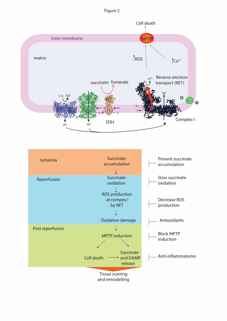
Q
+pHigh
QH2
H O2
succinate fumarate OO2
matrix
inner membrane
Reverse electron transport (RET)
Complex ISDH
1 O2/2
2
Q
QH2
2H+
e-
4H+
–4H
+
Q
QH2
Ca2+
MPTP
ROS
Cell death
Figure 2
Succinate accumulation
Ischemia
Reperfusion
Prevent succinate accumulation
Decrease ROSproduction
Block MPTPinduction
Succinate and DAMP release
Succinate oxidation
ROS production at compex I
by RET
Oxidative damage
MPTP induction
Cell death
Tissue scarring and remodelling
Slow succinate oxidation
Anti-in�ammatories
Antioxidants
Post reperfusion
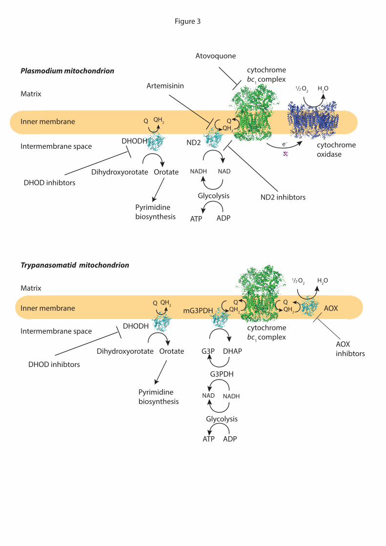
Matrix
Inner membrane
Intermembrane space
Figure 3
Trypanasomatid mitochondrion
1/2 O2 H2O
QQH2
QQH2
NADH
DHODH
DHAPG3P
mG3PDH AOX
NAD
QH2Q
G3PDH
Glycolysis
ADPATP
Dihydroxyorotate Orotate
Matrix
Inner membrane
Intermembrane space
Plasmodium mitochondrion
1/2 O2 H2O
QQH2
e–DHODH ND2
Atovoquone
Artemisinin
QH2Q
NADH NAD
Glycolysis
ADPATP
Dihydroxyorotate Orotate
Pyrimidine biosynthesis
ND2 inhibtors
DHOD inhibtors
DHOD inhibtors
AOX inhibtors
cytochrome oxidase
cytochrome bc1 complex
cytochrome bc1 complex
Pyrimidine biosynthesis


![Richard J. Murphy Oral History Interview JFK#1, 5/10/1967 ... · Richard J. Murphy– JFK #1 Table of Contents Page Topic 1 John F. Kennedy’s [JFK] appeal to the Young Democrats](https://static.fdocuments.net/doc/165x107/5c6732e709d3f2bf4a8b8cfd/richard-j-murphy-oral-history-interview-jfk1-5101967-richard-j-murphy.jpg)


