Methods to measure the binding of therapeutic monoclonal antibodies to the human Fc receptor...
6
Journal of Pharmaceutical and Biomedical Analysis 63 (2012) 23–28 Contents lists available at SciVerse ScienceDirect Journal of Pharmaceutical and Biomedical Analysis jou rn al h om epage: www.elsevier.com/locate/jpba Methods to measure the binding of therapeutic monoclonal antibodies to the human Fc receptor FcRIII (CD16) using real time kinetic analysis and flow cytometry Alice Harrison, Zhe Liu, Schona Makweche, Kevin Maskell, Hong Qi, Geoff Hale ∗ Millipore BioPharma Services, 91 Milton Park, Abingdon, Oxfordshire OX14 4RY, UK a r t i c l e i n f o Article history: Received 21 September 2011 Received in revised form 3 January 2012 Accepted 20 January 2012 Available online 30 January 2012 Keywords: FcRIII CD16 Surface plasmon resonance Flow cytometry Therapeutic antibody a b s t r a c t Two different methods have been developed to measure the binding of therapeutic antibodies to the low affinity human Fc receptor FcRIII (CD16). The first measures binding of antibody to recombinant soluble receptor by surface plasmon resonance and the second uses flow cytometry to measure antibody binding to cells which express the receptor. Both methods have been formatted as parallel line assays and show high levels of accuracy, precision and linearity, making them suitable for comparability, potency and stability assays. They are both readily able to detect structural differences such as glycosylation, which affect Fc receptor binding. The same approaches can be used to measure the binding of any antibody to any Fc receptor. These assays show greater internal precision and long-term reproducibility than traditional cell-based assays such as antibody-dependent cell-mediated cytotoxicity. A combinational approach with a target binding might be appropriate for routine drug batch release as these assays are likely to be significantly more sensitive to small changes in drug structure or activity. © 2012 Elsevier B.V. All rights reserved. 1. Introduction Monoclonal antibodies comprise the largest category of biotech- produced drugs in development. The physiologic properties which make them so useful, namely multivalent binding to unique target antigens and multifunctional interactions with natural effector systems, provide special challenges to the analytical scientist. Reproducible and precise assays for measuring drug potency are the cornerstone of quality control of manufac- tured products. Developing a suitable method to quantify the complete physiological activity of an antibody is usually not fea- sible. In this article, the therapeutic antibody alemtuzumab (Campath ® ) is used to illustrate how a complex cell-based assay can be broken down into simpler steps which are amenable to precise measurements of molecular interactions. Increasing Abbreviations: ADCC, antibody-dependent cell-mediated cytotoxicity; BSA, bovine serum albumin; CFSE, carboxyfluorescein diacetate succinimidyl ester; CMC, complement-mediated cytotoxicity; CV, coefficient of variation; FBS, foetal bovine serum; GPI, glycosylphosphatidylinositol; HBS-EP, Hepes-buffered saline containing EDTA and surfactant P20; IMDM, Iscove’s modified Dulbecco’s medium; MFI, median fluorescence intensity; PBMC, peripheral blood mononuclear cells; PBS, phosphate buffered saline; PI, propidium iodide; PL, parameter logistic; SPR, surface plasmon resonance. ∗ Correspondence to: Managing Director, Millipore BioPharma Services, 91 Milton Park, Abingdon, Oxon, OX14 4RY. Tel.: +44 0 1235 444100. precision of analysis leads to a better ability to determine lot to lot variability in the manufactured product and allows the assessment of drug stability and the impact of process changes. Alemtuzumab was the first humanised therapeutic antibody [1]. It is a human IgG1 which recognises the lymphocyte anti- gen CD52 and is remarkably effective at killing cells both by complement-mediated cytotoxicity (CMC) and antibody- dependent cell-mediated cytotoxicity (ADCC). In 2001, alem- tuzumab was approved for the treatment of patients with chronic lymphocytic leukaemia who had failed prior chemotherapy. Early batch release assays measured its CMC activity [2]. However, ADCC is thought to be the more important mechanism of tumour clear- ance and it is known that this can be markedly affected by changes in glycosylation that might not affect CMC in the same way. ADCC assays were considered difficult to standardise due to the require- ment for a consistent source of human effector cells. With the availability of suitable cell lines or commercial sources of cryopre- served peripheral blood mononuclear cells (PBMC) this has become more practicable but the variability inherent in the method still limits its routine application for drug batch release. An alternative procedure for quantifying the activity of the Fc domain is to measure the functional interactions of the antibody with recombinant human Fc receptors. The most important recep- tor for ADCC is believed to be CD16 (FcRIII). CD16 exists in two isoforms, CD16A (a heterodimeric transmembrane protein) on lym- phocytes and CD16B (GPI-anchored) on neutrophils. There are also different alleles at amino acid position 158 with 158Val showing 0731-7085/$ – see front matter © 2012 Elsevier B.V. All rights reserved. doi:10.1016/j.jpba.2012.01.029
-
Upload
alice-harrison -
Category
Documents
-
view
223 -
download
4
Transcript of Methods to measure the binding of therapeutic monoclonal antibodies to the human Fc receptor...

Mhc
AM
a
ARRAA
KFCSFT
1
pmtesptcs
(at
bcsEflbr
P
0d
Journal of Pharmaceutical and Biomedical Analysis 63 (2012) 23– 28
Contents lists available at SciVerse ScienceDirect
Journal of Pharmaceutical and Biomedical Analysis
jou rn al h om epage: www.elsev ier .com/ locate / jpba
ethods to measure the binding of therapeutic monoclonal antibodies to theuman Fc receptor Fc�RIII (CD16) using real time kinetic analysis and flowytometry
lice Harrison, Zhe Liu, Schona Makweche, Kevin Maskell, Hong Qi, Geoff Hale ∗
illipore BioPharma Services, 91 Milton Park, Abingdon, Oxfordshire OX14 4RY, UK
r t i c l e i n f o
rticle history:eceived 21 September 2011eceived in revised form 3 January 2012ccepted 20 January 2012vailable online 30 January 2012
a b s t r a c t
Two different methods have been developed to measure the binding of therapeutic antibodies to the lowaffinity human Fc receptor Fc�RIII (CD16). The first measures binding of antibody to recombinant solublereceptor by surface plasmon resonance and the second uses flow cytometry to measure antibody bindingto cells which express the receptor. Both methods have been formatted as parallel line assays and showhigh levels of accuracy, precision and linearity, making them suitable for comparability, potency and
eywords:c�RIIID16urface plasmon resonancelow cytometryherapeutic antibody
stability assays. They are both readily able to detect structural differences such as glycosylation, whichaffect Fc receptor binding. The same approaches can be used to measure the binding of any antibodyto any Fc receptor. These assays show greater internal precision and long-term reproducibility thantraditional cell-based assays such as antibody-dependent cell-mediated cytotoxicity. A combinationalapproach with a target binding might be appropriate for routine drug batch release as these assays arelikely to be significantly more sensitive to small changes in drug structure or activity.
. Introduction
Monoclonal antibodies comprise the largest category of biotech-roduced drugs in development. The physiologic properties whichake them so useful, namely multivalent binding to unique
arget antigens and multifunctional interactions with naturalffector systems, provide special challenges to the analyticalcientist. Reproducible and precise assays for measuring drugotency are the cornerstone of quality control of manufac-ured products. Developing a suitable method to quantify theomplete physiological activity of an antibody is usually not fea-ible.
In this article, the therapeutic antibody alemtuzumab
Campath®) is used to illustrate how a complex cell-basedssay can be broken down into simpler steps which are amenableo precise measurements of molecular interactions. IncreasingAbbreviations: ADCC, antibody-dependent cell-mediated cytotoxicity; BSA,ovine serum albumin; CFSE, carboxyfluorescein diacetate succinimidyl ester; CMC,omplement-mediated cytotoxicity; CV, coefficient of variation; FBS, foetal bovineerum; GPI, glycosylphosphatidylinositol; HBS-EP, Hepes-buffered saline containingDTA and surfactant P20; IMDM, Iscove’s modified Dulbecco’s medium; MFI, medianuorescence intensity; PBMC, peripheral blood mononuclear cells; PBS, phosphateuffered saline; PI, propidium iodide; PL, parameter logistic; SPR, surface plasmonesonance.∗ Correspondence to: Managing Director, Millipore BioPharma Services, 91 Milton
ark, Abingdon, Oxon, OX14 4RY. Tel.: +44 0 1235 444100.
731-7085/$ – see front matter © 2012 Elsevier B.V. All rights reserved.oi:10.1016/j.jpba.2012.01.029
© 2012 Elsevier B.V. All rights reserved.
precision of analysis leads to a better ability to determine lot to lotvariability in the manufactured product and allows the assessmentof drug stability and the impact of process changes.
Alemtuzumab was the first humanised therapeutic antibody[1]. It is a human IgG1 which recognises the lymphocyte anti-gen CD52 and is remarkably effective at killing cells bothby complement-mediated cytotoxicity (CMC) and antibody-dependent cell-mediated cytotoxicity (ADCC). In 2001, alem-tuzumab was approved for the treatment of patients with chroniclymphocytic leukaemia who had failed prior chemotherapy. Earlybatch release assays measured its CMC activity [2]. However, ADCCis thought to be the more important mechanism of tumour clear-ance and it is known that this can be markedly affected by changesin glycosylation that might not affect CMC in the same way. ADCCassays were considered difficult to standardise due to the require-ment for a consistent source of human effector cells. With theavailability of suitable cell lines or commercial sources of cryopre-served peripheral blood mononuclear cells (PBMC) this has becomemore practicable but the variability inherent in the method stilllimits its routine application for drug batch release.
An alternative procedure for quantifying the activity of the Fcdomain is to measure the functional interactions of the antibodywith recombinant human Fc receptors. The most important recep-
tor for ADCC is believed to be CD16 (Fc�RIII). CD16 exists in twoisoforms, CD16A (a heterodimeric transmembrane protein) on lym-phocytes and CD16B (GPI-anchored) on neutrophils. There are alsodifferent alleles at amino acid position 158 with 158Val showing
2 ical an
hfAbotw
dACghtbwawtIbbd
ca
ftNFtF
2
2
acoOs(Chhf
2
pps5b1fiwFoa
4 A. Harrison et al. / Journal of Pharmaceut
igher affinity for IgG than 158Phe. Both alleles occur with similarrequency but donors homozygous for 158Val show higher levels ofDCC. Hitherto, CD16A was difficult to express in transfected cellsut Armour et al. [3] have constructed hybrid molecules consistingf the extracellular domain of CD16A-158Val or CD16A-158Phe andhe transmembrane (lipid-anchored) domain of CD16B. CHO cellshich express these receptors are ideal for analysis of IgG binding.
It is known that the precise structure of the N-linked carbohy-rate on the Fc domain significantly affects binding to CD16 andDCC [4]. It is also established for rituximab that the affinity ofD16–antibody interaction correlates well with the extent of tar-et cell death mediated by ADCC [5]. Although the same correlationas not yet been shown for alemtuzumab, experiments in CD52ransgenic mice provided evidence for the importance of CD16y showing that ADCC mediated by NK cells and/or neutrophilsas responsible for lymphocyte depletion in vivo [6]. Therefore,
n assay which measures the interaction of therapeutic antibodiesith CD16 provides a sensitive and functionally relevant indica-
or of the consistency and quality of the antibody in this regard.t is also useful as a stability-indicating test since the interactionetween IgG Fc and CD16 is normally of low affinity, but is likely toe enhanced by antibody aggregation, which is an important type ofeterioration that might be seen under certain storage conditions.
This paper describes two complementary methodologies whichan be used to accurately measure IgG–FcR interactions and aremenable to validation as drug batch release assays.
Glycoengineered forms of alemtuzumab have been preparedrom cells transfected with glycosyltransferases to enable secre-ion of antibody with an increased proportion of bisecting-acetylgalactosamine and a reduced proportion of fucose in thec-linked carbohydrate [7,8]. These variations of glycosylated alem-uzumab are used as models to illustrate the correlation betweencR binding and functional ADCC.
. Materials and methods
.1. Antibodies and cells
Glyco-engineered alemtuzumab (Glyco1 and Glyco2), chimeric,glycosylated CD3 (otelixizumab or ChAglyCD3) [9] and Wien133ells (which expresses a high level of CD52 antigen) were gener-usly provided by Prof Herman Waldmann, University of Oxford,xford, UK. The cell line A4, created by transfecting Chinese Ham-
ter Ovary (CHO) cells with a gene encoding the high affinity alleleCD16A-V158) for human IgG Fc [3] was obtained from Dr Mikelark, University of Cambridge, Cambridge, UK, who also providedelpful advice on the initial assay development. Cryopreserveduman peripheral blood mononuclear cells (PBMC) were obtained
rom CTL-Europe GmbH, Bonn, Germany.
.2. ADCC assay
Wien133 target cells were labelled with CFSE [10] and resus-ended in culture medium (IMDM containing 10% FBS, 100 U/mLenicillin and 100 �g/mL streptomycin). Serial dilutions of testamples (50 �L) were added to a round bottom microplate and0 �L of the target cells (5 × 10⁵ cell/mL) were added and incu-ated for 30 min at 37 ◦C, 5% CO2. Effector cells (50 �L PBMC at–3 × 10⁷ cells/mL) were added and the plate was incubated for aurther 4 h. 50 �L of PBS containing 2 mM EDTA, 10 �g/mL propid-um iodide and 6000 latex beads/mL was added and the samples
ere promptly analysed by flow cytometry on a Beckman CoulterC500. The unstained latex beads were added so that the numberf viable target cells (CFSE+, PI−) could be normalised to eliminateny variability between samples due to changes in the flow rate
d Biomedical Analysis 63 (2012) 23– 28
or sample volume. It was not possible to count dead target cells(CFSE+, PI+) reliably because many of them appeared to disinte-grate. Use of serum in the culture medium raised the possibilitythat complement-mediated cytotoxicity could contribute to thekilling. However, any such effect was likely to be minimal sincethe concentration of alemtuzumab required for CMC (half maxi-mal at 500–700 ng/mL) [2] is substantially higher than required forADCC (half maximal at less than 20 ng/mL).
2.3. Measurement of protein concentrations
Concentrations of antibody stock solutions were measured byabsorbance at 280 nm using a Nanodrop 1000 (Thermo-Fisher,Loughborough, UK) taking the extinction coefficient (E1%) of alem-tuzumab to be 13.2 (based on its protein sequence) and assumingthe same coefficient for other samples.
2.4. SPR assay – Biacore
Monoclonal anti-His4 antibody (Qiagen Crawley, UK, cat no34670) was coupled to a Biacore CM5 chip (Biacore, Uppsala,Sweden) by standard amine chemistry according to the man-ufacturer’s instructions. His-tagged recombinant human CD16A(R&D Systems Abingdon, UK, cat no 4325-FC) at 1 �g/mLwas injected for 2 min at 5 �L/min to be captured by theanti-His antibody. Independent serial dilutions of the testsamples were prepared in triplicate in a microplate. Eachsample was injected for 5 min at 5 �L/min over two flow cells; onecontrol and one which had been exposed to the CD16A. The dif-ference in binding responses was measured at the end of injection.After each sample, the chip was regenerated by injection of 20 �L of10 mM glycine pH 3.0 and re-charged with CD16A. All experimentswere carried out in HBS-EP buffer (Biacore) at a constant temper-ature of 25.0 ◦C using Biacore 2000 and Biacore 3000 instruments.BIAcontrol software (Versions 3.2 and 4.1 respectively) was usedfor data collection and BIAevaluation software (Version 4.1) wasused for export of data.
2.5. SPR assay – ProteOn
Essentially identical experimental conditions were used for SPRexperiments conducted on the ProteOn XPR36 instrument (Bio-Rad, kindly made available by Anton van der Merwe, Universityof Oxford, Oxford, UK). However, this instrument has 36 bindingspots (comparable to four flow cells in the Biacore) and so a degreeof parallel processing was possible. Anti-His4 was coupled to all sixspots of channels 1, 2, 3, 5, 6 of a ProteOn GLM chip according tothe manufacturer’s instructions. Channel 4 was sham-coupled as areference. His-tagged recombinant human CD16A at 1 �g/mL wasinjected over channel 1 for 2 min at 25 �L/min to be captured bythe anti-His antibody. Independent serial dilutions of the test sam-ples were prepared in triplicate in a microplate. Each sample wasinjected for 5 min at 25 �L/min over six spots; one which had beenexposed to the CD16A, four controls and one reference. Six sam-ples were processed in parallel. The difference in binding responsesbetween the specific and the reference was measured at the end ofinjection. After each sample, the chip was regenerated by injectionof 25 �L of 10 mM glycine pH 3.0 and re-charged with CD16A. Allexperiments were carried out in HBS-EP buffer at a constant tem-perature of 25.0 ◦C. ProteOn Manager software (Version 2.1.0.38)was used for data collection and export.
2.6. Flow cytometry assay
Independent serial dilutions of the test samples (final volume50 �L) were prepared in triplicate in a microplate. A4 cells were

ical and Biomedical Analysis 63 (2012) 23– 28 25
sscwatc2tawUfl
2
tuapdsf
3
3
Geracaei
FPpaps5
Fig. 2. Antibody-dependent cell-mediated cytotoxicity of Wien133 cells by human
A. Harrison et al. / Journal of Pharmaceut
uspended in assay buffer (PBS containing 0.1% BSA and 0.05%odium azide) to give a concentration of 6 × 10⁵ cells/mL. 50 �L ofell suspension was added to each well of the microplate. The plateas incubated for 30 min at 25 ◦C and then washed four times with
ssay buffer by centrifugation. The cells were resuspended in detec-ion reagent, FITC-conjugated goat F(ab′)2 anti-human kappa lighthain (AbD Serotec, Kidlington, UK cat no STAR101FB), diluted 1 in5 with assay buffer. The plate was incubated and washed threeimes as previously. Finally the cells were resuspended in 100 �Lssay buffer and 50 �L of 3% formaldehyde was added. The cellsere analysed using a FC500 flow cytometer (Beckman Coulter,K). Cells were gated by forward and side scatter and the medianuorescence intensity (MFI) was recorded.
.7. Statistical analysis
Curve fitting using weighted four and five parameter logis-ic models (4PL, 5PL) and parallel line analysis was carried outsing StatLIA version 3.2 [11]. The chi-squared statistic was useds a measure of parallelism at a probability p = 0.05. Weightingarameters for Biacore and flow experiments were experimentallyetermined using all the available data from reference and testamples of alemtuzumab. Default weighting parameters were usedor ProteOn experiments due to the comparatively small dataset.
. Results and discussion
.1. ADCC assay
Samples of alemtuzumab and its glyco-engineered variantslyco1 and Glyco2 were analysed by ADCC using human PBMC asffectors and Wien133 cells as targets at a ratio of approx 40:1. Theesults are shown in Figs. 1 and 2. As expected, the glyco-engineeredntibodies were more potent in this assay and gave half maximal
ell killing at a concentration of 7.4 ng/mL whereas 17.9 ng/mL oflemtuzumab was required to obtain half-maximal killing. How-ver, the variability of this assay was relatively high with CVs of thendividual triplicates in the range 0.2–18.6% (mean 5.4%). Betweenig. 1. Antibody-dependent cell-mediated cytotoxicity of Wien133 cells by humanBMC in the presence of alemtuzumab. The starting concentration of the test sam-les was adjusted to simulate samples of different potency. The data are plotteds if the test samples had the same concentration as the reference sample so thatotency differences show as a relative shift in the dose response curves. Error barshow the standard deviation of the triplicate measurements. � Reference (100%) �0% © 100%.
PBMC in the presence of different glycoforms of alemtuzumab. Error bars show thestandard deviation of the triplicate measurements. � Wild-type alemtuzumab �Glyco1 © Glyco2.
2217 and 4655 events (live target cells) were acquired for each sam-ple, giving a theoretical mean CV (based on Poisson distribution) of1.8%. The relatively high level of assay variability and the appar-ent “hook effect” observed at the ends of the dose response madeit difficult to model by conventional 4PL or 5PL equations with asufficient degree of accuracy to detect deviations from parallelism.It is likely that the ADCC assay method might be improved withfurther work. Perhaps a suitable human cell line could be used asa source of effectors and a homogeneous readout of cell lysis couldreduce the tendency for assay drift and stochastic errors inherentin the sequential analysis of cells by flow cytometry. Nevertheless,it is unlikely that a cell based assay can attain the same degree ofaccuracy and precision as a ligand binding assay.
3.2. SPR assay – Biacore
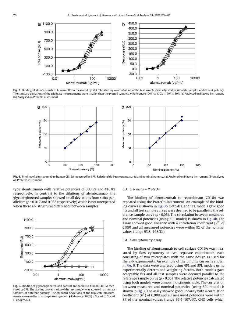
The binding of alemtuzumab to recombinant CD16A was mea-sured by SPR in three separate experiments, each using a differentpair of flow cells. In each experiment three test samples werecompared to a reference sample. The test samples and referencesamples were all prepared from a single batch of alemtuzumab butthe concentrations of the test samples were adjusted to simulatedrug samples of differing nominal potencies: 50%, 70%, 80%, 100%,120%, 130% and 150% of the reference sample. (Nominal potencyis the concentration at which the drug sample was prepared rela-tive to the reference; measured potency is the concentration whichis measured relative to the reference.) Each sample was analysedin triplicate. An example of the binding curves is shown in Fig. 3a.Analysis using a 4PL model gave poor fit probabilities by the �2 test(p < 0.03) therefore this model was deemed to be unsuitable. In con-trast, analysis using a 5PL model with experimentally determinedweighting factors gave good fit probabilities and all test samplecurves were deemed to be parallel to the reference sample curve(p > 0.05). The correlation between measured and nominal poten-cies is shown in Fig. 4a. The assay showed good linearity with acorrelation coefficient (R2) of 0.993 and all measured potencieswere within 9% of the nominal values (range 91.4–100.6%).
A negative control antibody, ChAglyCD3, engineered to elimi-nate binding to CD16A, showed no response in this assay (Fig. 5).However, the glycoengineered antibodies Glyco1 and Glyco2showed significantly enhanced CD16A binding compared to wild
26 A. Harrison et al. / Journal of Pharmaceutical and Biomedical Analysis 63 (2012) 23– 28
Fig. 3. Binding of alemtuzumab to human CD16A measured by SPR. The starting concentration of the test samples was adjusted to simulate samples of different potency.The standard deviations of the triplicate measurements were smaller than the plotted symbols. � Reference (100%) � 130% © 70% � 50%. (a) Analysed on Biacore instrument.(b) Analysed on ProteOn instrument.
F tweeno
trgaw
Fssm�
ig. 4. Binding of alemtuzumab to human CD16A measured by SPR. Relationship ben ProteOn instrument.
ype alemtuzumab with relative potencies of 300.5% and 410.8%espectively. In contrast to the dilutions of alemtuzumab, the
lycoengineered samples showed small deviations from strict par-llelism (p = 0.017 and 0.038 respectively) which is not unexpectedhen there are structural differences between samples.ig. 5. Binding of glycoengineered and control antibodies to human CD16A mea-ured by SPR. The starting concentration of the test samples was adjusted to simulateamples of different potency. The standard deviations of the triplicate measure-ents were smaller than the plotted symbols. � Reference (100%) � Glyco2 © Glyco1
ChAglyCD3.
measured and nominal potency. (a) Analysed on Biacore instrument. (b) Analysed
3.3. SPR assay – ProteOn
The binding of alemtuzumab to recombinant CD16A wasrepeated using the ProteOn instrument. An example of the bind-ing curves is shown in Fig. 3b. Both 4PL and 5PL models gave goodfits and all test sample curves were deemed to be parallel to the ref-erence sample curve (p > 0.05). The correlation between measuredand nominal potencies (using 5PL model) is shown in Fig. 4b. Theassay showed good linearity with a correlation coefficient (R2) of0.990 and all measured potencies were within 9% of the nominalvalues (range 93.8–108.3%).
3.4. Flow cytometry assay
The binding of alemtuzumab to cell-surface CD16A was mea-sured by flow cytometry in two separate experiments, eachconsisting of two microplates with the same design as used forthe SPR experiments. An example of the binding curves is shownin Fig. 6. The data were analysed using 4PL and 5PL models usingexperimentally determined weighting factors. Both models gaveacceptable fits and all test samples were deemed parallel to thereference sample curve (p > 0.05). The relative potencies calculatedusing both models were almost indistinguishable. The correlation
between measured and nominal potencies (using 5PL model) isshown in Fig. 7. The assay showed good linearity with a correlationcoefficient (R2) of 0.988 and all measured potencies were within8% of the nominal values (range 97.4–107.4%). CHO cells which
A. Harrison et al. / Journal of Pharmaceutical an
Fig. 6. Binding of alemtuzumab to human CD16A measured by flow cytometry.The starting concentration of the test samples was adjusted to simulate samplesof different potency. The standard deviations of the triplicate measurements weresmaller than the plotted symbols. � Reference (100%) � 130% © 70% � 50%.
Fig. 7. Binding of alemtuzumab to human CD16A measured by flow cytometry.Relationship between measured and nominal potency.
Fig. 8. Binding of glycoengineered and control antibodies to human CD16A mea-sured by flow cytometry. The starting concentration of the test samples was adjustedto simulate samples of different potency. The standard deviations of the triplicatemeasurements were smaller than the plotted symbols. � Reference (100%) � Glyco2© Glyco1 � ChAglyCD3.
d Biomedical Analysis 63 (2012) 23– 28 27
expressed CD16A-158Phe bound alemtuzumab with low affinity,giving responses substantially lower than the CD16A-158Val cells(A4) (data not shown). The glycoengineered antibodies and controlantibody were also compared to the reference antibody. The resultsare shown in Fig. 8. The control antibody ChAlgyCD3 gave no mea-surable response. Both Glyco1 and Glyco2 gave response curveswhich were distinctly non-parallel to the reference (p < 0.0001) asthey reached a higher plateau of binding. It was therefore not possi-ble to calculate true relative potencies by the parallel line method.
4. Conclusion
Accurate and precise measurement of potency is critical for con-trol of drug quality. It is desirable that the analytical method shouldreflect, as closely as practicable, the biological activity of the drug.However appropriate bioassays, whether in vivo, or cell-based, canbe cumbersome and lack essential reproducibility required for reli-able comparisons over the lifetime of a drug product. This dilemmais exemplified by monoclonal antibodies for tumour therapy, suchas alemtuzumab. In this case, ADCC using tumour cells as tar-gets and fresh human PBMC as effectors, is a good model of thepresumed physiological mode of action. However, the long termreliability of such an assay is questionable due to the potential forvariability in the donor or effector cells as well as the complexityof the procedure.
In contrast, the cell-based assay for binding to CD16 showedexcellent accuracy, precision and linearity. The precision ofreplicate measurements makes the method sensitive to small dif-ferences in drug structure (such as glycosylation) which wouldmake the binding curves non-parallel.
Equally good results were obtained with the SPR assays and theassay was independent of platform, since the results from Biacoreand ProteOn were very similar. Either the SPR or the flow methodis suitable for accurate determination of the Fc binding propertiesof a therapeutic antibody. The throughput of the cell based assaywas potentially higher as several microplates could be processedin one day, whereas the Biacore instrument could only process onemicroplate in 24 h. In practice, the choice between them is likelyto be determined by issues such as the availability of reagents andequipment.
These methods are equally suitable for drug comparability, sta-bility or lot release assays. Measurement of FcR binding alone isnot a complete substitute for the analysis of physiological activ-ity by ADCC. However, analogous methods can be developed foraccurate and precise measurement of the binding of antibody toantigen, either by SPR or by flow cytometry, and the combina-tion of the methods may well provide an acceptable substitutefor the laborious and much less precise measurement of activityby ADCC.
References
[1] L. Riechmann, M. Clark, H. Waldmann, G. Winter, Reshaping human antibodiesfor therapy, Nature 332 (1988) 323–327.
[2] J. Phillips, A. Drumm, P. Harrison, P. Bird, K. Bhamra, E. Berrie, G. Hale,Manufacture and quality control of CAMPATH-1 antibodies for clinical trials,Cytotherapy 3 (2001) 233–242.
[3] K.L. Armour, C.S. Smith, M.R. Clark, Expression of human Fc�RIIIa as a GPI-linked molecule on CHO cells to enable measurement of human IgG binding, J.Immunol. Methods 354 (2010) 20–33.
[4] R.M. Lifely, C. Hale, S. Boyce, M.J. Keen, J. Phillips, Glycosylation and biologi-cal activity of CAMPATH-1H expressed in different cell lines and grown underdifferent culture conditions, Glycobiology 5 (1995) 813–822.
[5] E. Hatjiharissi, L. Xu, D.D. Santos, Z.R. Hunter, B.T. Ciccarelli, S. Verselis, M.
Modica, Y. Cao, R.J. Manning, X. Leleu, E.A. Dimmock, A. Kortsaris, C. Mitsi-ades, K.C. Anderson, E.A. Fox, S.P. Treon, Increased natural killer cell expressionof CD16 augmented binding and ADCC activity to rituximab among individu-als expressing the Fc�RIIIa-158V/V and V/F polymorphism, Blood 110 (2007)2561–2564.
2 ical an
8 A. Harrison et al. / Journal of Pharmaceut[6] P. Hu, M.J. Turner, J. Shields, M.S. Gale, E. Hutto, B.L. Roberts, W.M. Siders, J.M.Kaplan, Investigation of the mechanism of action of alemtuzumab in a humanCD52 transgenic mouse model, Immunology 128 (2009) 260–270.
[7] P. Umana, J. Jean-Mairet, R. Moudry, H. Amstutz, J.E. Bailey, Engineered gly-coforms of an antineuroblastoma IgG1 with optimized antibody dependentcellular cytotoxic activity, Nat. Biotechnol. 17 (1999) 1276–2180.
[8] M.J.S. Dyer, S. Moser, P. Brünker, P. Bird, N. Almond, U. Puentener, L.M.W.Wheat, E. Bolam, E. Berrie, R. Grau, E. Buckby, B. Kennedy, R. Stebbings,G. Hale, P. Umana, Enhanced potency of glycoengineered anti-cd52 mono-clonal antibodies, Blood (ASH Annual Meeting Abstracts) 106 (2005), Abstract2958.
[
[
d Biomedical Analysis 63 (2012) 23– 28
[9] B. Keymeulen, E. Vandemeulebroucke, A.G. Ziegler, C. Mathieu, L. Kaufman, G.Hale, F. Gorus, M. Goldman, M. Walter, S. Candon, L. Schandene, L. Crenier, C.De Block, J-M. Seigneurin, P. De Pauw, D. Pierard, I. Weets, P. Rebello, P. Bird, E.Berrie, M. Frewin, H. Waldmann, J-F. Bach, D. Pipeleers, L. Chatenoud, Insulinneeds after CD3-antibody therapy in new-onset type I diabetes, N. Engl. J. Med.352 (2005) 2598–2608.
10] M. Bracher, H.J. Gould, B.J. Sutton, D. Dombrowicz, S.N. Karagiannis, Three-colour cytometric method to measure antibody-dependent tumour cell killingby cytotoxicity and phagocytosis, J. Immunol. Methods 323 (2007) 160–171.
11] P.G. Gottschalk, J.R. Dunn, Measuring parallelism, linearity, and relative potencyin bioassay and immunoassay data, J. Biopharm. Sci. 15 (2005) 437–463.
![6 Understanding Clinical Flow Cytometry€¦ · tions are available that detail the principles ofcytometry and the operation offlow cytometers [6-12]. 6.2 MONOCLONAL ANTIBODIES The](https://static.fdocuments.net/doc/165x107/5eddcb8bad6a402d6668fd4e/6-understanding-clinical-flow-cytometry-tions-are-available-that-detail-the-principles.jpg)